Immune Responses of Carp A molecular and cellular approach to infections Maria Forlenza

Welcome message from author
This document is posted to help you gain knowledge. Please leave a comment to let me know what you think about it! Share it to your friends and learn new things together.
Transcript
Immune Responses of Carp
A molecular and cellular approach to infections
Maria Forlenza
Thesis committee
Thesis supervisorsProf. Dr. Ir. Huub F.J. SavelkoulProfessor of Cell Biology and Immunology Wageningen University
Dr. Ir. Geert F. WiegertjesAssociate Professor, Cell Biology and Immunology GroupWageningen University
Other members:Prof. Dr. Giuseppe Scapigliati (University of Tuscia, Italy) Prof. Dr. Chris J. Secombes (University of Aberdeen, Scotland)Prof. Dr. Ir. Stefan Magez (Vrije Universiteit Brussel, Belgium)Prof. Dr. Ir. Jaap Keijer (Wageningen University, The Netherlands)
This research was conducted under the auspices of the Graduate School of the Wageningen Institute of Animal Sciences
ThesisSubmitted in partial fulfilment of the requirements for the degree of doctor
at the Wageningen University by the authority of the Rector Magnificus
Prof. dr. M.J. Kropff,in the presence of the
Thesis Committee appointed by the Doctorate Boardto be defended in public
on Friday 30 October 2009at 1.30 PM in the Aula.
Immune Responses of Carp
A molecular and cellular approach to infections
Maria Forlenza

Maria ForlenzaImmune responses of carp: a molecular and cellular approach to infections
Thesis, Wageningen University, Wageningen, NL (2009). With References, with summaries in Dutch and English
ISBN: 978-90-8585-512-5

To my father. He would have been proud

General Introduction: Parasite infections revisited.Aim and outline of the thesis
The use of Real-Time quantitative PCR (RT-qPCR) for the analysis of cytokine mRNA levels
Transcriptional analysis of the common carp (Cyprinus carpio L.) immune response to the fish louse Argulus japonicus Thiele(Crustacea: Branchiura)
Transcription of signal-3 cytokines, IL-12 and IFNαβ, coincides with the timing of CD8αβ up-regulation during viral infection of common carp (Cyprinus carpio L.)
DNA vaccination strategies in common carp (Cyprinus carpio L.) against spring viraemia of carp virus (SVCV)
Differential contribution of neutrophilic granulocytes and macrophages to nitrosative stress in a host-parasite infection model
Nitric oxide hinders antibody clearance from the surface of Trypanoplasma borreli and increases susceptibility to complement-mediated lysis
“Fishing” for antibodies to identify T cells in carp
Receptor-mediated and lectin-like activities of carp TNFα
General discussion
Chapter 1
Chapter 2
Chapter 3
Chapter 4
Chapter 5
Chapter 6
Chapter 7
Chapter 8
Chapter 9
Chapter 10
Summary (English)
Samenvatting (Dutch)
Acknowledgements
List of Publications
Curriculum vitae
9 21
25
43
53
73
99
113
125
153
169
193
197
201
206
208
Contents

chapter
General Introduction:Parasitic infections revisited
Geert F. Wiegertjes, Maria Forlenza, Maaike Joerink and Jörn Scharsack
Developmental and Comparative Immunology2005 (29), 749-758.
1

10
parasite infections revisited
ch
apt
er 1
11
ch
apt
er 1
chapter 1
Review
Parasite infections revisited
Geert F. Wiegertjes*, Maria Forlenza, Maaike Joerink, Jorn P. Scharsack
Cell Biology and Immunology Group, Department of Animal Sciences, Wageningen Institute of Animal Sciences,
Wageningen University, P.O. Box 338, Wageningen AH 6700, The Netherlands
Received 7 July 2004; revised 27 December 2004; accepted 19 January 2005
Available online 17 March 2005
Abstract
Studying parasites helps reveal basic mechanisms in immunology. For long this has been recognized for studies on the
immune system of mice and man. But it is not true for immunological studies on fish. To support this argument we discuss
selected examples of parasite infections not only in warm-blooded but also in cold-blooded vertebrates. We point out that
parasite infections deserve more attention as model systems in comparative immunology.
q 2005 Elsevier Ltd. All rights reserved.
Keywords: Parasites; Immunology; Fishes
1. Introduction
Parasite infections of cold-blooded vertebrates
certainly deserve their place in comparative
immunological studies, not just to solve problems
in, e.g. aquaculture but to exploit these infections as
models for a better understanding of immunological
concepts. After all, despite all molecular analyses
and studies on cellular responses to well-described
antigens and mitogens, whole animal infection
experiments remain essential for a true understanding
of the immune system of all vertebrates. Here, we
Developmental and Comparative Immunology 29 (2005) 749–758
www.elsevier.com/locate/devcompimm
Contents
1. Introduction . . . . . . . . . . . . . . . . . . . . . . . . . . . . . . . . . . . . . . . . . . . . . . . . . . . . . . . . . . . . . . . . . . . . . . . . . . . . . 749
2. Parasite infection models have defined the current paradigm . . . . . . . . . . . . . . . . . . . . . . . . . . . . . . . . . . . . . . . . . . 750
3. Infections with schistosomes . . . . . . . . . . . . . . . . . . . . . . . . . . . . . . . . . . . . . . . . . . . . . . . . . . . . . . . . . . . . . . . . . 751
4. Infections with trypanosomes . . . . . . . . . . . . . . . . . . . . . . . . . . . . . . . . . . . . . . . . . . . . . . . . . . . . . . . . . . . . . . . . 752
5. Parasite-driven immunogenetic diversity . . . . . . . . . . . . . . . . . . . . . . . . . . . . . . . . . . . . . . . . . . . . . . . . . . . . . . . . 754
6. Future perspective . . . . . . . . . . . . . . . . . . . . . . . . . . . . . . . . . . . . . . . . . . . . . . . . . . . . . . . . . . . . . . . . . . . . . . . . . 755
Acknowledgements . . . . . . . . . . . . . . . . . . . . . . . . . . . . . . . . . . . . . . . . . . . . . . . . . . . . . . . . . . . . . . . . . . . . . . . . 755
References . . . . . . . . . . . . . . . . . . . . . . . . . . . . . . . . . . . . . . . . . . . . . . . . . . . . . . . . . . . . . . . . . . . . . . . . . . . . . . 755
0145-305X/$ - see front matter q 2005 Elsevier Ltd. All rights reserved.
doi:10.1016/j.dci.2005.01.005
* Corresponding author. Tel.: C31 317 482732; fax: C31 317
483955.
E-mail address: [email protected] (G.F. Wiegertjes).
12
parasite infections revisited
ch
apt
er 1
highlight only some of the exciting new develop-
ments in the comparative immuno-parasitological
research area.
Comparative immunology is advancing rapidly but
progress is definitely hampered by the fact that
frequently, different laboratories study different ani-
mal (often fish) species. In fact, when realizing that fish
species belonging to the Salmonidae, Cyprinidae,
Ictaluridae or Gadidae should be considered evolutio-
narily distant, it is surprising that the research area is
progressing as rapidly as it is today. This can for a large
part be ascribed to selected genome initiatives in puffer
fish (Fugu rubripes) and zebra fish (Danio rerio) that
brought awealth of information on immuno-regulatory
genes. Database mining and the design of degenerate
primers based on conserved regions in immuno-
regulatory genes has allowed for a rapid expansion of
gene information for fish.
Parasite biology can be enormously complex,
eukaryote parasite life cycles often involve multiple
hosts. Vertebrates can act as the definitive host, i.e. the
host in which the parasite reproduces sexually, such as
with schistosome parasites. Here, invertebrate snails
act as intermediate hosts, i.e. the host in which larval
or asexual stages of the parasite develop. But in other
cases vertebrates can act as intermediate hosts. This is
true for the malaria parasite which completes its
sexual stages in mosquitoes. And last but not
necessarily least, they may be just one host in the
cycle of an organism which does not show sexual
reproduction as such. Trypanosomes and Leishmania,
e.g. need to undergo sequential developmental
changes in the bodies of at least two different species.
This complexity of host–parasite interactions may be
one explanation for the relatively limited number of
research groups that study the immune responses to
fish parasites. This certainly does not mean, however,
that such studies are not worth undertaking.
Although the present review focuses on parasite
infections of vertebrate hosts it is good to realize the
emerging interest of comparative immunologists in
studying insect parasites. The malaria (e.g. Plasmo-
dium falciparum) parasite interacts with two different
hosts: the intermediate vertebrate and the final
mosquito (e.g. Anopheles gambiae) host. Mosquitoes
have for long been considered mere intermediates in
the route to infection of humans, but are now
recognized as highly informative model organisms.
This is mainly because many novel molecular
technological developments (such as genome sequen-
cing projects and production of micro-arrays) are now
being applied not only to the parasite but also to the
mosquito [1–4]. This allows for a better understanding
of the molecular mechanisms mediating the physio-
logical responses of insects to parasite invasion [5].
Thus, at present, the mosquito as a model organism
for directly studying antiparasitic innate immune
responses in insects, complements studies of defense
reactions against bacteria and fungi in the fruit fly,
Drosophila melanogaster [6].
We do not aim at an extensive overview of the
immune defense reactions to all parasite infections in
vertebrates. For recent overviews of the immune
responses to well-known parasites of warm- and cold-
blooded vertebrates and of invertebrates, the reader is
referred to some excellent reviews [7–10,3]. Instead
we discuss selected examples of parasite infections
with a special interest to immunologists with an
evolutionary focus. We argue that these studies are
bringing us a wealth of information on the functioning
of the immune system in vertebrates and that there-
fore, parasite infections deserve immediate attention
as model systems in comparative immunology.
2. Parasite infection models have definedthe current paradigm
At present, laboratories interested in comparative
immunology strongly focus on molecular immuno-
logical research, sometimes extending to cellular
immunology. But, with the progression of the
genomics era into a proteomics era, it is the concept
of studying whole animals in vivo that deserves
renewed interest. This is true for endothermic
vertebrates, where a large research effort goes into
studies on the laboratory mouse, but no less true for
ectothermic vertebrates. High quality infection
models, of which some clearly have paved the way
for much of modern immunology, require well-
designed animal experiments. This has been achieved
through the use of genetically well-defined animals
such as inbred and knock-out mice. But it is important
to realize that investigations on mice usually are
models for investigations on humans. Often these
animal models do not describe natural host–pathogen
G.F. Wiegertjes et al. / Developmental and Comparative Immunology 29 (2005) 749–758750
13
ch
apt
er 1
chapter 1
combinations, where the pathogen’s host specificity
has arisen from progressive selection over thousands
of years. Typically, the natural situation is character-
ized by chronic infection with low pathogen loads
to ensure survival of both host and parasite.
Experimental models, however, frequently have
death as an outcome. Most comparative parasite
infection models do not suffer from this disadvantage
because these infections are studied in the target
animal species. Also, sometimes the mouse model
systems have been used simply because they had been
in use for many years already, defining the current
paradigm. Or just because they are easy to work with
[11]. Of course, the mere fact that certain mouse
model systems define a paradigm demonstrates the
enormous scientific impact the use of inbred mouse
lines have had as model systems.
It is often not well appreciated how much of our
present understanding of the immune system has
come from studying host–parasite interactions. For
example, the murine model of infection with Leish-
mania major (Protozoa, Kinetoplastida) provided
the first in vivo correlation between protective
immunity and an expansion of CD4C T helper 1
(Th1) lymphocytes and between progressive disease
and the development of a Th2 response [9]. Although
Th cell differentiation in human infection with
Leishmania is not always as clear as in mice infected
with L. major, the description of the molecular
mechanisms necessary for polarization of Th
responses in mice did bring a wealth of information
on the vertebrate immune response to pathogens.
Variation between individuals often disturbs the
outcome of our experiments. Scientists working with
warm-blooded vertebrates have found the answer to
genetic variation in focusing on inbred strains of mice.
Comparative immunologists, however, have even
been suffering from the lack of focus on a single
common species. They have chosen to study invert-
ebrates, or fish. Frequently these (commercial) fish
species have relatively long generation intervals. This
severely reduce the possibilities to produce inbred
strains via mating of close relatives, for which
approximately 20 generations are needed. The excep-
tion to the rule, of course, is zebra fish (D. rerio).
Zebra fish do have the advantage of a short generation
time allowing for the development of genetically
uniform strains by classical inbreeding, while also
haploid and diploid androgenetic or gynogenetic fish
are available [12]. As a result, zebra fish have gained
broad utility over the last 10–20 years. Considered a
representative vertebrate developmental model, large-
scale mutagenesis screens have created thousands of
mutant zebra fish lines for developmental studies [13].
In addition, antisense morpholinos have proven
specific and highly effective translational inhibitors
to study targeted gene-knock down in zebra fish [14].
As a consequence, to date, zebra fish are becoming
increasingly attractive for immunological studies,
also because of the recognition that zebra fish share
many orthologous genes or regions with the
human genome [15].
Although the small size of zebra fish limits their
use for cellular assays, considerable progress is made
also towards a functional analysis of immune-relevant
genes. For example, recently in zebra fish 19 putative
TOLL-like receptor (TLR) variants were described, of
which two are homologous to TLR4, showing that the
lack of TLR4 as reported for the puffer fish [16] is not
general for fish [17]. In addition, expression analysis
showed that a subset, including TLR1 and TLR2 are
expressed at higher levels following infection of zebra
fish with Mycobacterium marinum [18]. Clearly, with
renewed interest in innate immunity and associated
pattern recognition receptors (PRRs) such as the
TLRs, fish increasingly prove to be a highly interest-
ing animal species for comparative immunological
studies. Unfortunately, although zebra fish are natural
hosts to a number of parasites, including nematodes,
microsporidians and dinoflagellates [15], none of
these infections are presently exploited as parasite
infection models for fish.
3. Infections with schistosomes
A parasite infection model that has enormously
increased our understanding of the immune system is
infection of mice with Schistosoma mansoni (Platy-
helminthes, Digenea). Schistosomes (e.g. S. mansoni)
infect their mammalian hosts as aquatic cercariae
released from snails (often Biomphalaria glabrata).
Relatively little genetic information is available on
the snail as the intermediate host or its immune
defense against schistosome although snails certainly
G.F. Wiegertjes et al. / Developmental and Comparative Immunology 29 (2005) 749–758 751
14
parasite infections revisited
ch
apt
er 1
are interesting from a comparative immunological
point of view [19].
Following their production in the sporocyst stages,
in the snail intermediate host, cercariae actively
penetrate the skin of vertebrates. Eggs laid by female
worms finally become entrapped in, e.g. the liver
where they are not easily cleared by phagocytic cells.
As a reaction granulomas are formed around the
schistosome eggs. Often macrophages fuse to form
giant cells recruiting T cells from a systemically
activated T cell pool to the granuloma. Interleukin
(IL)-4 operates as the key cytokine driving the Th2
response to schistosome eggs, including an alternative
activation of macrophages [20]. In the absence of IL-4
or IL-10 an overproduction of pro-inflammatory
mediators including interferon (IFN)-g, tumor
necrosis factor (TNF)-a and nitric oxide (NO) cause
excessive liver damage. Also, schistosome granulo-
mas produce IL-12 and transforming growth factor
(TGF)-b both of which have an important role in
controlling IFN-g synthesis [21]. Typically, granulo-
mas containing schistosome eggs represent a localized
Th2 response, whereas granulomas containing,
e.g. Mycobacteria represent a clear Th1 response.
As such, granulomas represent unique models for
immunological research [20].
The present interest in innate immunity and its
recognition receptors lead to an increased research
focus on pathogen associated molecular patterns
(PAMPS) in parasites, able to stimulate these
receptors [22]. Studies on S. mansoni have shown
the importance of the surface syncytial layer in the
survival of this parasite in the mammalian host [23].
Several of these surface layer components are
candidates for protective capacity, including the
large subunit of calpain, Sm-p80. DNA immunization
protocols using Sm-p80 alone or with plasmids
encoding granulocyte-macrophage colony-stimulat-
ing factor (GM-CSF) and IL-4 [24] or a combination
of IL-2 and IL-12 [25] suggest that Sm-p80 is an
excellent vaccine candidate. Interestingly, these
studies [24,25] suggest that the balance between
Th1/Th2 responses to this antigen can be influenced
by the different cytokines included in the vaccine.
This clearly shows the potential of DNA vaccines to
not only induce protection against parasite infections
but also to be included in studies on type I and type II
immune responses. With the current success of DNA
vaccines in protecting fish against viruses [26] in
mind, this opens up new research areas for studying
the fish immune response to DNA vaccination against
parasites.
Fish can be infected with blood flukes of the family
Sanguinicolidae, a model which is much under-
appreciated for comparative immunological studies
since the fish host immune reaction shows clear
analogies to the mouse’s reactions to schistosome
infections. Snails (Lymnaea peregra) host Sanguini-
cola inermis, a blood fluke pathogenic to common
carp, and release cercariae that penetrate the fish skin
[27]. The eggs become entrapped in a number of
organs including the gills and mesonephros, leading to
local granuloma formation. Eggs are encapsulated by
different types of phagocytic cells [28]. The fish host
immune reaction to this parasite has not been studied
in great detail. Most studies describe cellular changes
following in vivo infection [28–31], some show in
vitro polarisation of lymphocytes in response to this
parasite [32]. The most recent studies focused on the
humoral C-reactive protein response to S. inermis
[33]. This parasite model in fish certainly deserves
further immunological characterization. Especially
now that several of the chemical messengers involved
in the Th2 response in warm-blooded vertebrates,
such as IL-10 and TGF-b have been described for fish,
this model holds great potential for studying a putative
type II response in cold-blooded vertebrates.
4. Infections with trypanosomes
Studies on infections with kinetoplastid parasites
have brought much understanding of host–parasite
interactions. Antigenic variation (e.g. Trypanosoma
brucei) as well as intracellular hiding (e.g. Trypano-
soma cruzi) have become schoolbook examples of
parasite adaptations to the host. At present, emerging
insecticide resistance in vectors and drug resistance in
parasites have led to a renewed research interest in
these infections models. As a consequence, to date
many of the state-of-the-art molecular techniques are
being applied to both parasite (e.g. T. brucei) [34] and
vector (e.g. tsetse fly) [35], bringing a wealth of
genetic information.
The Kinetoplastida, Protozoa with a kinetoplastid
organelle containing the mitochondrial DNA, are
G.F. Wiegertjes et al. / Developmental and Comparative Immunology 29 (2005) 749–758752
15
ch
apt
er 1
chapter 1
sub-divided into two suborders. The Trypanosomatina
contain the important mammalian trypanosome para-
sites with a single flagellum, whereas the parasites in
the second suborder (Bodonina) have two flagella.
Often, the Trypanosomatina are sub-divided into
salivarian parasites (transmitted via saliva) such as
T. brucei and stercorarian (transmitted via faeces)
parasites such as T. cruzi. The two groups diverged
some 200–300 million years ago [36]. In fish
representatives of both suborders are studied. Trypa-
nosoma danilewskyi (syn. Trypanosoma carassii)
(infects cyprinids) belongs to the ‘aquatic clade’
within the Trypanosomatina [36] while Trypano-
plasma borreli (cyprinids) and Cryptobia salmositica
(salmonids) both belong to the Bodonina.
Most of our present understanding of immune
reactions to trypanosomiasis has been obtained from
infections of mice which are not a natural host to the
tsetse-transmitted trypanosomes. This clearly is not
the case for the kinetoplastid parasites that infect fish
since they all represent natural parasite–host inter-
actions. In the aqueous environment, blood-sucking
leeches act as vectors between fishes for transmitting
kinetoplastid parasites. A disadvantage of the fish
host–parasite model is that little is known about the
leech vector. For some fish kinetoplastid parasites,
leeches just act as a vector while for others they are
obligatory intermediate hosts [37]. Like the Salivaria,
fish kinetoplasts are believed to be exclusively extra
cellular, and are found in the blood and tissue fluids of
their fish hosts [38,39]. The African trypanosomes
show antigenic variation of the variant surface
glycoprotein (VSG), their counterparts in fish do not
show evidence of antigenic variation [38]. Although
different in this respect, analogies of the fish’ immune
response to kinetoplasts with the mouse immune
response to the extracellular blood stage of trypano-
somes can be found. For example, peritoneal
macrophages from T. brucei infected mice produce
trypanostatic NO radicals in the presence of L-arginine
in vitro [40], although in vivo parasites proliferate in
the vicinity of macrophages. In these mice, the role
of parasite-specific antibodies may be crucial by
mediating the attachment of trypanosomes to
activated macrophages, thereby facilitating NO-
mediated trypanolysis [41]. In carp, similarly,
T. borreli induces the production of trypanostatic
concentrations of NO in vitro [42] but non-effective or
even immuno-suppressive concentrations in vivo
[43]. The effect of parasite-specific antibodies med-
iating attachment of these parasites to activated carp
macrophages, facilitating NO-mediated effects on
T. borreli, could be similar to those seen in mice,
and are presently under investigation in our labora-
tory. The glycosylphosphatidylinositol (GPI) anchors
found in protozoa are PAMPs that are recognized as
foreign [44], with divergent GPIs having evolved to
the advantage of the parasites to manipulate the
endogenous signaling pathways of the host [45].
The non-Salivarian trypanosomes are character-
ized by surfaces dominated by carbohydrate-rich
coats of GPI-anchored mucin-like glycoproteins,
which are not subject to antigen variation. The fish
trypanosomes (T. danilewskyi) and probably also the
Bodonina (T. borreli) have a surface coat highly
comparable to that of the non-Salivaria [46]. It has
been confirmed for T. danilewskyi that the carbo-
hydrate moiety of the mucins contains sialic acid, a
monosaccharide that in other trypanosomes is trans-
ferred from host glycoconjugates to parasite surface
molecules by trans-sialidase (TS). TS has recently
been detected in the fish trypanosome T. danilewskyi
[47]. Sialylated mucins are considered to be essential
for the survival of the parasite in the host. These
PAMPs in T. danilewskyi seem to provide a protective
layer against initial attack by the alternative comp-
lement pathway while specific antibodies against
these PAMPs may be needed for antibody-mediated
lysis [48]. For T. borreli infections in carp, a heat-
labile fraction that could be GPI-anchored proteins is
one of the PAMPs responsible for the induction of NO
in vitro [49].
A particular trypanosome PAMP has been shown
responsible for the activation of murine macrophages:
the cysteine protease cruzipain from T. cruzi. This
protease is found in every developmental form of the
parasite as a glycoprotein of about 52–58 KDa with a
highly mannose glycosylated C-terminal domain. For
C. salmositica, a close relative of T. borreli, a cysteine
protease with chemical properties similar to that of
cruzipain has been described [50]. Interestingly,
cruzipain when injected into mice, induces an
increase of urea associated with a decrease in nitrate
levels, suggesting a preferential up-regulation of the
arginase pathway associated with alternatively acti-
vated macrophages [51]. It could therefore, be of great
G.F. Wiegertjes et al. / Developmental and Comparative Immunology 29 (2005) 749–758 753
16
parasite infections revisited
ch
apt
er 1
interest to study fish macrophage activation by the
cysteine protease of C. salmositica.
Macrophages play an essential role in trypanolytic
events. These versatile cells are able to respond to a
variety of micro-environmental signals including
PAMPs and many cytokines. Typical intracellular
parasites such as Leishmania spp. and T. cruzi have
brought much understanding of what is presently re-
named the ‘classical’ activation of macrophages. This
particular activation integrates the cytokines tumor
necrosis factor alpha (TNF-a) and gamma interferon
(IFN-g), among others, in the type I response. More
recently, it has been recognized that particular
cytokines from Th2 cells can induce an ‘alternative’
activation of macrophages that induces distinct func-
tional activities as part of the type II response [52–55].
It is evident that an effective immune response against
a particular parasite requires a balanced differentiation
between classically activated macrophages (type I
response) and alternatively activated macrophages
(type II response). In fact, mouse resistance toT. brucei
is dependent on their ability to produce IFN-g, TNF-abut also NO (type I response) early during infection
and the production of IL-4 and IL-10 (type II response)
during the chronic phase. Imbalance induces tissue
damage (over stimulation of classically activated
macrophages) or a failure to control early pathogen
replication [56]. In a similar manner, in fish, the high
production of NO during T. borreli infection of carp
[57] and related immuno-suppression could explain
part of the pathology associated with these infections
[58]. The potential role of TNF-a, which has also beendescribed for fish, in inducing tissue damage after
T. borreli infection is still unclear and remains to be
investigated. What is clear, however, is that the
concept of classically versus alternatively activated
macrophages deserves more attention as a concept of
type I versus type II responses in fish. Conveniently,
both inducible nitric oxide synthase and arginase
activities can bemeasured in fishmacrophages [42,59].
5. Parasite-driven immunogenetic diversity
The Major Histocompatibility Complex (MHC)
genes, at least in humans, are the most polymorphic
genes known to date, and the MHC consists of a single
region of 4 Mb comprising the MHC class I and class
II but also other immunologically relevant genes [60].
A number of genes within the MHC code for the
peptide binding region (PBR), which is the most
polymorphic portion of the MHC class I and class II
molecules. In this area, non-synonymous mutations
occur at a higher rate than can be expected by random
events, suggesting there must be a selective pressure
affecting these genes. Since the PBR interacts with
pathogen-derived molecules, pathogens must have
driven MHC selection, most likely through mechan-
isms such as heterozygote advantage and balancing
selection [61].
In humans, many studies have tried to establish
possible associations between infectious disease and
MHC polymorphisms, but firm conclusions have been
obscured by the MHC redundancy (multiple co-
dominantly expressed genes) and a confounding
pattern of linkage (class I and class II genes in a
complex). In fact, the most compelling evidence has
come from chickens infected with a viral pathogen,
Mareks disease virus [62]. Chickens possess a
relatively simple MHC with single dominantly
expressed class I and class II loci [63], which may
be the reason for the clear association between the
presence of particular MHC haplotypes and resistance
to Mareks disease. The organization of the MHC in
teleosts is different from that in mammals or chicken.
Interestingly, fishes do not follow the paradigm of one
complex of genes on a single chromosome but have
taken an evolutionary route where the MHC class I
and class II genes are located on different linkage
groups [64,65]. This aspect, unique to teleost fishes,
allows for studies on associations of MH class I and
class II polymorphisms and disease resistance without
the confounding pattern of linkage found in warm-
blooded vertebrates [66].
Studies in Atlantic salmon (Salmo salar) indeed
have shown an association between MH polymorph-
ism and resistance to the bacterium Aeromonas
salmonicida [67]. Using denaturing gradient gel
electrophoresis to identify alleles, at least one MH
class II b allele was significantly more prevalent in
resistant families. The relative importance of this
particular allele in conferring resistance of Atlantic
salmon to A. salmonicida has been confirmed by
Grimholt et al. [68] using sequence-based typing to
identify alleles. In addition, using the same strategy,
these authors identified two class I a and two class II a
G.F. Wiegertjes et al. / Developmental and Comparative Immunology 29 (2005) 749–758754
17
ch
apt
er 1
chapter 1
alleles associated with increased resistance of Atlantic
salmon to infectious salmon anaemia virus. There are
not many studies in fish that have focused on
associations between MH polymorphisms and resist-
ance to parasite infections, however.
One of the reasons that associations between MH
polymorphisms in fish and resistance to parasites have
not often been studied may be the recognized
complexity of parasite biology. Another reason may
be that few parasite infections in teleosts have had
enough economic impact to drive research towards
investigating an immunogenetic approach. Although
this is not true for the salmon louse (e.g.
Lepeophtheirus salmonis), which has detrimental
effects (including economic ones) on farmed Atlantic
salmon, to our knowledge, no data in this species have
been published so far. One of the few published
studies is on simultaneous infections from multiple
parasite species in three-spined sticklebacks (Gaster-
osteus aculeatus). These fish are particularly suited to
test for optimal rather than maximal MH diversity
because their class II genotypes can differ markedly in
the number of class II B alleles. These studies have
demonstrated a consistent relationship between para-
site diversity among different habitats and MH
diversity and fitness [69,70] and are a good example
of how parasite models in fish can help to resolve
fundamental scientific questions.
6. Future perspective
Now that, at least for fish, the molecular charac-
terization of several messengers potentially involved
in type I (IFN, IL-1, TNF) or type II (IL-10, TGF)
immune responses is rapidly bridging the gap with
mammalian immunology, the rationale for a sub-
sequent functional analysis is stronger than ever.
Alternative macrophage activation routes seem to
offer new opportunities for analyzing type II immune
responses in fish. The need for well-defined parasite
infection models in fish becomes even more evident
taking into account that parasites (e.g. L. major,
S. mansoni) especially, have contributed so much to
the understanding of the Th1/Th2 concept in warm-
blooded vertebrates.
There are at least two infection models in fish that
hold a great promise in that they seem to induce
immune responses in fish with clear similarities to the
immune responses of mammals. S. inermis is a
parasite of carp that induces granuloma formation
comparable to what is seen in the mammalian
response to schistosomes. Also, the fish’ immune
response to trypanosomes shows clear similarities to
the mammalian immune response to extra cellular
stages of mammalian trypanosomes. Therefore, these
parasite models provide unique opportunities for
comparative immunologists.
There are several parasite infection models, such as
infection of fish with Ichthyophthirius multifiliis, that
have not been discussed here. This particular parasite
model has proven highly informative for studies on
GPI-anchored membrane proteins [71] and certainly
holds a future potential as model system for studying
mucosal immunity. We did not aim at an extensive
overview of all immune defense reactions to parasites
in teleosts. Instead we have pointed out that these
studies are bringing us a wealth of information on the
functioning of the immune system in both warm- and
cold-blooded vertebrates and that therefore, parasite
infections deserve immediate attention as model
systems in comparative immunology.
Acknowledgements
The authors would like to acknowledge the critical
reading of the manuscript by Dr Rene J.M. Stet. This
work was supported in part by the European
Community’s Improving Human Potential Pro-
gramme under contracts [HPRN-CT-2001-00214],
[PARITY] and [QLK5-CT-2001-50988].
References
[1] Ghosh A, Srinivasan P, Abraham EG, Fujioka H, Jacobs-
Lorena M. Molecular strategies to study Plasmodium-
mosquito interactions. Trends Parasitol 2003;19(2):94–101.
[2] Craig A, Kyes S, Ranson H, Hemingway J. Malaria parasite
and vector genomes: partners in crime. Trends Parasitol 2003;
19(8):356–62.
[3] Blandin S, Levashina EA. Mosquito immune responses
against malaria parasites. Curr Opin Immunol 2004;16(1):
16–20.
[4] Dimopoulos G, Christophides GK, Meister S, Schultz J,
White KP, Barillas-Mury C, et al. Genome expression analysis
G.F. Wiegertjes et al. / Developmental and Comparative Immunology 29 (2005) 749–758 755
18
parasite infections revisited
ch
apt
er 1
of Anopheles gambiae: responses to injury, bacterial chal-
lenge, and malaria infection. Proc Natl Acad Sci USA 2002;
99(13):8814–9.
[5] Land KM. The mosquito genome: perspectives and possibi-
lities. Trends Parasitol 2003;19(3):103–5.
[6] Dimopoulos G, Muller H-M, Levashina EA, Kafatos FC.
Innate immune defense against malaria infection in the
mosquito. Cun Opin Immunol 2001;13(1):79–88.
[7] Carton, Y, Nappi, AJ, Poirie, M. Genetics of anti-parasite
resistance in invertebrates. Dev Comp Immunol
2005;29:9–32.
[8] Maizels RM, Yazdanbakhsh M. Immune regulation by
helminth parasites: cellular and molecular mechanisms. Nat
Rev Immunol 2003;3(9):733–44.
[9] Gumy A, Louis JA, Launois P. The murine model of infection
with Leishmania major and its importance for the deciphering
of mechanisms underlying differences in Th cell differen-
tiation in mice from different genetic backgrounds. Int
J Parasitol 2004;34(4):433–44.
[10] Jones SRM. The occurrence and mechanisms of innate
immunity against parasites in fish. Dev Comp Immunol
2001;25(8–9):841–52.
[11] Druilhe P, Hagan P, Rook GAW. The importance of models of
infection in the study of disease resistance. Trends Microbiol
2002;10(10 Suppl.):S38–S46.
[12] Brandhorst BP, Corley-Smith GE. Production of haploid and
diploid androgenetic zebrafish. Methods Mol Biol 2004;254:
255–70.
[13] Yeh J-RJ, Crews CM. Chemical genetics: adding to the
developmental biology toolbox. Dev Cell 2003;5(1):11–19.
[14] Nasevicius A, Ekker SC. Effective targeted gene ‘knockdown’
in zebrafish. Nat Genet 2000;26(2):216–20.
[15] Yoder JA, Nielsen ME, Amemiya CT, Litman GW. Zebrafish
as an immunological model system. Microbes Infect 2002;
4(14):1469–78.
[16] Oshiumi H, Tsujita T, Shida K, MatsumotoM, Ikeo K, Seya T.
Prediction of the prototype of the human Toll-like receptor
gene family from the pufferfish, Fugu rubripes, genome.
Immunogenetics 2003;54(11):791–800.
[17] Jault C, Pichon L, Chluba J. Toll-like receptor gene family and
TIR-domain adapters in Danio rerio. Mol Immunol 2004;
40(11):759–71.
[18] Meijer AH, Gabby Krens SF, Medina Rodriguez IA, He S,
Bitter W, Snaar-Jagalska BE, Spaink HP. Expression analysis
of the Toll-like receptor and TIR domain adaptor families of
zebrafish. Mol Immunol 2004;40(11):773–83.
[19] Raghavan N, Miller AN, Gardner M, FitzGerald PC,
Kerlavage AR, Johnston DA, Lewis FA, Knight M. Com-
parative gene analysis of Biomphalaria glabrata hemocytes
pre- and post-exposure to miracidia of Schistosoma mansoni.
Mol Biochem Parasitol 2003;126(2):181–91.
[20] Sandor M, Weinstock JV, Wynn TA. Granulomas in
schistosome and mycobacterial infections: a model of local
immune responses. Trends Immunol 2003;24(1):44–52.
[21] Rakasz E, Blum AM, Metwali A, Elliott DE, Li J, Ballas ZK,
Qadir K, Lynch R, Weinstock JV. Localization and regulation
of IFN-gamma production within the granulomas of murine
schistosomiasis in IL-4-deficient and control mice. J Immunol
1998;160(10):4994–9.
[22] Medzhitov R, Janeway Jr CA. Innate immunity: impact on the
adaptive immune response. CurrOpin Immunol 1997;9(1):4–9.
[23] Siddiqui AA, Podesta RB, Clarke MW. Schistosoma mansoni:
characterization and identification of calcium-binding proteins
associated with the apical plasma membrane and envelope.
Exp Parasitol 1991;72(1):63–8.
[24] Siddiqui AA, Phillips T, Charest H, Podesta RB, Quinlin ML,
Pinkston JR, Woyd JD, Paz M, Villalovos RM, Pampa J.
Induction of protective immunity against Schistosoma man-
soni via DNA priming and boosting with the large subunit of
calpain (Sm-p80): adjuvant effects of granulocyte-macro-
phage colony-stimulating factor and interleukin-4. Infect
Immun 2003;71(7):3844–51.
[25] Siddiqui AA, Phillips T, Charest H, Podesta RB, Quinlin ML,
Pinkston JR, Woyd JD, Pompa J, Villalovos RM, Paz M.
Enhancement of Sm-p80 (large subunit of calpain) induced
protective immunity against Schistosoma mansoni through co-
delivery of interleukin-2 and interleukin-12 in a DNA vaccine
formulation. Vaccine 2003;21(21–22):2882–9.
[26] Lorenzen N, Lorenzen E, Einer-Jensen K, LaPatra SE. DNA
vaccines as a tool for analysing the protective immune
response against rhabdoviruses in rainbow trout. Fish Shellfish
Immunol 2002;12(5):439–53.
[27] Kirk RS, Lewis JW. Histopathology of Sanguinicola inermis
infection in carp Cyprinus carpio. J Helminthol 1998;72(1):
33–8.
[28] Richards DT, Hoole D, Lewis JW, Ewens E, Arme C.
Ultrastructural observations on the cellular response of carp,
Cyprinus carpio L., to eggs of the blood fluke Sanguinicola
inermis Plehn, 1905 (Trematoda: Sanguinicolidae). J Fish Dis
1994;17:439–46.
[29] Richards DT, Hoole D, Lewis JW, Ewens E, Arme C. Changes
in the cellular composition of the spleen and pronephros of
carp Cyprinus carpio infected with the blood fluke Sanguini-
cola inermis (Trematoda: Sanguinicolidae). Dis Aquat Organ
1994;19:173–9.
[30] Richards DT, Hoole D, Lewis JW, Ewens E, Arme C.
Stimulation of carp Cyprinus carpio lymphocytes by the blood
fluke Sanguinicola inermis (Trematoda: Sanguinicolidae). Dis
Aquat Organ 1996;25:87–93.
[31] Richards DT, Hoole D, Lewis JW, Ewens E, Arme C. In vitro
polarization of carp leucocytes in response to the blood fluke
Sanguinicola inermis Plehn, 1905 (Trematoda: Sanguinicoli-
dae). Parasitology 1996;112(Pt 5):509–13.
[32] Richards DT, Hoole D, Lewis JW, Ewens B, Arme C.
Adherence of carp leucocytes to adults and cercariae of the
blood fluke Sanguinicola inermis. J Helminthol 1996;70:
63–7.
[33] Hoole D, Lewis JW, Schuwerack PM, Chakravarthy C,
Shrive AK, Greenhough TJ, Cartwright JR. Inflammatory
interactions in fish exposed to pollutants and parasites: a role
for apoptosis and C reactive protein. Parasitology 2003;
126(Suppl.):S71–S85.
G.F. Wiegertjes et al. / Developmental and Comparative Immunology 29 (2005) 749–758756
19
ch
apt
er 1
chapter 1
[34] Davila AMR, Majiwa PAO, Grisard EC, Aksoy S,
Melville SE. Comparative genomics to uncover the secrets
of tsetse and livestock-infective trypanosomes. Trends Para-
sitol 2003;19(10):436–9.
[35] Butler D. African labs win major role in tsetse-fly genome
project. Nature 2004;427(6973):384.
[36] Stevens JR, Noyes HA, Schofield CJ, Gibson W. The
molecular evolution of Trypanosomatidae. Adv Parasitol
2001;48:1–56.
[37] Joerink M, Saeij JPJ, Stafford JL, Belosevic M,
Wiegertjes GF. Animal models for the study of innate
immunity: protozoan infections in fish. In: Wiegertjes GF,
Flik G, editors. Host–parasite interactions. Abingdon, UK:
BIOS Sci Publ; 2004. p. 67–89.
[38] Overath P, Haag J, Mameza MG, Lischke A. Freshwater fish
trypanosomes: definition of two types, host control by
antibodies and lack of antigenic variation. Parasitology
1999;119(Pt 6):591–601.
[39] Woo PTK. Cryptobia (Trypanoplasma) salmositica and
salmonid cryptobiosis. J Fish Dis 2003;26(11–12):627–46.
[40] Kaushik RS, Uzonna JE, Gordon JR, Tabel H. Innate
resistance to Trypanosoma congolense infections: differential
production of nitric oxide by macrophages from susceptible
BALB/c and resistant C57Bl/6 mice. Exp Parasitol 1999;
92(2):131–43.
[41] Gobert AP, Daulouede S, Lepoivre M, Boucher JL,
Bouteille B, Buguet A, et al. L-Arginine availability
modulates local nitric oxide production and parasite killing
in experimental trypanosomiasis. Infect Immun 2000;68(8):
4653–7.
[42] Saeij JPJ, Stet RJM, Groeneveld A, Verburg-van
Kemenade BM, van Muiswinkel WB, Wiegertjes GF.
Molecular and functional characterization of a fish induci-
ble-type nitric oxide synthase. Immunogenetics 2000;51(4–
5):339–46.
[43] Saeij JPJ, van Muiswinkel WB, Groeneveld A, Wiegertjes GF.
Immune modulation by fish kinetoplastid parasites: a role for
nitric oxide. Parasitology 2002;124(Pt 1):77–86.
[44] Tachado SD, Gerold P, Schwarz R, Novakovic S,
McConville M, Schofield L. Signal transduction in macro-
phages by glycosylphosphatidylinositols of Plasmodium,
Trypanosoma, and Leishmania: activation of protein tyrosine
kinases and protein kinase C by inositolglycan and diacylgly-
cerol moieties. Proc Natl Acad Sci USA 1997;94(8):4022–7.
[45] Tachado SD, Mazhari-Tabrizi R, Schofield L. Specificity in
signal transduction among glycosylphosphatidylinositols of
Plasmodium falciparum, Trypanosoma brucei, Trypanosoma
cruzi and Leishmania spp. Parasite Immunol 1999;21(12):
609–17.
[46] Overath P, Haag J, Lischke A, O’HUigin C. The surface
structure of trypanosomes in relation to their molecular
phylogeny. Int J Parasitol 2001;31(5–6):468–71.
[47] Aguero F, Campo V, Cremona L, Jager A, Di Noia JM,
Overath P, Sanchez DO, Carlos Frasch A. Gene discovery in
the freshwater fish parasite Trypanosoma carassii: identifi-
cation of trans-sialidase-like and mucin-like genes. Infect
Immun 2002;70(12):7140–4.
[48] Lischke A, Klein C, Stierhof Y-D, Hempel M, Mehlert A,
Almeida IC, Ferguson MAJ, Overath P. Isolation and
characterization of glycosylphosphatidylinositol-anchored,
mucin-like surface glycoproteins from bloodstream forms of
the freshwater-fish parasite Trypanosoma carassii. Biochem J
2000;345(Pt 3):693–700.
[49] Saeij JPJ, de Vries BJ, Wiegertjes GF. The immune response
of carp to Trypanoplasma borreli: kinetics of immune gene
expression and polyclonal lymphocyte activation. Dev Comp
Immunol 2003;27(10):859–74.
[50] Zuo X, Woo PTK. Characterization of purified metallo- and
cysteine proteases from the pathogenic haemoflagellate
Cryptobia salmositica Katz 1951. Parasitol Res 1998;84(6):
492–8.
[51] Giordanengo L, Guinazu N, Stempin C, Fretes R, Cerban F,
Gea S. Cruzipain, a major Trypanosoma cruzi antigen,
conditions the host immune response in favor of parasite.
Eur J Immunol 2002;32(4):1003–11.
[52] Mills CD. Macrophage arginine metabolism to ornithine/urea
or nitric oxide/citrulline: a life or death issue. Crit Rev
Immunol 2001;21(5):399–425.
[53] Gordon S. Alternative activation of macrophages. Nat Rev
Immunol 2003;3(1):23–35.
[54] Mantovani A, Sozzani S, Locati M, Allavena P, Sica A.
Macrophage polarization: tumor-associated macrophages as a
paradigm for polarized M2 mononuclear phagocytes. Trends
Immunol 2002;23(11):549–55.
[55] Noel W, Raes G, Hassanzadeh Ghassabeh G, De
Baetselier P, Beschin A. Alternatively activated macro-
phages during parasite infections. Trends Parasitol 2004;
20(3):126–33.
[56] Baetselier PD, Namangala B, Noel W, Brys L, Pays E,
Beschin A. Alternative versus classical macrophage activation
during experimental African trypanosomosis. Int J Parasitol
2001;31(5–6):575–87.
[57] Saeij JPJ, VanMuiswinkelWB, Groeneveld A,Wiegertjes GF.
Immune modulation by fish kinetoplastid parasites: a role for
nitric oxide. Parasitology 2002;124(Pt 1):77–86.
[58] Bunnajirakul S, Steinhagen D, Hetzel U, Korting W,
Drommer W. A study of sequential histopathology of
Trypanoplasma borreli (Protozoa: Kinetoplastida) in suscep-
tible common carp Cyprinus carpio. Dis Aquat Organ 2000;
39(3):221–9.
[59] Wiegertjes GF, Joerink M. Macrophage polarization in the
immune response to parasites. Bull Eur Assoc Fish Pathol
2004;24(1):5–10.
[60] Beck S, Trowsdale J. The human major histocompatability
complex: lessons from the DNA sequence. Annu Rev
Genomics Hum Genet 2000;1:117–37.
[61] Parham P, Ohta T. Population biology of antigen presen-
tation by MHC class I molecules. Science 1996;272(5258):
67–74.
[62] Kaufman J, Salomonsen J. The ‘minimal essential MHC’
revisited: both peptide-binding and cell surface expression
level of MHC molecules are polymorphisms selected by
pathogens in chickens. Hereditas 1997;127(1–2):67–73.
G.F. Wiegertjes et al. / Developmental and Comparative Immunology 29 (2005) 749–758 757
20
parasite infections revisited
ch
apt
er 1
[63] KaufmanJ,MilneS,GobelTW,WalkerBA,JacobJP,AuffrayC,
et al. The chicken B locus is a minimal essential major
histocompatibility complex. Nature 1999;401(6756):923–5.
[64] Bingulac-Popovic J, Figueroa F, Sato A, Talbot WS,
Johnson SL, Gates M, et al. Mapping of mhc class I and
class II regions to different linkage groups in the zebrafish,
Danio rerio. Immunogenetics 1997;46(2):129–34.
[65] Sato A, Figueroa F, Murray BW, Malaga-Trillo E, Zaleska-
Rutczynska Z, Sultmann H, Toyosawa S, Wedekind C,
Steck N, Klein J. Nonlinkage of major histocompatibility
complex class I and class II loci in bony fishes. Immunoge-
netics 2000;51(2):108–16.
[66] Stet RJM, Kruiswijk CP, Dixon B. Major histocompatibility
lineages and immune gene function in teleost fishes: the road
not taken. Crit Rev Immunol 2003;23(5–6):441–71.
[67] Langefors A, Lohm J, Grahn M, Andersen O, von Schantz T.
Association between major histocompatibility complex class
IIB alleles and resistance to Aeromonas salmonicida in
Atlantic salmon. Proc R Soc Lond B Biol Sci 2001;
268(1466):479–85.
[68] Grimholt U, Drablos F, Jorgensen SM, Hoyheim B,
Stet RJM. The major histocompatibility class I locus in
Atlantic salmon (Salmo salar L.): polymorphism, linkage
analysis and protein modelling. Immunogenetics 2002;54(8):
570–81.
[69] Wegner KM, Reusch TBH, Kalbe M. Multiple parasites are
driving major histocompatibility complex polymorphism in
the wild. J Evol Biol 2003;16(2):224–32.
[70] Wegner KM, Kalbe M, Kurtz J, Reusch TBH, Milinski M.
Parasite selection for immunogenetic optimality. Science
2003;301(5638):1343.
[71] Dickerson H, Clark T. Ichthyophthirius multifiliis: a model of
cutaneous infection and immunity in fishes. Immunol Rev
1998;166:377–84.
G.F. Wiegertjes et al. / Developmental and Comparative Immunology 29 (2005) 749–758758
21
ch
apt
er 1
chapter 1
Aim and outline
22
aim and outline
ch
apt
er 1
The hypothesis pertinent to this thesis is that homologous (naturally occurring) infection models are fundamental to a proper extrapolation of experimental data towards a practical implementation of prophylactic strategies such as immunomodulation and vaccination. Protection against infection relies on the integrated activity of innate and adaptive parts of the immune system and requires three fundamental components: 1) a molecular recognition system to identify the presence of the infectious organism, 2) an eliminating system to destroy the invaders at both molecular and cellular levels and 3) a communication system to coordinate these activities, that consists of soluble and cell-bound molecules.
All living organisms can be hosts and therefore can house parasites. The only limit is size: the smaller an organism, the more limited the list of parasites it can house. It may be most correct to differentiate between microparasites (viruses, bacteria, fungi and protists) and macroparasites (helminths, arthropods and other metazoans). In this thesis, we study three fundamentally different homologous infection models of common carp, including microparasitic infections with spring viraemia of carp virus (SVCV) and the protist Trypanoplasma borreli as well as macroparasitic infections with the ectoparasite Argulus japonicus (arthropod). The first aim of the research described in this thesis is to develop both molecular and cellular tools to be implemented in the characterization of the innate and adaptive immune response of carp to infections. The second aim of this thesis is to integrate molecular and cellular approaches to investigate the immune response of carp to infections, taking into account the nature of the pathogen. In the second part of this thesis, we focus on one model in particular, i.e. infections with the extracellular blood parasite T. borreli (Parabodonida; Kinetoplastida).
The potential of the T. borreli infection model of carp is extensively discussed in chapter 1 (general introduction). To date, the discipline of comparative immunology is receiving increased attention and is advancing rapidly, especially because of the developments in molecular biology and the progress made with molecular techniques. These developments include wide access to genome information on a number of fish species including pufferfish, stickleback and zebrafish, a close relative of carp. The combination of an increased amount of sequence information and the development of more sensitive and accurate methods to measure gene expression in real-time (chapter 2), has pushed forward the discipline of comparative immunology. In this thesis, we use the real-time quantitative PCR technique to describe the kinetics of the immune response to the ectoparasite Argulus japonicus (chapter 3) and to infection with the spring viraemia of carp virus (SVCV; chapter 4). In the latter study, we not only report the cloning of cell surface markers (CD8α and CD8β) for carp cytotoxic T cells (CTL), and the possible involvement of CTL in the immune response against this viral infection, but also point out the importance of interleukin-12, a cytokine with a crucial role in the development of an effective CTL response. In chapter 5, we therefore examine and discuss the possibility to co-administer an IL-12 expression plasmid in combination with a plasmid encoding for the G-protein of SVCV with the aim to improve DNA vaccination strategies against this virus. The experimental data obtained with the SVCV infection model provide a valuable example of how a thorough understanding of the immune response could lead to the successful design of an improved prophylactic strategy for vaccination in aquaculture practice.
The second part of this thesis focuses in particular on the T. borreli infection model. Experimental
23
ch
apt
er 1
chapter 1
infection of carp with T. borreli results in pathological changes also associated with trypanosome infections of warm-blooded vertebrates such as anaemia, splenomegaly and polyclonal lymphocyte activation. Typical of T. borreli infections only, is the induction of extremely elevated serum nitrite levels. Previous studies suggested the ability of T. borreli to induce elevated nitric oxide (NO) would represent a strategy of the parasite to induce immunosuppression and evade the host immune system. The strong immune reaction would thus be disadvantageous to the host. In this thesis, we challenge this view and hypothesize that a timely production of NO could also be beneficial to the host and that a balance between the host’s immune response and immune evasion strategies of the parasite would serve best both, host and parasite.
In chapter 6, we use an anti-nitrotyrosine antibody in combination with monoclonal antibodies that specifically recognize carp leukocyte sub-populations of macrophages and neutrophilic granulocytes to examine the relative contribution of these two cell types to the nitration process in vivo. In the same chapter, we examine, in vitro, the effect of reactive nitrogen species on parasite motility and viability, using NO and peroxynitrite donors. We also examine, in vivo, the effects of nitrosative stress on the parasite itself using T. borreli-specific antibodies for immuno-histochemistry. Given the phylogenetic relationship of T. borreli with other mammalian trypanosomes and the similarity observed in the pathology of these parasitic infections in warm-blooded vertebrates and carp, in the same chapter we examine the potential of T. borreli to serve as an alternative tool for pharmacological studies on human anti-trypanosome drugs. In chapter 7, we hypothesize that during the early phase of infection, when antibody titers and nitrite concentrations are still moderate, the trypanostatic effects of NO could favor rather than hinder the host defense mechanism. In this scenario, reduced parasite motility would hinder the hydrodynamic flow-mediated removal of IgM from the parasite surface. As a result, parasite-bound surface IgM concentrations would remain high, favoring complement activation and parasite lysis, resulting in clearance of the parasite from the bloodstream.
Owing to the use of a monoclonal antibody specific for B-lymphocytes, we could visualize a massive B cell proliferation in the spleen of T. borreli-infected fish (chapter 6). The T cell response, however, has not been investigated. In chapter 8, we report the cloning of three new sequences, additional to the cell surface markers reported in chapter 4, that are all specific for the T-cell-lineage in carp: CD3ε, Lck and ZAP-70. The identification of conserved epitopes in the newly described sequences allows for the selection of antibodies developed for use in mammalian immunology and specific for the corresponding human proteins. The selected antibodies are used for the identification of T cells in carp and for a preliminary investigation of the T cell response during T. borreli infection. In addition, using a different approach, affinity-purified polyclonal antibodies specifically directed against selected peptides in the extracellular domain of carp CD4 and CD8, are also produced. These antibodies are required for a proper identification of T cell subsets in carp during infection.
In the last part of this thesis (chapter 9), a comprehensive in vitro and in vivo analysis of the biological activities of the cytokine tumor necrosis factor-alpha (TNFα) is performed. A bacterial recombinant carp TNFα is produced and receptor-dependent as well as receptor-independent activities of carp TNFα are investigated, in vitro. The role of TNFα during T. borreli infections, is examined using three fundamentally different but complementary approaches: 1) inhibition of TNFα gene expression,
24
aim and outline
ch
apt
er 1 2) overexpression of TNFα and 3) inhibition of membrane-bound TNFα (mTNFα) shedding. Inhibition
of TNFα gene expression in vivo allowed us to discriminate between parasite-derived components and TNFα with respect to their contribution to the high nitrite levels associated with T. borreli infections. The yet unexploited role in fish of mTNFα and its role in protection against T. borreli infection are discussed. The molecular, histological and cellular methodologies used throughout this study are integrated in an attempt to emphasize the value of homologous infection models for the discipline of comparative immunology. Chapter 10 discusses the progress made in the understanding of the protective immune response of carp to the parasite T. borreli as well as future perspective concerning the exploitation and implementation of the zebrafish animal model alongside the carp animal model.
The studies described in this thesis, showed that the identification of more components of the fish innate and adaptive immune system increasingly permits to study functional activities within the protective immune response. Whole animal infection experiments are essential for a true understanding of the immune system and parasite infections in particular, thus deserve a more central role as model systems to help understand basic questions in comparative immunology.
chapter
The use of Real-Time quantitative PCR (RT-qPCR) for the analysis of cytokine
mRNA levels
Maria Forlenza, Thomas Kaiser, Huub F.J. Savelkouland Geert F. Wiegertjes
Parts of this chapter will be published in Cytokine Protocols (Methods in Molecular Biology), Marc De Ley (Editor), 2009.
Humana Press Inc.
2

26
real-time quantitative pcr
ch
apt
er 2
ch
apt
er 2
27
chapter 2
1
The use of Real-Time quantitative PCR (RT-qPCR) for the analysis
of cytokine mRNA levels
Maria Forlenza1, Thomas Kaiser2, Huub F.J. Savelkoul1 and Geert F. Wiegertjes1
1 Cell Biology and Immunology Group, Wageningen Institute of Animal Sciences, Wageningen
University, Wageningen, The Netherlands.
2 Corbett Life Science, Australia
28
real-time quantitative pcr
ch
apt
er 2
2
1. Introduction
Over the last decade, Real Time-quantitative PCR
(RT-qPCR) analysis has become the method of
choice for quantitative and accurate measurement
of mRNA expression levels but also for sensitive
detection of rare or mutated DNA species in
diagnostic research (1, 2). RT-qPCR is based on
the standard principles of PCR amplification in
addition to the use of specific probes or
intercalating dyes. Various probe systems are
available among which TaqMan probes,
Molecular Beacons, MGB probes, and others
increasing specificity and sensitivity of the Real
Time assays. RT-qPCR using intercalating dyes
that become fluorescent upon binding to double-
stranded DNA, has the advantage of running
melting curve analysis after each run in order to
check specificity. In most cases Sybr-Green I is
used, but other dyes are available including Eva-
Green, Syto9, etc.
Under optimal conditions, every PCR cycle should
result in a doubling of the amplification product.
At the end of every cycle the intercalating dye will
bind to all double-stranded DNA. Ideally, the
increase in amount of template will be directly
proportional to the increase in fluorescence.
Fluorescence data are collected during each cycle
allowing for real-time monitoring of
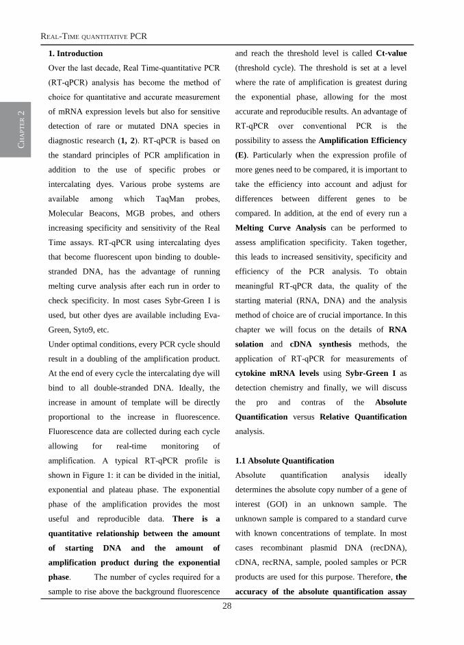
amplification. A typical RT-qPCR profile is
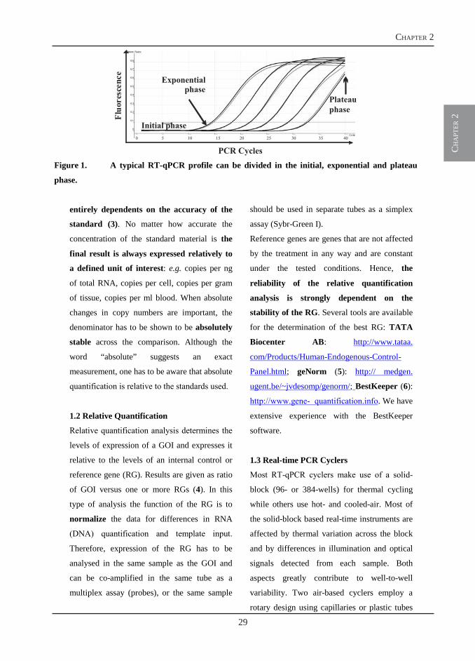
shown in Figure 1: it can be divided in the initial,
exponential and plateau phase. The exponential
phase of the amplification provides the most
useful and reproducible data. There is a
quantitative relationship between the amount
of starting DNA and the amount of
amplification product during the exponential
phase. The number of cycles required for a
sample to rise above the background fluorescence
and reach the threshold level is called Ct-value
(threshold cycle). The threshold is set at a level
where the rate of amplification is greatest during
the exponential phase, allowing for the most
accurate and reproducible results. An advantage of
RT-qPCR over conventional PCR is the
possibility to assess the Amplification Efficiency
(E). Particularly when the expression profile of
more genes need to be compared, it is important to
take the efficiency into account and adjust for
differences between different genes to be
compared. In addition, at the end of every run a
Melting Curve Analysis can be performed to
assess amplification specificity. Taken together,
this leads to increased sensitivity, specificity and
efficiency of the PCR analysis. To obtain
meaningful RT-qPCR data, the quality of the
starting material (RNA, DNA) and the analysis
method of choice are of crucial importance. In this
chapter we will focus on the details of RNA
solation and cDNA synthesis methods, the
application of RT-qPCR for measurements of
cytokine mRNA levels using Sybr-Green I as
detection chemistry and finally, we will discuss
the pro and contras of the Absolute
Quantification versus Relative Quantification
analysis.
1.1 Absolute Quantification
Absolute quantification analysis ideally
determines the absolute copy number of a gene of
interest (GOI) in an unknown sample. The
unknown sample is compared to a standard curve
with known concentrations of template. In most
cases recombinant plasmid DNA (recDNA),
cDNA, recRNA, sample, pooled samples or PCR
products are used for this purpose. Therefore, the
accuracy of the absolute quantification assay
ch
apt
er 2
29
chapter 2
3
Figure 1. A typical RT-qPCR profile can be divided in the initial, exponential and plateau
phase.
entirely dependents on the accuracy of the
standard (3). No matter how accurate the
concentration of the standard material is the
final result is always expressed relatively to
a defined unit of interest: e.g. copies per ng
of total RNA, copies per cell, copies per gram
of tissue, copies per ml blood. When absolute
changes in copy numbers are important, the
denominator has to be shown to be absolutely
stable across the comparison. Although the
word “absolute” suggests an exact
measurement, one has to be aware that absolute
quantification is relative to the standards used.
1.2 Relative Quantification
Relative quantification analysis determines the
levels of expression of a GOI and expresses it
relative to the levels of an internal control or
reference gene (RG). Results are given as ratio
of GOI versus one or more RGs (4). In this
type of analysis the function of the RG is to
normalize the data for differences in RNA
(DNA) quantification and template input.
Therefore, expression of the RG has to be
analysed in the same sample as the GOI and
can be co-amplified in the same tube as a
multiplex assay (probes), or the same sample
should be used in separate tubes as a simplex
assay (Sybr-Green I).
Reference genes are genes that are not affected
by the treatment in any way and are constant
under the tested conditions. Hence, the
reliability of the relative quantification
analysis is strongly dependent on the
stability of the RG. Several tools are available
for the determination of the best RG: TATA
Biocenter AB: http://www.tataa.
com/Products/Human-Endogenous-Control-
Panel.html; geNorm (5): http:// medgen.
ugent.be/~jvdesomp/genorm/; BestKeeper (6):
http://www.gene- quantification.info. We have
extensive experience with the BestKeeper
software.
1.3 Real-time PCR Cyclers
Most RT-qPCR cyclers make use of a solid-
block (96- or 384-wells) for thermal cycling
while others use hot- and cooled-air. Most of
the solid-block based real-time instruments are
affected by thermal variation across the block
and by differences in illumination and optical
signals detected from each sample. Both
aspects greatly contribute to well-to-well
variability. Two air-based cyclers employ a
rotary design using capillaries or plastic tubes
PCR Cycles
Fluo
resc
ence
Initial phase
Plateauphase
Exponentialphase
0 5 10 15 20 25 30 35 40
30
real-time quantitative pcr
ch
apt
er 2
4
and one of them uses a centrifuge, which
guarantees optimal thermal and optical
uniformity. Samples are continuously rotating
in the thermal chamber, guaranteeing minimal
temperature variation between tubes in contrast
to positional effects such as the recognized
“edge effect” observed in block-based designs.
In addition, every tube moves past the identical
excitation light source and detection pathway,
which guarantees optical uniformity. In our
laboratory we have extensive experience with
the Rotor-Gene 6000TM.
2. Material:
2.1 RNA isolation and cDNA synthesis
1. RNA isolation including on column DNase
treatment: RNeasy Mini Kit and RNase-
free DNase set (QIAgen)
2. cDNA synthesis including DNase
treatment: DNase I, Amplification Grade;
SuperscriptTM III First Strand Synthesis
Systems for RT-PCR Systems (Invitrogen).
3. Nuclease-free water (Promega)
4. NanoDrop spectrophotometer (Thermo
Scientific)
2.2 Plasmid construction and isolation:
1. Luria Bertani (LB) medium (1L)
2. LB plates
3. E. coli JM109 High Efficiency Competent
Cells (Promega)
4. pGEM-T easy Ligation Kit (Promega)
5. QIA prep Spin Miniprep kit (QIAgen)
6. Gel Extraction Kit (QIAgen)
2.3 RT-qPCR Master mix: ABsolute™
QPCR SYBR® Green Mix (ABgene)
2.4 Thermal cycler: Rotor-Gene 6000TM
(Corbett Research)
More details information to any RT-qPCR
topic can be found on the following web site:
http://www.gene-quantification.info
3. Methods:
3.1 RNA isolation and quantification
Isolation and quantification of good quality
RNA (see Note 1 at the end of the chapter) is
of extreme importance to obtain meaningful
gene expression data by RT-qPCR. Several
commercial kits are available; for RNA
isolation from small (30 mg) fresh-frozen or
RNA-later stored tissue samples and from
primary cells or cell lines (107 cells) we
obtained high-quality results with the RNeasy
kit from Qiagen.
1. Isolate RNA according to the
manufacture’s instructions. Work fast,
clean, wear gloves and use RNase-free
tubes and tips. To reduce genomic DNA
(gDNA) contaminations, include an on-
column DNase digestion step. Elute RNA
in 30-50 µl RNase-free water.
2. Use 1 to 2 µl of the eluted sample to
determine RNA concentration (OD
measurement at 260 nm) and RNA quality
(OD 260/280 ratio) with the NanoDrop
spectrophotometer. An OD 260/280 ratio
greater than 1.8 is usually considered an
acceptable indicator of good RNA quality.
The presence of gDNA in the sample will
ch
apt
er 2
31
chapter 2
5
lead to an overestimation of the RNA
concentration.
3. RNA integrity and the absence of gDNA
can be assessed by loading 1-2 µl of RNA
sample on a 1% agarose gel. Two major
bands corresponding to the 28S and 18S
rRNA should be clearly visible. In case of
gDNA contaminations, an additional band
of higher molecular weight than the two
rRNA bands can be observed.
3.2 cDNA synthesis
Several kits are available for cDNA synthesis.
We routinely use the SuperScript TM III First
strand cDNA synthesis kit with random
primers from Invitrogen.
1. Prior to cDNA synthesis from 1 µg of total
RNA (Note 2), perform a second DNase
digestion step using the DNase I
Amplification Grade Kit (Invitrogen).
2. Proceed with the cDNA synthesis protocol
according to the manufacturer’s
instructions. For each sample, always
include a control for gDNA
contaminations: in this sample the same
amount of RNA is used but no Reverse
Transcriptase is added to the mix (-RT
control).
3. After cDNA synthesis the final volume for
each sample is 20 µl. We routinely bring
the volume up to 100 µl and consider this
our stock sample solution. Depending on
the organ or cell type, we further dilute the
stock 5 to 10 times. This will allow
performing up to 200 reactions for each
sample when using 5 µl of template in each
PCR reaction.
3.3 Construction of recombinant plasmid
DNA (recDNA)
The calibration curves used in absolute
quantification can be based on known
concentrations of DNA standard molecules,
e.g. recDNA, gDNA, RT-PCR product,
commercially synthesized big oligonucleotide
(Note 3). In this section we will describe how
to construct a recombinant plasmid DNA
containing the sequence of any GOI.
1. Design primers to amplify a large (500-
1000 bp) fragment of the gene. The region
should of course contain the sequence to
which the primers designed for RT-qPCR
will anneal. Amplify the large product by
conventional PCR or Reverse
Transcriptase-PCR.
2. Gel-purify the product using the QIAgen
Gel Extraction Kit and elute in 30 µl of
water.
3. Ligate the product into the vector by
combining 3.5 µl of the gel purified
product to 5 µl of 2x Ligation buffer, 0.5
µl (25 ng) of pGEM-T easy and 1 µl (3UI)
of T4 DNA ligase. Mix by pipetting, and
incubate for 1h at room temperature or
overnight at 4°C for the maximum number
of transformants.
3.4 Amplification and quantification of
recDNA
1. Prepare LB agar plates containing
ampicillin, X-Gal and IPTG.
2. Centrifuge the ligation reactions briefly.
Add 2-5 µl of each ligation reaction to a
sterile 10 ml tube on ice.
32
real-time quantitative pcr
ch
apt
er 2
6
3. Thaw one vial (200 µl) of JM109 High
Efficiency Competent Cells on ice. When
just thawed, mix the cells by gently
flicking the tube. Carefully transfer 50 µl
of cells to the ligation tube from step 2.
Gently flick the tube and incubate on ice
for 20 min.
4. Heat-shock the cells for 45-50 sec in
water bath at exactly 42°C. DO NOT
SHAKE. Immediately return the tube to
ice for 2 min.
5. Add 950 µl room temperature SOC
medium to each reaction tube. Incubate
for 1.5h at 37°C with shacking
(~150rpm).
6. Transfer the total volume of the
transformation reaction to an Eppendorf
tube, centrifuge for 10 min at 2000 rpm.
Remove 900 µl of medium and resuspend
the bacterial pellet in the remaining 100 µl.
Spread 90 µl and 10 µl of cell suspension
onto two LB agar plates containing
ampicillin, X-Gal and IPTG and incubate
overnight at 37°C.
7. With a sterile pipette tip, tick-pick 5-8
white colonies and transfer each of them in
4 ml LB medium containing ampicillin
(50µg/ml). Grow overnight with shacking
at 300 rpm.
8. Isolate plasmid from 3 ml of the overnight
culture using the QIAgen QIA prep Spin
Miniprep kit. Elute plasmid in 50 µl of
water.
9. Make glycerol stocks by combining the
remaining 1 ml overnight culture to 200 µl
100% glycerol.
10. Load 1-2 µl of isolated plasmid on a 1%
agarose gel. Three bands of high molecular
weight corresponding to the linear, circular
and supercoiled form of the plasmid should
be visible.
11. Linearize the plasmid by combining 30 µl
of purified plasmid to 3 µl of restriction
enzyme of choice, 5 µl of the appropriate
10x reaction buffer and water up to a final
volume of 50 µl.
12. Gel purify the linearized plasmid using the
QIAgen Gel Extraction Kit and elute in 30
µl of water.
13. Determine plasmid concentration using the
NanoDrop. Take an average out of at least
five measurements (better ten) and perform
the measurement at multiple template
dilutions. The concentration of the
plasmid has to be calculated very
accurately because this measurement
will determine the outcome of the
absolute quantification analysis. For use
in RT-qPCR, prepare the plasmid as
described below.
3.5 Calculation of plasmid copy number and
preparation of the standard curve
Once the size of the plasmid containing the
GOI is known, it is possible to calculate the
number of grams/molecule, also known as
copy number, as in the following example:
Weight in Daltons (g/mol) =
(bp size of plasmid+insert)(330 Da x 2
nucleotide/bp)
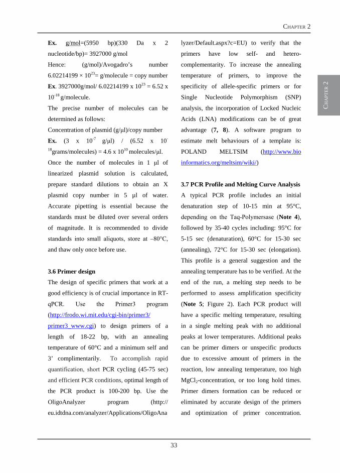
ch
apt
er 2
33
chapter 2
7
Ex. g/mol=(5950 bp)(330 Da x 2
nucleotide/bp)= 3927000 g/mol
Hence: (g/mol)/Avogadro’s number
6.02214199 × 1023= g/molecule = copy number
Ex. 3927000g/mol/ 6.02214199 x 1023 = 6.52 x
10-18 g/molecule.
The precise number of molecules can be
determined as follows:
Concentration of plasmid (g/l)/copy number
Ex. (3 x 10⁻⁷ g/l) / (6.52 x 10-
18grams/molecules) = 4.6 x 10¹⁰ molecules/l.
Once the number of molecules in 1 l of
linearized plasmid solution is calculated,
prepare standard dilutions to obtain an X
plasmid copy number in 5 µl of water.
Accurate pipetting is essential because the
standards must be diluted over several orders
of magnitude. It is recommended to divide
standards into small aliquots, store at –80°C,
and thaw only once before use.
3.6 Primer design
The design of specific primers that work at a
good efficiency is of crucial importance in RT-
qPCR. Use the Primer3 program
(http://frodo.wi.mit.edu/cgi-bin/primer3/
primer3_www.cgi) to design primers of a
length of 18-22 bp, with an annealing
temperature of 60°C and a minimum self and
3’ complimentarily. To accomplish rapid
quantification, short PCR cycling (45-75 sec)
and efficient PCR conditions, optimal length of
the PCR product is 100-200 bp. Use the
OligoAnalyzer program (http://
eu.idtdna.com/analyzer/Applications/OligoAna
lyzer/Default.aspx?c=EU) to verify that the
primers have low self- and hetero-
complementarity. To increase the annealing
temperature of primers, to improve the
specificity of allele-specific primers or for
Single Nucleotide Polymorphism (SNP)
analysis, the incorporation of Locked Nucleic
Acids (LNA) modifications can be of great
advantage (7, 8). A software program to
estimate melt behaviours of a template is:
POLAND MELTSIM (http://www.bio
informatics.org/meltsim/wiki/)
3.7 PCR Profile and Melting Curve Analysis
A typical PCR profile includes an initial
denaturation step of 10-15 min at 95°C,
depending on the Taq-Polymersase (Note 4),
followed by 35-40 cycles including: 95°C for
5-15 sec (denaturation), 60°C for 15-30 sec
(annealing), 72°C for 15-30 sec (elongation).
This profile is a general suggestion and the
annealing temperature has to be verified. At the
end of the run, a melting step needs to be
performed to assess amplification specificity
(Note 5; Figure 2). Each PCR product will
have a specific melting temperature, resulting
in a single melting peak with no additional
peaks at lower temperatures. Additional peaks
can be primer dimers or unspecific products
due to excessive amount of primers in the
reaction, low annealing temperature, too high
MgCl2-concentration, or too long hold times.
Primer dimers formation can be reduced or
eliminated by accurate design of the primers
and optimization of primer concentration.
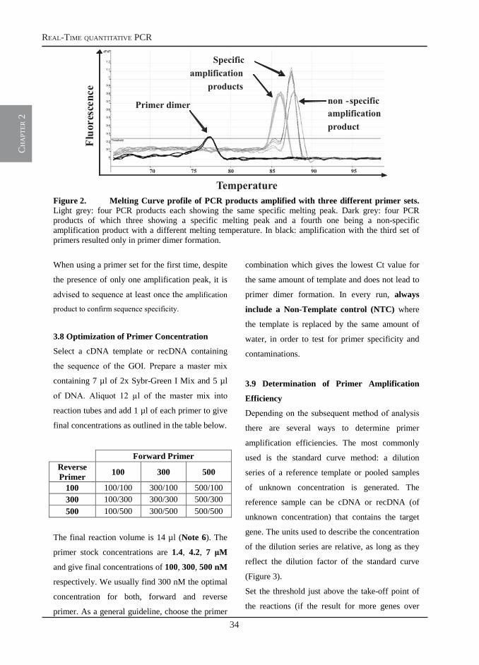
34
real-time quantitative pcr
ch
apt
er 2
8
Figure 2. Melting Curve profile of PCR products amplified with three different primer sets.Light grey: four PCR products each showing the same specific melting peak. Dark grey: four PCR products of which three showing a specific melting peak and a fourth one being a non-specific amplification product with a different melting temperature. In black: amplification with the third set of primers resulted only in primer dimer formation.
When using a primer set for the first time, despite
the presence of only one amplification peak, it is
advised to sequence at least once the amplification
product to confirm sequence specificity.
3.8 Optimization of Primer Concentration
Select a cDNA template or recDNA containing
the sequence of the GOI. Prepare a master mix
containing 7 µl of 2x Sybr-Green I Mix and 5 µl
of DNA. Aliquot 12 µl of the master mix into
reaction tubes and add 1 µl of each primer to give
final concentrations as outlined in the table below.
Forward Primer Reverse Primer 100 300 500
100 100/100 300/100 500/100 300 100/300 300/300 500/300 500 100/500 300/500 500/500
The final reaction volume is 14 µl (Note 6). The
primer stock concentrations are 1.4, 4.2, 7 M
and give final concentrations of 100, 300, 500 nM
respectively. We usually find 300 nM the optimal
concentration for both, forward and reverse
primer. As a general guideline, choose the primer
combination which gives the lowest Ct value for
the same amount of template and does not lead to
primer dimer formation. In every run, always
include a Non-Template control (NTC) where
the template is replaced by the same amount of
water, in order to test for primer specificity and
contaminations.
3.9 Determination of Primer Amplification
Efficiency
Depending on the subsequent method of analysis
there are several ways to determine primer
amplification efficiencies. The most commonly
used is the standard curve method: a dilution
series of a reference template or pooled samples
of unknown concentration is generated. The
reference sample can be cDNA or recDNA (of
unknown concentration) that contains the target
gene. The units used to describe the concentration
of the dilution series are relative, as long as they
reflect the dilution factor of the standard curve
(Figure 3).
Set the threshold just above the take-off point of
the reactions (if the result for more genes over
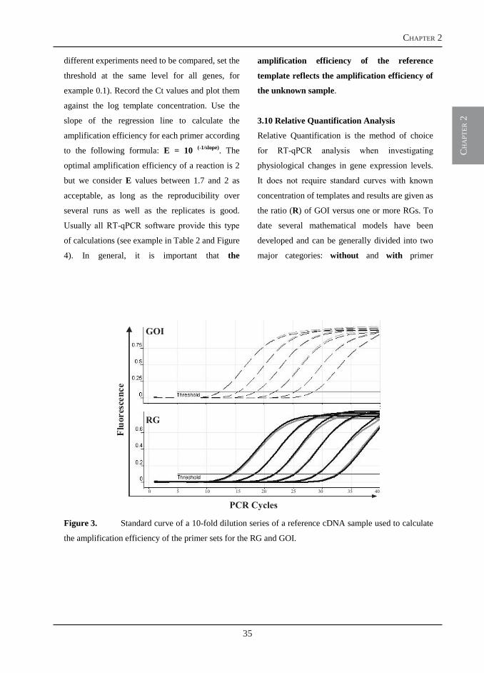
ch
apt
er 2
35
chapter 2
9
different experiments need to be compared, set the
threshold at the same level for all genes, for
example 0.1). Record the Ct values and plot them
against the log template concentration. Use the
slope of the regression line to calculate the
amplification efficiency for each primer according
to the following formula: E = 10 (-1/slope). The
optimal amplification efficiency of a reaction is 2
but we consider E values between 1.7 and 2 as
acceptable, as long as the reproducibility over
several runs as well as the replicates is good.
Usually all RT-qPCR software provide this type
of calculations (see example in Table 2 and Figure
4). In general, it is important that the
amplification efficiency of the reference
template reflects the amplification efficiency of
the unknown sample.
3.10 Relative Quantification Analysis
Relative Quantification is the method of choice
for RT-qPCR analysis when investigating
physiological changes in gene expression levels.
It does not require standard curves with known
concentration of templates and results are given as
the ratio (R) of GOI versus one or more RGs. To
date several mathematical models have been
developed and can be generally divided into two
major categories: without and with primer
Figure 3. Standard curve of a 10-fold dilution series of a reference cDNA sample used to calculate
the amplification efficiency of the primer sets for the RG and GOI.
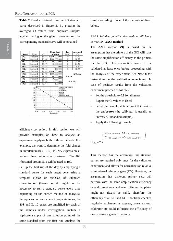
36
real-time quantitative pcr
ch
apt
er 2
10
Table 2 Results obtained from the RG standard
curve described in figure 3. By plotting the
averaged Ct values from duplicate samples
against the log of the given concentration, the
corresponding standard curve will be obtained
efficiency correction. In this section we will
provide examples on how to analyse an
experiment applying both of those methods. For
example, we want to determine the fold change
in interleukin-10 (IL-10) mRNA expression at
various time points after treatment. The 40S
ribosomal protein S11 will be used as RG.
Set up the first run of the day by amplifying a
standard curve for each target gene using a
template cDNA or recDNA of unknown
concentration (Figure 4; it might not be
necessary to run a standard curve every time
depending on the chosen method of analysis).
Set up a second run where in separate tubes, the
40S and IL-10 genes are amplified for each of
the samples under investigation. Include a
triplicate sample of one dilution point of the
same standard from the first run. Analyse the
results according to one of the methods outlined
below.
3.10.1 Relative quantification without efficiency
correction: Ct method
The Ct method (9) is based on the
assumption that the primers of the GOI will have
the same amplification efficiency as the primers
for the RG. This assumption needs to be
validated at least once before proceeding with
the analysis of the experiment. See Note 8 for
instructions on the validation experiment. In
case of positive results from the validation
experiment proceed as follows:
- Set the threshold to 0.1 for all genes.
- Export the Ct values to Excel
- Select the sample at time point 0 (zero) as
the calibrator (the calibrator is usually an
untreated, unhandled sample).
- Apply the following formula:
This method has the advantage that standard
curves are required only once for the validation
experiment and allows for normalization relative
to an internal reference gene (RG). However, the
assumption that different primer sets will
perform with the same amplification efficiency
over different runs and over different templates
might not always be valid. Therefore, the
efficiency of all RG and GOI should be checked
regularly, as changes in reagents, concentrations,
calibrator etc could influence the efficiency of
one or various genes differently.
Ct 40S (calibrator) - Ct IL-10 (calibrator)
Ct 40S (sample 1 -7) - Ct IL-10 (sample 1-7)
R IL-10 = 2
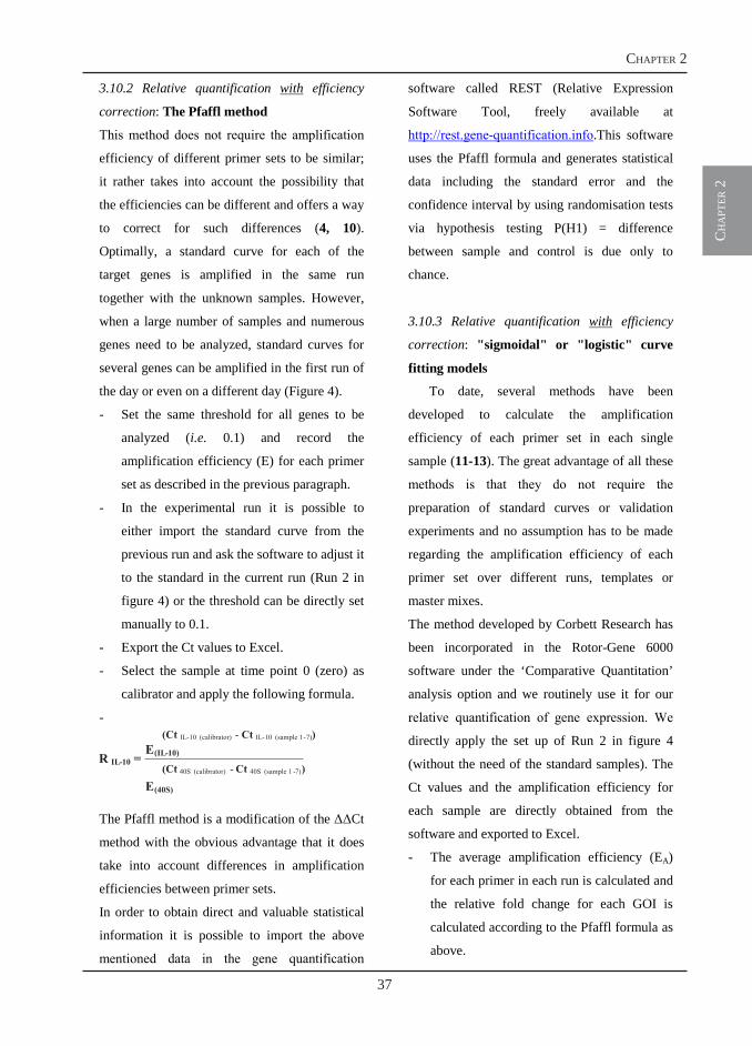
ch
apt
er 2
37
chapter 2
11
3.10.2 Relative quantification with efficiency
correction: The Pfaffl method
This method does not require the amplification
efficiency of different primer sets to be similar;
it rather takes into account the possibility that
the efficiencies can be different and offers a way
to correct for such differences (4, 10).
Optimally, a standard curve for each of the
target genes is amplified in the same run
together with the unknown samples. However,
when a large number of samples and numerous
genes need to be analyzed, standard curves for
several genes can be amplified in the first run of
the day or even on a different day (Figure 4).
- Set the same threshold for all genes to be
analyzed (i.e. 0.1) and record the
amplification efficiency (E) for each primer
set as described in the previous paragraph.
- In the experimental run it is possible to
either import the standard curve from the
previous run and ask the software to adjust it
to the standard in the current run (Run 2 in
figure 4) or the threshold can be directly set
manually to 0.1.
- Export the Ct values to Excel.
- Select the sample at time point 0 (zero) as
calibrator and apply the following formula.
-
The Pfaffl method is a modification of the Ct
method with the obvious advantage that it does
take into account differences in amplification
efficiencies between primer sets.
In order to obtain direct and valuable statistical
information it is possible to import the above
mentioned data in the gene quantification
software called REST (Relative Expression
Software Tool, freely available at
http://rest.gene-quantification.info.This software
uses the Pfaffl formula and generates statistical
data including the standard error and the
confidence interval by using randomisation tests
via hypothesis testing P(H1) = difference
between sample and control is due only to
chance.
3.10.3 Relative quantification with efficiency
correction: "sigmoidal" or "logistic" curve
fitting models
To date, several methods have been
developed to calculate the amplification
efficiency of each primer set in each single
sample (11-13). The great advantage of all these
methods is that they do not require the
preparation of standard curves or validation
experiments and no assumption has to be made
regarding the amplification efficiency of each
primer set over different runs, templates or
master mixes.
The method developed by Corbett Research has
been incorporated in the Rotor-Gene 6000
software under the ‘Comparative Quantitation’
analysis option and we routinely use it for our
relative quantification of gene expression. We
directly apply the set up of Run 2 in figure 4
(without the need of the standard samples). The
Ct values and the amplification efficiency for
each sample are directly obtained from the
software and exported to Excel.
- The average amplification efficiency (EA)
for each primer in each run is calculated and
the relative fold change for each GOI is
calculated according to the Pfaffl formula as
above.
(Ct IL-10 (calibrator) - Ct IL-10 (sample 1-7))
(Ct 40S (calibrator) - Ct 40S (sample 1 -7))R IL-10 = E(IL-10)
E(40S)
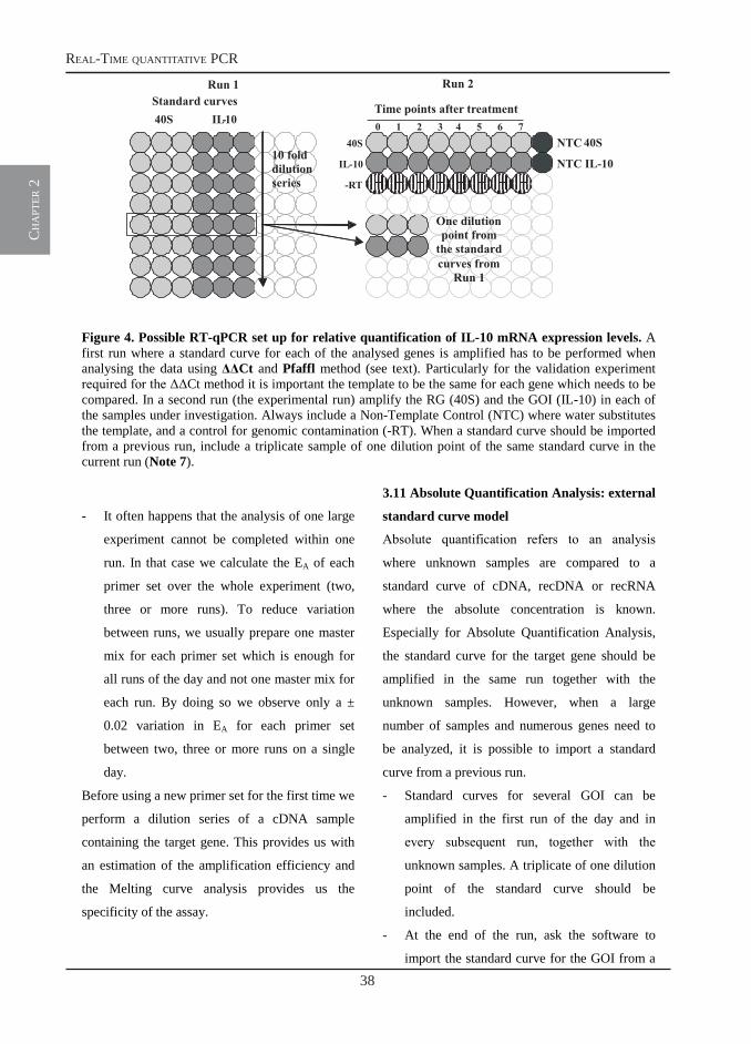
38
real-time quantitative pcr
ch
apt
er 2
12
Figure 4. Possible RT-qPCR set up for relative quantification of IL-10 mRNA expression levels. A first run where a standard curve for each of the analysed genes is amplified has to be performed when analysing the data using Ct and Pfaffl method (see text). Particularly for the validation experiment required for the Ct method it is important the template to be the same for each gene which needs to be compared. In a second run (the experimental run) amplify the RG (40S) and the GOI (IL-10) in each of the samples under investigation. Always include a Non-Template Control (NTC) where water substitutes the template, and a control for genomic contamination (-RT). When a standard curve should be imported from a previous run, include a triplicate sample of one dilution point of the same standard curve in the current run (Note 7).
- It often happens that the analysis of one large
experiment cannot be completed within one
run. In that case we calculate the EA of each
primer set over the whole experiment (two,
three or more runs). To reduce variation
between runs, we usually prepare one master
mix for each primer set which is enough for
all runs of the day and not one master mix for
each run. By doing so we observe only a ±
0.02 variation in EA for each primer set
between two, three or more runs on a single
day.
Before using a new primer set for the first time we
perform a dilution series of a cDNA sample
containing the target gene. This provides us with
an estimation of the amplification efficiency and
the Melting curve analysis provides us the
specificity of the assay.
3.11 Absolute Quantification Analysis: external
standard curve model
Absolute quantification refers to an analysis
where unknown samples are compared to a
standard curve of cDNA, recDNA or recRNA
where the absolute concentration is known.
Especially for Absolute Quantification Analysis,
the standard curve for the target gene should be
amplified in the same run together with the
unknown samples. However, when a large
number of samples and numerous genes need to
be analyzed, it is possible to import a standard
curve from a previous run.
- Standard curves for several GOI can be
amplified in the first run of the day and in
every subsequent run, together with the
unknown samples. A triplicate of one dilution
point of the standard curve should be
included.
- At the end of the run, ask the software to
import the standard curve for the GOI from a
40S
IL-10
NTC40S
NTC IL-10
0 1 2 3 4 5 6 7Time points after treatment
-RT
Run 1 Run 2Standard curves40S IL-10
One dilutionpoint fromthe standardcurves from
Run 1
10 folddilutionseries
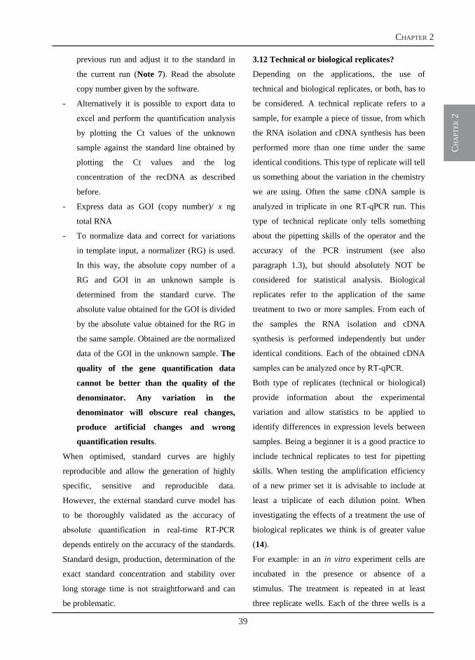
ch
apt
er 2
39
chapter 2
13
previous run and adjust it to the standard in
the current run (Note 7). Read the absolute
copy number given by the software.
- Alternatively it is possible to export data to
excel and perform the quantification analysis
by plotting the Ct values of the unknown
sample against the standard line obtained by
plotting the Ct values and the log
concentration of the recDNA as described
before.
- Express data as GOI (copy number)/ x ng
total RNA
- To normalize data and correct for variations
in template input, a normalizer (RG) is used.
In this way, the absolute copy number of a
RG and GOI in an unknown sample is
determined from the standard curve. The
absolute value obtained for the GOI is divided
by the absolute value obtained for the RG in
the same sample. Obtained are the normalized
data of the GOI in the unknown sample. The
quality of the gene quantification data
cannot be better than the quality of the
denominator. Any variation in the
denominator will obscure real changes,
produce artificial changes and wrong
quantification results.
When optimised, standard curves are highly
reproducible and allow the generation of highly
specific, sensitive and reproducible data.
However, the external standard curve model has
to be thoroughly validated as the accuracy of
absolute quantification in real-time RT-PCR
depends entirely on the accuracy of the standards.
Standard design, production, determination of the
exact standard concentration and stability over
long storage time is not straightforward and can
be problematic.
3.12 Technical or biological replicates?
Depending on the applications, the use of
technical and biological replicates, or both, has to
be considered. A technical replicate refers to a
sample, for example a piece of tissue, from which
the RNA isolation and cDNA synthesis has been
performed more than one time under the same
identical conditions. This type of replicate will tell
us something about the variation in the chemistry
we are using. Often the same cDNA sample is
analyzed in triplicate in one RT-qPCR run. This
type of technical replicate only tells something
about the pipetting skills of the operator and the
accuracy of the PCR instrument (see also
paragraph 1.3), but should absolutely NOT be
considered for statistical analysis. Biological
replicates refer to the application of the same
treatment to two or more samples. From each of
the samples the RNA isolation and cDNA
synthesis is performed independently but under
identical conditions. Each of the obtained cDNA
samples can be analyzed once by RT-qPCR.
Both type of replicates (technical or biological)
provide information about the experimental
variation and allow statistics to be applied to
identify differences in expression levels between
samples. Being a beginner it is a good practice to
include technical replicates to test for pipetting
skills. When testing the amplification efficiency
of a new primer set it is advisable to include at
least a triplicate of each dilution point. When
investigating the effects of a treatment the use of
biological replicates we think is of greater value
(14).
For example: in an in vitro experiment cells are
incubated in the presence or absence of a
stimulus. The treatment is repeated in at least
three replicate wells. Each of the three wells is a
40
real-time quantitative pcr
ch
apt
er 2
14
biological replicate; however, the cells are derived
from a single individual. More relevant would be
to repeat the same in vitro experiment on cells
isolated from three different individuals, each of
them being a biological replicate.
Notes:
1. The extraction and purification procedure of
total RNA must fulfill the following criteria:
free of protein (absorbance 260 nm/280 nm);
free of genomic DNA; should be non
degraded (28S:18S ratio should be roughly
between 1.8 and 2.0, with low amount of
short fragments); free of enzymatic inhibitors
for RT and PCR reaction, which is strongly
dependent on the purification and clean-up
methods; free of any substances which
complex essential reaction co-factors, like
Mg2+ or Mn2+; free of nucleases for extended
storage (15).
2. From 0.1 ng up to 5 µg total RNA can be
transcribed into cDNA using this kit.
Optimally, 1 µg of total RNA is used. In
general, it is important to use the same
amount of starting RNA material for each
sample within the same experiment. This will
greatly reduce the sample-to-sample variation
due to differences in cDNA synthesis
efficiency and will simplify the subsequent
analysis, particularly when absolute
quantification is used. In some cases not all
samples (within the same experiment) would
yield RNA amounts sufficient to use 1 µg of
RNA/sample, it is possible to lower the
amounts down to 0.1 µg, but again this
amount should be used for all samples within
the same experiment.
3. Cloned recDNA and gDNA are very stable
and generate highly reproducible standard
curves even after a long storage time.
Furthermore, the longer templates derived
from recDNA and gDNA mimic the average
native mRNA length of about 2 kb better than
shorter templates derived from RT-PCR
product or oligonucleotides. A problem with
DNA-based calibration curves is that they are
subject to the PCR step only, unlike the
unknown mRNA samples that must first be
reverse transcribed. This increases the
potential for variability of the RT-PCR results
and the amplification results may not be
strictly comparable with the results from the
unknown samples (3).
4. The initial denaturation time depends on the
type of Taq Polymerase present in the master
mix. We strongly advise Hot-Start Taq
Polymerases that require 2 to 15 min at 95°C,
depending on the Taq-Polymerase. This
allows performing the preparation and
aliquoting of the master mix on the bench at
room temperature.
5. At the end of a run, after the last annealing
step, all amplification products will be present
as double-stranded (ds) DNA and Sybr Green
I will be bound to it. During the Melting step
the decrease in fluorescence is measured due
to melting of dsDNA products and
consequent release of the fluorescent dye.
Each product will melt at a specific
temperature. Primer dimers usually have a
lower melting temperature than PCR products
ranging between the 80 and 200 bp.
6. Usually companies advise a final volume of
50 µl but the reaction can easily be scaled
down to save costs. We always try to add at
ch
apt
er 2
41
chapter 2
15
least 5 µl of template. Lower volumes might
increase the chance of pipetting errors.
7. The slope of the calibration curve is more
reproducible than the intercept, and the slope
directly correlates with PCR efficiency.
Hence only a single standard point will be
required to “re-register” a previously
performed calibration curve level for the new
unknown samples. However, this assumes
that the efficiency in a given run is the same
as in a previous run.
8. Amplify a standard curve as described in the
paragraph “Determination of Primer
Amplification Efficiency”. In this case the
reference template has to be the same for
9. both primer sets, and preferably one of the
cDNA samples which is going to be used for
the subsequent experiment.
- After having set the threshold (0.1) export
the Ct values to EXCEL and average the Ct of
replicate samples.
- Calculate the LOG10 of the given arbitrary
concentration (LOGconc).
- Obtain the Ct: for each dilution point
calculate the difference between the Ct(RG) and
Ct(GOI). Plot the LOGconc vs Ct and obtain
the equation of the curve.
If the efficiencies of the two primer sets are
approximately equal, the obtained curve
should be a nearly horizontal line with a slope
< ± 0.1. If this is the case the experiment can
be analysed with the Ct method.
References
1. Nolan, T., Hands, R. E., and Bustin, S. A. (2006) Quantification of mRNA using real-time RT-PCR. Nat. Protocols 1, 1559-1582.
2. Bustin, S. A., Benes, V., Nolan, T., and Pfaffl, M. W. (2005) Quantitative real-time RT-PCR - a perspective. J Mol Endocrinol 34, 597-601.
3. Pfaffl, M. W., and Hageleit, M. (2001) Validities of mRNA quantification using recombinant RNA and recombinant DNA external calibration curves in real-time RT-PCR. Biotechnology Letters 23, 275-282.
4. Pfaffl, M. W. (2001) A new mathematical model for relative quantification in real-time RT-PCR. Nucleic Acids Res 29, e45.
5. Vandesompele, J., De Preter, K., Pattyn, F., Poppe, B., Van Roy, N., De Paepe, A., and Speleman, F. (2002) Accurate normalization of real-time quantitative RT-PCR data by geometric averaging of multiple internal control genes. Genome Biol 3, RESEARCH0034.
6. Pfaffl, M. W., Tichopad, A., Prgomet, C., and Neuvians, T. P. (2004) Determination of stable housekeeping genes, differentially regulated target genes and sample integrity: BestKeeper--Excel-based tool using pair-wise correlations. Biotechnol Lett 26, 509-15.
7. Latorra, D., Arar, K., and Hurley, J. M. (2003) Design considerations and effects of LNA in PCR primers. Mol Cell Probes 17, 253-9.
8. Latorra, D., Campbell, K., Wolter, A., and Hurley, J. M. (2003) Enhanced allele-specific PCR discrimination in SNP genotyping using 3' locked nucleic acid (LNA) primers. Hum Mutat 22, 79-85.
9. Livak, K. J., and Schmittgen, T. D. (2001) Analysis of Relative Gene Expression Data Using Real-Time Quantitative PCR and the 2-[Delta][Delta]CT Method. Methods 25, 402-408.
10. Pfaffl, M. W., Horgan, G. W., and Dempfle, L. (2002) Relative expression software tool (REST) for group-wise comparison and statistical analysis of relative expression results in real-time PCR. Nucleic Acids Res30, e36.
11. Liu, W., and Saint, D. A. (2002) A new quantitative method of real time reverse transcription polymerase chain reaction assay based on simulation of polymerase chain reaction kinetics. Anal Biochem 302, 52-9.
12. Ramakers, C., Ruijter, J. M., Deprez, R. H., and Moorman, A. F. (2003) Assumption-free analysis of quantitative real-time polymerase chain reaction (PCR) data. Neurosci Lett 339, 62-6.
13. Tichopad, A., Dilger, M., Schwarz, G., and Pfaffl, M. W. (2003) Standardized determination of real-time PCR efficiency from a single reaction set-up. Nucleic Acids Res 31, e122.
14. Forlenza, M., de Carvalho Dias, J. D., Vesely, T., Pokorova, D., Savelkoul, H. F., and Wiegertjes, G. F. (2008) Transcription of signal-3 cytokines, IL-12 and IFN alpha beta, coincides with the timing of CD8
42
real-time quantitative pcr
ch
apt
er 2
16
alpha beta up-regulation during viral infection of common carp (Cyprinus carpio L). Mol Immunol 45, 1531-47.
15. Fleige, S., and Pfaffl, M. W. (2006) RNA integrity and the effect on the real-time qRT-PCR performance. Molecular Aspects of Medicine 27, 126-139.
chapter
Transcriptional analysis of the commoncarp (Cyprinus carpio L.) immune response
to the fish louse Argulus japonicus Thiele(Crustacea:Branchiura)
Maria Forlenza*, Peter D. Walker*, Beitske J. de Vries, Sjoerd E. Wendelaar Bonga and Geert F. Wiegertjes
Fish and Shellfish Immunology, 2008 (25), 76-83
* Both authors contributed equally to the work.
3

44
carp immune response to the fish louse argulus
ch
apt
er 3
45
chapter 3
ch
apt
er 3
Transcriptional analysis of the common carp(Cyprinus carpio L.) immune response to the fishlouse Argulus japonicus Thiele (Crustacea:Branchiura)
Maria Forlenza a,1, Peter D. Walker b,1, Beitske J. de Vries a, Sjoerd E.Wendelaar Bonga b, Geert F. Wiegertjes a,*
a Cell Biology and Immunology Group, Wageningen Institute of Animal Sciences, Wageningen University, PO Box 338,6700 AH Wageningen, The Netherlandsb Department of Animal Ecology and Ecophysiology, Faculty of Science, Radboud University of Nijmegen,Toernooiveld 1, 6525 ED Nijmegen, The Netherlands
Received 25 September 2007; revised 12 December 2007; accepted 23 December 2007Available online 10 January 2008
KEYWORDSCarp;Skin;Argulus;Cytokine;Chemokine;Ectoparasite;Immune response
Abstract In the present study we investigated changes in transcription levels of a panel ofselected immune relevant genes in peripheral blood leucocytes (PBL) and skin samples col-lected from carp exposed to larval Argulus japonicus. We show that in skin up-regulation ofgene transcription of the chemokine CXCa, and to a lesser extent the chemokine receptorCXCR1 and the cytokine TNFa, are good indicators of parasite-induced skin damage at 2 dayspost-parasite exposure. Up-regulation of gene transcription corresponded well with an in-crease in leucocytes, probably neutrophilic granulocyte numbers in skin samples collectedat the sites of infection. We show that time-point controls are essential when studying geneexpression, especially in peripheral blood leucocytes (PBL). In addition, we demonstrate thatnon-infected control samples isolated from the skin of infected fish are suitable autologouscontrols, at least until after larval A. japonicus have undergone their first moult and begunto demonstrate increased mobility over their host’s integument. The observed results areindicative of A. japonicus affecting the skin as a whole organ, particularly after the parasites’first moult, a phenomenon which has a great impact on correct skin sampling for RNA isolation.ª 2008 Elsevier Ltd. All rights reserved.
Introduction
Parasite-induced damage and disease in fish is currently re-ceiving more intensive research focus from the scientificcommunity. Fish lice have been shown to be a threat to
* Correspondingauthor. Tel.:þ31317482732; fax:þ31317483955.E-mail address: [email protected] (G.F. Wiegertjes).
1 Both the authors contributed equally to the work.
1050-4648/$ - see front matter ª 2008 Elsevier Ltd. All rights reserved.doi:10.1016/j.fsi.2007.12.013
ava i lab le a t www.sc iencedi rec t .com
journa l homepage : www.e lsev ie r . com/ loca te / f s i
Fish & Shellfish Immunology (2008) 25, 76e83
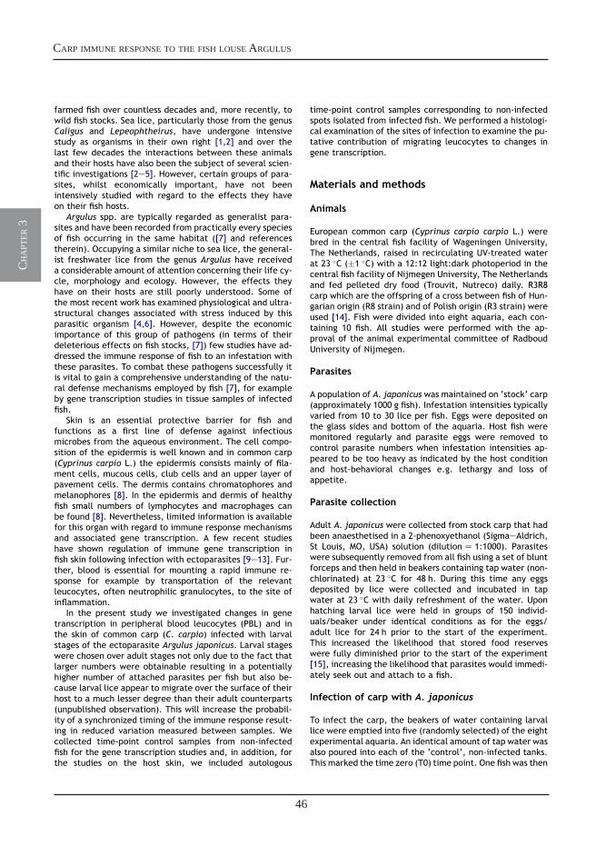
46
carp immune response to the fish louse argulus
ch
apt
er 3
farmed fish over countless decades and, more recently, towild fish stocks. Sea lice, particularly those from the genusCaligus and Lepeophtheirus, have undergone intensivestudy as organisms in their own right [1,2] and over thelast few decades the interactions between these animalsand their hosts have also been the subject of several scien-tific investigations [2e5]. However, certain groups of para-sites, whilst economically important, have not beenintensively studied with regard to the effects they haveon their fish hosts.
Argulus spp. are typically regarded as generalist para-sites and have been recorded from practically every speciesof fish occurring in the same habitat ([7] and referencestherein). Occupying a similar niche to sea lice, the general-ist freshwater lice from the genus Argulus have receiveda considerable amount of attention concerning their life cy-cle, morphology and ecology. However, the effects theyhave on their hosts are still poorly understood. Some ofthe most recent work has examined physiological and ultra-structural changes associated with stress induced by thisparasitic organism [4,6]. However, despite the economicimportance of this group of pathogens (in terms of theirdeleterious effects on fish stocks, [7]) few studies have ad-dressed the immune response of fish to an infestation withthese parasites. To combat these pathogens successfully itis vital to gain a comprehensive understanding of the natu-ral defense mechanisms employed by fish [7], for exampleby gene transcription studies in tissue samples of infectedfish.
Skin is an essential protective barrier for fish andfunctions as a first line of defense against infectiousmicrobes from the aqueous environment. The cell compo-sition of the epidermis is well known and in common carp(Cyprinus carpio L.) the epidermis consists mainly of fila-ment cells, mucous cells, club cells and an upper layer ofpavement cells. The dermis contains chromatophores andmelanophores [8]. In the epidermis and dermis of healthyfish small numbers of lymphocytes and macrophages canbe found [8]. Nevertheless, limited information is availablefor this organ with regard to immune response mechanismsand associated gene transcription. A few recent studieshave shown regulation of immune gene transcription infish skin following infection with ectoparasites [9e13]. Fur-ther, blood is essential for mounting a rapid immune re-sponse for example by transportation of the relevantleucocytes, often neutrophilic granulocytes, to the site ofinflammation.
In the present study we investigated changes in genetranscription in peripheral blood leucocytes (PBL) and inthe skin of common carp (C. carpio) infected with larvalstages of the ectoparasite Argulus japonicus. Larval stageswere chosen over adult stages not only due to the fact thatlarger numbers were obtainable resulting in a potentiallyhigher number of attached parasites per fish but also be-cause larval lice appear to migrate over the surface of theirhost to a much lesser degree than their adult counterparts(unpublished observation). This will increase the probabil-ity of a synchronized timing of the immune response result-ing in reduced variation measured between samples. Wecollected time-point control samples from non-infectedfish for the gene transcription studies and, in addition, forthe studies on the host skin, we included autologous
time-point control samples corresponding to non-infectedspots isolated from infected fish. We performed a histologi-cal examination of the sites of infection to examine the pu-tative contribution of migrating leucocytes to changes ingene transcription.
Materials and methods
Animals
European common carp (Cyprinus carpio carpio L.) werebred in the central fish facility of Wageningen University,The Netherlands, raised in recirculating UV-treated waterat 23 �C (�1 �C) with a 12:12 light:dark photoperiod in thecentral fish facility of Nijmegen University, The Netherlandsand fed pelleted dry food (Trouvit, Nutreco) daily. R3R8carp which are the offspring of a cross between fish of Hun-garian origin (R8 strain) and of Polish origin (R3 strain) wereused [14]. Fish were divided into eight aquaria, each con-taining 10 fish. All studies were performed with the ap-proval of the animal experimental committee of RadboudUniversity of Nijmegen.
Parasites
A population of A. japonicus was maintained on ‘stock’ carp(approximately 1000 g fish). Infestation intensities typicallyvaried from 10 to 30 lice per fish. Eggs were deposited onthe glass sides and bottom of the aquaria. Host fish weremonitored regularly and parasite eggs were removed tocontrol parasite numbers when infestation intensities ap-peared to be too heavy as indicated by the host conditionand host-behavioral changes e.g. lethargy and loss ofappetite.
Parasite collection
Adult A. japonicus were collected from stock carp that hadbeen anaesthetised in a 2-phenoxyethanol (SigmaeAldrich,St Louis, MO, USA) solution (dilutionZ 1:1000). Parasiteswere subsequently removed from all fish using a set of bluntforceps and then held in beakers containing tap water (non-chlorinated) at 23 �C for 48 h. During this time any eggsdeposited by lice were collected and incubated in tapwater at 23 �C with daily refreshment of the water. Uponhatching larval lice were held in groups of 150 individ-uals/beaker under identical conditions as for the eggs/adult lice for 24 h prior to the start of the experiment.This increased the likelihood that stored food reserveswere fully diminished prior to the start of the experiment[15], increasing the likelihood that parasites would immedi-ately seek out and attach to a fish.
Infection of carp with A. japonicus
To infect the carp, the beakers of water containing larvallice were emptied into five (randomly selected) of the eightexperimental aquaria. An identical amount of tap water wasalso poured into each of the ‘control’, non-infected tanks.This marked the time zero (T0) time point. One fish was then
Carp immune response to the fish louse Argulus 77
47
chapter 3
ch
apt
er 3
removed from each of the tanks at T0, 10 h, 24 h (1 day),48 h (2 days), 72 h (3 days) and 6 days post infection. Uponremoval, the fish were irreversibly anaesthetised in 2-phenoxyethanol (dilutionZ 1:500) and subsequentlyweighed, measured (standard length) and the number of at-tached parasites recorded.
Peripheral blood leucocytes (PBL) and skin isolation
Blood was collected via puncture of the caudal vessel anddiluted 1:1 with cRPMI (RPMI 1640 (Cambrex, Verviers,Belgium) adjusted to 270 mOsmol/kg) containing 50 IU ml-1
of heparin (Leo Pharmaceutical Products, Weesp, The Neth-erlands). After centrifugation at 600 g for 10 min, the buffy-coat containing leucocytes was collected and layered on5 ml of Ficoll-Paque� Plus (Amersham Biosciences, Up-psala, Sweden). Following subsequent centrifugation at800 g for 25 min, the PBL at the interface were collectedand washed three times with cRPMI. Cell pellets were col-lected, immediately snap frozen in liquid nitrogen andstored at �80 �C until used for RNA isolation.
Several 5� 5 mm samples of skin were carefully re-moved from the ventral region of the flanks of uninfected(control) and infected fish. For infected fish, the sampleswere taken from sites of parasite attachment/feeding (in-fected spots) and also from sites distant to the sites ofinfection (autologous controls). The number of infectedspots differed between individual fish. Skin samples werethen immediately snap frozen in liquid nitrogen and storedat �80 �C until use or immersed in Bouin’s fixative for sub-sequent processing and histological analysis.
Histological analysis
Skin samples for histological analyses were fixed fora minimum of 24 h in Bouin’s fixative. Samples were subse-quently dehydrated through a graded series of ethanol andembedded in paraffin. Five mm sections were mounted ongelatinized glass slides and dried overnight in an oven at40 �C. Slides were then stained with hematoxylin and eosinand examined microscopically for evidence of parasite-induced damage and host inflammatory responses (i.e. infil-tration of leucocytes).
RNA isolation and quantification
For real-time quantitative polymerase chain reaction (RT-qPCR) analysis total RNA was isolated from PBL and skinusing the RNAeasy Mini Kit (Qiagen, Leusden, The Nether-lands) according to the manufacturer’s instructions. On-column DNase treatment with the RNase-free DNase set(Qiagen) was also included.
RNA was isolated, separately from three skin pieces fromeach of the three non-infected controls or from five pieces(autologous controls) and one to four pieces (infected spots)from each of the five infected fish. The RNA isolated frommore pieces from the same individual was never pooled andeach piece was handled and analyzed separately.
The concentration of RNA was measured spectrophoto-metrically (GeneQuant, Pharmacia Biotech) at OD260 nm and
the purity determined as the OD260 nm/OD280 nm ratio withexpected values between 1.8 and 2.0. The integrity ofRNA was determined by electrophoresis on 1% agarose gelcontaining 0.5 mg ml-1 ethidium bromide at 100 V. TotalRNA was stored at �80 �C until further use.
cDNA synthesis
Prior to cDNA synthesis, a second DNase treatment was per-formed using DNase I, amplification grade (Invitrogen,Breda, The Netherlands). Briefly, 1 mg of RNA from eachsample was combined with 1 ml 10� DNase reaction bufferand 1 U DNase I up to a final volume of 10 ml, mixed and in-cubated at RT for 15 min, followed by the inactivation ofDNase I by adding 1 ml of 25 mM EDTA. Synthesis of cDNAwas performed with Invitrogen’s Superscript� III FirstStrand Synthesis Systems for RT-PCR, according to the man-ufacturer’s instructions. Briefly, DNase I-treated RNA sam-ples (11 ml) were mixed with 5� first strand buffer, 300 ngrandom primers, 10 mM dNTPs, 0.1 M DTT, 10 U RNase inhib-itor, and 200 U Superscript III Reverse Transcriptase(Invitrogen) up to a final volume of 20 ml. The mixturewas incubated at 37 �C for 60 min followed by an inactiva-tion step at 70 �C for 15 min. A non-reverse transcriptasecontrol was included for each sample. Before use as tem-plate in RT-qPCR experiments, the cDNA was further diluted25 times in nuclease-free water.
Real-time quantitative polymerase chain reaction(RT-qPCR)
RT-qPCR using SYBR Green I technology was performedusing Rotor-Gene� 2000 (Corbett Research, Mortlake,Sydney, Australia) with the Brilliant� SYBR� Green QPCR(Stratagene, La Jolla, CA, USA) as detection chemistry.The primers used for RT-qPCR are listed in Table 1. PCRconditions were optimized by analyzing the melting curvesof the products and product specificity was assessed byanalysis on a 1% agarose gel. Master-mix for each PCR runwas prepared as follows: 0.32 ml of water, 0.84 ml of eachprimer (5 mM), 7 ml Master SYBR Green I mix. Finally, 5 mlof diluted cDNA was added to 9 ml of master-mix and trans-ferred to a 0.1 ml tube. The following amplification pro-gram was used: after 15 min of denaturation at 95 �C, 40cycles of RT-qPCR with three-step amplification were per-formede15 s at 95 �C for denaturation, 30 s at 60 �C for an-nealing and 30 s at 72 �C for elongation followed by a finalholding step of 1 min at 60 �C. A melting step was then per-formed with continuous fluorescence acquisition starting at60 �C with a rate of 1 �C/5 s up to 99 �C to determine theamplification specificity. In all cases, the amplificationswere specific and no amplification was observed in the neg-ative controls (non-template control and non-reverse tran-scriptase control). Fluorescence data from RT-qPCRexperiments were analyzed using Rotor-Gene version6.0.21 software and exported to Microsoft Excel. The cyclethreshold Ct for each sample and the reaction efficiencies(E ) for each primer set were obtained upon comparativequantitation analysis from the Rotor-Gene version 6.0.21software. Briefly, the E for each primer set was recordedper sample and an average E (EA) was then calculated for
78 M. Forlenza et al.
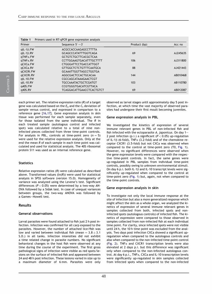
48
carp immune response to the fish louse argulus
ch
apt
er 3
each primer set. The relative expression ratio (R) of a targetgene was calculated based on the EA and the Ct deviation ofsample versus control, and expressed in comparison to areference gene [16,17]. Gene expression analysis in skintissue was performed for each sample separately, evenfor those isolated from the same individual. The R ineach treated sample (autologous control and infectedspot) was calculated relative to a total of nine non-infected pieces collected from three time-point controls.For analysis in PBL, controls at time-point zero (nZ 5)were used for the relative expression analysis. Only at theend the mean R of each sample in each time point was cal-culated and used for statistical analysis. The 40S ribosomalprotein S11 was used as an internal reference gene.
Statistics
Relative expression ratios (R) were calculated as describedabove. Transformed values (ln(R)) were used for statisticalanalysis in SPSS software (version 15.0). Homogeneity ofvariance was analyzed using the Levene’s test. Significantdifferences (P< 0.05) were determined by a two-way AN-OVA followed by a Sidak test. In case of unequal variancesbetween groups, the two-way ANOVA was followed bya GameseHowell test.
Results
General observations
Larval parasites were found attached to fish just 2 h post in-fection. Infection was confirmed for all carp exposed to theparasites. However, the number of attached lice/fish waslow and varied between individual fish (meanZ 3.8� 3.1S.D.) in all tanks. Infection intensities did not exhibita time related change in parasite numbers. No significantbehavioral changes in the host fish were observed at anytime during the course of the experiment. The first grosspathological signs of infection were visible as red spots/le-sions on the surface of infected fish and appeared between24 and 48 h post infection. These lesions varied in size up toa maximum diameter of 6 mm. Parasites were only
observed as larval stages until approximately day 5 post in-fection, at which time the vast majority of observed para-sites had undergone their first moult becoming juveniles.
Gene expression analysis in PBL
We investigated the kinetics of expression of severalimmune relevant genes in PBL of non-infected fish andfish infected with the ectoparasite A. japonicus. On day 1e2 post infection (p.i.) a significant (P< 0.05) up-regulationof IL-1b (6-fold), TNFa (3.2-fold) and of the chemokine re-ceptor CXCR1 (3.5-fold) but not CXCa was observed whencompared to the control at time-point zero (T0, Fig. 1).However, no significant differences were observed whenthe gene expression levels were compared with the respec-tive time-point controls. In fact, the same genes wereup-regulated in PBL samples from individual time-pointcontrols, possibly owing to unknown environmental stimuli.On day 6 p.i. both IL-1b and IL-10 transcript levels were sig-nificantly up-regulated when compared to the control attime-point zero (Fig. 1) but, again, not when compared tothe time-point controls.
Gene expression analysis in skin
To investigate not only the local immune response at thesite of infection but also a more generalized response whichmight affect the skin as a whole organ, we analyzed the ki-netics of expression of several immune relevant genes insamples collected from both, infected spots and non-infected spots (autologous controls) of infected fish. The ki-netics of expression were compared to those observed insamples collected from non-infected fish at each individualtime point. For clarity, since infected spots were not visibleuntil 24 h, the 10 h time point was excluded from the anal-ysis. Two days post infection CXCa showed a significant up-regulation when compared to the autologous controls andalso when compared to the non-infected time-point control(Fig. 2). TNFa and CXCR1 transcription levels were alsoelevated at 2 days p.i. but this difference was significantonly when compared to the non-infected autologous con-trol. At day 6 p.i., TNFa, CXCa and IL-10 transcription levelswere significantly up-regulated in skin samples collectedfrom infected spots when compared to the non-infected
Table 1 Primers used in RT-qPCR gene expression analysis
Primer Sequence 50/30 Product (bp) Acc no
qIL-1b.FW ACGCCACCAAGAGCCTTTTAqIL-1b.RV GCAGCCCATATTTGGTCAGA 69 AJ245635qTNFa.FW GCTGTCTGCTTCACGCTCAAqTNFa.RV CCTTGGAAGTGACATTTGCTTTT 106 AJ311800qCXCa.FW CTGGGATTCCTGACCATTGGTqCXCa.RV GTTGGCTCTCTGTTTCAATGCA 88 AJ421443qCXCRI.FW GCAAATTGGTTAGCCTGGTGAqCXCRI.RV AGGCGACTCCACTGCACAA 144 AB010468qIL-10.FW CGCCAGCATAAAGAACTCGTqIL-10.RV TGCCAAATACTGCTCGATGT 103 AB110780q40S.FW CCGTGGGTGACATCGTTACAq40S.RV TCAGGACATTGAACCTCACTGTCT 69 AB012087
Carp immune response to the fish louse Argulus 79
49
chapter 3
ch
apt
er 3
time-point control. At the same time point, only for TNFaand CXCa, significantly elevated transcription levels wereobserved in the autologous control samples when comparedto the respective non-infected time-point control.
Histological analysis
To investigate whether chemokine (CXCa) up-regulation ofgene transcription could be related to increased transcrip-tion activity in leucocytes already present in the skin or to anincreased number of leucocytes in the skin following in-fection we performed a histological analysis of the skinsamples. Fig. 3B shows a massive infiltration of leucocytes atthe site of infection. A large amount of inflammatory infil-trate can be observed not only in both the mildly hyperplas-tic epidermal and dermal tissues but also throughout thewhole subcutaneous fatty layer. A very low number of leuco-cytes could be observed in the control skin samples (Fig. 3A).
Discussion
We investigated the immune response of common carp tofreshwater lice from the genus Argulus. The most pro-nounced up-regulation of gene transcription was observed
for the chemokine CXCa in the skin at 1e2 days post infec-tion with larval A. japonicus. At the same time a massive in-filtration of leucocytes, most likely neutrophilicgranulocytes, at the site of infection could be observed inhistological sections of the skin. At day 6 post infection im-mune gene transcription was up-regulated not only in theinfected skin but also, although to a lesser extent, in autol-ogous skin sample controls collected from non-infectedspots of infected fish. This suggests that the response toA. japonicus larval stages is initially restricted to the siteof infection but is extended to a generalized responsethroughout the skin as a whole organ at a later stage ofthe infection. In PBL, transcription levels of the investi-gated genes varied greatly in control fish, emphasizingthe importance of time-point control samples for gene tran-scription studies.
During our experiment, the first visible red, inflamedspots were not observed until approximately 2 days postinfection, despite the attachment of several larval lice tothe skin of exposed fish as early as 2 h post infection. Inother experiments, the first notable response to argulidinfestations, observed as small red areas (up to severalmm in diameter), has been noted even within a few hourspost infection [7]. Although we could confirm infection
IL-1β
0
2
4
6
8
10
12
14
0 10h 1d 2d 3d 6d
IL-10
0
1
2
3
4
5
6
0 10h 1d 2d 3d 6d
TNFα
0
1
2
3
4
5
6
0 10h 1d 2d 3d 6d
CXCa
0
1
2
3
4
5
6
0 10h 1d 2d 3d 6d
CXCR1
0
1
2
3
4
5
6
0 10h 1d 2d 3d 6d
Rel
ativ
e to
40S
Rel
ativ
e to
40S
Rel
ativ
e to
40S
Rel
ativ
e to
40S
Rel
ativ
e to
40S
time post infection
time post infection
Control
Infected
**
* *
*
*
*
**,a *
**
*
Figure 1 Kinetics of gene expression in PBL after Argulus japonicus infection of common carp (Cyprinus carpio L.). Fish wereexposed to 150 larval parasites/tank by bath challenge. PBL from non-infected and infected fish were collected at differenttime points post infection. Expression was determined by real-time quantitative PCR and expressed relative to S11 protein ofthe 40S subunit. Data represent mean values of nZ 5 time-zero controls, nZ 5 infected fish and nZ 3 time-point controls(�SD). Symbol ‘*’ represents a significant difference as compared to non-infected controls at time-point zero. ‘a’ indicates a sig-nificant difference as compared to the non-infected time-point control.
80 M. Forlenza et al.

50
carp immune response to the fish louse argulus
ch
apt
er 3
for each individual fish, the infection pressure in our exper-iment was relatively mild as shown by a mean infection in-tensity of 3.8 attached parasites per fish. Further, we choseto investigate the immune response to larval rather than ju-venile or adult lice. Adult argulids are quite mobile beingable to glide over the surface of their hosts with relativeease using their maxillary suckers [18]. This can result inmultiple sites of infection on the same host being causedby one parasite at different times. However, larval stagestend to be less mobile and remain relatively stationary, an-choring themselves to their hosts integument via the use oftheir hook-like second antennae [7]. Argulus spp. cause di-rect damage to the fish skin through their attachment andfeeding mechanisms although, typically, the craters donot penetrate much deeper than the epidermis [19]. Skindamage is the result of mechanical actions of the maxillarysuckers in adult lice and hooks or spines in larval and juve-nile stages and the sharp mandibles. In addition, damageresults from various toxins or digestive enzymes secretedvia the pre-oral stylet and labial spines [20].
Two days post infection, levels of CXCa transcriptswere up-regulated in skin samples collected at the sitesof infection (infected spots), and were significantly
different from the levels measured in skin isolated notonly from non-infected control fish but also from autolo-gous skin samples collected from non-infected spots of in-fected fish. At the same time point, TNFa and CXCreceptor-1 (CXCR1) transcription were found to be up-regulated with respect to the autologous control. In com-parison to the response observed 1e2 days post infection,at 6 days post infection a clear up-regulation of CXCa,TNFa and, to a lesser extent, that IL-10 transcripts wasobserved in skin samples collected from both infectedand non-infected spots of the infected fish. These resultsindicate that whilst the immune response to A. japonicuslarval stages (2 days post infection) is restricted to thesite of infection, the response to juvenile stages (6 dayspost infection) is extended throughout the skin as a wholeorgan. For A. japonicus, in fact, a succession of moultstakes place approximately every 5 days, depending onthe ambient temperature [21]. It is likely, also duringour experiment, after 5 days A. japonicus larval stagesstarted to moult and migrate over the skin of the fishhost. This would explain why the change in gene expres-sion profile was comparable, although smaller, betweenautologous skin sample controls and infected samples.
IL-1β
0
1
2
3
4
5
6
7
24h 2d 3d 6d
IL-10
0
1
2
3
4
5
6
7
24h 2d 3d 6d
time post infection
TNFα
0
1
2
3
4
5
6
7
24h 2d 3d 6d
CXCa
0
1
2
3
4
5
6
7
24h 2d 3d 6d
CXCR1
0
1
2
3
4
5
6
7
24h 2d 3d 6d
time post infectionR
elat
ive
to 4
0SR
elat
ive
to 4
0S
Rel
ativ
e to
40S
Rel
ativ
e to
40S
Rel
ativ
e to
40S Control
Non-infected spot
Infected spot
a a
**
* **,a
*
a
Figure 2 Kinetics of gene expression in skin after Argulus japonicus infection of common carp (Cyprinus carpio L.). Fish wereexposed to 150 larval parasites/tank by bath challenge. Skin samples were collected at different time points post infectionfrom non-infected fish, non-infected spots of infected fish (autologous control) and infected spots of infected fish. Expressionwas determined by real-time quantitative PCR and expressed relative to S11 protein of the 40S subunit. Data represent mean valuesof nZ 5 infected fish and nZ 3 time-point controls (�SD). Symbol ‘*’ represents a significant difference as compared to thenon-infected time-point control. ‘a’ indicates a significant difference as compared to the autologous control.
Carp immune response to the fish louse Argulus 81
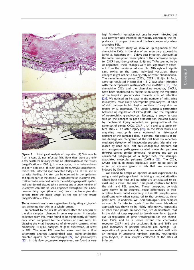
51
chapter 3
ch
apt
er 3
The observed results are suggestive of migrating A. japoni-cus affecting the skin as a whole organ.
In contrast to the results obtained after the analysis ofthe skin samples, changes in gene expression in samplescollected from PBL were found to be significantly differentonly when compared to the controls at time zero, in-dicating the vital importance of time-point controls whenemploying RT-qPCR analyses of gene expression, at leastin PBL. The same PBL samples were used for a flowcytometric analysis (unpublished data) using antibodiesdirected against carp macrophages [22] and granulocytes[23]. In this flow cytometer experiment we found a very
high fish-to-fish variation not only between infected butalso between non-infected individuals, confirming the im-portance of proper time-point controls, especially whenanalyzing PBL.
In the present study we show an up-regulation of thechemokine CXCa in the skin of common carp exposed tolarval A. japonicus at 1e2 days post infection. Although atthe same time-point transcription of the chemokine recep-tor CXCR1 and the cytokines IL-1b and TNFa seemed to beup-regulated, these changes were not significantly differ-ent from the non-infected controls. Although not signifi-cant owing to the large individual variation, thesechanges might reflect a biologically relevant phenomenon.The same immune genes (CXCa, CXCR1, IL-1b), in fact,were up-regulated in carp skin 1.5e2 days after infectionwith the ectoparasite Ichthyophthirius multifiliis [13]. Thechemokine CXCa and the chemokine receptor, CXCR1,have been implicated as factors stimulating the migrationof neutrophilic granulocytes towards sites of infection[24]. We noticed an increase in the number of infiltratingleucocytes, most likely neutrophilic granulocytes, at sitesof skin damage in histological sections of carp skin in-fected by A. japonicus. This would suggest a correlationbetween up-regulation of CXCa (CXR1) and the migrationof neutrophilic granulocytes. Recently, a study in carpskin on the changes in gene transcription induced purelyby mechanical injury reported an up-regulation of thesame set of genes: CXCa, CXCR1, IL-1b and to a lesser ex-tent TNFa 2e3 h after injury [25]. In the latter study alsomigrating neutrophils were observed in histologicalsections of the damaged skin. In general, the results of tis-sue damage are recognized at the cell level via receptor-mediated detection of intracellular proteins (alarmins) re-leased by dead cells. Not only endogenous alarmins butalso exogenous pathogen-associated molecular patterns(PAMPs) convey similar (immune) responses and can beconsidered subgroups of a larger set, the damage-associated molecular patterns (DAMPs) [26]. The CXCa,CXCR1 and IL-1b genes especially seem to be part ofa set of immune genes in fish that are commonlyinduced by DAMPs.
We aimed to design an optimal animal experiment byusing a mild pathogen load mimicking a natural situationwhere both the host and parasite are anticipated to co-exist and survive. We used time-point controls for boththe skin and PBL samples. These time-point controlswere shown to be essential since differences in tran-scription levels noted especially in the PBL samples weresignificant only when compared to the controls at time-point zero. In addition, we used autologous skin samplesas controls for infected spots from the same fish whoseapproach was shown to be highly informative, especiallyat later time points. In conclusion, we demonstrate thatin the skin of carp exposed to larval/juvenile A. japoni-cus up-regulation of gene transcription for the chemo-kine CXCa and to a lesser extent the chemokinereceptor CXCR1 and the cytokines IL-1b and TNFa aregood indicators of parasite-induced skin damage. Up-regulation of gene transcription corresponded well withan increase in leucocyte numbers, possibly neutrophilicgranulocytes, in skin samples collected at the sites ofinfection.
Figure 3 Histological analysis of carp skin. (A) Skin samplefrom a control, non-infected fish. Note that there are onlya few scattered leucocytes and no inflammation of the tissues(magnificationZ 1000�), LZ leucocytes, mZmelanophoresand acZ club cells. (B) Skin sample from Argulus japonicus in-fected fish. Infected spot collected 2 days p.i. at the site ofparasite feeding. A crater can be observed in the epidermisand apical part of the dermis. A high degree of leucocyte infil-tration can be observed in both the mildly hyperplastic epider-mal and dermal tissues (thick arrows) and a large number ofleucocytes can also be seen dispersed throughout the subcu-taneous fatty layer (thin arrows). Note the leucocytes dis-persing from the blood vessel at the top of the image(magnificationZ 300�).
82 M. Forlenza et al.
52
carp immune response to the fish louse argulus
ch
apt
er 3
Acknowledgements
This work was funded by the European Community’sImproving Human Potential Programme under contractHPRN-CT-2001-00214, PARITY, including a fellowship forMF and PDW. The authors also wish to thank T. Spanings forfish husbandry and M. Joerink, J.J. Taverne-Thiele,S. Gomez and W. Abbink for assistance with fish sampling.
References
[1] Boxshall GA, Defaye D. Pathogens of wild and farmedfish e sea lice. Francis: Chichester, UK; 1993.
[2] Johnson SC, Albright LJ. The developmental stages of Lep-eophtheirus salmonis (Krøyer, 1837) (Copepoda: Caligidae).Can J Zool 1991;69:929e50.
[3] Krkosek M, Lewis MA, Volpe JP. Transmission dynamics of par-asitic sea lice from farm to wild salmon. Proc R Soc Lond Ser BBiol Sci 2004;272:689e96.
[4] Nolan DT, van der Salm AL, Wendelaar Bonga SE. The hosteparasite relationship between the rainbow trout (Oncorhyn-chus mykiss) and the ectoparasite Argulus foliaceus (Crusta-cea: Branchiura): epithelial mucous cell response, cortisoland factors which may influence parasite establishment. Con-trib Zool 2000;69:57e63.
[5] Wells A, Grierson CE, MacKenzie M, Russon IJ, Reinardy H,Middlemiss C, et al. Physiological effects of simultaneous,abrupt seawater entry and sea lice (Lepeophtheirus salmonis)infestation of wild, sea-run brown trout (Salmo trutta) smolts.Can J Fish Aquat Sci 2006;63:2809e21.
[6] Van der Salm AL, Nolan DT, Spannings FAT, WendelaarBonga SE. Effects of infection with the ectoparasite Argulusjaponicus (Thiele) and administration of cortisol on cellularproliferation and apoptosis in the epidermis of common carpCyprinus carpio (L.) skin. J Fish Dis 2000;23:173e84.
[7] Walker PD, Flik G, Wendelaar Bonga SE. The biology of para-sites from the genus Argulus and a review of the interactionswith its host. In: Wiegertjes GF, Flik G, editors. Hosteparasiteinteractions. Abingdon, UK: Garland/BIOS ScientificPublishers; 2004. p. 107e29.
[8] Iger Y, Lock RA, van der Meij JC, Wendelaar Bonga SE. Effectsof water-borne cadmium on the skin of the common carp(Cyprinus carpio). Arch Environ Contam Toxicol 1994;26:342e50.
[9] Lindenstrøm T, Buchmann K, Secombes CJ. Gyrodactylus der-javini infection elicits IL-1beta expression in rainbow troutskin. Fish Shellfish Immunol 2003;15:107e15.
[10] Lindenstrøm T, Secombes CJ, Buchmann K. Expression of im-mune response genes in rainbow trout skin induced by Gyro-dactylus derjavini infections. Vet Immunol Immunopathol2004;97:137e48.
[11] Sigh J, Lindenstrøm T, Buchmann K. The parasitic ciliate Ich-thyophthirius multifiliis induces expression of immune rele-vant genes in rainbow trout, Oncorhynchus mykiss(Walbaum). J Fish Dis 2004;27:409e17.
[12] Sigh J, Lindenstrøm T, Buchmann K. Expression of pro-inflam-matory cytokines in rainbow trout (Oncorhynchus mykiss) dur-ing an infection with Ichthyophthirius multifiliis. Fish ShellfishImmunol 2004;17:75e86.
[13] Gonzalez SF, Buchmann K, Nielsen ME. Real-time gene expres-sion analysis in carp (Cyprinus carpio L.) skin: inflammatoryresponses caused by the ectoparasite Ichthyophthirius multi-filiis. Fish Shellfish Immunol 2007;22:641e50.
[14] Irnazarow I. Genetic variability of Polish and Hungarian carplines. Aquacult Res 1995;129:215e9.
[15] Tam Q, Avenant-Oldewage A. The digestive system of larvalArgulus japonicus (Branchiura). J Crustacean Biol 2006;26:447e54.
[16] Pfaffl MW. A new mathematical model for relative quantifica-tion in real-time RT-PCR. Nucleic Acids Res 2001;29:e45.
[17] Tichopad A, Dilger M, Schwarz G, Pfaffl MW. Standardized de-termination of real-time PCR efficiency from a single reactionset-up. Nucleic Acids Res 2003;31:122.
[18] Kearn GC. Leeches, lice and lampreys: a natural history of skinand gill parasites of fishes. Dordrecht: Springer; 2004.
[19] Lester RJG, Roubal F. Phylum Arthropoda. In: Woo PTK, editor.Fish diseases and disorders. Wallingford, UK: CAB Interna-tional; 1995. p. 475e598.
[20] Shimura S, Inoue K. Toxic effects of extract from the mouth-parts of Argulus coregoni Thorell (Crustacea: Branchiura).Bull Jpn Soc Sci Fish 1984;50:729.
[21] Fryer G. The parasitic Copepoda and Branchiura of Britishfreshwater fishes: a handbook and key. Freshwater BiologicalAssociation, Scientific Publications; 1982.
[22] Weyts FAA, Rombout JHWM, Flik G, Verburg-VanKemenade BML. A common carp (Cyprinus carpio L.) leuco-cyte cell line shares morphological and functional charac-teristics with macrophages. Fish Shellfish Immunol 1997;7:123e33.
[23] Nakayasu C, Omori M, Hasegawa S, Kurata O, Okamoto N. Pro-duction of a monoclonal antibody for carp (Cyprinus carpio L.)phagocytic cells and separation of the cells. Fish ShellfishImmunol 1998;8:91e100.
[24] Huising MO, Stolte E, Flik G, Savelkoul HF, Verburg-vanKemenade BML. CXC chemokines and leukocyte chemotaxisin common carp (Cyprinus carpio L.). Dev Comp Immunol2003;27:875e88.
[25] Gonzalez SF, Huising MO, Stakauskas R, Forlenza M, Verburg-van Kemenade BML, Buchmann K, et al. Real-time gene ex-pression analysis in carp (Cyprinus carpio L.) skin: inflamma-tory responses to injury mimicking infection withectoparasites. Dev Comp Immunol 2007;31:244e54.
[26] Bianchi ME. DAMPs, PAMPs and alarmins: all we need to knowabout danger. J Leukoc Biol 2007;81:1e5.
Carp immune response to the fish louse Argulus 83
Transcription of signal-3 cytokines, IL-12 and IFNαβ, coincides with the timing
of CD8αβ up-regulation during viral infection of common carp (Cyprinus carpio)
chapter
4
Maria Forlenza, João D.A. de Cavalho Dias, Tomas Veselý, Dagmar Pokorová, Huub F.J. Savelkoul and Geert F. Wiegertjes
Molecular Immunology, 2008 (45), 1531-1547
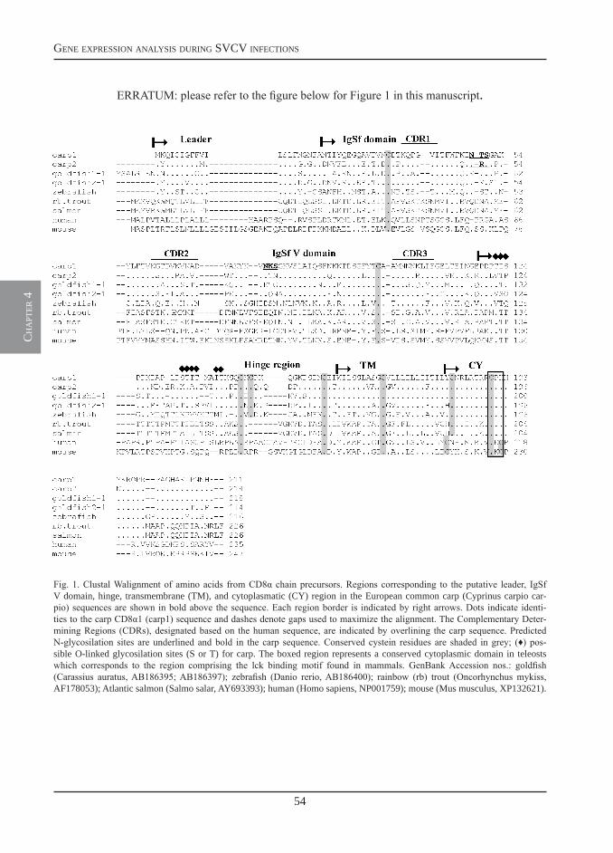
54
gene expression analysis during svcv infections
ch
apt
er 4
ERRATUM: please refer to the figure below for Figure 1 in this manuscript.
Fig. 1. Clustal Walignment of amino acids from CD8α chain precursors. Regions corresponding to the putative leader, IgSf V domain, hinge, transmembrane (TM), and cytoplasmatic (CY) region in the European common carp (Cyprinus carpio car-pio) sequences are shown in bold above the sequence. Each region border is indicated by right arrows. Dots indicate identi-ties to the carp CD8α1 (carp1) sequence and dashes denote gaps used to maximize the alignment. The Complementary Deter-mining Regions (CDRs), designated based on the human sequence, are indicated by overlining the carp sequence. Predicted N-glycosilation sites are underlined and bold in the carp sequence. Conserved cystein residues are shaded in grey; (♦) pos-sible O-linked glycosilation sites (S or T) for carp. The boxed region represents a conserved cytoplasmic domain in teleosts which corresponds to the region comprising the lck binding motif found in mammals. GenBank Accession nos.: goldfish (Carassius auratus, AB186395; AB186397); zebrafish (Danio rerio, AB186400); rainbow (rb) trout (Oncorhynchus mykiss, AF178053); Atlantic salmon (Salmo salar, AY693393); human (Homo sapiens, NP001759); mouse (Mus musculus, XP132621).
55
chapter 4
ch
apt
er 4
Available online at www.sciencedirect.com
Molecular Immunology 45 (2008) 1531–1547
Transcription of signal-3 cytokines, IL-12 and IFN��, coincideswith the timing of CD8�� up-regulation during viral
infection of common carp (Cyprinus carpio L.)
Maria Forlenza a, Joao D.A. de Carvalho Dias a, Tomas Vesely b, Dagmar Pokorova b,Huub F.J. Savelkoul a, Geert F. Wiegertjes a,∗
a Department of Animal Sciences, Cell Biology and Immunology Group, Wageningen Institute of Animal Sciences,Wageningen University, P.O. Box 338, 6700 AH, Wageningen, The Netherlands
b Veterinary Research Institute, Hudcova 70, 621 32 Brno, Czech Republic
Received 14 September 2007; received in revised form 5 October 2007; accepted 6 October 2007Available online 19 November 2007
Abstract
Mammalian naıve CD8+ T cells are activated by antigen (signal 1) and CD28 costimulation (signal 2) to undergo several rounds of cell division,but programming for survival, effector function and memory requires a third signal that can be provided by IL-12 and/or type I interferons.Functional studies indicate that the route of antigen presentation and costimulation are conserved from fish to mammals. However, the potential ofIL-12 and IFN�� to act as signal-3 cytokines in infections inducing a CTL response has not been examined in fish. We report the cloning of CD8�and CD8� homologues, each present in duplicate copies and of two TCR-C� isoforms in European common carp. The identification of (cytotoxic)T cell marker sequences and the availability of sequences coding for the signal-3 cytokines in the same fish species, allowed us to investigate byRT-qPCR their kinetics of gene expression during viral and parasitic infection. Our results show that transcription of signal-3 cytokines occurredconcomitantly with CD8�� up-regulation exclusively at 4 days post-primary viral infection. No regulation of IL-12 and IFN�� was observed afterparasitic infection. Our data provide evidences for an evolutionary conservation of function for IL-12 and IFN�� to act as third signal during CTLactivation. In addition, we suggest that a CD8�2/�1 and a p35p40b association could be the preferred combinations for the formation of a functionalCD8 co-receptor and an IL-12p70 heterodimer during viral infection. The relevance of our findings to future vaccination strategies in fish is discussed.© 2007 Elsevier Ltd. All rights reserved.
Keywords: CD8; IL-12; Type I interferon; Cytotoxic T cells; Fish; Virus
1. Introduction
Mammalian naıve CD8+ T cells are activated by antigen (Ag,signal 1) and CD28 costimulation (signal 2) to undergo severalrounds of cell division. However, programming for survival (i.e.clonal expansion), effector function and memory requires a thirdsignal delivered by cytokines. These three signals together formthe basis for CD8+ T cell-mediated immunity. The third signal isprovided by interleukin (IL)-12 and/or type I interferons (IFNs),reviewed in (Mescher et al., 2006).
In fish, cytotoxic cells are allospecific (Fischer et al., 1998;Hasegawa et al., 1998; Boudinot et al., 2001) and their cytotoxic
∗ Corresponding author.E-mail address: [email protected] (G.F. Wiegertjes).
functions are restricted to MHC class I-matched target cells(Utke et al., 2007). The presence of fish homologues to TCR,CD3, CD8, CD28 and MHC-I, among others, suggests thatthe route of antigen presentation (signal 1), costimulation(signal 2) and thus, the mechanisms at the base of cell-mediatedcytotoxicity are conserved from fish to mammals (Bernard et al.,2006a,b; Fischer et al., 2006). At present, CD8 gene expressionis the best tool available to monitor cytotoxic T lymphocyte(CTL) activity in fish. For example, in trout, CD8� expressiondirectly correlates with an increased antiviral cytotoxic activityagainst MHC class I-matched cells and with the inductionof alloreactive cytotoxic cells, both in vitro and in vivo, aftersensitisation with the allotarget (Fischer et al., 2003; Utke et al.,2007). In Ginbuna crucian carp TCR� and CD8� kinetics ofgene expression correspond well with second scale allografting(Somamoto et al., 2005) and with virus-specific cell-mediated
0161-5890/$ – see front matter © 2007 Elsevier Ltd. All rights reserved.doi:10.1016/j.molimm.2007.10.010
56
gene expression analysis during svcv infections
ch
apt
er 4
1532 M. Forlenza et al. / Molecular Immunology 45 (2008) 1531–1547
cytotoxic activity (Somamoto et al., 2006). However, despitethe increase in information on T cell-mediated immuneresponses, coordinated studies on the signal-3 requirements forT cell-mediated immunity have not been performed in fish.
In mammals, signal-3 is provided by IL-12 and/or type IIFNs (Mescher et al., 2006) and acts as a switch that determineswhether exposure to Ag results in tolerance or in full activationand development of memory (Hernandez et al., 2002; Curtsingeret al., 2003b). Effects of IL-12 and type I IFNs result from theirdirect action on CD8+ T cells rather than on an indirect influenceon levels of antigen or costimulatory molecules (B7-1 ligand) onantigen presenting cells (Curtsinger et al., 2003a, 2005). Recentin vivo studies suggest that the relative importance of IL-12and/or IFNs to act as signal-3 cytokine is dependent on the typeof response under examination. For example, in lymphocyticchoriomeningitis virus infection, type I IFNs are the predomi-nant third signal required to support the CD8+ T cell response(Kolumam et al., 2005). In contrast, in acute vaccinia virus infec-tion it is likely that both IL-12 and type I IFNs contribute to theCD8+ T cell response (van Den Broek et al., 2000), while IL-12 alone provides the necessary third signal during allospecificCD8+ T cell responses to transplants (Filatenkov et al., 2005).
In fish, the potential of IL-12 and IFN�� to act as signal-3cytokines in infections inducing a CTL response has not beenexamined. The involvement of type I IFN and IFN-inducedgenes (e.g. GTPase Mx protein) has been shown in immuneresponses induced by dsRNA, CpGs or during in vivo viralinfections, (Robertsen, 2006), but never in correlation with CD8gene expression. The sequence for the European common carp(Cyprinus carpio carpio L.) type I interferon (designated ascarp IFN��) has been described only very recently (Gonzalez etal., 2007). Sequences homologous to the mammalian IL-12p35and IL-12p40 genes have been described for a number of fishspecies (Yoshiura et al., 2003; Huising et al., 2006; Nascimentoet al., 2007) but only few studies on the function of fish IL-12 have been performed. In vitro, IL-12 regulation is seen inmacrophages after stimulation with, e.g. poly(I:C), LPS, ConA and PMA (Yoshiura et al., 2003; Huising et al., 2006). Incarp, three highly distinct IL-12p40 genes (named IL-12p40a,IL-12p40b and IL-12p40c) were described and their in vitroexpression profiles differed profoundly (Huising et al., 2006).In sea bass, up-regulation of IL-12p40 expression, but not IL-12p35, was observed in vivo after injection of UV-killed but notlive bacteria. In the latter case a more prominent up-regulationof IL-1� was observed (Nascimento et al., 2007).
In the present study, we report the cloning of four cDNAsequences coding for two CD8� and two CD8� genes in theEuropean common carp. The CD8� sequences are the firstreported so far in a cyprinid species. Two sequences coding forthe constant domain of the TCR� chain are also cloned. Theidentification of sequences coding for the signal-3 cytokines,IL-12 and type I IFN, as well as for (cytotoxic) T cell markers inthe same fish species, allowed us to investigate their kinetics ofgene expression during primary and secondary infection of carpwith spring viraemia of carp virus (SVCV) (Ahne et al., 2002).To further investigate whether signal-3 cytokines would play arole in infections other than viral, where a CTL response is not
required, we analysed IL-12 and IFN�� gene expression duringprimary infection with the extracellular parasite Trypanoplasmaborreli. Taken together, our results strongly suggest that both,IL-12 and IFN�� signal-3 cytokines might be required exclu-sively during primary viral, but not parasitic infection, when alsoup-regulation of CD8 transcription is observed. The implicationsof our findings for future vaccination strategies are discussed.
2. Materials and methods
2.1. Animals
European (C. carpio carpio) and East-Asian (C. carpiohaematopterus) common carp diverged more than 500,000 yearsago (Kohlmann et al., 2003; Zhou et al., 2003; Thai et al., 2004)and both subspecies are commonly studied in fish research. Inthe present study, European common carp of the R3 × R8 strainwhich are the offspring of a cross between fish of Hungarianorigin (R8 strain) and of Polish origin (R3 strain) were used(Irnazarow, 1995). In this study, we refer to the European com-mon carp subspecies as carp, unless stated otherwise. Carp werebred in the central fish facility of Wageningen University, TheNetherlands, and raised either in this facility or at the VeterinaryResearch Institute (VRI), Brno, Czech Republic, in recirculatingUV-treated water and fed pelleted dry food (Trouvit, Nutreco)daily. Al studies were performed with the approval of the animalexperimental committee of Wageningen University.
2.2. Identification of carp CD8α and CD8β cDNAs
Degenerate primers (Table 1) were designed on con-served regions of known fish CD8� and CD8� sequences(GenBank Accession nos. CD8�: AAF32266; AAW33878;BAD89371–BAD89374; 001015040; CD8�: AY563420;AY693392; AY701524). (Anchored) PCR was performed on a�gt10 cDNA library of carp thymus. The library was constructedusing mRNA isolated from ten 5-month-old individuals of asecond generation gynogenetic European common carp line(van Erp et al., 1996). Aliquots (300 ng) of the library werecombined in Taq buffer with 1 U Taq polymerase (Goldstar,Eurogentec S.A., Seraing, Belgium), MgCl2 (1.5 mM), dNTPs(200 �M) and primers (400 nM each) in a total volume of50 �l. The PCR was performed using a GeneAmp PCR system9700 (PE Applied Biosystems, Foster City, CA, USA) underthe following conditions: 4 min at 94 ◦C, followed by 35cycles of 30 s at 94 ◦C, 30 s at 55 ◦C and 2 min at 72 ◦C,finally an extension step of 7 min at 72 ◦C was performed.Degenerate primers, CD8�.FW4 and CD8�.RV5, were used incombination to amplify a product with 82–91% of identity withother known cyprinid CD8�1 sequences. The specific primercycaCD8�.FW3 was used in combination with �gt10.RVspecific primer for the amplification of CD8� 3�UTR. Similarly,cycaCD8�.RV3 in combination with �gt10.FW primer wasused for the amplification of the 5� UTR. The second reactionled to the amplification of a second CD8� sequence.
A similar strategy was used to amplify the CD8� gene.Degenerate primers CD8�.FW1, CD8�.FW2 were used as
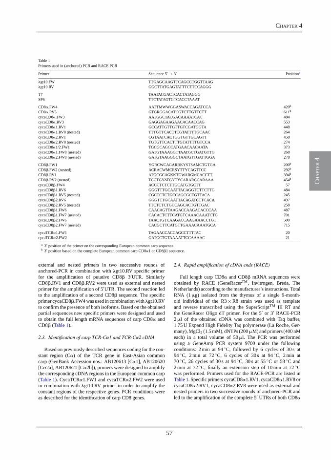
57
chapter 4
ch
apt
er 4
M. Forlenza et al. / Molecular Immunology 45 (2008) 1531–1547 1533
Table 1Primers used in (anchored) PCR and RACE PCR
Primer Sequence 5� → 3� Positiona
�gt10.FW TTGAGCAAGTTCAGCCTGGTTAAG�gt10.RV GGCTTATGAGTATTTCTTCCAGGG
T7 TAATACGACTCACTATAGGGSP6 TTCTATAGTGTCACCTAAAT
CD8�.FW4 AATTMWWGGASWACCAGATCCA 420b
CD8�.RV5 GTGRGGACATCGTCTTGTTCTT 611b
cycaCD8�.FW3 AATGGCTACGACAAAATCAC 484cycaCD8�.RV3 GAGGAGAAGAACACAACCAG 553cycaCD8�1.RV1 GCCATTGTTGTTGTCGATGGTA 448cycaCD8�1.RV8 (nested) TTTGTTCACTTTGTATTTTGCAAC 264cycaCD8�2.RV1 CGTAATCACTGGTGTTGCAGTT 458cycaCD8�2.RV8 (nested) TGTGTTCACTTTGTATTTTGTCCA 274cycaCD8�1/2.FW1 TGCGCAGCCATGAACAACAATA 373cycaCD8�1.FW8 (nested) GATGTAAAGGTTAATGCTGATGTTG 268cycaCD8�2.FW8 (nested) GATGTAAGGGCTAATGTTGATTGGA 278
CD8�.FW1 YGRCWCAGARRKYSTSAMCTGTGA 200b
CD8�.FW2 (nested) ACRACWMCRSYTTYCAGTTCC 292b
CD8�.RV1 ATGCGCAGKKTGWARGMCACCTT 394b
CD8�.RV2 (nested) TCCTGYATGYTYCARARCCARAAA 454b
cycaCD8�.FW4 ACCCTCTCTTGCATGTGCTT 57cycaCD8�1.RV6 GGGTTTGCAATTACAGGTCTTCTTG 484cycaCD8�1.RV5 (nested) CGCTCTCTGCCAGCGCTGTTACA 245cycaCD8�2.RV6 GGGTTTGCAATTACAGATCTTCACA 497cycaCD8�2.RV5 (nested) TTCTCTCTGCCAGCACTGTTGAC 258cycaCD8�1.FW6 CAACAGTTAAGACCAAGACACCCAA 487cycaCD8�1.FW7 (nested) CACACTCTTCATGTCAAACAAATCTG 701cycaCD8�2.FW6 TAACTGTCAAGACCAAGAAACCTGT 500cycaCD8�2.FW7 (nested) CACGCTTCATGTTGAAACAAATGCA 715
cycaTCR�1.FW1 TAGAACCACCAGCCTTTTAC 20cycaTCR�2.FW2 GATGCTGTAAAATTCCAAAAC 21
a 3� position of the primer on the corresponding European common carp sequence.b 3� position based on the complete European common carp CD8�1 or CD8�1 sequence.
external and nested primers in two successive rounds ofanchored-PCR in combination with �gt10.RV specific primerfor the amplification of putative CD8� 3�UTR. SimilarlyCD8�.RV1 and CD8�.RV2 were used as external and nestedprimer for the amplification of 5�UTR. The second reaction ledto the amplification of a second CD8� sequence. The specificprimer cycaCD8�.FW4 was used in combination with �gt10.RVto confirm the presence of both isoforms. Based on the obtainedpartial sequences new specific primers were designed and usedto obtain the full length mRNA sequences of carp CD8� andCD8� (Table 1).
2.3. Identification of carp TCR-Cα1 and TCR-Cα2 cDNA
Based on previously described sequences coding for the con-stant region (C�) of the TCR gene in East-Asian commoncarp (GenBank Accession nos.: AB120613 [C�1], AB120620[C�2a], AB120621 [C�2b]), primers were designed to amplifythe corresponding cDNA regions in the European common carp(Table 1). CycaTCR�1.FW1 and cycaTCR�2.FW2 were usedin combination with �gt10.RV primer in order to amplify theconstant regions of the respective genes. PCR conditions wereas described for the identification of carp CD8 genes.
2.4. Rapid amplification of cDNA ends (RACE)
Full length carp CD8� and CD8� mRNA sequences wereobtained by RACE (GeneRacerTM, Invitrogen, Breda, TheNetherlands) according to the manufacturer’s instructions. TotalRNA (1 �g) isolated from the thymus of a single 9-month-old individual of the R3 × R8 strain was used as templateand reverse transcribed using the SuperScriptTM III RT andthe GeneRacer Oligo dT primer. For the 5� or 3� RACE-PCR2 �l of the obtained cDNA was combined with Taq buffer,1.75 U Expand High Fidelity Taq polymerase (La Roche, Ger-many), MgCl2 (1.5 mM), dNTPs (200 �M) and primers (400 nMeach) in a total volume of 50 �l. The PCR was performedusing a GeneAmp PCR system 9700 under the followingconditions: 2 min at 94 ◦C, followed by 6 cycles of 30 s at94 ◦C, 2 min at 72 ◦C, 6 cycles of 30 s at 94 ◦C, 2 min at70 ◦C, 26 cycles of 30 s at 94 ◦C, 30 s at 55 ◦C or 58 ◦C and2 min at 72 ◦C, finally an extension step of 10 min at 72 ◦Cwas performed. Primers used for the RACE-PCR are listed inTable 1. Specific primers cycaCD8�1.RV1, cycaCD8�1.RV8 orcycaCD8�2.RV1, cycaCD8�2.RV8 were used as external andnested primers in two successive rounds of anchored-PCR andled to the amplification of the complete 5� UTRs of both CD8�
58
gene expression analysis during svcv infections
ch
apt
er 4
1534 M. Forlenza et al. / Molecular Immunology 45 (2008) 1531–1547
isoforms. Similarly, cycaCD8�1/2.FW1, cycaCD8�1.FW8 orcycaCD8�1/2.FW1, cycaCD8�2.FW8 specific primers wereused for the amplification of the complete 3� UTRs. Asimilar strategy was used to amplify the complete CD8�gene. Specific primers cycaCD8�1.RV6, cycaCD8�1.RV5 orcycaCD8�2.RV6, cycaCD8�2.RV5 were used as external andnested primers in two successive rounds of anchored-PCR andled to the amplification of the complete 5� UTRs of both CD8�isoforms. Similarly cycaCD8�1.FW6, cycaCD8�1.FW7 andcycaCD8�2.FW6, cycaCD8�2.FW7 specific primers were usedfor the amplification of the complete 3� UTRs.
2.5. Cloning, sequencing and protein analysis
Products amplified by PCR were ligated and cloned inJM109 cells using the pGEM-T easy kit (Promega, Leiden,The Netherlands) according to the standard protocol. Positiveclones were selected by blue-white screening on agar platescontaining 100 �g/ml Ampicillin. At least eight clones for eachPCR product were selected for subsequent colony-PCR usingT7 and SP6 plasmid-specific primers. A standard PCR programwas performed as described for the identification of carp CD8genes. From each PCR product both strands were sequencedusing the ABI Prism Big Dye Terminator Cycle SequencingReady Reaction kit and analysed using a 3730 DNA analyser.Nucleotide sequences were translated using the ExPASy trans-late tool (http://us.expasy.org/tools/dna.html) and aligned withClustal W and BLAST software (http://www.ebi.ac.uk/clustalwand http://www.ncbi.nlm.nih.gov/BLAST). The signal peptidecleavage site and the transmembrane region were predictedby using the SignalP 3.0 (http://www.cbs.dtu.dk/services/SignalP/) and the TMHMM 2.0 (http://www.cbs.dtu.dk/services/TMHMM-2.0/) servers. Post-translational modifi-cations were predicted using the NetOGlyc 3.1 (http://www.cbs.dtu.dk/services/NetOGlyc/) and the NetNGlyc 1.0(http://www.cbs.dtu.dk/services/NetNGlyc/) servers.
2.6. Viral infection of carp
Spring viraemia of carp virus (SVCV) strain CAPM V 539(Koutna et al., 2003) was propagated in EPC (Epithelioma Papu-losum Cyprini, (Fijan et al., 1983)) cells at 15 ◦C. Cells weregrown in Eagle�s Minimal Essential Medium (MEM) contain-ing 2% fetal bovine serum (FBS) and standard concentrationof antibiotics. The virus titers, given as tissue culture infectivedose (TCID50/ml), were calculated by the method of Reed andMuench (1938).
Ten-month-old carp were raised at 15 ◦C to an average weightof 30–40 g. This temperature is optimal for SVCV infectivity(Ahne et al., 2002). Fish (n= 110) were exposed, by immer-sion, to SVCV-infected tissue culture (103 TCID50/ml) for 2 h.Control fish (n= 62) were treated similarly and exposed to con-trol cultures only. Infected fish were divided over two replicatetanks for subsequent blood and tissue sampling. At 0, 6 h, 1 day,2 days, 4 days, 7 days, 2 weeks, 3 weeks and 4 weeks post-infection (p.i.), six fish were killed to collect blood and tissuesamples. Eight weeks after the first infection, survivors (n= 74)
were reinfected using the same conditions as for the primaryinfection and divided over two replicate tanks. Samples weretaken at the same time points and from the same number of fishas after the primary infection. Control fish were exposed to con-trol cultures only. Infection of carp with SVCV was performedat the central fish facility of the Veterinary Research Institute(VRI), Brno, Czech Republic.
2.7. Parasitic infection of carp
T. borreli was cloned and characterized before (Steinhagenet al., 1989) and maintained by syringe passage through carp.Parasites were monitored in blood (10× diluted in RPMI 1640(Cambrex, Verviers, Belgium) adjusted to 270 mOsmol kg−1,cRPMI) using a Burker counting chamber. The minimum detec-tion limit of this method was 105 parasites/ml of blood.
Six-month-old carp were raised at 23 ◦C to an averageweight of approximately 160 g, and acclimatized for 2 weeksto 20 ◦C. This temperature is optimal for parasite growth(unpublished data). Fish (n= 90) were anaesthetised in 0.3 g/lTricaine Methane Sulfonate (TMS, Crescent Research Chemi-cals, Phoenix, USA) and intra-peritoneally (i.p.) injected witha dose of 104 parasites/fish, PBS-injected individuals served asunchallenged control group. Infected and non-infected fish wereequally divided over 6 tanks. Infected fish were distinguishedfrom the controls by a tattooed number. At 0, 1, 3, 10 days, 3, 4,and 6 weeks, n= 5 infected and n= 3 control fish were killed tocollect blood and tissue samples. Infection of carp with T. bor-reli was performed at the central fish facility of the WageningenUniversity, The Netherlands.
2.8. Organ and peripheral blood leukocytes (PBL) isolation
Fish were killed by an overdose of TMS. Fish were bledby puncture of the caudal blood vessel. After viral infection,organs were collected and immediately transferred to RNAlater(Ambion, Inc., Austin, USA). Following an overnight incubationat 4 ◦C the samples were stored at −20 ◦C until use. Alterna-tively, after parasitic infection organs were snap frozen in liquidnitrogen and stored at −80 ◦C until use.
For blood collection, carp were anaesthetised in 0.3 g/l TMS.Blood was collected by puncture of the caudal blood vessel anddiluted 1:1 with cRPMI medium containing 50 IU/ml of hep-arin (Leo Pharmaceutical products, Weesp, The Netherlands).After centrifugation at 600 × g for 10 min, the buffy-coat con-taining the leukocytes was collected and layered on 5 ml ofFicoll-PaqueTM Plus (Amersham Biosciences, Uppsala, Swe-den). Following subsequent centrifugation at 800 × g for 25 min,the leukocyte layer at the interface was collected and washedthree times with cRPMI. Cell pellets were collected and eitherstored in RNAlater, directly lysed prior to RNA isolation orimmediately used for magnetic activated cell sorting.
2.9. Magnetic activated cell sorting (MACS)
After Ficoll separation, PBL were collected and washedtwice in cRPMI. The primary antibody (WCI-12) was added
59
chapter 4
ch
apt
er 4
M. Forlenza et al. / Molecular Immunology 45 (2008) 1531–1547 1535
in a 1:50 dilution and incubated for 30 min on ice. WCI-12is a mouse monoclonal antibody directed against the heavychain of carp IgM and was used to separate IgM bearingcells (Koumans-van Diepen et al., 1995). Next, the cells werewashed twice with MACS-buffer (0.5% BSA in cRPMI). Thesecondary antibody (phycoerythrin (PE)-conjugated goat anti-mouse, DAKO, Glostrup, Denmark) was then added in a 1:50dilution and incubated for 30 min on ice. After extensive wash-ing, total cell number was determined with a Burker chamber,and 10 �l of magnetic beads (anti-PE Microbeads, MiltenyiBiotec, GmbH, Germany) was added per 1 × 10⁸ cells. Afterincubation for 15 min at 4 ◦C, cells were washed and finallyresuspended in MACS buffer. The magnetic separation wasperformed on LS-MidiMACS Columns according to the manu-facturer’s instructions. The purity of the WCI-12+ and WCI-12−fractions was assessed by flow cytometric analysis using aFACScan® flow cytometer (Becton Dickinson, Mountain View,CA, USA). Cell pellets were collected and directly lysed priorto RNA isolation.
2.10. RNA isolation and quantification
For real-time quantitative polymerase chain reaction (RT-qPCR) analysis total RNA was isolated from thymus, headkidney, mid kidney, PBL, spleen, liver, gills and gut usingthe RNAeasy Mini Kit (Qiagen, Leusden, The Netherlands)according to the manufacturer’s instructions. On-column DNasetreatment with the RNase-free DNase set (Qiagen) was alsoincluded. Prior to RNA isolation from liver a ProteinaseK treatment was also included. The concentration of RNAwas measured on a spectrophotometer (GeneQuant, Pharma-cia Biotech) at OD260 nm. The purity of the extracted RNA wasdetermined as the OD260 nm/OD280 nm ratio with expected valuesbetween 1.8 and 2.0. The integrity of RNA was determined byelectrophoresis on 1% agarose gel containing 0.5 �g/ml ethid-ium bromide at 100 V. Total RNA was stored at −80 ◦C until use.
2.11. cDNA synthesis
Prior to cDNA synthesis, a second DNase treatment was per-formed using DNase I, Amplification Grade (Invitrogen, Breda,The Netherlands). Briefly, 1 �g of RNA from each sample wascombined with 10X DNase reaction buffer and 1 U DNase I,mixed and incubated at RT for 15 min, followed by inactiva-tion of DNase I by adding 1 �l of 25 mM EDTA. Synthesis ofcDNA was performed with Invitrogen’s SuperScriptTM III FirstStrand Synthesis Systems for RT-PCR Systems, according tothe manufacturer’s instructions. Briefly, DNase I-treated RNAsamples were mixed with 5× first strand buffer, 300 ng randomprimers, 10 mM dNTPs, 0.1 M DTT, 10 U RNase inhibitor, and200 U SuperScript III Reverse Transcriptase (Invitrogen) up toa final volume of 20 �l. The mixture was incubated at 37 ◦Cfor 60 min followed by an inactivation step at 70 ◦C for 15 min.A non-reverse transcriptase control was included for each sam-ple. cDNA samples were further diluted 50 times (25 times forPBL samples) in nuclease-free water before use as template inreal-time PCR experiments.
2.12. Detection of SVCV
To confirm that all fish used in our experiment were infectedby the SVC virus, virus-specific primers were used in a single-tube reverse transcription (RT)-PCR and nested PCR reaction(Koutna et al., 2003).
For RT-PCR the SuperScript One-Step RT-PCR system(Invitrogen) was used. Briefly, 0.5 �g of total RNA isolatedfrom head kidney of infected or control fish was combinedwith 25 �l 2X rection buffer, 20 pmol of the external primers(extFW: 5�-GCCTAAATGTGTTGATGGAACG-3�; extRV: 5�-GGATAATATCGGCTTGGAAAGC-3�), 40 U RNase inhibitor,100 U SuperScript II RT/Taq and diethyl pyrocarbonate (DEPC)-treated water up to a final volume of 25 �l. RT-PCR was carriedout under the following conditions: cDNA synthesis for 30 minat 50 ◦C, one cycle of 2 min 30 s at 94 ◦C, followed by 35cycles of 30 s at 94 ◦C, 1 min at 50 ◦C, and 1 min at 68 ◦C, withfinal extension step for 7 min at 72 ◦C. Nested PCR reactionswere performed by combining 2 �l of the amplification productobtained by RT-PCR with Taq buffer (10×), 1 U Taq poly-merase, MgCl2 (1.5 mM), 20 pmol of each of the internal primers(intFW: 5�-CAAGAGAAGCTGACATCAGTGG-3�; intRV: 5�-GACAATAGGTCCCTCTACTTCG-3�), 200 �M dNTPs, andDEPC water up to a final volume of 50 �l. PCR conditionsincluded one cycle of 4 min at 94 ◦C, followed by 25 cyclesof 30 s at 94 ◦C, 30 s at 55 ◦C and 30 s at 72 ◦C followed by afinal extension step for 7 min at 72 ◦C. RT-PCR and nested PCRwere run using a GeneAmp PCR system 9700.
2.13. Real-time quantitative polymerase chain reaction(RT-qPCR)
RT-qPCR using SYBR Green I technology was performedusing a Rotor-GeneTM 2000 (Corbett Research, Mortlake,Sydney, Australia) with the Brilliant® SYBR® Green QPCR(Stratagene, La Jolla, CA, USA) as detection chemistry. Theprimers used for RT-qPCR are listed in Table 2. PCR conditionswere optimized by analyzing the melting curves of the prod-ucts (Ririe et al., 1997) and product specificity was assessedby analysis on a 1% agarose gel. Master-mix for each PCRrun was prepared as follows: 0.32 �l of water, 0.84 �l of eachprimer (5 �M), 7 �l Master SYBR Green I mix. Finally, 5 �lof diluted cDNA was added to 9 �l of master mix and trans-ferred to a 0.1 ml tube. The following amplification programwas used: after 15 min of denaturation at 95 ◦C, 40 cycles ofRT-qPCR with three-step amplification were performed: 15 s at95 ◦C for denaturation, 30 s at 60 ◦C for annealing and 30 s at72 ◦C for elongation followed by a final holding step of 1 min at60 ◦C. A melting step was then performed with continuous flu-orescence acquisition starting at 60 ◦C with a rate of 1 ◦C/5 sup to 99 ◦C to determine the amplification specificity. In allcases, the amplifications were specific and no amplification wasobserved in negative controls (non-template control and non-reverse transcriptase control). Fluorescence data from RT-qPCRexperiments were analysed using Rotor-Gene version 6.0.21software and exported to Microsoft Excel. The cycle thresh-old Ct for each sample and the reaction efficiencies (E) for each
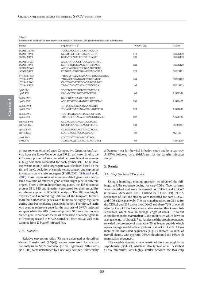
60
gene expression analysis during svcv infections
ch
apt
er 4
1536 M. Forlenza et al. / Molecular Immunology 45 (2008) 1531–1547
Table 2Primers used in RT-qPCR gene expression analysis + indicates LNA (locked nucleic acid) substitutions
Primer Sequence 5� → 3� Product (bp) Acc no
qCD8�1/2.FW1 TGCGCAGCCATGAACAACAATAqCD8�1.RV1 GCCATTGTTGTTGTCGATGGTA 119 EU025118qCD8�2.RV1 CGTAATCACTGGTGTTGCAGTT 129 EU025119
qCD8�1.FW3 AATCAA+CGGCT+CGGAAACTATCqCD8�1.RV3 CGCTCTCTGCCAGCGCTGTTACA 132 EU025120qCD8�2.FW3 AAT+CAATGGCT+CGGAAGTTCTGqCD8�2.RV3 CCAGCA+CTGTTGA+CATACACTAA 123 EU025121
qTCR�1.FW2 TTCACA+CAA+CATAATG+CTGTAAATGAqTCR�1.RV2 TTGA+CTGGATGATCCTGACATGC 144 EU025122qTCR�2.FW2 CAGTG+CGATATGCAGAACGAAGTqTCR�2.RV2 TTGACTGGATGAT+CCTTGCTGA 95 EU025123
qp35.FW1 TGCTTCTCTGTCTCTGTGATGGAqp35.RV1 CACAGCTGCAGTCGTTCTTGA 86 AJ580354
qp40a.FW1 GAGCGCATCAACCTGACCATqp40a.RV1 AGGATCGTGGATATGTGACCTCTAC 151 AJ621425
qp40b.FW2 TCTTGCACCGCAAGAAACTATGqp40b.RV2 TGCAGTTGATGAGACTAGAGTTTCG 121 AJ628699
qp40c.FW1 TGGTTGATAAGGTTCACCCTTCTCqp40c.RV1 TATCTGTTCTACAGGTCAGGGTAACG 127 AJ628700
qIFN��.FW1 GGCAGATATG+GGACGGTGAGqIFN��.RV1 GTCCTCCA+CCTCAGCTTTGTC 132 EC393381
q�Act.FW2 GCTATGTGGCTCTTGACTTCGAq�Act.RV2 CCGTCAGGCAGCTCATAGCT 89 M24113
q40S.FW1 CCGTGGGTGACATCGTTACAq40S.RV1 TCAGGACATTGAACCTCACTGTCT 69 AB012087
primer set were obtained upon Comparative Quantitation Anal-ysis from the Rotor-Gene version 6.0.21 software. Briefly, theE for each primer set was recorded per sample and an averageE (EA) was then calculated for each primer set. The relativeexpression ratio (R) of a target gene was calculated based on theEA and the Ct deviation of sample versus control, and expressedin comparison to a reference gene (Pfaffl, 2001; Tichopad et al.,2003). Basal expression of immune-related genes was calcu-lated as a ratio of reference gene versus target gene in differentorgans. Three different house keeping genes, the 40S ribosomalprotein S11, 18S and �-actin, were tested for their suitabilityas reference genes in RT-qPCR analysis. The 18S was highlyexpressed and required high dilution of the template, further-more both ribosomal genes were found to be highly regulatedduring viral but not during parasitic infection. Therefore, �-actinwas used as reference gene for the analysis of SVCV infectedsamples while the 40S ribosomal protein S11 was used as ref-erence gene to calculate the basal expression of a target gene indifferent organs and in MACS sorted cell fractions, as well as insamples from T. borreli-infected fish.
2.14. Statistics
Relative expression ratios (R) were calculated as describedabove. Transformed (LN(R)) values were used for statisti-cal analysis in SPSS Software (15.0). Significant differences(P< 0.05) were determined by a one-way ANOVA followed by
a Dunnett t-test for the viral infection study and by a two-wayANOVA followed by a Sidak’s test for the parasite infectionstudy.
3. Results
3.1. Carp has two CD8α genes
Using a homology cloning approach we obtained the full-length mRNA sequence coding for carp CD8�. Two isoformswere identified and were designated as CD8�1 and CD8�2(GenBank Accession nos.: EU025118; EU025119). cDNAsequences of 949 and 948 bp were identified for carp CD8�1and CD8�2, respectively. The translated peptides are 211 aa forthe CD8�1 and 214 aa for the CD8�2 and share 73% of overallidentity. Carp CD8� has a comparable size to other known fishsequences, which have an average length of about 197 aa butis smaller than the mammalian CD8� molecules which have anaverage length of about 217 aa. Analysis of the protein sequencesrevealed the presence of a putative 20 aa leader peptide whichupon cleavage would release proteins of about 21.1 kDa. Align-ment of the translated sequences (Fig. 1) showed 54–80% ofoverall identity with cyprinid, 26% with salmonid and 16% withmammalian sequences.
The variable domain, characteristic of the immunoglobulinsuperfamily (IgSf V), which is also typical of all describedCD8� molecules, was highly similar between the two carp
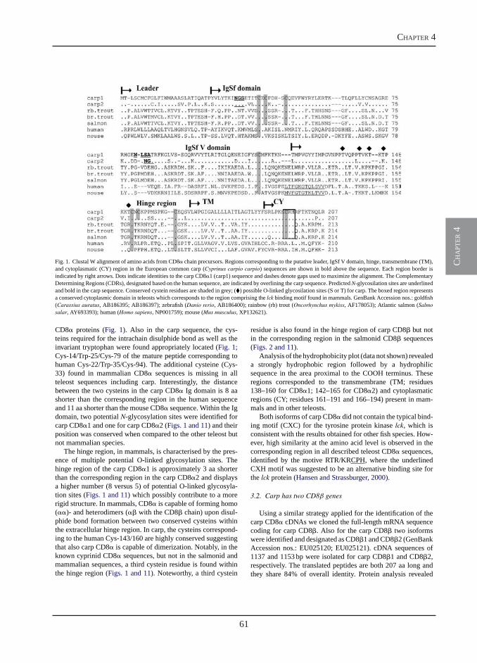
61
chapter 4
ch
apt
er 4
M. Forlenza et al. / Molecular Immunology 45 (2008) 1531–1547 1537
Fig. 1. Clustal W alignment of amino acids from CD8� chain precursors. Regions corresponding to the putative leader, IgSf V domain, hinge, transmembrane (TM),and cytoplasmatic (CY) region in the European common carp (Cyprinus carpio carpio) sequences are shown in bold above the sequence. Each region border isindicated by right arrows. Dots indicate identities to the carp CD8�1 (carp1) sequence and dashes denote gaps used to maximize the alignment. The ComplementaryDetermining Regions (CDRs), designated based on the human sequence, are indicated by overlining the carp sequence. PredictedN-glycosilation sites are underlinedand bold in the carp sequence. Conserved cystein residues are shaded in grey; (�) possible O-linked glycosilation sites (S or T) for carp. The boxed region representsa conserved cytoplasmic domain in teleosts which corresponds to the region comprising the lck binding motif found in mammals. GenBank Accession nos.: goldfish(Carassius auratus, AB186395; AB186397); zebrafish (Danio rerio, AB186400); rainbow (rb) trout (Oncorhynchus mykiss, AF178053); Atlantic salmon (Salmosalar, AY693393); human (Homo sapiens, NP001759); mouse (Mus musculus, XP132621).
CD8� proteins (Fig. 1). Also in the carp sequence, the cys-teins required for the intrachain disulphide bond as well as theinvariant tryptophan were found appropriately located (Fig. 1;Cys-14/Trp-25/Cys-79 of the mature peptide corresponding tohuman Cys-22/Trp-35/Cys-94). The additional cysteine (Cys-33) found in mammalian CD8� sequences is missing in allteleost sequences including carp. Interestingly, the distancebetween the two cysteins in the carp CD8� Ig domain is 8 aashorter than the corresponding region in the human sequenceand 11 aa shorter than the mouse CD8� sequence. Within the Igdomain, two potential N-glycosylation sites were identified forcarp CD8�1 and one for carp CD8�2 (Figs. 1 and 11) and theirposition was conserved when compared to the other teleost butnot mammalian species.
The hinge region, in mammals, is characterised by the pres-ence of multiple potential O-linked glycosylation sites. Thehinge region of the carp CD8�1 is approximately 3 aa shorterthan the corresponding region in the carp CD8�2 and displaysa higher number (8 versus 5) of potential O-linked glycosyla-tion sites (Figs. 1 and 11) which possibly contribute to a morerigid structure. In mammals, CD8� is capable of forming homo(��)- and heterodimers (�� with the CD8� chain) upon disul-phide bond formation between two conserved cysteins withinthe extracellular hinge region. In carp, the cysteins correspond-ing to the human Cys-143/160 are highly conserved suggestingthat also carp CD8� is capable of dimerization. Notably, in theknown cyprinid CD8� sequences, but not in the salmonid andmammalian sequences, a third cystein residue is found withinthe hinge region (Figs. 1 and 11). Noteworthy, a third cystein
residue is also found in the hinge region of carp CD8� but notin the corresponding region in the salmonid CD8� sequences(Figs. 2 and 11).
Analysis of the hydrophobicity plot (data not shown) revealeda strongly hydrophobic region followed by a hydrophilicsequence in the area proximal to the COOH terminus. Theseregions corresponded to the transmembrane (TM; residues138–160 for CD8�1; 142–165 for CD8�2) and cytoplasmaticregions (CY; residues 161–191 and 166–194) present in mam-mals and in other teleosts.
Both isoforms of carp CD8� did not contain the typical bind-ing motif (CXC) for the tyrosine protein kinase lck, which isconsistent with the results obtained for other fish species. How-ever, high similarity at the amino acid level is observed in thecorresponding region in all described teleost CD8� sequences,identified by the motive RTR/KRCPH, where the underlinedCXH motif was suggested to be an alternative binding site forthe lck protein (Hansen and Strassburger, 2000).
3.2. Carp has two CD8β genes
Using a similar strategy applied for the identification of thecarp CD8� cDNAs we cloned the full-length mRNA sequencecoding for carp CD8�. Also for the carp CD8� two isoformswere identified and designated as CD8�1 and CD8�2 (GenBankAccession nos.: EU025120; EU025121). cDNA sequences of1137 and 1153 bp were isolated for carp CD8�1 and CD8�2,respectively. The translated peptides are both 207 aa long andthey share 84% of overall identity. Protein analysis revealed
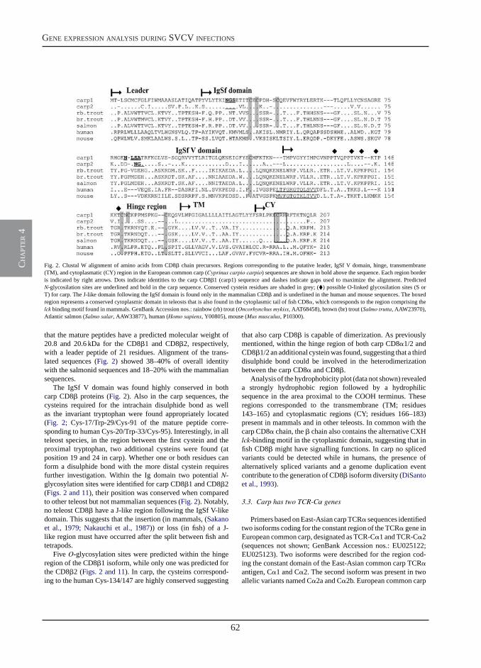
62
gene expression analysis during svcv infections
ch
apt
er 4
1538 M. Forlenza et al. / Molecular Immunology 45 (2008) 1531–1547
Fig. 2. Clustal W alignment of amino acids from CD8� chain precursors. Regions corresponding to the putative leader, IgSf V domain, hinge, transmembrane(TM), and cytoplasmatic (CY) region in the European common carp (Cyprinus carpio carpio) sequences are shown in bold above the sequence. Each region borderis indicated by right arrows. Dots indicate identities to the carp CD8�1 (carp1) sequence and dashes indicate gaps used to maximize the alignment. PredictedN-glycosilation sites are underlined and bold in the carp sequence. Conserved cystein residues are shaded in grey; (�) possible O-linked glycosilation sites (S orT) for carp. The J-like domain following the IgSf domain is found only in the mammalian CD8� and is underlined in the human and mouse sequences. The boxedregion represents a conserved cytoplasmic domain in teleosts that is also found in the cytoplasmic tail of fish CD8�, which corresponds to the region comprising thelck binding motif found in mammals. GenBank Accession nos.: rainbow (rb) trout (Oncorhynchus mykiss, AAT68458), brown (br) trout (Salmo trutta, AAW23970),Atlantic salmon (Salmo salar, AAW33877), human (Homo sapiens, Y00805), mouse (Mus musculus, P10300).
that the mature peptides have a predicted molecular weight of20.8 and 20.6 kDa for the CD8�1 and CD8�2, respectively,with a leader peptide of 21 residues. Alignment of the trans-lated sequences (Fig. 2) showed 38–40% of overall identitywith the salmonid sequences and 18–20% with the mammaliansequences.
The IgSf V domain was found highly conserved in bothcarp CD8� proteins (Fig. 2). Also in the carp sequences, thecysteins required for the intrachain disulphide bond as wellas the invariant tryptophan were found appropriately located(Fig. 2; Cys-17/Trp-29/Cys-91 of the mature peptide corre-sponding to human Cys-20/Trp-33/Cys-95). Interestingly, in allteleost species, in the region between the first cystein and theproximal tryptophan, two additional cysteins were found (atposition 19 and 24 in carp). Whether one or both residues canform a disulphide bond with the more distal cystein requiresfurther investigation. Within the Ig domain two potential N-glycosylation sites were identified for carp CD8�1 and CD8�2(Figs. 2 and 11), their position was conserved when comparedto other teleost but not mammalian sequences (Fig. 2). Notably,no teleost CD8� have a J-like region following the IgSf V-likedomain. This suggests that the insertion (in mammals, (Sakanoet al., 1979; Nakauchi et al., 1987)) or loss (in fish) of a J-like region must have occurred after the split between fish andtetrapods.
Five O-glycosylation sites were predicted within the hingeregion of the CD8�1 isoform, while only one was predicted forthe CD8�2 (Figs. 2 and 11). In carp, the cysteins correspond-ing to the human Cys-134/147 are highly conserved suggesting
that also carp CD8� is capable of dimerization. As previouslymentioned, within the hinge region of both carp CD8�1/2 andCD8�1/2 an additional cystein was found, suggesting that a thirddisulphide bond could be involved in the heterodimerizationbetween the carp CD8� and CD8�.
Analysis of the hydrophobicity plot (data not shown) revealeda strongly hydrophobic region followed by a hydrophilicsequence in the area proximal to the COOH terminus. Theseregions corresponded to the transmembrane (TM; residues143–165) and cytoplasmatic regions (CY; residues 166–183)present in mammals and in other teleosts. In common with thecarp CD8� chain, the � chain also contains the alternative CXHlck-binding motif in the cytoplasmic domain, suggesting that infish CD8� might have signalling functions. In carp no splicedvariants could be detected while in humans, the presence ofalternatively spliced variants and a genome duplication eventcontribute to the generation of CD8� isoform diversity (DiSantoet al., 1993).
3.3. Carp has two TCR-Cα genes
Primers based on East-Asian carp TCR� sequences identifiedtwo isoforms coding for the constant region of the TCR� gene inEuropean common carp, designated as TCR-C�1 and TCR-C�2(sequences not shown; GenBank Accession nos.: EU025122;EU025123). Two isoforms were described for the region cod-ing the constant domain of the East-Asian common carp TCR�antigen, C�1 and C�2. The second isoform was present in twoallelic variants named C�2a and C�2b. European common carp
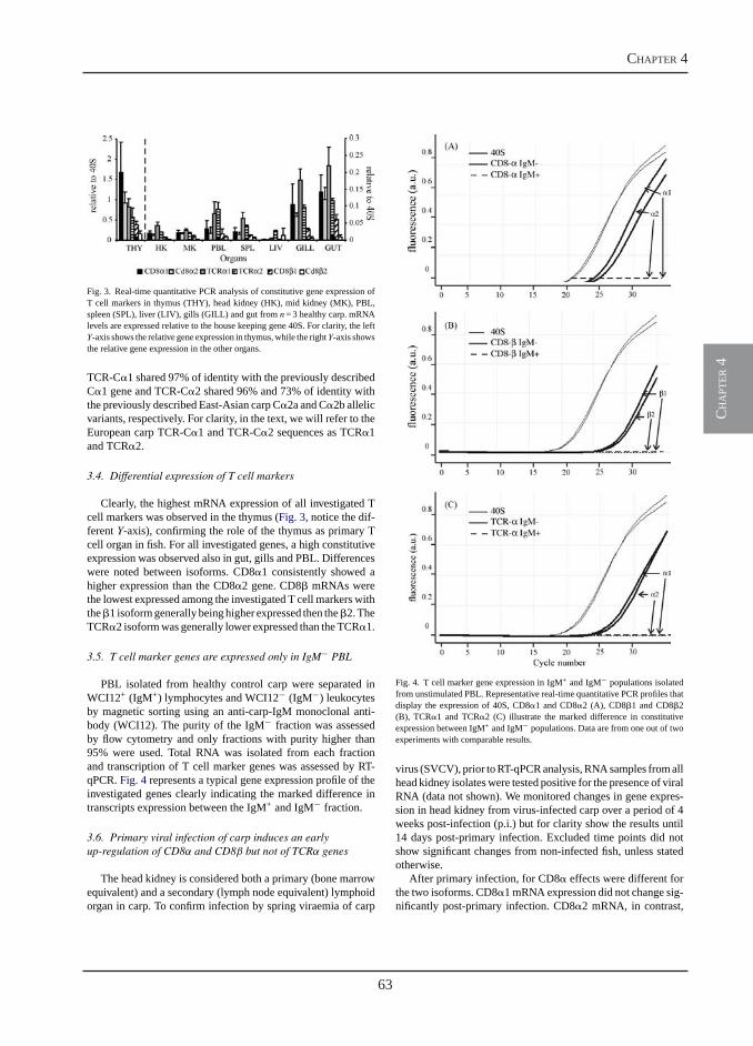
63
chapter 4
ch
apt
er 4
M. Forlenza et al. / Molecular Immunology 45 (2008) 1531–1547 1539
Fig. 3. Real-time quantitative PCR analysis of constitutive gene expression ofT cell markers in thymus (THY), head kidney (HK), mid kidney (MK), PBL,spleen (SPL), liver (LIV), gills (GILL) and gut from n= 3 healthy carp. mRNAlevels are expressed relative to the house keeping gene 40S. For clarity, the leftY-axis shows the relative gene expression in thymus, while the rightY-axis showsthe relative gene expression in the other organs.
TCR-C�1 shared 97% of identity with the previously describedC�1 gene and TCR-C�2 shared 96% and 73% of identity withthe previously described East-Asian carp C�2a and C�2b allelicvariants, respectively. For clarity, in the text, we will refer to theEuropean carp TCR-C�1 and TCR-C�2 sequences as TCR�1and TCR�2.
3.4. Differential expression of T cell markers
Clearly, the highest mRNA expression of all investigated Tcell markers was observed in the thymus (Fig. 3, notice the dif-ferent Y-axis), confirming the role of the thymus as primary Tcell organ in fish. For all investigated genes, a high constitutiveexpression was observed also in gut, gills and PBL. Differenceswere noted between isoforms. CD8�1 consistently showed ahigher expression than the CD8�2 gene. CD8� mRNAs werethe lowest expressed among the investigated T cell markers withthe �1 isoform generally being higher expressed then the �2. TheTCR�2 isoform was generally lower expressed than the TCR�1.
3.5. T cell marker genes are expressed only in IgM− PBL
PBL isolated from healthy control carp were separated inWCI12+ (IgM+) lymphocytes and WCI12− (IgM−) leukocytesby magnetic sorting using an anti-carp-IgM monoclonal anti-body (WCI12). The purity of the IgM− fraction was assessedby flow cytometry and only fractions with purity higher than95% were used. Total RNA was isolated from each fractionand transcription of T cell marker genes was assessed by RT-qPCR. Fig. 4 represents a typical gene expression profile of theinvestigated genes clearly indicating the marked difference intranscripts expression between the IgM+ and IgM− fraction.
3.6. Primary viral infection of carp induces an earlyup-regulation of CD8α and CD8β but not of TCRα genes
The head kidney is considered both a primary (bone marrowequivalent) and a secondary (lymph node equivalent) lymphoidorgan in carp. To confirm infection by spring viraemia of carp
Fig. 4. T cell marker gene expression in IgM+ and IgM− populations isolatedfrom unstimulated PBL. Representative real-time quantitative PCR profiles thatdisplay the expression of 40S, CD8�1 and CD8�2 (A), CD8�1 and CD8�2(B), TCR�1 and TCR�2 (C) illustrate the marked difference in constitutiveexpression between IgM+ and IgM− populations. Data are from one out of twoexperiments with comparable results.
virus (SVCV), prior to RT-qPCR analysis, RNA samples from allhead kidney isolates were tested positive for the presence of viralRNA (data not shown). We monitored changes in gene expres-sion in head kidney from virus-infected carp over a period of 4weeks post-infection (p.i.) but for clarity show the results until14 days post-primary infection. Excluded time points did notshow significant changes from non-infected fish, unless statedotherwise.
After primary infection, for CD8� effects were different forthe two isoforms. CD8�1 mRNA expression did not change sig-nificantly post-primary infection. CD8�2 mRNA, in contrast,
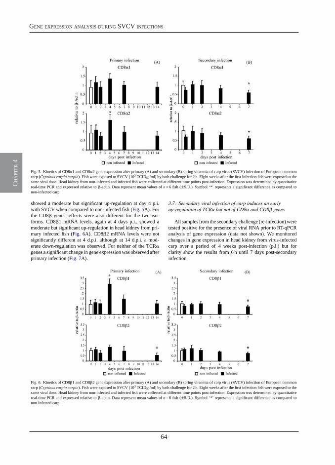
64
gene expression analysis during svcv infections
ch
apt
er 4
1540 M. Forlenza et al. / Molecular Immunology 45 (2008) 1531–1547
Fig. 5. Kinetics of CD8�1 and CD8�2 gene expression after primary (A) and secondary (B) spring viraemia of carp virus (SVCV) infection of European commoncarp (Cyprinus carpio carpio). Fish were exposed to SVCV (103 TCID50/ml) by bath challenge for 2 h. Eight weeks after the first infection fish were exposed to thesame viral dose. Head kidney from non-infected and infected fish were collected at different time points post-infection. Expression was determined by quantitativereal-time PCR and expressed relative to �-actin. Data represent mean values of n= 6 fish (±S.D.). Symbol ‘*’ represents a significant difference as compared tonon-infected carp.
showed a moderate but significant up-regulation at day 4 p.i.with SVCV when compared to non-infected fish (Fig. 5A). Forthe CD8� genes, effects were also different for the two iso-forms. CD8�1 mRNA levels, again at 4 days p.i., showed amoderate but significant up-regulation in head kidney from pri-mary infected fish (Fig. 6A). CD8�2 mRNA levels were notsignificantly different at 4 d.p.i. although at 14 d.p.i. a mod-erate down-regulation was observed. For neither of the TCR�genes a significant change in gene expression was observed afterprimary infection (Fig. 7A).
3.7. Secondary viral infection of carp induces an earlyup-regulation of TCRα but not of CD8α and CD8β genes
All samples from the secondary challenge (re-infection) weretested positive for the presence of viral RNA prior to RT-qPCRanalysis of gene expression (data not shown). We monitoredchanges in gene expression in head kidney from virus-infectedcarp over a period of 4 weeks post-infection (p.i.) but forclarity show the results from 6 h until 7 days post-secondaryinfection.
Fig. 6. Kinetics of CD8�1 and CD8�2 gene expression after primary (A) and secondary (B) spring viraemia of carp virus (SVCV) infection of European commoncarp (Cyprinus carpio carpio). Fish were exposed to SVCV (103 TCID50/ml) by bath challenge for 2 h. Eight weeks after the first infection fish were exposed to thesame viral dose. Head kidney from non-infected and infected fish were collected at different time points post-infection. Expression was determined by quantitativereal-time PCR and expressed relative to �-actin. Data represent mean values of n= 6 fish (±S.D.). Symbol ‘*’ represents a significant difference as compared tonon-infected carp.
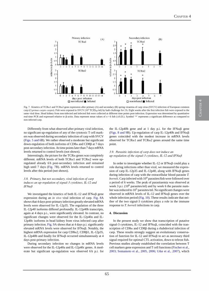
65
chapter 4
ch
apt
er 4
M. Forlenza et al. / Molecular Immunology 45 (2008) 1531–1547 1541
Fig. 7. Kinetics of TCR�1 and TCR�2 gene expression after primary (A) and secondary (B) spring viraemia of carp virus (SVCV) infection of European commoncarp (Cyprinus carpio carpio). Fish were exposed to SVCV (103 TCID50/ml) by bath challenge for 2 h. Eight weeks after the first infection fish were exposed to thesame viral dose. Head kidney from non-infected and infected fish were collected at different time points post-infection. Expression was determined by quantitativereal-time PCR and expressed relative to �-actin. Data represent mean values of n= 6 fish (±S.D.). Symbol ‘*’ represents a significant difference as compared tonon-infected carp.
Differently from what observed after primary viral infection,no significant up-regulation of any of the cytotoxic T cell mark-ers was observed during secondary infection of carp with SVCV(Figs. 5 and 6B). We rather observed a moderate but significantdown-regulation of both isoforms of CD8� and CD8� at 7 dayspost-secondary infection. At time points later than 7 days mRNAlevels returned to control levels (not shown).
Interestingly, the picture for the TCR� genes was completelydifferent. mRNA levels of both TCR�1 and TCR�2 were up-regulated already 6 h post-secondary infection and remainedhigh until 7 days (Fig. 7B). mRNA levels returned to controllevels after this period (not shown).
3.8. Primary, but not secondary, viral infection of carpinduces an up-regulation of signal-3 cytokines, IL-12 andIFNαβ
We investigated the kinetics of both IL-12 and IFN�� geneexpression during an in vivo viral infection of carp. Fig. 8Ashows that 4 days post-primary infection greatly elevated mRNAlevels were observed for IL-12p35. The regulation of the threeIL-12p40 isoforms differed profoundly. IL-12p40b transcripts,again at 4 days p.i., were significantly elevated. In contrast, nosignificant changes were observed for the IL-12p40a and IL-12p40c isoforms in head kidney from virus infected carp afterprimary infection. Fig. 9A shows that at 4 days p.i. significantlyelevated mRNA levels were observed for IFN��. Notably, thehighest mRNA expression for carp CD8�2, CD8�1, IL-12p35,IL-12p40b and finally for IFN�� occurred simultaneously at 4days post-primary infection.
During secondary infection no changes in mRNA levelswere observed for the IL-12p40a and IL-12p40c genes. A mod-erate but significant up-regulation was observed 6 h p.i. for
the IL-12p40b gene and at 1 day p.i. for the IFN�� gene(Figs. 8 and 9B). Up-regulation of carp IL-12p40b and IFN��genes coincided with the modest increase in mRNA levelsobserved for TCR�1 and TCR�2 genes around the same timepoint.
3.9. Parasitic infection of carp does not induce anup-regulation of the signal-3 cytokines, IL-12 and IFNαβ
In order to investigate whether IL-12 or IFN�� could play arole during infections other than viral, we measured the expres-sion of carp IL-12p35 and IL-12p40, along with IFN�� genesduring infection of carp with the extracellular blood parasite T.borreli. Carp infected with 104 parasites/fish were followed overa period of 6 weeks. The peak of parasitaemia was observed atweek 3 p.i. (108 parasites/ml) and by week 6 the parasite num-ber was reduced to 105 parasites/ml. No significant changes wereobserved in mRNA levels of IL-12 and IFN�� genes over thewhole infection period (Fig. 10). These results indicate that nei-ther of the two signal-3 cytokines plays a role in the immuneresponse to T. borreli infections in carp.
4. Discussion
In the present study we show that transcription of putativesignal-3 cytokines, IL-12 and IFN��, coincided with the tran-scription of CD8� and CD8� during a rhabdoviral infection ofcarp. These results strongly suggest an evolutionary conserva-tion of function for IL-12 and IFN�� to act as necessary thirdsignal required for optimal CTL activation, down to teleost fish.Previous studies already established the correlation between Tcell markers gene expression and T cell functions (Fischer et al.,2003; Somamoto et al., 2005, 2006; Utke et al., 2007), which
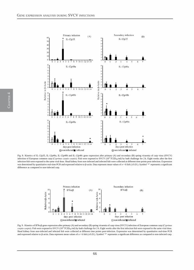
66
gene expression analysis during svcv infections
ch
apt
er 4
1542 M. Forlenza et al. / Molecular Immunology 45 (2008) 1531–1547
Fig. 8. Kinetics of IL-12p35, IL-12p40a, IL-12p40b and IL-12p40c gene expression after primary (A) and secondary (B) spring viraemia of carp virus (SVCV)infection of European common carp (Cyprinus carpio carpio). Fish were exposed to SVCV (103 TCID50/ml) by bath challenge for 2 h. Eight weeks after the firstinfection fish were exposed to the same viral dose. Head kidney from non-infected and infected fish were collected at different time points post-infection. Expressionwas determined by quantitative real-time PCR and expressed relative to �-actin. Data represent mean values of n= 6 fish (±S.D.). Symbol ‘*’ represents a significantdifference as compared to non-infected carp.
Fig. 9. Kinetics of IFN�� gene expression after primary (A) and secondary (B) spring viraemia of carp virus (SVCV) infection of European common carp (Cyprinuscarpio carpio). Fish were exposed to SVCV (103 TCID50/ml) by bath challenge for 2 h. Eight weeks after the first infection fish were exposed to the same viral dose.Head kidney from non-infected and infected fish were collected at different time points post-infection. Expression was determined by quantitative real-time PCRand expressed relative to �-actin. Data represent mean values of n= 6 fish (±S.D.). Symbol ‘*’ represents a significant difference as compared to non-infected carp.
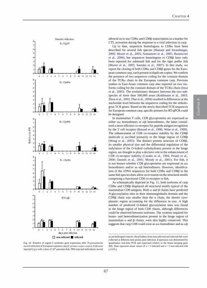
67
chapter 4
ch
apt
er 4
M. Forlenza et al. / Molecular Immunology 45 (2008) 1531–1547 1543
Fig. 10. Kinetics of signal-3 cytokines gene expression after Trypanoplasmaborreli infection of European common carp (Cyprinus carpio carpio). Fish wereinjected (i.p.) with a dose of 104 parasites/fish, PBS-injected individuals served
allowed us to use CD8� and CD8� transcription as a marker forCTL activation during the response to a viral infection in carp.
Up to date, sequences homologous to CD8� have beendescribed for several fish species (Hansen and Strassburger,2000; Moore et al., 2005; Somamoto et al., 2005; Buonocoreet al., 2006), but sequences homologous to CD8� have onlybeen reported for salmonid fish and for the tiger puffer fish(Moore et al., 2005; Suetake et al., 2007). In this study, wereport the cloning of both CD8� and CD8� genes for the Euro-pean common carp, each present in duplicate copies. We confirmthe presence of two sequences coding for the constant domainof the TCR� chain in the European common carp. Previousstudies in East-Asian common carp also reported on two iso-forms coding for the constant domain of the TCR� chain (Imaiet al., 2005). The evolutionary distance between the two sub-species of more than 500,000 years (Kohlmann et al., 2003;Zhou et al., 2003; Thai et al., 2004) resulted in differences at thenucleotide level between the sequences coding for the ortholo-gous TCR genes. Based on the newly described TCR sequencesfor European common carp, specific primers for RT-qPCR couldbe designed.
In mammalian T cells, CD8 glycoproteins are expressed aseither �� homodimers or �� heterodimers, the latter consid-ered a more efficient co-receptor for peptide antigen recognitionby the T cell receptor (Renard et al., 1996; Witte et al., 1999).The enhancement of CD8 co-receptor stability by the CD8�molecule is ascribed primarily to the hinge region of CD8�(Wong et al., 2003). The distinct protein structure of CD8�,its smaller physical size and the differential regulation of thesialylation of the O-linked carbohydrates present in the hingeregion, are thought to play a decisive role in the enhancement ofCD8 co-receptor stability (Casabo et al., 1994; Priatel et al.,2000; Daniels et al., 2001; Moody et al., 2001). For fish, itis not known whether CD8 glycoproteins are expressed as ��homodimers and/or as �� heterodimers. However, identifica-tion of the cDNA sequences for both CD8� and CD8� in thesame fish species does allow us to reason on the structural motifscomposing a functional CD8 co-receptor in fish.
As schematically depicted in Fig. 11, both isoforms of carpCD8� and CD8� displayed all structural motifs typical of themammalian CD8 antigens. Both � and � chains have predictedN-glycosylation sites in their immunoglobulin domain and theCD8� chain was smaller than the � chain, the shorter cyto-plasmic region accounting for the difference in size. A highnumber of predicted O-linked glycosylation sites was foundin the hinge region of both CD8 chains, although differencescould be observed between isoforms. The cysteins required forhomo- and heterodimerization present in the hinge region ofmammalian � and � chains, were also highly conserved. Thissuggests that carp CD8 could exist as �� homodimer and as ��
as unchallenged controls. Head kidney from non-infected and infected fish werecollected at different time points post-infection. Expression was determined byquantitative real-time PCR and expressed relative to the house keeping gene40S. Data represent mean values of n= 5 infected and n= 3 non-infected fish(±S.D.).
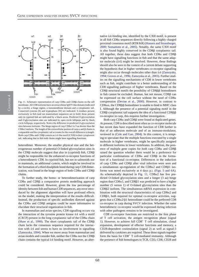
68
gene expression analysis during svcv infections
ch
apt
er 4
1544 M. Forlenza et al. / Molecular Immunology 45 (2008) 1531–1547
Fig. 11. Schematic representation of carp CD8� and CD8� chains on the cellmembrane. All CD8 isoforms have an extracellular IgSf V-like domain (indicatedby a circle), a hinge region, a transmembrane domain and a cytoplasmic tail.Conserved cysteins (S) and tryptophans (W) are indicated. S residues presentexclusively in fish (and not mammalian) sequences are in bold; those presentonly in cyprinid fish are indicated by a black arrow. Predicted N-glycosylationand O-glycosylation sites are indicated by open circle lollipops and by blackcircle lollipops, respectively. Notice the difference in predictedO-glycosylationsites between isoforms. The hinge region of carp CD8�1 is 3 aa shorter than theCD8�2 isoform. The length of the extracellular portion of carp � and � chains iscomparable and the cytoplasmic tail accounts for the overall difference in length.Both carp CD8� and CD8� contain an lck-like motif (CXH) in their cytoplasmictail, indicating that in fish both chains might have signalling functions.
heterodimer. Moreover, the smaller physical size and the het-erogeneous number of potential O-linked glycosylation sites inthe CD8� molecule suggest that also in (cyprinid) fish, CD8�might be responsible for the enhanced co-receptor functions ofa heterodimeric CD8. In cyprinid fish, but not in salmonids norin mammals, an additional cystein, which might be involved inthe formation of a third disulphide bond during carp CD8 dimer-ization, was found in the hinge region of both CD8� and CD8�chains.
To further study, the homo- or heterodimerization of carpCD8� and CD8� a comparative protein modelling approachcould be considered. However, given the low percentage ofidentity between fish and human CD8 sequences, any error intro-duced by the alignment algorithm will have profound effectson the model, making the interpretation of the results difficult.Instead, the production of specific antibodies directed againstthe CD8� and CD8� antigens could be more informative toelucidate their structural organization in fish.
In mammalian and avian species, CD8 signalling occurs viathe interaction of the tyrosine protein kinase lck with a motif(CXCP) present in the long cytoplasmic tail of the CD8� chain(Shaw et al., 1990). The short cytoplasmic tail of the CD8�chain lacks the consensus sequence required for the interac-tion with lck and seems to have no involvement in signalling(Zamoyska, 1994). When we move away from mammalian andavian models and consider fish, neither the CD8� nor the CD8�chain contains the typical lck binding motif. However, an alter-
native lck-binding site, identified by the CXH motif, is presentin all fish CD8� sequences directly following a highly chargedproximal consensus motif (RTR/KR) (Hansen and Strassburger,2000; Somamoto et al., 2005). Notably, the same CXH motifis also found highly conserved in the CD8� cytoplasmic tail.All together, these data suggest that both CD8� and CD8�might have signalling functions in fish and that the same adap-tor molecule (lck) might be involved. However, these findingsshould also be seen in the context of a current debate supportingthe hypothesis that in higher vertebrates co-receptor signallingmight also occur through molecules other than lck (Zamoyska,1994; Groves et al., 1996; Zamoyska et al., 2003). Further stud-ies on the signalling mechanisms of CD8 in lower vertebratessuch as fish, might contribute to a better understanding of theCD8 signalling pathways of higher vertebrates. Based on theCD8� structural motifs the possibility of CD8�� homodimersin fish cannot be excluded. Human, but not mouse, CD8� canbe expressed on the cell surface without the need of CD8�coexpression (Devine et al., 2000). However, in contrast toCD8��, the CD8�� homodimer is unable to bind to MHC classI. Although the presence of a potential signaling motif in theCD8� cytoplasmic tail supports the idea of a functional CD8��co-receptor in carp, this requires further investigation.
Both carp CD8� and CD8� were found as duplicated genes.At present, CD8 is described most often as co-receptor moleculebut recent data have expanded the function of CD8 further tothat of an adhesion molecule and of an immuno-modulator,reviewed in (Cole and Gao, 2004). In this context, it is tempt-ing to speculate that the multiple functions executed by a singlemolecule in higher vertebrates, might be specifically assignedto different isoforms in lower vertebrates. In addition, the pres-ence of multiple gene copies for both carp CD8� and CD8�raised the question whether there would be a preference forparticular combinations of CD8� and CD8� in the forma-tion of a functional co-receptor. Differences in the inductionof carp CD8� and CD8� after viral infection were seen anda simultaneous up-regulation of the CD8�2 and CD8�1 iso-forms was noted exclusively at 4 days p.i. (Figs. 5 and 6A).As schematically depicted in Fig. 11, CD8�2 has five pre-dicted O-linked glycosylation sites and a longer (3 aa) hingeregion than CD8�1, and CD8�1 was predicted to have a highernumber (5 versus 1) of O-linked glycosylation sites than theCD8�2 isoform. The simultaneous mRNA expression in com-bination with the structural characteristics of carp CD8�2 andCD8�1, both required for optimal co-receptor activities, sug-gests that a CD8�2/�1 heterodimer could be the preferred CD8co-receptor in carp during SVCV infection. Whether the sameheterodimeric co-receptor would be expressed during infectionwith other pathogens remains to be investigated.
CD8 co-receptor functions are restricted to the first phaseof T cell activation, the antigen recognition phase (signal1). However, to achieve full CD8+ T cell stimulation, clonalexpansion, development of effector functions and memory, aCD28-dependent costimulation (signal 2) as well as signal-3delivered by cytokines are required. These three signals togetherform the basis for CD8+ T cell-mediated immunity. Althoughthe presence of fish homologues to TCR, CD3, CD8, CD28 and
69
chapter 4
ch
apt
er 4
M. Forlenza et al. / Molecular Immunology 45 (2008) 1531–1547 1545
MHC-I, among others, suggests that the presence of signal 1and signal 2 is conserved from fish to mammals (Bernard etal., 2006a,b; Fischer et al., 2006), in fish, the potential of IL-12and IFN�� to act as signal-3 cytokines in infections inducing aCTL response has not been examined. We used a natural viralinfection of carp to study the timing of mRNA expression ofthe CD8 cytotoxic T cell markers and of the putative signal-3cytokines IL-12 and IFN��. Our results show that both, IL-12and IFN��, are involved during the onset of a CTL responsewhich strongly suggests that also in fish these cytokines mightprovide the necessary third signal for complete T cell activa-tion. In fact, during primary SVCV infection, up-regulation ofcarp CD8 gene expression occurred simultaneously with an up-regulation of IL-12 and IFN��. In mammals, in vitro and in vivostudies examining the timing requirements for exposure to Ag,costimulation and signal-3 cytokine showed that optimal overallstimulation and development of function required Ag/B7 to bepresent for about 36 h, with IL-12 being present as early as 12 h,up to 3 days (van Stipdonk et al., 2001; Curtsinger et al., 2003a).Coordinated timing of expression of carp CD8, IL-12 and IFN��was always seen 4 days post-infection, corresponding well to thetiming observed in higher vertebrates.
We used a natural parasite infection of carp (Wiegertjes et al.,2005) to assess whether IL-12 and IFN�� signal-3 cytokineswould be upregulated in infections other than viral, where aCTL response is not required. After infection with the extra-cellular blood flagellate T. borreli, no changes in IL-12 or inIFN�� gene transcription could be observed in head kidneyfrom parasite-infected carp. In contrast, a significant (6.6-fold)up-regulation of TNF� was observed concomitantly with thepeak of parasitaemia (unpublished data), while TNF� mRNAexpression was only moderately (1.6-fold) increased 4 days post-primary viral infection (data not shown). This suggests that theTNF� cytokine plays a more important role during parasitic thanduring viral infections of carp. In mammals, in fact, the signalrequired for optimal CTL stimulation cannot be provided by thecytokines IL-1, IL-2, IL-4, IL-7, IL-15, IL-18, tumor necrosisfactor-� (TNF-�) and IFN-� (Mescher et al., 2006). All together,our results suggest that IL-12 and IFN�� gene expression arerequired during viral, but not during parasitic infection.
During secondary infection (re-infection) with SVCV, no up-regulation of the CD8 genes and only a marginal increase inmRNA expression of the signal-3 cytokines IL-12 (IL-1240b)and IFN�� was observed. The latter finding is not surprisingsince memory CD8+ T cells do not require a third signal todevelop effector functions (Curtsinger et al., 1999; Schmidt andMescher, 2002) and confirms that also in carp, signal-3 cytokinesare not involved during the course of a secondary infection. Theabsence of a clear up-regulation of any of the CD8 genes mightbe indicative of the fact that other types of effector mechanisms,such as B cell or non-CTL-mediated T cell responses, mightbe involved during secondary infection. This is supported bythe observation that transcription of both TCR isoforms wasclearly elevated during second exposure to the virus. Similarresults were observed after primary and secondary ranavirusinfection of Xenopus laevis (Morales and Robert, 2007). Atpresent, sequences coding for genes homologous to CD3� or
CD4 are not yet available in carp; their identification could pro-vide a better insight into the role played by CD4+ T cells duringthe course of a secondary SVCV infection. Acutely cytopathicviruses, such as vaccinia virus, promote CTL responses uponprimary infection and humoral responses involving IgM neu-tralizing antibodies early upon secondary infection. In the lattercase, virus-specific CTL are neither necessary nor sufficient tocontrol infection, reviewed in (Hangartner et al., 2006). SVC isa severe infection of cyprinid carp that is widespread through-out Europe, causing high mortalities during spring (Ahne et al.,2002). SVCV belongs to the group of acutely cytopathic virusesand it has been tentatively assigned to the genus Vesiculovirusof the Rhabdoviridae family. Outbreaks of SVCV can resultin mortality rates between 40 and 90% and only recently dataare becoming available indicating that DNA vaccination ofcarp can protect against SVCV (Kanellos et al., 2006). It isbecoming clear that during primary SVCV infection an earlymoderate cytotoxic response occurs (Kanellos et al., 2006), fol-lowed by a later development of neutralizing antibodies directedagainst the glycoprotein of the virus (Ahne et al., 2002). Uponinfection following DNA vaccination (Kanellos et al., 2006) orafter secondary SVCV infection a protective humoral response,rather than a cytotoxic, response is achieved (Ahne et al., 2002;Kanellos et al., 2006).In vivo peptide immunization studies in mice have shown that
administration of IL-12 along with the immunizing peptide hadthe same effect as adjuvant in preventing tolerance, stimulating aCTL response and establishing a memory development. In carpIL-12p35 was reported as a single copy gene while three highlydistinct IL-12p40 genes, with distinct gene expression patterns,were identified (Huising et al., 2006). Coexpression of IL-12p35and IL-12p40 genes is required in order to yield the functionalIL-12p70 heterodimer (Gubler et al., 1991). Thus, for IL-12 tobe used as an immunostimulant in carp, the identification of thefunctional IL-12p70 heterodimer is required. We found neitherregulation of IL-12p40a and IL-12p40c expression nor after pri-mary nor after secondary SVCV infection, while we observedsimultaneous expression of the IL-12p35 and IL-12p40b genes4 days post-primary infection, in head kidney from infected fish.Our findings, therefore, suggest that it is the p35p40b associa-tion that leads to the formation of a functional carp IL-12p70heterodimer. Given the potential role of IL-12 as adjuvant, ourfindings should be taken into consideration for future vaccinationstrategies against viral infection of fish. Of course, the possibil-ity that other heterodimeric combinations may be formed duringthe immune response to infections other than the one examinedin this study, cannot be excluded. Future studies on the coex-pression of IL-12p35 and the different IL-12p40 isoforms arerequired to confirm the preference for a p35p40b association incarp.
In conclusion, we report the cloning of CD8� and CD8�homologues, each present in duplicate copies, and confirmthe presence of two TCR-C� isoforms in European commoncarp (C. carpio carpio). Based on structural analysis, carpCD8 molecules could exist as �� and �� dimers although thepresence of CD8�� homodimers cannot be excluded. The pres-ence of potential signalling motifs in the cytoplasmic tail of
70
gene expression analysis during svcv infections
ch
apt
er 4
1546 M. Forlenza et al. / Molecular Immunology 45 (2008) 1531–1547
both CD8� and CD8� indicates that both chains might havesignalling functions in fish. Coordinated kinetics of mRNAtranscription suggest that a CD8�2/�1 heterodimer could bethe preferred CD8 co-receptor in carp. Similarly, our expres-sion study provides evidences that it is the association betweenthe p35p40b molecules which will lead to the formation of afunctional IL-12p70 heterodimer during viral infection of carp.Further, transcription of signal-3 cytokines coincided with thetiming of CD8�� up-regulation during viral infection of carp.This strongly suggests that IL-12 and IFN�� signal-3 cytokinesare particularly required after primary viral infection when alsoup-regulation of cytotoxic T cell markers is observed.
Acknowledgments
We thank the central fish facilities, “De Haar-Vissen,” for tak-ing care of the carp. We thank Ellen Stolte and Greetje Castelijnfor help with MACS sorting and Silvia Paolucci for her techni-cal assistance. Uwe Fischer, Mark Huising, Jan Rombout andVirgil Schijns are acknowledged for their comments on themanuscript. This work was supported by the European Com-mission within the project IMAQUANIM (EC contract numberFOOD-CT-2005-007103).
References
Ahne, W., Bjorklund, H.V., Essbauer, S., Fijan, N., Kurath, G., Winton, J.R.,2002. Spring viremia of carp (SVC). Dis. Aquat. Org. 52, 261–272.
Bernard, D., Riteau, B., Hansen, J.D., Phillips, R.B., Michel, F., Boudinot, P.,Benmansour, A., 2006a. Costimulatory receptors in a teleost fish: typicalCD28, elusive CTLA4. J. Immunol. 176, 4191–4200.
Bernard, D., Six, A., Rigottier-Gois, L., Messiaen, S., Chilmonczyk, S., Quillet,E., Boudinot, P., Benmansour, A., 2006b. Phenotypic and functional simi-larity of gut intraepithelial and systemic T cells in a teleost fish. J. Immunol.176, 3942–3949.
Boudinot, P., Boubekeur, S., Benmansour, A., 2001. Rhabdovirus infectioninduces public and private T cell responses in teleost fish. J. Immunol. 167,6202–6209.
Buonocore, F., Randelli, E., Bird, S., Secombes, C.J., Costantini, S., Facchiano,A., Mazzini, M., Scapigliati, G., 2006. The CD8alpha from sea bass (Dicen-trarchus labrax L.): cloning, expression and 3D modelling. Fish ShellfishImmunol. 20, 637–646.
Casabo, L.G., Mamalaki, C., Kioussis, D., Zamoyska, R., 1994. T cell activationresults in physical modification of the mouse CD8 beta chain. J. Immunol.152, 397–404.
Cole, D.K., Gao, G.F., 2004. CD8: adhesion molecule, co-receptor and immuno-modulator. Cell. Mol. Immunol. 1, 81–88.
Curtsinger, J.M., Johnson, C.M., Mescher, M.F., 2003a. CD8 T cell clonal expan-sion and development of effector function require prolonged exposure toantigen, costimulation, and signal 3 cytokine. J. Immunol. 171, 5165–5171.
Curtsinger, J.M., Lins, D.C., Mescher, M.F., 2003b. Signal 3 determines toler-ance versus full activation of naive CD8 T cells: dissociating proliferationand development of effector function. J. Exp. Med. 197, 1141–1151.
Curtsinger, J.M., Schmidt, C.S., Mondino, A., Lins, D.C., Kedl, R.M., Jenkins,M.K., Mescher, M.F., 1999. Inflammatory cytokines provide a third signalfor activation of naive CD4+ and CD8+ T cells. J. Immunol. 162, 3256–3262.
Curtsinger, J.M., Valenzuela, J.O., Agarwal, P., Lins, D., Mescher, M.F., 2005.Type I IFNs provide a third signal to CD8 T cells to stimulate clonal expan-sion and differentiation. J. Immunol. 174, 4465–4469.
Daniels, M.A., Devine, L., Miller, J.D., Moser, J.M., Lukacher, A.E., Altman,J.D., Kavathas, P., Hogquist, K.A., Jameson, S.C., 2001. CD8 binding toMHC class I molecules is influenced by T cell maturation and glycosylation.Immunity 15, 1051–1061.
Devine, L., Kieffer, L.J., Aitken, V., Kavathas, P.B., 2000. Human CD8 beta,but not mouse CD8 beta, can be expressed in the absence of CD8 alpha as abeta beta homodimer. J. Immunol. 164, 833–838.
DiSanto, J.P., Smith, D., de Bruin, D., Lacy, E., Flomenberg, N., 1993. Tran-scriptional diversity at the duplicated human CD8 beta loci. Eur. J. Immunol.23, 320–326.
Fijan, N., Sulimanovic, D., Bearzotti, M., Muzinic, D., Zwillenberg, L.O.,Chilmonczyk, S., Vautherot, J.F., de kinkelin, P., 1983. Some propertiesof the Epithelioma papulosum cyprini (EPC) cell line from carp Cyprinuscarpio. Ann. Virol. (Institue Pasteur) 134, 207–220.
Filatenkov, A.A., Jacovetty, E.L., Fischer, U.B., Curtsinger, J.M., Mescher, M.F.,Ingulli, E., 2005. CD4 T cell-dependent conditioning of dendritic cells toproduce IL-12 results in CD8-mediated graft rejection and avoidance oftolerance. J. Immunol. 174, 6909–6917.
Fischer, U., Ototake, M., Nakanishi, T., 1998. In vitro cell-mediated cytotoxicityagainst allogeneic erythrocytes in ginbuna crucian carp and goldfish using anon-radioactive assay. Dev. Comp. Immunol. 22, 195–206.
Fischer, U., Utke, K., Ototake, M., Dijkstra, J.M., Kollner, B., 2003. Adaptivecell-mediated cytotoxicity against allogeneic targets by CD8-positive lym-phocytes of rainbow trout (Oncorhynchus mykiss). Dev. Comp. Immunol.27, 323–337.
Fischer, U., Utke, K., Somamoto, T., Kollner, B., Ototake, M., Nakanishi, T.,2006. Cytotoxic activities of fish leucocytes. Fish Shellfish Immunol. 20,209–226.
Gonzalez, S.F., Chatziandreou, N., Nielsen, M.E., Li, W., Rogers, J., Taylor, R.,Santos, Y., Cossins, A., 2007. Cutaneous immune responses in the commoncarp detected using transcript analysis. Mol. Immunol. 44, 1664–1679.
Groves, T., Smiley, P., Cooke, M.P., Forbush, K., Perlmutter, R.M., Guidos, C.J.,1996. Fyn can partially substitute for Lck in T lymphocyte development.Immunity 5, 417–428.
Gubler, U., Chua, A.O., Schoenhaut, D.S., Dwyer, C.M., McComas, W., Motyka,R., Nabavi, N., Wolitzky, A.G., Quinn, P.M., Familletti, P.C., et al., 1991.Coexpression of two distinct genes is required to generate secreted bioactivecytotoxic lymphocyte maturation factor. Proc. Natl. Acad. Sci. U.S.A. 88,4143–4147.
Hangartner, L., Zinkernagel, R.M., Hengartner, H., 2006. Antiviral antibodyresponses: the two extremes of a wide spectrum. Nat. Rev. Immunol. 6,231–243.
Hansen, J.D., Strassburger, P., 2000. Description of an ectothermic TCR core-ceptor, CD8 alpha, in rainbow trout. J. Immunol. 164, 3132–3139.
Hasegawa, S., Nakayasu, C., Yoshitomi, T., Nakanishi, T., Okamoto, N., 1998.Specific cell-mediated cytotoxicity against an allogeneic target cell line inisogeneic ginbuna crucian carp. Fish Shellfish Immunol. 8, 303–313.
Hernandez, J., Aung, S., Marquardt, K., Sherman, L.A., 2002. Uncouplingof proliferative potential and gain of effector function by CD8(+) T cellsresponding to self-antigens. J. Exp. Med. 196, 323–333.
Huising, M.O., van Schijndel, J.E., Kruiswijk, C.P., Nabuurs, S.B., Savelkoul,H.F.J., Flik, G., Verburg-van Kemenade, B.M.L., 2006. The presence ofmultiple and differentially regulated interleukin-12p40 genes in bony fishessignifies an expansion of the vertebrate heterodimeric cytokine family. Mol.Immunol. 43, 1519–1533.
Imai, E., Ishikawa, J., Moritomo, T., Tomana, M., 2005. Characterisation of Tcell antigen receptor alpha chain isotypes in the common carp. Fish ShellfishImmunol. 19, 205–216.
Irnazarow, I., 1995. Genetic variability of Polish and Hungarian carp lines.Aquacult. Res. 129, 215–219.
Kanellos, T., Sylvester, I.D., D’Mello, F., Howard, C.R., Mackie, A., Dixon,P.F., Chang, K.C., Ramstad, A., Midtlyng, P.J., Russell, P.H., 2006. DNAvaccination can protect Cyprinus Carpio against spring viraemia of carpvirus. Vaccine 24, 4927–4933.
Kohlmann, K., Gross, R., Murakaeva, A., Kersten, P., 2003. Genetic variabilityand structure of common carp (Cyprinus carpio) populations throughout thedistribution range inferred from allozyme, microsatellite and mitochondrialDNA markers. Aquat. Living Res. 16, 421–431.
Kolumam, G.A., Thomas, S., Thompson, L.J., Sprent, J., Murali-Krishna, K.,2005. Type I interferons act directly on CD8 T cells to allow clonal expansionand memory formation in response to viral infection. J. Exp. Med. 202,637–650.
71
chapter 4
ch
apt
er 4
M. Forlenza et al. / Molecular Immunology 45 (2008) 1531–1547 1547
Koumans-van Diepen, J.E., Egberts, E., Peixoto, B.R., Taverne, N., Rombout,J.H.W.M., 1995. B cell and immunoglobulin heterogeneity in carp (Cypri-nus carpio L.); An immuno(cyto)chemical study. Dev. Comp. Immunol. 19,97–108.
Koutna, M., Vesely, T., Psikal, I., Hulova, J., 2003. Identification of springviraemia of carp virus (SVCV) by combined RT-PCR and nested PCR. Dis.Aquat. Org. 55, 229–235.
Mescher, M.F., Curtsinger, J.M., Agarwal, P., Casey, K.A., Gerner, M., Hammer-beck, C.D., Popescu, F., Xiao, Z., 2006. Signals required for programmingeffector and memory development by CD8+ T cells. Immunol. Rev. 211,81–92.
Moody, A.M., Chui, D., Reche, P.A., Priatel, J.J., Marth, J.D., Reinherz, E.L.,2001. Developmentally regulated glycosylation of the CD8alphabeta core-ceptor stalk modulates ligand binding. Cell 107, 501–512.
Moore, L.J., Somamoto, T., Lie, K.K., Dijkstra, J.M., Hordvik, I., 2005. Char-acterisation of salmon and trout CD8alpha and CD8beta. Mol. Immunol. 42,1225–1234.
Morales, H.D., Robert, J., 2007. Characterization of primary and memory CD8T-cell responses against ranavirus (FV3) in Xenopus laevis. J. Virol. 81,2240–2248.
Nakauchi, H., Shinkai, Y., Okumura, K., 1987. Molecular cloning of Lyt-3, amembrane glycoprotein marking a subset of mouse T lymphocytes: molec-ular homology to immunoglobulin and T-cell receptor variable and joiningregions. Proc. Natl. Acad. Sci. U.S.A. 84, 4210–4214.
Nascimento, D.S., Vale, A.d., Tomas, A.M., Zou, J., Secombes, C.J., dos Santos,N.M.S., 2007. Cloning, promoter analysis and expression in response tobacterial exposure of sea bass (Dicentrarchus labrax L.) interleukin-12 p40and p35 subunits. Mol. Immunol. 44, 2277–2291.
Pfaffl, M.W., 2001. A new mathematical model for relative quantification inreal-time RT-PCR. Nucleic Acids Res. 29, e45.
Priatel, J.J., Chui, D., Hiraoka, N., Simmons, C.J., Richardson, K.B., Page,D.M., Fukuda, M., Varki, N.M., Marth, J.D., 2000. The ST3Gal-I sialyltrans-ferase controls CD8+ T lymphocyte homeostasis by modulating O-glycanbiosynthesis. Immunity 12, 273–283.
Reed, R.J., Muench, H., 1938. A simple method of estimating 50% endpoints.Am. J. Hyg. 27, 493–497.
Renard, V., Romero, P., Vivier, E., Malissen, B., Luescher, I.F., 1996. CD8 betaincreases CD8 coreceptor function and participation in TCR-ligand binding.J. Exp. Med. 184, 2439–2444.
Ririe, K.M., Rasmussen, R.P., Wittwer, C.T., 1997. Product differentiation byanalysis of DNA melting curves during the polymerase chain reaction. Anal.Biochem. 245, 154–160.
Robertsen, B., 2006. The interferon system of teleost fish. Fish ShellfishImmunol. 20, 172–191.
Sakano, H., Huppi, K., Heinrich, G., Tonegawa, S., 1979. Sequences at thesomatic recombination sites of immunoglobulin light-chain genes. Nature280, 288–294.
Schmidt, C.S., Mescher, M.F., 2002. Peptide antigen priming of naive, but notmemory, CD8 T cells requires a third signal that can be provided by IL-12.J. Immunol. 168, 5521–5529.
Shaw, A.S., Chalupny, J., Whitney, J.A., Hammond, C., Amrein, K.E., Kavathas,P., Sefton, B.M., Rose, J.K., 1990. Short related sequences in the cytoplasmicdomains of CD4 and CD8 mediate binding to the amino-terminal domain ofthe p56lck tyrosine protein kinase. Mol. Cell. Biol. 10, 1853–1862.
Somamoto, T., Yoshiura, Y., Nakanishi, T., Ototake, M., 2005. Molecular cloningand characterization of two types of CD8alpha from ginbuna crucian carp,Carassius auratus langsdorfii. Dev. Comp. Immunol. 29, 693–702.
Somamoto, T., Yoshiura, Y., Sato, A., Nakao, M., Nakanishi, T., Okamoto, N.,Ototake, M., 2006. Expression profiles of TCRbeta and CD8alpha mRNAcorrelate with virus-specific cell-mediated cytotoxic activity in Ginbunacrucian carp. Virology 348, 370–377.
Steinhagen, D., Kruse, P., Korting, W., 1989. The parasitemia of clonedTrypanoplasma borreli Laveran and Mesnil, 1901 in laboratory-infectedcommon carp (Cyprinus carpio L.). J. Parasitol. 75, 685–689.
Suetake, H., Araki, K., Akatsu, K., Somamoto, T., Dijkstra, J.M., Yoshiura, Y.,Kikuchi, K., Suzuki, Y., 2007. Gene organization and expression of CD8�
and CD8� genes in fugu Takifugu rubripes. Fish Shellfish Immunol. 23,1107–1118.
Thai, B.T., Burridge, C.P., Pham, T.A., Austin, C.M., 2004. Using mitochondrialnucleotide sequences to investigate diversity and genealogical relationshipswithin common carp (Cyprinus carpio L.). Anim. Gen. 36, 23–28.
Tichopad, A., Dilger, M., Schwarz, G., Pfaffl, M.W., 2003. Standardized deter-mination of real-time PCR efficiency from a single reaction set-up. NucleicAcids Res. 31, e122.
Utke, K., Bergmann, S., Lorenzen, N., Kollner, B., Ototake, M., Fischer, U.,2007. Cell-mediated cytotoxicity in rainbow trout, Oncorhynchus mykiss,infected with viral haemorrhagic septicaemia virus. Fish Shellfish Immunol.22, 182–196.
van Den Broek, M., Bachmann, M.F., Kohler, G., Barner, M., Escher, R.,Zinkernagel, R., Kopf, M., 2000. IL-4 and IL-10 antagonize IL-12-mediatedprotection against acute vaccinia virus infection with a limited role of IFN-gamma and nitric oxide synthetase 2. J. Immunol. 164, 371–378.
van Erp, S.H., Dixon, B., Figueroa, F., Egberts, E., Stet, R.J.M., 1996. Identifi-cation and characterization of a new major histocompatibility complex classI gene in carp (Cyprinus carpio L.). Immunogenetics 44, 49–61.
van Stipdonk, M.J., Lemmens, E.E., Schoenberger, S.P., 2001. Naive CTLsrequire a single brief period of antigenic stimulation for clonal expansionand differentiation. Nat. Immunol. 2, 423–429.
Wiegertjes, G.F., Forlenza, M., Joerink, M., Scharsack, J.P., 2005. Parasite infec-tions revisited. Dev. Comp. Immunol. 29, 749–758.
Witte, T., Spoerl, R., Chang, H.C., 1999. The CD8beta ectodomain contributesto the augmented coreceptor function of CD8alphabeta heterodimers relativeto CD8alphaalpha homodimers. Cell Immunol. 191, 90–96.
Wong, J.S., Wang, X., Witte, T., Nie, L., Carvou, N., Kern, P., Chang, H.C.,2003. Stalk region of beta-chain enhances the coreceptor function of CD8.J. Immunol. 171, 867–874.
Yoshiura, Y., Kiryu, I., Fujiwara, A., Suetake, H., Suzuki, Y., Nakanishi, T.,Ototake, M., 2003. Identification and characterization of Fugu orthologuesof mammalian interleukin-12 subunits. Immunogenetics 55, 296–306.
Zamoyska, R., 1994. The CD8 coreceptor revisited: one chain good, two chainsbetter. Immunity 1, 243–246.
Zamoyska, R., Basson, A., Filby, A., Legname, G., Lovatt, M., Seddon, B.,2003. The influence of the src-family kinases, Lck and Fyn, on T celldifferentiation, survival and activation. Immunol. Rev. 191, 107–118.
Zhou, J.F., Wu, Q.J., Ye, Y.Z., Tong, J.G., 2003. Genetic devergence betweenCyprinus carpio carpio and Cyprinus carpio haematopterus as assessed bymitochondrial DNA analysis, with enphasis on origin of European domesticcarp. Genetica 119, 93–97.

72

DNA vaccination strategies in common carp (Cyprinus carpio L.) against spring
viraemia of carp virus (SVCV)
chapter
5
Maria Forlenza, Tomas Veselý, Dagmar Pokorová, Niels Lorenzen, Katja Einer-Jensen, Anne Kozijn, Huub F.J. Savelkoul and Geert F. Wiegertjes
Manuscript in preparation

74
dna vaccination strategies against svcv
ch
apt
er 5
75
chapter 5
ch
apt
er 5
1
DNA vaccination strategies in carp (Cyprinus carpio) against
spring viraemia of carp virus (SVCV)
Maria Forlenza1, Tomas Vesely2, Dasa Pokorova2, Niels Lorenzen3, Katja Einer-Jensen3,
Anne Kozijn1, Huub F.J. Savelkoul1 and Geert Wiegertjes1
1 Department of Animal Sciences, Cell Biology and Immunology Group, Wageningen Institute of Animal
Sciences, Wageningen University, P.O. Box 338, 6700 AH, Wageningen, The Netherlands 2 Veterinary Research Institute, Hudcova 70, 621 32 Brno, Czech Republic3 Department of Fish, National Veterinary Institute, Technical University of Denmark, DK-8200 Arhus N,
Denmark
Abstract
Spring viraemia of carp virus (SVCV; Rhabdoviridae) causes a severe infection of carp with high mortalities especially at reduced water temperatures during spring. DNA vaccination can protect carp against SVC, although the mechanisms of protection have not been elucidated. We report the cloning of a carp myxovirus resistance (Mx1) cDNA and describe the changes in gene expression of a number of additional immune genes after DNA vaccination and after bath challenge with SVCV. Surprisingly, Mx1 up-regulation was observed not only in fish that received the vaccine plasmid, but also in carp that received the empty plasmid only, indicating the induction of an innate immune response, not specifically triggered by the viral G protein. The possibility that CpG motifs contained in the backbone of the pcDNA3 plasmid could induce a species-specific recognition of these pathogen-associated molecular patterns, is discussed. Besides a clear up-regulation of Mx1 gene expression, DNA vaccination itself, but also challenge with SVCV of vaccinated carp did not induce strong changes in the expression of other investigated genes. Challenge of naïve carp, however, showed an up-regulation of several genes including IFNαβ, Mx, IL-12p35, IFNγ2 and IL-10, particularly at 4 days post-challenge. The timing of up-regulation confirms our previous observation that infection of naïve fish with SVCV induces an early, simultaneous up-regulation of putative signal-3 cytokines IFNαβ and IL-12. DNA vaccination resulted in 85% survival after bath challenge, with a relative percent survival (RPS) of 66.7%. DNA vaccination against other rhabdoviruses such as VHS or IHNV, can result in RPS values of 90% or higher. Therefore, despite the promising results observed in this study, the DNA vaccine shows potential for further improvement. In mammals, IL-12 has been shown to serve as a potent immunoadjuvant when administered along with the immunizing antigen, sustaining cell-mediated immunity in particular. In fish, evidence points towards a role for cell-mediated responses in the protection against SVCV. We therefore constructed a plasmid coding for carp IL-12 composed of the p40b subunit fused to the p35 subunit and show that we can detect secreted, and possibly bio-active IL-12 in the supernatant of transfected cells. Administration of the IL-12p40bLp35 construct along with the DNA vaccine as a means of improving the efficacy of the SVCV DNA vaccine is discussed.
ý á

76
dna vaccination strategies against svcv
ch
apt
er 5
2
Introduction
Spring viraemia of carp (SVC) is a severe
viral infection of carp that is widespread
throughout Europe, causing high mortalities
during spring (1). SVCV belongs to the group
of acutely cytopathic viruses and it has been
tentatively assigned to the genus Vesiculovirus
of the Rhabdoviridae family. Outbreaks of
SVCV can result in mortality rates between 40
and 90% and only recently new data indicate
that DNA vaccination can protect carp against
this viral infection (2). In general, DNA
vaccination is particularly effective in fish and
notably the first DNA vaccine commercialized
for an edible animal species (Apex-IHN) is
directed against a fish virus and protects
Atlantic salmon from infectious hematopoietic
necrosis (IHN) (3). Other successful examples
include DNA vaccines against viral
haemorrhagic septicaemia virus (VHSV) in
trout (4) against hirame rhabdovirus (HIRRV)
and VHSV in Japanese flounder (5, 6) and
against SVCV in carp (2, 7). The DNA
vaccines developed against these fish
rhabdoviruses are based on the intramuscular
injection of microgram quantities of naked
DNA plasmid encoding the viral glycoprotein
(G protein). Co-vaccination with DNA
plasmids encoding the G protein of VHSV and
the G protein of IHNV has been shown to
successfully induce specific divalent protection
against both viruses (8). In general, DNA
vaccines against VHSV and IHNV and similar
vaccines against other fish rhabdoviruses
induce an early, transient, non-specific anti-
viral immunity, followed in time by a
protective antibody response (4, 9-15).
A number of studies have provided further
insights in the immune response induced by
DNA vaccines using microarray and real-time
quantitative PCR describing the kinetics of
gene expression following DNA vaccination
(5, 6, 16-18). Most of these studies have been
performed in salmonid fish species or in
Japanese flounder but in carp, gene expression
following DNA vaccination against SVCV has
not been investigated. Despite differences in
the fold change or peak of gene expression,
often due to differences in the sampling time
points, tissue origin and temperature at
vaccination, a factor common to all DNA
vaccination studies is the early up-regulation
of type I interferon (IFN)-induced genes such
as Mx (myxovirus resistance gene). The
expression of IFN and Mx has been correlated
temporally with the non-specific protection
conferred during the early phase after
vaccination. Sequences for Mx genes have
been reported for most fish species, including
rainbow trout (19), Atlantic salmon (20, 21),
Japanese flounder (22), halibut (23), channel
catfish (24), grouper (25), pufferfish (26)
crucian carp (27) and zebrafish (28), but not
common carp. In the present study we first
report the cloning of a carp Mx gene
homologous to other Mx1 genes described for
teleost fish. Next, following DNA vaccination
and challenge with SVCV, we examined the
kinetics of carp Mx1 gene expression, together
with the expression of other relevant genes
such as type I interferon (referred to as IFNαβ
in carp), IL-12p35 and IL-12p40, IL-1β1,
CxCa, IL-10 and IFNγ2 to provide further
77
chapter 5
ch
apt
er 5
3
insight in the immune response of carp
induced by DNA vaccination.
We recently reported on the cloning of CD8α
and CD8β homologues, each present in
duplicate copies, and on the kinetics of
expression of these genes during primary and
secondary infection of carp with SVCV (29).
In the same study we noted that the expression
of putative signal-3 cytokines such as IL-12
and IFN-αβ was also upregulated,
concomitantly with the CD8α and CD8β gene
expression, all at 4 days after primary
infection. In mammals, signal-3 cytokines are
required for optimal programming for survival,
effector function and memory of CD8+ T cells
(30). IL-12 is a heterodimeric protein
composed of a p35 and a p40 subunit. In carp
three p40 subunits, designated p40a, p40b and
p40c have been identified (31) and during
primary viral infection, expression of p35 was
up-regulated concomitantly with the
expression of the p40b subunit only (1). As a
preliminary work, in the present study, we
report the construction of a DNA vector
encoding carp IL-12, obtained by fusing the
p35 and p40b subunits with an interspacing
linker (p40bLp35). We show that recombinant
carp IL-12 can be expressed in a carp cell line
resulting in a soluble protein of the expected
size of 80 kDa. Future applications of the carp
IL-12 DNA plasmid as molecular adjuvant to
improve DNA vaccination of carp against
SVCV will be discussed.
Materials and methods
Animals
European common carp (Cyprinus carpio
carpio) of the R3 x R8 strain, which are the
offspring of a cross between fish of Hungarian
origin (R8 strain) and of Polish origin (R3
strain), were used (32). In this study, the
European common carp subspecies will be
referred to as carp, unless stated otherwise.
Carp were produced in the central fish facility
of Wageningen University, The Netherlands,
then transported to the Veterinary Research
Institute (VRI), Brno, Czech Republic, and
raised in recirculating UV-treated water and
fed pelleted dry food (Trouvit, Nutreco) daily.
SVCV-vaccination and challenge experiments
were performed in the latter facility. Adult
carp (16 months) with an average weight of
30-40 g were used.
DNA vaccination
Carp were raised at 15°C, the optimal
temperature for SVCV infectivity (1) and
randomly assigned to one of three groups:
pcDNA3 (empty plasmid), pcDNA3-SVCV-G
and an unhandled group. pcDNA3-SVCV-G
was prepared as described previously (9).
Plasmid-injected groups were divided over two
duplicate tanks (n=57 fish/tank). Fish were
anesthetized in 100 mg/l ethyl p-amino-
benzoate (Benzocaine). Except for the
unhandled group, fish in each group were
injected with 10 g plasmid DNA by two
intramuscular injections in a total volume of
50 l. At time points 0, 1 day (d), 2 d, 3 d, 4 d,
5 d, 1 week and 2 weeks post vaccination, n=6
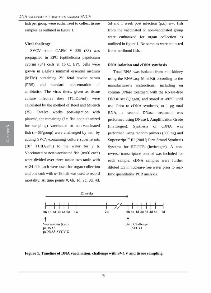
78
dna vaccination strategies against svcv
ch
apt
er 5
4
fish per group were euthanized to collect tissue
samples as outlined in figure 1.
Viral challenge
SVCV strain CAPM V 539 (33) was
propagated in EPC (epithelioma papulosum
cyprini (34) cells at 15°C. EPC cells were
grown in Eagle’s minimal essential medium
(MEM) containing 2% fetal bovine serum
(FBS) and standard concentration of
antibiotics. The virus titers, given as tissue
culture infective dose (TCID50/ml), were
calculated by the method of Reed and Muench
(35). Twelve weeks post-injection with
plasmid, the remaining (i.e. fish not euthanized
for sampling) vaccinated or non-vaccinated
fish (n=66/group) were challenged by bath by
adding SVCV-containing culture supernatants
(10-3 TCID50/ml) to the water for 2 h.
Vaccinated or non-vaccinated fish (n=66 each)
were divided over three tanks: two tanks with
n=24 fish each were used for organ collection
and one tank with n=18 fish was used to record
mortality. At time points 0, 6h, 1d, 2d, 3d, 4d,
5d and 1 week post infection (p.i.), n=6 fish
from the vaccinated or non-vaccinated group
were euthanized for organ collection as
outlined in figure 1. No samples were collected
from moribund fish.
RNA isolation and cDNA synthesis
Total RNA was isolated from mid kidney
using the RNAeasy Mini Kit according to the
manufacturer’s instructions, including on
column DNase treatment with the RNase-free
DNase set (Qiagen) and stored at -80ºC until
use. Prior to cDNA synthesis, to 1 g total
RNA, a second DNase treatment was
performed using DNase I, Amplification Grade
(Invitrogen). Synthesis of cDNA was
performed using random primers (300 ng) and
SuperscriptTM III (200U) First Strand Synthesis
Systems for RT-PCR (Invitrogen). A non-
reverse transcriptase control was included for
each sample. cDNA samples were further
diluted 1:5 in nuclease-free water prior to real-
time quantitative PCR analysis.
Figure 1. Timeline of DNA vaccination, challenge with SVCV and tissue sampling.
0h 4d3d2d1d 5d 1w 0h 4d3d2d1d 5d 7d2w 6h
Vaccination (i.m.)pcDNA3pcDNA3-SVCV-G
Bath Challenge(SVCV)
12 weeks
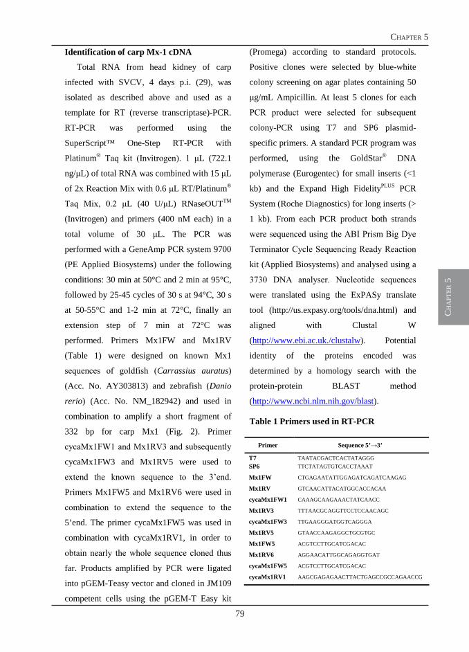
79
chapter 5
ch
apt
er 5
5
Identification of carp Mx-1 cDNA
Total RNA from head kidney of carp
infected with SVCV, 4 days p.i. (29), was
isolated as described above and used as a
template for RT (reverse transcriptase)-PCR.
RT-PCR was performed using the
SuperScript™ One-Step RT-PCR with
Platinum® Taq kit (Invitrogen). 1 L (722.1
ng/L) of total RNA was combined with 15 L
of 2x Reaction Mix with 0.6 L RT/Platinum®
Taq Mix, 0.2 L (40 U/L) RNaseOUTTM
(Invitrogen) and primers (400 nM each) in a
total volume of 30 L. The PCR was
performed with a GeneAmp PCR system 9700
(PE Applied Biosystems) under the following
conditions: 30 min at 50°C and 2 min at 95°C,
followed by 25-45 cycles of 30 s at 94°C, 30 s
at 50-55°C and 1-2 min at 72°C, finally an
extension step of 7 min at 72°C was
performed. Primers Mx1FW and Mx1RV
(Table 1) were designed on known Mx1
sequences of goldfish (Carrassius auratus)
(Acc. No. AY303813) and zebrafish (Danio
rerio) (Acc. No. NM_182942) and used in
combination to amplify a short fragment of
332 bp for carp Mx1 (Fig. 2). Primer
cycaMx1FW1 and Mx1RV3 and subsequently
cycaMx1FW3 and Mx1RV5 were used to
extend the known sequence to the 3’end.
Primers Mx1FW5 and Mx1RV6 were used in
combination to extend the sequence to the
5’end. The primer cycaMx1FW5 was used in
combination with cycaMx1RV1, in order to
obtain nearly the whole sequence cloned thus
far. Products amplified by PCR were ligated
into pGEM-Teasy vector and cloned in JM109
competent cells using the pGEM-T Easy kit
(Promega) according to standard protocols.
Positive clones were selected by blue-white
colony screening on agar plates containing 50
g/mL Ampicillin. At least 5 clones for each
PCR product were selected for subsequent
colony-PCR using T7 and SP6 plasmid-
specific primers. A standard PCR program was
performed, using the GoldStar® DNA
polymerase (Eurogentec) for small inserts (<1
kb) and the Expand High FidelityPLUS PCR
System (Roche Diagnostics) for long inserts (>
1 kb). From each PCR product both strands
were sequenced using the ABI Prism Big Dye
Terminator Cycle Sequencing Ready Reaction
kit (Applied Biosystems) and analysed using a
3730 DNA analyser. Nucleotide sequences
were translated using the ExPASy translate
tool (http://us.expasy.org/tools/dna.html) and
aligned with Clustal W
(http://www.ebi.ac.uk./clustalw). Potential
identity of the proteins encoded was
determined by a homology search with the
protein-protein BLAST method
(http://www.ncbi.nlm.nih.gov/blast).
Table 1 Primers used in RT-PCR
Primer Sequence 5’→3’
T7 TAATACGACTCACTATAGGG SP6 TTCTATAGTGTCACCTAAAT
Mx1FW CTGAGAATATTGGAGATCAGATCAAGAG
Mx1RV GTCAACATTACATGGCACCACAA
cycaMx1FW1 CAAAGCAAGAAACTATCAACC
Mx1RV3 TTTAACGCAGGTTCCTCCAACAGC
cycaMx1FW3 TTGAAGGGATGGTCAGGGA
Mx1RV5 GTAACCAAGAGGCTGCGTGC
Mx1FW5 ACGTCCTTGCATCGACAC
Mx1RV6 AGGAACATTGGCAGAGGTGAT
cycaMx1FW5 ACGTCCTTGCATCGACAC
cycaMx1RV1 AAGCGAGAGAACTTACTGAGCCGCCAGAACCG
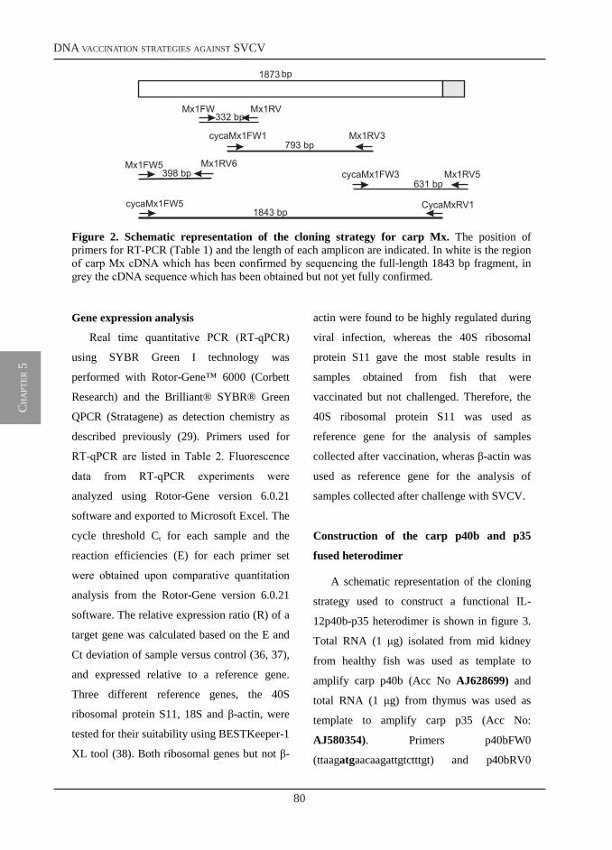
80
dna vaccination strategies against svcv
ch
apt
er 5
6
Figure 2. Schematic representation of the cloning strategy for carp Mx. The position of primers for RT-PCR (Table 1) and the length of each amplicon are indicated. In white is the region of carp Mx cDNA which has been confirmed by sequencing the full-length 1843 bp fragment, in grey the cDNA sequence which has been obtained but not yet fully confirmed.
Gene expression analysis
Real time quantitative PCR (RT-qPCR)
using SYBR Green I technology was
performed with Rotor-Gene™ 6000 (Corbett
Research) and the Brilliant® SYBR® Green
QPCR (Stratagene) as detection chemistry as
described previously (29). Primers used for
RT-qPCR are listed in Table 2. Fluorescence
data from RT-qPCR experiments were
analyzed using Rotor-Gene version 6.0.21
software and exported to Microsoft Excel. The
cycle threshold Ct for each sample and the
reaction efficiencies (E) for each primer set
were obtained upon comparative quantitation
analysis from the Rotor-Gene version 6.0.21
software. The relative expression ratio (R) of a
target gene was calculated based on the E and
Ct deviation of sample versus control (36, 37),
and expressed relative to a reference gene.
Three different reference genes, the 40S
ribosomal protein S11, 18S and β-actin, were
tested for their suitability using BESTKeeper-1
XL tool (38). Both ribosomal genes but not β-
actin were found to be highly regulated during
viral infection, whereas the 40S ribosomal
protein S11 gave the most stable results in
samples obtained from fish that were
vaccinated but not challenged. Therefore, the
40S ribosomal protein S11 was used as
reference gene for the analysis of samples
collected after vaccination, wheras β-actin was
used as reference gene for the analysis of
samples collected after challenge with SVCV.
Construction of the carp p40b and p35
fused heterodimer
A schematic representation of the cloning
strategy used to construct a functional IL-
12p40b-p35 heterodimer is shown in figure 3.
Total RNA (1 g) isolated from mid kidney
from healthy fish was used as template to
amplify carp p40b (Acc No AJ628699) and
total RNA (1 g) from thymus was used as
template to amplify carp p35 (Acc No:
AJ580354). Primers p40bFW0
(ttaagatgaacaagattgtctttgt) and p40bRV0
CycaMxRV1cycaMx1FW5
cycaMx1FW3 Mx1RV5Mx1RV6Mx1FW5
cycaMx1FW1 Mx1RV3
1873 bp
793 bp
631 bp398 bp
1843 bp
332 bpMx1FW Mx1RV
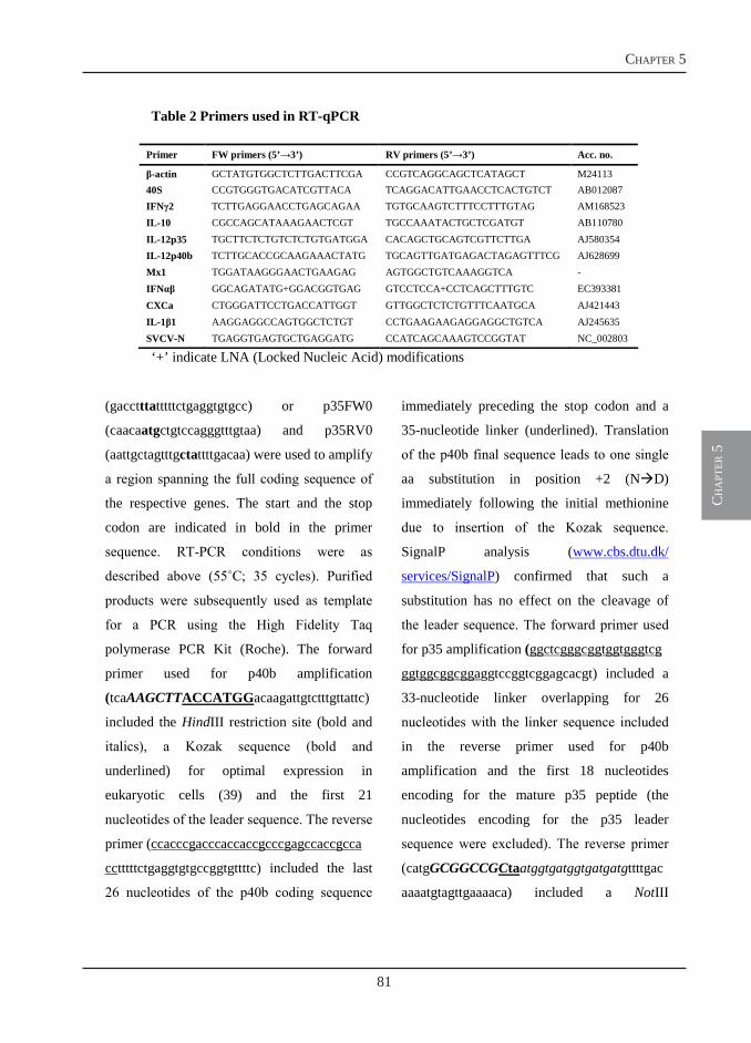
81
chapter 5
ch
apt
er 5
7
Table 2 Primers used in RT-qPCR
‘+’ indicate LNA (Locked Nucleic Acid) modifications
(gacctttatttttctgaggtgtgcc) or p35FW0
(caacaatgctgtccagggtttgtaa) and p35RV0
(aattgctagtttgctattttgacaa) were used to amplify
a region spanning the full coding sequence of
the respective genes. The start and the stop
codon are indicated in bold in the primer
sequence. RT-PCR conditions were as
described above (55˚C; 35 cycles). Purified
products were subsequently used as template
for a PCR using the High Fidelity Taq
polymerase PCR Kit (Roche). The forward
primer used for p40b amplification
(tcaAAGCTTACCATGGacaagattgtctttgttattc)
included the HindIII restriction site (bold and
italics), a Kozak sequence (bold and
underlined) for optimal expression in
eukaryotic cells (39) and the first 21
nucleotides of the leader sequence. The reverse
primer (ccacccgacccaccaccgcccgagccaccgcca
cctttttctgaggtgtgccggtgttttc) included the last
26 nucleotides of the p40b coding sequence
immediately preceding the stop codon and a
35-nucleotide linker (underlined). Translation
of the p40b final sequence leads to one single
aa substitution in position +2 (ND)
immediately following the initial methionine
due to insertion of the Kozak sequence.
SignalP analysis (www.cbs.dtu.dk/
services/SignalP) confirmed that such a
substitution has no effect on the cleavage of
the leader sequence. The forward primer used
for p35 amplification (ggctcgggcggtggtgggtcg
ggtggcggcggaggtccggtcggagcacgt) included a
33-nucleotide linker overlapping for 26
nucleotides with the linker sequence included
in the reverse primer used for p40b
amplification and the first 18 nucleotides
encoding for the mature p35 peptide (the
nucleotides encoding for the p35 leader
sequence were excluded). The reverse primer
(catgGCGGCCGCtaatggtgatggtgatgatgttttgac
aaaatgtagttgaaaaca) included a NotIII
Primer FW primers (5’→3’) RV primers (5’→3’) Acc. no.
β-actin GCTATGTGGCTCTTGACTTCGA CCGTCAGGCAGCTCATAGCT M24113 40S CCGTGGGTGACATCGTTACA TCAGGACATTGAACCTCACTGTCT AB012087 IFNγ2 TCTTGAGGAACCTGAGCAGAA TGTGCAAGTCTTTCCTTTGTAG AM168523 IL-10 CGCCAGCATAAAGAACTCGT TGCCAAATACTGCTCGATGT AB110780 IL-12p35 TGCTTCTCTGTCTCTGTGATGGA CACAGCTGCAGTCGTTCTTGA AJ580354 IL-12p40b TCTTGCACCGCAAGAAACTATG TGCAGTTGATGAGACTAGAGTTTCG AJ628699 Mx1 TGGATAAGGGAACTGAAGAG AGTGGCTGTCAAAGGTCA - IFNαβ GGCAGATATG+GGACGGTGAG GTCCTCCA+CCTCAGCTTTGTC EC393381 CXCa CTGGGATTCCTGACCATTGGT GTTGGCTCTCTGTTTCAATGCA AJ421443 IL-1β1 AAGGAGGCCAGTGGCTCTGT CCTGAAGAAGAGGAGGCTGTCA AJ245635 SVCV-N TGAGGTGAGTGCTGAGGATG CCATCAGCAAAGTCCGGTAT NC_002803
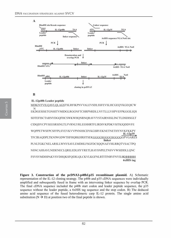
82
dna vaccination strategies against svcv
ch
apt
er 5
8
Figure 3. Construction of the pcDNA3-p40bLp35 recombinant plasmid. A) Schematic representation of the IL-12 cloning strategy. The p40b and p35 cDNA sequences were individually amplified and subsequently fused in frame with an intervening linker sequence by overlap PCR. The final cDNA sequence included the p40b start codon and leader peptide sequence, the p35 sequence without the leader peptide, a 6xHIS tag sequence and the stop codon. B) The deduced amino acid sequence of the fused heterodimeric carp IL-12 protein. The single amino acid substitution (N D) at position two of the final peptide is shown.
ATG TAA
HindIII site/Kozak sequence
linker sequence
IL-12p40b ATG TGA
Linker sequence
6xHIS sequence/TGA/NotI site
IL-12p35Leaderpeptide
PCR
ATG IL-12p40bHindIII
linker
PCR
6xHISlinker
NotITGA
Denaturation andoverlap PCR
ATGHindIIIlinker
6xHIS NotITGA
ATGATG IL-12p40bHindIII linker6xHIS NotI
TGA
cloning in pcDNA3
IL-12p35
MDKIVFVILQAVLQLAGS
GGGGSGGGGSGGGG
HHHHHH
TSLRFIKPNVVALEVSDLSSFEVSLHCGEQYKGEQICW
EKDGESISETGNHITVMIDGLRGGNFTCHRPNRDLLNYTLLLVHPVEFPKGGILIQS
SDTEFISCTARNYDGQFHCSWKWHQNRNQRAVVYSTAIRNSSLINCTLDSDISGLT
CIDQDYCPYSEESRSINLTLFVRNLYRLEEHHRTFLIRDIVKPDKVSITKIQDDVFE
WQPPETWSFPCSFFPLSYEVKVVPNNHSCDYKGSRVEKNETNETHYNVKFKKPY
TFCIRAQDPLTKNIWGDWTHFHQRKHRHTSEK GPVGARES
PLNLTGKCNELARSLLWNVSAVLEMDHLFSGFDCSQQNAEVHLRRQTVSACTPQ
NSNCAHSAVLNIDENECLQRILEDLHYYRETLRAYSNPELTNSVVWSIDDLLQNC
FSVSVMDISPAKVSVDHQKSFQERLQLCKVLKGFNLRTITINRVFNYILSK
linker
IL-12p40b Leader peptide
6xHIS tag
IL-12p35
N
B
A
Leaderpeptide
Leaderpeptide
83
chapter 5
ch
apt
er 5
9
restriction site (bold and italics), the p35 stop
codon (underlined), an 18-nucleotide sequence
coding for a 6xHistidine tag (6HIS) and the
last 25 nucleotides of the p35 coding sequence.
Products were amplified under the
following conditions. Five l of purified RT-
PCR products were combined with 1.7 U High
Fidelity Enzyme (Roche), dNTPs (200 M),
forward and reverse primers (300 nM each), 5
µl 10X buffer and nuclease-free water up to a
final volume of 50 µl. Reaction was performed
with the following profile: 94˚C for 4 min
followed by 30 PCR cycles at 94˚C for 15 sec,
50˚C for 30 sec and 72˚C for 1 min and a final
extension step at 72˚C for 7 min. Purified
products were directly cloned in pGEM-T easy
vector (Promega) and transformed in JM109
competent cells (Promega). The recombinant
plasmids were isolated using the QIAGEN
plasmid purification kit and the sequence was
confirmed using the ABI Prism Big Dye
Terminator Cycle Sequencing Ready Reaction
kit (Applied Biosystems) and analysed using a
3730 DNA analyser. p40b and p35 sequences
were re-amplified using high fidelity enzyme
and 1 µl of the respective plasmids as
templates. Following amplification of the
individual subunits, the p40 and p35 products
containing the linker sequences were amplified
together by overlap PCR using high fidelity
enzyme and the forward p40b and reverse p35
primers. The amplification product containing
the p40b subunit fused in frame with the p35
subunit was cloned in the HindIII and NotI
sites of the pcDNA3 plasmid (pcDNA3-
p40bLp35) and its correct sequence was
verified. Plasmids were isolated using the
endotoxin-free midi isolation kit (Invitrogen).
Nucleoporation of EPC (epithelioma
papulosum cyprini) cells
EPC cells were cultured in RPMI 1640
(Gibco) supplemented with 10% foetal calf
serum (FCS; Gibco), 2 mM L-Glutamin, 100
U/ml Penicillin-G and 50 mg/ml Streptomycin
sulphate and grown to confluency in 75 cm2
flasks. Two days before nucleoporation cells
were passaged in a 1:3 ratio. On the day of
nucleoporation, cells were washed twice in
carp PBS (cPBS, 150 mM NaCl, 2 mM KCl,
20 mM Na2HPO4, 2 mM KH2PO4) and
trypsinized using a solution of 0. 25% (w/v)
trypsin and 0.1% (w/v) EDTA in cPBS.
Reaction was stopped by addition of 20% FCS
and the single cell suspension was collected
and washed twice in cPBS. Cells (1 x 10⁶)
were resuspended in 100 µl solution V
(Amaxa) and nucleoporated with 2.5 g or 5
g of pcDNA3-p40bLp35 plasmid using the
T20 program, according to the manufacturer’s
instructions (Amaxa). Empty pcDNA3 plasmid
(5 g) was used as negative control and 2.5 g
of pGFPMax (Amaxa) was used as positive
control. Transfected cells were immediately
transferred to a 6 well plate in 2 ml complete
medium supplemented with 7.5% FCS and left
undisturbed for 5h. At this time point, medium
was replaced with complete medium
supplemented with 2.5% FCS.
84
dna vaccination strategies against svcv
ch
apt
er 5
10
Carp IL-12 (p40bLp35) protein expression
First, at time point 72h post-transfection,
cells treated with pGFPMax plasmid were
analyzed by flow cytometry for GFP
expression to assess nucleoporation efficiency.
Then, two approaches were taken to analyze
IL-12 protein expression. In the first approach,
supernatants of EPC cells transfected with
pcDNA3-p40bLp35 or pcDNA3 were
collected after 72h, centrifuged for 10 min at
800 x g and immediately used for Western blot
analysis, or stored at -20˚C. Twenty l of
supernatants from nucleoporated cells were
resolved on a 10% SDS-PAGE and proteins
were subsequently transferred to a
nitrocellulose membrane (Protran®, Schleicher
& Schuell, Bioscience). Membranes were
incubated with a 1:1500 dilution of mouse
anti-6xHIS antibody (Qiagen) in TBS-BSA
(10 mM Tris-Cl, pH 7.5, 150 mM NaCl, 3%
(w/v) BSA) overnight at 4˚C. After washing
twice in TBS and once in TBS-Tween/Triton
(TBS, 0.05% (v/v) Tween, 0.2% (v/v) Triton
X-100), membranes were incubated in a
1:1000 dilution of horseradish peroxydase
conjugated goat-anti-mouse antibody (Dako)
in 5% (w/v) non-fat dried milk in TBS for 1h
at room temperature. After four washes in
TBS-Tween/Triton, proteins were visualized
by chemiluminescence detection (Amersham,
ECL detection system for Western blot).
In the second approach, IL-12 protein
expression was shown by immuno
histochemical detection of transfected EPC
monolayers, using a mouse anti-TetraHIS
antibody (1:10; Qiagen) followed by
incubation with an alkaline phosphatase-
conjugated rabbit anti-mouse antibody
(1:1000) as previously described (40).
Statistical analysis
Relative expression ratios (R) were
calculated as described above. Transformed
(LN(R)) values were used for statistical
analysis in SPSS Software (15.0). Significant
differences (P < 0.05) were determined by a
one-way ANOVA followed by the Games-
Howell test for samples from both, the
vaccination and challenge experiment.
Results
Cloning of carp Mx1
Using a homology cloning approach we
identified a carp Mx sequence of 1843 bp long
covering the almost complete full length
coding sequence. Sequence analysis revealed
high similarity to known Mx1 sequences. The
sequence encodes for a protein of 624 amino
acids and misses short aa stretches at both
ends. Carp Mx1 has all characteristics of the
dynamin superfamily (Fig. 4): the N-terminal
GTPase domain containing a dynamin family
signature at amino acid positions 56-65 and the
tripartite GTP-binding domain located at
positions 33–50, 139–142, and 208–211; the
dynamin central domain, stretching from
position 221-516; the C-terminal GTPase
effector domain (GED) including a putative
leucine zippers motif, situated in a coiled-coil
region and represented by a stretch of Leu
residues spaced seven amino acid residues
apart. Similar to human MxA and other fish
Mx1 sequences, carp Mx1 does not contain a
nuclear localization signal (NLS), suggesting
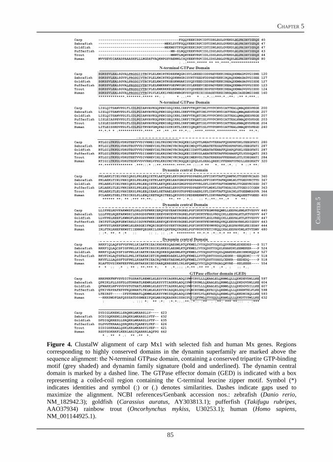
85
chapter 5
ch
apt
er 5
11
Figure 4. ClustalW alignment of carp Mx1 with selected fish and human Mx genes. Regions corresponding to highly conserved domains in the dynamin superfamily are marked above the sequence alignment: the N-terminal GTPase domain, containing a conserved tripartite GTP-binding motif (grey shaded) and dynamin family signature (bold and underlined). The dynamin central domain is marked by a dashed line. The GTPase effector domain (GED) is indicated with a box representing a coiled-coil region containing the C-terminal leucine zipper motif. Symbol (*) indicates identities and symbol (:) or (.) denotes similarities. Dashes indicate gaps used to maximize the alignment. NCBI references/Genbank accession nos.: zebrafish (Danio rerio, NM_182942.3); goldfish (Carassius auratus, AY303813.1); pufferfish (Takifugu rubripes, AAO37934) rainbow trout (Oncorhynchus mykiss, U30253.1); human (Homo sapiens, NM_001144925.1).
Carp ----------------------------------------FSQQYEEKIRPCIDTIDRLRSLGVEKDLALPAIAVIGDQS 40Zebrafish ---------------------------------MEKLSYTFSQQYEEKIRPCIDTIDNLRSLGVEKDLALPAIAVIGDQS 47Goldfish ---------------------------------MEKMSYTFSQHYEEKIRPCIDTIDNLRSLGVEKDLALPAIAVIGDQS 47Pufferfish ------------------------------------MN-SLNQQYEEKVRPCIDLIDSLRSLGVEKDLALPAIAVIGDQS 43Trout ------------------------------------MNNTLNQHYEEKVRPCIDLIDSLRSLGVEKDLALPAIAVIGDQS 44Human MVVSEVDIAKADPAAASHPLLLNGDATVAQKNPGSVAENNLCSQYEEKVRPCIDLIDSLRALGVEQDLALPAIAVIGDQS 80
: .:****:***** ** **:****:**************
Carp SGKSSVLEALSGVA CPLELKMIRTKDEEKWQGRISYLGKEEDIDDPAEVEKKIREAQDEMAGPGVGISNE 120Zebrafish SGKSSVLEALSGVP CPLELKMIRTKDQDKWHGRISYKTYEEDFDDPAEVEKKIRQAQDEMAGAGVGISEE 127Goldfish SGKSSVLEALSGVA CPLELKMIRTKGEGKWHAKISYQDYEEDIDDPAEVEKKIREAQDEMAGAGVGISDE 127Pufferfish SGKSSVLEALSGVA CPLELKMKRRKVGEPWYGNISYLEKEEVIEDPADVEKKIKEAQDEMAGVGVGISDD 123Trout SGKSSVLEALSGVA CPLELKMKRKKEGEEWHGKISYQDHEEEIEDPSDVEKKIREAQDEMAGVGVGISDD 124Human SGKSSVLEALSGVA CPLVLKLKKLVNEDKWRGKVSYQDYEIEISDASEVEKEINKAQNAIAGEGMGISHE 160
*************.*******:***** **: : * ..:** * :.*.::***:*.:**: :** *:***.:
Carp LISLQITSANVPDLTLIDLPGIARVAVKGQPENIGDQIKRLIRKFVTKQETINLVVVPCNVDIATTEALQMAQEEDPEGE 200Zebrafish LISLQITSANVPDLTLIDLPGIARVAVKGQPENIGDQIKRLIRKFVTRQETINLVVVPCNVDIATTEALQMAQAEDPDGE 207Goldfish LISLQITSANVPDLTLIDLPGIARVAVKGQPENIGDQIKRLIRKFVTKQETINLVVVPCNVDIATTEALQMAQEEDPEGE 207Pufferfish LISLEIASPEVPDLTLIDLPGIARVAVKGQPENIGEQIKRLIRKFITKQETISLVVVPCNVDIATTEALKMAQEVDPDGE 203Trout LISLEIGSPDVPDLTLIDLPGIARVAVKGQPENIGEQIKRLIRKFIMKQETISLVVVPCNVDIATTEALKMAQEVDPEGE 204Human LITLEISSRDVPDLTLIDLPGITRVAVGNQPADIGYKIKTLIKKYIQRQETISLVVVPSNVDIATTEALSMAQEVDPEGD 240
**:*:* * :************:**** .** :** :** **:*:: :****.*****.**********.*** **:*:
Carp RTLGILTKPDLVDKGTEETVVDIVHNEIIHLTKGYMIVRCRGQKEILDQVTLNEATVTEKAFFQDHPHFSKLYEEGFATI 280Zebrafish RTLGILTKPDLVDKGTEGTVVDIVHNEVIHLTKGYMIVRCRGQKEIMDQVTLNEATETESAFFKDHPHFSKLYEEGFATI 287Goldfish RTLGILTKPDLVDKGTEGTVVDIVHNEVIHLTKGYMIVRCRGQKEIIDHVTLNEATATENAFFQDHPQFSKLYEEGFATI 287Pufferfish RTLGILTKPDLVDKGTEETVVDIIHNEVIHLKKGYMIVRCRGQKEIIDKVSLAEATETETAFFRDHAHFQTLYDDGQATI 283Trout RTLGILTKPDLVDKGTEETVVDIVHNEVIHLTKGYMIVKCRGQKEIMERVSLTEATEREKAFFKEHAHLSTLYDEGHATI 284Human RTIGILTKPDLVDKGTEDKVVDVVRNLVFHLKKGYMIVKCRGQQEIQDQLSLSEALQREKIFFENHPYFRDLLEEGKATV 320
**:************** .***:::* ::**.******:****:** ::::* ** *. **.:*. : * ::* **:
Carp PKLAEKLTIELVHHIQRSLPRLEEQIETKLAETQKELEKYGNGPPSDPAERLSFFIDKVTAFTQDMFNLTTGEEVKCASD 360Zebrafish PKLAEKLTIELVHHIQKSLPRLEEQIETKLAETQKELEAYGNGPPSEPAARLSFFIDKVTAFNQDMLNLTTGEDVKCTTD 367Goldfish PKLAEKLTIELVHHIQKSLPRLEEQIETKLAETQKELEAYGNGPPSDPVVRLSFLIDKVTAFTHDMFNLTTGEEVKCSSD 367Pufferfish LKLAEKLTLELVNHIEKSLPRLEEQIEEKLAHTRAELERYGTGPPSDPAEKVTFLMDKLTAFTHDAISLTTGEDICCGEN 363Trout PKLAEKLTLELVHHIEKSLPRLEEQIEAKLSETHAELERYGTGPPEDSAERLYFLIDKVTAFTQDAINLSTGEEMKSGVR 364Human PCLAEKLTSELITHICKSLPLLENQIKETHQRITEELQKYGVDIPEDENEKMFFLIDKVNAFNQDITALMQGEETVGEED 400
****** **: ** :*** **:**: . . **: ** . *.: :: *::**:.**.:* * **:
Carp LLIFPELRDVFAKWNSFLDRLGVSFNKKIEKQVGDYEAKYRGRELPGFINYKTFEGMVREQMKLLEEPALKMLKTVSDVV 440Zebrafish LLLFPELRQEFAKWSHILDRSGDSFNKKIEKEVDNYEVKYRGRELPGFINYKTFEGLVRDQIKLLEEPALKTLKTVSDVV 447Goldfish LLVFPELREEFLKWNGFLERSGHSFNKKIEKEVDNYEAKYRGRELPGFSNYKTLEGLVREQIKLLEEPALKTLKTVSDVV 447Pufferfish INIFSTLRQKFGEWIRHLDISGVKFNERLEREVQEYEVKYRGRELPGFINYKTFEYMVKEQVKQLEEPAVRNLKDIGDDV 443Trout LNVFSTLRKEFGKWKLHLERSGEIFNQRIEGEVDDYEKTYRGRELPGFINYKTFEVMVKDQIKQLEGPAVKKLKEISDAV 444Human IRLFTRLRHEFHKWSTIIENNFQEGHKILSRKIQKFENQYRGRELPGFVNYRTFETIVKQQIKALEEPAVDMLHTVTDMV 480
: :*. **. * :* :: :: :. :: .:* ********* **:*:* :*::*:* ** **: *: : * *
Carp RKKFIQLAQFSFVGFPNLLKIAKTKIEAIKHDKESQAESMLRTQFKMELIVYSQDGTYSQSLQDVKEMLEDEEDES---S 517Zebrafish RKKFIQLAQCSFIGFPNLLKIAKTKIEGIKLNKESLAESMLKTQFKMELIVYSQDGTYSQSLKHAKDKLEEMEKER---P 524Goldfish RKKFIQLAQYSFIGFPNLLKIAKTKIEAIKQDKESQAESMLRTQFKMELIVYSQDGTYSQSLQHAKDKLEEIENDKQQLP 527Pufferfish RKVFIHLAQTSFAGLPNLIKTAKAKIETIKQRMEKDAEKLLRTQFKMELLVYTQDRTYSSSLDESKK--EEQEDKC---V 518Trout RKVFLLLAQSSFTGFPNLLKSAKTKIEAIKQVNESTAESMLRTQFKMELIVYTQDSTYSHSLCERKR--EEDEDQ----P 518Human RLAFTDVSIKNFEEFFNLHRTAKSKIEDIRAEQEREGEKLIRLHFQMEQIVYCQDQVYRGALQKVRE--KELEEEK---- 554
* * :: .* : ** : **:*** *: * .*.::: :*:** :** ** .* :* . : :: *..
Carp HRKPKKFNFVSVDICTGSRATLREMKLHLESYYKIASKRLADQIPMVIRYLLLQEAALELQRNMLQLLQDKDEVDNLLKE 597Zebrafish QPKIKLPLLSSFDLGTDNHATLREMRLHLKSYYTIASKRLADQIPMVIRYMLLQEAALELQRNMLQLLQDKDGVDNLLKE 604Goldfish QFNAKKLNFVSVDVSTGTHATLREMRLHLESYYTIASKRLADQIPMVIRYLLLQEAALELQRNMLQLLQDKDGVDILLKE 607Pufferfish QTKIVRPPAFRFPFKQDNHATLTELMLHLKSYYRIASQRLSDQIPLMIRYHMLQEFAIQLQREMLQLLQDRENTELFLKE 598Trout LTEIRST-----IFSTDNHATLQEMMLHLKSYYWISSQRLADQIPMVIRYLVLQEFASQLQREMLQTLQEKDNIEQLLKE 593Human --KKKSWDFGAFQSSSATDSSMEEIFQHLMAYHQEASKRISSHIPLIIQFFMLQTYGQQLQKAMLQLLQDKDTYSWLLKE 632
: . ::: *: ** :*: :*:*::.:**::*:: :** . :**: *** **::: . :***
Carp DYDIGLKRENLLSRQNRLMKARSLLV---- 623Zebrafish DCDIGQKRENLLSRQKRLMKARSLLVTF-- 632Goldfish DFDIGQKRESLLSRQKRLMKARSLLVTF-- 635Pufferfish DLDVGTKRAALQSRQKRLTQAREYLVKF-- 626Trout DIDIGSKRAALQSKLKRLMKARSYLVEF-- 621Human RSDTSDKRKFLKERLARLTQARRRLAQFPG 662
* . ** * .: ** :** *.
LPRGSGIITRLPRGSGIVTRLPRGSGIVTRLPRGSGIVTRLPRGSGIVTRLPRGSGIVTR
N-terminal GTPase Domain
N-terminal GTPase Domain
Dynamin central Domain
Dynamin central Domain
Dynamin central Domain
GTPase effector domain (GED)
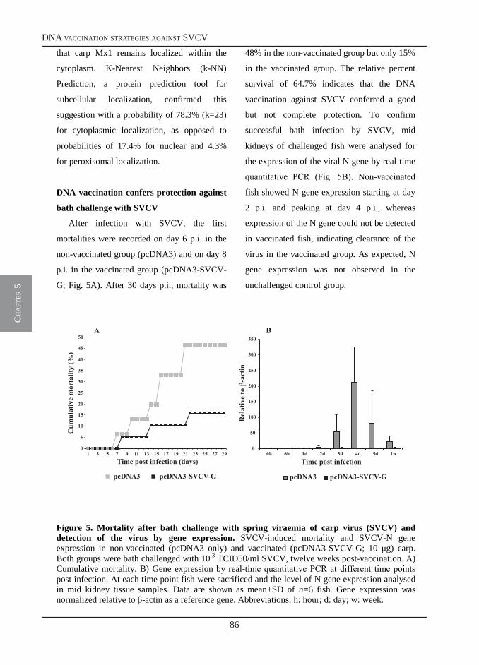
86
dna vaccination strategies against svcv
ch
apt
er 5
12
that carp Mx1 remains localized within the
cytoplasm. K-Nearest Neighbors (k-NN)
Prediction, a protein prediction tool for
subcellular localization, confirmed this
suggestion with a probability of 78.3% (k=23)
for cytoplasmic localization, as opposed to
probabilities of 17.4% for nuclear and 4.3%
for peroxisomal localization.
DNA vaccination confers protection against
bath challenge with SVCV
After infection with SVCV, the first
mortalities were recorded on day 6 p.i. in the
non-vaccinated group (pcDNA3) and on day 8
p.i. in the vaccinated group (pcDNA3-SVCV-
G; Fig. 5A). After 30 days p.i., mortality was
48% in the non-vaccinated group but only 15%
in the vaccinated group. The relative percent
survival of 64.7% indicates that the DNA
vaccination against SVCV conferred a good
but not complete protection. To confirm
successful bath infection by SVCV, mid
kidneys of challenged fish were analysed for
the expression of the viral N gene by real-time
quantitative PCR (Fig. 5B). Non-vaccinated
fish showed N gene expression starting at day
2 p.i. and peaking at day 4 p.i., whereas
expression of the N gene could not be detected
in vaccinated fish, indicating clearance of the
virus in the vaccinated group. As expected, N
gene expression was not observed in the
unchallenged control group.
Figure 5. Mortality after bath challenge with spring viraemia of carp virus (SVCV) and detection of the virus by gene expression. SVCV-induced mortality and SVCV-N gene expression in non-vaccinated (pcDNA3 only) and vaccinated (pcDNA3-SVCV-G; 10 g) carp. Both groups were bath challenged with 10-3 TCID50/ml SVCV, twelve weeks post-vaccination. A) Cumulative mortality. B) Gene expression by real-time quantitative PCR at different time points post infection. At each time point fish were sacrificed and the level of N gene expression analysed in mid kidney tissue samples. Data are shown as mean+SD of n=6 fish. Gene expression was normalized relative to β-actin as a reference gene. Abbreviations: h: hour; d: day; w: week.
B
0
50
100
150
200
250
300
350
0h 6h 1d 2d 3d 4d 5d 1wTime post infection
pcDNA3 pcDNA3-SVCV-G
0
5
10
15
20
25
30
35
40
45
50
1 3 5 7 9 11 13 15 17 19 21 23 25 27 29Time post infection (days)
pcDNA3 pcDNA3-SVCV-G
Cumulativemortality(%)
Relative to-actin
A
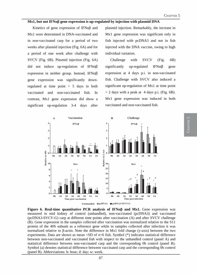
87
chapter 5
ch
apt
er 5
13
Mx1, but not IFNαβ gene expression is up-regulated by injection with plasmid DNA
Kinetics of gene expression of IFNαβ and
Mx1 were determined in DNA-vaccinated and
in non-vaccinated carp for a period of two
weeks after plasmid injection (Fig. 6A) and for
a period of one week after challenge with
SVCV (Fig. 6B). Plasmid injection (Fig. 6A)
did not induce up-regulation of IFNαβ
expression in neither group. Instead, IFNαβ
gene expression was significantly down-
regulated at time point > 5 days in both
vaccinated and non-vaccinated fish. In
contrast, Mx1 gene expression did show a
significant up-regulation 3-4 days after
plasmid injection. Remarkably, the increase in
Mx1 gene expression was significant only in
fish injected with pcDNA3 and not in fish
injected with the DNA vaccine, owing to high
individual variation.
Challenge with SVCV (Fig. 6B)
significantly up-regulated IFNαβ gene
expression at 4 days p.i. in non-vaccinated
fish. Challenge with SVCV also induced a
significant up-regulation of Mx1 at time point
> 2 days with a peak at 4 days p.i. (Fig. 6B).
Mx1 gene expression was induced in both
vaccinated and non-vaccinated fish.
Figure 6. Real-time quantitative PCR analysis of IFNαβ and Mx1. Gene expression was measured in mid kidney of control (unhandled), non-vaccinated (pcDNA3) and vaccinated (pcDNA3-SVCV-G) carp at different time points after vaccination (A) and after SVCV challenge (B). Gene expression in the samples collected after vaccination was normalized relative to the S11 protein of the 40S subunit as a reference gene while in samples collected after infection it was normalized relative to β-actin. Note the difference in Mx1 fold change (y-axis) between the two experiments. Data are shown as mean +SD of n=6 fish. Symbol (*) indicates statistical difference between non-vaccinated and vaccinated fish with respect to the unhandled control (panel A) and statistical difference between non-vaccinated carp and the corresponding 0h control (panel B). Symbol (a) denotes statistical difference between vaccinated carp and the corresponding 0h control (panel B). Abbreviations: h: hour; d: day; w: week.
IFNαβ
0
1
2
3
4
0h 1d 2d 3d 4d 5d 1w 2w
* *
*
IFNαβ
0
1
2
3
4
0h 6h 1d 2d 3d 4d 5d 1w
*
Mx1
0
2
4
6
8
10
0h 1d 2d 3d 4d 5d 1w 2w
**
Mx1
0
5
10
15
20
25
30
0h 6h 1d 2d 3d 4d 5d 1w
a
a
a
a a*
*
*
* *
Time post vaccination Time post infection
pcDNA3 pcDNA3-SVCV-Gunhandled
A B
Rel
ati
ve
to-A
ct
Rel
ati
ve
to4
0S
Vaccination Challenge
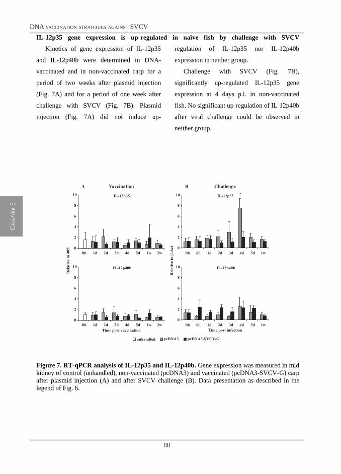
88
dna vaccination strategies against svcv
ch
apt
er 5
14
IL-12p35 gene expression is up-regulated in naïve fish by challenge with SVCV
Kinetics of gene expression of IL-12p35
and IL-12p40b were determined in DNA-
vaccinated and in non-vaccinated carp for a
period of two weeks after plasmid injection
(Fig. 7A) and for a period of one week after
challenge with SVCV (Fig. 7B). Plasmid
injection (Fig. 7A) did not induce up-
regulation of IL-12p35 nor IL-12p40b
expression in neither group.
Challenge with SVCV (Fig. 7B),
significantly up-regulated IL-12p35 gene
expression at 4 days p.i. in non-vaccinated
fish. No significant up-regulation of IL-12p40b
after viral challenge could be observed in
neither group.
Figure 7. RT-qPCR analysis of IL-12p35 and IL-12p40b. Gene expression was measured in mid kidney of control (unhandled), non-vaccinated (pcDNA3) and vaccinated (pcDNA3-SVCV-G) carp after plasmid injection (A) and after SVCV challenge (B). Data presentation as described in the legend of Fig. 6.
IL-12p35
0
2
4
6
8
10
0h 1d 2d 3d 4d 5d 1w 2w
IL-12p40b
0
2
4
6
8
10
0h 1d 2d 3d 4d 5d 1w 2w
IL-12p35
0
2
4
6
8
10
0h 6h 1d 2d 3d 4d 5d 1w
*
IL-12p40b
0
2
4
6
8
10
0h 6h 1d 2d 3d 4d 5d 1wTime post vaccination Time post infection
Vaccination ChallengeA B
Relativeto-Act
Relative to40S
pcDNA3 pcDNA3-SVCV-Gunhandled
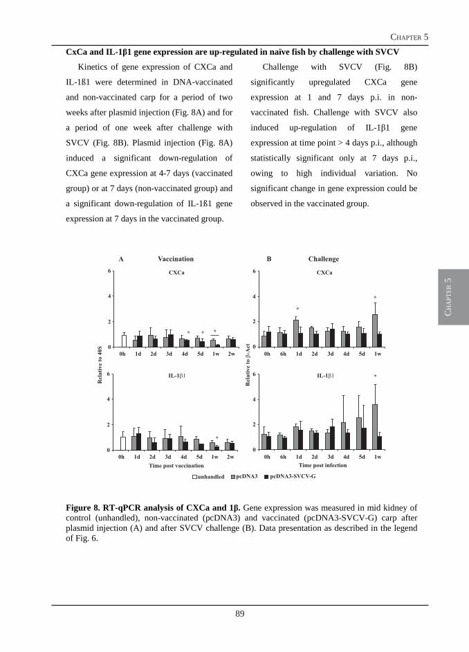
89
chapter 5
ch
apt
er 5
15
CxCa and IL-1β1 gene expression are up-regulated in naïve fish by challenge with SVCV
Kinetics of gene expression of CXCa and
IL-1ß1 were determined in DNA-vaccinated
and non-vaccinated carp for a period of two
weeks after plasmid injection (Fig. 8A) and for
a period of one week after challenge with
SVCV (Fig. 8B). Plasmid injection (Fig. 8A)
induced a significant down-regulation of
CXCa gene expression at 4-7 days (vaccinated
group) or at 7 days (non-vaccinated group) and
a significant down-regulation of IL-1ß1 gene
expression at 7 days in the vaccinated group.
Challenge with SVCV (Fig. 8B)
significantly upregulated CXCa gene
expression at 1 and 7 days p.i. in non-
vaccinated fish. Challenge with SVCV also
induced up-regulation of IL-1β1 gene
expression at time point > 4 days p.i., although
statistically significant only at 7 days p.i.,
owing to high individual variation. No
significant change in gene expression could be
observed in the vaccinated group.
Figure 8. RT-qPCR analysis of CXCa and 1β. Gene expression was measured in mid kidney of control (unhandled), non-vaccinated (pcDNA3) and vaccinated (pcDNA3-SVCV-G) carp after plasmid injection (A) and after SVCV challenge (B). Data presentation as described in the legend of Fig. 6.
CXCa
0
2
4
6
0h 1d 2d 3d 4d 5d 1w 2w
* **
IL-1
0
2
4
6
0h 1d 2d 3d 4d 5d 1w 2w
*
CXCa
0
2
4
6
0h 6h 1d 2d 3d 4d 5d 1w
*
*
IL-1
0
2
4
6
0h 6h 1d 2d 3d 4d 5d 1w
*
Time post vaccination Time post infection
Vaccination ChallengeA B
Rel
ativ
eto
-Act
Rel
ativ
e to
40S
pcDNA3 pcDNA3-SVCV-Gunhandled
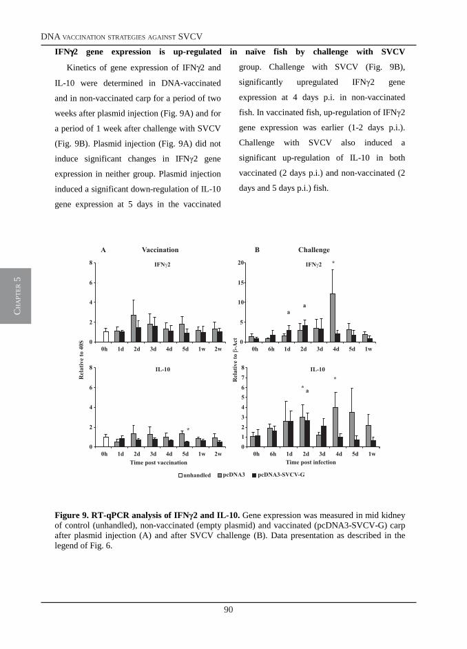
90
dna vaccination strategies against svcv
ch
apt
er 5
16
IFNγγγγ2 gene expression is up-regulated in naïve fish by challenge with SVCV
Kinetics of gene expression of IFNγ2 and
IL-10 were determined in DNA-vaccinated
and in non-vaccinated carp for a period of two
weeks after plasmid injection (Fig. 9A) and for
a period of 1 week after challenge with SVCV
(Fig. 9B). Plasmid injection (Fig. 9A) did not
induce significant changes in IFNγ2 gene
expression in neither group. Plasmid injection
induced a significant down-regulation of IL-10
gene expression at 5 days in the vaccinated
group. Challenge with SVCV (Fig. 9B),
significantly upregulated IFNγ2 gene
expression at 4 days p.i. in non-vaccinated
fish. In vaccinated fish, up-regulation of IFNγ2
gene expression was earlier (1-2 days p.i.).
Challenge with SVCV also induced a
significant up-regulation of IL-10 in both
vaccinated (2 days p.i.) and non-vaccinated (2
days and 5 days p.i.) fish.
Figure 9. RT-qPCR analysis of IFNγ2 and IL-10. Gene expression was measured in mid kidney of control (unhandled), non-vaccinated (empty plasmid) and vaccinated (pcDNA3-SVCV-G) carp after plasmid injection (A) and after SVCV challenge (B). Data presentation as described in the legend of Fig. 6.
IFN 2
0
2
4
6
8
0h 1d 2d 3d 4d 5d 1w 2w
IL-10
0
2
4
6
8
0h 1d 2d 3d 4d 5d 1w 2w
*
IFN 2
0
5
10
15
20
0h 6h 1d 2d 3d 4d 5d 1w
aa
*
IL-10
012345678
0h 6h 1d 2d 3d 4d 5d 1w
a
**
Time post vaccination Time post infection
Vaccination ChallengeA B
Relativet
o-A
ct
Relative t
o40
S
pcDNA3 pcDNA3-SVCV-Gunhandled
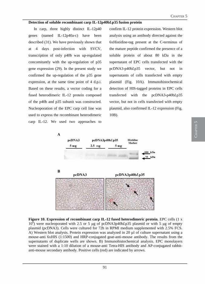
91
chapter 5
ch
apt
er 5
17
Detection of soluble recombinant carp IL-12p40bLp35 fusion protein
In carp, three highly distinct IL-12p40
genes (named IL-12p40a-c) have been
described (31). We have previously shown that
at 4 days post-infection with SVCV,
transcription of only p40b was up-regulated
concomitantly with the up-regulation of p35
gene expression (29). In the present study we
confirmed the up-regulation of the p35 gene
expression, at the same time point of 4 d.p.i.
Based on these results, a vector coding for a
fused heterodimeric IL-12 protein composed
of the p40b and p35 subunit was constructed.
Nucleoporation of the EPC carp cell line was
used to express the recombinant heterodimeric
carp IL-12. We used two approaches to
confirm IL-12 protein expression. Western blot
analysis using an antibody directed against the
6xHistidine-tag present at the C-terminus of
the mature peptide confirmed the presence of a
soluble protein of about 80 kDa in the
supernatant of EPC cells transfected with the
pcDNA3-p40bLp35 vector, but not in
supernatants of cells transfected with empty
plasmid (Fig. 10A). Immunohistochemical
detection of HIS-tagged proteins in EPC cells
transfected with the pcDNA3-p40bLp35
vector, but not in cells transfected with empty
plasmid, also confirmed IL-12 expression (Fig.
10B).
Figure 10. Expression of recombinant carp IL-12 fused heterodimeric protein. EPC cells (1 x 10⁶) were nucleoporated with 2.5 or 5 g of pcDNA3p40bLp35 plasmid or with 5 g of empty plasmid (pcDNA3). Cells were cultured for 72h in RPMI medium supplemented with 2.5% FCS. A) Western blot analysis. Protein expression was analyzed in 20 µl of culture supernatant using a mouse-anti 6xHIS (1:1500) and HRP-conjugated goat-anti-mouse antibody. The results from the supernatants of duplicate wells are shown. B) Immunohistochemical analysis. EPC monolayers were stained with a 1:10 dilution of a mouse-anti Tetra-HIS antibody and AP-conjugated rabbit-anti-mouse secondary antibody. Positive cells (red) are indicated by arrows.
92
dna vaccination strategies against svcv
ch
apt
er 5
18
Discussion
In the present study we report the cloning
of a carp Mx1 sequence homologous to other
known Mx1 sequences together with the gene
expression analysis of several other immune
relevant genes following DNA vaccination and
following challenge of vaccinated carp with
spring viraemia of carp virus (SVCV). This is
the first report describing changes in gene
expression in a cyprinid fish after DNA
vaccination and challenge with SVCV.
Only two other studies have reported on the
efficacy of a DNA vaccine in (koi) carp
against SVCV (2, 7), partly because
experimental challenge with SVCV has proven
difficult to establish because of low mortality
(41). In general, SVC has a seasonal nature
and under farming conditions the disease is
most severe at temperatures between 11-17 oC
and challenge conditions should respect this
temperature range. Of course, the rate of
development of a protective immune response
following vaccination is temperature
dependent in fish. For example, after infection,
neutralizing antibodies have been detected
after 4 weeks at 20 oC, after 7 weeks at 15 oC
but not before 10 weeks at 10 oC (41). In the
DNA vaccination study by Kannellos et al. (2),
carp were vaccinated at 20˚C and after 6
weeks, challenged at 15 oC by intraperitoneal
(i.p.) injection with a heterologous strain of
SVCV. Challenge at 15 oC killed 64% of the
control fish, and the strongest protection was
observed in carp that received the full-length
G-gene expressed by plasmids driven by a
CMV-Intron-A rather than the most commonly
used CMV promoter, with a relative
percentage survival (RPS) of 48%. In the
second study by Emmenegger (7), high value
ornamental varieties of carp (koi) received a
comparable treatment with DNA vaccination
at 20 oC and SVCV challenge at 10-12 oC by
i.p. injection. Although the RPS values in this
study (50-80%) were higher than in the first
study (48%) fish were challenged at 4 weeks
post-vaccination already, therefore a role for a
non-specific (interferon-induced) immune
response cannot be excluded. As shown in
several studies in salmonids, and tentatively in
carp (this study), DNA vaccination induces an
early non-specific anti-viral protection
mediated by a type I interferon response that
can last for several weeks (11).
Ideally, a test challenge method should
closely mimic natural exposure to the pathogen
and ensure that mucosal immune mechanisms
are triggered. Bath and co-habitation
challenges best fulfill this requirement, but are
more difficult methods to control and
standardize than i.p. injection challenge
methods. Although Emmenegger et al. (7) did
develop a bath challenge with SVCV, the dose
required to obtain 67% mortality of naïve fish
was relatively high (>105 PFU/ml) and
challenge of vaccinated fish was finally
performed via the i.p. route. We used 10-3
TCID50 SVCV/ml for our bath challenge
which led to 50% mortality of naïve fish. Our
DNA vaccination resulted in 85% survival
after bath challenge, with a RPS value of
66.7%. Despite these promising results,
indeed, our bath challenge has been proven
difficult to control and standardize and awaits
succesful replication. DNA vaccination of
93
chapter 5
ch
apt
er 5
19
salmonids against members of the genus
Novirhabdoviridae (VHS, IHNV and HIRRV)
has yielded very strong protection with RPS
values of 90% or more, also using bath
challenges as the preferred method (11, 15).
Although a member of the family
Rhabdoviridaea, taxonomically, SVCV is
classified in the genus Vesiculoviridae and not
Novirhabdoviridae (1). Structural and
functional differences between these two
genera (1) may help to explain the lower
protection generally observed after DNA
vaccination against SVCV. In the present
study, owing to the lack of suitable antibodies
for cytological staining of the SVCV-G
protein, expression levels of this protein could
not be quantified. However, our construct did
confer protection. Possibly, the expression and
presentation to the cell surface of the SVCV-G
protein might not occur less efficiently than
VHS-G protein expression. In the future, the
use of a different promoter, such as the CMV-
IntronA, or different leader sequences might
increase protein expression thereby improving
vaccination efficacy.
Extensive gene expression and microarray
analysis have been performed in salmonids and
Japanese flounder after DNA vaccination
against novirhabdoviruses (5, 6, 16-18). As
mentioned above, DNA vaccination often
induces an early type I interferon and
associated Mx response, resulting in non-
specific anti-viral protection (11, 15, 42). In
these studies, the early Mx up-regulation (4-7
days) was always observed exclusively in fish
receiving the DNA vaccine containing the G-
protein and not (or to a considerably lower
extent) in control fish treated with an empty
plasmid. Surprisingly, in our study, significant
Mx1 up-regulation was observed also in carp
that received the empty plasmid. Although up-
regulation of Mx gene expression in response
to empty plasmid DNA contrast with the
general findings in salmonids and Japanese
flounder, this observation has also been made
in DNA vaccination experiments with channel
catfish (43). The results in channel catfish and
carp indicate the induction of an innate
immune response, not triggered by the viral G
protein but by plasmid components, in
particular fish species. Likely candidates are
the several (n=25) CpG motifs contained in the
backbone of the pcDNA3 plasmid (11). Since
the salmonids and Japanese flounder have been
found unresponsive to the same CpG motifs in
pcDNA3, this would indicate a species-
specific recognition of these pathogen-
associated molecular patterns by, for example,
TOLL-like receptor 9 (44). Interestingly, a
recent study using a different plasmid (pAE6)
for DNA vaccination of rainbow trout showed
increased TLR9 gene expression at the
injection site, also after injection of plasmid
DNA only (45). Preliminary studies in our
laboratory have suggested that unmethylated
CpG oligodeoxy- nucleotides (CpG-ODNs),
previously shown to induce type I interferon
expression and cell proliferation in leukocytes
from Atlantic salmon (46), apparently fail to
have the same effect in carp (C.M.S. Ribeiro,
personal communication). Thus, although
salmonids have not been found to respond to
the CpG motifs in pcDNA3, it could well be
94
dna vaccination strategies against svcv
ch
apt
er 5
20
the case that carp leukocytes are responsive to
the CpG-ODN motifs of pcDNA3.
Beside the above-described up-regulation
of Mx1 gene expression, none of the other
immune relevant genes tested was up-
regulated after injection with neither the empty
plasmid nor the vaccine plasmid. Nevertheless,
carp clearly showed improved protection
against bath challenge with SVCV. Although
the Mx1 gene expression induced by the empty
plasmid suggests the induction of a non-
specific immune response, the period of 12
weeks between vaccination and challenge
makes it unlikely that the observed protection
could be ascribed to an increased non-specific
immune response only. As stated, none of the
other investigated immune genes was up-
regulated in mid kidney after vaccination. On
the contrary, a down-regulation of IFNαβ,
CXCa, IL-1β1 and IL-10 was observed.
Similar results have been obtained in other fish
species where, beside a clear Mx gene up-
regulation, expression of other immune
relevant genes in peripheral organs upon DNA
vaccination remained low, especially when
compared to the fold changes observed after
viral challenge of naïve fish (5, 6, 17, 18). A
recent study in trout muscle following DNA
vaccination against IHNV, however, clearly
shows that local gene expression at the site of
injection is much higher (>10 fold) than gene
expression in peripheral organs (16). This
strongly argues for future studies on gene
expression focusing at local sites of plasmid
injection in the muscle rather than gene
expression studies in peripheral immune
organs.
Challenge with SVCV of vaccinated carp
did not induce a very strong change in gene
expression. Mx1, but also IFNγ2 and IL-10
were upregulated at 1-2 days p.i. with the latter
possibly indicating the initiation of a rapid
adaptive immune response in vaccinated fish.
This is in line with the fact that expression of
the SVCV-N gene could not be detected in any
of the vaccinated fish after challenge,
indicating an immediate clearance of the virus
by a protective immune response. Challenge
with SVCV of non-vaccinated carp, however,
showed an up-regulation of several genes
including IFNαβ, Mx, p35, IFNγ2 and IL-10,
particularly at 4 days p.i. This confirms our
previous observation that infection of naïve
fish with SVCV induces a simultaneous up-
regulation of IFNαβ, p35 and p40b all at 4
days p.i. (29). In the present study we were
unable to confirm the up-regulation of p40b,
possibly because of the generally low fold
changes in gene expression observed in this
study. Analysis of the level of SVCV-N gene
expression in challenged naïve carp confirmed
that the viral load during the present study was
at least 100 times lower than during the
previous study (data not shown), although
performed under the same conditions. A lower
viral load might account for the smaller fold
change in gene expression and consequently,
for the undetectable change in p40b
expression. The clear up-regulation of the p35
subunit, however, again confirms a role for IL-
12 during SVCV infection.
In mammals, IL-12 has been shown to
serve as a potent immunoadjuvant when
administered along with the immunizing
95
chapter 5
ch
apt
er 5
21
antigen. IL-12 was able to mimic the same
phenotype, clonal expansion, effector function,
and establishment of memory in CD8+ T cells
induced by conventional adjuvants. In the
present study we constructed a plasmid coding
for a carp IL-12 composed of the p40b subunit
fused to the p35 subunit through an
interspacing linker. A similar strategy has been
previously adopted to produce bioactive IL-12
fusion protein in mice, humans and dogs (47,
48). We could detect secreted, and possibly
bio-active IL-12 in the supernatant of EPC
cells transfected with the pcDNA3-p40bLp35
vector. In carp the type of immune response
(antibody- or cell-mediated) responsible for
protection has not been fully elucidated.
Whereas in salmonids, DNA vaccination
against IHNV and VHS induces high levels of
neutralizing antibodies, in carp several
observations point out a major role for cell-
mediated immunity in protection against
SVCV. In our previous study (29) we observed
an up-regulation of cytotoxic T cell (CTL)
markers (CD8αβ) after infection with SVCV.
In the study by Kannellos et al. (2) the same
DNA vaccine which conferred the highest
protection in carp after SVCV challenge,
consistently induced lymphocyte proliferation
rather than neutralizing antibodies in goldfish.
In addition, despite the absence of detectable
neutralizing antibodies protection against
SVCV has been observed after vaccination
with inactivated viruses (41). Given the ability
of IL-12 to sustain cell-mediated immunity
especially, we strongly suggest that
administration of the IL-12p40bLp35 construct
along with the DNA vaccine, will most likely
lead to a more robust cell-mediated immune
response thereby improving the efficacy of the
SVCV DNA vaccine.
Acknowledgements
This work was supported by the European
Commission through contract FP6007103
IMAQUANIM.
References
1. Ahne, W., H. V. Bjorklund, S. Essbauer, N. Fijan, G. Kurath, and J. R. Winton. 2002. Spring viremia of carp (SVC). DAO 52:261-272.
2. Kanellos, T., I. D. Sylvester, F. D'Mello, C. R. Howard, A. Mackie, P. F. Dixon, K. C. Chang, A. Ramstad, P. J. Midtlyng, and P. H. Russell. 2006. DNA vaccination can protect Cyprinus carpioagainst spring viraemia of carp virus. Vaccine 24:4927-4933.
3. Simard, N., C. Lyngoy, V. Funk, G. Traxler, S. LaPatra, and K. Salonius. 2006. Research to market: meeting safety and efficacy requirements for a DNA vaccine used in Atlantic salmon. In International Veterinary Vaccine and Diagnostics Conference. I. Olsen, and T. Gjøen, eds. Reprosentralen, University of Oslo, Oslo, Norway, Oslo, Norway. 46.
4. Lorenzen, N., and S. E. LaPatra. 2005. DNA vaccines for aquacultured fish. Rev Sci Tech 24:201-213.
5. Byon, J. Y., T. Ohira, I. Hirono, and T. Aoki. 2006. Comparative immune responses in Japanese flounder, Paralichthys olivaceus after vaccination with viral hemorrhagic septicemia virus (VHSV) recombinant glycoprotein and DNA vaccine using a microarray analysis. Vaccine 24:921-930.
6. Takano, T., A. Iwahori, I. Hirono, and T. Aoki. 2004. Development of a DNA vaccine against hirame rhabdovirus and analysis of the expression of immune-related genes after vaccination. Fish Shellfish Immunol 17:367-374.
96
dna vaccination strategies against svcv
ch
apt
er 5
22
7. Emmenegger, E. J., and G. Kurath. 2008. DNA vaccine protects ornamental koi (Cyprinus carpio koi) against North American spring viremia of carp virus. Vaccine 26:6415-6421.
8. Einer-Jensen, K., L. Delgado, E. Lorenzen, G. Bovo, Ø. Evensen, S. E. LaPatra, and N. Lorenzen. 2009. Dual DNA vaccination of rainbow trout (Oncorhynchus mykiss) against two different rhabdoviruses, VHSV and IHNV, induces specific divalent protection. Vaccine 27:1248-1253.
9. Utke, K., H. Kock, H. Schuetze, S. M. Bergmann, N. Lorenzen, K. Einer-Jensen, B. Kollner, R. A. Dalmo, T. Veselý, M. Ototake, and U. Fischer. 2008. Cell-mediated immune responses in rainbow trout after DNA immunization against the viral hemorrhagic septicemia virus. Dev Comp Immunol 32:239-252.
10. Boudinot, P., D. Bernard, S. Boubekeur, M. I. Thoulouze, M. Bremont, and A. Benmansour. 2004. The glycoprotein of a fish rhabdovirus profiles the virus-specific T-cell repertoire in rainbow trout. J Gen Virol 85:3099-3108.
11. Kim, C. H., M. C. Johnson, J. D. Drennan, B. E. Simon, E. Thomann, and J. A. Leong. 2000. DNA vaccines encoding viral glycoproteins induce nonspecific immunity and Mx protein synthesis in fish. J Virol 74:7048-7054.
12. LaPatra, S. E., S. Corbeil, G. R. Jones, W. D. Shewmaker, N. Lorenzen, E. D. Anderson, and G. Kurath. 2001. Protection of rainbow trout against infectious hematopoietic necrosis virus four days after specific or semi-specific DNA vaccination. Vaccine 19:4011-4019.
13. Lorenzen, N., E. Lorenzen, K. Einer-Jensen, and S. E. LaPatra. 2002. DNA vaccines as a tool for analysing the protective immune response against rhabdoviruses in rainbow trout. Fish Shellfish Immunol 12:439-453.
14. Lorenzen, N., E. Lorenzen, K. Einer-Jensen, and S. E. LaPatra. 2002. Immunity induced shortly after DNA vaccination of rainbow trout against rhabdoviruses protects against heterologous virus but not against bacterial pathogens. Dev Comp Immunol 26:173-179.
15. McLauchlan, P. E., B. Collet, E. Ingerslev, C. J. Secombes, N. Lorenzen, and A. E. Ellis. 2003. DNA vaccination against viral haemorrhagic septicaemia (VHS) in rainbow trout: size, dose, route of injection and duration of protection-early protection correlates with Mx expression. Fish Shellfish Immunol 15:39-50.
16. Purcell, M. K., K. M. Nichols, J. R. Winton, G. Kurath, G. H. Thorgaard, P. Wheeler, J. D. Hansen, R. P. Herwig, and L. K. Park. 2006. Comprehensive gene expression profiling following DNA vaccination of rainbow trout against infectious hematopoietic necrosis virus. Mol Immunol 43:2089-2106.
17. Acosta, F., A. Petrie, K. Lockhart, N. Lorenzen, and A. E. Ellis. 2005. Kinetics of Mx expression in rainbow trout (Oncorhynchus mykiss) and Atlantic salmon (Salmo salar L.) parr in response to VHS-DNA vaccination. Fish Shellfish Immunol 18:81-89.
18. Purcell, M. K., G. Kurath, K. A. Garver, R. P. Herwig, and J. R. Winton. 2004. Quantitative expression profiling of immune response genes in rainbow trout following infectious haematopoietic necrosis virus (IHNV) infection or DNA vaccination. Fish Shellfish Immunol 17:447-462.
19. Trobridge, G. D., and J. A. Leong. 1995. Characterization of a rainbow trout Mx gene. J Interferon Cytokine Res 15:691-702.
20. Kibenge, M. J., K. Munir, and F. S. Kibenge. 2005. Constitutive expression of Atlantic salmon Mx1 protein in CHSE-214 cells confers resistance to infectious salmon anaemia virus. Virol J 2:75.
21. Robertsen, B., G. Trobridge, and J. A. Leong. 1997. Molecular cloning of double-stranded RNA inducible Mx genes from Atlantic salmon (Salmo salar L.). Dev Comp Immunol 21:397-412.
22. Lee, J.-Y., I. Hirono, and T. Aoki. 2000. Cloning and analysis of expression of Mx cDNA in Japanese flounder, Paralichthys olivaceus. Dev Comp Immunol 24:407-415.
23. Jensen, V., and B. Robertsen. 2000. Cloning of an Mx cDNA from Atlantic Halibut (Hippoglossus hippoglossus) and characterization of Mx mRNA expression in response to double-stranded RNA or infectious pancreatic necrosis virus. J Interferon Cytokine Res 20:701-710.
24. Plant, K. P., and R. L. Thune. 2004. Cloning and characterisation of a channel catfish (Ictalurus punctatus) Mx gene. Fish Shellfish Immunol 16:391-405.
25. Lin, C. H., J. A. Christopher John, C. H. Lin, and C. Y. Chang. 2006. Inhibition of nervous necrosis virus propagation by fish Mx proteins. Biochem Biophys Res Commun 351:534-539.
26. Yap, W. H., A. Tay, S. Brenner, and B. Venkatesh. 2003. Molecular cloning of the pufferfish (Takifugu rubripes) Mx gene and functional characterization of its promoter. Immunogenetics 54:705-713.
97
chapter 5
ch
apt
er 5
23
27. Zhang, Y. B., Q. Li, and J. F. Gui. 2004. Differential expression of two Carassius auratus Mx genes in cultured CAB cells induced by grass carp hemorrhage virus and interferon. Immunogenetics 56:68-75.
28. Altmann, S. M., M. T. Mellon, M. C. Johnson, B. H. Paw, N. S. Trede, L. I. Zon, and C. H. Kim. 2004. Cloning and characterization of an Mx gene and its corresponding promoter from the zebrafish, Danio rerio. Dev Comp Immunol 28:295-306.
29. Forlenza, M., J. D. de Carvalho Dias, T. Veselý, D. Pokorová, H. F. J. Savelkoul, and G. F. Wiegertjes. 2008. Transcription of signal-3 cytokines, IL-12 and IFN alpha beta, coincides with the timing of CD8 alpha beta up-regulation during viral infection of common carp (Cyprinus carpio L). Mol Immunol 45:1531-1547.
30. Mescher, M. F., J. M. Curtsinger, P. Agarwal, K. A. Casey, M. Gerner, C. D. Hammerbeck, F. Popescu, and Z. Xiao. 2006. Signals required for programming effector and memory development by CD8+ T cells. Immunol Rev 211:81-92.
31. Huising, M. O., J. E. van Schijndel, C. P. Kruiswijk, S. B. Nabuurs, H. F. J. Savelkoul, G. Flik, and B. M. L. Verburg-van Kemenade. 2006. The presence of multiple and differentially regulated interleukin-12p40 genes in bony fishes signifies an expansion of the vertebrate heterodimeric cytokine family. Mol Immunol 43:1519-1533.
32. Irnazarow, I. 1995. Genetic variability of Polish and Hungarian carp lines. Aquaculture Res 129:215-219.
33. Koutna, M., T. Vesely, I. Psikal, and J. Hulova. 2003. Identification of spring viraemia of carp virus (SVCV) by combined RT-PCR and nested PCR. Dis Aquat Organ 55:229-235.
34. Fijan, N., D. Sulimanovic, M. Bearzotti, D. Muzinic, L. O. Zwillenberg, S. Chilmonczyk, J. F. Vautherot, and P. de Kinkelin. 1983. Some properties of the Epithelioma papulosum cyprini (EPC) cell line from carp Cyprinus carpio. Ann. Virol. (Institue Pasteur) 134:207-220.
35. Reed, R. J., and H. Müench. 1938. A simple method of estimating fifty percent endpoints. Am. J. Hyg. 27:493-497.
36. Pfaffl, M. W. 2001. A new mathematical model for relative quantification in real-time RT-PCR. Nucleic Acids Res 29:e45.
37. Tichopad, A., M. Dilger, G. Schwarz, and M. W. Pfaffl. 2003. Standardized determination of real-time PCR efficiency from a single reaction set-up. Nucleic Acids Res 31:e122.
38. Pfaffl, M. W., A. Tichopad, C. Prgomet, and T. P. Neuvians. 2004. Determination of stable housekeeping genes, differentially regulated target genes and sample integrity: BestKeeper--Excel-based tool using pair-wise correlations. Biotechnol Lett 26:509-515.
39. Kozak, M. 1987. At least six nucleotides preceding the AUG initiator codon enhance translation in mammalian cells. J Mol Biol 196:947-950.
40. Heppell, J., N. Lorenzen, N. K. Armstrong, T. Wu, E. Lorenzen, K. Einer-Jensen, J. Schorr, and H. L. Davis. 1998. Development of DNA vaccines for fish: vector design, intramuscular injection and antigen expression using viral haemorrhagic septicaemia virus genes as model. Fish Shellfish Immunol 8:271-286.
41. Dixon, P. 1997. Immunization with viral antigens: viral diseases of carp and catfish. Dev Biol Stand 90:221-232.
42. Sommerset, I., E. Lorenzen, N. Lorenzen, H. Bleie, and A. H. Nerland. 2003. A DNA vaccine directed against a rainbow trout rhabdovirus induces early protection against a nodavirus challenge in turbot. Vaccine 21:4661-4667.
43. Harbottle, H., K. P. Plant, and R. L. Thune. 2005. DNA vaccination against Channel Catfish Virus results in minimal immune response and is not efficacious against challenge. J. Aquat Anim Health 17:251-262.
44. Kumagai, Y., O. Takeuchi, and S. Akira. 2008. TLR9 as a key receptor for the recognition of DNA. Adv Drug Delivery Rev 60:795-804.
45. Ortega-Villaizan, M., V. Chico, A. Falco, L. Perez, J. M. Coll, and A. Estepa. 2009. The rainbow trout TLR9 gene and its role in the immune responses elicited by a plasmid encoding the glycoprotein G of the viral haemorrhagic septicaemia rhabdovirus (VHSV). Mol Immunol 46:1710-1717.
46. Strandskog, G., T. Ellingsen, and J. B. Jorgensen. 2007. Characterization of three distinct CpG oligonucleotide classes which differ in ability to induce IFN alpha/beta activity and cell proliferation in Atlantic salmon (Salmo salar L.) leukocytes. Dev Comp Immunol 31:39-51.
98
dna vaccination strategies against svcv
ch
apt
er 5
24
47. Lieschke, G. J., P. K. Rao, M. K. Gately, and R. C. Mulligan. 1997. Bioactive murine and human interleukin-12 fusion proteins which retain antitumor activity in vivo. Nat Biotech 15:35-40.
48. Saldarriaga, O. A., L. E. Perez, B. L. Travi, and P. C. Melby. 2006. Selective enhancement of the type 1 cytokine response by expression of a canine interleukin (IL)-12 fused heterodimeric DNA. Vet Immunol Immunopathol 110:377-388.
Differential contribution of neutrophilic granulocytes and macrophages
to nitrosative stress in a host-parasite infection model
chapter
6
Maria Forlenza, Jörn P. Scharsack, Neli M. Kachamakova, Anja J. Taverne-Thiele, Jan H.W.M. Rombout and Geert F. Wiegertjes
Molecular Immunology, 2008 (45), 3178-3189

100
nitrosative stress during parasitic infection of carp
ch
apt
er 6
101
chapter 6
ch
apt
er 6
Molecular Immunology 45 (2008) 3178–3189
Contents lists available at ScienceDirect
Molecular Immunology
journa l homepage: www.e lsev ier .com/ locate /mol imm
Differential contribution of neutrophilic granulocytes and macrophages tonitrosative stress in a host–parasite animal model
Maria Forlenzaa, Joern P. Scharsackb, Neli M. Kachamakovac, Anja J. Taverne-Thielea,Jan H.W.M. Rombouta, Geert F. Wiegertjesa,∗
a Department of Animal Sciences, Cell Biology and Immunology Group, Wageningen Institute of Animal Sciences, Wageningen University,P.O. Box 338, 6700 AH Wageningen, The Netherlandsb University of Munster, Institute for Evolution and Biodiversity, Animal Evolutionary Ecology Group, Huefferstr. 1, D-48149 Munster, Germanyc Polish Academy of Sciences, Institute of Ichthyobiology & Aquaculture in Golysz, Zaborze, ul. Kalinowa 2, 43-520 Chybie, Poland
a r t i c l e i n f o
Article history:Received 16 January 2008Accepted 29 February 2008Available online 14 April 2008
Keywords:NitrotyrosineMacrophagesGranulocytesNitrosative stressParasitesMyeloperoxidasePeroxynitriteNitric oxideCarpTeleosts
a b s t r a c t
Tyrosine nitration is a hallmark for nitrosative stress caused by the release of reactive oxygen and nitrogenspecies by activated macrophages and neutrophilic granulocytes at sites of inflammation and infection.In the first part of the study, we used an informative host–parasite animal model to describe the dif-ferential contribution of macrophages and neutrophilic granulocytes to in vivo tissue nitration. To thispurpose commoncarp (Cyprinus carpio)were infectedwith the extracellular bloodparasite Trypanoplasmaborreli (Kinetoplastida). After infection, serum nitrite levels significantly increased concurrently to theupregulation of inducible nitric oxide synthase (iNOS) gene expression. Tyrosine nitration, as measuredby immunohistochemistry using an anti-nitrotyrosine antibody, dramatically increased in tissues fromparasite-infectedfish,demonstrating that elevatedNOproductionduringT. borreli infectioncoincideswithnitrosative stress in immunologically active tissues. The combined use of an anti-nitrotyrosine antibodywith a panel of monoclonal antibodies specific for several carp leukocytes, revealed that fish neutrophilicgranulocytes strongly contribute to in vivo tissue nitration most likely through both, a peroxynitrite- andan MPO-mediated mechanism. Conversely, fish macrophages, by restricting the presence of radicals andenzymes to their intraphagosomal compartment, contribute to amuch lesser extent to in vivo tissue nitra-tion. In the second part of the study, we examined the effects of nitrosative stress on the parasite itself.Peroxynitrite, but not NO donor substances, exerted strong cytotoxicity on the parasite in vitro. In vivo,however, nitration of T. borreliwas limited if not absent despite the presence of parasites in highly nitratedtissue areas. Further, we investigated parasite susceptibility to the human anti-trypanosome drug Melar-soprol (Arsobal), which directly interferes with the parasite-specific trypanothione anti-oxidant system.Arsobal treatment strongly decreased T. borreli viability both, in vitro and in vivo. All together, our datasuggest an evolutionary conservation in modern bony fish of the function of neutrophilic granulocytesand macrophages in the nitration process and support the common carp as a suitable animal model forinvestigations on nitrosative stress in host–parasite interactions. The potential of T. borreli to serve as analternative tool for pharmacological studies on human anti-trypanosome drugs is discussed.
© 2008 Elsevier Ltd. All rights reserved.
1. Introduction
Both activatedneutrophilic granulocytes andmacrophages havethe ability to engulf extracellular pathogens into intracellularphagosomes and, through a series of events that lead to the mat-uration of the vacuole, destroy the invading agent in the newly‘armed’ phagolysosome (Vieira et al., 2002). To the antimicrobialproperties of a phagolysosome, greatly contribute the presence ofmicrobicidal peptides, enzymes such as (myelo)peroxidase, elas-
∗ Corresponding author. Tel.: +31 317 482732; fax: +31 317 482718.E-mail address: [email protected] (G.F. Wiegertjes).
tase and defensins, a very low pH and the production of highlyreactive oxygen species (e.g. H2O2, O2
•−). The production of nitricoxide (NO•) by the cytokine-inducible NO•-synthase (iNOS) alsoplays an important part in the microbicidal activity of professionalphagocytes (Evans et al., 1996; Xia and Zweier, 1997; Linares et al.,2001). However, the same oxidative agents and enzymes can bereleased outside the cell, where they not only attack extracellularpathogens but also contribute to nitrosative stress and tissue injury(Ricevuti, 1997; Klebanoff, 2005).
The rapid recruitment of professional phagocytes to infectionand inflammatory sites can cause tyrosine nitration, a hallmark oftissue injury (Hurst, 2002). The simultaneous generation of NO•
and O2•− can lead to the formation of peroxynitrite (ONOO−), a
0161-5890/$ – see front matter © 2008 Elsevier Ltd. All rights reserved.doi:10.1016/j.molimm.2008.02.025
102
nitrosative stress during parasitic infection of carp
ch
apt
er 6
M. Forlenza et al. / Molecular Immunology 45 (2008) 3178–3189 3179
powerful oxidant known to nitrate phenolic compounds, includ-ing the tyrosine ring, leading to the formation of nitrotyrosine(Radi et al., 2001). Tyrosine nitration has been associated not onlywith infectious diseases (e.g. human and murine trypanosomiasis,murine leishmaniasis andbacterial infections) but alsowith several(neuro)degenerative and inflammatory diseases (Ischiropoulos,1998; Naviliat et al., 2005). However, significant participationof peroxynitrite in tyrosine nitration has also been questioned(Pfeiffer et al., 2001a,b). Although tyrosine nitration certainly is aspecific (bio)marker for nitrosative stress it has often been con-sidered, erroneously, a marker exclusively for peroxynitrite (Hurst,2002). Themostwidely discussed alternativemechanism for in vivotyrosine nitration is the myeloperoxidase (MPO)-catalyzed oxida-tion of nitrite (NO2
−), a catabolic end-product of NO• (Eiserich etal., 1998; Hazen et al., 1999; van Dalen et al., 2000; Brennan etal., 2002; Gaut et al., 2002). To date, several pharmacological andgenetic studies in various mammalian animal and cellular modelsof disease (Ischiropoulos, 1998; Radi et al., 2001; Radi, 2004) havebeen applied to identify the biological mechanisms of nitrationand preferential nitration pathway, but the responses have beenhighly model-dependent. Therefore, additional animal models andstrategies are needed to study the balance between protection andpathogenesis caused by nitrosative stress.
Zebrafish (Danio rerio) have been found highly valuable forembryologically and genetically tractable diseases (Lieschke andCurrie, 2007), and increasingly are also recognized as animalmodelfor immunological studies (van der Sar et al., 2006; Kari et al.,2007; Lin et al., 2007; Meijer et al., 2008). The teleost commoncarp (Cyprinus carpio L.) is genetically very close to the zebrafishbut its large body size easily allows for cellular studies. The avail-ability of monoclonal antibodies specifically recognizing variouscarp leukocytes further supports the use of common carp as animalspecies suitable for immunological studies. We use the commoncarp as an animal model to study the balance between protectionand pathogenesis caused by nitrosative stress in vivo (Saeij et al.,2002, 2003a; Scharsack et al., 2003). To this end, infections withthe extracellular blood parasite Trypanoplasma borreli (Protozoa,Kinetoplastida) can be highly informative (Wiegertjes et al., 2005).Similar to Trypanosoma brucei infections in mice, typical featuresinclude the induction of extremely high serum nitrite levels, poly-clonal lymphocyte activation, splenomegaly and, in the late stagesof infection, anaemia (Saeij et al., 2002, 2003a). Nitrosative agentsand enzymes, released outside the cell, can attack these extracel-lular parasites but also contribute to nitrosative stress and tissueinjury. NO• production was detrimental to the host as suggestedby observationswhere T. borreli-infected carp treatedwith an iNOSinhibitor had higher survival than infected control carp (Saeij et al.,2002). NO• resulted in apoptosis of carp peripheral blood leuko-cytes (PBL) and inhibited proliferation of blood, spleen and headkidney (HK) leukocytes (Saeij et al., 2002, 2003b; Scharsack et al.,2003). In two separate studies, effects of NO• on the parasite itselfwere also investigated (Saeij et al., 2002; Scharsack et al., 2003),however the results were in apparent contradiction, possibly dueto the different read-out systems used by the authors. In conclu-sion, T. borreli infections of common carp are especially suitable toinvestigate the immuno-modulatory effects of nitrosative stress onboth, host and parasite.
The first aim of the present study was to investigate the rela-tive contribution of neutrophilic granulocytes and macrophages tonitrosative stress in vivo. To this end,we infected commoncarpwiththe parasite T. borreli and followed iNOS gene expression over time.We used an anti-nitrotyrosine antibody to investigate the kineticsof nitrotyrosine formation in spleen from infected fish. The anti-nitrotyrosine antibody was also used in combination with a panelof antibodies to probe the pattern and the extent of tyrosine nitra-
tion of neutrophilic granulocytes, macrophages and B cells as wellas of the parasites. The most widely discussed alternative for invivo tyrosine nitration by peroxynitrite is MPO-catalyzed oxidationof nitrite. We carefully compared the patterns of tyrosine nitrationand a staining specific forMPOenzymatic activity to investigate thecontribution of MPO to the nitration process in vivo.
The second aim of this study was to examine the immuno-modulatory effects of nitrosative stress on the parasite itself.Studies have demonstrated that different kinetoplastid parasites,among which Trypanosoma brucei (Mabbott et al., 1994), Try-panosoma cruzi (Denicola et al., 1993) and Leishmania major(Assreuy et al., 1994), show different susceptibilities to NO• andONOO−. We studied the effects of nitrosative stress on T. borreliitself by measuring parasite viability after in vitro exposure to NO•
or ONOO− donors. Further, we investigated parasite susceptibil-ity to the human anti-trypanosome drug Melarsoprol (Arsobal),which directly interferes with the trypanothione anti-oxidant sys-tem (Harder et al., 2001). The trypanothione system is exclusive toparasitic protozoa of the order Kinetoplastida, replacing the nearlyubiquitous glutathione/glutathione reductase (GR) couple presentin their mammalian hosts (Krauth-Siegel et al., 2003). Melarso-prol is believed to form a stable complex with trypanothione itselfthereby inhibiting the activity of the trypanothione reductase (TR)and the ability of theparasite to survive oxidative/nitrosative stress.
Our results support the commoncarp as a suitable animalmodelfor studies on the relative contribution of neutrophilic granulocytesand macrophages to nitrosative stress. We also provide evidencesfor an evolutionary conservation in modern bony fish of the func-tion of neutrophilic granulocytes andmacrophages in the nitrationprocess. The suitability of the T. borreliparasite as an alternative toolfor pharmacological studies on human anti-trypanosome drugs isdiscussed.
2. Materials and methods
2.1. Animals
European common carp (Cyprinus carpio carpio L.) were bredand raised in the central fish facility atWageningen University, TheNetherlands, at 23 ◦C in recirculating UV-treatedwater and fed pel-leted dry food (Trouvit, Nutreco) daily. R3xR8 carp which are theoffspring of a cross betweenfish of Hungarian origin (R8 strain) andof Polish origin (R3 strain) were used (Irnazarow, 1995).
2.2. Parasites and parasitic infection of carp
T. borreli was cloned and characterized by Steinhagen et al.(1989) and maintained by syringe passage through carp followingintra-peritoneal (i.p.) injectionswith 1×10⁴ parasites per fish. Par-asitaemia was monitored using a Burker counting chamber afterdilution of blood 1:1 in carp RPMI medium (cRPMI, RPMI 1640[Gibco] adjusted to 280mOsmol kg−1) containing 50 IU/ml of hep-arin (Leo Pharmaceutical Products, Weesp, The Netherlands). Forparasite isolation, blood was collected from heavily infected carpand after centrifugation T. borreli were collected from the buffycoat and purified on a 1 cm×12 cm ion-exchange column chro-matography using diethylaminoethyl cellulose (DE52, Whatman®
International Ltd., Maidstone, England) (Overath et al., 1998). Afterisolation, parasites were harvested by centrifugation and resus-pended in fresh complete HML medium (Steinhagen et al., 2000)supplemented with 5% pooled carp serum, l-Glutamine (2mM),Penicillin-G (100 IU/ml) and streptomycin sulphate (50mg/l)).
Before infection with T. borreli, carp (9-month-old, weighing160±20g)were tattooedwith individual numbers, acclimatized to

103
chapter 6
ch
apt
er 6
3180 M. Forlenza et al. / Molecular Immunology 45 (2008) 3178–3189
a quarantine unit for 3 weeks and kept at 20 ◦C. Fish (n=52) wereanaesthetised in 0.3 g/l Tricaine Methane Sulfonate (TMS, CrescentResearch Chemicals, Phoenix, USA) and i.p. injected with a doseof 1×10⁴ T. borreli/fish, PBS-injected individuals served as non-infected control group. Infected and non-infected fishwere equallydividedover six tanks.At timepoint0d,n=5controlfishwerekilledand at various time points after infection, n=5 infected and n=3non-infected fish were killed with an overdose of TMS to collectserum, blood and tissue samples.
2.3. Isolation of organs and peripheral blood leukocytes
Bloodwas collected by puncture of the caudal vessel and diluted1:1 with cRPMI medium containing heparin (50 IU/ml). Head kid-ney, liver and spleen were dissected, immediately frozen in liquidnitrogen and stored at −80 ◦C until use for RNA isolation and(immuno)histochemistry. For isolation of PBL, heparinised bloodwas centrifuged at 600× g for 10min at 4 ◦C and the buffy coatcontaining leukocytes was collected and layered on 5ml of Ficoll-PaqueTM Plus (AmershamBiosciences,Uppsala, Sweden). Followingsubsequent centrifugation at 800× g for 25min at 4 ◦C, the leuko-cyte layer at the interface was collected and washed three timeswith cRPMI. Cell pellets were stored at −80 ◦C until further use.
2.4. Isolation of total RNA and cDNA synthesis
Total RNA was isolated from head kidney, spleen, liver and PBLusing the RNeasy Mini Kit (Qiagen, The Netherlands) accordingto the manufacturer’s instructions, including on column DNasetreatmentwith the RNase-free DNase set (Qiagen). During RNA iso-lation from liver samples a proteinase-K treatment was includedas well. RNA concentration was measured spectrophotometrically(GeneQuant, Pharmacia Biotech, Sweden) at OD260nm. The integrityof RNA was determined by electrophoresis on 1% agarose gel. TotalRNA from individual sampleswas stored at−80 ◦Cuntil further use.
Prior to cDNA synthesis, a second DNase treatment was per-formed using DNase I, Amplification Grade (Invitrogen, Carlsbad,CA). Briefly, 1�g of RNA was combined with 10× DNase reactionbuffer and 1U DNase I, mixed and incubated at room temperature(rT) for 15min, followed by inactivation of DNase I by additionof 25mM EDTA. Synthesis of cDNA was adapted from Invitro-gen’s SuperscriptTM III First Strand Synthesis Systems for RT-PCR.Briefly, DNase I-treated RNA samples were mixed with 5× firststrand buffer, 300ng random primers, 10mM dNTPs, 0.1M DTT,40U RNase inhibitor, followed by incubation at rT for 10min andat 37 ◦C for 2min. Superscript III Reverse Transcriptase (200U) wasadded and themixture was incubated at 37 ◦C for 50min. The reac-tion was stopped at 70 ◦C for 15min. A non-reverse transcriptasecontrol was included for each sample. cDNA samples were diluted1:50 (PBL samples diluted1:25) in nuclease-freewater before usingas template in real-time quantitative PCR analysis.
2.5. Real-time quantitative polymerase chain reaction (RT-qPCR)
RT-qPCR using SYBR Green I technology was performed withRotor-GeneTM 2000 (Corbett Research,Mortlake, Sydney, Australia)and the Brilliant® SYBR® Green QPCR (Stratagene) as detec-tion chemistry as described previously (Forlenza et al., 2008).Briefly, master mix for each PCR run was prepared as follows:0.32�l of water, 0.84�l of each primer (5�M) and 7�l Mas-ter SYBR Green I mix were combined. Finally, 5�l of dilutedcDNA were added to 9�l of master mix and transferred to a0.1ml tube. Specific primers for iNOS and for the S11 protein ofthe 40S subunit (qiNOSFW aacaggtctgaaagggaatcca; qiNOSRV cat-tatctctcatgtccagagtctcttct; q40SFW ccgtgggtgacatcgttaca; q40SRV
tcaggacattgaacctcactgtct) were used under the following condi-tions: 95 ◦C for 15min; 40 cycles at 95 ◦C for 15 s, 60 ◦C for 30 sand 72 ◦C for 30 s followed by a final holding step at 60 ◦C for1min. A melting step was then performed with continuous fluo-rescence acquisition from 60 ◦C, with a rate of 1 ◦C/5 s, up to 99 ◦Cto confirm amplification specificity. In all cases, amplification wasnot observed in negative controls (non-template control and non-reverse transcriptase control). The cycle threshold (CT) values foreach sample and the reaction efficiencies (E) for each primer setwere obtained upon Comparative Quantitation analysis from theRotor-Gene Version 6.0.21 software. The relative expression ratio(R) of a target gene was calculated based on the averaged E and theCT deviation of sample versus control, and expressed in compari-son to a reference gene (Pfaffl, 2001; Tichopad et al., 2003). The 40Sribosomal protein S11 was used as internal reference gene.
2.6. Primary antibodies
The mouse monoclonal antibody WCI-12 binds to the heavychain of carp IgM and was used to identify B cells (Secombes et al.,1983; Koumans-van Diepen et al., 1995). The TCL-BE8 monoclonalantibody binds to carp neutrophilic granulocytes (strong affinity),monocytes (low affinity) (Nakayasu et al., 1998), and basophilicgranulocytes (intermediate affinity; unpublished results). In tissuesections, the monoclonal antibody WCL-15 reacts with the cyto-plasm of monocytes andmacrophages (Weyts et al., 1997; Romanoet al., 1998). A polyclonal rabbit antiserum against T. borreli wasused as described elsewhere (Joerink et al., 2007). For the detectionof nitrotyrosine, anti-nitrotyrosine rabbit immunoaffinity purifiedIgG (Bio-connect, Upstate, The Netherlands) was used.
2.7. (Immuno)histochemistry
Cryosections (7�m) of spleen tissue were mounted on poly-l-lysine-coated glass slides (BDH Laboratory Supplies, Poole, UK),air-dried for 60min and incubated in a 0.3% H2O2 solution inmethanol for 20min to inactivate endogenous peroxidase. Follow-ing steps were performed at rT unless stated otherwise. Sectionswerewashed for 5minwithPBS, thenwithdistilledwater and incu-bated in proteinase-K solution (50�g/ml proteinase-K in distilledwater) for 10min at 37 ◦C. Samples were fixed in 4% paraformalde-hyde in PBS for 5min at 4 ◦C followed by washing in 0.1% TritonPBS (PBS-T) for 5min at 4 ◦C and subsequently in PBS-T for 7minat rT. A blocking solution of 5% normal goat serumwas then addedonto the slides and incubated for 30min. Anti-nitrotyrosine anti-body (1:20) was then added alone or in combination with WCI-12(1:20)orTCL-BE8 (1:20)orWCL-15 (1:50) inPBS for1h.Afterwash-ing twice for 10min in PBS-T, sections were incubated for 1h in a1:200 dilution in PBS of horseradish peroxidase-conjugated goat-anti-rabbit antibody (HRP-GAR, Dako, Glostrup, Denmark) alone orin combination with the same dilution of alkaline phosphatase-conjugated goat-anti-mouse antibody (AP-GAM, Dako).When onlyHRP-GAR antibodies were used, after washing twice in PBS-T, sec-tionswere incubated for 10min in 0.05Msodiumacetate buffer, pH5 and following addition of 0.4mg/ml 3-amino-9-ethyl-carbazole(AEC; Sigma–Aldrich, St. Louis, USA) in sodium acetate buffer con-taining 0.03% H2O2 incubated again for 25min. Sections were thenrinsed four times in distilled water. Alternatively, when both sec-ondary antibodies were used (double-staining), sections were firstincubated in AP-buffer (0.1M Tris–Cl, 0.1M NaCl, 0.05M MgCl2,pH 9.5) for 10min and then stained using AP-substrate (4.5�l/mlnitro-blue-tetrazoleum [Roche Applied Science] and 3.5�l/ml5�-bromo-4�-chloro-3�-indolyl phosphatase [BCIP; Roche AppliedScience] in AP-buffer) for 2–5min followed by four washes in dis-tilledwater and subsequentAEC stainedasdescribedabove. Finally,
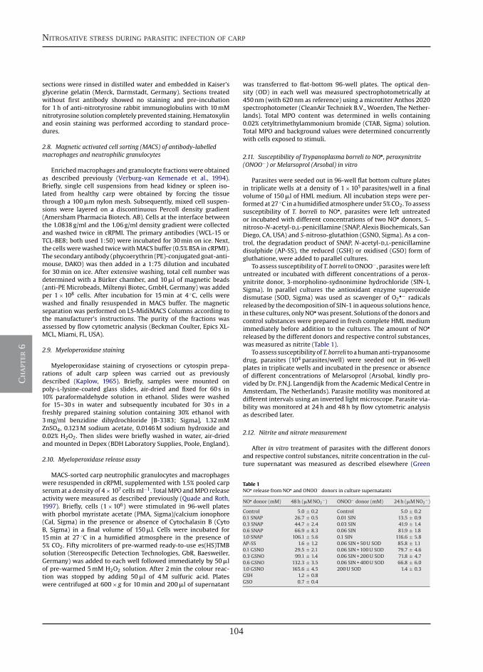
104
nitrosative stress during parasitic infection of carp
ch
apt
er 6
M. Forlenza et al. / Molecular Immunology 45 (2008) 3178–3189 3181
sections were rinsed in distilled water and embedded in Kaiser’sglycerine gelatin (Merck, Darmstadt, Germany). Sections treatedwithout first antibody showed no staining and pre-incubationfor 1h of anti-nitrotyrosine rabbit immunoglobulins with 10mMnitrotyrosine solution completely prevented staining. Hematoxylinand eosin staining was performed according to standard proce-dures.
2.8. Magnetic activated cell sorting (MACS) of antibody-labelledmacrophages and neutrophilic granulocytes
Enrichedmacrophages and granulocyte fractionswere obtainedas described previously (Verburg-van Kemenade et al., 1994).Briefly, single cell suspensions from head kidney or spleen iso-lated from healthy carp were obtained by forcing the tissuethrough a 100�m nylon mesh. Subsequently, mixed cell suspen-sions were layered on a discontinuous Percoll density gradient(Amersham Pharmacia Biotech. AB). Cells at the interface betweenthe 1.0838g/ml and the 1.06g/ml density gradient were collectedand washed twice in cRPMI. The primary antibodies (WCL-15 orTCL-BE8; both used 1:50) were incubated for 30min on ice. Next,the cells werewashed twicewithMACS buffer (0.5% BSA in cRPMI).The secondary antibody (phycoerythrin (PE)-conjugated goat-anti-mouse, DAKO) was then added in a 1:75 dilution and incubatedfor 30min on ice. After extensive washing, total cell number wasdetermined with a Burker chamber, and 10�l of magnetic beads(anti-PE Microbeads, Miltenyi Biotec, GmbH, Germany) was addedper 1×10⁸ cells. After incubation for 15min at 4 ◦C, cells werewashed and finally resuspended in MACS buffer. The magneticseparation was performed on LS-MidiMACS Columns according tothe manufacturer’s instructions. The purity of the fractions wasassessed by flow cytometric analysis (Beckman Coulter, Epics XL-MCL, Miami, FL, USA).
2.9. Myeloperoxidase staining
Myeloperoxidase staining of cryosections or cytospin prepa-rations of adult carp spleen was carried out as previouslydescribed (Kaplow, 1965). Briefly, samples were mounted onpoly-l-lysine-coated glass slides, air-dried and fixed for 60 s in10% paraformaldehyde solution in ethanol. Slides were washedfor 15–30 s in water and subsequently incubated for 30 s in afreshly prepared staining solution containing 30% ethanol with3mg/ml benzidine dihydrochloride [B-3383; Sigma], 1.32mMZnSO4, 0.123M sodium acetate, 0.0146M sodium hydroxide and0.02% H2O2. Then slides were briefly washed in water, air-driedand mounted in Depex (BDH Laboratory Supplies, Poole, England).
2.10. Myeloperoxidase release assay
MACS-sorted carp neutrophilic granulocytes and macrophageswere resuspended in cRPMI, supplemented with 1.5% pooled carpserum at a density of 4×10⁷ cellsml−1. TotalMPO andMPO releaseactivity were measured as described previously (Quade and Roth,1997). Briefly, cells (1×10⁶) were stimulated in 96-well plateswith phorbol myristate acetate (PMA, Sigma)/calcium ionophore(CaI, Sigma) in the presence or absence of Cytochalasin B (CytoB, Sigma) in a final volume of 150�l. Cells were incubated for15min at 27 ◦C in a humidified atmosphere in the presence of5% CO2. Fifty microliters of pre-warmed ready-to-use es(HS)TMBsolution (Stereospecific Detection Technologies, GbR, Baesweiler,Germany) was added to each well followed immediately by 50�lof pre-warmed 5mM H2O2 solution. After 2min the colour reac-tion was stopped by adding 50�l of 4M sulfuric acid. Plateswere centrifuged at 600× g for 10min and 200�l of supernatant
was transferred to flat-bottom 96-well plates. The optical den-sity (OD) in each well was measured spectrophotometrically at450nm (with 620nm as reference) using a microtiter Anthos 2020spectrophotometer (CleanAir Techniek B.V., Woerden, The Nether-lands). Total MPO content was determined in wells containing0.02% cetyltrimethylammonium bromide (CTAB, Sigma) solution.Total MPO and background values were determined concurrentlywith cells exposed to stimuli.
2.11. Susceptibility of Trypanoplasma borreli to NO•, peroxynitrite(ONOO−) or Melarsoprol (Arsobal) in vitro
Parasites were seeded out in 96-well flat bottom culture platesin triplicate wells at a density of 1×10⁵ parasites/well in a finalvolume of 150�l of HML medium. All incubation steps were per-formed at 27 ◦C in a humidified atmosphere under 5%CO2. To assesssusceptibility of T. borreli to NO•, parasites were left untreatedor incubated with different concentrations of two NO• donors, S-nitroso-N-acetyl-d,l-penicillamine (SNAP, Alexis Biochemicals, SanDiego, CA, USA) and S-nitroso-glutathion (GSNO, Sigma). As a con-trol, the degradation product of SNAP, N-acetyl-d,l-penicillaminedisulphide (AP-SS), the reduced (GSH) or oxidised (GSO) form ofgluthatione, were added to parallel cultures.
To assess susceptibility of T. borreli toONOO−, parasiteswere leftuntreated or incubated with different concentrations of a perox-ynitrite donor, 3-morpholino-sydnonimine hydrochloride (SIN-1,Sigma). In parallel cultures the antioxidant enzyme superoxidedismutase (SOD, Sigma) was used as scavenger of O2
•− radicalsreleased by the decomposition of SIN-1 in aqueous solutions hence,in these cultures, onlyNO• was present. Solutions of the donors andcontrol substances were prepared in fresh complete HML mediumimmediately before addition to the cultures. The amount of NO•
released by the different donors and respective control substances,was measured as nitrite (Table 1).
Toassess susceptibilityofT. borreli toahumananti-trypanosomedrug, parasites (104 parasites/well) were seeded out in 96-wellplates in triplicate wells and incubated in the presence or absenceof different concentrations of Melarsoprol (Arsobal, kindly pro-vided by Dr. P.N.J. Langendijk from the Academic Medical Centre inAmsterdam, The Netherlands). Parasite motility was monitored atdifferent intervals using an inverted light microscope. Parasite via-bility was monitored at 24h and 48h by flow cytometric analysisas described later.
2.12. Nitrite and nitrate measurement
After in vitro treatment of parasites with the different donorsand respective control substances, nitrite concentration in the cul-ture supernatant was measured as described elsewhere (Green
Table 1NO• release from NO• and ONOO− donors in culture supernatants
NO• donor (mM) 48h (�MNO2−) ONOO− donor (mM) 24h (�MNO2
−)
Control 5.0 ± 0.2 Control 5.0 ± 0.20.1 SNAP 26.7 ± 0.5 0.01 SIN 13.5 ± 0.90.3 SNAP 44.7 ± 2.4 0.03 SIN 41.9 ± 1.40.6 SNAP 66.9 ± 8.3 0.06 SIN 81.9 ± 1.81.0 SNAP 106.1 ± 5.6 0.1 SIN 116.6 ± 5.8AP-SS 1.6 ± 1.2 0.06 SIN+50U SOD 85.8 ± 1.10.1 GSNO 29.5 ± 2.1 0.06 SIN+100U SOD 79.7 ± 4.60.3 GSNO 99.1 ± 1.4 0.06 SIN+200U SOD 71.8 ± 4.70.6 GSNO 132.3 ± 3.5 0.06 SIN+400U SOD 66.8 ± 6.01.0 GSNO 165.6 ± 4.5 200U SOD 1.4 ± 0.3GSH 1.2 ± 0.8GSO 0.7 ± 0.4
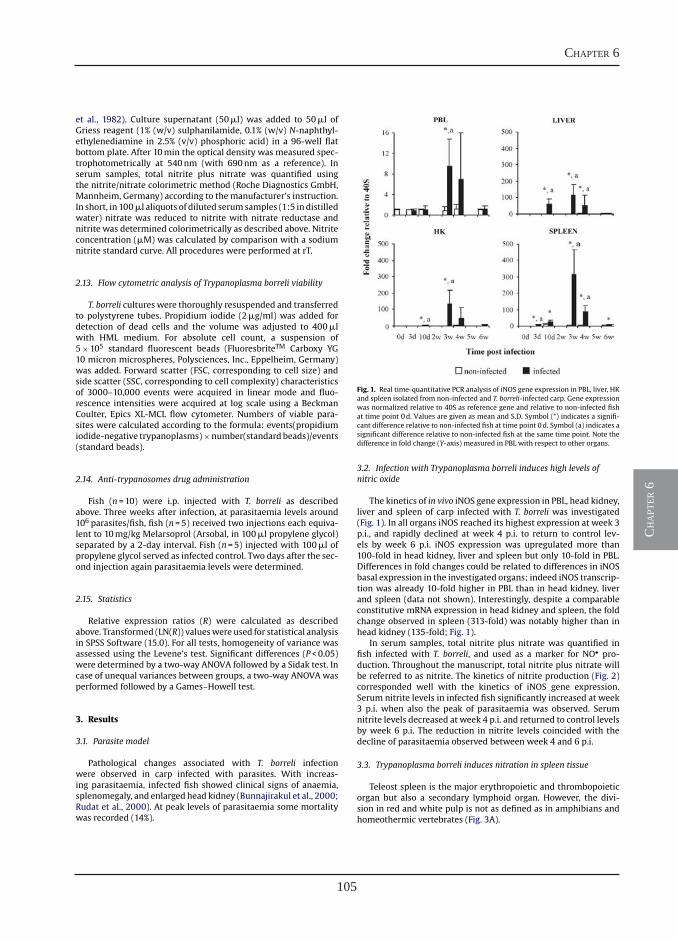
105
chapter 6
ch
apt
er 6
3182 M. Forlenza et al. / Molecular Immunology 45 (2008) 3178–3189
et al., 1982). Culture supernatant (50�l) was added to 50�l ofGriess reagent (1% (w/v) sulphanilamide, 0.1% (w/v) N-naphthyl-ethylenediamine in 2.5% (v/v) phosphoric acid) in a 96-well flatbottom plate. After 10min the optical density was measured spec-trophotometrically at 540nm (with 690nm as a reference). Inserum samples, total nitrite plus nitrate was quantified usingthe nitrite/nitrate colorimetric method (Roche Diagnostics GmbH,Mannheim, Germany) according to themanufacturer’s instruction.In short, in 100�l aliquots of diluted serumsamples (1:5 in distilledwater) nitrate was reduced to nitrite with nitrate reductase andnitrite was determined colorimetrically as described above. Nitriteconcentration (�M) was calculated by comparison with a sodiumnitrite standard curve. All procedures were performed at rT.
2.13. Flow cytometric analysis of Trypanoplasma borreli viability
T. borreli cultures were thoroughly resuspended and transferredto polystyrene tubes. Propidium iodide (2�g/ml) was added fordetection of dead cells and the volume was adjusted to 400�lwith HML medium. For absolute cell count, a suspension of5×105 standard fluorescent beads (FluoresbriteTM Carboxy YG10 micron microspheres, Polysciences, Inc., Eppelheim, Germany)was added. Forward scatter (FSC, corresponding to cell size) andside scatter (SSC, corresponding to cell complexity) characteristicsof 3000–10,000 events were acquired in linear mode and fluo-rescence intensities were acquired at log scale using a BeckmanCoulter, Epics XL-MCL flow cytometer. Numbers of viable para-sites were calculated according to the formula: events(propidiumiodide-negative trypanoplasms)×number(standard beads)/events(standard beads).
2.14. Anti-trypanosomes drug administration
Fish (n=10) were i.p. injected with T. borreli as describedabove. Three weeks after infection, at parasitaemia levels around106 parasites/fish, fish (n=5) received two injections each equiva-lent to 10mg/kg Melarsoprol (Arsobal, in 100�l propylene glycol)separated by a 2-day interval. Fish (n=5) injected with 100�l ofpropylene glycol served as infected control. Two days after the sec-ond injection again parasitaemia levels were determined.
2.15. Statistics
Relative expression ratios (R) were calculated as describedabove. Transformed (LN(R)) valueswere used for statistical analysisin SPSS Software (15.0). For all tests, homogeneity of variance wasassessed using the Levene’s test. Significant differences (P<0.05)were determined by a two-way ANOVA followed by a Sidak test. Incase of unequal variances between groups, a two-way ANOVA wasperformed followed by a Games–Howell test.
3. Results
3.1. Parasite model
Pathological changes associated with T. borreli infectionwere observed in carp infected with parasites. With increas-ing parasitaemia, infected fish showed clinical signs of anaemia,splenomegaly, and enlarged head kidney (Bunnajirakul et al., 2000;Rudat et al., 2000). At peak levels of parasitaemia some mortalitywas recorded (14%).
Fig. 1. Real time-quantitative PCR analysis of iNOS gene expression in PBL, liver, HKand spleen isolated from non-infected and T. borreli-infected carp. Gene expressionwas normalized relative to 40S as reference gene and relative to non-infected fishat time point 0d. Values are given as mean and S.D. Symbol (*) indicates a signifi-cant difference relative to non-infected fish at time point 0d. Symbol (a) indicates asignificant difference relative to non-infected fish at the same time point. Note thedifference in fold change (Y-axis) measured in PBL with respect to other organs.
3.2. Infection with Trypanoplasma borreli induces high levels ofnitric oxide
The kinetics of in vivo iNOS gene expression in PBL, head kidney,liver and spleen of carp infected with T. borreli was investigated(Fig. 1). In all organs iNOS reached its highest expression at week 3p.i., and rapidly declined at week 4 p.i. to return to control lev-els by week 6 p.i. iNOS expression was upregulated more than100-fold in head kidney, liver and spleen but only 10-fold in PBL.Differences in fold changes could be related to differences in iNOSbasal expression in the investigated organs; indeed iNOS transcrip-tion was already 10-fold higher in PBL than in head kidney, liverand spleen (data not shown). Interestingly, despite a comparableconstitutive mRNA expression in head kidney and spleen, the foldchange observed in spleen (313-fold) was notably higher than inhead kidney (135-fold; Fig. 1).
In serum samples, total nitrite plus nitrate was quantified infish infected with T. borreli, and used as a marker for NO• pro-duction. Throughout the manuscript, total nitrite plus nitrate willbe referred to as nitrite. The kinetics of nitrite production (Fig. 2)corresponded well with the kinetics of iNOS gene expression.Serum nitrite levels in infected fish significantly increased at week3 p.i. when also the peak of parasitaemia was observed. Serumnitrite levels decreased at week 4 p.i. and returned to control levelsby week 6 p.i. The reduction in nitrite levels coincided with thedecline of parasitaemia observed between week 4 and 6 p.i.
3.3. Trypanoplasma borreli induces nitration in spleen tissue
Teleost spleen is the major erythropoietic and thrombopoieticorgan but also a secondary lymphoid organ. However, the divi-sion in red and white pulp is not as defined as in amphibians andhomeothermic vertebrates (Fig. 3A).
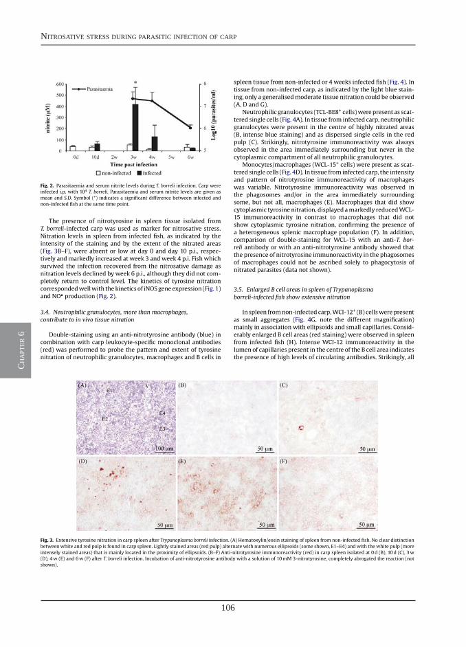
106
nitrosative stress during parasitic infection of carp
ch
apt
er 6
M. Forlenza et al. / Molecular Immunology 45 (2008) 3178–3189 3183
Fig. 2. Parasitaemia and serum nitrite levels during T. borreli infection. Carp wereinfected i.p. with 104 T. borreli. Parasitaemia and serum nitrite levels are given asmean and S.D. Symbol (*) indicates a significant difference between infected andnon-infected fish at the same time point.
The presence of nitrotyrosine in spleen tissue isolated fromT. borreli-infected carp was used as marker for nitrosative stress.Nitration levels in spleen from infected fish, as indicated by theintensity of the staining and by the extent of the nitrated areas(Fig. 3B–F), were absent or low at day 0 and day 10 p.i., respec-tively andmarkedly increased at week 3 andweek 4 p.i. Fish whichsurvived the infection recovered from the nitrosative damage asnitration levels declined by week 6 p.i., although they did not com-pletely return to control level. The kinetics of tyrosine nitrationcorrespondedwellwith thekinetics of iNOSgeneexpression (Fig. 1)and NO• production (Fig. 2).
3.4. Neutrophilic granulocytes, more than macrophages,contribute to in vivo tissue nitration
Double-staining using an anti-nitrotyrosine antibody (blue) incombination with carp leukocyte-specific monoclonal antibodies(red) was performed to probe the pattern and extent of tyrosinenitration of neutrophilic granulocytes, macrophages and B cells in
spleen tissue from non-infected or 4 weeks infected fish (Fig. 4). Intissue from non-infected carp, as indicated by the light blue stain-ing, only a generalisedmoderate tissue nitration could be observed(A, D and G).
Neutrophilic granulocytes (TCL-BE8+ cells)werepresent as scat-tered single cells (Fig. 4A). In tissue from infected carp, neutrophilicgranulocytes were present in the centre of highly nitrated areas(B, intense blue staining) and as dispersed single cells in the redpulp (C). Strikingly, nitrotyrosine immunoreactivity was alwaysobserved in the area immediately surrounding but never in thecytoplasmic compartment of all neutrophilic granulocytes.
Monocytes/macrophages (WCL-15+ cells) were present as scat-tered single cells (Fig. 4D). In tissue from infected carp, the intensityand pattern of nitrotyrosine immunoreactivity of macrophageswas variable. Nitrotyrosine immunoreactivity was observed inthe phagosomes and/or in the area immediately surroundingsome, but not all, macrophages (E). Macrophages that did showcytoplasmic tyrosine nitration, displayed amarkedly reducedWCL-15 immunoreactivity in contrast to macrophages that did notshow cytoplasmic tyrosine nitration, confirming the presence ofa heterogeneous splenic macrophage population (F). In addition,comparison of double-staining for WCL-15 with an anti-T. bor-reli antibody or with an anti-nitrotyrosine antibody showed thatthe presence of nitrotyrosine immunoreactivity in the phagosomesof macrophages could not be ascribed solely to phagocytosis ofnitrated parasites (data not shown).
3.5. Enlarged B cell areas in spleen of Trypanoplasmaborreli-infected fish show extensive nitration
In spleen fromnon-infected carp,WCI-12+ (B) cellswerepresentas small aggregates (Fig. 4G, note the different magnification)mainly in association with ellipsoids and small capillaries. Consid-erably enlarged B cell areas (red staining) were observed in spleenfrom infected fish (H). Intense WCI-12 immunoreactivity in thelumen of capillaries present in the centre of the B cell area indicatesthe presence of high levels of circulating antibodies. Strikingly, all
Fig. 3. Extensive tyrosine nitration in carp spleen after Trypanoplasma borreli infection. (A) Hematoxylin/eosin staining of spleen from non-infected fish. No clear distinctionbetween white and red pulp is found in carp spleen. Lightly stained areas (red pulp) alternate with numerous ellipsoids (some shown, E1–E4) and with the white pulp (moreintensely stained areas) that is mainly located in the proximity of ellipsoids. (B–F) Anti-nitrotyrosine immunoreactivity (red) in carp spleen isolated at 0d (B), 10d (C), 3w(D), 4w (E) and 6w (F) after T. borreli infection. Incubation of anti-nitrotyrosine antibody with a solution of 10mM 3-nitrotyrosine, completely abrogated the reaction (notshown).
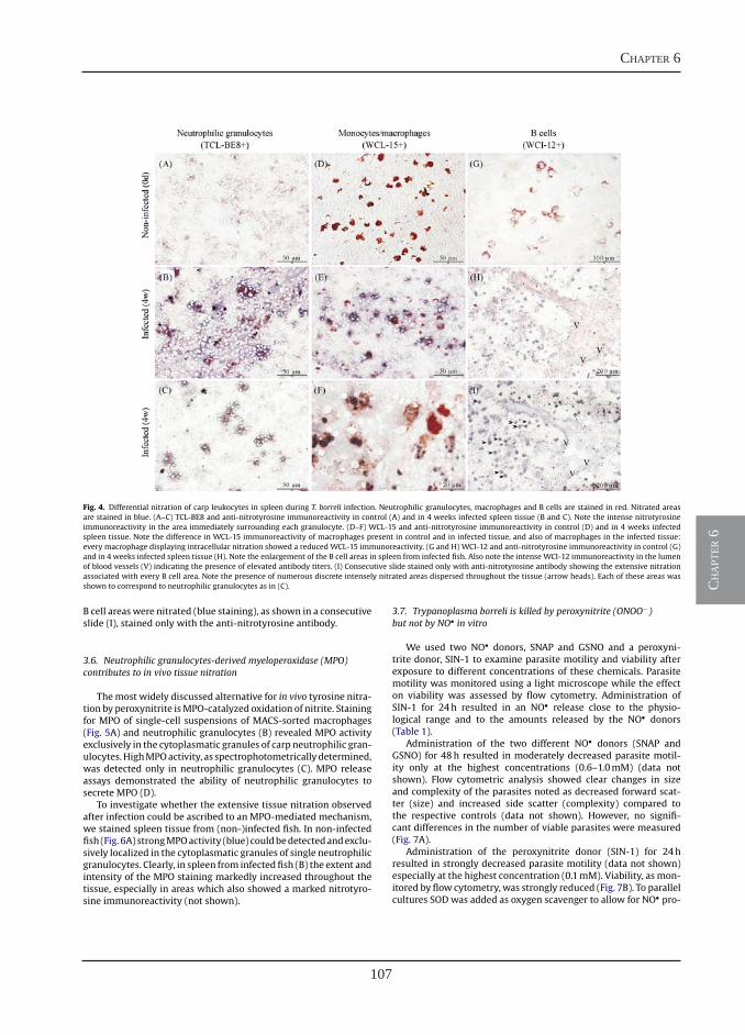
107
chapter 6
ch
apt
er 6
3184 M. Forlenza et al. / Molecular Immunology 45 (2008) 3178–3189
Fig. 4. Differential nitration of carp leukocytes in spleen during T. borreli infection. Neutrophilic granulocytes, macrophages and B cells are stained in red. Nitrated areasare stained in blue. (A–C) TCL-BE8 and anti-nitrotyrosine immunoreactivity in control (A) and in 4 weeks infected spleen tissue (B and C). Note the intense nitrotyrosineimmunoreactivity in the area immediately surrounding each granulocyte. (D–F) WCL-15 and anti-nitrotyrosine immunoreactivity in control (D) and in 4 weeks infectedspleen tissue. Note the difference in WCL-15 immunoreactivity of macrophages present in control and in infected tissue, and also of macrophages in the infected tissue:every macrophage displaying intracellular nitration showed a reduced WCL-15 immunoreactivity. (G and H) WCI-12 and anti-nitrotyrosine immunoreactivity in control (G)and in 4 weeks infected spleen tissue (H). Note the enlargement of the B cell areas in spleen from infected fish. Also note the intense WCI-12 immunoreactivity in the lumenof blood vessels (V) indicating the presence of elevated antibody titers. (I) Consecutive slide stained only with anti-nitrotyrosine antibody showing the extensive nitrationassociated with every B cell area. Note the presence of numerous discrete intensely nitrated areas dispersed throughout the tissue (arrow heads). Each of these areas wasshown to correspond to neutrophilic granulocytes as in (C).
B cell areaswere nitrated (blue staining), as shown in a consecutiveslide (I), stained only with the anti-nitrotyrosine antibody.
3.6. Neutrophilic granulocytes-derived myeloperoxidase (MPO)contributes to in vivo tissue nitration
Themost widely discussed alternative for in vivo tyrosine nitra-tion by peroxynitrite isMPO-catalyzed oxidation of nitrite. Stainingfor MPO of single-cell suspensions of MACS-sorted macrophages(Fig. 5A) and neutrophilic granulocytes (B) revealed MPO activityexclusively in the cytoplasmatic granules of carp neutrophilic gran-ulocytes.HighMPOactivity, as spectrophotometricallydetermined,was detected only in neutrophilic granulocytes (C). MPO releaseassays demonstrated the ability of neutrophilic granulocytes tosecrete MPO (D).
To investigate whether the extensive tissue nitration observedafter infection could be ascribed to an MPO-mediated mechanism,we stained spleen tissue from (non-)infected fish. In non-infectedfish (Fig. 6A) strongMPOactivity (blue) couldbedetectedandexclu-sively localized in the cytoplasmatic granules of single neutrophilicgranulocytes. Clearly, in spleen from infected fish (B) the extent andintensity of the MPO staining markedly increased throughout thetissue, especially in areas which also showed a marked nitrotyro-sine immunoreactivity (not shown).
3.7. Trypanoplasma borreli is killed by peroxynitrite (ONOO−)but not by NO• in vitro
We used two NO• donors, SNAP and GSNO and a peroxyni-trite donor, SIN-1 to examine parasite motility and viability afterexposure to different concentrations of these chemicals. Parasitemotility was monitored using a light microscope while the effecton viability was assessed by flow cytometry. Administration ofSIN-1 for 24h resulted in an NO• release close to the physio-logical range and to the amounts released by the NO• donors(Table 1).
Administration of the two different NO• donors (SNAP andGSNO) for 48h resulted in moderately decreased parasite motil-ity only at the highest concentrations (0.6–1.0mM) (data notshown). Flow cytometric analysis showed clear changes in sizeand complexity of the parasites noted as decreased forward scat-ter (size) and increased side scatter (complexity) compared tothe respective controls (data not shown). However, no signifi-cant differences in the number of viable parasites were measured(Fig. 7A).
Administration of the peroxynitrite donor (SIN-1) for 24hresulted in strongly decreased parasite motility (data not shown)especially at the highest concentration (0.1mM). Viability, as mon-itored by flow cytometry, was strongly reduced (Fig. 7B). To parallelcultures SOD was added as oxygen scavenger to allow for NO• pro-
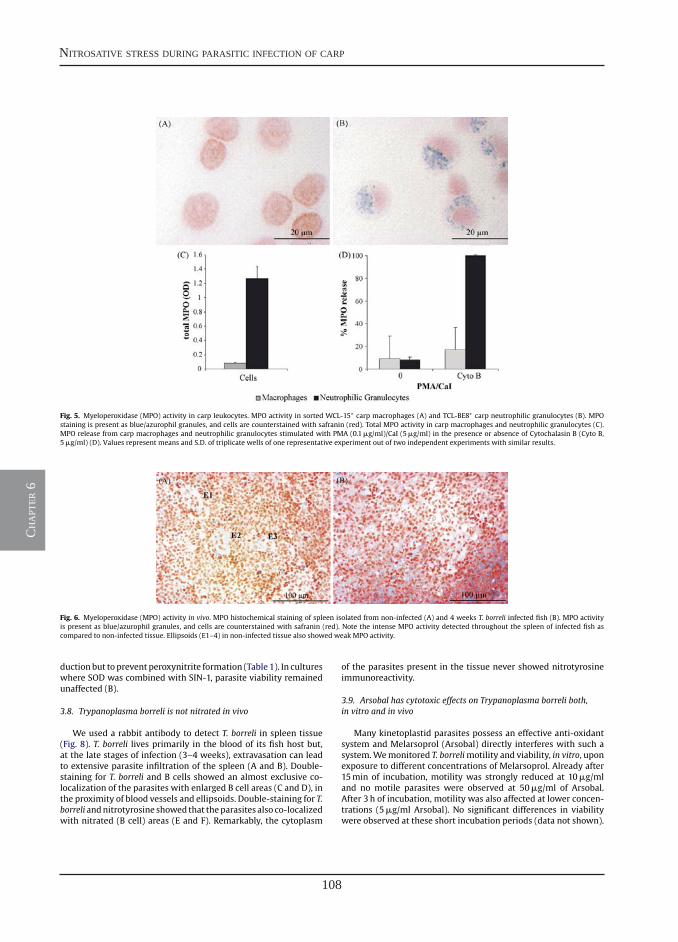
108
nitrosative stress during parasitic infection of carp
ch
apt
er 6
M. Forlenza et al. / Molecular Immunology 45 (2008) 3178–3189 3185
Fig. 5. Myeloperoxidase (MPO) activity in carp leukocytes. MPO activity in sorted WCL-15+ carp macrophages (A) and TCL-BE8+ carp neutrophilic granulocytes (B). MPOstaining is present as blue/azurophil granules, and cells are counterstained with safranin (red). Total MPO activity in carp macrophages and neutrophilic granulocytes (C).MPO release from carp macrophages and neutrophilic granulocytes stimulated with PMA (0.1�g/ml)/CaI (5�g/ml) in the presence or absence of Cytochalasin B (Cyto B,5�g/ml) (D). Values represent means and S.D. of triplicate wells of one representative experiment out of two independent experiments with similar results.
Fig. 6. Myeloperoxidase (MPO) activity in vivo. MPO histochemical staining of spleen isolated from non-infected (A) and 4 weeks T. borreli infected fish (B). MPO activityis present as blue/azurophil granules, and cells are counterstained with safranin (red). Note the intense MPO activity detected throughout the spleen of infected fish ascompared to non-infected tissue. Ellipsoids (E1–4) in non-infected tissue also showed weak MPO activity.
ductionbut to prevent peroxynitrite formation (Table 1). In cultureswhere SOD was combined with SIN-1, parasite viability remainedunaffected (B).
3.8. Trypanoplasma borreli is not nitrated in vivo
We used a rabbit antibody to detect T. borreli in spleen tissue(Fig. 8). T. borreli lives primarily in the blood of its fish host but,at the late stages of infection (3–4 weeks), extravasation can leadto extensive parasite infiltration of the spleen (A and B). Double-staining for T. borreli and B cells showed an almost exclusive co-localization of the parasites with enlarged B cell areas (C and D), inthe proximity of blood vessels and ellipsoids. Double-staining for T.borreli andnitrotyrosine showed that theparasites also co-localizedwith nitrated (B cell) areas (E and F). Remarkably, the cytoplasm
of the parasites present in the tissue never showed nitrotyrosineimmunoreactivity.
3.9. Arsobal has cytotoxic effects on Trypanoplasma borreli both,in vitro and in vivo
Many kinetoplastid parasites possess an effective anti-oxidantsystem and Melarsoprol (Arsobal) directly interferes with such asystem.Wemonitored T. borrelimotility and viability, in vitro, uponexposure to different concentrations of Melarsoprol. Already after15min of incubation, motility was strongly reduced at 10�g/mland no motile parasites were observed at 50�g/ml of Arsobal.After 3h of incubation, motility was also affected at lower concen-trations (5�g/ml Arsobal). No significant differences in viabilitywere observed at these short incubation periods (data not shown).
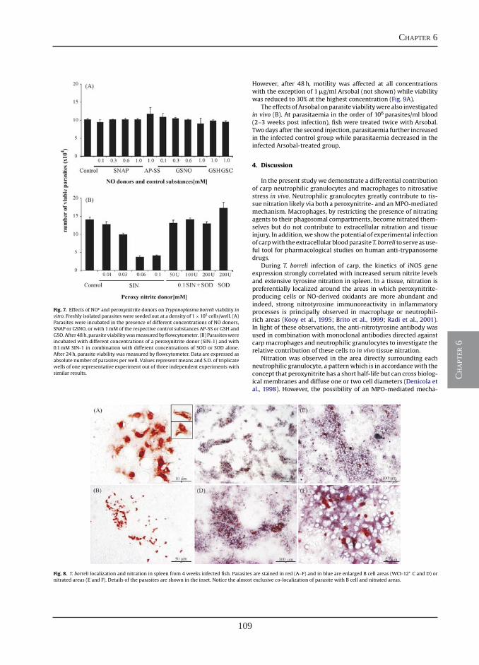
109
chapter 6
ch
apt
er 6
3186 M. Forlenza et al. / Molecular Immunology 45 (2008) 3178–3189
Fig. 7. Effects of NO• and peroxynitrite donors on Trypanoplasma borreli viability invitro. Freshly isolated parasites were seeded out at a density of 1×10⁵ cells/well. (A)Parasites were incubated in the presence of different concentrations of NO donors,SNAP or GSNO, or with 1mM of the respective control substances AP-SS or GSH andGSO.After 48h, parasite viabilitywasmeasuredbyflowcytometer. (B) Parasiteswereincubated with different concentrations of a peroxynitrite donor (SIN-1) and with0.1mM SIN-1 in combination with different concentrations of SOD or SOD alone.After 24h, parasite viability was measured by flowcytometer. Data are expressed asabsolute number of parasites per well. Values represent means and S.D. of triplicatewells of one representative experiment out of three independent experiments withsimilar results.
However, after 48h, motility was affected at all concentrationswith the exception of 1�g/ml Arsobal (not shown) while viabilitywas reduced to 30% at the highest concentration (Fig. 9A).
The effects ofArsobal onparasite viabilitywere also investigatedin vivo (B). At parasitaemia in the order of 106 parasites/ml blood(2–3 weeks post infection), fish were treated twice with Arsobal.Two days after the second injection, parasitaemia further increasedin the infected control group while parasitaemia decreased in theinfected Arsobal-treated group.
4. Discussion
In the present study we demonstrate a differential contributionof carp neutrophilic granulocytes and macrophages to nitrosativestress in vivo. Neutrophilic granulocytes greatly contribute to tis-sue nitration likely via both a peroxynitrite- and anMPO-mediatedmechanism. Macrophages, by restricting the presence of nitratingagents to their phagosomal compartments, become nitrated them-selves but do not contribute to extracellular nitration and tissueinjury. In addition, we show the potential of experimental infectionof carpwith theextracellularbloodparasiteT. borreli to serveasuse-ful tool for pharmacological studies on human anti-trypanosomedrugs.
During T. borreli infection of carp, the kinetics of iNOS geneexpression strongly correlated with increased serum nitrite levelsand extensive tyrosine nitration in spleen. In a tissue, nitration ispreferentially localized around the areas in which peroxynitrite-producing cells or NO-derived oxidants are more abundant andindeed, strong nitrotyrosine immunoreactivity in inflammatoryprocesses is principally observed in macrophage or neutrophil-rich areas (Kooy et al., 1995; Brito et al., 1999; Radi et al., 2001).In light of these observations, the anti-nitrotyrosine antibody wasused in combination with monoclonal antibodies directed againstcarpmacrophages and neutrophilic granulocytes to investigate therelative contribution of these cells to in vivo tissue nitration.
Nitration was observed in the area directly surrounding eachneutrophilic granulocyte, a patternwhich is in accordancewith theconcept that peroxynitrite has a short half-life but can cross biolog-ical membranes and diffuse one or two cell diameters (Denicola etal., 1998). However, the possibility of an MPO-mediated mecha-
Fig. 8. T. borreli localization and nitration in spleen from 4 weeks infected fish. Parasites are stained in red (A–F) and in blue are enlarged B cell areas (WCI-12+ C and D) ornitrated areas (E and F). Details of the parasites are shown in the inset. Notice the almost exclusive co-localization of parasite with B cell and nitrated areas.
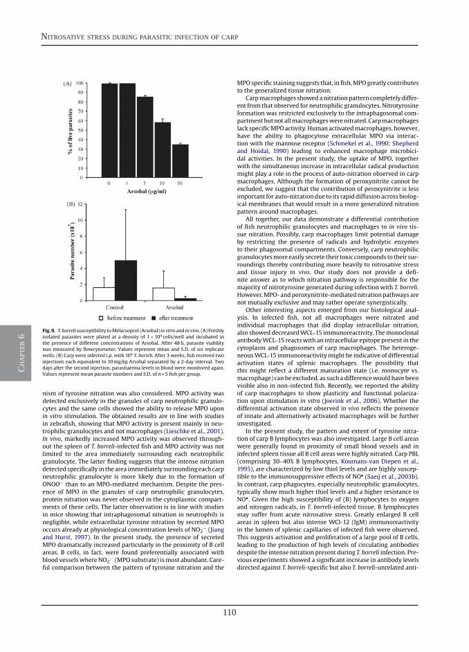
110
nitrosative stress during parasitic infection of carp
ch
apt
er 6
M. Forlenza et al. / Molecular Immunology 45 (2008) 3178–3189 3187
Fig. 9. T. borreli susceptibility toMelarsoprol (Arsobal) in vitro and in vivo. (A) Freshlyisolated parasites were plated at a density of 1×10⁴ cells/well and incubated inthe presence of different concentrations of Arsobal. After 48h, parasite viabilitywas measured by flowcytometer. Values represent mean and S.D. of six replicatewells. (B) Carp were infected i.p. with 104 T. borreli. After 3 weeks, fish received twoinjections each equivalent to 10mg/kg Arsobal separated by a 2-day interval. Twodays after the second injection, parasitaemia levels in blood were monitored again.Values represent mean parasite numbers and S.D. of n=5 fish per group.
nism of tyrosine nitration was also considered. MPO activity wasdetected exclusively in the granules of carp neutrophilic granulo-cytes and the same cells showed the ability to release MPO uponin vitro stimulation. The obtained results are in line with studiesin zebrafish, showing that MPO activity is present mainly in neu-trophilic granulocytes and not macrophages (Lieschke et al., 2001).In vivo, markedly increased MPO activity was observed through-out the spleen of T. borreli-infected fish and MPO activity was notlimited to the area immediately surrounding each neutrophilicgranulocyte. The latter finding suggests that the intense nitrationdetected specifically in thearea immediately surroundingeachcarpneutrophilic granulocyte is more likely due to the formation ofONOO− than to an MPO-mediated mechanism. Despite the pres-ence of MPO in the granules of carp neutrophilic granulocytes,protein nitration was never observed in the cytoplasmic compart-ments of these cells. The latter observation is in line with studiesin mice showing that intraphagosomal nitration in neutrophils isnegligible, while extracellular tyrosine nitration by secreted MPOoccurs already at physiological concentration levels of NO2
− (Jiangand Hurst, 1997). In the present study, the presence of secretedMPO dramatically increased particularly in the proximity of B cellareas. B cells, in fact, were found preferentially associated withblood vesselswhereNO2
− (MPO substrate) ismost abundant. Care-ful comparison between the pattern of tyrosine nitration and the
MPOspecific staining suggests that, in fish,MPOgreatly contributesto the generalized tissue nitration.
Carpmacrophages showedanitrationpattern completely differ-ent from that observed for neutrophilic granulocytes. Nitrotyrosineformation was restricted exclusively to the intraphagosomal com-partmentbutnot allmacrophageswerenitrated. Carpmacrophageslack specificMPOactivity.Humanactivatedmacrophages, however,have the ability to phagocytose extracellular MPO via interac-tion with the mannose receptor (Schmekel et al., 1990; Shepherdand Hoidal, 1990) leading to enhanced macrophage microbici-dal activities. In the present study, the uptake of MPO, togetherwith the simultaneous increase in intracellular radical productionmight play a role in the process of auto-nitration observed in carpmacrophages. Although the formation of peroxynitrite cannot beexcluded, we suggest that the contribution of peroxynitrite is lessimportant for auto-nitrationdue to its rapiddiffusion across biolog-ical membranes that would result in a more generalized nitrationpattern around macrophages.
All together, our data demonstrate a differential contributionof fish neutrophilic granulocytes and macrophages to in vivo tis-sue nitration. Possibly, carp macrophages limit potential damageby restricting the presence of radicals and hydrolytic enzymesto their phagosomal compartments. Conversely, carp neutrophilicgranulocytesmoreeasily secrete their toxic compounds to their sur-roundings thereby contributing more heavily to nitrosative stressand tissue injury in vivo. Our study does not provide a defi-nite answer as to which nitration pathway is responsible for themajority of nitrotyrosine generated during infection with T. borreli.However,MPO-andperoxynitrite-mediatednitrationpathways arenot mutually exclusive and may rather operate synergistically.
Other interesting aspects emerged from our histological anal-ysis. In infected fish, not all macrophages were nitrated andindividual macrophages that did display intracellular nitration,also showeddecreasedWCL-15 immunoreactivity. ThemonoclonalantibodyWCL-15 reactswith an intracellular epitope present in thecytoplasm and phagosomes of carp macrophages. The heteroge-neousWCL-15 immunoreactivitymight be indicative of differentialactivation states of splenic macrophages. The possibility thatthis might reflect a different maturation state (i.e. monocyte vs.macrophage) canbe excluded, as such adifferencewouldhavebeenvisible also in non-infected fish. Recently, we reported the abilityof carp macrophages to show plasticity and functional polariza-tion upon stimulation in vitro (Joerink et al., 2006). Whether thedifferential activation state observed in vivo reflects the presenceof innate and alternatively activated macrophages will be furtherinvestigated.
In the present study, the pattern and extent of tyrosine nitra-tion of carp B lymphocytes was also investigated. Large B cell areaswere generally found in proximity of small blood vessels and ininfected spleen tissue all B cell areas were highly nitrated. Carp PBL(comprising 30–40% B lymphocytes, Koumans-van Diepen et al.,1995), are characterized by low thiol levels and are highly suscep-tible to the immunosuppressive effects of NO• (Saeij et al., 2003b).In contrast, carp phagocytes, especially neutrophilic granulocytes,typically show much higher thiol levels and a higher resistance toNO•. Given the high susceptibility of (B) lymphocytes to oxygenand nitrogen radicals, in T. borreli-infected tissue, B lymphocytesmay suffer from acute nitrosative stress. Greatly enlarged B cellareas in spleen but also intense WCI-12 (IgM) immunoreactivityin the lumen of splenic capillaries of infected fish were observed.This suggests activation and proliferation of a large pool of B cells,leading to the production of high levels of circulating antibodiesdespite the intense nitration present during T. borreli infection. Pre-vious experiments showed a significant increase in antibody levelsdirected against T. borreli-specific but also T. borreli-unrelated anti-
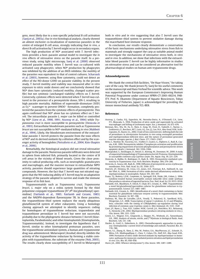
111
chapter 6
ch
apt
er 6
3188 M. Forlenza et al. / Molecular Immunology 45 (2008) 3178–3189
gens, most likely due to a non-specific polyclonal B cell activation(Saeij et al., 2003a). Our in vivo histological analysis, clearly showedan almost exclusive co-localization of numerous parasites in thecentre of enlarged B cell areas, strongly indicating that in vivo, adirect B cell activation by T. borrelimight occur in secondary organs.
The high production of NO• associated with T. borreli infec-tions provides a host–parasite animal model to investigate the invivo effects of nitrosative stress on the parasite itself. In a pre-vious study, using light microscopy, Saeij et al. (2000) observedreduced parasite motility when T. borreli was co-cultured withactivated carp phagocytes; when NO• production by phagocyteswas inhibited by the addition of an iNOS inhibitor, the motility ofthe parasites was equivalent to that of control cultures. Scharsacket al. (2003), however, using flow cytometry, could not detect aneffect of the NO-donor GSNO on parasite viability. In the presentstudy, T. borreli motility and viability was measured after in vitroexposure to nitric oxide donors and we conclusively showed thatNO• does have cytostatic (reduced motility, changed scatter pro-file) but not cytotoxic (unchanged viability) effects on T. borreli.Conversely, cytotoxic effects were detectedwhen T. borreliwas cul-tured in the presence of a peroxynitrite donor, SIN-1, resulting inhigh parasite mortality. Addition of superoxide-dismutase (SOD)as O2
•− scavenger to prevent ONOO− formation, completely pro-tected the parasites from the cytotoxic effects of peroxynitrite. Thisagain confirmed that NO• alone has no cytotoxic effect on T. bor-reli. The intracellular parasite L. major can be killed or controlledby NO• (Liew et al., 1990, 1991; Assreuy et al., 1994) while Try-panosoma cruzi is more susceptible to peroxynitrite (Denicola etal., 1993). Extracellular bloodstreamparasites such as Trypanosomabrucei are not susceptible to NO•-mediated killing in vivo (Mabbottet al., 1994). Likely, the bloodstream environment of the extracel-lular parasite T. borreli protects the parasite from the toxic effectsof NO• and its derived oxidants by a dilution effect and by the scav-enger activity of hemoglobin (Pietraforte et al., 2004; Kim-Shapiroet al., 2006).
Remarkably, the histological analysis did not reveal nitrosativedamage to theparasite.Numerousparasite infiltratesweredetectedin spleen from infected fish, primarily located in the centre of Bcell areas in the vicinity of blood vessels. Given the close prox-imity to radical producing cells, such as neutrophilic granulocytesand macrophages, and the massive increase in extracellular MPOactivity, parasites should experience large quantities of nitratingcompounds. However, the fact that T. borreli was not nitrated sug-gests that the NO-inducing ability of T. borrelimay be an adaptationstrategy of the parasite adopted to survive and evade the immuneresponse of its fish host.
Trypanosomatids such as Trypanosoma cruzi, Trypanosomabrucei, L. major rely on a redox system formed by the thiolpolyamine conjugate trypanothione [N1,N8-bis(glutathionyl) sper-midine] (Fairlamb et al., 1985; Fairlamb and Cerami, 1985)and the NADPH-dependent flavoenzyme TR. In these parasites,the trypanothione–thiol system replaces the nearly ubiquitousglutathione/GR system of other eukaryotes. Using a homologycloning approach we attempted to identify sequences codingfor homologues of trypanothione reductase, tryparedoxin andtrypanothione peroxidase in T. borreli but were not successful,probably due to the phylogenetic distance between T. borreli (Kine-toplastida, Parabodonida) and other kinetoplastids (Kinetoplastida,Trypanosomatida). Instead, to investigate the hypothesis that T.borreli, similar to other kinetoplastid protozoan parasites, usesthe trypanothione antioxidant system, a human anti-trypanosomedrug was administered. Melarsoprol (Arsobal) directly inhibits theactivity of the trypanothione reductase by forming a stable com-plex with trypanothione, the substrate of the enzyme (Nok, 2003).The results clearly show susceptibility of T. borreli to Melarsoprol
both in vitro and in vivo suggesting that also T. borreli uses thetrypanothione–thiol system to prevent oxidative damage duringthe exacerbated host immune response.
In conclusion, our results clearly demonstrate a conservationof the basic mechanisms underlying nitrosative stress from fish tomammals and strongly support the carp as suitable animal modelto investigate the mechanisms of nitrosative stress both, in vitroand in vivo. In addition, we show that infections with the extracel-lular blood parasite T. borreli can be highly informative in studieson nitrosative stress and can be considered an alternative tool forpharmacological studies on human anti-trypanosome drugs.
Acknowledgements
We thank the central fish facilities, “De Haar-Vissen,” for takingcare of the carp. We thank Jeroen P.J. Saeij for his useful commentson themanuscript andHans Verhoef for scientific advice. Thisworkwas supported by the European Commission’s Improving HumanPotential Programme under contract HPRN-CT-2001-00214, PAR-ITY. Prof. N. Okamoto (Department of Aquatic Biosciences, TokyoUniversity of Fisheries, Japan) is acknowledged for providing themouse monoclonal antibody TCL-BE8.
References
Assreuy, J., Cunha, F.Q., Epperlein, M., Noronha-Dutra, A., O’Donnell, C.A., Liew,F.Y., Moncada, S., 1994. Production of nitric oxide and superoxide by activatedmacrophages and killing of Leishmania major. Eur. J. Immunol. 24, 672–676.
Brennan, M.L., Wu, W., Fu, X., Shen, Z., Song, W., Frost, H., Vadseth, C., Narine, L.,Lenkiewicz, E., Borchers, M.T., Lusis, A.J., Lee, J.J., Lee, N.A., Abu-Soud, H.M., Ischi-ropoulos, H., Hazen, S.L., 2002. A tale of two controversies: defining both the roleof peroxidases in nitrotyrosine formation in vivo using eosinophil peroxidaseand myeloperoxidase-deficient mice, and the nature of peroxidase-generatedreactive nitrogen species. J. Biol. Chem. 277, 17415–17427.
Brito, C., Naviliat, M., Tiscornia, A.C., Vuillier, F., Gualco, G., Dighiero, G., Radi, R., Cay-ota, A.M., 1999. Peroxynitrite inhibits T lymphocyte activation and proliferationby promoting impairment of tyrosine phosphorylation andperoxynitrite-drivenapoptotic death. J. Immunol. 162, 3356–3366.
Bunnajirakul, S., Steinhagen, D., Hetzel, U., Korting, W., Drommer, W., 2000. A studyof sequential histopathology of Trypanoplasmaborreli (Protozoa: Kinetoplastida)in susceptible common carp Cyprinus carpio. Dis. Aquat. Org. 39, 221–229.
Denicola, A., Rubbo, H., Rodriguez, D., Radi, R., 1993. Peroxynitrite-mediated cyto-toxicity to Trypanosoma cruzi. Arch. Biochem. Biophys. 304, 279–286.
Denicola, A., Souza, J.M., Radi, R., 1998. Diffusion of peroxynitrite across erythrocytemembranes. Proc. Natl. Acad. Sci. 95, 3566–3571.
Eiserich, J.P., Hristova, M., Cross, C.E., Jones, A.D., Freeman, B.A., Halliwell, B., vander Vliet, A., 1998. Formation of nitric oxide-derived inflammatory oxidants bymyeloperoxidase in neutrophils. Nature 391, 393–397.
Evans, T.J., Buttery, L.D.K., Carpenter, A., Springall, D.R., Polak, J.M., Cohen, J., 1996.Cytokine-treated human neutrophils contain inducible nitric oxide synthasethat produces nitration of ingested bacteria. Proc. Natl. Acad. Sci. 93, 9553–9558.
Fairlamb, A.H., Blackburn, P., Ulrich, P., Chait, B.T., Cerami, A., 1985. Trypanothione:a novel bis(glutathionyl)spermidine cofactor for glutathione reductase in try-panosomatids. Science 227, 1485–1487.
Fairlamb, A.H., Cerami, A., 1985. Identification of a novel, thiol-containing co-factoressential for glutathione reductase enzyme activity in trypanosomatids. Mol.Biochem. Parasitol. 14, 187–198.
Forlenza, M., Dias de Carvalho, J.D.A., Vesely, T., Pokorova, D., Savelkoul, H.F.J.,Wiegertjes, G.F., 2008. Transcription of signal-3 cytokines, IL-12 and IFNalpha-beta, coincides with the timing of CD8alphabeta up-regulation during viralinfection of common carp (Cyprinus carpio L). Mol. Immunol. 45, 1531–1547.
Gaut, J.P., Byun, J., Tran, H.D., Lauber, W.M., Carroll, J.A., Hotchkiss, R.S., Belaaouaj,A., Heinecke, J.W., 2002. Myeloperoxidase produces nitrating oxidants in vivo. J.Clin. Invest. 109, 1311–1319.
Green, L.C., Wagner, D.A., Glogowski, J., Skipper, P.L., Wishnok, J.S., Tannenbaum,S.R., 1982. Analysis of nitrate, nitrite, and [15N]nitrate in biological fluids. Anal.Biochem. 126, 131–138.
Harder, A., Greif, G., Haberkorn, A., 2001. Chemotherapeutic approaches to proto-zoa: kinetoplastida—current level of knowledge and outlook. Parasitol. Res. 87,778–780.
Hazen, S.L., Zhang, R., Shen, Z., Wu, W., Podrez, E.A., MacPherson, J.C., Schmitt, D.,Mitra, S.N., Mukhopadhyay, C., Chen, Y., Cohen, P.A., Hoff, H.F., Abu-Soud, H.M.,1999. Formation of nitric oxide-derived oxidants by myeloperoxidase in mono-cytes:pathways formonocyte-mediatedproteinnitrationand lipidperoxidationin vivo. Circ. Res. 85, 950–958.
Hurst, J.K., 2002. Whence nitrotyrosine? J. Clin. Invest. 109, 1287–1289.
112
nitrosative stress during parasitic infection of carp
ch
apt
er 6
M. Forlenza et al. / Molecular Immunology 45 (2008) 3178–3189 3189
Irnazarow, I., 1995. Genetic variability of Polish and Hungarian carp lines. Aquacul-ture 129, 215–219.
Ischiropoulos, H., 1998. Biological tyrosine nitration: a pathophysiological functionof nitric oxide and reactive oxygen species. Arch. Biochem. Biophys. 356, 1–11.
Jiang, Q., Hurst, J.K., 1997. Relative chlorinating, nitrating, and oxidizing capabili-ties of neutrophils determined with phagocytosable probes. J. Biol. Chem. 272,32767–32772.
Joerink, M., Groeneveld, A., Ducro, B., Savelkoul, H.F.J., Wiegertjes, G.F., 2007. Mixedinfection with Trypanoplasma borreli and Trypanosoma carassii induces pro-tection: involvement of cross-reactive antibodies. Dev. Comp. Immunol. 31,903–915.
Joerink, M., Ribeiro, C.M., Stet, R.J.M., Hermsen, T., Savelkoul, H.F.J., Wiegertjes,G.F., 2006. Head kidney-derived macrophages of common carp (Cyprinus car-pio L.) show plasticity and functional polarization upon differential stimulation.J. Immunol. 177, 61–69.
Kaplow, L.S., 1965. Simplified myeloperoxidase stain using benzidine dihydrochlo-ride. Blood 26, 215–219.
Kari, G., Rodeck, U., Dicker, A.P., 2007. Zebrafish: an emerging model system forhuman disease and drug discovery. Clin. Pharmacol. Ther. 82, 70–80.
Kim-Shapiro, D.B., Schechter, A.N., Gladwin, M.T., 2006. Unraveling the reactions ofnitric oxide, nitrite, andhemoglobin inphysiologyand therapeutics.Arterioscler.Thromb. Vasc. Biol. 26, 697–705.
Klebanoff, S.J., 2005. Myeloperoxidase: friend and foe. J. Leukoc. Biol. 77, 598–625.
Kooy, N.W., Royall, J.A., Ye, Y.Z., Kelly, D.R., Beckman, J.S., 1995. Evidence for in vivoperoxynitrite production in human acute lung injury. Am. J. Respir. Crit. CareMed. 151, 1250–1254.
Koumans-van Diepen, J.E., Egberts, E., Peixoto, B.R., Taverne, N., Rombout, J.H.W.M.,1995. B cell and immunoglobulin heterogeneity in carp (Cyprinus carpio L); animmuno(cyto)chemical study. Dev. Comp. Immunol. 19, 97–108.
Krauth-Siegel, R.L., Meiering, S.K., Schmidt, H., 2003. The parasite-specific trypan-othionemetabolism of trypanosoma and leishmania. Biol. Chem. 384, 539–549.
Lieschke, G.J., Currie, P.D., 2007. Animal models of human disease: zebrafish swiminto view. Nat. Rev. Genet. 8, 353–367.
Lieschke, G.J., Oates, A.C., Crowhurst,M.O.,Ward, A.C., Layton, J.E., 2001.Morphologicand functional characterization of granulocytes andmacrophages in embryonicand adult zebrafish. Blood 98, 3087–3096.
Liew, F.Y., Li, Y., Moss, D., Parkinson, C., Rogers, M.V., Moncada, S., 1991. Resistance toLeishmaniamajor infection correlateswith the induction of nitric oxide synthasein murine macrophages. Eur. J. Immunol. 21, 3009–3014.
Liew, F.Y., Millott, S., Parkinson, C., Palmer, R.M., Moncada, S., 1990. Macrophagekilling of Leishmania parasite in vivo is mediated by nitric oxide from l-arginine.J. Immunol. 144, 4794–4797.
Lin, B., Chen, S., Cao, Z., Lin, Y., Mo, D., Zhang, H., Gu, J., Dong, M., Liu, Z., Xu,A., 2007. Acute phase response in zebrafish upon Aeromonas salmonicida andStaphylococcus aureus infection: striking similarities and obvious differenceswith mammals. Mol. Immunol. 44, 295–301.
Linares, E., Giorgio, S., Mortara, R.A., Santos, C.X.C., Yamada, A.T., Augusto, O.,2001. Role of peroxynitrite in macrophage microbicidal mechanisms in vivorevealed by protein nitration and hydroxylation. Free Radic. Biol. Med. 30, 1234–1242.
Mabbott, N.A., Sutherland, I.A., Sternberg, J.M., 1994. Trypanosomabrucei is protectedfrom the cytostatic effects of nitric oxide under in vivo conditions. Parasitol. Res.80, 687–690.
Meijer,A.H., vanderSar,A.M., Cunha,C., Lamers,G.E., Laplante,M.A., Kikuta,H., Bitter,W., Becker, T.S., Spaink, H.P., 2008. Identification and real-time imaging of amyc-expressing neutrophil population involved in inflammation and mycobacterialgranuloma formation in zebrafish. Dev. Comp. Immunol. 32, 36–49.
Nakayasu, C., Omori, M., Hasegawa, S., Kurata, O., Okamoto, N., 1998. Production of amonoclonal antibody for carp (Cyprinus carpioL.) phagocytic cells andseparationof the cells. Fish Shellfish Immunol. 8, 91–100.
Naviliat, M., Gualco, G., Cayota, A., Radi, R., 2005. Protein 3-nitrotyrosine formationduringTrypanosomacruzi infection inmice. Braz. J.Med. Biol. Res. 38, 1825–1834.
Nok, A.J., 2003. Arsenicals (melarsoprol), pentamidine and suramin in the treatmentof human African trypanosomiasis. Parasitol. Res. 90, 71–79.
Overath, P., Ruoff, J., Stierhof, Y.-D., Haag, J., Tichy, H., Dykova, I., Lom, J., 1998. Cul-tivation of bloodstream forms of Trypanosoma carassii, a common parasite offreshwater fish. Parasitol. Res. 84, 343–347.
Pfaffl,M.W., 2001. A newmathematicalmodel for relative quantification in real-timeRT-PCR. Nucleic Acids Res. 29, e45.
Pfeiffer, S., Lass, A., Schmidt, K., Mayer, B., 2001a. Protein tyrosine nitration incytokine-activated murine macrophages. Involvement of a peroxidase/nitritepathway rather than peroxynitrite. J. Biol. Chem. 276, 34051–34058.
Pfeiffer, S., Lass, A., Schmidt, K., Mayer, B., 2001b. Protein tyrosine nitration inmouseperitonealmacrophages activated in vitro and in vivo: evidence against an essen-tial role of peroxynitrite. FASEB J. 15, 2355–2364.
Pietraforte, D., Salzano, A.M., Scorza, G., Minetti, M., 2004. Scavenging of reactivenitrogen species by oxygenated hemoglobin: globin radicals and nitroty-rosines distinguish nitrite from nitric oxide reaction. Free Radic. Biol. Med. 37,1244–1255.
Quade,M.J., Roth, J.A., 1997. A rapid, direct assay tomeasure degranulation of bovineneutrophil primary granules. Vet. Immunol. Immunopathol. 58, 239–248.
Radi, R., 2004. Nitric oxide, oxidants, and protein tyrosine nitration. Proc. Natl. Acad.Sci. 101, 4003–4008.
Radi, R., Peluffo, G., Alvarez, M.N., Naviliat, M., Cayota, A., 2001. Unraveling perox-ynitrite formation in biological systems. Free Radic. Biol. Med. 30, 463–488.
Ricevuti, G., 1997. Host tissue damage by phagocytes. Ann. NY Acad. Sci. 832,426–448.
Romano, N., Picchietti, S., Taverne-Thiele, J.J., Taverne, N., Abelli, L., Mastrolia,L., Verburg-van Kemenade, B.M.L., Rombout, J.H.W.M., 1998. Distribution ofmacrophages during fish development: an immunohistochemical study in carp(Cyprinus carpio L.). Anat. Embryol. (Berl.) 198, 31–41.
Rudat, S., Steinhagen, D., Hetzel, U., Drommer, W., Korting, W., 2000. Cytopathologi-cal observationson renal tubule epitheliumcells in commoncarpCyprinus carpiounder Trypanoplasma borreli (Protozoa: Kinetoplastida) infection. Dis. Aquat.Org. 40, 203–209.
Saeij, J.P., Stet, R.J.M., Groeneveld, A., Verburg-van Kemenade, B.M.L., vanMuiswinkel,W.B.,Wiegertjes, G.F., 2000.Molecular and functional characteriza-tion of a fish inducible-type nitric oxide synthase. Immunogenetics 51, 339–346.
Saeij, J.P., Van Muiswinkel, W.B., Groeneveld, A., Wiegertjes, G.F., 2002. Immunemodulation by fish kinetoplastid parasites: a role for nitric oxide. Parasitology124, 77–86.
Saeij, J.P.J., de Vries, B.J., Wiegertjes, G.F., 2003a. The immune response of carp toTrypanoplasma borreli: kinetics of immune gene expression and polyclonal lym-phocyte activation. Dev. Comp. Immunol. 27, 859–874.
Saeij, J.P.J., van Muiswinkel, W.B., van de Meent, M., Amaral, C., Wiegertjes, G.F.,2003b. Different capacities of carp leukocytes to encounter nitric oxide-mediated stress: a role for the intracellular reducedglutathionepool. Dev. Comp.Immunol. 27, 555–568.
Scharsack, J.P., Steinhagen, D., Kleczka, C., Schmidt, J.O., Korting, W., Michael, R.D.,Leibold, W., Schuberth, H.J., 2003. The haemoflagellate Trypanoplasma borreliinduces the production of nitric oxide, which is associated with modulationof carp (Cyprinus carpio L.) leukocyte functions. Fish Shellfish Immunol. 14,207–222.
Schmekel, B., Hornblad, Y., Linden, M., Sundstrom, C., Venge, P., 1990. Myeloperox-idase in human lung lavage. II. Internalization of myeloperoxidase by alveolarmacrophages. Inflammation 14, 455–461.
Secombes, C.J., van Groningen, J.J., Egberts, E., 1983. Separation of lymphocytesubpopulations in carp Cyprinus carpio L. by monoclonal antibodies: immuno-histochemical studies. Immunology 48, 165–175.
Shepherd, V.L., Hoidal, J.R., 1990. Clearance of neutrophil-derived myeloperoxidaseby the macrophage mannose receptor. Am. J. Respir. Cell Mol. Biol. 2, 335–340.
Steinhagen, D., Hedderich, W., Skouras, A., Scharsack, J.P., Schuberth, J., Leibold, W.,Korting,W., 2000. In vitro cultivation of Trypanoplasma borreli (protozoa: kineto-plastida), a parasite from the blood of common carp Cyprinus carpio. Dis. Aquat.Org. 41, 195–201.
Steinhagen, D., Kruse, P., Korting,W., 1989. The parasitemia of cloned Trypanoplasmaborreli Laveran andMesnil, 1901, in laboratory-infected common carp (Cyprinuscarpio L.). J. Parasitol. 75, 685–689.
Tichopad, A., Dilger, M., Schwarz, G., Pfaffl, M.W., 2003. Standardized determinationof real-time PCR efficiency from a single reaction set-up. Nucleic Acids Res. 31,e122.
van Dalen, C.J., Winterbourn, C.C., Senthilmohan, R., Kettle, A.J., 2000. Nitrite asa substrate and inhibitor of myeloperoxidase. Implications for nitration andhypochlorus acid production at sites of inflammation. J. Biol. Chem. 275,11638–11644.
van der Sar, A.M., Stockhammer, O.W., van der Laan, C., Spaink, H.P., Bitter, W., Mei-jer, A.H., 2006. MyD88 innate immune function in a zebrafish embryo infectionmodel. Infect. Immun. 74, 2436–2441.
Verburg-van Kemenade, B.M.L., Groeneveld, A., van Rens, B.T.T.M., Rombout,J.H.W.M., 1994. Characterization of macrophages and neutrophilic granulocytesfrom the pronephros of carp (Cyprinus carpio). J. Exp. Biol. 187, 143–158.
Vieira, O.V., Botelho, R.J., Grinstein, S., 2002. Phagosome maturation: aging grace-fully. Biochem. J. 366, 689–704.
Weyts, F.A.A., Rombout, J.H.W.M., Flik, G., Verburg-Van Kemenade, B.M.L., 1997.A common carp (Cyprinus carpio L.) leucocyte cell line shares morphologicaland functional characteristics with macrophages. Fish Shellfish Immunol. 7,123–133.
Wiegertjes, G.F., Forlenza, M., Joerink, M., Scharsack, J.P., 2005. Parasite infectionsrevisited. Dev. Comp. Immunol. 29, 749–758.
Xia, Y., Zweier, J.L., 1997. Superoxide and peroxynitrite generation from induciblenitric oxide synthase in macrophages. Proc. Natl. Acad. Sci. 94, 6954–6958.
Nitric oxide hinders antibody clearance from the surface of Trypanoplasma borreli
and increases susceptibility to complement-mediated lysis
chapter
7
Maria Forlenza, Miki Nakao, Indra Wibowo, Maaike Joerink, Joop A.J. Arts, Huub F.J. Savelkoul and Geert F. Wiegertjes
Molecular Immunology In Press

114
effects of no on antibody clearance from the parasite surface
ch
apt
er 7
Please cite this article in press as: Forlenza, M., et al., Nitric oxide hinders antibody clearance from the surface of Trypanoplasma borreli andincreases susceptibility to complement-mediated lysis. Mol. Immunol. (2009), doi:10.1016/j.molimm.2009.08.011
ARTICLE IN PRESSGModel
MIMM-3233; No.of Pages10
Molecular Immunology xxx (2009) xxx–xxx
Contents lists available at ScienceDirect
Molecular Immunology
journa l homepage: www.e lsev ier .com/ locate /mol imm
Nitric oxide hinders antibody clearance from the surface of Trypanoplasma borreliand increases susceptibility to complement-mediated lysis
Maria Forlenzaa, Miki Nakaob, Indra Wibowoa, Maaike Joerinkc, Joop A.J. Artsa,Huub F.J. Savelkoula, Geert F. Wiegertjesa,∗
a Department of Animal Sciences, Cell Biology and Immunology Group, Wageningen Institute of Animal Sciences, Wageningen University,P.O. Box 338, 6700 AH Wageningen, The Netherlandsb Department of Bioscience and Biotechnology, Kyushu University, Hakozaki, Fukuoka 812-8581, Japanc Department of Medicine Clinical Allergy Research Unit, Karolinska University, Hospital Solna L2:04, SE-171 76 Stockholm, Sweden
a r t i c l e i n f o
Article history:Received 24 June 2009Received in revised form 7 August 2009Accepted 12 August 2009Available online xxx
Keywords:Nitric oxideImmune evasionTrypanosomesAntibody clearanceComplementCarpTeleost
a b s t r a c t
Trypanoplasma borreli is an extracellular blood parasite of carp belonging to the same Order (Kinetoplas-tida) as African trypanosomes. These mammalian parasites have developed different strategies to evadethe host immune system including antigenic variation, immunosuppression and clearance of surface-bound antibodies. The lattermechanismallows trypanosomes to use their swimmingmovement to causesurface-bound antibodies to ‘sail’ and accumulate at the posterior end of the parasite, to be internalizedvia the flagellar pocket and be degraded. There is no evidence that T. borreli shows antigenic variation,but during the late phases of infection NO-mediated immunosuppression is observed. High levels ofnitric oxide (NO) lead to extensive tissue nitration whereas the parasite itself is not affected. Therefore,the induction of NO has thus far been considered a parasite-driven response with immunosuppressiveeffects. In the present study, we show that the induction of NO, particularly during the early phase of T.borreli infections, should be re-considered an effective part of the host immune response. We show thatT. borreli rapidly removes surface-bound IgM. In addition, moderate concentrations of NO, by hinderingsurface antibody clearance, maintain high the concentrations of membrane-bound IgM, thereby favoringantibody-dependent complement-mediated parasite lysis. We performed a comprehensive quantitativegene expression analysis of in total seven different complement factors involved in all three activationpathways, differentiating between 1 and 4 isoforms for each complement gene. Our gene expressionanalysis supports an important role for antibody-dependent complement-mediated lysis of T. borreli invivo. To our knowledge, NO-dependent inhibition of antibody clearance from the surface of kinetoplastidparasites has not been investigated. Our data support a role forNOas an important player in host–parasiteinteractions, not only as immune suppressor (late response) but also as immune effector (early response)in infections with bloodstream parasites such as T. borreli.
© 2009 Elsevier Ltd. All rights reserved.
1. Introduction
Immuneevasion is anessentialmechanismbywhichapathogeninterferes, disrupts ormanipulates the immune response to ensureits survival and replication in the host (Schmid-Hempel, 2008).Some of the best studied paradigms of evasion are those describedfor African trypanosomes (e.g. Trypanosomabrucei), protozoanflag-ellates of the order Kinetoplastida (Donelson et al., 1998; Simpsonet al., 2006). For African trypanosomes at least three strategiesof immune evasion have been described: (1) antigenic variationof the variant surface glycoprotein (VSG) present on the parasitesurface (Antia et al., 1996; Cross, 1996), facilitating escape from
∗ Corresponding author. Tel.: +31 317 48 27 32; fax: +31 317 48 27 18.E-mail address: [email protected] (G.F. Wiegertjes).
the host humoral immune response; (2) immunosuppression ofthe host immune response, particularly of the T cell compartment(Donelson et al., 1998; Stijlemans et al., 2007); (3) rapid removalof antibodies from the parasite cell surface (Dean and Matthews,2007). Although the biological process by which trypanosomesremove antibodies from their surface was proposed already in1993 (McLintock et al., 1993), the mechanism of antibody clear-ancewasonly recently ascribed tohydrodynamic sorting (DeanandMatthews, 2007; Engstler et al., 2007). This mechanism allows try-panosomes (T. brucei) to use their swimmingmovement to create acurrent that causes surface-bound antibodies to ‘sail’ and accumu-late at the posterior end of the parasite, to be internalized via theflagellar pocket and be degraded. Of the three evasion strategies,antigenic variation and immunosuppression ensure the long-termpersistence of an infecting population. Antibody clearance seemsto be effectivemainly at low antibody concentrations and supports
0161-5890/$ – see front matter © 2009 Elsevier Ltd. All rights reserved.doi:10.1016/j.molimm.2009.08.011
115
chapter 7
ch
apt
er 7
Please cite this article in press as: Forlenza, M., et al., Nitric oxide hinders antibody clearance from the surface of Trypanoplasma borreli andincreases susceptibility to complement-mediated lysis. Mol. Immunol. (2009), doi:10.1016/j.molimm.2009.08.011
ARTICLE IN PRESSGModel
MIMM-3233; No.of Pages10
Molecular Immunology xxx (2009) xxx–xxx
Contents lists available at ScienceDirect
Molecular Immunology
journa l homepage: www.e lsev ier .com/ locate /mol imm
Nitric oxide hinders antibody clearance from the surface of Trypanoplasma borreliand increases susceptibility to complement-mediated lysis
Maria Forlenzaa, Miki Nakaob, Indra Wibowoa, Maaike Joerinkc, Joop A.J. Artsa,Huub F.J. Savelkoula, Geert F. Wiegertjesa,∗
a Department of Animal Sciences, Cell Biology and Immunology Group, Wageningen Institute of Animal Sciences, Wageningen University,P.O. Box 338, 6700 AH Wageningen, The Netherlandsb Department of Bioscience and Biotechnology, Kyushu University, Hakozaki, Fukuoka 812-8581, Japanc Department of Medicine Clinical Allergy Research Unit, Karolinska University, Hospital Solna L2:04, SE-171 76 Stockholm, Sweden
a r t i c l e i n f o
Article history:Received 24 June 2009Received in revised form 7 August 2009Accepted 12 August 2009Available online xxx
Keywords:Nitric oxideImmune evasionTrypanosomesAntibody clearanceComplementCarpTeleost
a b s t r a c t
Trypanoplasma borreli is an extracellular blood parasite of carp belonging to the same Order (Kinetoplas-tida) as African trypanosomes. These mammalian parasites have developed different strategies to evadethe host immune system including antigenic variation, immunosuppression and clearance of surface-bound antibodies. The lattermechanismallows trypanosomes to use their swimmingmovement to causesurface-bound antibodies to ‘sail’ and accumulate at the posterior end of the parasite, to be internalizedvia the flagellar pocket and be degraded. There is no evidence that T. borreli shows antigenic variation,but during the late phases of infection NO-mediated immunosuppression is observed. High levels ofnitric oxide (NO) lead to extensive tissue nitration whereas the parasite itself is not affected. Therefore,the induction of NO has thus far been considered a parasite-driven response with immunosuppressiveeffects. In the present study, we show that the induction of NO, particularly during the early phase of T.borreli infections, should be re-considered an effective part of the host immune response. We show thatT. borreli rapidly removes surface-bound IgM. In addition, moderate concentrations of NO, by hinderingsurface antibody clearance, maintain high the concentrations of membrane-bound IgM, thereby favoringantibody-dependent complement-mediated parasite lysis. We performed a comprehensive quantitativegene expression analysis of in total seven different complement factors involved in all three activationpathways, differentiating between 1 and 4 isoforms for each complement gene. Our gene expressionanalysis supports an important role for antibody-dependent complement-mediated lysis of T. borreli invivo. To our knowledge, NO-dependent inhibition of antibody clearance from the surface of kinetoplastidparasites has not been investigated. Our data support a role forNOas an important player in host–parasiteinteractions, not only as immune suppressor (late response) but also as immune effector (early response)in infections with bloodstream parasites such as T. borreli.
© 2009 Elsevier Ltd. All rights reserved.
1. Introduction
Immuneevasion is anessentialmechanismbywhichapathogeninterferes, disrupts ormanipulates the immune response to ensureits survival and replication in the host (Schmid-Hempel, 2008).Some of the best studied paradigms of evasion are those describedfor African trypanosomes (e.g. Trypanosomabrucei), protozoanflag-ellates of the order Kinetoplastida (Donelson et al., 1998; Simpsonet al., 2006). For African trypanosomes at least three strategiesof immune evasion have been described: (1) antigenic variationof the variant surface glycoprotein (VSG) present on the parasitesurface (Antia et al., 1996; Cross, 1996), facilitating escape from
∗ Corresponding author. Tel.: +31 317 48 27 32; fax: +31 317 48 27 18.E-mail address: [email protected] (G.F. Wiegertjes).
the host humoral immune response; (2) immunosuppression ofthe host immune response, particularly of the T cell compartment(Donelson et al., 1998; Stijlemans et al., 2007); (3) rapid removalof antibodies from the parasite cell surface (Dean and Matthews,2007). Although the biological process by which trypanosomesremove antibodies from their surface was proposed already in1993 (McLintock et al., 1993), the mechanism of antibody clear-ancewasonly recently ascribed tohydrodynamic sorting (DeanandMatthews, 2007; Engstler et al., 2007). This mechanism allows try-panosomes (T. brucei) to use their swimmingmovement to create acurrent that causes surface-bound antibodies to ‘sail’ and accumu-late at the posterior end of the parasite, to be internalized via theflagellar pocket and be degraded. Of the three evasion strategies,antigenic variation and immunosuppression ensure the long-termpersistence of an infecting population. Antibody clearance seemsto be effectivemainly at low antibody concentrations and supports
0161-5890/$ – see front matter © 2009 Elsevier Ltd. All rights reserved.doi:10.1016/j.molimm.2009.08.011
116
effects of no on antibody clearance from the parasite surface
ch
apt
er 7
Please cite this article in press as: Forlenza, M., et al., Nitric oxide hinders antibody clearance from the surface of Trypanoplasma borreli andincreases susceptibility to complement-mediated lysis. Mol. Immunol. (2009), doi:10.1016/j.molimm.2009.08.011
ARTICLE IN PRESSGModel
MIMM-3233; No.of Pages10
2 M. Forlenza et al. / Molecular Immunology xxx (2009) xxx–xxx
survival of individual parasites, especially during the initial phaseof a specific humoral response. In addition, it has been observedthat the presence of parasite-specific IgM or IgG initiates a cycleof aggregation–disaggregation of bloodstream forms of T. brucei(O’Beirne et al., 1998) and the ability of the parasite to disaggre-gate will ensure longer survival. Adaptation and evasion strategiesallowed theKinetoplastida topersist in almost all vertebrategroups(Simpsonet al., 2006) and infectionswith theseprotozoanparasitesare widespread not only among warm-blooded but also amongcold-blooded vertebrates. The Kinetoplastida is a group of flag-ellate protozoa that can be subdivided into two major subordersTrypanosomatida and Parabodonida that diverged some 200–300million years ago (Stevens et al., 2001). The Trypanosomatidainclude a number of important mammalian pathogens, such as T.brucei, that are transmitted by insect vectors. The Parabodonidaincludes thefishpathogens Trypanoplasmaborreli (Wiegertjes et al.,2005) an extracellular parasite that is transmitted by a leech vectorand is naturally found in the blood of cyprinid common carp (Cypri-nus carpio L.). Infections with this parasite showmost of the typicalpathological features also observed during African trypanosomisisin warm-blooded vertebrates such as splenomegaly, anemia andpolyclonal B cell activation.
Parasite-specific antibodies (IgM) are produced during T. borreliinfection (Wiegertjes et al., 1995; Saeij et al., 2003a) and in vitro,proof of antibody-dependent complement-mediated parasite lysissuggests that the concerted action of IgMand complement could beresponsible for parasite clearance (Saeij et al., 2003a). However, theimportance of antibody-dependent complement-mediated lysishas not yet conclusively been shown in vivo.
In teleost fish, and particularly in tetraploid species such ascommon carp, complement components are often encoded bymultiple genes (Nakao et al., 1998, 2000, 2001, 2002, 2003a,b,2006; Nagai et al., 2000; Kato et al., 2003; Mutsuro et al., 2005).It is important that in vivo studies take into account the pres-ence of different complement isoforms. In the present study, weperformed a comprehensive in vivo quantitative gene expressionanalysis, during infection with T. borreli, of in total seven differ-ent complement factors involved in all three activation pathways,differentiating between 1 and 4 isoforms for each complementgene. Most clearly, a significant up-regulation of factors involvedin the classical, but not lectin, complement activation pathwaywas observed at 3–4 weeks post-infection, concomitantly withthe onset of the specific antibody response. We believe thatour gene expression analysis further supports an important rolefor antibody-dependent complement-mediated lysis of T. borreliin vivo.
With regard to the immune evasion strategies, thus far thereis no evidence that T. borreli makes use of antigenic variation(1). Immunosuppression (2), however, is associated with T. borreliinfections and is observedas inhibitionof lymphocyteproliferation,most probably through a nitric oxide (NO)-dependent mechanism(Saeij et al., 2002, 2003c). Surface antibody clearance has not beenexamined for T. borreli, but there is no reason why hydrodynamicsorting of surface antibodies as an immune evasion strategy (3)should exist only in T. brucei. T. borreli is a flagellated parasite withtypical high motility in the bloodstream, and it is therefore likelythat also for this parasite, surface antibody clearance must alsooccur.
During T. borreli infections, the elevated nitrite levels lead toinflammation and nitrosative stress as demonstrated by the exten-sive tissue nitration observed in organs from infected fish, butthe parasite is not affected (Forlenza et al., 2008a). In vitro, highconcentrations of NO do not affect viability but do inhibit para-site motility (Saeij et al., 2000; Scharsack et al., 2003; Forlenzaet al., 2008a). Thus far, we considered the induction of high con-centration of NO an immune evasion mechanism induced by T.
borreli. However, the induction of high NO levels and the associ-ated reduction of parasite motility could also be considered a hostdefense mechanism. Reduced motility would hinder the hydrody-namic flow-mediated removal of IgM from the parasite surface.As a result, surface IgM concentrations would remain high, favor-ing complement activation and parasite lysis, resulting in rapidclearance of the parasite from the bloodstream. To our knowl-edge, the relation between NO-dependent inhibition of antibodyclearance from the parasite surface and the efficacy of the hostimmune response has not been investigated, not in mammals or infish.
2. Materials and methods
2.1. Animals
European common carp (C. carpio carpio L.)were bred and raisedin the central fish facility at Wageningen University, The Nether-lands, at23 ◦C in recirculatingUV-treated tapwater and fedpelleteddry food (Trouvit, Nutreco) daily. R3xR8 carp, which are the off-spring of a cross between fish of Hungarian origin (R8 strain) andof Polish origin (R3 strain), were used (Irnazarow, 1995).
2.2. Parasites and infection of carp with parasites
T. borreli was cloned and characterized by Steinhagen et al.(1989) and maintained by syringe passage through carp fol-lowing i.p. injection with 104 parasites/fish. Parasitemia wasmonitored in plasma using a Bürker counting chamber afterdilution of blood in RPMI medium adjusted to 280 mOsmolkg−1 containing 50 IU/ml of heparin (Leo Pharmaceutical prod-ucts). For parasite purification, blood was collected from heavilyinfected carp and after centrifugation, T. borreli were collectedfrom the buffy coat and purified on a 1 cm×12 cm ion-exchangecolumn chromatography using diethylaminoethyl cellulose (DE-52, Whatman® International. Ltd; Overath et al., 1998). Afterpurification, parasites were resuspended in fresh growth medium(Steinhagen et al., 2000) supplementedwith 5% pooled carp serum,l-Glutamin (2mM), Penicillin-G (100 IU/ml) and Streptomycin sul-phate (50mg/l).
Before infection with T. borreli, carp (6-months-old, weighing160±20g)were tattooedwith individual numbers, acclimatized toa quarantine unit for 3weeks and kept at 20 ◦C. For gene expressionstudies, fish (n=52) were anaesthetised in 0.3 g/l TricaineMethaneSulfonate (TMS, CrescentResearchChemicals) and injected i.p.with104 parasites/fish, PBS-injected individuals served as non-infectedcontrol group. Infected and non-infected fish were equally dividedover 6 tanks. At time point 0h, n=5 control fish and at various timepoints after infection,n=5 infected andn=3non-infectedfishwerekilled by an overdose of TMS. Fish were bled by vena puncture ofthe caudal vessel to collect serum or plasma (blood diluted 1:1 inheparinized medium). Tissue samples were collected and immedi-ately snap frozen in liquid nitrogen. Samples were stored at−80 ◦Cuntil use.
2.3. Quantification of nitrite and T. borreli-specific antibodylevels in serum
Total nitrite plus nitrate was quantified in serum samplesas described previously (Saeij et al., 2002) with a nitrite/nitratecolorimetric method according to the manufacturer’s (Roche Diag-nostics) instructions.
T. borreli-specific carp IgM was detected in serum samples byELISA as previously described (Wiegertjes et al., 1994). Briefly, 96-well ELISA plates were coated overnight at 37 ◦C with parasitelysate equivalent to 3×10⁴ parasites per well in coating buffer
Please cite this article in press as: Forlenza, M., et al., Nitric oxide hinders antibody clearance from the surface of Trypanoplasma borreli andincreases susceptibility to complement-mediated lysis. Mol. Immunol. (2009), doi:10.1016/j.molimm.2009.08.011
ARTICLE IN PRESSGModel
MIMM-3233; No.of Pages10
M. Forlenza et al. / Molecular Immunology xxx (2009) xxx–xxx 3
(bi-carbonate buffer, pH 9.6) in a humid chamber. If not stated oth-erwise all following steps were performed for 1h at 37 ◦C. Blockingwas performed in 1% BSA in TBS-T (10mM TrisCl, pH 7.5, 150mMNaCl, 0.1% Tween-20). Plates were then incubated with carp serumdiluted 1:100 in carp PBS (cPBS, 150mM NaCl, 2mM KCl, 20mMNa2HPO4, 2mM KH2PO4) and subsequently with mouse anti-carpIgM (WCI-12). Finally, goat anti-mouse horseradish peroxidaseconjugate (1:1000, GAM-HRP, Biorad) was used as detecting anti-body. Incubation with ABTS peroxidase substrate (Roche) wasallowed to proceed at room temperature until sufficient color haddeveloped and optical density was measured spectrophotomet-rically at 405nm (with 492nm as reference) using a microtiterAnthos2020spectrophotometer (CleanAir TechniekB.V.). Antibodytiters are expressed as arbitrary units calculated relative to serialdilutions of standard immune serum.
2.4. RNA isolation and cDNA synthesis
Total RNA was isolated from carp hepatopancreas using theRNAeasy Mini Kit according to the manufacturer’s instructions,including Proteinase K (Qiagen) and on column DNase treatmentwith the RNase-free DNase set (Qiagen) and stored at −80 ◦C untiluse. Prior to cDNA synthesis, 1�g of RNA from each sample wascombinedwith 10×DNase reactionbuffer and1U/�l DNaseAmpli-fication Grade (Invitrogen), mixed and incubated at RT for 15min,followed by inactivation of DNase by addition of 25mMEDTA. Syn-thesis of cDNA was performed using SuperscriptTM III First StrandSynthesis Systems for RT-PCR (Invitrogen). A non-reverse tran-scriptase controlwas included for each sample. cDNAsampleswerefurther diluted 1:10 in nuclease-freewater prior to real-time quan-titative PCR analysis.
2.5. Gene expression analysis
Based on previously described East-Asian common carp (C.carpio haematopterus) sequences coding for the investigated com-plement factors (GeneBank accession numbers are listed in Table 1)primerswere designed in order to amplify the corresponding cDNAregions in the European common carp. Products were sequencedand specific primers were designed to amplify European commoncarp genes (Table 1). RT-qPCR using SYBR Green I technology wasperformed with Rotor-GeneTM 6000 (Corbett Research) and theBrilliant® SYBR® Green QPCR (Stratagene) as detection chemistryas described previously (Forlenza et al., 2008b). Primers used forRT-qPCR are listed in Table 1. Fluorescence data from RT-qPCRexperiments were analyzed using Rotor-Gene version 6.0.21 soft-ware and exported to Microsoft Excel. The cycle threshold Ct foreach sample and the reaction efficiencies (E) for each primer setwere obtained upon Comparative Quantitation Analysis from theRotor-Gene version 6.0.21 software. The relative expression ratio(R) of a target gene was calculated based on the E and Ct deviationof sample versus control (Pfaffl, 2001; Tichopad et al., 2003), andnormalized relative to themRNA levels of the S11protein of the 40Ssubunit as reference gene. R of T. borreli-infected and non-infectedfish at the same time points were calculated relative to the 0h con-trol (n=5) according to the Pfaffl formula as indicated in Eq. (1) andused for statistical analysis
R = (EA target gene)�Ct target(0h(1−5)−sample)
(EA reference gene)�Ct ref(0h(1−5)−sample) (1)
For clarity, in the results section, R of T. borreli-infected fish calcu-lated relative to the non-infected fish at the same time point arepresented. Basal expression was calculated as a ratio of reference
Table 1Primers used for real-time quantitative PCR gene expression analysis.
Name Sequence EA Ct value Acc. No
qC1rs-A FW CAAGCCCATCTTGGCTCCTGG 1.72 20.0±0.8 AB042609qC1rs-A RV GTCCAGATCAAGCGGGGACGTqC1rs-B FW CAAGCCCATCTTGGCTCCACA 1.74 16.9±1.4 AB042610qC1rs-B RV GGCATATCTGTGAACATGGTGCGqB-C2A1FW ACGTCTGATATGGAAACTGACC 1.78 18.7±0.8 AB007004qB-C2A1RV CCTTTGCAGGCAACATCATCTGTTTqB-C1A2FW GCAGTGGTGGTATTGAACCAAAA 1.76 17.6±0.7 AB021177qB-C2A2RV ACCAACCTGAATCACTCGACCTTqB-C2A3FW CGGTCATGGGAAAAAGCATTGAGA 1.78 26.6±0.6 AB047361qB-C2A3RV GATATCTTTAGCATTTGTCGCAGqB-C2Bfw GCACCGCATCTCTGTCAGAAGTGA 1.78 21.3±1.5 AB007005qB-C2BRV CCACTTGGAAATAGCGCATCCTCTTqC4-1FW AGATGATACTGCGCTTATCTTGTGA 1.79 20.2±0.7 AB037278qC4-1 RV AGCACATACCTGAAATGTTGTCCqC4-2FW GGGTGTTTGCCAAGAGAAAGCACTG 1.73 19.7±0.9 AB037279qC4-2 RV CAACAGGTACTGCATGTTTCCqC5-1 Fw2 AGCAGAGAAGAGTCACTGGTGG 1.83 19.3±0.5 AB084635qC5-1 Rv2 AACCCAAATCACTTCGCTCAqC5-2 Fw2 TAAACGTGGACATATCAGTTCGCA 1.79 21.8±0.7 AB084636qC5-2 Rv2 CTGTATCTACGGCAGAAAAAGqfIA-FW2 ACGCCTCTGGTTTGTCGTATC 1.80 23.5±0.5 AB072912qfIA-RV2 CCAAGATGATTGGTTTCGCTGqfIB-FW2 CCGCTGGTGTGCACAGATGCG 1.79 19.9±0.7 AB072913qfIB RV2 GTTCGCCACATTTATCACCCCqC3H1FW TGCATGGAAAGACAGGCCG 1.81 15.7±0.6 AB016210qC3H1RV TCCTCTATAGCAGCAAAGTCATqC3S FW2 GGTTATCAAGGGGAGTTGAGCTAT 1.84 15.7±0.7 AB016215qC3S RV2 TGCTGCTTTGGGTGGATGGGTqMASP2 FW CAAGCTGTCCAAGGTGATTG 1.77 21.0±0.6 AB234294qMASP2 RV AGCAGTGAGGACCCAGTTGTq40S FW CCGTGGGTGACATCGTTACA 1.8 19.1±0.5 AB012087q40S RV TCAGGACATTGAACCTCACTGTCT
Ct value =Averaged threshold values in hepatopancreas of control fish (n=5).EA = averaged efficiency for each primer set. This valuewill be used for the calculation of relative expression ratios (R) according to the Pfaffl formula (Pfaffl, 2001) as indicatedin Eq. (1).According to this method, E=2=100%, therefore EA =1.8 =80%.
117
chapter 7
ch
apt
er 7
Please cite this article in press as: Forlenza, M., et al., Nitric oxide hinders antibody clearance from the surface of Trypanoplasma borreli andincreases susceptibility to complement-mediated lysis. Mol. Immunol. (2009), doi:10.1016/j.molimm.2009.08.011
ARTICLE IN PRESSGModel
MIMM-3233; No.of Pages10
M. Forlenza et al. / Molecular Immunology xxx (2009) xxx–xxx 3
(bi-carbonate buffer, pH 9.6) in a humid chamber. If not stated oth-erwise all following steps were performed for 1h at 37 ◦C. Blockingwas performed in 1% BSA in TBS-T (10mM TrisCl, pH 7.5, 150mMNaCl, 0.1% Tween-20). Plates were then incubated with carp serumdiluted 1:100 in carp PBS (cPBS, 150mM NaCl, 2mM KCl, 20mMNa2HPO4, 2mM KH2PO4) and subsequently with mouse anti-carpIgM (WCI-12). Finally, goat anti-mouse horseradish peroxidaseconjugate (1:1000, GAM-HRP, Biorad) was used as detecting anti-body. Incubation with ABTS peroxidase substrate (Roche) wasallowed to proceed at room temperature until sufficient color haddeveloped and optical density was measured spectrophotomet-rically at 405nm (with 492nm as reference) using a microtiterAnthos2020spectrophotometer (CleanAir TechniekB.V.). Antibodytiters are expressed as arbitrary units calculated relative to serialdilutions of standard immune serum.
2.4. RNA isolation and cDNA synthesis
Total RNA was isolated from carp hepatopancreas using theRNAeasy Mini Kit according to the manufacturer’s instructions,including Proteinase K (Qiagen) and on column DNase treatmentwith the RNase-free DNase set (Qiagen) and stored at −80 ◦C untiluse. Prior to cDNA synthesis, 1�g of RNA from each sample wascombinedwith 10×DNase reactionbuffer and1U/�l DNaseAmpli-fication Grade (Invitrogen), mixed and incubated at RT for 15min,followed by inactivation of DNase by addition of 25mMEDTA. Syn-thesis of cDNA was performed using SuperscriptTM III First StrandSynthesis Systems for RT-PCR (Invitrogen). A non-reverse tran-scriptase controlwas included for each sample. cDNAsampleswerefurther diluted 1:10 in nuclease-freewater prior to real-time quan-titative PCR analysis.
2.5. Gene expression analysis
Based on previously described East-Asian common carp (C.carpio haematopterus) sequences coding for the investigated com-plement factors (GeneBank accession numbers are listed in Table 1)primerswere designed in order to amplify the corresponding cDNAregions in the European common carp. Products were sequencedand specific primers were designed to amplify European commoncarp genes (Table 1). RT-qPCR using SYBR Green I technology wasperformed with Rotor-GeneTM 6000 (Corbett Research) and theBrilliant® SYBR® Green QPCR (Stratagene) as detection chemistryas described previously (Forlenza et al., 2008b). Primers used forRT-qPCR are listed in Table 1. Fluorescence data from RT-qPCRexperiments were analyzed using Rotor-Gene version 6.0.21 soft-ware and exported to Microsoft Excel. The cycle threshold Ct foreach sample and the reaction efficiencies (E) for each primer setwere obtained upon Comparative Quantitation Analysis from theRotor-Gene version 6.0.21 software. The relative expression ratio(R) of a target gene was calculated based on the E and Ct deviationof sample versus control (Pfaffl, 2001; Tichopad et al., 2003), andnormalized relative to themRNA levels of the S11protein of the 40Ssubunit as reference gene. R of T. borreli-infected and non-infectedfish at the same time points were calculated relative to the 0h con-trol (n=5) according to the Pfaffl formula as indicated in Eq. (1) andused for statistical analysis
R = (EA target gene)�Ct target(0h(1−5)−sample)
(EA reference gene)�Ct ref(0h(1−5)−sample) (1)
For clarity, in the results section, R of T. borreli-infected fish calcu-lated relative to the non-infected fish at the same time point arepresented. Basal expression was calculated as a ratio of reference
Table 1Primers used for real-time quantitative PCR gene expression analysis.
Name Sequence EA Ct value Acc. No
qC1rs-A FW CAAGCCCATCTTGGCTCCTGG 1.72 20.0±0.8 AB042609qC1rs-A RV GTCCAGATCAAGCGGGGACGTqC1rs-B FW CAAGCCCATCTTGGCTCCACA 1.74 16.9±1.4 AB042610qC1rs-B RV GGCATATCTGTGAACATGGTGCGqB-C2A1FW ACGTCTGATATGGAAACTGACC 1.78 18.7±0.8 AB007004qB-C2A1RV CCTTTGCAGGCAACATCATCTGTTTqB-C1A2FW GCAGTGGTGGTATTGAACCAAAA 1.76 17.6±0.7 AB021177qB-C2A2RV ACCAACCTGAATCACTCGACCTTqB-C2A3FW CGGTCATGGGAAAAAGCATTGAGA 1.78 26.6±0.6 AB047361qB-C2A3RV GATATCTTTAGCATTTGTCGCAGqB-C2Bfw GCACCGCATCTCTGTCAGAAGTGA 1.78 21.3±1.5 AB007005qB-C2BRV CCACTTGGAAATAGCGCATCCTCTTqC4-1FW AGATGATACTGCGCTTATCTTGTGA 1.79 20.2±0.7 AB037278qC4-1 RV AGCACATACCTGAAATGTTGTCCqC4-2FW GGGTGTTTGCCAAGAGAAAGCACTG 1.73 19.7±0.9 AB037279qC4-2 RV CAACAGGTACTGCATGTTTCCqC5-1 Fw2 AGCAGAGAAGAGTCACTGGTGG 1.83 19.3±0.5 AB084635qC5-1 Rv2 AACCCAAATCACTTCGCTCAqC5-2 Fw2 TAAACGTGGACATATCAGTTCGCA 1.79 21.8±0.7 AB084636qC5-2 Rv2 CTGTATCTACGGCAGAAAAAGqfIA-FW2 ACGCCTCTGGTTTGTCGTATC 1.80 23.5±0.5 AB072912qfIA-RV2 CCAAGATGATTGGTTTCGCTGqfIB-FW2 CCGCTGGTGTGCACAGATGCG 1.79 19.9±0.7 AB072913qfIB RV2 GTTCGCCACATTTATCACCCCqC3H1FW TGCATGGAAAGACAGGCCG 1.81 15.7±0.6 AB016210qC3H1RV TCCTCTATAGCAGCAAAGTCATqC3S FW2 GGTTATCAAGGGGAGTTGAGCTAT 1.84 15.7±0.7 AB016215qC3S RV2 TGCTGCTTTGGGTGGATGGGTqMASP2 FW CAAGCTGTCCAAGGTGATTG 1.77 21.0±0.6 AB234294qMASP2 RV AGCAGTGAGGACCCAGTTGTq40S FW CCGTGGGTGACATCGTTACA 1.8 19.1±0.5 AB012087q40S RV TCAGGACATTGAACCTCACTGTCT
Ct value =Averaged threshold values in hepatopancreas of control fish (n=5).EA = averaged efficiency for each primer set. This valuewill be used for the calculation of relative expression ratios (R) according to the Pfaffl formula (Pfaffl, 2001) as indicatedin Eq. (1).According to this method, E=2=100%, therefore EA =1.8 =80%.
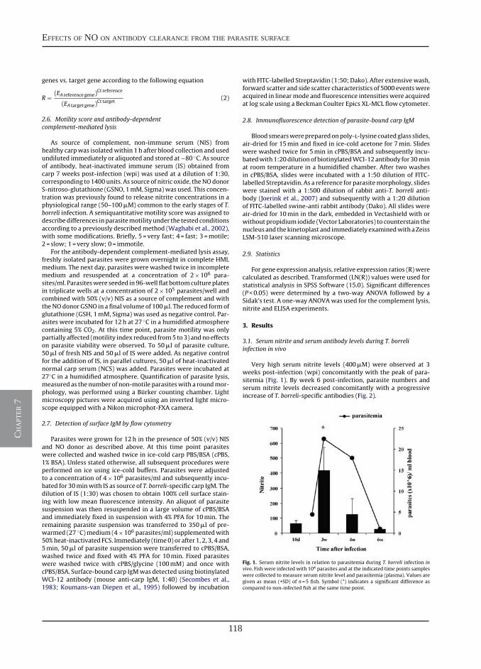
118
effects of no on antibody clearance from the parasite surface
ch
apt
er 7
Please cite this article in press as: Forlenza, M., et al., Nitric oxide hinders antibody clearance from the surface of Trypanoplasma borreli andincreases susceptibility to complement-mediated lysis. Mol. Immunol. (2009), doi:10.1016/j.molimm.2009.08.011
ARTICLE IN PRESSGModel
MIMM-3233; No.of Pages10
4 M. Forlenza et al. / Molecular Immunology xxx (2009) xxx–xxx
genes vs. target gene according to the following equation
R = (EA reference gene)Ct reference
(EA target gene)Ct target
(2)
2.6. Motility score and antibody-dependentcomplement-mediated lysis
As source of complement, non-immune serum (NIS) fromhealthy carpwas isolatedwithin 1h after blood collection and usedundiluted immediately or aliquoted and stored at−80 ◦C. As sourceof antibody, heat-inactivated immune serum (IS) obtained fromcarp 7 weeks post-infection (wpi) was used at a dilution of 1:30,corresponding to 1400units. As source of nitric oxide, theNOdonorS-nitroso-glutathione (GSNO, 1mM, Sigma)was used. This concen-tration was previously found to release nitrite concentrations in aphysiological range (50–100�M) common to the early stages of T.borreli infection. A semiquantitative motility score was assigned todescribedifferences inparasitemotilityunder the testedconditionsaccording to a previously describedmethod (Waghabi et al., 2002),with some modifications. Briefly, 5 = very fast; 4 = fast; 3 =motile;2 = slow; 1=very slow; 0= immotile.
For the antibody-dependent complement-mediated lysis assay,freshly isolated parasites were grown overnight in complete HMLmedium. The next day, parasites were washed twice in incompletemedium and resuspended at a concentration of 2×10⁶ para-sites/ml. Parasiteswere seeded in96-well flat bottomcultureplatesin triplicate wells at a concentration of 2×10⁵ parasites/well andcombined with 50% (v/v) NIS as a source of complement and withtheNOdonor GSNO in a final volume of 100�l. The reduced formofglutathione (GSH, 1mM, Sigma) was used as negative control. Par-asites were incubated for 12h at 27 ◦C in a humidified atmospherecontaining 5% CO2. At this time point, parasite motility was onlypartially affected (motility index reduced from5 to3) andnoeffectson parasite viability were observed. To 50�l of parasite culture,50�l of fresh NIS and 50�l of IS were added. As negative controlfor the addition of IS, in parallel cultures, 50�l of heat-inactivatednormal carp serum (NCS) was added. Parasites were incubated at27◦C in a humidified atmosphere. Quantification of parasite lysis,measured as the number of non-motile parasiteswith a roundmor-phology, was performed using a Bürker counting chamber. Lightmicroscopy pictures were acquired using an inverted light micro-scope equipped with a Nikon microphot-FXA camera.
2.7. Detection of surface IgM by flow cytometry
Parasites were grown for 12h in the presence of 50% (v/v) NISand NO donor as described above. At this time point parasiteswere collected and washed twice in ice-cold carp PBS/BSA (cPBS,1% BSA). Unless stated otherwise, all subsequent procedures wereperformed on ice using ice-cold buffers. Parasites were adjustedto a concentration of 4×10⁶ parasites/ml and subsequently incu-bated for 30minwith IS as source of T. borreli-specific carp IgM. Thedilution of IS (1:30) was chosen to obtain 100% cell surface stain-ing with low mean fluorescence intensity. An aliquot of parasitesuspension was then resuspended in a large volume of cPBS/BSAand immediately fixed in suspension with 4% PFA for 10min. Theremaining parasite suspension was transferred to 350�l of pre-warmed (27 ◦C)medium (4×10⁶ parasites/ml) supplementedwith50%heat-inactivated FCS. Immediately (time0) or after 1, 2, 3, 4 and5min, 50�l of parasite suspension were transferred to cPBS/BSA,washed twice and fixed with 4% PFA for 10min. Fixed parasiteswere washed twice with cPBS/glycine (100mM) and once withcPBS/BSA. Surface-bound carp IgMwas detected using biotinylatedWCI-12 antibody (mouse anti-carp IgM, 1:40) (Secombes et al.,1983; Koumans-van Diepen et al., 1995) followed by incubation
with FITC-labelled Streptavidin (1:50; Dako). After extensivewash,forward scatter and side scatter characteristics of 5000 eventswereacquired in linearmode and fluorescence intensitieswere acquiredat log scale using a Beckman Coulter Epics XL-MCL flow cytometer.
2.8. Immunofluorescence detection of parasite-bound carp IgM
Bloodsmearswerepreparedonpoly-l-lysinecoatedglass slides,air-dried for 15min and fixed in ice-cold acetone for 7min. Slideswere washed twice for 5min in cPBS/BSA and subsequently incu-batedwith1:20dilutionofbiotinylatedWCI-12antibody for30minat room temperature in a humidified chamber. After two washesin cPBS/BSA, slides were incubated with a 1:50 dilution of FITC-labelled Streptavidin. As a reference for parasitemorphology, slideswere stained with a 1:500 dilution of rabbit anti-T. borreli anti-body (Joerink et al., 2007) and subsequently with a 1:20 dilutionof FITC-labelled swine-anti rabbit antibody (Dako). All slides wereair-dried for 10min in the dark, embedded in Vectashield with orwithout propidium iodide (Vector Laboratories) to counterstain thenucleusand thekinetoplast and immediately examinedwithaZeissLSM-510 laser scanning microscope.
2.9. Statistics
For gene expression analysis, relative expression ratios (R) werecalculated as described. Transformed (LN(R)) values were used forstatistical analysis in SPSS Software (15.0). Significant differences(P<0.05) were determined by a two-way ANOVA followed by aSidak’s test. A one-way ANOVAwas used for the complement lysis,nitrite and ELISA experiments.
3. Results
3.1. Serum nitrite and serum antibody levels during T. borreliinfection in vivo
Very high serum nitrite levels (400�M) were observed at 3weeks post-infection (wpi) concomitantly with the peak of para-sitemia (Fig. 1). By week 6 post-infection, parasite numbers andserum nitrite levels decreased concomitantly with a progressiveincrease of T. borreli-specific antibodies (Fig. 2).
Fig. 1. Serum nitrite levels in relation to parasitemia during T. borreli infection invivo. Fish were infected with 104 parasites and at the indicated time points sampleswere collected to measure serum nitrite level and parasitemia (plasma). Values aregiven as mean (+SD) of n=5 fish. Symbol (*) indicates a significant difference ascompared to non-infected fish at the same time point.
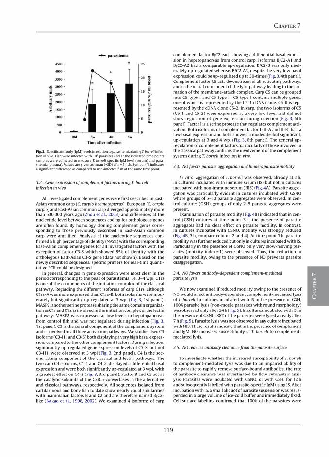
119
chapter 7
ch
apt
er 7
Please cite this article in press as: Forlenza, M., et al., Nitric oxide hinders antibody clearance from the surface of Trypanoplasma borreli andincreases susceptibility to complement-mediated lysis. Mol. Immunol. (2009), doi:10.1016/j.molimm.2009.08.011
ARTICLE IN PRESSGModel
MIMM-3233; No.of Pages10
M. Forlenza et al. / Molecular Immunology xxx (2009) xxx–xxx 5
Fig. 2. Specific antibody (IgM) levels in relation toparasitemiaduring T. borreli infec-tion in vivo. Fish were infected with 104 parasites and at the indicated time pointssamples were collected to measure T. borreli-specific IgM level (serum) and para-sitemia (plasma). Values are given as mean (+SD) of n=5 fish. Symbol (*) indicatesa significant difference as compared to non-infected fish at the same time point.
3.2. Gene expression of complement factors during T. borreliinfection in vivo
All investigated complement genes were first described in East-Asian common carp (C. carpio haematopterus). European (C. carpiocarpio) and East-Asian common carp diverged approximatelymorethan 500,000 years ago (Zhou et al., 2003) and differences at thenucleotide level between sequences coding for orthologous genesare often found. By homology cloning complement genes corre-sponding to those previously described in East-Asian commoncarp were amplified. Analysis of the nucleotide sequences con-firmed a high percentage of identity (>95%)with the correspondingEast-Asian complement genes for all investigated factors with theexception of factor C3-S which showed 85% of identity with theorthologous East-Asian C3-S gene (data not shown). Based on thenewly described sequences, specific primers for real-time quanti-tative PCR could be designed.
In general, changes in gene expression were most clear in theperiod corresponding to the peak of parasitemia, i.e. 3–4 wpi. C1rsis one of the components of the initiation complex of the classicalpathway. Regarding the different isoforms of carp C1rs, althoughC1rs-A was more expressed than C1rs-B, both isoforms were mod-erately but significantly up-regulated at 3 wpi (Fig. 3, 1st panel).MASP2, another serineprotease sharing the samedomainorganiza-tionasC1r andC1s, is involved in the initiation complexof the lectinpathway. MASP2 was expressed at low levels in hepatopancreasfrom control fish and was not regulated during infection (Fig. 3,1st panel). C3 is the central component of the complement systemand is involved in all three activation pathways.We studied two C3isoforms (C3-H1andC3-S) bothdisplayingaveryhighbasal expres-sion, compared to the other complement factors. During infection,significantly up-regulated gene expression levels of C3-S, but notC3-H1, were observed at 3 wpi (Fig. 3, 2nd panel). C4 is the sec-ond acting component of the classical and lectin pathways. Thetwo carp C4 isoforms, C4-1 and C4-2, displayed a differential basalexpression and were both significantly up-regulated at 3 wpi, witha greatest effect on C4-2 (Fig. 3, 3rd panel). Factor B and C2 act asthe catalytic subunits of the C3/C5-convertases in the alternativeand classical pathways, respectively. All sequences isolated fromcartilaginous and bony fish to date show nearly equal similaritieswith mammalian factors B and C2 and are therefore named B/C2-like (Nakao et al., 1998, 2002). We examined 4 isoforms of carp
complement factor B/C2 each showing a differential basal expres-sion in hepatopancreas from control carp. Isoforms B/C2-A1 andB/C2-A2 had a comparable up-regulation, B/C2-B was only mod-erately up-regulated whereas B/C2-A3, despite the very low basalexpression, could be up-regulated up to 30-times (Fig. 3, 4th panel).Complement factor C5 acts downstream of all activating pathwaysand is the initial component of the lytic pathway leading to the for-mation of the membrane-attack complex. Carp C5 can be groupedinto C5-type I and C5-type II. C5-type I contains multiple genes,one of which is represented by the C5-1 cDNA clone. C5-II is rep-resented by the cDNA clone C5-2. In carp, the two isoforms of C5(C5-1 and C5-2) were expressed at a very low level and did notshow regulation of gene expression during infection (Fig. 3, 5thpanel). Factor I is a serine protease that regulates complement acti-vation. Both isoforms of complement factor I (fI-A and fI-B) had alow basal expression and both showed a moderate, but significant,up-regulation at 3 and 4 wpi (Fig. 3, 6th panel). The general up-regulation of complement factors, particularly of those involved inthe classical pathway confirms the involvement of the complementsystem during T. borreli infection in vivo.
3.3. NO favors parasite aggregation and hinders parasite motility
In vitro, aggregation of T. borreli was observed, already at 3h,in cultures incubated with immune serum (IS) but not in culturesincubated with non-immune serum (NIS) (Fig. 4A). Parasite aggre-gation was particularly evident in cultures incubated with GSNOwhere groups of 5–10 parasite aggregates were observed. In con-trol cultures (GSH), groups of only 2–5 parasite aggregates werepresent.
Examination of parasite motility (Fig. 4B) indicated that in con-trol (GSH) cultures at time point 3h, the presence of parasiteaggregates had no clear effect on parasite motility. In contrast,in cultures incubated with GSNO, motility was strongly reduced(Fig. 4B, 3h, compare column 2 and 4). At time point 7h, parasitemotilitywas further reduced but only in cultures incubatedwith IS.Particularly in the presence of GSNO only very slow-moving par-asites (motility index=1) were observed. Thus, the reduction inparasite motility, owing to the presence of NO prevents parasitedisaggregation.
3.4. NO favors antibody-dependent complement-mediatedparasite lysis
We now examined if reduced motility owing to the presence ofNO would affect antibody-dependent complement-mediated lysisof T. borreli. In cultures incubated with IS in the presence of GSH,100% parasite lysis (non-motile parasites with round morphology)was observedonly after 24h (Fig. 5). In cultures incubatedwith IS inthe presence of GSNO, 88% of the parasites were lysed already after7h (Fig. 5). Parasite lysis was not observed in any culture incubatedwithNIS. These results indicate that in the presence of complementand IgM, NO increases susceptibility of T. borreli to complement-mediated lysis.
3.5. NO reduces antibody clearance from the parasite surface
To investigate whether the increased susceptibility of T. borrelito complement-mediated lysis was due to an impaired ability ofthe parasite to rapidly remove surface-bound antibodies, the rateof antibody clearance was investigated by flow cytometric anal-ysis. Parasites were incubated with GSNO, or with GSH, for 12hand subsequently labelledwith parasite-specific IgMusing IS. Afterincubationwith IS, a small aliquot of parasite suspensionwas resus-pended in a large volume of ice-cold buffer and immediately fixed.Cell surface labelling confirmed that 100% of the parasites were
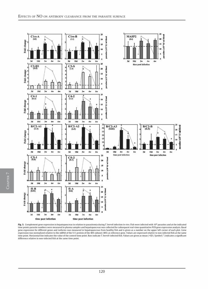
120
effects of no on antibody clearance from the parasite surface
ch
apt
er 7
Please cite this article in press as: Forlenza, M., et al., Nitric oxide hinders antibody clearance from the surface of Trypanoplasma borreli andincreases susceptibility to complement-mediated lysis. Mol. Immunol. (2009), doi:10.1016/j.molimm.2009.08.011
ARTICLE IN PRESSGModel
MIMM-3233; No.of Pages10
6 M. Forlenza et al. / Molecular Immunology xxx (2009) xxx–xxx
Fig. 3. Complement gene expression in hepatopancreas in relation to parasitemia during T. borreli infection in vivo. Fish were infected with 104 parasites and at the indicatedtime points parasite numbersweremeasured in plasma samples and hepatopancreaswas collected for subsequent real-time quantitative PCR gene expression analysis. Basalgene expression for different genes and isoforms was measured in hepatopancreas from healthy fish and is given as a number on the upper left corner of each plot. Geneexpression was normalized relative to the mRNA of the S11 protein of the 40S subunit (40S) as reference gene. Values are expressed relative to non-infected fish at the sametime point. Horizontal line indicates the value of the control time point. Bars indicate T. borreli-infected fish. Values are given asmean (+SD). Symbol (*) indicates a significantdifference relative to non-infected fish at the same time point.
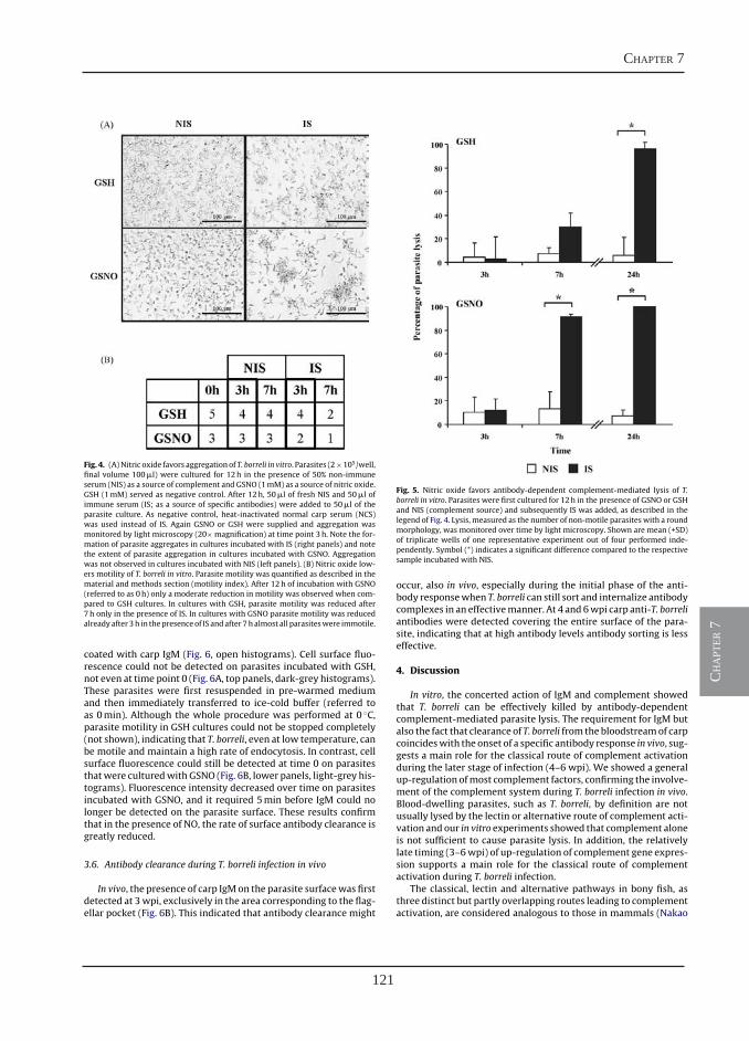
121
chapter 7
ch
apt
er 7
Please cite this article in press as: Forlenza, M., et al., Nitric oxide hinders antibody clearance from the surface of Trypanoplasma borreli andincreases susceptibility to complement-mediated lysis. Mol. Immunol. (2009), doi:10.1016/j.molimm.2009.08.011
ARTICLE IN PRESSGModel
MIMM-3233; No.of Pages10
M. Forlenza et al. / Molecular Immunology xxx (2009) xxx–xxx 7
Fig. 4. (A)Nitric oxide favors aggregation of T. borreli in vitro. Parasites (2×10⁵/well,final volume 100�l) were cultured for 12h in the presence of 50% non-immuneserum (NIS) as a source of complement and GSNO (1mM) as a source of nitric oxide.GSH (1mM) served as negative control. After 12h, 50�l of fresh NIS and 50�l ofimmune serum (IS; as a source of specific antibodies) were added to 50�l of theparasite culture. As negative control, heat-inactivated normal carp serum (NCS)was used instead of IS. Again GSNO or GSH were supplied and aggregation wasmonitored by light microscopy (20× magnification) at time point 3h. Note the for-mation of parasite aggregates in cultures incubated with IS (right panels) and notethe extent of parasite aggregation in cultures incubated with GSNO. Aggregationwas not observed in cultures incubated with NIS (left panels). (B) Nitric oxide low-ers motility of T. borreli in vitro. Parasite motility was quantified as described in thematerial and methods section (motility index). After 12h of incubation with GSNO(referred to as 0h) only a moderate reduction in motility was observed when com-pared to GSH cultures. In cultures with GSH, parasite motility was reduced after7h only in the presence of IS. In cultures with GSNO parasite motility was reducedalreadyafter 3h in thepresenceof IS andafter 7halmost all parasiteswere immotile.
coated with carp IgM (Fig. 6, open histograms). Cell surface fluo-rescence could not be detected on parasites incubated with GSH,not even at time point 0 (Fig. 6A, top panels, dark-grey histograms).These parasites were first resuspended in pre-warmed mediumand then immediately transferred to ice-cold buffer (referred toas 0min). Although the whole procedure was performed at 0 ◦C,parasite motility in GSH cultures could not be stopped completely(not shown), indicating that T. borreli, even at low temperature, canbe motile and maintain a high rate of endocytosis. In contrast, cellsurface fluorescence could still be detected at time 0 on parasitesthatwere culturedwith GSNO (Fig. 6B, lower panels, light-grey his-tograms). Fluorescence intensity decreased over time on parasitesincubated with GSNO, and it required 5min before IgM could nolonger be detected on the parasite surface. These results confirmthat in the presence of NO, the rate of surface antibody clearance isgreatly reduced.
3.6. Antibody clearance during T. borreli infection in vivo
In vivo, the presence of carp IgMon the parasite surfacewas firstdetected at 3wpi, exclusively in the area corresponding to the flag-ellar pocket (Fig. 6B). This indicated that antibody clearance might
Fig. 5. Nitric oxide favors antibody-dependent complement-mediated lysis of T.borreli in vitro. Parasites were first cultured for 12h in the presence of GSNO or GSHand NIS (complement source) and subsequently IS was added, as described in thelegend of Fig. 4. Lysis, measured as the number of non-motile parasiteswith a roundmorphology, was monitored over time by light microscopy. Shown are mean (+SD)of triplicate wells of one representative experiment out of four performed inde-pendently. Symbol (*) indicates a significant difference compared to the respectivesample incubated with NIS.
occur, also in vivo, especially during the initial phase of the anti-body responsewhen T. borreli can still sort and internalize antibodycomplexes in an effectivemanner. At 4 and 6wpi carp anti-T. borreliantibodies were detected covering the entire surface of the para-site, indicating that at high antibody levels antibody sorting is lesseffective.
4. Discussion
In vitro, the concerted action of IgM and complement showedthat T. borreli can be effectively killed by antibody-dependentcomplement-mediated parasite lysis. The requirement for IgM butalso the fact that clearance of T. borreli from the bloodstreamof carpcoincideswith the onset of a specific antibody response in vivo, sug-gests a main role for the classical route of complement activationduring the later stage of infection (4–6 wpi). We showed a generalup-regulation ofmost complement factors, confirming the involve-ment of the complement system during T. borreli infection in vivo.Blood-dwelling parasites, such as T. borreli, by definition are notusually lysed by the lectin or alternative route of complement acti-vation and our in vitro experiments showed that complement aloneis not sufficient to cause parasite lysis. In addition, the relativelylate timing (3–6wpi) of up-regulation of complement gene expres-sion supports a main role for the classical route of complementactivation during T. borreli infection.
The classical, lectin and alternative pathways in bony fish, asthree distinct but partly overlapping routes leading to complementactivation, are considered analogous to those in mammals (Nakao
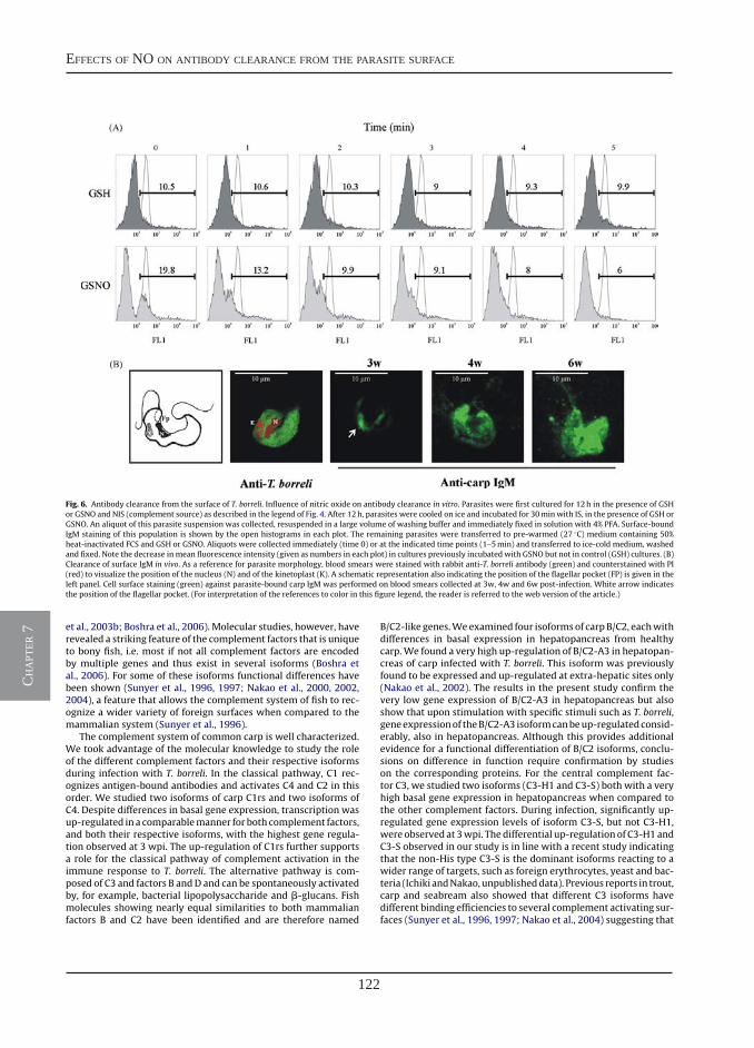
122
effects of no on antibody clearance from the parasite surface
ch
apt
er 7
Please cite this article in press as: Forlenza, M., et al., Nitric oxide hinders antibody clearance from the surface of Trypanoplasma borreli andincreases susceptibility to complement-mediated lysis. Mol. Immunol. (2009), doi:10.1016/j.molimm.2009.08.011
ARTICLE IN PRESSGModel
MIMM-3233; No.of Pages10
8 M. Forlenza et al. / Molecular Immunology xxx (2009) xxx–xxx
Fig. 6. Antibody clearance from the surface of T. borreli. Influence of nitric oxide on antibody clearance in vitro. Parasites were first cultured for 12h in the presence of GSHor GSNO and NIS (complement source) as described in the legend of Fig. 4. After 12h, parasites were cooled on ice and incubated for 30min with IS, in the presence of GSH orGSNO. An aliquot of this parasite suspension was collected, resuspended in a large volume of washing buffer and immediately fixed in solution with 4% PFA. Surface-boundIgM staining of this population is shown by the open histograms in each plot. The remaining parasites were transferred to pre-warmed (27 ◦C) medium containing 50%heat-inactivated FCS and GSH or GSNO. Aliquots were collected immediately (time 0) or at the indicated time points (1–5min) and transferred to ice-cold medium, washedand fixed. Note the decrease in mean fluorescence intensity (given as numbers in each plot) in cultures previously incubated with GSNO but not in control (GSH) cultures. (B)Clearance of surface IgM in vivo. As a reference for parasite morphology, blood smears were stained with rabbit anti-T. borreli antibody (green) and counterstained with PI(red) to visualize the position of the nucleus (N) and of the kinetoplast (K). A schematic representation also indicating the position of the flagellar pocket (FP) is given in theleft panel. Cell surface staining (green) against parasite-bound carp IgM was performed on blood smears collected at 3w, 4w and 6w post-infection. White arrow indicatesthe position of the flagellar pocket. (For interpretation of the references to color in this figure legend, the reader is referred to the web version of the article.)
et al., 2003b; Boshra et al., 2006). Molecular studies, however, haverevealed a striking feature of the complement factors that is uniqueto bony fish, i.e. most if not all complement factors are encodedby multiple genes and thus exist in several isoforms (Boshra etal., 2006). For some of these isoforms functional differences havebeen shown (Sunyer et al., 1996, 1997; Nakao et al., 2000, 2002,2004), a feature that allows the complement system of fish to rec-ognize a wider variety of foreign surfaces when compared to themammalian system (Sunyer et al., 1996).
The complement system of common carp is well characterized.We took advantage of the molecular knowledge to study the roleof the different complement factors and their respective isoformsduring infection with T. borreli. In the classical pathway, C1 rec-ognizes antigen-bound antibodies and activates C4 and C2 in thisorder. We studied two isoforms of carp C1rs and two isoforms ofC4. Despite differences in basal gene expression, transcription wasup-regulated in a comparablemanner for both complement factors,and both their respective isoforms, with the highest gene regula-tion observed at 3 wpi. The up-regulation of C1rs further supportsa role for the classical pathway of complement activation in theimmune response to T. borreli. The alternative pathway is com-posed of C3 and factors B andD and can be spontaneously activatedby, for example, bacterial lipopolysaccharide and �-glucans. Fishmolecules showing nearly equal similarities to both mammalianfactors B and C2 have been identified and are therefore named
B/C2-like genes.Weexamined four isoformsof carpB/C2, eachwithdifferences in basal expression in hepatopancreas from healthycarp.We found a very high up-regulation of B/C2-A3 in hepatopan-creas of carp infected with T. borreli. This isoform was previouslyfound to be expressed and up-regulated at extra-hepatic sites only(Nakao et al., 2002). The results in the present study confirm thevery low gene expression of B/C2-A3 in hepatopancreas but alsoshow that upon stimulation with specific stimuli such as T. borreli,geneexpressionof theB/C2-A3 isoformcanbeup-regulatedconsid-erably, also in hepatopancreas. Although this provides additionalevidence for a functional differentiation of B/C2 isoforms, conclu-sions on difference in function require confirmation by studieson the corresponding proteins. For the central complement fac-tor C3, we studied two isoforms (C3-H1 and C3-S) both with a veryhigh basal gene expression in hepatopancreas when compared tothe other complement factors. During infection, significantly up-regulated gene expression levels of isoform C3-S, but not C3-H1,were observed at 3wpi. Thedifferential up-regulation of C3-H1 andC3-S observed in our study is in line with a recent study indicatingthat the non-His type C3-S is the dominant isoforms reacting to awider range of targets, such as foreign erythrocytes, yeast and bac-teria (Ichiki andNakao,unpublisheddata). Previous reports in trout,carp and seabream also showed that different C3 isoforms havedifferent binding efficiencies to several complement activating sur-faces (Sunyer et al., 1996, 1997; Nakao et al., 2004) suggesting that
123
chapter 7
ch
apt
er 7
Please cite this article in press as: Forlenza, M., et al., Nitric oxide hinders antibody clearance from the surface of Trypanoplasma borreli andincreases susceptibility to complement-mediated lysis. Mol. Immunol. (2009), doi:10.1016/j.molimm.2009.08.011
ARTICLE IN PRESSGModel
MIMM-3233; No.of Pages10
M. Forlenza et al. / Molecular Immunology xxx (2009) xxx–xxx 9
C3 isoforms in teleost fishmay have different substrate specificitiesof the thioester.
In mammals the main mechanism that is believed to medi-ate control of trypanosome infections is the antibody-mediatedphagocytosis of opsonized parasites by liver macrophages, includ-ing Kupffer cells (Shi et al., 2004; Magez et al., 2006; Baral et al.,2007). In contrast, the presence of macrophages is not crucial forthe clearance of infections with T. borreli. Carp infected with T.borreli but depleted of macrophages by intraperitoneal injectionof clodronate-liposomes showed only a minor increase in para-sitaemia (Saeij et al., 2003b).We believe that in T. borreli infections,at least during the late phase of infection (>3 wpi), antibody-dependent complement-mediated lysis plays an important role inparasite clearance.
T. borreli infections of carp are characterized by anemia,splenomegaly and polyclonal B cell activation, all features incommon with African trypanosome infections of mammals. Theability to induce extremely elevated serum nitrite levels, however,appears a typical feature of T. borreli infections. Elevated nitritelevels result in tissue nitration and inflammation (Forlenza et al.,2008a) whereas administration, in vivo, of an iNOS inhibitor wasbeneficial to the host owing to lower parasitemia levels and highersurvival (Saeij et al., 2002). In vitro, high concentrations of NOwerefound to inhibit lymphocyte proliferation and to affect parasitemotility only, and never viability (Saeij et al., 2002, 2003c; Forlenzaet al., 2008a). Hence, all evidence so far suggested that the highNO-inducing ability of T. borreli would be a strategy of the parasite toimmunosuppress and thus evade the host immune system.
In the present study we examined in more detail the effects ofNO on parasite motility, re-considering the high induction of NOduring infections with T. borreli also as a host defense mechanism.We recreated, in vitro, a physiological situation corresponding tothe early stages of infection when serum complement factors arenot limiting, serum nitrite concentrations are still moderate andthehost is initiating a specific antibody response.Our results clearlyindicate thatwhile incubation of T. borreli in thepresence of specificantibodies and complement leads to parasite lysis within a periodof 24h, incubation under the same conditions but in the presenceof NO leads to complete parasite lysis within 7h. In mammals, acrucial role for NO in parasite control has been described duringT. congolense infections where NO in combination with IFN-� andIgG2a/3 is a determining factor for the resolution of the infection(Magez et al., 2006, 2007). However, a direct effect ofNOon thepar-asite itself has not been reported. It would therefore be interestingto investigate whether the NO-mediated parasite killing mecha-nism observed during T. congolense infection can be explained bythe ability of NO to affect parasite motility and consequently theremoval of surface-bound IgG complexes.
All African trypanosomes have developed immune evasionstrategies that rely on antigenic variation, immunosuppression,and on the rapid removal of surface-bound antibodies by hydro-dynamic sorting (McLintock et al., 1993; Donelson et al., 1998;Engstler et al., 2007). While scrutinizing the effects of NO on para-sitemotilitywemeasured an increased susceptibility of T. borreli toantibody-dependent complement-mediated lysis in the presenceof NO, leading us to investigate whether NOwould indirectly affectthe ability of T. borreli to remove surface-bound antibody com-plexes. Flow cytometric analysis confirmed that NO, by hinderingmotility, affects the ability of T. borreli to remove IgM from its sur-face. Incubation with NO increased the rate of antibody clearancefrom a half-time of seconds to a half-time of minutes, suggestingthat host-derived NO can interfere with the immune evasion strat-egy of the parasite. We could also confirm that, during the earlystages of infection, surface-bound antibodies are located almostexclusively at the level of the flagellar pocket of these parasites,indicating that antibody sorting might occur on the surface of T.
borrelialso in vivo. Antibodyremovalmustbecontinuous, andclear-ance of trypanosomes can only occur when the host is producingimmunoglobulins faster than the trypanosomes can degrade them.Carp IgM, once taken up in the flagellar pocket is likely transportedto the endosome and digested at low pH by a cysteine proteinaseof T. borreliwith cathepsin L-like activity (Ruszczyk et al., 2008a,b).Later during T. borreli infection (>3 wpi), surface-bound IgM wasdetected on the entire cell surface of the parasite indicating thatIgM removal and degradation is less effective when antibody titersincreased.
We have shown that antibody clearance occurs also in T. bor-reli, butwhether themechanisms can be ascribed to hydrodynamicflow-mediated sorting still needs to be confirmed. The flagellum ofT. brucei is attached and runs along the body, propelling theparasitein the direction away from the flagellar pocket, causing a move-ment of immune complexes by hydrodynamic drag forces in thedirection of the flagellar pocket. Owing to the basic mechanismdescribed for hydrodynamic sorting, i.e. directional drag forces,it has also been proven that trypanosomes can more effectivelyremove IgM-VSG rather than IgG-VSG complexes, simply due tothe larger ligand size (Engstler et al., 2007). This endocytic pro-cess occurs most effectively at 37 ◦C and is completely abolished attemperatures lower than 24 ◦C. Carp do not produce IgG and areectothermic animals that generally live at temperatures between4 ◦C and 25 ◦C.We noticed, in vitro, that even at 0 ◦C antibody clear-ance from the surface of T. borreli could not be halted, suggestingthis parasite has adapted to lower temperatures. Also taking intoaccount that T. borreli (Bodonida) has two instead of one flagel-lum, comparison in amore quantitativemanner of cellularmotilityand rate of endocytosis in representatives of the Bodonida and Try-panosomatida should provide insight into the evolution of immuneevasion strategies developed by Kinetoplastida.
All together, our data demonstrate that host-derived NO inter-feres with clearance of surface IgM as a parasite immune evasionstrategy. Particularly during the early phase of an immune responseto T. borreli this allows the host to gain enough time to mount aspecific antibody response, required to develop effective antibody-dependent complement-mediated lysis of T. borreli during thelate phase of infection. On the other hand, it is essential that thehost restricts the production of NO to limit suppressive effects onlymphocyte proliferation. The homologus host–parasite infectionmodel of carp with T. borreli provides a clear example of how a bal-ance between parasite virulence and effectiveness of the immuneresponse is required for long-term persistence of an infective par-asite population as well as survival of the host.
Acknowledgements
The authors wish to thank the central fish facilities, “De Haar-Vissen,” for taking care of the carp. We also thank Anja Tavernefor her technical assistance. This work was partly supported bythe European Commission’s Improving Human Potential Programunder contract HPRN-CT-2001-00214 and by the European Com-mission through contract FP6007103 IMAQUANIM.
References
Antia, R., Nowak,M.A., Anderson, R.M., 1996.Antigenic variation and thewithin-hostdynamics of parasites. Proc. Natl. Acad. Sci. U.S.A. 93, 985–989.
Baral, T.N., De Baetselier, P., Brombacher, F., Magez, S., 2007. Control of Trypanosomaevansi infection is IgM mediated and does not require a type I inflammatoryresponse. J. Infect. Dis. 195, 1513–1520.
Boshra, H., Li, J., Sunyer, J.O., 2006. Recent advances on the complement system ofteleost fish. Fish Shellfish Immunol. 20, 239–262.
Cross, G.A., 1996. Antigenic variation in trypanosomes: secrets surface slowly. Bioes-says 18, 283–291.
Dean, S.D., Matthews, K.R., 2007. Restless gossamers: antibody clearance by hydro-dynamic flow forces generated at the surface of motile trypanosome parasites.Cell Host Microbe 2, 279–281.
124
effects of no on antibody clearance from the parasite surface
ch
apt
er 7
Please cite this article in press as: Forlenza, M., et al., Nitric oxide hinders antibody clearance from the surface of Trypanoplasma borreli andincreases susceptibility to complement-mediated lysis. Mol. Immunol. (2009), doi:10.1016/j.molimm.2009.08.011
ARTICLE IN PRESSGModel
MIMM-3233; No.of Pages10
10 M. Forlenza et al. / Molecular Immunology xxx (2009) xxx–xxx
Donelson, J.E., Hill, K.L., El-Sayed, N.M., 1998. Multiple mechanisms of immune eva-sion by African trypanosomes. Mol. Biochem. Parasitol. 91, 51–66.
Engstler, M., Pfohl, T., Herminghaus, S., Boshart, M., Wiegertjes, G., Heddergott, N.,Overath, P., 2007. Hydrodynamic flow-mediated protein sorting on the cell sur-face of trypanosomes. Cell 131, 505–515.
Forlenza, M., Scharsack, J.P., Kachamakova, N.M., Taverne-Thiele, A.J., Rombout, J.H.,Wiegertjes, G.F., 2008a. Differential contribution of neutrophilic granulocytesand macrophages to nitrosative stress in a host–parasite animal model. Mol.Immunol. 45, 3178–3189.
Forlenza, M., Walker, P.D., de Vries, B.J., Wendelaar Bonga, S.E., Wiegertjes, G.F.,2008b. Transcriptional analysis of the common carp (Cyprinus carpio L.) immuneresponse to the fish louse Argulus japonicus Thiele (Crustacea: Branchiura). FishShellfish Immunol. 25, 76–83.
Irnazarow, I., 1995. Genetic variability of Polish and Hungarian carp lines. Aquac.Res. 129, 215–219.
Joerink, M., Groeneveld, A., Ducro, B., Savelkoul, H.F., Wiegertjes, G.F., 2007. Mixedinfection with Trypanoplasma borreli and Trypanosoma carassii induces pro-tection: involvement of cross-reactive antibodies. Dev. Comp. Immunol. 31,903–915.
Kato, Y., Nakao, M., Mutsuro, J., Zarkadis, I.K., Yano, T., 2003. The complement com-ponent C5 of the common carp (Cyprinus carpio): cDNA cloning of two distinctisotypes that differ in a functional site. Immunogenetics 54, 807–815.
Koumans-van Diepen, J.C., Egberts, E., Peixoto, B.R., Taverne, N., Rombout, J.H.,1995. B cell and immunoglobulin heterogeneity in carp (Cyprinus carpio L.); animmuno(cyto)chemical study. Dev. Comp. Immunol. 19, 97–108.
Magez, S., Radwanska, M., Drennan, M., Fick, L., Baral, T.N., Allie, N., Jacobs, M.,Nedospasov, S., Brombacher, F., Ryffel, B., De Baetselier, P., 2007. Tumor necrosisfactor (TNF) receptor-1 (TNFp55) signal transduction and macrophage-derivedsoluble TNF are crucial for nitric oxide-mediated Trypanosoma congolense para-site killing. J. Infect. Dis. 196, 954–962.
Magez, S., Radwanska,M., Drennan,M., Fick, L., Baral, T.N., Brombacher, F., Baetselier,P.D., 2006. Interferon-� and nitric oxide in combination with antibodies are keyprotective host immune factors during Trypanosoma congolense Tc13 Infections.J. Infect. Dis. 193, 1575–1583.
McLintock, L.M., Turner, C.M., Vickerman, K., 1993. Comparison of the effects ofimmune killing mechanisms on Trypanosoma brucei parasites of slender andstumpy morphology. Parasite Immunol. 15, 475–480.
Mutsuro, J., Tanaka,N., Kato, Y., Dodds, A.W., Yano, T., Nakao,M., 2005. Twodivergentisotypes of the fourth complement component from a bony fish, the commoncarp (Cyprinus carpio). J. Immunol. 175, 4508–4517.
Nagai, T., Mutsuro, J., Kimura, M., Kato, Y., Fujiki, K., Yano, T., Nakao, M., 2000. Anovel truncated isoform of the mannose-binding lectin-associated serine pro-tease (MASP) from the common carp (Cyprinus carpio). Immunogenetics 51,193–200.
Nakao, M., Fushitani, Y., Fujiki, K., Nonaka, M., Yano, T., 1998. Two diverged com-plement factor B/C2-like cDNA sequences from a teleost, the common carp(Cyprinus carpio). J. Immunol. 161, 4811–4818.
Nakao,M.,Hisamatsu, S.,Nakahara,M.,Kato,Y., Smith, S.L., Yano, T., 2003a.Molecularcloning of the complement regulatory factor I isotypes from the common carp(Cyprinus carpio). Immunogenetics 54, 801–806.
Nakao, M., Kajiya, T., Sato, Y., Somamoto, T., Kato-Unoki, Y., Matsushita, M., Nakata,M., Fujita, T., Yano, T., 2006. Lectin pathway of bony fish complement: identifi-cation of two homologs of the mannose-binding lectin associated with MASP2in the common carp (Cyprinus carpio). J. Immunol. 177, 5471–5479.
Nakao, M., Matsumoto, M., Nakazawa,M., Fujiki, K., Yano, T., 2002. Diversity of com-plement factor B/C2 in the common carp (Cyprinus carpio): three isotypes ofB/C2-A expressed in different tissues. Dev. Comp. Immunol. 26, 533–541.
Nakao, M., Miura, C., Itoh, S., Nakahara, M., Okumura, K., Mutsuro, J., Yano, T., 2004.A complement C3 fragment equivalent to mammalian C3d from the commoncarp (Cyprinus carpio): generation in serum after activation of the alternativepathway and detection of its receptor on the lymphocyte surface. Fish ShellfishImmunol. 16, 139–149.
Nakao, M., Mutsuro, J., Nakahara, M., Kato, Y., Yano, T., 2003b. Expansion of genesencoding complement components in bony fish: biological implications of thecomplement diversity. Dev. Comp. Immunol. 27, 749–762.
Nakao,M.,Mutsuro, J., Obo,R., Fujiki, K.,Nonaka,M., Yano, T., 2000.Molecular cloningand protein analysis of divergent forms of the complement component C3 froma bony fish, the common carp (Cyprinus carpio): presence of variants lacking thecatalytic histidine. Eur. J. Immunol. 30, 858–866.
Nakao, M., Osaka, K., Kato, Y., Fujiki, K., Yano, T., 2001. Molecular cloning of the com-plement C1r/C1s/MASP2-like serine proteases from the common carp (Cyprinuscarpio). Immunogenetics 52, 255–263.
O’Beirne, C., Lowry, C.M., Voorheis, H.P., 1998. Both IgM and IgG anti-VSG anti-bodies initiate a cycle of aggregation–disaggregation of bloodstream forms ofTrypanosoma brucei without damage to the parasite. Mol. Biochem. Parasitol.91, 165–193.
Overath, P., Ruoff, J., Stierhof, Y.-D., Haag, J., Tichy, H., Dyková, I., Lom, J., 1998. Cul-tivation of bloodstream forms of Trypanosoma carassii, a common parasite offreshwater fish. Parasitol. Res. 84, 343–347.
Pfaffl,M.W., 2001.Anewmathematicalmodel for relativequantification in real-timeRT-PCR. Nucleic Acids Res. 29, e45.
Ruszczyk, A., Forlenza, M., Joerink, M., Ribeiro, C.M., Jurecka, P., Wiegertjes, G.F.,2008a. Trypanoplasma borreli cysteine proteinase activities support a conserva-tion of function with respect to digestion of host proteins in common carp. Dev.Comp. Immunol. 32, 1348–1361.
Ruszczyk, A., Forlenza, M., Savelkoul, H.F., Wiegertjes, G.F., 2008b. Molecularcloning and functional characterisation of a cathepsin l-like proteinase fromthe fish kinetoplastid parasite Trypanosoma carassii. Fish Shellfish Immunol. 24,205–214.
Saeij, J.P., de Vries, B.J., Wiegertjes, G.F., 2003a. The immune response of carp toTrypanoplasma borreli: kinetics of immune gene expression and polyclonal lym-phocyte activation. Dev. Comp. Immunol. 27, 859–874.
Saeij, J.P., Groeneveld,A., VanRooijen,N.,Haenen,O.L.,Wiegertjes,G.F., 2003b.Minoreffect of depletion of resident macrophages from peritoneal cavity on resis-tance of common carp Cyprinus carpio to blood flagellates. Dis. Aquat. Organ. 57,67–75.
Saeij, J.P., Stet, R.J., Groeneveld, A., Verburg-van Kemenade, L.B., van Muiswinkel,W.B., Wiegertjes, G.F., 2000. Molecular and functional characterization of a fishinducible-type nitric oxide synthase. Immunogenetics 51, 339–346.
Saeij, J.P., Van Muiswinkel, W.B., Groeneveld, A., Wiegertjes, G.F., 2002. Immunemodulation by fish kinetoplastid parasites: a role for nitric oxide. Parasitology124, 77–86.
Saeij, J.P., vanMuiswinkel,W.B., vandeMeent,M., Amaral, C.,Wiegertjes, G.F., 2003c.Different capacities of carp leukocytes to encounter nitric oxide-mediatedstress: a role for the intracellular reducedglutathionepool.Dev. Comp. Immunol.27, 555–568.
Scharsack, J.P., Steinhagen, D., Kleczka, C., Schmidt, J.O., Korting, W., Michael, R.D.,Leibold, W., Schuberth, H.J., 2003. The haemoflagellate Trypanoplasma borreliinduces the production of nitric oxide, which is associated with modulationof carp (Cyprinus carpio L.) leucocyte functions. Fish Shellfish Immunol. 14,207–222.
Schmid-Hempel, P., 2008. Parasite immune evasion: a momentous molecular war.Trends Ecol. Evol. 23, 318–326.
Secombes, C.J., van Groningen, J.J., Egberts, E., 1983. Separation of lymphocytesubpopulations in carp Cyprinus carpio L. by monoclonal antibodies: immuno-histochemical studies. Immunology 48, 165–175.
Shi, M., Wei, G., Pan, W., Tabel, H., 2004. Trypanosoma congolense infections:antibody-mediated phagocytosis by Kupffer cells. J. Leukoc. Biol. 76, 399–405.
Simpson, A.G.B., Stevens, J.R., Lukes, J., 2006. The evolution and diversity of kineto-plastid flagellates. Trends Parasitol. 22, 168–174.
Steinhagen, D., Hedderich, W., Skouras, A., Scharsack, J.P., Schuberth, J., Leibold, W.,Körting,W., 2000. In vitro cultivation of Trypanoplasmaborreli (protozoa: kineto-plastida), a parasite from the blood of common carp Cyprinus carpio. Dis. Aquat.Org. 41, 195–201.
Steinhagen, D., Kruse, P., Körting,W., 1989. Theparasitemia of cloned Trypanoplasmaborreli Laveran andMesnil, 1901, in laboratory-infected common carp (Cyprinuscarpio L.). J. Parasitol. 75, 685–689.
Stevens, J.R., Noyes, H.A., Schofield, C.J., Gibson, W., 2001. The molecular evolutionof Trypanosomatidae. Adv. Parasitol. 48, 1–56.
Stijlemans, B., Guilliams, M., Raes, G., Beschin, A., Magez, S., De Baetselier, P.,2007. African trypanosomosis: from immune escape and immunopathology toimmune intervention. Vet. Parasitol. 148, 3–13.
Sunyer, J.O., Tort, L., Lambris, J.D., 1997. Diversity of the third form of complement,C3, in fish: functional characterization of five forms of C3 in the diploid fishSparus aurata. Biochem. J. 326 (Pt 3), 877–881.
Sunyer, J.O., Zarkadis, I.K., Sahu,A., Lambris, J.D., 1996.Multiple formsof complementC3 in trout that differ in binding to complement activators. Proc. Natl. Acad. Sci.U.S.A. 93, 8546–8551.
Tichopad, A., Dilger, M., Schwarz, G., Pfaffl, M.W., 2003. Standardized determinationof real-time PCR efficiency from a single reaction set-up. Nucleic Acids Res. 31,e122.
Waghabi, M.C., Coutinho, C.M., Soeiro, M.N., Pereira, M.C., Feige, J.J., Keramidas, M.,Cosson, A., Minoprio, P., Van Leuven, F., Araujo-Jorge, T.C., 2002. Increased Try-panosoma cruzi invasion and heart fibrosis associated with high transforminggrowth factor beta levels in mice deficient in alpha(2)-macroglobulin. Infect.Immun. 70, 5115–5123.
Wiegertjes, G.F., Forlenza, M., Joerink, M., Scharsack, J.P., 2005. Parasite infectionsrevisited. Dev. Comp. Immunol. 29, 749–758.
Wiegertjes, G.F., Groeneveld, A., van Muiswinkel, W.B., 1995. Genetic variation insusceptibility to Trypanoplasma borreli infection in common carp (Cyprinus car-pio L.). Vet. Immunol. Immunopathol. 47, 153–161.
Wiegertjes, G.F., Stet, R.J., Van Muiswinkel, W.B., 1994. Divergent selection for anti-body production in common carp (Cyprinus carpio L.) using gynogenesis. Anim.Genet. 25, 251–257.
Zhou, J.F., Wu, Q.J., Ye, Y.Z., Tong, J.G., 2003. Genetic devergence between Cyprinuscarpio carpio and Cyprinus carpio haematopterus as assessed by mitochondrialDNA analysis, with enphasis on origin of European domestic carp. Genetica 119,93–97.
“Fishing” for antibodies to identify T cells in carp
chapter
8
Maria Forlenza, Jan H.W.M. Rombout, Anja J. Taverne-Thiele, Huub F.J. Savelkoul and Geert F. Wiegertjes
Manuscript in preparation

126
identification of t cells in carp
ch
apt
er 8
127
chapter 8
ch
apt
er 8
1
“Fishing” for antibodies to identify T cells in carp
Maria Forlenza1, Jan H.W.M. Rombout1, Anja J. Taverne-Thiele1, Huub F.J. Savelkoul1 and
Geert F. Wiegertjes1
1 Cell Biology and Immunology Group, Wageningen Institute of Animal Sciences, Wageningen University, Wageningen, The Netherlands.
Abstract The TCR complex consists of a single TCRαβ heterodimer associated with at least three CD3 dimers including γε, δε and ξξ and includes the CD4 or CD8 co-receptors. The protein tyrosine kinases Lck and ZAP-70 provide essential signals in a cascade of events which will finally lead to T cell signalling and activation. In the present study we report the cloning of a full-length mRNA sequence for carp CD3ε and partial mRNA sequences for carp Lck and ZAP-70. Gene expression of the newly described sequences was detected only in thymocytes and not in sorted B cells, granulocytes and macrophages confirming their suitability as T-cell-lineage specific marker. Despite the genetic information available for almost all components of the TCR complex, the number of antibodies available for the identification of T cells in carp is very limited. Commercially available antibodies directed against a highly conserved prolin-rich region in the cytoplasmic tail of hCD3ε, the N-terminal region of ZAP-70 and the C-terminal region of Lck were tested for their ability to specifically recognize carp T cells. The anti-hZAP-70 antibody in particular, recognized carp T cells in both immunohistochemistry and flow cytometry and a 70 kDa protein by Western blot analysis. Two antibodies directed against selected antigenic epitopes of carp CD4 and CD8α were raised in rabbits. Immunohistochemical analysis, using the anti-hCD3ε, anti-hZAP-70, anti-cCD4 and anti-cCD8α antibodies revealed a conserved picture of the structure of carp thymus. The high cell density and intense staining observed in the thymic cortex indicates the presence of double-positive CD4+/CD8+ immature T cells (CD3ε+/ZAP-70+) in the cortical region, similar to what is observed in mammals and other fish. Despite the suitability for immunohistochemistry, both anti-cCD4 and anti-cCD8 they did not show immunoreactivity to live T cells in flow cytometry. Future strategies for an improved production of monoclonal antibodies specifically recognizing carp CD4+ and CD8+ T cells, based on very recent successful attempts in other fish species, are discussed. The anti-hCD3ε and anti-hZAP-70 were used to study T cell distribution in the spleen of Trypanoplasma borreli-infected carp. We could show the presence of both enlarged B cell and T cell areas and their co-localization in the spleen tissue. Double staining revealed that B and T cells lie intermingled to each other, with no clear distinction between a B or T cell zone, especially during infection.
128
identification of t cells in carp
ch
apt
er 8
2
Introduction
Comparative genomics has shown that the
basic mechanisms responsible for genetic
diversification of immunoglobulin (Ig) and T
cell receptors, including rag-1 and rag-2
homologues, are present in all jawed
vertebrates, including fish (1). The genomic
information crucial to an understanding of the
immune system of fish, therefore, is well
developed. However, where the technological
development in mammalian immunology has
generated an array of resources in the form of
cell lines and monoclonal antibodies,
comparative immunologists still have a
considerable number of technical hurdles to
take. Especially the proper phenotypic
classification of the different leukocyte cell
types has remained a technical challenge (2-4).
Where the development of monoclonal
antibodies against the major Ig type in fish has
allowed for the recognition of IgM-bearing B
cells, the identification of the major T cell
subtypes i.e. cytotoxic T cells (CTL) and T
helper/regulatory T cells (Th), remains
problematic in fish. Maybe the oldest (1995)
and best characterized T cell-specific
monoclonal antibody (DLT15) produced
against sea bass (Dicentrarchus labrax)
peripheral T cells (5), was only recently
characterized as a putative marker for CD8+ T
cells (6). In carp (Cyprinus carpio), the
monoclonal antibodies WCL9 and WCL38
have been shown to recognize immature
cortical thymocytes and putative mucosal T
cells, respectively, (7, 8). Although these
antibodies have been proven useful for specific
research questions regarding ontogeny of the
immune system (9) and mucosal immunity, the
identification of mature, peripheral T cells in
carp that would allow for studies into the role
of cell-mediated immunity in infectious
diseases, remains problematic.
To date, homology cloning has led to the
identification of all sequences coding for T-
cell-lineage specific surface markers (Fig. 1),
including TCR(αβγδ), CD3(εγδξ), CD4 and
CD8(αβ) in several fish species. This has
allowed for studies on T cell and thymic
development in fish and several in situ
hybridization studies confirmed a general
similarity between cold- and warm-blooded
vertebrates in the development and functional
anatomy of the thymus (10-15). Also in fish,
immature T cells (rag+) localise to the thymic
cortex, whereas mature T cells (rag-) localize in
the medulla of the adult thymus (11, 15, 16).
Although it is important to respect age-
dependent difference as well as differences in T
cell development or thymus organisation
between fish species (17), T cell development
in teleost fish appears to be similar to that of
warm-blooded vertebrates.
Comparative sequence analyses clearly
indicate that the extracellular domains of
recognition molecules such as the B and T cell
receptors are poorly conserved across species.
However, the signalling domains (cytoplasmic
regions) and even more so, the signalling
molecules (transcription factors, adaptor
molecules and kinases) involved in the
initiation and progression of the signalling
cascade, generally do show a high percentage
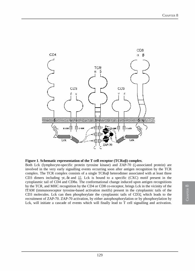
129
chapter 8
ch
apt
er 8
3
Figure 1. Schematic representation of the T cell receptor (TCRαβ) complex. Both Lck (lymphocyte-specific protein tyrosine kinase) and ZAP-70 (ξ-associated protein) are involved in the very early signalling events occurring soon after antigen recognition by the TCR complex. The TCR complex consists of a single TCRαβ heterodimer associated with at least three CD3 dimers including γε, δε and ξξ. Lck is bound to a specific (CXC) motif present in the cytoplasmic tail of CD4 and CD8α. The conformational change induced upon antigen recognitions by the TCR, and MHC recognition by the CD4 or CD8 co-receptor, brings Lck in the vicinity of the ITAM (immunoreceptor tyrosine-based activation motifs) present in the cytoplasmic tails of the CD3 molecules. Lck can then phosphorylate the cytoplasmic tails of CD3ξ which leads to the recruitment of ZAP-70. ZAP-70 activation, by either autophosphorylation or by phosphorylation by Lck, will initiate a cascade of events which will finally lead to T cell signalling and activation.

130
identification of t cells in carp
ch
apt
er 8
4
of sequence similarity across species (personal
observation). In fact, recognition molecules are
often associated with conserved signalling
cascades throughout phylogeny (18). We made
use of the general conservation of intracellular
domains and signalling molecules, first by
cloning the coding sequences for carp CD3ε,
ZAP-70 and Lck and subsequently by
identifying commercially available antibodies
that would likely recognize conserved epitopes
in the corresponding sequences of carp CD3ε,
Lck or ZAP-70 proteins.
Antibodies directed against a highly
conserved prolin-rich region in the cytoplasmic
tail of human CD3ε (hCD3ε), the N-terminal
region of ZAP-70 (hZAP-70) and the C-
terminal region of Lck (hLck) were tested for
their ability to specifically recognize
lymphocytes belonging to the T cell lineage of
carp. The antibody against hCD3ε has been
previously shown to react with blood
leukocytes in snapper (Pagrus auratus) (19),
Japanese flounder (Paralichthys olivaceus)
(20), Atlantic salmon (Salmo salar) (21) and
with T cells of the amphibian Xenopus laevis
(22). For the production of antibodies
recognizing carp CD4 (cCD4) (23) and cCD8α
(24), a different approach was used. Antigenic
peptides selected from the extracellular region
of the molecules were used for immunization of
rabbits and subsequent affinity purification of
rabbit IgG. Among the several components of
the TCR complex, CD3ε is specifically
expressed on (αβ+ or γδ+) T cells and is
therefore considered a useful pan-T cell
marker. Lck and ZAP-70 are expressed in T but
also in NK cells and are thus considered T-cell-
lineage-specific markers, CD8α is expressed on
CTL, γδ+ T cells and on some NK cell types,
whereas CD4 is expressed on T cells (Th) but
can also be expressed on monocytes (at least in
humans and rats but not in mice) (25, 26). In
addition, abnormal ZAP-70 expression is
regarded as a marker for B cell chronic
lymphocytic leukaemia (27). Therefore, the use
of only one single marker is not sufficient for
an unequivocal identification of T cell-subtypes
which requires a combination of pan-T cell and
T cell-subtype-specific antibodies.
We have previously shown that upon
infection with the extracellular parasite
Trypanoplasma borreli, extremely enlarged B
cell areas can be observed in the spleen of
heavily infected fish (28). Owing to the lack of
specific antibodies, the relative distribution and
involvement of T cells during this parasitic
infection was not investigated. Among the
panel of antibodies investigated in the present
study, the anti-hCD3ε and anti-hZAP-70
antibodies were selected for further use and
applied in combination with a monoclonal
antibody specifically recognizing IgM-bearing
B cells (WCI12) for an immunohistochemical
examination of spleen from T. borreli-infected
carp. This study confirmed the presence of not
only proliferating B cells but also T cells in the
spleen of infected fish. The evident T cell
proliferation could be indicative of polyclonal
activation similar to the polyclonal activation
observed for B cells during T. borreli
infections. In the immuno-histochemical
analysis, the enlarged T and B cell areas almost
completely overlapped and double staining
confirmed that B and T cells lie intermingled in
131
chapter 8
ch
apt
er 8
5
the spleen, particularly in infected spleen
tissue.
In this study, the cloning of new markers
for the carp T-cell-lineage, as well as the
characterization of a number of antibodies with
potential use for future comparative studies, is
reported. The overall high conservation of the
N-terminal region of ZAP-70 makes the anti-
hZAP-70 antibody highly suitable for use as a
pan T cell marker in carp. This antibody was
found highly suitable for the identification of
cells from the carp T cell lineage not only in
immunohistochemical and Western blot, but
also in flow cytometric applications. Future
strategies for an improved production of
monoclonal antibodies specifically recognizing
carp CD4+ and CD8+ T cells and development
of additional methods to detect activated T cells
in fish will be discussed.
Material and Methods
Animals
European common carp (Cyprinus carpio
carpio L.) were bred and raised to the age of
nine months in the central fish facility at
Wageningen University, The Netherlands, at
23°C in recirculating UV-treated tap water and
fed pelleted dry food (Trouvit, Nutreco) daily.
In this study, the European common carp
subspecies will be referred to as carp, unless
stated otherwise. R3xR8 carp, which are the
offspring of a cross between fish of Hungarian
origin (R8 strain) and of Polish origin (R3
strain), were used (29).
Nine-month-old carp were infected with the
parasite Trypanoplasma borreli (30) as
previously described (28). Fish were killed by
an overdose of anaesthetic 4 weeks post-
infection at parasitemia of 1 x 10⁷ parasites/ml.
Spleen was collected, immediately snap-frozen
in liquid nitrogen and stored at -80˚C for
histological analysis.
Identification of carp T cell-related cDNA
sequences: CD3ε, ZAP-70 and Lck
To identify carp CD3ε, first a degenerate
reverse primer (CD3εRV4; see Table 1) was
designed in the conserved region spanning the
transmembrane and cytoplasmic region of
known fish (Paralichthys olivaceus:
AB044572, AB044573; Takifugu rubripes:
AB166798, AB166799; Acipenser ruthenus:
AJ242941) CD3ε sequences. To obtain the carp
CD3ε 5’-end, an anchored-PCR using
CD3εRV4 in combination with λgt10FW was
performed on cDNA from a λgt10 library of
carp thymus from 5-month-old individuals
(31). The library (300 ng) was combined with 1
IU Taq polymerase (Goldstar, Eurogentec),
MgCl2 (1.5 mM), dNTPs (200 M), primers
(400 nM each), 5 µl 10x Buffer and water up to
a final volume of 50 l. The PCR was
performed using a GeneAmp PCR system 9700
(PE Applied Biosystems) under the following
conditions: 4 min at 94ºC, followed by 35
cycles of 30s at 94ºC, 30s at 50ºC and 2 min at
72ºC, finally an extension step of 7 min at 72ºC
was performed. The reaction gave a PCR
product of 287 bp including the start codon and
the 5’ untranslated region (UTR) and was used
to design the specific primer cycaCD3εFW3.
Under similar PCR conditions, cycaCD3εFW3
was used in combination with λgt10RV to
extend the 3’-end of the sequence. The
132
identification of t cells in carp
ch
apt
er 8
6
obtained PCR product of 300 bp included the
stop codon and the 3’ UTR. The full-length
coding sequence of 549 bp was confirmed
using the specific primers cycaCD3εFW0 and
cycaCD3εRV0.
To identify carp ZAP-70, first a forward
and reverse primer were designed based on a
known zebrafish sequence (NP_001018425) in
areas showing the highest percentage of
identity to human and mouse ZAP-70. RNA (1
g) from whole thymus was used as template in
a Reverse Transcriptase-PCR (RT-PCR) using
the primers ZAP70FW1 in combination with
ZAP70RV1 (Table 1). RT-PCR was performed
using the SuperScript™ One-Step RT-PCR
with Platinum® Taq kit (Invitrogen). Total
RNA (722.1 ng/l) was combined with 2x
Reaction Mix, RNaseOUTTM (40 U/l)
(Invitrogen) primers (400 nM each) and water
up to a final volume of 30 l. The PCR was
performed under the following conditions: 30
min at 50°C and 2 min at 95°C, followed by
25-45 cycles of 30 s at 94°C, 30 s at 50-55°C
and 1-2 min at 72°C, finally an extension step
of 7 min at 72°C was performed. Products were
purified and 5 µl was used as template for a
second PCR using nested primers ZAP70FW2
and ZAP70RV2. The obtained product showed
the expected size of 634 bp.
To identify carp Lck, a forward and reverse
primer were designed based on a known
zebrafish sequence (NM_001001596) in areas
showing the highest percentage of identity to
human and mouse Lck. RNA (1 g) from
whole thymus was used as template in an RT-
PCR using the primers LckFW3 in combination
with LckRV1 (Table 1). The obtained product
showed the expected size of 830 bp.
All amplified products were ligated into
pGEM-Teasy vector and cloned in JM109
competent cells using the pGEM-T Easy kit
(Promega) according to standard protocols.
Positive clones were selected by blue-white
colony screening on agar plates containing 50
g/ml Ampicillin. At least 8 clones for each
PCR product were selected for subsequent
colony-PCR using T7 and SP6 plasmid-specific
primers. A standard PCR program was
performed, using the GoldStar® DNA
polymerase. From each PCR product both
strands were sequenced using the ABI Prism
Big Dye Terminator Cycle Sequencing Ready
Reaction kit (Applied Biosystems) and
analysed using a 3730 DNA analyser.
Nucleotide sequences were translated using the
ExPASy translate tool
(http://us.expasy.org/tools/dna.html) and
aligned with Clustal W
(http://www.ebi.ac.uk/clustalw) and BLAST
software
(http://www.ncbi.nlm.nih.gov/BLAST).
RNA isolation and cDNA synthesis
Total RNA was isolated from carp thymus
or carp peripheral blood leukocytes (PBL),
using the RNAeasy Mini Kit according to the
manufacturer’s (Qiagen) instructions, including
on-column DNase treatment with the RNase-
free DNase set and stored at -80ºC until use.
Prior to cDNA synthesis, to 0.25-1 g total
RNA, a second DNase treatment was
performed using DNase I, Amplification Grade
(Invitrogen). Synthesis of cDNA was

133
chapter 8
ch
apt
er 8
7
performed using random primers (300 ng) and
SuperscriptTM III First Strand Synthesis
Systems for RT-PCR (Invitrogen). A non-
reverse transcriptase control was included for
each sample. cDNA samples were further
diluted 1:5 in nuclease-free water prior to RT-
qPCR analysis.
Table 1 Primers used for identification of
carp CD3ε, ZAP-70 and Lck cDNA
Gene expression analysis by real-time
quantitative PCR (RT-qPCR)
RT-qPCR using SYBR Green I technology
was performed with Rotor-Gene™ 6000
(Corbett Research) and the Brilliant® SYBR®
Green QPCR (Stratagene) as detection
chemistry as described previously (24). Primers
used for RT-qPCR are listed in Table 2.
Fluorescence data from RT-qPCR experiments
were analyzed using Rotor-Gene version 6.0.21
software and exported to Microsoft Excel. The
cycle threshold Ct for each sample and the
reaction efficiencies (E) for each primer set
were obtained upon Comparative Quantitation
Analysis from the Rotor-Gene version 6.0.21
software. The relative expression ratio (R) of a
target gene was calculated relative to the S11
protein of the 40S subunit as reference gene
(32, 33).
Primary antibodies
Monoclonal mouse antibody WCI12 binds
to the heavy chain of IgM on carp B cells (34,
35). Monoclonal mouse antibody TCL-BE8
binds to carp neutrophilic granulocytes (strong
affinity), monocytes (low affinity) (36), and
basophilic granulocytes (intermediate affinity;
unpublished results). Monoclonal mouse
antibody WCL15 strongly reacts with the
cytoplasm of carp monocytes and macrophages
in tissue sections (37, 38). On live cells
WCL15 stains the surface of carp monocytes
and macrophages and shows a weak cross-
reactivity to thrombocytes (39). Monoclonal
mouse antibody WCL6 recognizes a 90 kDa
membrane molecule on carp thrombocytes
(40). Monoclonal rabbit antibody anti-hZAP-70
recognizes human ZAP-70 and was purchased
from Cell Signaling technology (Cat No:
99F2).
Affinity-purified polyclonal rabbit antibody
anti-hCD3ε was purchased from Dako (Cat No
Primer primers (5’→3’)
λgt10.FW TTGAGCAAGTTCAGCCTGGTTAAG
λgt10.RV GGCTTATGAGTATTTCTTCCAGGG CD3εRV4 VAHGCAGTTYTCACACACTTT cycaCD3εFW3 AGCACACCTTCTACCTCAAA cycaCD3εFW0 TTCAGGACGTTACACCATG cycaCD3εRV0 AACAGCATCTATTTATTG ZAP70FW1 CTCGGGACCTCCATCAT ZAP70RV1 ACTGTTGTCATTCTATCCTGTC ZAP70FW2 CGAGAGAGAGATGAAATGG ZAP70RV2 GGTAGAGATGGGCATGAA
LckFW3 CCAAAAACCCTGGTGGCA
LckRV1 TGCTCCTGGTACTGTCCCTCTGT
Table 2 Primers used for Real-Time quantitative PCR
Primer FW primers (5’→3’) RV primers (5’→3’) Acc. no. q40S CCGTGGGTGACATCGTTACA TCAGGACATTGAACCTCACTGTCT AB012087 qCD3ε TGAGTTGAGCGGTTTGACTG TGGCAGCACAGAAGTAGATGA - qZAP70 GGAACAAGCCATCATTAGCC GTCGTCTCTCACCCTCCTG - qLck CAAGTGGACGGCACCTGA GCAATCACCTCTGGGTTTGT -
134
identification of t cells in carp
ch
apt
er 8
8
A0452). Polyclonal rabbit antibodies anti-carp
CD4 (cCD4) and cCD8α were produced against
synthetic peptides coupled to keyhole limpet
hemocyanin (KLH), according to a 3-months
standard protocol (Eurogentec). Affinity
purification of rabbit IgG was performed
against purified peptides and specificity
assessed by ELISA (Eurogentec). For cCD4,
amino acids 254-268 (PQGADNKVKAEKRE)
were chosen for immunization. For cCD8α
amino acids 79-93 (GKVSLAIQSFNKKTD),
positioned in a region conserved between carp
CD8α1 and CD8α2, were chosen for
immunization. Both peptides were chosen in
the extracellular region of the respective
protein.
Purification of carp leukocyte subtypes
Fish were killed by an overdose of
anaesthetic (0.3 g/l Tricaine Methane Sulfonate
(TMS), Crescent Research Chemicals) in
aquarium water buffered with 0.6 g/l sodium
bicarbonate. Blood was collected by puncture
of the caudal vein and immediately diluted 1:1
with carp RPMI (cRPMI-1640 (Gibco) adjusted
to 280 mOsmol kg-1) containing 50 IU of
heparin (Leo Pharmaceutical products). Total
peripheral blood leukocytes (PBL) were
isolated by centrifugation on Ficoll-Paque™
Plus (Amersham Biosciences) as previously
described (24). Thymus and head kidney were
aseptically removed and a single cell
suspension was obtained by forcing the tissue
through a 100 µm nylon mesh. Total cell
suspensions from thymus were layered on a
1.02, 1.06, 1.0838 g/ml discontinuous Percoll
density gradient (Amersham Pharmacia
Biotech. AB) and cells at the interface between
the 1.0838-1.06 g/ml gradient layers were
collected and washed twice in cRPMI. Total
cell suspensions from head kidney were layered
on a 1.02, 1.06, 1.07, 1.0838 g/ml
discontinuous Percoll density gradient and cells
at the interface of each layer were collected,
washed twice and used as primary source for a
second purification step using Magnetic
Activated Cell Sorting (MACS) of B cells
(1.02-1.06 g/ml), macrophages (1.06-1.07 g/ml)
and granulocytes (1.07-1.838 g/ml).
For MACS, cells (1 x 10⁷/ml) were
incubated for 30 min on ice with the primary
antibodies WCI12, WCL15 or TCL-BE8 to
specifically label B cells, macrophages and
granulocytes, respectively. Cells were washed
twice with MACS-buffer (0.5% BSA in
cRPMI) and subsequently incubated for 30 min
on ice with a 1:50 dilution of Phycoerythrin
(PE)-conjugated goat-anti-mouse (GAM-PE)
secondary antibody (Dako). Cells were
extensively washed and incubated for 15 min at
4ºC with 10 µl/108 cells magnetic beads (anti-
PE Microbeads, Miltenyi Biotec). Cells were
washed, resuspended in MACS buffer and
sorted on LS-MidiMACS Columns according
to the manufacturer’s instructions. Purity of the
different carp leukocyte subtypes was assessed
by flow cytometric analysis (Beckman Coulter,
Epics XL-MCL, Miami, FL, USA).
Human Jurkat T cell line
For some experiments, the human Jurkat T
cell line (ATCC: TIB-152), was used as
positive control. Cells were cultured in RPMI-
1640 supplemented with 25 mM HEPES, 10%
135
chapter 8
ch
apt
er 8
9
(v/v) Fetal Bovine Serum (FBS), L-Glutamin (2
mM), Penicillin-G (100 IU/ml) and
Streptomycin sulphate (50 mg/l), and
maintained at a cell density of 1-1.5 x 10⁶
cells/ml.
Western blot
Carp thymocytes and magnetically-sorted B
cells, macrophages and granulocytes were
isolated as described above. Total cell lysates
from carp leukocytes or human Jurkat cells
were obtained by resuspending cell pellets in
lysis buffer (20 M Tris-Cl, pH 7.2, 100 M
NaCl, 1 M EDTA, 0.5% (v/v) Triton X-100, 1
M PMSF) at a concentration of 10 µl/ 2 x 10⁶
cells. Lysates were homogenized on ice by 6-7
passages through a 22 gauge needle and
subsequently incubated on ice for 15 min in
lysis buffer to ensure complete lysis. Lysates
were centrifuged at max speed (21000 x g) for
10 minutes at 4oC to pellet nuclei. Supernatants
were carefully collected and protein
concentration measured spectrophotometrically
(NanoDrop® ND-1000, Labtech International
Ltd.) at OD280nm. Total cell lysates (10 g) were
resolved on a 10% SDS-PAGE and proteins
transferred to nitrocellulose membranes
(Protrans, Schleicher & Schuell, Bioscience
GmbH). Membranes were incubated overnight
at 4˚C with a 1:1000 dilution of anti-hZAP-70
antibody with gentile agitation. Horseradish
peroxidase (HRP)-conjugated goat-anti-rabbit
antibody (1:2000; GAR-HRP, Dako) was used
as secondary antibody. Proteins were visualized
by chemiluminescence detection (Amersham,
ECL detection system for Western blot) on X-
ray films.
Flow cytometry
Carp PBL or carp thymocytes were
adjusted to a concentration of 1 x 10⁷ cells/ml
in MACS-buffer. Cell suspensions (100 µl)
were incubated on ice for 30 min with WCI12
(1:50), WCL15 (1:50), TCL-BE8 (1:50) or
WCL6 (1:100). After washing, cells were
incubated for 30 min on ice with a 1:50 dilution
of GAM-PE as secondary antibody. For
intracellular staining of ZAP-70 or CD3ε, cells
were first permeabilized by incubation for 15
min on ice in Cytofix/Cytoperm buffer (BD
Bioscience). After a washing step with 1x
Perm/Wash buffer (BD Bioscience), cells were
resuspended in the same buffer and incubated
for 30 min on ice with anti-hZAP-70 or anti-
hCD3ε (1:20). After washing, cells were
incubated for 30 min on ice with fluorescein
isothiocyanate (FITC)-conjugated swine-anti-
rabbit antibody (1:20; SWAR-FITC, Dako) as
secondary antibody. For double-staining, first
the procedure for extracellular staining was
performed, followed by permeabilization and
intracellular staining. After extensive washing,
fluorescence intensities of 104 events were
acquired in log scale using a Beckman Coulter
Epics XL-MCL flow cytometer.
Laser scanning microscopy
Cell surface and intracellular staining was
performed as described above. After washing,
cells were centrifuged for 5 min at 100 x g on
poly-L-lysine coated glass slides (BDH
Laboratory supplies) and subsequently fixed for
7 min in ice-cold 4% (w/v) paraformaldehyde
in 100% ethanol solution. Slides were
embedded in Vecta-Shield Mounting medium
136
identification of t cells in carp
ch
apt
er 8
10
(Vector laboratories) and cell fluorescence
examined with a laser scanning microscope
(LSM 510, Carl Zeiss, Germany). A FITC
signal was excited with a 488 nm argon laser
and detected using a band-pass filter (505-530
nm), whereas a PE signal was excited with a
543 nm helium-neon laser and detected using a
long-pass filter (585 nm).
Immunohistochemistry
Cryosections (4-7 m) of spleen tissue were
mounted on poly-L-lysine-coated glass slides,
air-dried for 30-60 min and incubated in a 0.3%
H2O2 solution in methanol for 20 min to
inactivate endogenous peroxidase. Following
steps were performed at room temperature (rT)
unless stated otherwise. Sections were washed
for 5 min with PBS, then with distilled water
and incubated for 10 min at 37°C in proteinase-
K (Promega) solution (50 µg/ml in distilled
water). Samples were fixed for 5 min at 4°C in
4% (w/v) paraformaldehyde followed by
washing first for 10 min at 4°C and then for 7
min at rT in PBS-Triton (PBS-T, 150 mM
NaCl, 2 mM KCl, 20 mM Na2HPO4, 2 mM
KH2PO4, 0.1% (v/v) Triton X-100). A blocking
solution of 5% normal goat serum (Dako) was
then added and slides were incubated for 30
min. Primary antibodies WCI12 (1:50), anti-
hCD3ε (1:50), anti-hZAP-70 (1:50), anti-cCD4
(1:10) or anti-cCD8α (1:10) were diluted in
PBS-T and slides were incubated for 1h. After
washing twice for 10 min in PBS-T, sections
were incubated for 1h with GAR-HRP or
GAM-AP (1:200 in PBS) as secondary
antibody. For single-staining with GAR-HRP
antibodies, sections were incubated for 10 min
in 0.05 M sodium acetate buffer, pH 5 and
following addition of 0.4 mg/ml 3-amino-9-
ethyl-carbazole (AEC; Sigma-Aldrich) for 25
min in 0.03% H2O2 in sodium acetate buffer.
Rinsed sections (4x in distilled water) were
embedded in Kaiser’s glycerin gelatin (Merck).
For single-staining with GAM-AP, sections
were incubated for 10 min in AP-buffer (0.1 M
Tris-Cl, 0.1 M NaCl, 0.05 M MgCl2, pH 9.5)
and then stained using AP-substrate (4.5 µl/ml
nitro-blue-tetrazoleum and 3.5 µl/ml 5’-bromo-
4’-chloro-3’-indolyl phosphatase [BCIP] all
from Roche Applied Science) diluted in AP-
buffer for 2-5 min. Rinsed sections (4x in
distilled water) were embedded in Aquatext
(Merk). For double-staining, sections were first
stained for AP and then for AEC, rinsed in
distilled water and embedded in Kaiser’s
glycerin gelatin. Sections treated identically but
without the presence of the first antibody
showed no staining.
Results
Carp CD3ε sequence analysis
Using a homology cloning approach a carp
CD3ε cDNA sequence of 581 bp covering the
complete full-length mRNA sequence was
identified. The sequence encodes for a protein
of 173 amino acids (Fig. 2) with a putative 20
aa leader peptide which upon cleavage would
release a protein of about 17 kDa. Alignment of
the translated carp sequence indicates 20-26%
overall identity with other fish CD3ε sequences
and 17-20% identity with mammalian CD3ε
sequences. Particularly the transmembrane and
cytoplasmatic regions showed a slightly higher
degree of conservation with an overall identity
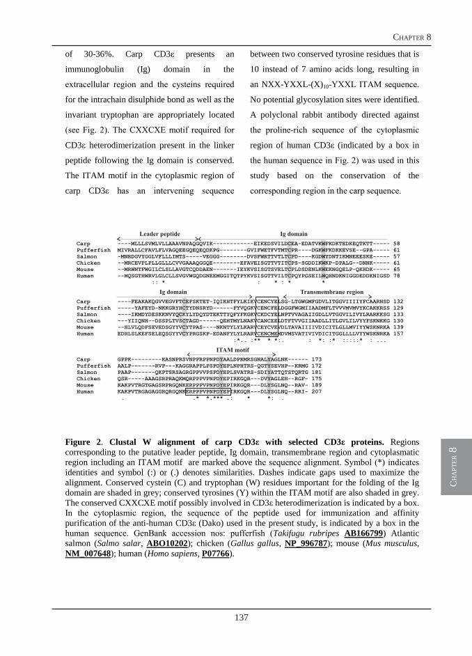
137
chapter 8
ch
apt
er 8
11
of 30-36%. Carp CD3ε presents an
immunoglobulin (Ig) domain in the
extracellular region and the cysteins required
for the intrachain disulphide bond as well as the
invariant tryptophan are appropriately located
(see Fig. 2). The CXXCXE motif required for
CD3ε heterodimerization present in the linker
peptide following the Ig domain is conserved.
The ITAM motif in the cytoplasmic region of
carp CD3ε has an intervening sequence
between two conserved tyrosine residues that is
10 instead of 7 amino acids long, resulting in
an NXX-YXXL-(X)10-YXXL ITAM sequence.
No potential glycosylation sites were identified.
A polyclonal rabbit antibody directed against
the proline-rich sequence of the cytoplasmic
region of human CD3ε (indicated by a box in
the human sequence in Fig. 2) was used in this
study based on the conservation of the
corresponding region in the carp sequence.
Figure 2. Clustal W alignment of carp CD3ε with selected CD3ε proteins. Regions corresponding to the putative leader peptide, Ig domain, transmembrane region and cytoplasmatic region including an ITAM motif are marked above the sequence alignment. Symbol (*) indicates identities and symbol (:) or (.) denotes similarities. Dashes indicate gaps used to maximize the alignment. Conserved cystein (C) and tryptophan (W) residues important for the folding of the Ig domain are shaded in grey; conserved tyrosines (Y) within the ITAM motif are also shaded in grey. The conserved CXXCXE motif possibly involved in CD3ε heterodimerization is indicated by a box. In the cytoplasmic region, the sequence of the peptide used for immunization and affinity purification of the anti-human CD3ε (Dako) used in the present study, is indicated by a box in the human sequence. GenBank accession nos: pufferfish (Takifugu rubripes AB166799) Atlantic salmon (Salmo salar, ABO10202); chicken (Gallus gallus, NP_996787); mouse (Mus musculus, NM_007648); human (Homo sapiens, P07766).
Leader peptide Ig domain
Ig domain Transmembrane region
ITAM motif
Carp ----MLLLSVMLVLLAAAVNPAQGQVIK------------EIKEDSVILDCEA-EDATVKWFKDKTEDKEQTKTT----- 58Pufferfish MIVRALLCFAVLFLVAGQEEGQEQEQDKPG--------GVIFWETFVTMTCPR----DGKWFKDKKEVSE--GPA----- 61Salmon -MNRDGVYGGLVFLLLIMTS-----VEGGG--------DVSFWRTTVTLTCPD----KGDWYDNTIKMNEEESKE----- 57Chicken --MRCEVPLPLLGLLLCVVGAAAQGGQE--------EFAVEISGTTVTITCPS-SGDDIKWKP-DPALG--DNNK----- 61Mouse --MRWNTFWGILCLSLLAVGTCQDDAEN-------IEYKVSISGTSVELTCPLDSDENLKWEKNGQELP-QKHDK----- 65Human --MQSGTHWRVLGLCLLSVGVWGQDGNEEMGGITQTPYKVSISGTTVILTCPQYPGSEILWQHNDKNIGGDEDDKNIGSD 78
:: * : * : * *
Carp ----FEAKAKQGVVEGVFTCEFSKTET-IQIKHTFYLKIKVCENCYELSG-LTGWGMFGDVLITGGVIIIIYFCAARNSD 132Pufferfish -----YAFEYD-NKKGRYHCTYDNSRYD------FYVQGKVCENCFELDGGFWGMIIAADMFLTVVVMVMVYKCAKKRSS 129Salmon ----IKMDYDESKKNVYQCKYLYDQYDTEKTTYQFYFKGKVCKDCYELNPTVVAGAIIGDLLVTGGVILIVYLRARKKSG 133Chicken ---YIIQNH--DSSPLTVSCTAGD----- QEHTMYLNAKVCANCEELDTFTVVGIIAADLLITLGVLILVYYFSKNKKG 130Mouse --HLVLQDFSEVEDSGYYVCYTPAS----NKNTYLYLKARVCEYCVEVDLTAVAIIIIVDICITLGLLMVIYYWSKNRKA 139Human EDHLSLKEFSELEQSGYYVCYPRGSKP-EDANFYLYLRARVCENCMEMDVMSVATIVIVDICITGGLLLLVYYWSKNRKA 157
:*.. :** * *:. : *: :* :::::* : ...
Carp GPPK---------KASNPRSVNPPRPPNPDYAALDPKMRSGNALYAGLNK------ 173Pufferfish AALP-------RVP---KAGGRAPPLPSPDYEPLNPHTRS-QGTYSEVHP--KRMG 172Salmon PAAP-------QKPTSRSAGRGPPVVPSPDYEPLSVATRS-SDIYATTQTSTQRTG 181Chicken QSR-----AAAGSRPRAQKMQRPPPVPNPDYEPIRKGQR---DVYAGLEH--RGF- 175Mouse KAKPVTRGTGAGSRPRGQNKERPPPVPNPDYEPIRKGQR---DLYSGLNQ--RAV- 189Human KAKPVTRGAGAGGRQRGQNKERPPPVPNPDYEPIRKGQR---DLYSGLNQ--RRI- 207
. .* *.*** .: * *: .
-
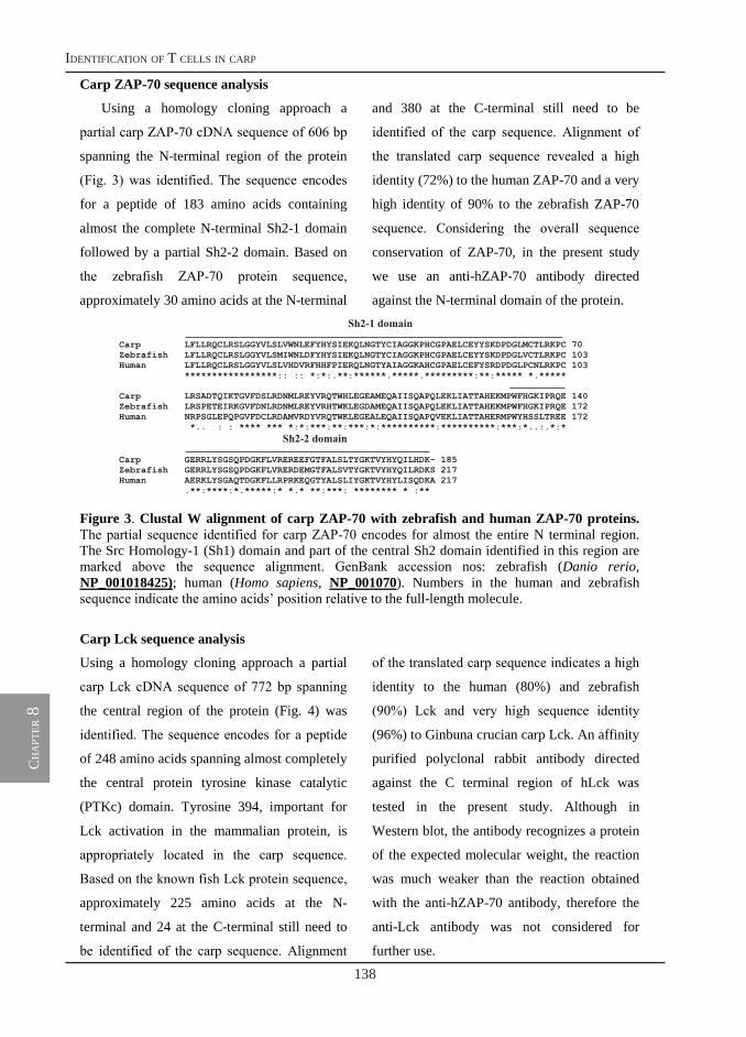
138
identification of t cells in carp
ch
apt
er 8
12
Carp ZAP-70 sequence analysis
Using a homology cloning approach a
partial carp ZAP-70 cDNA sequence of 606 bp
spanning the N-terminal region of the protein
(Fig. 3) was identified. The sequence encodes
for a peptide of 183 amino acids containing
almost the complete N-terminal Sh2-1 domain
followed by a partial Sh2-2 domain. Based on
the zebrafish ZAP-70 protein sequence,
approximately 30 amino acids at the N-terminal
and 380 at the C-terminal still need to be
identified of the carp sequence. Alignment of
the translated carp sequence revealed a high
identity (72%) to the human ZAP-70 and a very
high identity of 90% to the zebrafish ZAP-70
sequence. Considering the overall sequence
conservation of ZAP-70, in the present study
we use an anti-hZAP-70 antibody directed
against the N-terminal domain of the protein.
Figure 3. Clustal W alignment of carp ZAP-70 with zebrafish and human ZAP-70 proteins.The partial sequence identified for carp ZAP-70 encodes for almost the entire N terminal region. The Src Homology-1 (Sh1) domain and part of the central Sh2 domain identified in this region are marked above the sequence alignment. GenBank accession nos: zebrafish (Danio rerio, NP_001018425); human (Homo sapiens, NP_001070). Numbers in the human and zebrafish sequence indicate the amino acids’ position relative to the full-length molecule.
Carp Lck sequence analysis
Using a homology cloning approach a partial
carp Lck cDNA sequence of 772 bp spanning
the central region of the protein (Fig. 4) was
identified. The sequence encodes for a peptide
of 248 amino acids spanning almost completely
the central protein tyrosine kinase catalytic
(PTKc) domain. Tyrosine 394, important for
Lck activation in the mammalian protein, is
appropriately located in the carp sequence.
Based on the known fish Lck protein sequence,
approximately 225 amino acids at the N-
terminal and 24 at the C-terminal still need to
be identified of the carp sequence. Alignment
of the translated carp sequence indicates a high
identity to the human (80%) and zebrafish
(90%) Lck and very high sequence identity
(96%) to Ginbuna crucian carp Lck. An affinity
purified polyclonal rabbit antibody directed
against the C terminal region of hLck was
tested in the present study. Although in
Western blot, the antibody recognizes a protein
of the expected molecular weight, the reaction
was much weaker than the reaction obtained
with the anti-hZAP-70 antibody, therefore the
anti-Lck antibody was not considered for
further use.
Carp LFLLRQCLRSLGGYVLSLVWNLEFYHYSIEKQLNGTYCIAGGKPHCGPAELCEYYSKDPDGLMCTLRKPC 70Zebrafish LFLLRQCLRSLGGYVLSMIWNLDFYHYSIEKQLNGTYCIAGGKPHCGPAELCEYYSKDPDGLVCTLRKPC 103Human LFLLRQCLRSLGGYVLSLVHDVRFHHFPIERQLNGTYAIAGGKAHCGPAELCEFYSRDPDGLPCNLRKPC 103
*****************:: :: *:*:.**:******.*****.*********:**:***** *.*****
Carp LRSADTQIKTGVFDSLRDNMLREYVRQTWHLEGEAMEQAIISQAPQLEKLIATTAHEKMPWFHGKIPRQE 140Zebrafish LRSPETEIRKGVFDNLRDNMLREYVRHTWKLEGDAMEQAIISQAPQLEKLIATTAHEKMPWFHGKIPRQE 172Human NRPSGLEPQPGVFDCLRDAMVRDYVRQTWKLEGEALEQAIISQAPQVEKLIATTAHERMPWYHSSLTREE 172
*.. : : **** *** *:*:***:**:***:*:**********:**********:***:*..:.*:*
Carp GERRLYSGSQPDGKFLVREREEFGTFALSLTYGKTVYHYQILHDK- 185Zebrafish GERRLYSGSQPDGKFLVRERDEMGTFALSVTYGKTVYHYQILRDKS 217Human AERKLYSGAQTDGKFLLRPRKEQGTYALSLIYGKTVYHYLISQDKA 217
.**:****:*.*****:* *.* **:***: ******** * :**
Sh2-1 domain
Sh2-2 domain
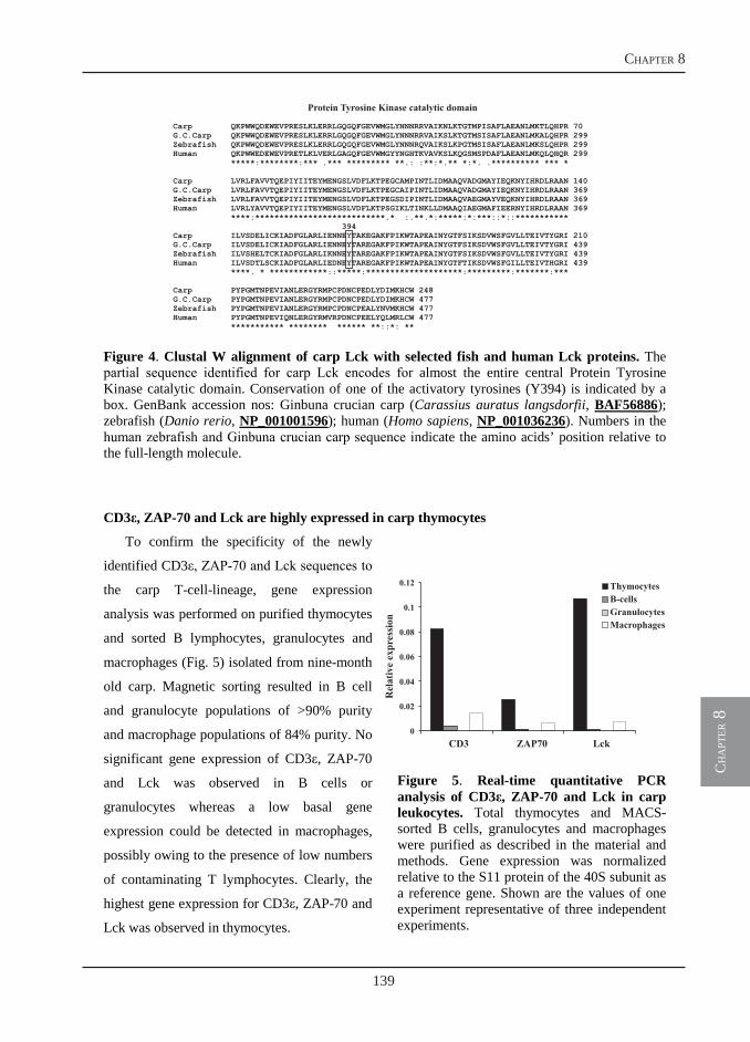
139
chapter 8
ch
apt
er 8
13
Figure 4. Clustal W alignment of carp Lck with selected fish and human Lck proteins. The partial sequence identified for carp Lck encodes for almost the entire central Protein Tyrosine Kinase catalytic domain. Conservation of one of the activatory tyrosines (Y394) is indicated by a box. GenBank accession nos: Ginbuna crucian carp (Carassius auratus langsdorfii, BAF56886);zebrafish (Danio rerio, NP_001001596); human (Homo sapiens, NP_001036236). Numbers in the human zebrafish and Ginbuna crucian carp sequence indicate the amino acids’ position relative to the full-length molecule.
CD3ε, ZAP-70 and Lck are highly expressed in carp thymocytes
To confirm the specificity of the newly
identified CD3ε, ZAP-70 and Lck sequences to
the carp T-cell-lineage, gene expression
analysis was performed on purified thymocytes
and sorted B lymphocytes, granulocytes and
macrophages (Fig. 5) isolated from nine-month
old carp. Magnetic sorting resulted in B cell
and granulocyte populations of >90% purity
and macrophage populations of 84% purity. No
significant gene expression of CD3ε, ZAP-70
and Lck was observed in B cells or
granulocytes whereas a low basal gene
expression could be detected in macrophages,
possibly owing to the presence of low numbers
of contaminating T lymphocytes. Clearly, the
highest gene expression for CD3ε, ZAP-70 and
Lck was observed in thymocytes.
Figure 5. Real-time quantitative PCR analysis of CD3ε, ZAP-70 and Lck in carp leukocytes. Total thymocytes and MACS-sorted B cells, granulocytes and macrophages were purified as described in the material and methods. Gene expression was normalized relative to the S11 protein of the 40S subunit as a reference gene. Shown are the values of one experiment representative of three independent experiments.
Carp QKPWWQDEWEVPRESLKLERRLGQGQFGEVWMGLYNNNRRVAIKNLKTGTMPISAFLAEANLMKTLQHPR 70G.C.Carp QKPWWQDEWEVPRESLKLERRLGQGQFGEVWMGLYNNNRRVAIKSLKTGTMSISAFLAEANLMKALQHPR 299Zebrafish QKPWWQDEWEVPRESLKLERRLGQGQFGEVWMGLYNNNRQVAIKSLKPGTMSISAFLAEANLMKSLQHPR 299Human QKPWWEDEWEVPRETLKLVERLGAGQFGEVWMGYYNGHTKVAVKSLKQGSMSPDAFLAEANLMKQLQHQR 299
*****:********:*** .*** ********* **.: :**:*.** *:*. .********** *** *
Carp LVRLFAVVTQEPIYIITEYMENGSLVDFLKTPEGCAMPINTLIDMAAQVADGMAYIEQKNYIHRDLRAAN 140G.C.Carp LVRLFAVVTQEPIYIITEYMENGSLVDFLKTPEGCAIPINTLIDMAAQVADGMAYIEQKNYIHRDLRAAN 369Zebrafish LVRLFAVVTQEPIYIITEYMENGSLVDFLKTPEGSDIPINTLIDMAAQVAEGMAYVEQKNYIHRDLRAAN 369Human LVRLYAVVTQEPIYIITEYMENGSLVDFLKTPSGIKLTINKLLDMAAQIAEGMAFIEERNYIHRDLRAAN 369
****:***************************.* :.**.*:*****:*:***::*::***********394
Carp ILVSDELICKIADFGLARL
FPIKWTAPEAINYGTFSIKSDVWSFGVLLTEIVTYGRI 439Human ILVSDTLSCKIADFGLARLIEDNEYTAREGAKFPIKWTAPEAINYGTFTIKSDVWSFGILLTEIVTHGRI 439
****. * ************::*****:********************:*********:*******:***
Carp PYPGMTNPEVIANLERGYRMPCPDNCPEDLYDIMKHCW 248G.C.Carp PYPGMTNPEVIANLERGYRMPCPDNCPEDLYDIMKHCW 477Zebrafish PYPGMTNPEVIANLERGYRMPCPDNCPEALYNVMKHCW 477Human PYPGMTNPEVIQNLERGYRMVRPDNCPEELYQLMRLCW 477
*********** ******** ****** **::*: **
Protein Tyrosine Kinase catalytic domain
IENNE TAKEGAKFPIKWTAPEAINYGTFSIKSDVWSFGVLLTEIVTYGRI 210G.C.Carp ILVSDELICKIADFGLARLIENNEYTAREGAKFPIKWTAPEAINYGTFSIKSDVWSFGVLLTEIVTYGRI 439Zebrafish ILVSHELTCKIADFGLARLIKNNEYTAREGAK
Y
0
0.02
0.04
0.06
0.08
0.1
0.12
CD3 ZAP70 Lck
ThymocytesB-cellsGranulocytesMacrophages
Relativeexpression
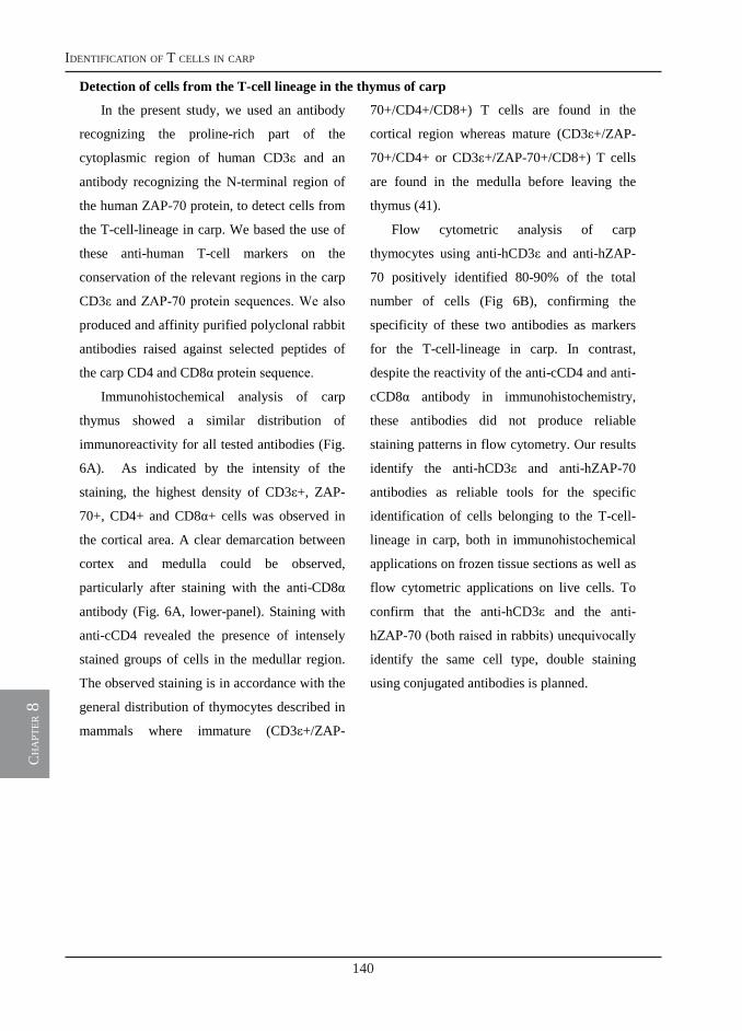
140
identification of t cells in carp
ch
apt
er 8
14
Detection of cells from the T-cell lineage in the thymus of carp
In the present study, we used an antibody
recognizing the proline-rich part of the
cytoplasmic region of human CD3ε and an
antibody recognizing the N-terminal region of
the human ZAP-70 protein, to detect cells from
the T-cell-lineage in carp. We based the use of
these anti-human T-cell markers on the
conservation of the relevant regions in the carp
CD3ε and ZAP-70 protein sequences. We also
produced and affinity purified polyclonal rabbit
antibodies raised against selected peptides of
the carp CD4 and CD8α protein sequence.
Immunohistochemical analysis of carp
thymus showed a similar distribution of
immunoreactivity for all tested antibodies (Fig.
6A). As indicated by the intensity of the
staining, the highest density of CD3ε+, ZAP-
70+, CD4+ and CD8α+ cells was observed in
the cortical area. A clear demarcation between
cortex and medulla could be observed,
particularly after staining with the anti-CD8α
antibody (Fig. 6A, lower-panel). Staining with
anti-cCD4 revealed the presence of intensely
stained groups of cells in the medullar region.
The observed staining is in accordance with the
general distribution of thymocytes described in
mammals where immature (CD3ε+/ZAP-
70+/CD4+/CD8+) T cells are found in the
cortical region whereas mature (CD3ε+/ZAP-
70+/CD4+ or CD3ε+/ZAP-70+/CD8+) T cells
are found in the medulla before leaving the
thymus (41).
Flow cytometric analysis of carp
thymocytes using anti-hCD3ε and anti-hZAP-
70 positively identified 80-90% of the total
number of cells (Fig 6B), confirming the
specificity of these two antibodies as markers
for the T-cell-lineage in carp. In contrast,
despite the reactivity of the anti-cCD4 and anti-
cCD8α antibody in immunohistochemistry,
these antibodies did not produce reliable
staining patterns in flow cytometry. Our results
identify the anti-hCD3ε and anti-hZAP-70
antibodies as reliable tools for the specific
identification of cells belonging to the T-cell-
lineage in carp, both in immunohistochemical
applications on frozen tissue sections as well as
flow cytometric applications on live cells. To
confirm that the anti-hCD3ε and the anti-
hZAP-70 (both raised in rabbits) unequivocally
identify the same cell type, double staining
using conjugated antibodies is planned.
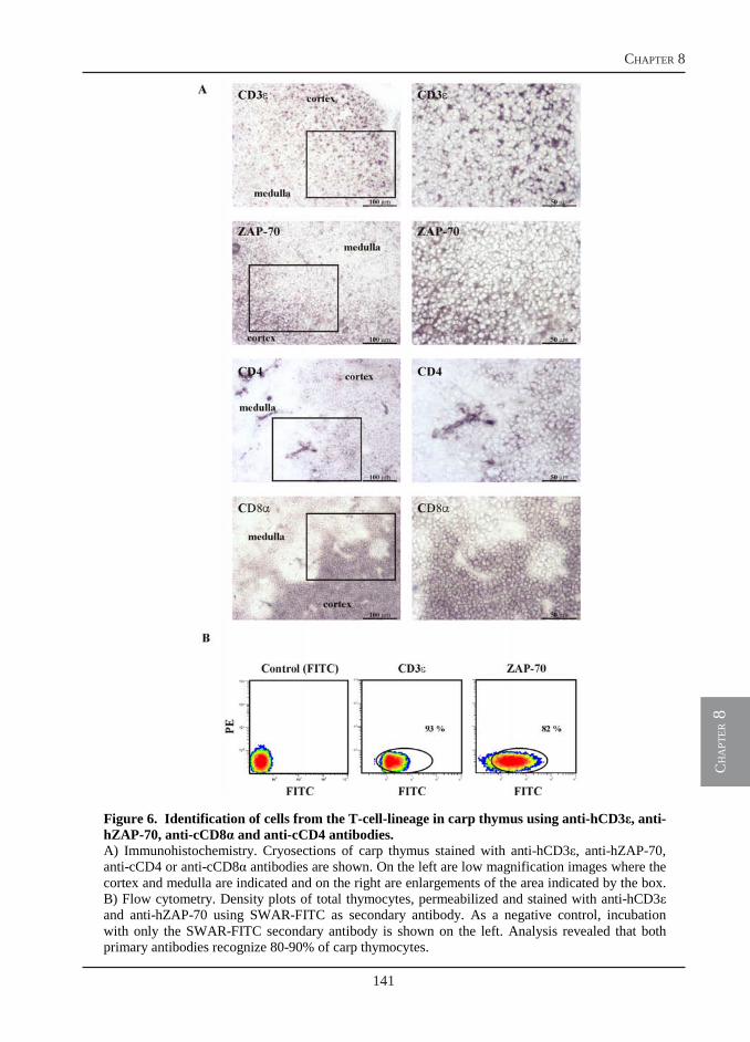
141
chapter 8
ch
apt
er 8
15
Figure 6. Identification of cells from the T-cell-lineage in carp thymus using anti-hCD3ε, anti-hZAP-70, anti-cCD8α and anti-cCD4 antibodies. A) Immunohistochemistry. Cryosections of carp thymus stained with anti-hCD3ε, anti-hZAP-70, anti-cCD4 or anti-cCD8α antibodies are shown. On the left are low magnification images where the cortex and medulla are indicated and on the right are enlargements of the area indicated by the box. B) Flow cytometry. Density plots of total thymocytes, permeabilized and stained with anti-hCD3εand anti-hZAP-70 using SWAR-FITC as secondary antibody. As a negative control, incubation with only the SWAR-FITC secondary antibody is shown on the left. Analysis revealed that both primary antibodies recognize 80-90% of carp thymocytes.
142
identification of t cells in carp
ch
apt
er 8
16
Detection of cells from the T-cell lineage in carp blood
Anti-hCD3ε and anti-hZAP-70 antibodies
were used in combination with previously
described antibodies that recognize carp B cells
(WCI12), monocytes (WCL15), neutrophilic
granulocytes (TCL-BE8) or thrombocytes
(WCL6) to identify the relative percentage of
PBL that belong to the T-cell-lineage.
Intracellular staining (CD3ε; ZAP-70) of
carp PBL was performed along with cell
surface staining (WCI12; WCL15; TCL-BE8;
WCL6). Permeabilization for intracellular
staining resulted in cross-reactivity of the anti-
hCD3ε antibody with a subpopulation of B
cells and some monocytes/macrophages (data
not shown). In contrast, permeabilization for
intracellular staining did not affect the
specificity of the anti-hZAP-70 antibody and
single-staining revealed the presence of about
5-15% ZAP-70+ cells in the blood of healthy
carp (Fig. 7A, upper panel). Double staining
confirmed the specificity of the ZAP-70
staining, revealing negligible cross-reactivity
with other leukocyte cell types in the blood
(Fig. 7A, lower panel).
Laser scanning microscopical analysis of
carp PBL upon single- or double-staining with
WCI12 (PE, red) and anti-hZAP-70 (FITC,
green), confirmed the specificity of ZAP-70 to
cells of the T-cell-lineage (Fig. 7B). As
expected, staining with the anti-hZAP-70
antibody revealed intracellular staining of
lymphocyte-like cells with a large nucleus and
a thin rim of cytoplasm whereas staining with
the anti-IgM antibody WCI12, revealed a
typical surface staining with patches of IgM+
areas (42) on lymphocytes.
Western blot analysis using the anti-hZAP-
70 antibody on total cell lysates of sorted
leukocyte sub-populations confirmed the
presence of a protein band with expected
molecular weight of 70 kDa in carp thymocytes
and human T cells, but not in other carp
leukocyte cell types (Fig. 7C). All together, our
results indicate the suitability of the anti-hZAP-
70 antibody to serve as tool for
immunohistochemical, flow cytometric and
Western blot analysis of cells belonging to the
T-cell-lineage in carp.
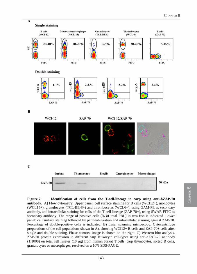
143
chapter 8
ch
apt
er 8
17
Figure 7. Identification of cells from the T-cell-lineage in carp using anti-hZAP-70 antibody. A) Flow cytometry. Upper panel: cell surface staining for B cells (WCI12+), monocytes (WCL15+), granulocytes (TCL-BE-8+) and thrombocytes (WCL6+), using GAM-PE as secondary antibody, and intracellular staining for cells of the T-cell-lineage (ZAP-70+), using SWAR-FITC as secondary antibody. The range of positive cells (% of total PBL) in n=4 fish is indicated. Lower panel: cell surface staining followed by permeabilization and intracellular staining against ZAP-70. Percentage of double-positive cells is indicated. B) Laser scanning microscopy. Cytocentrifuge preparations of the cell populations shown in A), showing WCI12+ B cells and ZAP-70+ cells after single and double staining. Phase-contrast image is shown on the right. C) Western blot analysis. ZAP-70 protein expression in different carp leukocyte cell-types using anti-hZAP-70 antibody (1:1000) on total cell lysates (10 g) from human Jurkat T cells, carp thymocytes, sorted B cells, granulocytes or macrophages, resolved on a 10% SDS-PAGE.
144
identification of t cells in carp
ch
apt
er 8
18
Distribution of T cells in spleen of T. borreli-infected carp
We have previously shown the presence of
extremely enlarged B cell areas in spleen of
carp heavily infected with T. borreli (28). The
marked B cell proliferation may be explained
by the polyclonal B cell activation typically
associated with T. borreli infections (43).
Owing to the lack of suitable T cell markers,
the distribution and the involvement of T cells
during the immune response of carp to T.
borreli has not been investigated. We used the
WCI12 antibody to specifically target carp B
cells and both the anti-hCD3ε and anti-hZAP-
70 antibodies to identify T cells in carp spleen
during infection with T. borreli (Fig. 8).
In spleen from naïve, non-infected, carp
only few and small B or T cell areas can be
observed, particularly around small ellipsoids
(Fig. 8A, left panel). During infection, not only
enlarged B cell (WCI12+) but also enlarged T
cell areas (CD3ε+ and ZAP-70+) can be
observed in spleen (Fig. 8A, right panels). The
location of B and T cell areas within the spleen
overlapped in consecutive sections indicative of
a co-localization of proliferating B cells and T
cells during T. borreli infection.
Double-staining using WCI12 in
combination with either anti-hCD3ε or anti-
hZAP-70 confirmed specificity of the staining
indicating the presence of intermingled single-
positive WCI12 and single-positive CD3ε or
ZAP-70 cells in the same area. In addition, the
co-localization of B and T cell areas confirms
the lack of a clear demarcation between B and
T cell areas in the spleen of fish, particularly
during an infection.
Discussion
The TCR complex consists of a single
TCRαβ heterodimer associated with at least
three CD3 dimers including γε, δε and ξξ and
the CD4 or CD8 co-receptors. The protein
tyrosine kinases Lck and ZAP-70 provide
essential signals in the cascade of events which
will finally lead to T cell signalling and
activation. In the present study we report the
cloning of a full-length mRNA sequence for
carp CD3ε and partial mRNA sequences for
carp Lck and ZAP-70. Only single transcripts
were found for these newly described
molecules. Duplicate copies for the constant
region of carp TCRα (24, 44), CD8α and CD8β
(24), but so far only a single copy for carp CD4
has been described (23). Two CD3ε isoforms
have been reported for Japanese flounder
(Paralichthys olivaceus) (20) and fugu
(Takifugu rubripes) (45) and two isoforms of
Lck have been reported for rainbow trout
(Oncorhynchus mykiss) (46). Common carp is
considered an allotetraploid species (47) and
genes often occur in duplicated form in the
genome. Thus, we expect that isoforms of
several, if not all, molecules of the TCR
complex could exist in duplicate copies in carp.
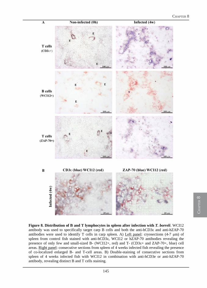
145
chapter 8
ch
apt
er 8
19
Figure 8. Distribution of B and T lymphocytes in spleen after infection with T. borreli. WCI12 antibody was used to specifically target carp B cells and both the anti-hCD3ε and anti-hZAP-70 antibodies were used to identify T cells in carp spleen. A) Left panel: cryosections (4-7 m) of spleen from control fish stained with anti-hCD3ε, WCI12 or hZAP-70 antibodies revealing the presence of only few and small-sized B- (WCI12+, red) and T- (CD3ε+ and ZAP-70+, blue) cell areas. Right panel: consecutive sections from spleen of 4 weeks infected fish revealing the presence of co-localized enlarged B- and T-cell areas. B) Double-staining of consecutive sections from spleen of 4 weeks infected fish with WCI12 in combination with anti-hCD3e or anti-hZAP-70 antibody, revealing distinct B and T cells staining.
146
identification of t cells in carp
ch
apt
er 8
20
Despite the amount of genetic information
on the TCR complex, the number of
monoclonal antibodies (MAbs) available for
the identification and functional
characterization of T cells in fish is very
limited. For example, MAbs that have been
produced against recombinant rainbow trout
TCRα and TCRβ were cross-reactive with other
lymphocyte subpopulations and could not be
used to specifically target T lymphocytes (2).
Attempts to identify cross-reactive MAbs
developed for use in mammalian immunology
that would specifically recognize fish antigens
also have been without much success. Of a
panel of 213 antibodies directed against various
human leukocyte surface antigens, none proved
reliably cross-reactive to trout antigens (48).
Another attempt with a panel of 377 MAbs
directed against, among other, human CD
antigens yielded only four MAbs that clearly
reacted with trout PBL and two antibodies that
reacted with carp PBL (4). The latter attempts
emphasize the phylogenetic distance between
warm- and cold-blooded vertebrates and
confirm the generally low conservation of
extracellular domains of cell surface molecules.
In the present study we selected two
antibodies developed for use in mammalian
immunology based not on the extracellular but
on the intracellular epitopes recognized. The
anti-hCD3ε antibody had previously been used
also in other fish species (19, 21, 49) and in
Xenopus laevis (22) whereas the anti-hZAP-70
antibody had not been considered before. In
carp, the anti-hCD3ε antibody can reliably be
used in immunohistochemical but not in flow
cytometric assays where it was found to cross-
react with a subpopulation of B cells and
monocytes in the blood (not shown). In
contrast, the anti-hZAP-70 specifically
recognizes T lymphocytes in both
immunohistochemistry and flow cytometry
with negligible cross-reactivity. Western blot
analysis confirmed recognition of a carp
protein of expected molecular weight.
Therefore, owing to the general high
conservation of the fish ZAP-70s (zebrafish:
NP_001018425; pufferfish: CAG00734) when
compared to the mammalian homologues, the
antibody used in this study will most likely be
applicable to more fish species. A drawback
concerning the use of this antibody, however, is
the permeabilization step, which does not allow
for immunopurification of live cells.
Upon TCR engagement, ZAP-70 is
activated either by autophosphorylation or by
phosphorylation by Lck (see Fig. 1). ZAP-70
phosphorylation, therefore, is one of the earliest
signalling events and detection of phospho-
ZAP-70 (pZAP-70) is commonly regarded a
reliable marker for T cell activation (50). The
combined use of anti-hZAP-70 and anti-pZAP-
70 antibodies would not only allow for
detection of changes in the distribution of T
cells throughout the organism but also for
detection of their activation state both, in vitro
and in vivo. The latter possibility, focusing on
the very early signalling events, rather than on
downstream products such as IL-2 or on T cell
proliferation, would allow for an unequivocal
detection of activated T cells, at least in vitro.
At present, we are therefore investigating the
147
chapter 8
ch
apt
er 8
21
use of antibodies directed against
phosphorylated tyrosines (Y) of ZAP-70, in
particular Y319, as marker for activated T cells
of carp.
In this study affinity purified rabbit
antibodies directed against carp CD4 and CD8α
were also produced. Antigenic epitopes present
in the extracellular region of these molecules
were selected for peptide synthesis and
immunization. Both anti-cCD4 and anti-
cCD8α were suitable for immunohistochemical
analysis. Structural analysis of the carp thymus
using a combination of the anti-hCD3ε, anti-
hZAP-70, anti-cCD4 and anti-cCD8α
antibodies revealed a conserved picture of the
general distribution and development of carp
thymocytes. The high cell density and intense
staining observed in the thymic cortex with all
antibodies, most likely indicates the presence of
double-positive CD4+/CD8+ immature T cells
(CD3ε+/ZAP-70+) in the cortical region. This
is similar to what is generally observed in
mammals and has also been observed in sea
bass, using in situ hybridization to demonstrate
staining for CD4 and CD8α in overlapping
areas in the cortex, but not in the medulla (51).
Despite the suitability for immuno
histochemistry, both anti-cCD4 and anti-cCD8
did not show reactivity to live cells in flow
cytometry. The epitopes chosen for
immunization were selected on an extracellular
region where no predicted glycosylation sites
were found. However, it can not be excluded
that neighbouring glycosylation sites might
interfere with the accessibility of the antibody
to the protein backbone.
New strategies for the production of
antibodies suitable for T cell recognition have
been finally developed. Most recently, Köllner
and colleagues established a panel of MAbs
which specifically recognize peripheral
rainbow trout T cells (52). Magnetic sorting
using one selected MAb confirmed the
expression of TCRα and β, CD8α and β, CD4
and major histocompatibility class II genes in
the purified lymphocyte population, indicating
this MAb can be considered a pan-specific T
cell marker. Key to their succes was the use of
activated leukocytes from mixed leukocyte
cultures for the immunization protocol in mice.
Another successful and highly promising
approach, used a retrovirus-mediated gene
transfer system, resulting in rat MAbs
recognizing two distinct populations of CD8α+
and CD4+ lymphocytes in Ginbuna crucian
carp (Carassius auratus langsdorfii) (53).
Although this species and the common carp are
closely related fish species, these antibodies do
not seem to recognize T cells of carp in flow
cytometry (Nakanishi T, pers comm; and
unpublished data).
We have previously shown that infections
with T. borreli are characterized by polyclonal
B cell activation as determined by ELISA on
serum samples collected from infected fish
(43). Immunohistochemical analysis showed
the presence of extremely enlarged B cell areas
in the spleen of infected fish (28). In the
present study, the anti-hCD3ε and anti-hZAP
were used to study T cell distribution in the
spleen of T. borreli-infected carp. We could
show the presence of both enlarged B cell and
T cell areas and their co-localization in the
148
identification of t cells in carp
ch
apt
er 8
22
spleen of infected fish. Double-staining
revealed that B and T cells are intermingled to
each other, with no clear distinction between a
B or T cell zone, especially during infection.
Changes in the typical distribution of B and T
cells in the white pulp were observed in mice
infected with Plasmodium chabaudi chabaudi,
where B cells and T cells are also found
intermingled to each other (54). Future
differentiation between T cell subtype (Th or
CTL) during T. borreli infection in fish,
requires the development of anti-cCD4 and
anti-cCD8α antibodies. The most recent
successful attempts in rainbow trout and
Ginbuna crucian carp hold a promise to
comparative immunologists that the long-
desired T cell-specific MAbs will soon become
available. In addition, application of forward
and reverse genetic approaches in zebrafish
(Danio rerio) also provides an elegant example
of in vivo tracking of T cells. Transgenic
zebrafish expressing GFP under the Lck
promoter enabled the actual (real-time)
visualization of T cell development, homing
and hematopoietic origin (55-57). Exploitation
and implementation of the zebrafish animal
model alongside the carp animal model should
rapidly burst the study of T cell function in fish
as well as help to bridge the gap of knowledge
between mammalian and comparative
immunology.
Acknowledgements
This work was supported by the European
Commission through contract FP6007103
IMAQUANIM.
References 1. Litman, G. W., and M. D. Cooper. 2007. Why study the evolution of immunity? Nat Immunol 8:547-
548. 2. Jansson, E., K. O. Gronvik, A. Johannisson, K. Naslund, E. Westergren, and L. Pilström. 2003.
Monoclonal antibodies to lymphocytes of rainbow trout (Oncorhynchus mykiss). Fish Shellfish Immunol 14:239-257.
3. Timmusk, S., E. Jansson, and L. Pilström. 2003. The generation of monoclonal antibodies by genetic immunisation: antibodies against trout TCRalpha and IgL isotypes. Fish Shellfish Immunol 14:187-206.
4. Fischer, U., and B. Köllner. 2007. Cross-reactivity of human leukocyte differentiation antigen monoclonal antibodies on carp and rainbow trout cells. Vet Immunol Immunopathol 119:142-155.
5. Scapigliati, G., M. Mazzini, L. Mastrolia, N. Romano, and L. Abelli. 1995. Production and characterisation of a monoclonal antibody against the thymocytes of the sea bass Dicentrarchus labrax (L.) (Teleostea, Percicthydae). Fish Shellfish Immunol 5:393-405.
6. Scapigliati, G., I. Boschi, E. Randelli, S. Picchietti, C. Bernini, and F. Buonocore. 2009. "In vitro" cell-mediated cytotoxic activities in the sea bass Dicentrarchus labrax, and immunopurification of cytotoxic leukocytes. In 11th Congress of the International Society of Developmental and Comparative Immunology, Prague, Czech Republic.
7. Rombout, J. H. W. M., J. W. van de Wal, A. Companjen, N. Taverne, and A. J. Taverne-Thiele. 1997. Characterization of a T cell lineage marker in carp (Cyprinus carpio L.). Dev Comp Immunol 21:35-46.
8. Rombout, J. H. W. M., P. H. M. Joosten, M. Y. Engelsma, A. P. Vos, N. Taverne, and J. J. Taverne-Thiele. 1998. Indications for a distinct putative T cell population in mucosal tissue of carp (Cyprinus carpio L.). Dev Comp Immunol 22:63-77.
9. Huttenhuis, H. B. T., N. Romano, C. Van Oosterhoud, A. Taverne-Thiele, L. Mastrolia, W. B. Van Muiswinkel, and J. H. W. M. Rombout. 2006. The ontogeny of mucosal immune cells in common carp (Cyprinus carpio L.). Anat Embryol 211:19-29.
149
chapter 8
ch
apt
er 8
23
10. Huttenhuis, H. B. T., M. O. Huising, T. van der Meulen, C. N. van Oosterhoud, N. A. Sanchez, A. J. Taverne-Thiele, H. W. Stroband, and J. H. W. M. Rombout. 2005. Rag expression identifies B and T cell lymphopoietic tissues during the development of common carp (Cyprinus carpio). Dev Comp Immunol 29:1033-1047.
11. Romano, N., A. J. Taverne-Thiele, M. Fanelli, M. R. Baldassini, L. Abelli, L. Mastrolia, W. B. Van Muiswinkel, and J. H. M. W. Rombout. 1999. Ontogeny of the thymus in a teleost fish, Cyprinus carpio L.: developing thymocytes in the epithelial microenvironment. Dev Comp Immunol 23:123-137.
12. Lam, S. H., H. L. Chua, Z. Gong, T. J. Lam, and Y. M. Sin. 2004. Development and maturation of the immune system in zebrafish, Danio rerio: a gene expression profiling, in situ hybridization and immunological study. Dev Comp Immunol 28:9-28.
13. Zapata, A., B. Diez, T. Cejalvo, C. Gutiérrez-de Frías, and A. Cortés. 2006. Ontogeny of the immune system of fish. Fish Shellfish Immunol 20:126-136.
14. Romano, N., F. Rossi, L. Abelli, E. Caccia, R. Piergentili, L. Mastrolia, E. Randelli, and F. Buonocore. 2007. Majority of TCRbeta+ T-lymphocytes located in thymus and midgut of the bony fish, Dicentrarchus labrax (L.). Cell Tissue Res 329:479-489.
15. Danilova, N., V. S. Hohman, F. Sacher, T. Ota, C. E. Willett, and L. A. Steiner. 2004. T cells and the thymus in developing zebrafish. Dev Comp Immunol 28:755-767.
16. Lam, S. H., H. L. Chua, Z. Gong, Z. Wen, T. J. Lam, and Y. M. Sin. 2002. Morphologic transformation of the thymus in developing zebrafish. Dev Dynamics 225:87-94.
17. Bowden, T. J., P. Cook, and J. H. W. M. Rombout. 2005. Development and function of the thymus in teleosts. Fish Shellfish Immunol 19:413-427.
18. Hoffmann, J. A., and J. M. Reichhart. 2002. Drosophila innate immunity: an evolutionary perspective. Nat Immunol 3:121-126.
19. Cook, M. T., R. N. Morrison, R. Wilkinson, B. F. Nowak, P. J. Hayball, and J. D. Hayball. 2001. A screen of mammalian antibodies on snapper (Pagrus auratus, Sparidae) peripheral blood leukocytes reveals cross reactivity of an anti-human CD3 antibody with a population of mIg- cells. Dev Comp Immunol 25:553-559.
20. Park, C. I., I. Hirono, J. Enomoto, B. H. Nam, and T. Aoki. 2001. Cloning of Japanese flounder Paralichthys olivaceus CD3 cDNA and gene, and analysis of its expression. Immunogenetics 53:130-135.
21. Bakke-McKellep, A. M., M. K. Frøystad, E. Lilleeng, F. Dapra, S. Refstie, K. Å, and T. Landsverk. 2007. Response to soy: T-cell-like reactivity in the intestine of Atlantic salmon, Salmo salar L. J Fish Dis 30:13-25.
22. Göbel, T. W. F., E. L. Meier, and L. Du Pasquier. 2000. Biochemical analysis of the Xenopus laevisTCR/CD3 complex supports the "stepwise evolution" model. Eur J Immunol 30:2775-2781.
23. Sun, X. F., N. Shang, W. Hu, Y. P. Wang, and Q. L. Guo. 2007. Molecular cloning and characterization of carp (Cyprinus carpio L.) CD8beta and CD4-like genes. Fish Shellfish Immunol 23:1242-1255.
24. Forlenza, M., J. D. de Carvalho Dias, T. Veselý, D. Pokorová, H. F. Savelkoul, and G. F. Wiegertjes. 2008. Transcription of signal-3 cytokines, IL-12 and IFNαβ, coincides with the timing of CD8αβup-regulation during viral infection of common carp (Cyprinus carpio L). Mol Immunol 45:1531-1547.
25. Kazazi, F., J. M. Mathijs, P. Foley, and A. L. Cunningham. 1989. Variations in CD4 expression by human monocytes and macrophages and their relationships to infection with the human immunodeficiency virus. J Gen Virol 70 ( Pt 10):2661-2672.
26. Chan, L. S. 2003. Animal models of human inflammatory skin diseases. CRC Press. 27. Amin, S., A. Parker, and J. Mann. 2008. ZAP70 in chronic lymphocytic leukaemia. Int J Biochem
Cell Biol 40:1654-1658. 28. Forlenza, M., J. P. Scharsack, N. M. Kachamakova, A. J. Taverne-Thiele, J. H. W. M. Rombout, and
G. F. Wiegertjes. 2008. Differential contribution of neutrophilic granulocytes and macrophages to nitrosative stress in a host-parasite animal model. Mol Immunol 45:3178-3189.
29. Irnazarow, I. 1995. Genetic variability of Polish and Hungarian carp lines. Aquaculture Research 129:215-219.
30. Steinhagen, D., P. Kruse, and W. Körting. 1989. The parasitemia of cloned Trypanoplasma borreli Laveran and Mesnil, 1901, in laboratory-infected common carp (Cyprinus carpio L.). J Parasitol 75:685-689.
31. van Erp, S. H., B. Dixon, F. Figueroa, E. Egberts, and R. J. M. Stet. 1996. Identification and characterization of a new major histocompatibility complex class I gene in carp (Cyprinus carpio L.). Immunogenetics 44:49-61.
150
identification of t cells in carp
ch
apt
er 8
24
32. Pfaffl, M. W. 2001. A new mathematical model for relative quantification in real-time RT-PCR. Nucleic Acids Res 29:e45.
33. Tichopad, A., M. Dilger, G. Schwarz, and M. W. Pfaffl. 2003. Standardized determination of real-time PCR efficiency from a single reaction set-up. Nucleic Acids Res 31:e122.
34. Secombes, C. J., J. J. van Groningen, and E. Egberts. 1983. Separation of lymphocyte subpopulations in carp Cyprinus carpio L. by monoclonal antibodies: immunohistochemical studies. Immunology 48:165-175.
35. Koumans-van Diepen, J. C. E., E. Egberts, B. R. Peixoto, N. Taverne, and J. H. W. M. Rombout. 1995. B cell and immunoglobulin heterogeneity in carp (Cyprinus carpio L); An immuno(cyto)chemical study. Developmental & Comparative Immunology 19:97-108.
36. Nakayasu, C., M. Omori, S. Hasegawa, O. Kurata, and N. Okamoto. 1998. Production of a monoclonal antibody for carp (Cyprinus carpio L.) phagocytic cells and separation of the cells. Fish Shellfish Immunol 8:91-100.
37. Weyts, F. A. A., J. H. W. M. Rombout, G. Flik, and B. M. L. Verburg-Van Kemenade. 1997. A common carp (Cyprinus carpio L.) leucocyte cell line shares morphological and functional characteristics with macrophages. Fish Shellfish Immunol 7:123-133.
38. Romano, N., S. Picchietti, J. J. Taverne-Thiele, N. Taverne, L. Abelli, L. Mastrolia, B. M. L. Verburg-van Kemenade, and J. H. W. M. Rombout. 1998. Distribution of macrophages during fish development: an immunohistochemical study in carp (Cyprinus carpio, L.). Anat Embryol (Berl) 198:31-41.
39. Romano, N., J. J. Taverne-Thiele, J. C. Van Maanen, and J. H. M. W. Rombout. 1997. Leucocyte subpopulations in developing carp (Cyprinus carpio L.): immunocytochemical studies. Fish Shellfish Immunol 7:439-453.
40. Rombout, J. H. W. M., J. C. E. Koumans-van Diepen, P. M. Emmer, J. J. Taverne-Thiele, and N. Taverne. 1996. Characterization of carp thrombocytes with specific monoclonal antibodies. J Fish Biol 49:521-531.
41. Rothenberg, E. V., J. E. Moore, and M. A. Yui. 2008. Launching the T-cell-lineage developmental programme. Nat Rev Immunol 8:9-21.
42. van Diepen, J. C. E., G. T. M. Wagenaar, and J. H. W. M. Rombout. 1991. Immunocytochemical detection of membrane antigens of carp leucocytes using light and electron microscopy. Fish Shellfish Immunol 1:47-57.
43. Saeij, J. P. J., B. J. de Vries, and G. F. Wiegertjes. 2003. The immune response of carp to Trypanoplasma borreli: kinetics of immune gene expression and polyclonal lymphocyte activation. Dev Comp Immunol 27:859-874.
44. Imai, E., J. Ishikawa, T. Moritomo, and M. Tomana. 2005. Characterisation of T cell antigen receptor α chain isotypes in the common carp. Fish Shellfish Immunol 19:205-216.
45. Araki, K., H. Suetake, K. Kikuchi, and Y. Suzuki. 2005. Characterization and expression analysis of CD3ε and CD3γ/δ in fugu, Takifugu rubripes. Immunogenetics 57:158-163.
46. Laing, K. J., S. Dutton, and J. D. Hansen. 2007. Molecular and biochemical analysis of rainbow trout LCK suggests a conserved mechanism for T-cell signaling in gnathostomes. Mol Immunol 44:2737-2748.
47. Zhang, Y., L. Liang, P. Jiang, D. Li, C. Lu, and X. Sun. 2008. Genome evolution trend of common carp (Cyprinus carpio L.) as revealed by the analysis of microsatellite loci in a gynogenetic family. J Genet Genomics 35:97-103.
48. Brodersen, R., F. Bijlsma, K. Gori, K. T. Jensen, W. Chen, J. Dominguez, K. Haverson, P. F. Moore, A. Saalmuller, D. Sachs, W. J. Slierendrecht, C. Stokes, O. Vainio, F. Zuckermann, and B. Aasted. 1998. Analysis of the immunological cross reactivities of 213 well characterized monoclonal antibodies with specificities against various leucocyte surface antigens of human and 11 animal species. Vet Immunol Immunopathol 64:1-13.
49. Park, C.-I., I. Hirono, and T. Aoki. 2005. Molecular characterization of the Japanese flounder, Paralichthys olivaceus, CD3ε and evolution of the CD3 cluster. Dev Comp Immunol 29:123-133.
50. Mustelin, T., and K. Taskén. 2003. Positive and negative regulation of T-cell activation through kinases and phosphatases. Biochem. J. 371:15-27.
51. Picchietti, S., L. Guerra, F. Buonocore, E. Randelli, A. M. Fausto, and L. Abelli. 2009. Lymphocyte differentiation in sea bass thymus: CD4 and CD8-alpha gene expression studies. Fish Shellfish Immunol 27:50-56.
52. Köllner, B. 2009. T-lymphocytes in rainbow trout: investigation with newly established T-cell specific monoclonal antibodies. In 11th Congress of the International Society of Developmental and Comparative Immunology, Prague, Czech Republic. 23.
151
chapter 8
ch
apt
er 8
25
53. Toda, H., Y. Shibasaki, T. Koike, M. Ohtani, F. Takizawa, M. Ototake, T. Moritomo, and T. Nakanishi. 2009. Alloantigen-specific killing is mediated by CD8-positive T cells in fish. Dev Comp Immunol 33:646-652.
54. Achtman, A. H., M. Khan, I. C. MacLennan, and J. Langhorne. 2003. Plasmodium chabaudi chabaudi infection in mice induces strong B cell responses and striking but temporary changes in splenic cell distribution. J Immunol 171:317-324.
55. Langenau, D. M., A. A. Ferrando, D. Traver, J. L. Kutok, J. P. Hezel, J. P. Kanki, L. I. Zon, A. T. Look, and N. S. Trede. 2004. In vivo tracking of T cell development, ablation, and engraftment in transgenic zebrafish. Proc Natl Acad Sci U S A 101:7369-7374.
56. Traver, D., A. Winzeler, H. M. Stern, E. A. Mayhall, D. M. Langenau, J. L. Kutok, A. T. Look, and L. I. Zon. 2004. Effects of lethal irradiation in zebrafish and rescue by hematopoietic cell transplantation. Blood 104:1298-1305.
57. Langenau, D. M., and L. I. Zon. 2005. The zebrafish: a new model of T-cell and thymic development. Nat Rev Immunol 5:307-317.

152
identification of t cells in carp
ch
apt
er 8
Receptor-mediated and lectin-like activities of carp TNFα
chapter
9
Maria Forlenza, Stefan Magez, Jörn P. Scharsack, Adrie Westphal, Huub F.J. Savelkoul and Geert F. Wiegertjes
Journal of Immunology, 2009 (183), 5319–5332

154
Carp TNFa activities
ch
apt
er 9
155
chapter 9
ch
apt
er 9
Receptor-Mediated and Lectin-Like Activities of Carp(Cyprinus carpio) TNF-�1,2
Maria Forlenza,* Stefan Magez,† Jorn P. Scharsack,‡ Adrie Westphal,§ Huub F. J. Savelkoul,*and Geert F. Wiegertjes3*
Functional characterization of TNF-� in species other than mammalian vertebrates is limited, and TNF-� has been studied in alimited number of fish species, primarily in vitro using recombinant proteins. Studies on TNF-� from different fish species so farpointed to several inconsistencies, in particular with respect to some receptor-mediated activities of fish TNF-�, such as the abilityto directly activate phagocytes. In the present study a comprehensive analysis of in vitro as well as in vivo biological activities oftwo isoforms of carp TNF-� was performed. Our results show that carp TNF-� directly primes carp phagocytes and indirectlypromotes typical receptor-mediated activities such as phagocyte activation by acting via endothelial cells. Additionally, for the firsttime in nonmammalian vertebrate species, the lectin-like activity of fish TNF-� homologs was investigated. Our results show anevolutionary conservation of function of this receptor-independent activity of TNF-� not only in cyprinid fish, but also in perci-form and salmonid fish. The role of TNF-� in vivo, during infections of carp with the blood parasite Trypanoplasma borreli, wasexamined using three fundamentally different but complementary approaches: (1) inhibition of TNF-� expression, (2) overex-pression of TNF-�, and (3) inhibition of shedding of membrane-bound TNF-�. Our results show that, also in fish, a tight regulationof TNF-� expression is important, since depletion or excess of TNF-� can make an important difference to survival of infection.Finally, we demonstrate a crucial protective role for membrane-bound TNF-�, which has a yet unexploited function in fish. TheJournal of Immunology, 2009, 183: 5319–5332.
T umor necrosis factor-� is a pleiotropic cytokine and amajor regulator of leukocyte trafficking and inflammation(1). Under most conditions monocytes/macrophages are
the major producers of TNF-�, but in response to specific stimulisuch as polyclonal activation, PMA, or LPS stimulation, also T andB lymphocytes, NK cells, endothelial cells, and other immune cellscan produce TNF-� (2–6).
So far, TNF-� homologs have been identified only in the mam-malian warm-blooded vertebrates. In birds, neither TNF-� norlymphotoxin-� has been identified (7, 8). In amphibians, only veryrecently the first TNF-� sequence was published (9), with a limitedfunctional characterization. In contrast, TNF-� homologs havebeen reported for at least 10 different teleost fish species (10–23)
with two or sometimes three isoforms of TNF-� found in severalfish species, including the common carp (Cyprinus carpio) (11, 13,14, 16–18, 22). In vitro studies using recombinant proteins haveshown that fish TNF-� is active at concentrations at least 1000times higher than its mammalian counterpart (20–22, 24–27) andhave evidenced crucial differences between TNF-� of cold- andwarm-blooded vertebrates. Furthermore, activities of fish TNF-�appear species-specific in particular with respect to the ability offish TNF-� to activate phagocytes directly. For example, both inrainbow trout (Oncorhynchus mykiss) (24) and goldfish (Carassiusauratus) (22), TNF-� stimulates phagocyte activity in vitro, albeitat different concentrations. In contrast, seabream (Sparus aurata)TNF-� is a potent activator of endothelial cells but does not acti-vate phagocytes directly (27). Instead, supernatant from seabreamendothelial cells treated with TNF-� promoted the adhesion, mi-gration, and activation of leukocytes. This is in line with receptor-mediated functions of TNF-� in mammalian vertebrates, wherestimulation of endothelial cells with TNF-� increases the expressionof adhesion molecules and the synthesis of chemotactic mediatorssuch as IL-8. Thereby, TNF-� promotes leukocyte recruitment andactivation and contributes to the inflammatory process. In the presentstudy we show that carp (C. carpio) TNF-� primes but does not di-rectly activate phagocytes and thereby promotes phagocyte activation,indirectly, via stimulation of endothelial cells.
TNF-� fulfills important functions in host-pathogen interactionsand is required for protective immunity against intracellular bac-teria and fungi but also against intracellular and extracellular pro-tozoan parasites (28, 29). An additional, but perhaps less well-known activity of TNF-�, is its lectin-like activity first recognizedin 1988 (30) when TNF-� was reported to directly interact withspecific oligosaccharides through its lectin-like domain. The lectindomain is located at the top of the pyramid-shaped TNF-� mole-cule, also referred to as TIP domain, and is spatially and function-ally distinct from the receptor binding region. TNF-� binds
*Cell Biology and Immunology Group, Wageningen Institute of Animal Sciences,Wageningen University, Wageningen, The Netherlands; †Unit of Cellular and Mo-lecular Immunology, Vrije Universiteit Brussel, Brussels, Belgium, and Departmentof Molecular and Cellular Interactions, Institute for Biotechnology, Ghent, Belgium;‡Institute for Evolution and Biodiversity, Animal Evolutionary Ecology Group, Uni-versity of Munster, Munster, Germany; and §Laboratory of Biochemistry, Departmentof Agrotechnology and Food Sciences, Wageningen University, Wageningen, TheNetherlands
Received for publication June 4, 2009. Accepted for publication August 6, 2009.
The costs of publication of this article were defrayed in part by the payment of pagecharges. This article must therefore be hereby marked advertisement in accordancewith 18 U.S.C. Section 1734 solely to indicate this fact.1 This work was partly supported by the European Commission’s Improving HumanPotential Program under Contract HPRN-CT-2001-00214 and QLK5-CT-2001-50988and by the European Commission through Contract FP6007103 (Improved Immunityof Aquacultured Animals, IMAQUANIM).2 The sequence presented in this article has been submitted to GenBank under acces-sion no. GQ231486.3 Address correspondence and reprint requests to Dr. Geert F. Wiegertjes. Cell Bi-ology and Immunology Group, Department of Animal Sciences, Wageningen Uni-versity, P.O. Box 338, 6700 AH, Wageningen, The Netherlands. E-mail address:[email protected]
Copyright © 2009 by The American Association of Immunologists, Inc. 0022-1767/09/$2.00
The Journal of Immunology
www.jimmunol.org/cgi/doi/10.4049/jimmunol.0901780
156
Carp TNFa activities
ch
apt
er 9
through its lectin-like domain to mannose moieties present in theflagellar pocket of some African trypanosomes and can cause di-rect parasite lysis (31–34). The lectin-like activity of TNF-�, a yetunexamined receptor-independent function of fish TNF-�, was in-vestigated in a trypanolytic assay in vitro. Our results clearly showthat not only carp and zebrafish (Danio rerio) (both Cyprinidae),but also rainbow trout (Salmonidae) and seabream (Perciformes)TNF-� directly lyse Trypanosoma brucei, indicating that the lec-tin-like activity of TNF-� is evolutionary conserved.
Studies with TNF-�-deficient knockout mice, with transgenicmice overexpressing TNF-�, or studies administrating recombi-nant TNF-� all pointed out a dual role of TNF-� in immune pro-tection as well as pathology (35). The “double-edged sword” roleof TNF-� has most clearly been shown in studies on malaria andtrypanosome infections where TNF-� was identified as a key me-diator in both control of parasitemia as well as infection-associatedpathology (28, 29, 36–38). In fish, due to a general lack of knock-out and transgenic animals, the function of TNF-� in vivo islargely unknown. Only very recently, by the use of TNFR1 mor-phant zebrafish, TNF-� signaling has been shown to mediate re-sistance to mycobacteria by inhibiting bacterial growth and pre-venting macrophage death (39). To further investigate the in vivorole played by TNF-� with respect to protection or infection-as-sociated pathology, we used a natural host-parasite infectionmodel. Experimental infection of carp with the extracellular bloodparasite Trypanoplasma borreli (Kinetoplastida) presents most ofthe pathological features associated with trypanosome infections inmammalian vertebrates such as anemia, splenomegaly, polyclonal Bcell activation, as well as extremely elevated serum nitrite levels andassociated tissue nitration (40 – 44). Similarly, T. borreli infec-tion of carp can be treated with the human anti-trypanosomedrug melarsoprol (Arsobal) (44). In the present study we showthat inhibition of TNF-� gene expression in T. borreli-infectedcarp treated with the TNF-� inhibitor pentoxifylline results inextremely high parasitemia and increased mortality, whereasoverexpression of TNF-� by injection of plasmid DNA leads toincreased mortality, possibly owing to an exacerbation of theinflammatory response. Our results indicate a functional con-servation in fish of the dual role of TNF-� in control of para-sitemia and in infection-associated pathology.
TNF-� is present as a soluble and membrane-bound (mTNF-�)4
form, with both exhibiting unique and overlapping activities.While solunle TNF-� mainly signals via TNFRI (p55), mTNF-� isthe prime activating ligand of TNFR2 (p75) (45). Membrane-bound TNF-� has been shown to be involved in several biologicalactivities, such as cytotoxicity, polyclonal activation of B cells,induction of IL-10 by monocytes, induction of chemokines, andICAM-1 expression on endothelial cells (46–48). mTNF-� hasbeen implicated in the control of Listeria monocytogenes (49),mycobacterial infection (50, 51), and possibly in the pathogenesisof experimental cerebral malaria (52). In the present study, weused an inhibitor of TNF-�-converting enzyme (TACE) to inves-tigate the contribution of mTNF-� to the clearance of T. borreli ininfected carp. In vitro, treatment of LPS-stimulated carp leuko-cytes with TACE inhibitor increased the number of cells bearingTNF-� on their surface. In vivo, fish treated with the TACE in-hibitor were fully protected and cleared the parasite within daysafter treatment. Additionally, splenomegaly, plasma nitrite levels,
and tissue nitration were all considerably reduced in fish treatedwith the TACE inhibitor, suggesting a so far unrecognized pro-tective role for membrane-bound TNF-� in fish.
Materials and MethodsAnimals
European common carp (Cyprinus carpio carpio L.) were bred and raisedin the central fish facility at Wageningen University, The Netherlands, at23°C in recirculating UV-treated tap water and fed pelleted dry food (Trou-vit; Nutreco) daily. R3xR8 carp, which are the offspring of a cross betweenfish of Hungarian origin (R8 strain) and of Polish origin (R3 strain), wereused (53). All experiments were performed with the approval of the animalexperimental committee of Wageningen University.
Cell isolation
Fish were killed by an overdose of anesthetic (0.3 g/L tricaine methanesulfonate; Crescent Research Chemicals) in aquarium water bufferedwith 0.6 g/L sodium bicarbonate (Sigma-Aldrich). PBL, head kidneyleukocytes (HKL), or enriched phagocyte fractions (macrophages andneutrophilic granulocytes) were isolated as described previously (54,55). After isolation, cells were adjusted to the appropriate concentrationwith carp complete RPMI 1640 medium (cRPMI (Invitrogen) adjustedto 280 mOsmol/kg, supplemented with 1.5% (v/v) heat inactivated-pooled carp serum, 2 mM L-glutamine, 100 U/ml penicillin G, and 50mg/ml streptomycin sulfate).
Isolation of cardiac endothelial cells (EC) was performed as published(56, 57) with some modifications. Briefly, hearts from 9- to 12-mo-old carpwere aseptically removed and transferred to 20 ml of ice-cold cRPMI sup-plemented with 5 IU/ml heparin (Leo Pharmaceutical Products). All sub-sequent steps were performed at room temperature. Excess blood was re-moved by slowly injecting into the atrium 10 ml of perfusion buffer (56)supplemented with 5 IU/ml heparin. Atria were dissected and several in-cisions were made to ensure complete access of the following buffers. Atriawere incubated by shaking at 250 rpm for 30 min in carp PBS (cPBS, 150mM NaCl, 2 mM KCl, 20 mM Na2HPO4, 2 mM KH2PO4) for 5 min incPBS containing 0.5 mg/ml trypsin and 0.1 mg/ml EDTA and for 30 minin 0.7% saline solution containing 0.5 mg/ml collagenase and 0.7 mg/mlCaCl2. After each incubation step, cells were centrifuged for 5 min at400 � g. After collagenase treatment, the suspension was transferred to asterile petri dish and flushed 5–10 times through the jet of a 10-ml syringe.Large fragments were discarded by sedimentation at 1 � g for 8–10 min.The resulting cell suspension was centrifuged for 5 min at 400 � g and cellpellets were resuspended in complete EC medium (RPMI 1640 supple-mented with 10% FCS, 2 mM L-glutamine, 100 U/ml penicillin G, 50mg/ml streptomycin sulfate, and 50 �g/ml gentamicin). The cell suspen-sion obtained from n � 4 atria was seeded in two collagen type I-coated6-well plates or two 24-well plates (Greiner, catalog nos. 657950 and662950) and incubated at 27°C in a humidified atmosphere of 5% CO2 toallow cells to adhere. After 24–48 h, cell debris and nonadherent cellswere removed by washing twice with complete EC medium and then cul-tures were left undisturbed.
Parasites
T. borreli was cloned and characterized Steinhagen et al. (58) and main-tained by syringe passage through carp following i.p. injection with 1 �104 parasites/fish. Parasitemia was monitored using a Burker countingchamber after dilution of blood 1/1 in RPMI 1649 medium adjusted to 280mOsmol/kg containing 50 IU/ml of heparin. For parasite isolation, bloodwas collected from heavily infected carp and, after centrifugation, T. bor-reli were collected from the buffy coat and purified on a 1 � 12-cm ion-exchange column chromatography using DEAE cellulose (DE-52; What-man International) (59). After isolation, parasites were harvested bycentrifugation and resuspended in fresh complete HML medium (60) sup-plemented with 5% pooled carp serum, L-glutamine (2 mM), penicillin G(100 IU/ml), and streptomycin sulfate (50 mg/L)).
Bacterial expression constructs
For bacterial expression of soluble TNF-�, the sequence corresponding tothe mature peptide of carp TNF-�1 (accession no. AJ311800, aa 77–237)or TNF-�2 (accession no. AJ311801, aa 70–231) was amplified by stan-dard PCR using a proofreading Taq polymerase (Expand high-fidelity Taqpolymerase; Roche) and cDNA from LPS-stimulated HKL as template. Allprimers used for cloning are listed in Table I. PCR products were directlycloned into pQE-30UA plasmid (Qiagen) downstream of the sequence
4 Abbreviations used in this paper: mTNF-�, membrane-bound TNF-�; cPBS, carpPBS; cRPMI, carp complete RPMI 1640 medium; DHR, dihydrorhodamine; EC,endothelial cell; EPC, Epithelioma papulosum cyprinid; HKL, head kidney leukocyte;iNOS, inducible NO synthase; LB, lysis buffer; PTX, pentoxifylline; TACE, TNF-�-converting enzyme.
2 RECEPTOR-MEDIATED AND LECTIN-LIKE ACTIVITIES OF CARP TNF-�
157
chapter 9
ch
apt
er 9
coding for a 6� histidine tag. The primers used for the amplification werepQETNF1_FW or pQETNF2_FW in combination with pQETNF_RV.
Eukaryotic expression constructs
For eukaryotic expression of soluble TNF-�1 or TNF-�2, the mature pep-tides (excluding the TNF-� leader peptide and transmembrane sequence)were cloned in the pIRES-EGFP plasmid (Promega), downstream of theleader peptide region of the carp TGF� (AF136947). The transmembraneregion was removed to favor direct TNF-� secretion. The TGF� leaderpeptide was inserted upstream of the mature TNF-� peptide to increase therate of TNF-� secretion, as the use of a TGF� leader peptide has beenpreviously shown to effectively increase protein secretion in carp cells (61).TNF-�1 or TNF-�2 sequences were amplified as described above usingIRES_EcoRI_TNF1_FW or IRES_EcoRI_TNF2_FW in combination withIRES_BamHI_TNF_RV (see Table I). The nucleotide sequence encodingfor the leader peptide region of the carp TGF� was amplified usingXhoI_TGFleader_FW and EcoRIHisLeader_RV, with part of the RV prim-ers containing the sequence encoding for a 6� histidine tag. The leadersequence was first cloned into the XhoI site of the pIRES-EGFP plasmid,followed by restriction and ligation of the TNF-�1 or TNF-�2 sequencebetween the EcoRI site, downstream of the leader-His sequence, and theBamHI site of the pIRES-EGFP plasmid. All constructs were transformedinto M15 competent Escherichia coli cells, and positive clones were se-lected on agar plates containing ampicillin (100 �g/ml) and kanamicin (25�g/ml). At least eight clones for each construct were sequenced. Expres-sion of mature carp TNF-� was confirmed by Western blot analysis ofsupernatants from transfected Epithelioma papulosum cyprini (EPC) cells(62) using an anti-histidine Ab (Qiagen).
Recombinant carp TNF-�
Transformed E. coli M15 cells were grown on agar plates supplementedwith antibiotics (100 �g/ml ampicillin and 25 �g/ml kanamicin) overnightat 37°C. One single colony was transferred to 20 ml of Luria-Bertani me-dium (1% (w/v) tryptone, 0.5% (w/v) yeast extract, 0.5% (w/v) NaCl (pH7.5)) supplemented with antibiotic and grown overnight at 37°C with shak-ing. The overnight culture was transferred to 1 liter of prewarmed TBmedium (tryptone 2.4% (w/v), yeast extract 1.2% (w/v), NaCl 0.5% (w/v),glycerol 4% (v/v) (pH 7.5)) and incubated at 30°C with shaking. At OD600
of 0.6–0.8, protein production was induced with 1 mM isopropyl �-D-thiogalactoside and the culture was incubated for a further 4 h at 30°C.Bacterial pellets were collected by centrifugation at 5270 � g for 15 minat 4°C and stored at �80°C until use. Proteins were purified under nativeconditions and all steps were conducted at 4°C unless stated otherwise.Bacterial pellets from 1-liter culture were resuspended in 20–40 ml of lysisbuffer (LB; 50 mM Tris-Cl (pH 7.5), 500 mM NaCl, 5 mM MgCl2, 10 mMimidazole, 10 mM 2-ME, 10% glycerol (v/v), 0.1% Triton X-114 (v/v))supplemented with protease inhibitor cocktail (Sigma-Aldrich, catalog no.P8849) and lysozyme (1 mg/ml; Merck) and the suspension was incubatedfor 30 min on ice. Lysates were sonicated and subsequently centrifuged at10,000 � g for 20 min at 4°C. Supernatants were collected and combinedwith 500 �l (for TNF-�1) or 750 �l (for TNF-�2) of Ni-NTA matrix(Qiagen). Samples were incubated for 30 min at 4°C while rotating andsubsequently loaded on a 12-ml Poly-Prep chromatography column (Bio-Rad, catalog no. 7311550). All washing steps were performed under the fol-lowing conditions: 300 � g for 4.5 min at 4°C. Columns were washed fivetimes with 5 ml of wash buffer A (LB containing 20 mM imidazole), five timeswith 5 ml of wash buffer B (LB containing 30 mM imidazole), three times with5 ml of wash buffer C (LB containing 50 mM imidazole, without TritonX-114), once with 5 ml of wash buffer D (LB containing 100 mM imida-zole, without Triton X-114), and three times with 5 ml of wash buffer E
(500 mM NaCl, 2 mM KCl, 20 mM Na2HPO4, 2 mM KH2PO4, 100 mMimidazole). After an empty centrifugation step, proteins were eluted inelution buffer (wash buffer E containing 250 mM imidazole). Eluted pro-teins were subsequently dialyzed against cPBS, and endotoxins were mea-sured by end-point chromogenic Limulus amebocyte lysate test (CharlesRiver Laboratories). If necessary, further removal of endotoxin wasachieved by Triton X-114 phase separation (63). All preparations contained�0.015 EU/ml. Unless stated otherwise, heat-treated TNF-� samples wereused as negative control in all stimulation assays.
Western blot
To confirm overexpression of carp TNF-� in plasmid-injected fish, �100mg of tissue collected from the injection site was resuspended in 1 ml ofRIPA lysis buffer (50 mM Tris-HCl (pH 7.4), 1% Nonidet P-40, 0.25%sodium deoxycholate, 150 mM NaCl, 1 mM EDTA, 1 mM PMSF, andprotease inhibitor cocktail (Sigma-Aldrich, catalog no. P8849) and homog-enized on ice by sonication (3 min, on for 30 s, off for 30 s, power 5, duty30%). Lysates were cleared by centrifugation at 21,000 � g for 20 min at4°C. Supernatants were carefully collected and 25 �l was resolved on a12.5% SDS-PAGE. Proteins were transferred to nitrocellulose membranes(Protran; Schleicher & Schuell BioScience) and incubated overnight at 4°Cwith a 1/2000 dilution of affinity-purified polyclonal rabbit-anti-cTNF� Abwith gentile agitation. HRP-conjugated goat-anti-rabbit Ab (1/2000; Dako)was used as secondary Ab. Proteins were visualized by chemiluminescencedetection (ECL detection system for Western blot; Amersham Biosciences)on x-ray films.
Affinity-purified polyclonal rabbit IgG anti-carp TNF-�1 or anti-TNF-�2 was produced by immunization of rabbits with purified bacterialrecombinant protein, according to a 3-mo standard protocol (Eurogentec).Affinity-purified IgG recognized both TNF-�1 and TNF-�2. Anti-TNF-�Abs were used for subsequent protein detection.
Cell stimulation
PBL, HKL, or enriched phagocyte fractions were resuspended in completemedium at a concentration of 1 � 107 cells/ml and 100 �l was transferredto 96-well plates. Cells were incubated overnight at 27°C in a humidifiedatmosphere of 5% CO2. The following day cells were stimulated withvarious concentration of TNF-�, in the presence or absence of LPS in afinal volume of 200 �l.
EC were cultured in 24-well plates until confluent and subsequentlystimulated with various concentrations of TNF-� in a final volume of 500�l. After 4 h cells were harvested for gene expression analysis. Alterna-tively, EC were cultured in 6-well plates in complete EC medium untilconfluent. The day before stimulation the medium was replaced with com-plete cRPMI containing 1.5% pooled carp serum, and cells were subse-quently stimulated with 1 �g/ml TNF-� in a final volume of 1.5 ml. After24 h cell supernatants were collected and cleared through a 0.45-�m filter.Supernatants were used immediately or stored at �80°C until use.
RNA isolation and cDNA synthesis
Total RNA was isolated from spleen, PBL, or enriched phagocyte fractionsusing the RNeasy Mini Kit according to the manufacturer’s instructions,including an on-column DNase treatment with the RNase-free DNase set(Qiagen), and stored at �80°C until use. Before cDNA synthesis, to 0.25–1�g of total RNA, a second DNase treatment was performed using DNaseI, amplification grade (Invitrogen). Synthesis of cDNA was performed us-ing SuperScript III first-strand synthesis systems for RT-PCR (Invitrogen)using random primers. A nonreverse transcriptase control was included foreach sample. cDNA samples were further diluted 1/5 in nuclease-free waterbefore real-time quantitative PCR analysis.
Gene expression analysis
Real-time quantitative PCR using SYBR Green I technology was per-formed with Rotor-Gene 6000 (Corbett Research) and the Brilliant SYBRGreen quantitative PCR (Stratagene) as detection chemistry as describedpreviously (55). Primers used for real-time quantitative PCR are listed inTable II. Fluorescence data from real-time quantitative PCR experimentswere analyzed using Rotor-Gene version 6.0.21 software and exported toMicrosoft Excel. The cycle threshold (Ct) for each sample and the reactionefficiencies (E) for each primer set were obtained upon comparative quan-titation analysis from the Rotor-Gene version 6.0.21 software. The relativeexpression ratio (R) of a target gene was calculated based on the E and Ct
deviation of sample vs control (64, 65), and it was expressed relative to theS11 protein of the 40S subunit as reference gene.
Table I. Primers used for TNF-� cloning in bacterial or eukaryoticplasmid
Primer Sequence 5� 3 3�a
pQETNF1_FW ctttcaaaagcaaatgtcpQETNF2_FW ctttcaaaagaaaatgtcpQETNF_RV taaagcaaacaccccaaaIRES_EcoRI_TNF1_FW aagagaattcctttcaaaagcaaatgtcIRES_EcoRI_TNF2_FW aagagaattcctttcaaaag�aaaatgtcIRES_BamHI_TNF RV agagggatcctcataaagcaaacaccccaaXhoI_TGFleader_FW cggacgctcgagatgagggtggagagtttattaEcoRIHisLeader_RV ctccaggaattcgtgatggtgatggtgatgtgc
tccgctatagtgcacaaatccc
a �, Locked nucleic acid modifications.
3The Journal of Immunology
158
Carp TNFa activities
ch
apt
er 9
Respiratory burst assay
Enriched phagocyte fractions (1 � 106) were stimulated for 1 h at 27°C ina humidified atmosphere of 5% CO2 with increasing concentrations ofrecombinant TNF-� in the presence of 0.1 �g/ml dihydrorhodamine (DHR;Sigma-Aldrich). In all set-ups, PMA (Sigma-Aldrich) was added 15 minbefore measurement at a final concentration of 0.05 �g/ml. Each treatmentwas conducted in quadruplicate. Cells were analyzed by flow cytometry inthe presence of 1 �g/ml propidium iodide to exclude necrotic cells. For-ward scatter and side scatter characteristics of 1 � 104 events were ac-quired in linear mode and fluorescence intensities were acquired at logscale using a Beckman Coulter Epics XL-MCL flow cytometer.
Phagocytosis assay
Heat-killed, formalin-fixed bacteria (Staphylococcus aureus; Pansorbin cellsstandardized; Calbiochem/EMD Biosciences, catalog no. 507861) were incu-bated overnight with 5 �g/ml FITC in carbonate/bicarbonate buffer (pH 9.4) atroom temperature, while rotating and protected from light. Excess FITC wasremoved by washing five times in cPBS, and bacteria were resuspended incRPMI and were ready to use in the phagocytosis assay.
Total HKL (0.5 � 106) were transferred to flow cytometry tubes andstimulated for 30 min at 27°C in a humidified atmosphere of 5% CO2
with increasing concentrations of recombinant carp TNF-� in the pres-ence of FITC-labeled bacteria (5 � 106). Each treatment was conductedin quadruplicate. Phagocytosis was stopped by placing the tubes on iceand by adding 800 �l of ice-cold 1.5% paraformaldehyde. Two-hundredmicroliters of cell suspension was combined with 200 �l of cPBS con-taining 1.5% PFA and 200 �g/ml trypan blue and measured by flowcytometry as described above. Live cell gating excluded free bacteriafrom the measurement.
NO assay
Total HKL (1 � 106) were stimulated with various concentrations ofTNF-� in the presence or absence of LPS (20 �g/ml). Each treatment wasconducted in triplicate. After 72 h of incubation at 27°C in a humidifiedatmosphere of 5% CO2, 75 �l of cell culture supernatant was combinedwith 100 �l of 1% (w/v) sulfanilamide in 2.5% (v/v) phosphoric acid and100 �l of 0.1% (w/v) N-naphthyl-ethylenediamine in 2.5% (v/v) phospho-ric acid. Absorbance values at 540 nm were acquired and nitrite concen-tration was measured using a sodium nitrite standard curve. In plasmasamples, total nitrite plus nitrate was quantified as previously described(44) using a nitrite/nitrate colorimetric method (Roche Diagnostics, catalogno. 1746081) according to the manufacturer’s instruction.
Migration assay
Cell migration was analyzed using a 10-well transmigration chamber (Neu-roprobe, catalog no. AA10) as described previously (66). Total HKL (4 �106) were layered in the upper well of a transmigration chamber and TNF-�-treated EC supernatants were layered in the lower well. The upper andlower wells were separated by a polyvinylpyrrolidone-pretreated polycar-bonate filter with 3-�m pores. Pooled carp serum (at 10%) was used aspositive control, and EC supernatants stimulated with heat-treated TNF-�served as negative controls. After 3 h cells were harvested and an absolutecell count was performed by flow cytometry, as described previously (44).
Three-dimensional modeling of carp TNF-�
The structure of mouse TNF-� with a resolution of 1.41 Å (Protein DataBank entry 2tnf) was used as a template to model the carp TNF-�1 andTNF-�2 with the program MODELLER (version 9v5) (67, 68) using theconsistent valence force field (69). The models were verified after severalrounds of sequence alignment adjustments and energy minimization. Ste-reochemical quality of the homology models was assessed using the pro-gram PROCHECK (70). Protein folding quality was verified using theprogram PROSAII (71), which independently evaluates the compatibilityof each residue to its environment.
Trypanolysis assay
Trypanolysis assay was performed as described previously (32). Briefly,AnTat1.1 T. brucei parasites were purified on a DE-52 ion-exchange col-umn and resuspended to a concentration of 2 � 106 parasites/ml in PSG(PBS (pH 8.0) supplemented with 0.1% glucose). One hundred microlitersof parasite suspension was combined with recombinant TNF-� or withTNF-� preincubated for 30 min with increasing concentration of N,N�-diacetylchitobiose (Sigma-Aldrich, catalog no. D1523) and then incubatedat 30°C. Percentage cell death was calculated by light microscopy countsof remaining parasites after 4 h of incubation. Recombinant rainbow troutTNF-� (accession no. NM_001124357) was provided by Dr. J. Zou (De-partment of Zoology, University of Aberdeen, Aberdeen, U.K.) and re-combinant zebrafish (accession no. NM_212859) and seabream (accessionno. AJ413189) TNF-� were provided by Dr. F. Roca (Department of CellBiology and Histology, University of Murcia, Murcia, Spain).
Inhibition of TNF-� expression in vivo
Nine-month-old carp (weighing 65 � 0.3 g) were infected with 1 � 104 T.borreli per fish. Starting at 1 wk after infection, and for a period of 4 wk,fish (n � 11) received a daily dose (i.p.) of pentoxifylline (PTX; Sigma-Aldrich, catalog no. P1784) of 50 mg/ml dissolved in 100 �l cPBS. PBS-injected fish (n � 11) served as infected control, and noninfected fish (n �5) injected with PTX only (same concentration, same volume) served asnegative control for adverse side effects due to PTX administration. In aparallel experiment, spleen tissue from n � 5 fish from each group wascollected to confirm inhibition of TNF-� expression in PTX-treated fish.
Overexpression of TNF-� in vivo
Six-month-old carp (n � 77, weighing 16 � 0.2 g) were infected with 1 �104 T. borreli per fish; at the same time, fish received two i.m. injectionsin the dorsal muscle of 25 �l of PBS containing plasmid DNA: fish weredivided over seven groups (n � 11) and injected with a high (640 ng/�l)or low dose (64 ng/�l) of plasmid DNA encoding either TNF-�1 orTNF-�2 (pIRES-TNF-�1-EGFP or pIRES-TNF-�2-EGFP) or with equiv-alent doses of a mixture of the two plasmids. Control fish received theequivalent of a high dose of empty plasmid (pIRES-EGFP). To confirmoverexpression of carp TNF-� in plasmid-injected fish, muscle tissue fromthe injection site was collected from n � 3 fish from each group at 48 hafter injection and used for TNF-� detection by Western blot.
Overexpression of mTNF� in vivo
Nine-month-old carp (weighing 70 � 2.2 g) were infected with 1 � 104 T.borreli per fish. After 2.5 wk when parasitemia levels reached 2 � 106
parasites/ml of blood, fish (n � 9) received three consecutive daily injec-tions of 100 �l (i.p.) of TACE inhibitor (PKF242-484, 10 mg/ml).PKF242–484 was provided by Dr. A. Trifilieff and was synthesized at theNovartis Institute for BioMedical Research, Basel, Switzerland. AsPKF242–484 was shown to inhibit various matrix metalloproteinases, FN-439 (10 mg/ml, 100 �l, i.p.), a broad-range matrix metalloproteinase in-hibitor that has no activity on TACE, was used as negative control (72, 73).After 3 days parasitemia was measured and plasma collected for subse-quent measurement of nitrite levels. Spleen tissues were collected andsnap-frozen in liquid nitrogen for subsequent histological analysis.
In a parallel in vitro experiment, head kidney leukocytes were stimu-lated with LPS (50 �g/ml) in the presence or absence of PKF242-hy-phen]484 (50 �M). At various time points after stimulation, cells werecollected and washed once in cold cRPMI medium. To remove any recep-tor-bound soluble TNF-�, cells were first acid-treated as described previ-ously (74) and subsequently analyzed by flow cytometry. Surface-boundTNF-� was detected using affinity-purified rabbit-anti-carp TNF-� IgG.
Immunohistochemistry
For the detection of nitrotyrosine, anti-nitrotyrosine rabbit polyclonal im-munoaffinity-purified IgG (Bio-connect; Upstate Biotechnology, catalog
Table II. Primers used for quantitative real-time PCR analysis
Primer Sequence 5� 3 3� Accession No.
q40S.FW1 ccgtgggtgacatcgttaca AB012087q40S.RV1 tcaggacattgaacctcactgtctqTNFa1_FW gagcttcacgaggactaatagacagt AJ311800qTNFa1_RV ctgcggtaagggcagcaatcqTNFa2_FW cggcacgaggagaaaccgagc AJ311801qTNFa2_RV catcgttgtgtctgttagtaagttcqIL_10_FW cgccagcataaagaactcgt AB110780qIL-10_RV tgccaaatactgctcgatgtqIL_1�_FW aaggaggccagtggctctgt CCA245635qIL-1�_RV cctgaagaagaggaggctgtcaqiNOS_FW aacaggtctgaaagggaatcca AJ242906qiNOS_RV cattatctctcatgtccagagtctcttctqCXCa_FW ctgggattcctgaccattggt AJ421443qCXCa_RV gttggctctctgtttcaatgcaqIL1RI_FW acgccaccaagagcctttta AJ843873qIL1RI_RV gcagcccatatttggtcagaqsele_FW ggaaagaataatgaagactgtgtgg GQ231486qsele_RV caggatgccgtgtagcagag
4 RECEPTOR-MEDIATED AND LECTIN-LIKE ACTIVITIES OF CARP TNF-�
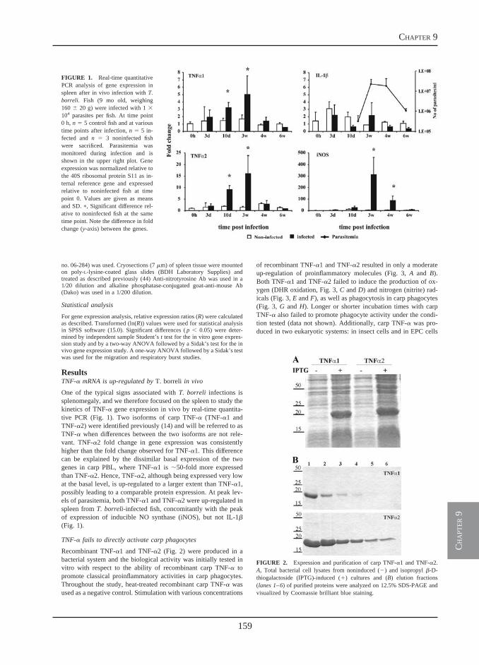
159
chapter 9
ch
apt
er 9
no. 06-284) was used. Cryosections (7 �m) of spleen tissue were mountedon poly-L-lysine-coated glass slides (BDH Laboratory Supplies) andtreated as described previously (44) Anti-nitrotyrosine Ab was used in a1/20 dilution and alkaline phosphatase-conjugated goat-anti-mouse Ab(Dako) was used in a 1/200 dilution.
Statistical analysis
For gene expression analysis, relative expression ratios (R) were calculatedas described. Transformed (ln(R)) values were used for statistical analysisin SPSS software (15.0). Significant differences ( p � 0.05) were deter-mined by independent sample Student’s t test for the in vitro gene expres-sion study and by a two-way ANOVA followed by a Sidak’s test for the invivo gene expression study. A one-way ANOVA followed by a Sidak’s testwas used for the migration and respiratory burst studies.
ResultsTNF-� mRNA is up-regulated by T. borreli in vivo
One of the typical signs associated with T. borreli infections issplenomegaly, and we therefore focused on the spleen to study thekinetics of TNF-� gene expression in vivo by real-time quantita-tive PCR (Fig. 1). Two isoforms of carp TNF-� (TNF-�1 andTNF-�2) were identified previously (14) and will be referred to asTNF-� when differences between the two isoforms are not rele-vant. TNF-�2 fold change in gene expression was consistentlyhigher than the fold change observed for TNF-�1. This differencecan be explained by the dissimilar basal expression of the twogenes in carp PBL, where TNF-�1 is �50-fold more expressedthan TNF-�2. Hence, TNF-�2, although being expressed very lowat the basal level, is up-regulated to a larger extent than TNF-�1,possibly leading to a comparable protein expression. At peak lev-els of parasitemia, both TNF-�1 and TNF-�2 were up-regulated inspleen from T. borreli-infected fish, concomitantly with the peakof expression of inducible NO synthase (iNOS), but not IL-1�(Fig. 1).
TNF-� fails to directly activate carp phagocytes
Recombinant TNF-�1 and TNF-�2 (Fig. 2) were produced in abacterial system and the biological activity was initially tested invitro with respect to the ability of recombinant carp TNF-� topromote classical proinflammatory activities in carp phagocytes.Throughout the study, heat-treated recombinant carp TNF-� wasused as a negative control. Stimulation with various concentrations
of recombinant TNF-�1 and TNF-�2 resulted in only a moderateup-regulation of proinflammatory molecules (Fig. 3, A and B).Both TNF-�1 and TNF-�2 failed to induce the production of ox-ygen (DHR oxidation, Fig. 3, C and D) and nitrogen (nitrite) rad-icals (Fig. 3, E and F), as well as phagocytosis in carp phagocytes(Fig. 3, G and H). Longer or shorter incubation times with carpTNF-� also failed to promote phagocyte activity under the condi-tion tested (data not shown). Additionally, carp TNF-� was pro-duced in two eukaryotic systems: in insect cells and in EPC cells
FIGURE 1. Real-time quantitativePCR analysis of gene expression inspleen after in vivo infection with T.borreli. Fish (9 mo old, weighing160 � 20 g) were infected with 1 �104 parasites per fish. At time point0 h, n � 5 control fish and at varioustime points after infection, n � 5 in-fected and n � 3 noninfected fishwere sacrificed. Parasitemia wasmonitored during infection and isshown in the upper right plot. Geneexpression was normalized relative tothe 40S ribosomal protein S11 as in-ternal reference gene and expressedrelative to noninfected fish at timepoint 0. Values are given as meansand SD. �, Significant difference rel-ative to noninfected fish at the sametime point. Note the difference in foldchange (y-axis) between the genes.
FIGURE 2. Expression and purification of carp TNF-�1 and TNF-�2.A, Total bacterial cell lysates from noninduced (�) and isopropyl �-D-thiogalactoside (IPTG)-induced (�) cultures and (B) elution fractions(lanes 1–6) of purified proteins were analyzed on 12.5% SDS-PAGE andvisualized by Coomassie brilliant blue staining.
5The Journal of Immunology
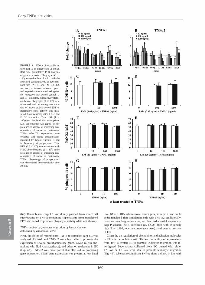
160
Carp TNFa activities
ch
apt
er 9
(62). Recombinant carp TNF-�, affinity purified from insect cellsupernatants or TNF-�-containing supernatants from transfectedEPC also failed to promote phagocyte activity (data not shown).
TNF-� indirectly promotes migration of leukocytes viaactivation of endothelial cells
Next, the ability of recombinant TNF-� to stimulate carp EC wasanalyzed. TNF-�1 and TNF-�2 were both able to promote theexpression of several proinflammatory genes, CXCa (a fish che-mokine with IL-8 characteristics), and adhesion molecules in EC(Fig. 4A). TNF-�2 was more potent than TNF-�1 in promotinggene expression. iNOS gene expression was present at low basal
level (R � 0.0045, relative to reference gene) in carp EC and couldbe up-regulated after stimulation, only with TNF-�2. Additionally,based on homology sequencing, we identified a partial sequence ofcarp P-selectin (Sele, accession no. GQ231486) with extremelyhigh (R � 1.395, relative to reference gene) basal gene expressionin EC.
Given the up-regulation of chemokines and adhesion moleculesin EC after stimulation with TNF-�, the ability of supernatantsfrom TNF-�-treated EC to promote leukocyte migration was in-vestigated. Supernatants collected from EC treated with eitherTNF-�1 or TNF-�2 were able to promote leukocyte migration(Fig. 4B), whereas recombinant TNF-� alone did not. In line with
FIGURE 3. Effects of recombinantcarp TNF-� on phagocytes: A and B,Real-time quantitative PCR analysisof gene expression. Phagocytes (1 �106) were stimulated for 3 h with theindicated concentrations of recombi-nant carp TNF-�1 and TNF-�2. 40Swas used as internal reference gene,and expression was normalized againstthe respective heat-treated control. Cand D, Respiratory burst activity (DHRoxidation). Phagocytes (1 � 106) werestimulated with increasing concentra-tion of native or heat-treated TNF-�.Respiratory burst activity was mea-sured fluorometrically after 1 h. E andF, NO production. Total HKL (1 �106) were stimulated with a suboptimalLPS concentration (20 �g/ml) in thepresence or absence of increasing con-centrations of native or heat-treatedTNF-�. After 72 h supernatants werecollected and nitrite concentrationsmeasured by Griess reaction. G andH, Percentage of phagocytosis. TotalHKL (0.5 � 106) were stimulated withFITC-labeled bacteria (5 � 106) in thepresence or absence of increasing con-centrations of native or heat-treatedTNF-�. Percentage of phagocytosiswas determined fluorometrically after30 min.
6 RECEPTOR-MEDIATED AND LECTIN-LIKE ACTIVITIES OF CARP TNF-�
the difference observed at the gene expression level, TNF-�2-treated supernatants were more potent than TNF-�1-treated super-natants in promoting leukocyte migration.
TNF-� directly primes and indirectly promotes phagocyteactivation by acting on endothelial cells
To investigate the indirect activation of phagocytes by carpTNF-�, supernatants from TNF-�-treated EC were used to pro-mote the production of oxygen radicals (DHR oxidation) in carpphagocytes (Fig. 5). Clearly, increased DHR oxidation was ob-served in granulocytes (Fig. 5A, gate B) in samples incubated for30–60 min with supernatants from TNF-�-treated EC (Fig. 5, Aand B, gray bars). Preincubation of phagocytes for as short as 10min with carp TNF-�1 or TNF-�2 resulted in a more rapid (15min) DHR oxidation of granulocytes (Fig. 5, A and B, black bars).Preincubation with TNF-�2 resulted in a higher production of ox-ygen radicals than did preincubation with TNF-�1. The differencein oxidation of DHR in phagocytes prestimulated, or not, withTNF-� was less pronounced after 30 min. However, after 60 min,oxidation of DHR in prestimulated cells was significantly lowerthan in non-prestimulated phagocytes, with a greater effect visiblein TNF-�1-prestimulated cells (compare gray and black bars inFig. 5B). This indicates that although carp TNF-� was not able todirectly promote the production of oxygen radicals in phagocytes,TNF-� can prime phagocytes to respond faster to EC-derivedmediators.
Lectin-like activity of carp TNF-�
Carp TNF-� is homologous to mammalian TNF-� and presentsmany conserved structural features of its mammalian counterpart
(14). We produced a putative three-dimensional model based onthe crystal structure of mouse TNF-� (75), confirming that alsocarp TNF-� might exist as a homotrimer, with each subunit con-sisting of anti-parallel �-sheets organized in a “jelly-roll” motifwith a pyramid shape (Fig. 6). Mammalian TNF-� has a lectindomain located at the top of the pyramid-shaped molecule alsoreferred to as TIP domain. Carp TNF-� has a conserved three-dimensional structure clearly identifying a TIP domain (Fig. 6A).More detailed analysis of the three-dimensional structure and ofthe primary sequence corresponding to the TIP region evidencedconsiderable differences between TNF-�1 and TNF-�2 (Fig. 6, Band C).
We investigated whether the lectin-like activity ascribed tomammalian TNF-�, and known to act through its TIP domain (31,32), would be conserved in the homologous carp TNF-� molecule.The ability of carp TNF-�1 and TNF-�2 to kill the bloodstreamform of T. brucei (AnTat 1.1 clone) was investigated in a trypano-lytic assay. Both TNF-�1 and TNF-�2 killed T. brucei in a dose-and time-dependent manner (data not shown) where, maximally, 1�g/ml carp TNF-� killed 100% of parasites within 4 h (Fig. 7, Aand B). Trypanolytic activity of carp TNF-� against T. bruceicould be blocked by preincubation with increasing concentrationsof N,N�-diacetylchitobiose (Fig. 7, A and B), confirming thatTNF-� activity acted via a lectin-like interaction. In parasite cul-tures incubated with TNF-�1, N,N�-diacetylchitobiose completelyblocked TNF-�1 activity at concentrations �1 ng/ml, whereas incultures incubated with TNF-�2 even the highest concentration ofN,N�-diacetylchitobiose could not completely block TNF-�2 activity.Such a difference could be due to the dissimilarities observed in the
FIGURE 4. Effects of recombinant carp TNF-� on EC. A, Real-time quantitative PCR analysis of gene expression. Endothelial cells were cultured in24-well plates until confluent and then stimulated for 4 h with the indicated concentrations of carp TNF-�1 and TNF-�2. Gene expression was normalizedrelative to the 40S ribosomal protein S11 as internal reference gene and was expressed relative to the respective heat-treated control. Values are given asmeans and SD (n � 4). �, Statistical differences relative to the heat-treated control. B, Migration of leukocytes. Total HKL (4 � 106) were layered on theupper well of a transmigration chamber, and migration toward TNF-� alone or toward supernatants from TNF-�-treated EC was recorded after 3 h. Pooledcarp serum (10%) was used as positive control and complete cRPMI as negative control. The total number of cells that migrated toward supernatants fromEC stimulated with native TNF-� was corrected for the number of cells that migrated toward supernatants from EC stimulated with heat-treated TNF-�.�, Significant differences relative to the medium control or to supernatants form samples stimulated with the respective heat-treated control; #, significantdifferences between supernatants treated with TNF-�1 and TNF-�2. P-sele indicates carp P-selectin; SPN, supernatant.
7The Journal of Immunology
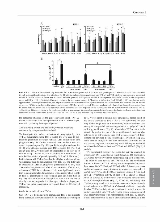
161
chapter 9
ch
apt
er 9
(62). Recombinant carp TNF-�, affinity purified from insect cellsupernatants or TNF-�-containing supernatants from transfectedEPC also failed to promote phagocyte activity (data not shown).
TNF-� indirectly promotes migration of leukocytes viaactivation of endothelial cells
Next, the ability of recombinant TNF-� to stimulate carp EC wasanalyzed. TNF-�1 and TNF-�2 were both able to promote theexpression of several proinflammatory genes, CXCa (a fish che-mokine with IL-8 characteristics), and adhesion molecules in EC(Fig. 4A). TNF-�2 was more potent than TNF-�1 in promotinggene expression. iNOS gene expression was present at low basal
level (R � 0.0045, relative to reference gene) in carp EC and couldbe up-regulated after stimulation, only with TNF-�2. Additionally,based on homology sequencing, we identified a partial sequence ofcarp P-selectin (Sele, accession no. GQ231486) with extremelyhigh (R � 1.395, relative to reference gene) basal gene expressionin EC.
Given the up-regulation of chemokines and adhesion moleculesin EC after stimulation with TNF-�, the ability of supernatantsfrom TNF-�-treated EC to promote leukocyte migration was in-vestigated. Supernatants collected from EC treated with eitherTNF-�1 or TNF-�2 were able to promote leukocyte migration(Fig. 4B), whereas recombinant TNF-� alone did not. In line with
FIGURE 3. Effects of recombinantcarp TNF-� on phagocytes: A and B,Real-time quantitative PCR analysisof gene expression. Phagocytes (1 �106) were stimulated for 3 h with theindicated concentrations of recombi-nant carp TNF-�1 and TNF-�2. 40Swas used as internal reference gene,and expression was normalized againstthe respective heat-treated control. Cand D, Respiratory burst activity (DHRoxidation). Phagocytes (1 � 106) werestimulated with increasing concentra-tion of native or heat-treated TNF-�.Respiratory burst activity was mea-sured fluorometrically after 1 h. E andF, NO production. Total HKL (1 �106) were stimulated with a suboptimalLPS concentration (20 �g/ml) in thepresence or absence of increasing con-centrations of native or heat-treatedTNF-�. After 72 h supernatants werecollected and nitrite concentrationsmeasured by Griess reaction. G andH, Percentage of phagocytosis. TotalHKL (0.5 � 106) were stimulated withFITC-labeled bacteria (5 � 106) in thepresence or absence of increasing con-centrations of native or heat-treatedTNF-�. Percentage of phagocytosiswas determined fluorometrically after30 min.
6 RECEPTOR-MEDIATED AND LECTIN-LIKE ACTIVITIES OF CARP TNF-�
the difference observed at the gene expression level, TNF-�2-treated supernatants were more potent than TNF-�1-treated super-natants in promoting leukocyte migration.
TNF-� directly primes and indirectly promotes phagocyteactivation by acting on endothelial cells
To investigate the indirect activation of phagocytes by carpTNF-�, supernatants from TNF-�-treated EC were used to pro-mote the production of oxygen radicals (DHR oxidation) in carpphagocytes (Fig. 5). Clearly, increased DHR oxidation was ob-served in granulocytes (Fig. 5A, gate B) in samples incubated for30–60 min with supernatants from TNF-�-treated EC (Fig. 5, Aand B, gray bars). Preincubation of phagocytes for as short as 10min with carp TNF-�1 or TNF-�2 resulted in a more rapid (15min) DHR oxidation of granulocytes (Fig. 5, A and B, black bars).Preincubation with TNF-�2 resulted in a higher production of ox-ygen radicals than did preincubation with TNF-�1. The differencein oxidation of DHR in phagocytes prestimulated, or not, withTNF-� was less pronounced after 30 min. However, after 60 min,oxidation of DHR in prestimulated cells was significantly lowerthan in non-prestimulated phagocytes, with a greater effect visiblein TNF-�1-prestimulated cells (compare gray and black bars inFig. 5B). This indicates that although carp TNF-� was not able todirectly promote the production of oxygen radicals in phagocytes,TNF-� can prime phagocytes to respond faster to EC-derivedmediators.
Lectin-like activity of carp TNF-�
Carp TNF-� is homologous to mammalian TNF-� and presentsmany conserved structural features of its mammalian counterpart
(14). We produced a putative three-dimensional model based onthe crystal structure of mouse TNF-� (75), confirming that alsocarp TNF-� might exist as a homotrimer, with each subunit con-sisting of anti-parallel �-sheets organized in a “jelly-roll” motifwith a pyramid shape (Fig. 6). Mammalian TNF-� has a lectindomain located at the top of the pyramid-shaped molecule alsoreferred to as TIP domain. Carp TNF-� has a conserved three-dimensional structure clearly identifying a TIP domain (Fig. 6A).More detailed analysis of the three-dimensional structure and ofthe primary sequence corresponding to the TIP region evidencedconsiderable differences between TNF-�1 and TNF-�2 (Fig. 6, Band C).
We investigated whether the lectin-like activity ascribed tomammalian TNF-�, and known to act through its TIP domain (31,32), would be conserved in the homologous carp TNF-� molecule.The ability of carp TNF-�1 and TNF-�2 to kill the bloodstreamform of T. brucei (AnTat 1.1 clone) was investigated in a trypano-lytic assay. Both TNF-�1 and TNF-�2 killed T. brucei in a dose-and time-dependent manner (data not shown) where, maximally, 1�g/ml carp TNF-� killed 100% of parasites within 4 h (Fig. 7, Aand B). Trypanolytic activity of carp TNF-� against T. bruceicould be blocked by preincubation with increasing concentrationsof N,N�-diacetylchitobiose (Fig. 7, A and B), confirming thatTNF-� activity acted via a lectin-like interaction. In parasite cul-tures incubated with TNF-�1, N,N�-diacetylchitobiose completelyblocked TNF-�1 activity at concentrations �1 ng/ml, whereas incultures incubated with TNF-�2 even the highest concentration ofN,N�-diacetylchitobiose could not completely block TNF-�2 activity.Such a difference could be due to the dissimilarities observed in the
FIGURE 4. Effects of recombinant carp TNF-� on EC. A, Real-time quantitative PCR analysis of gene expression. Endothelial cells were cultured in24-well plates until confluent and then stimulated for 4 h with the indicated concentrations of carp TNF-�1 and TNF-�2. Gene expression was normalizedrelative to the 40S ribosomal protein S11 as internal reference gene and was expressed relative to the respective heat-treated control. Values are given asmeans and SD (n � 4). �, Statistical differences relative to the heat-treated control. B, Migration of leukocytes. Total HKL (4 � 106) were layered on theupper well of a transmigration chamber, and migration toward TNF-� alone or toward supernatants from TNF-�-treated EC was recorded after 3 h. Pooledcarp serum (10%) was used as positive control and complete cRPMI as negative control. The total number of cells that migrated toward supernatants fromEC stimulated with native TNF-� was corrected for the number of cells that migrated toward supernatants from EC stimulated with heat-treated TNF-�.�, Significant differences relative to the medium control or to supernatants form samples stimulated with the respective heat-treated control; #, significantdifferences between supernatants treated with TNF-�1 and TNF-�2. P-sele indicates carp P-selectin; SPN, supernatant.
7The Journal of Immunology
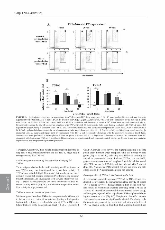
162
Carp TNFa activities
ch
apt
er 9
TIP region. Collectively, these results indicate that both isoforms ofcarp TNF-� have lectin-like activities and that TNF-�2 might have astronger activity than TNF-�1.
Evolutionary conservation of the lectin-like activity of fishTNF-�
To investigate whether the lectin-like activity would be limited tocarp TNF-� only, we investigated the trypanolytic activity ofTNF-� from zebrafish (both Cyprinidae) but also from two moredistantly related fish species, seabream (Perciformes) and rainbowtrout (Salmonidae). All investigated TNF-� were effective in kill-ing T. brucei at concentrations and time comparable to that ob-served for carp TNF-� (Fig. 7C), further confirming that the lectin-like activity is highly conserved.
TNF-� is essential to control parasitemia
We investigated the role of TNF-� in vivo particularly with respectto fish survival and control of parasitemia. Starting at 1 wk postin-fection, infected fish received a daily dose of PTX, a TNF-� in-hibitor that acts at the transcriptional level (14, 76). Fish injected
with PTX showed lower survival and higher parasitemia at all timepoints after infection when compared with the infected controlgroup (Fig. 8, A and B), indicating that TNF-� is critically in-volved in parasitemia control. Reduced TNF-�, but not iNOS,gene expression was observed in spleen from infected fish treatedwith PTX, but not in PBS-injected fish infected with T. borreli(Fig. 8C). Noninfected PTX-injected fish did not show any sideeffects due to PTX administration (data not shown).
Overexpression of TNF-� is detrimental to the host
A recombinant plasmid expressing TNF-�1 or TNF-�2 was con-structed to investigate the immunomodulatory activity of excessTNF-� during in vivo T. borreli infection. Fish treated with var-ious doses of recombinant plasmid encoding either TNF-�1 orTNF-�2 all showed lower survival than the infected control group,with the group injected with a high dose of TNF-�2 plasmid show-ing the lowest survival (Fig. 8D). Despite the clear effect on sur-vival, parasitemia was not significantly affected. For clarity, onlythe parasitemia curve of the group injected with a high dose ofTNF-�2 plasmid is shown (Fig. 8E). TNF-� plasmid-injected fish
FIGURE 5. Activation of phagocytes by supernatants from TNF-�-treated EC. Carp phagocytes (1 � 106) were incubated for the indicated time withsupernatants collected from TNF-�-treated EC in the presence of DHR (0.1 �g/ml). Alternatively, cells were first preincubated for 10 min with 1 �g/mlcarp TNF-�1 or TNF-�2. For the last 15 min, PMA was added to the culture and fluorescence values of 104 events were acquired fluorometrically. A,Representative scatter dot plot of phagocytes stimulated with TNF-�2-treated EC supernatants. Cells were directly incubated with TNF-�2-treated ECsupernatants (upper panel) or pretreated with TNF-�2 and subsequently stimulated with the respective supernatant (lower panel). Gate A indicates totalDHR� cells and gate B indicates a granulocyte subpopulation with increased fluorescence intensity. B, Positive cells in gate B in phagocyte cultures directlystimulated with EC supernatants (gray bars) or preincubated with TNF-� and subsequently stimulated with the respective supernatant (black bars).Measurements were performed in quadruplicate. Values are given as means and SD. �, Significant differences with respect to supernatant from ECstimulated with heat-treated TNF-�; #, significant differences between prestimulated and non-prestimulated phagocytes. Shown is one representativeexperiment of two independent experiments performed.
8 RECEPTOR-MEDIATED AND LECTIN-LIKE ACTIVITIES OF CARP TNF-�
showed high TNF-� protein expression in muscle tissue at theinjection site as detected by Western blot (Fig. 8F). Parallel ex-periments performed by i.p. injection of high and low doses ofrecombinant carp TNF-�1 and TNF-�2 showed similar results,where all groups treated with TNF-� had lower survival than didthe infected control group (data not shown).
Membrane-bound TNF-� is crucial for full protection of thehost
An inhibitor of TACE was used in vivo during T. borreli infectionto prevent TNF-� shedding, thereby increasing mTNF-� levels(73). About 2.5 wk (18 days) after infection fish reached a para-sitemia of 2 � 106 parasites/ml of blood and received TACE in-hibitor. After 3 days of treatment only, in all fish treated with theTACE inhibitor, complete clearance of the parasite was observed,while in fish treated with a general matrix metalloproteinase in-hibitor, which has no effect on TACE, parasitemia progressed nor-mally (Fig. 8, G and H). In vitro, stimulation of carp leukocyteswith LPS in the presence of the TACE inhibitor significantly in-creased the number of positive cells with detectable surface-boundTNF-� in a time-dependent manner (Fig. 8I). Additionally, fishtreated with the TACE inhibitor displayed extremely reducedspleen size as compared with the control group (data not shown).Neither the TACE inhibitor nor the control compound showed di-rect parasite toxicity in vitro in a concentration range from 0.2 to
100 �M (data not shown). The effects of the TACE inhibitor onparasitemia and spleen size led us to investigate whether otherparameters generally associated with pathology of infection, suchas plasma nitrite levels and tissue nitration, would also be affected.As expected, plasma nitrite levels were strongly reduced, althoughnot significantly owing to high variation between individuals, infish treated with the TACE inhibitor (Fig. 9A). Tissue nitration, asmeasured by immunohistochemistry using an anti-nitrotyrosine Ab(44), was also reduced by treatment with TACE inhibitor, as in-dicated by the lower nitrotyrosine staining in the spleen of treatedfish (Fig. 9B).
FIGURE 6. Three-dimensional analysis of carp TNF-�. A, Side view ofcarp TNF-�1 with the individual monomers shown in magenta, blue, andgreen. At the top of the pyramid-shaped molecule is the TIP domain. B,Top view of superimposed TNF-�1 (orange) and TNF-�2 (gray) trimericmolecules showing the different orientation of the loops forming the TIPdomain in each of the two molecules. C, ClustalW alignment of the aminoacid portion corresponding to the TIP region of TNF-�1 and TNF-�2.Despite the high similarity in the surrounding region, the TIP region (yel-low) shows a considerable degree of variation.
FIGURE 7. Lysis of bloodstream forms of T. brucei by fish TNF-�.Freshly isolated parasites (2 � 106/ml) were incubated in PSG (pH 8.0)with TNF-� (1 �g/ml) for 4 h at 37°C. Parasites were incubated with carpTNF-�1 (A) or carp TNF-�2 (B) alone or after preincubation with increas-ing concentrations of N,N�-diacetylchitobiose or (C) with zebrafish, sea-bream, or trout.
9The Journal of Immunology
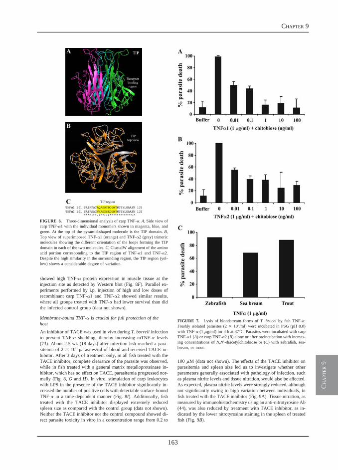
163
chapter 9
ch
apt
er 9
TIP region. Collectively, these results indicate that both isoforms ofcarp TNF-� have lectin-like activities and that TNF-�2 might have astronger activity than TNF-�1.
Evolutionary conservation of the lectin-like activity of fishTNF-�
To investigate whether the lectin-like activity would be limited tocarp TNF-� only, we investigated the trypanolytic activity ofTNF-� from zebrafish (both Cyprinidae) but also from two moredistantly related fish species, seabream (Perciformes) and rainbowtrout (Salmonidae). All investigated TNF-� were effective in kill-ing T. brucei at concentrations and time comparable to that ob-served for carp TNF-� (Fig. 7C), further confirming that the lectin-like activity is highly conserved.
TNF-� is essential to control parasitemia
We investigated the role of TNF-� in vivo particularly with respectto fish survival and control of parasitemia. Starting at 1 wk postin-fection, infected fish received a daily dose of PTX, a TNF-� in-hibitor that acts at the transcriptional level (14, 76). Fish injected
with PTX showed lower survival and higher parasitemia at all timepoints after infection when compared with the infected controlgroup (Fig. 8, A and B), indicating that TNF-� is critically in-volved in parasitemia control. Reduced TNF-�, but not iNOS,gene expression was observed in spleen from infected fish treatedwith PTX, but not in PBS-injected fish infected with T. borreli(Fig. 8C). Noninfected PTX-injected fish did not show any sideeffects due to PTX administration (data not shown).
Overexpression of TNF-� is detrimental to the host
A recombinant plasmid expressing TNF-�1 or TNF-�2 was con-structed to investigate the immunomodulatory activity of excessTNF-� during in vivo T. borreli infection. Fish treated with var-ious doses of recombinant plasmid encoding either TNF-�1 orTNF-�2 all showed lower survival than the infected control group,with the group injected with a high dose of TNF-�2 plasmid show-ing the lowest survival (Fig. 8D). Despite the clear effect on sur-vival, parasitemia was not significantly affected. For clarity, onlythe parasitemia curve of the group injected with a high dose ofTNF-�2 plasmid is shown (Fig. 8E). TNF-� plasmid-injected fish
FIGURE 5. Activation of phagocytes by supernatants from TNF-�-treated EC. Carp phagocytes (1 � 106) were incubated for the indicated time withsupernatants collected from TNF-�-treated EC in the presence of DHR (0.1 �g/ml). Alternatively, cells were first preincubated for 10 min with 1 �g/mlcarp TNF-�1 or TNF-�2. For the last 15 min, PMA was added to the culture and fluorescence values of 104 events were acquired fluorometrically. A,Representative scatter dot plot of phagocytes stimulated with TNF-�2-treated EC supernatants. Cells were directly incubated with TNF-�2-treated ECsupernatants (upper panel) or pretreated with TNF-�2 and subsequently stimulated with the respective supernatant (lower panel). Gate A indicates totalDHR� cells and gate B indicates a granulocyte subpopulation with increased fluorescence intensity. B, Positive cells in gate B in phagocyte cultures directlystimulated with EC supernatants (gray bars) or preincubated with TNF-� and subsequently stimulated with the respective supernatant (black bars).Measurements were performed in quadruplicate. Values are given as means and SD. �, Significant differences with respect to supernatant from ECstimulated with heat-treated TNF-�; #, significant differences between prestimulated and non-prestimulated phagocytes. Shown is one representativeexperiment of two independent experiments performed.
8 RECEPTOR-MEDIATED AND LECTIN-LIKE ACTIVITIES OF CARP TNF-�
showed high TNF-� protein expression in muscle tissue at theinjection site as detected by Western blot (Fig. 8F). Parallel ex-periments performed by i.p. injection of high and low doses ofrecombinant carp TNF-�1 and TNF-�2 showed similar results,where all groups treated with TNF-� had lower survival than didthe infected control group (data not shown).
Membrane-bound TNF-� is crucial for full protection of thehost
An inhibitor of TACE was used in vivo during T. borreli infectionto prevent TNF-� shedding, thereby increasing mTNF-� levels(73). About 2.5 wk (18 days) after infection fish reached a para-sitemia of 2 � 106 parasites/ml of blood and received TACE in-hibitor. After 3 days of treatment only, in all fish treated with theTACE inhibitor, complete clearance of the parasite was observed,while in fish treated with a general matrix metalloproteinase in-hibitor, which has no effect on TACE, parasitemia progressed nor-mally (Fig. 8, G and H). In vitro, stimulation of carp leukocyteswith LPS in the presence of the TACE inhibitor significantly in-creased the number of positive cells with detectable surface-boundTNF-� in a time-dependent manner (Fig. 8I). Additionally, fishtreated with the TACE inhibitor displayed extremely reducedspleen size as compared with the control group (data not shown).Neither the TACE inhibitor nor the control compound showed di-rect parasite toxicity in vitro in a concentration range from 0.2 to
100 �M (data not shown). The effects of the TACE inhibitor onparasitemia and spleen size led us to investigate whether otherparameters generally associated with pathology of infection, suchas plasma nitrite levels and tissue nitration, would also be affected.As expected, plasma nitrite levels were strongly reduced, althoughnot significantly owing to high variation between individuals, infish treated with the TACE inhibitor (Fig. 9A). Tissue nitration, asmeasured by immunohistochemistry using an anti-nitrotyrosine Ab(44), was also reduced by treatment with TACE inhibitor, as in-dicated by the lower nitrotyrosine staining in the spleen of treatedfish (Fig. 9B).
FIGURE 6. Three-dimensional analysis of carp TNF-�. A, Side view ofcarp TNF-�1 with the individual monomers shown in magenta, blue, andgreen. At the top of the pyramid-shaped molecule is the TIP domain. B,Top view of superimposed TNF-�1 (orange) and TNF-�2 (gray) trimericmolecules showing the different orientation of the loops forming the TIPdomain in each of the two molecules. C, ClustalW alignment of the aminoacid portion corresponding to the TIP region of TNF-�1 and TNF-�2.Despite the high similarity in the surrounding region, the TIP region (yel-low) shows a considerable degree of variation.
FIGURE 7. Lysis of bloodstream forms of T. brucei by fish TNF-�.Freshly isolated parasites (2 � 106/ml) were incubated in PSG (pH 8.0)with TNF-� (1 �g/ml) for 4 h at 37°C. Parasites were incubated with carpTNF-�1 (A) or carp TNF-�2 (B) alone or after preincubation with increas-ing concentrations of N,N�-diacetylchitobiose or (C) with zebrafish, sea-bream, or trout.
9The Journal of Immunology
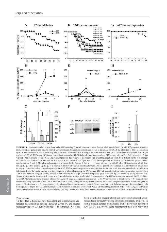
164
Carp TNFa activities
ch
apt
er 9
DiscussionTo date, TNF-� homologs have been identified in mammalian ver-tebrates, one amphibian species (Xenopus laevis) (9), and severalteleost species (10–23) but not in birds (7, 8). Although TNF-� has
been identified in several teleost fish species its biological activi-ties and role particularly during infections are largely unknown. Infish, a limited number of functional studies have been performed(20–22, 24–27), mostly using recombinant TNF-� in vitro, and
FIGURE 8. Immunomodulation by soluble and mTNF-� during T. borreli infection in vivo. At time 0 fish were infected i.p. with 104 parasites. Mortality(top panels) and parasitemia (middle panels) were monitored. Control experiments are shown in the lower panels. A–C, Inhibition of TNF-� expressionby PTX administration. A and B, Mortality and parasitemia in infected fish. Starting 1 wk after infection, fish (n � 12) received a daily dose of PTX (50mg/kg) or PBS. C, TNF-� and iNOS gene expression (quantitative RT-PCR) in spleen of nontreated and PTX-treated infected fish. Spleen from n � 5 fishwas collected at 24 days postinfection. Shown are expression data relative to the noninfected fish at the same time point. Note that for clarity, fold changesof TNF-�1 and TNF-�2 are indicated on the left axis and iNOS of the right axis. D–F, Overexpression of TNF-� by recombinant plasmid DNAadministration. D and E, Mortality and parasitemia in infected fish. At time 0, fish (n � 11) were injected i.m. with 25 �l of PBS containing a high dose(10 �g/10 g), a low dose (1 �g/10 g), or a mixture of the two of plasmid encoding for carp TNF-�1 (a1) or TNF-�2 (a2). Fish injected with a high doseof empty plasmid served as infected negative control. F, TNF-� protein expression at the injection site in muscle of plasmid-injected fish. Muscle fromfish injected with the empty plasmid or with a high dose of plasmid encoding for TNF-�1 and TNF-�2 was collected for protein expression analysis. CarpTNF-� was detected using an affinity-purified rabbit anti-carp TNF-� IgG and HRP-conjugated goat-anti-rabbit IgG as secondary Ab by Western blot.Shown are the results from one fish out of n � 3 tested showing similar results. G–I, Overexpression of mTNF-� by administration of TACE inhibitor.G and H, Mortality and parasitemia in infected fish. After 18 days, when parasitemia reached �2 � 106 parasites/ml of blood, fish (n � 9) received threeconsecutive daily injections of the TACE inhibitor (PKF242-484, 10 mg/ml) or equivalent doses of the control compound (FN439). Values are given asmean � SD of at least n � 7 measurements. �, Significant differences with respect to the infected control group. I, Relative increase of the number of cellsbearing surface-bound TNF-�. Carp leukocytes were stimulated in triplicate wells with LPS (50 �g/ml) in the presence of PKF242-484 (50 �M) and valuesare expressed relative to leukocytes stimulated with LPS only. Shown are results from one representative experiment out of three performed independently.
10 RECEPTOR-MEDIATED AND LECTIN-LIKE ACTIVITIES OF CARP TNF-�
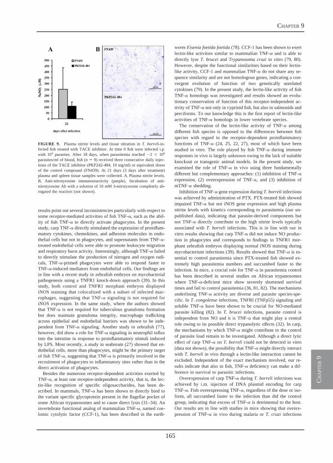
165
chapter 9
ch
apt
er 9
DiscussionTo date, TNF-� homologs have been identified in mammalian ver-tebrates, one amphibian species (Xenopus laevis) (9), and severalteleost species (10–23) but not in birds (7, 8). Although TNF-� has
been identified in several teleost fish species its biological activi-ties and role particularly during infections are largely unknown. Infish, a limited number of functional studies have been performed(20–22, 24–27), mostly using recombinant TNF-� in vitro, and
FIGURE 8. Immunomodulation by soluble and mTNF-� during T. borreli infection in vivo. At time 0 fish were infected i.p. with 104 parasites. Mortality(top panels) and parasitemia (middle panels) were monitored. Control experiments are shown in the lower panels. A–C, Inhibition of TNF-� expressionby PTX administration. A and B, Mortality and parasitemia in infected fish. Starting 1 wk after infection, fish (n � 12) received a daily dose of PTX (50mg/kg) or PBS. C, TNF-� and iNOS gene expression (quantitative RT-PCR) in spleen of nontreated and PTX-treated infected fish. Spleen from n � 5 fishwas collected at 24 days postinfection. Shown are expression data relative to the noninfected fish at the same time point. Note that for clarity, fold changesof TNF-�1 and TNF-�2 are indicated on the left axis and iNOS of the right axis. D–F, Overexpression of TNF-� by recombinant plasmid DNAadministration. D and E, Mortality and parasitemia in infected fish. At time 0, fish (n � 11) were injected i.m. with 25 �l of PBS containing a high dose(10 �g/10 g), a low dose (1 �g/10 g), or a mixture of the two of plasmid encoding for carp TNF-�1 (a1) or TNF-�2 (a2). Fish injected with a high doseof empty plasmid served as infected negative control. F, TNF-� protein expression at the injection site in muscle of plasmid-injected fish. Muscle fromfish injected with the empty plasmid or with a high dose of plasmid encoding for TNF-�1 and TNF-�2 was collected for protein expression analysis. CarpTNF-� was detected using an affinity-purified rabbit anti-carp TNF-� IgG and HRP-conjugated goat-anti-rabbit IgG as secondary Ab by Western blot.Shown are the results from one fish out of n � 3 tested showing similar results. G–I, Overexpression of mTNF-� by administration of TACE inhibitor.G and H, Mortality and parasitemia in infected fish. After 18 days, when parasitemia reached �2 � 106 parasites/ml of blood, fish (n � 9) received threeconsecutive daily injections of the TACE inhibitor (PKF242-484, 10 mg/ml) or equivalent doses of the control compound (FN439). Values are given asmean � SD of at least n � 7 measurements. �, Significant differences with respect to the infected control group. I, Relative increase of the number of cellsbearing surface-bound TNF-�. Carp leukocytes were stimulated in triplicate wells with LPS (50 �g/ml) in the presence of PKF242-484 (50 �M) and valuesare expressed relative to leukocytes stimulated with LPS only. Shown are results from one representative experiment out of three performed independently.
10 RECEPTOR-MEDIATED AND LECTIN-LIKE ACTIVITIES OF CARP TNF-�
results point out several inconsistencies particularly with respect tosome receptor-mediated activities of fish TNF-�, such as the abil-ity of fish TNF-� to directly activate phagocytes. In the presentstudy, carp TNF-� directly stimulated the expression of proinflam-matory cytokines, chemokines, and adhesion molecules in endo-thelial cells but not in phagocytes, and supernatants from TNF-�-treated endothelial cells were able to promote leukocyte migrationand respiratory burst activity. Interestingly, although TNF-� failedto directly stimulate the production of nitrogen and oxygen radi-cals, TNF-�-primed phagocytes were able to respond faster toTNF-�-induced mediators from endothelial cells. Our findings arein line with a recent study in zebrafish embryos on mycobacterialpathogenesis using a TNFR1 knock-down approach (39). In thisstudy, both control and TNFR1 morphant embryos displayediNOS staining that colocalized with a subset of infected mac-rophages, suggesting that TNF-� signaling is not required foriNOS expression. In the same study, where the authors showedthat TNF-� is not required for tuberculous granuloma formationbut does maintain granuloma integrity, macrophage traffickingacross epithelial and endothelial barriers was shown to be inde-pendent from TNF-� signaling. Another study in zebrafish (77),however, did show a role for TNF-� signaling in neutrophil influxinto the intestine in response to proinflammatory stimuli inducedby LPS. Most recently, a study in seabream (27) showed that en-dothelial cells, more than phagocytes, might be the primary targetof fish TNF-�, suggesting that TNF-� is primarily involved in therecruitment of phagocytes to inflammatory sites rather than in thedirect activation of phagocytes.
Besides the numerous receptor-dependent activities exerted byTNF-�, at least one receptor-independent activity, that is, the lec-tin-like recognition of specific oligosaccharides, has been de-scribed. In mammals, TNF-� has been shown to directly bind tothe variant specific glycoprotein present in the flagellar pocket ofsome African trypanosomes and to cause direct lysis (31–34). Aninvertebrate functional analog of mammalian TNF-�, named coe-lomic cytolytic factor (CCF-1), has been described in the earth-
worm Eisenia foetida foetida (78). CCF-1 has been shown to exertlectin-like activities similar to mammalian TNF-� and is able todirectly lyse T. brucei and Trypanosoma cruzi in vitro (79, 80).However, despite the functional similarities based on their lectin-like activity, CCF-1 and mammalian TNF-� do not share any se-quence similarity and are not homologous genes, indicating a con-vergent evolution of function of two genetically unrelatedcytokines (79). In the present study, the lectin-like activity of fishTNF-� homologs was investigated and results showed an evolu-tionary conservation of function of this receptor-independent ac-tivity of TNF-� not only in cyprinid fish, but also in salmonids andperciforms. To our knowledge this is the first report of lectin-likeactivities of TNF-� homologs in lower vertebrate species.
The conservation of the lectin-like activity of TNF-� amongdifferent fish species is opposed to the differences between fishspecies with regard to the receptor-dependent proinflammatoryfunctions of TNF-� (24, 25, 22, 27), most of which have beenstudied in vitro. The role played by fish TNF-� during immuneresponses in vivo is largely unknown owing to the lack of suitableknockout or transgenic animal models. In the present study, weexamined the role of TNF-� in vivo using three fundamentallydifferent but complementary approaches: (1) inhibition of TNF-�expression, (2) overexpression of TNF-�, and (3) inhibition ofmTNF-� shedding.
Inhibition of TNF-� gene expression during T. borreli infectionswas achieved by administration of PTX. PTX-treated fish showedimpaired TNF-� but not iNOS gene expression and high plasmanitrite levels with kinetics corresponding to parasitemia (our un-published data), indicating that parasite-derived components butnot TNF-� directly contribute to the high nitrite levels typicallyassociated with T. borreli infections. This is in line with our invitro results showing that carp TNF-� did not induce NO produc-tion in phagocytes and corresponds to findings in TNFR1 mor-phant zebrafish embryos displaying normal iNOS staining duringmycobacterium infections (39). Results showed that TNF-� is es-sential to control parasitemia since PTX-treated fish showed ex-tremely high parasitemia numbers and succumbed faster to theinfection. In mice, a crucial role for TNF-� in parasitemia controlhas been described in several studies on African trypanosomeswhere TNF-�-deficient mice show severely shortened survivaltimes and fail to control parasitemia (36, 81, 82). The mechanismsunderlining TNF-� activity are diverse and parasite species-spe-cific. In T. congolense infections, TNFRI (TNFp55) signaling andsoluble TNF-� have been shown to be crucial for NO-mediatedparasite killing (82). In T. brucei infections, parasite control isindependent from NO and it is TNF-� that might play a centralrole owing to its possible direct trypanolytic effects (32). In carp,the mechanisms by which TNF-� might contribute to the controlof parasite load remain to be investigated. Although a direct lyticeffect of carp TNF-� on T. borreli could not be detected in vitro(data not shown), the possibility that TNF-� might directly interactwith T. borreli in vivo through a lectin-like interaction cannot beexcluded. Independent of the exact mechanism involved, our re-sults indicate that also in fish, TNF-� deficiency can make a dif-ference in survival to parasitic infections.
Overexpression of carp TNF-� during T. borreli infections wasachieved by i.m. injection of DNA plasmid encoding for carpTNF-�. Fish overexpressing TNF-�, regardless of the dose or iso-form, all succumbed faster to the infection than did the controlgroup, indicating that excess of TNF-� is detrimental to the host.Our results are in line with studies in mice showing that overex-pression of TNF-� in vivo during malaria or T. cruzi infections
FIGURE 9. Plasma nitrite levels and tissue nitration in T. borreli-in-fected fish treated with TACE inhibitor. At time 0 fish were infected i.p.with 104 parasites. After 18 days, when parasitemia reached �2 � 106
parasites/ml of blood, fish (n � 9) received three consecutive daily injec-tions of the TACE inhibitor (PKF242-484, 10 mg/ml) or equivalent dosesof the control compound (FN439). At 21 days (3 days after treatment)plasma and spleen tissue samples were collected. A, Plasma nitrite levels.B, Anti-nitrotyrosine immunoreactivity (purple). Incubation of anti-nitrotyrosine Ab with a solution of 10 mM 3-nitrotyrosine completely ab-rogated the reaction (not shown).
11The Journal of Immunology
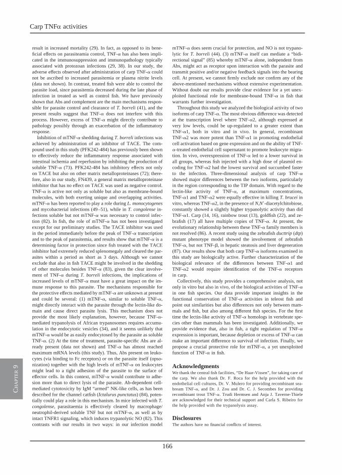
166
Carp TNFa activities
ch
apt
er 9
result in increased mortality (29). In fact, as opposed to its bene-ficial effects on parasiteamia control, TNF-� has also been impli-cated in the immunosuppression and immunopathology typicallyassociated with protozoan infections (29, 38). In our study, theadverse effects observed after administration of carp TNF-� couldnot be ascribed to increased parasitemia or plasma nitrite levels(data not shown). In contrast, treated fish were able to control theparasite load, since parasitemia decreased during the late phase ofinfection in treated as well as control fish. We have previouslyshown that Abs and complement are the main mechanisms respon-sible for parasite control and clearance of T. borreli (41), and thepresent results suggest that TNF-� does not interfere with thisprocess. However, excess of TNF-� might directly contribute topathology possibly through an exacerbation of the inflammatoryresponse.
Inhibition of mTNF-� shedding during T. borreli infections wasachieved by administration of an inhibitor of TACE. The com-pound used in this study (PFK242-484) has previously been shownto effectively reduce the inflammatory response associated withintestinal ischemia and reperfusion by inhibiting the production ofsoluble TNF-� (73). PFK242-484 has inhibitory effects not onlyon TACE but also on other matrix metalloproteinases (72); there-fore, also in our study, FN439, a general matrix metalloproteinaseinhibitor that has no effect on TACE was used as negative control.TNF-� is active not only as soluble but also as membrane-boundmolecules, with both exerting unique and overlapping activities.mTNF-� has been reported to play a role during L. monocytogenesand mycobacterial infections (49–51), while in T. congolense in-fections soluble but not mTNF-� was necessary to control infec-tion (82). In fish, the role of mTNF-� has not been investigatedexcept for our preliminary studies. The TACE inhibitor was usedin the period immediately before the peak of TNF-� transcriptionand to the peak of parasitemia, and results show that mTNF-� is adetermining factor in protection since fish treated with the TACEinhibitor had extremely reduced splenomegaly and cleared the par-asites within a period as short as 3 days. Although we cannotexclude that also in fish TACE might be involved in the sheddingof other molecules besides TNF-� (83), given the clear involve-ment of TNF-� during T. borreli infections, the implications ofincreased levels of mTNF-� must have a great impact on the im-mune response to this parasite. The mechanisms responsible forthe protective effects mediated by mTNF-� are unknown at presentand could be several: (1) mTNF-�, similar to soluble TNF-�,might directly interact with the parasite through the lectin-like do-main and cause direct parasite lysis. This mechanism does notprovide the most likely explanation, however, because TNF-�-mediated trypanolysis of African trypanosomes requires accumu-lation in the endocytotic vesicles (34), and it seems unlikely thatmTNF-� would be as easily endocytosed by the parasite as solubleTNF-�. (2) At the time of treatment, parasite-specific Abs are al-ready present (data not shown) and TNF-� has almost reachedmaximum mRNA levels (this study). Thus, Abs present on leuko-cytes (via binding to Fc receptors) or on the parasite itself (opso-nization) together with the high levels of mTNF-� on leukocytesmight lead to a tight adhesion of the parasite to the surface ofeffector cells. In this context, mTNF-� would contribute to adhe-sion more than to direct lysis of the parasite. Ab-dependent cell-mediated cytotoxicity by IgM “armed” NK-like cells, as has beendescribed for the channel catfish (Ictalurus punctatus) (84), poten-tially could play a role in this mechanism. In mice infected with T.congolense, parasitaemia is effectively cleared by macrophage/neutrophil-derived soluble TNF but not mTNF-�, as well as byintact TNFR1 signaling, which induces trypanolytic NO (82). Thiscontrasts with our results in two ways: in our infection model
mTNF-� does seem crucial for protection, and NO is not trypano-lytic for T. borreli (44). (3) mTNF-� itself can mediate a “bidi-rectional signal” (85) whereby mTNF-� alone, independent fromAbs, might act as receptor upon interaction with the parasite andtransmit positive and/or negative feedback signals into the bearingcell. At present, we cannot firmly exclude nor confirm any of theabove-mentioned mechanisms without extensive experimentation.Without doubt our results provide clear evidence for a yet unex-ploited functional role for membrane-bound TNF-� in fish thatwarrants further investigation.
Throughout this study we analyzed the biological activity of twoisoforms of carp TNF-�. The most obvious difference was detectedat the transcription level where TNF-�2, although expressed atvery low levels, could be up-regulated to a greater extent thanTNF-�1, both in vitro and in vivo. In general, recombinantTNF-�2 was more potent than TNF-�1 in promoting endothelialcell activation based on gene expression and on the ability of TNF-�-treated endothelial cell supernatant to promote leukocyte migra-tion. In vivo, overexpression of TNF-� led to a lower survival inall groups, whereas fish injected with a high dose of plasmid en-coding for TNF-�2 had the lowest survival and succumbed fasterto the infection. Three-dimensional analysis of carp TNF-�showed major differences between the two isoforms, particularlyin the region corresponding to the TIP domain. With regard to thelectin-like activity of TNF-�, at maximum concentrations,TNF-�1 and TNF-�2 were equally effective in killing T. brucei invitro, whereas TNF-�2, in the presence of N,N�-diacetylchitobiose,constantly showed a slightly higher trypanolytic activity than didTNF-�1. Carp (14, 16), rainbow trout (13), goldfish (22), and ze-brafish (17) all have multiple copies of TNF-�. At present, theevolutionary relationship between these TNF-� family members isnot resolved (86). A recent study using the zebrafish ducttrip (dtp)mutant phenotype model showed the involvement of zebrafishTNF-�, but not TNF-�, in hepatic steatosis and liver degeneration(87). Our results show that both carp TNF-� isoforms examined inthis study are biologically active. Further characterization of thebiological relevance of the differences between TNF-�1 andTNF-�2 would require identification of the TNF-� receptorsin carp.
Collectively, this study provides a comprehensive analysis, notonly in vitro but also in vivo, of the biological activities of TNF-�in one fish species. Our data provide important insights in thefunctional conservation of TNF-� activities in teleost fish andpoint out similarities but also differences not only between mam-mals and fish, but also among different fish species. For the firsttime the lectin-like activity of TNF-� homologs in vertebrate spe-cies other than mammals has been investigated. Additionally, weprovide evidence that, also in fish, a tight regulation of TNF-�expression is important, because depletion or excess of TNF-� canmake an important difference to survival of infection. Finally, wepropose a crucial protective role for mTNF-�, a yet unexploitedfunction of TNF-� in fish.
AcknowledgmentsWe thank the central fish facilities, “De Haar-Vissen”, for taking care ofthe carp. We also thank Dr. F. Roca for the help provided with theendothelial cell cultures, Dr. V. Mulero for providing recombinant sea-bream TNF-�, and Dr. J. Zou and Dr. C. J. Secombes for providingrecombinant trout TNF-�. Trudi Hermsen and Anja J. Taverne-Thieleare acknowledged for their technical support and Carla S. Ribeiro forthe help provided with the trypanolysis assay.
DisclosuresThe authors have no financial conflicts of interest.
12 RECEPTOR-MEDIATED AND LECTIN-LIKE ACTIVITIES OF CARP TNF-�
167
chapter 9
ch
apt
er 9
References1. Grell, M., and P. Scheurich. 1997. Tumor necrosis factor. In Growth Factors and
Cytokines in Health and Disease, Vol. 2B. D. LeRoith and C. Bondy, eds. JAIpress, Greenwich, pp. 669–726.
2. Peters, P. M., J. R. Ortaldo, M. R. Shalaby, L. P. Svedersky, G. E. Nedwin,T. S. Bringman, P. E. Hass, B. B. Aggarwal, R. B. Herberman, D. V. Goeddel,et al. 1986. Natural killer-sensitive targets stimulate production of TNF-� but notTNF-� (lymphotoxin) by highly purified human peripheral blood large granularlymphocytes. J. Immunol. 137: 2592–2598.
3. Cuturi, M. C., M. Murphy, M. P. Costa-Giomi, R. Weinmann, B. Perussia, andG. Trinchieri. 1987. Independent regulation of tumor necrosis factor and lym-photoxin production by human peripheral blood lymphocytes. J. Exp. Med. 165:1581–1594.
4. Sung, S. S., J. M. Bjorndahl, C. Y. Wang, H. T. Kao, and S. M. Fu. 1988.Production of tumor necrosis factor/cachectin by human T cell lines and periph-eral blood T lymphocytes stimulated by phorbol myristate acetate and anti-CD3antibody. J. Exp. Med. 167: 937–953.
5. Andersson, U., G. Adolf, M. Dohlsten, G. Moller, and H. O. Sjogren. 1989.Characterization of individual tumor necrosis factor �- and �-producing cellsafter polyclonal T cell activation. J. Immunol. Methods 123: 233–240.
6. Chan, E. L., S. B. Haudek, B. P. Giroir, and J. T. Murphy. 2001. Human coronaryendothelial cell activation by endotoxin is characterized by NF-�B activation andTNF-� synthesis. Shock 16: 349–354.
7. Takimoto, T., K. Takahashi, K. Sato, and Y. Akiba. 2005. Molecular cloningand functional characterizations of chicken TL1A. Dev. Comp. Immunol. 29:895–905.
8. Takimoto, T., K. Sato, Y. Akiba, and K. Takahashi. 2008. Role of chicken TL1Aon inflammatory responses and partial characterization of its receptor. J. Immu-nol. 180: 8327–8332.
9. Mawaribuchi, S., K. Tamura, S. Okano, S. Takayama, Y. Yaoita, T. Shiba,N. Takamatsu, and M. Ito. 2008. Tumor necrosis factor-� attenuates thyroidhormone-induced apoptosis in vascular endothelial cell line XLgoo establishedfrom Xenopus tadpole tails. Endocrinology 149: 3379–3389.
10. Hirono, I., B. H. Nam, T. Kurobe, and T. Aoki. 2000. Molecular cloning, char-acterization, and expression of TNF cDNA and gene from Japanese flounderParalychthys olivaceus. J. Immunol. 165: 4423–4427.
11. Laing, K. J., T. Wang, J. Zou, J. Holland, S. Hong, N. Bols, I. Hirono, T. Aoki,and C. J. Secombes. 2001. Cloning and expression analysis of rainbow troutOncorhynchus mykiss tumour necrosis factor-�. Eur J. Biochem. 268:1315–1322.
12. Garcia-Castillo, J., P. Pelegrin, V. Mulero, and J. Meseguer. 2002. Molecularcloning and expression analysis of tumor necrosis factor � from a marine fishreveal its constitutive expression and ubiquitous nature. Immunogenetics 54:200–207.
13. Zou, J., T. Wang, I. Hirono, T. Aoki, H. Inagawa, T. Honda, G. I. Soma,M. Ototake, T. Nakanishi, A. E. Ellis, and C. J. Secombes. 2002. Differentialexpression of two tumor necrosis factor genes in rainbow trout, Oncorhynchusmykiss. Dev. Comp. Immunol. 26: 161–172.
14. Saeij, J. P., R. J. Stet, B. J. de Vries, W. B. van Muiswinkel, and G. F. Wiegertjes.2003. Molecular and functional characterization of carp TNF: a link betweenTNF polymorphism and trypanotolerance? Dev. Comp. Immunol. 27: 29–41.
15. Zou, J., C. J. Secombes, S. Long, N. Miller, L. W. Clem, and V. G. Chinchar.2003. Molecular identification and expression analysis of tumor necrosis factor inchannel catfish (Ictalurus punctatus). Dev. Comp. Immunol. 27: 845–858.
16. Savan, R., and M. Sakai. 2004. Presence of multiple isoforms of TNF-� in carp(Cyprinus carpio L.): genomic and expression analysis. Fish Shellfish Immunol.17: 87–94.
17. Savan, R., T. Kono, D. Igawa, and M. Sakai. 2005. A novel tumor necrosis factor(TNF) gene present in tandem with the TNF-� gene on the same chromosome inteleosts. Immunogenetics 57: 140–150.
18. Haugland, Ø., I. S. Mercy, K. Romøren, J. Torgersen, and Ø. Evensen. 2007.Differential expression profiles and gene structure of two tumor necrosis factor-�variants in Atlantic salmon (Salmo salar L.). Mol. Immunol. 44: 1652–1663.
19. Nascimento, D. S., P. J. B. Pereira, M. I. R. Reis, A. do Vale, J. Zou, M. T. Silva,C. J. Secombes, and N. M. S. dos Santos. 2007. Molecular cloning and expressionanalysis of sea bass (Dicentrarchus labrax L.) tumor necrosis factor-� (TNF-�).Fish Shellfish Immunol. 23: 701–710.
20. Ordas, M. C., M. M. Costa, F. J. Roca, G. Lopez-Castejon, V. Mulero,J. Meseguer, A. Figueras, and B. Novoa. 2007. Turbot TNF� gene: molecularcharacterization and biological activity of the recombinant protein. Mol. Immu-nol. 44: 389–400.
21. Xiao, J., Z. C. Zhou, C. Chen, W. L. Huo, Z. X. Yin, S. P. Weng, S. M. Chan,X. Q. Yu, and J. G. He. 2007. Tumor necrosis factor-� gene from mandarin fish,Siniperca chuatsi: molecular cloning, cytotoxicity analysis and expression pro-file. Mol. Immunol. 44: 3615–3622.
22. Grayfer, L., J. G. Walsh, and M. Belosevic. 2008. Characterization and functionalanalysis of goldfish (Carassius auratus L.) tumor necrosis factor-�. Dev. Comp.Immunol. 32: 532–543.
23. Covello, J. M., S. Bird, R. N. Morrison, S. C. Battaglene, C. J. Secombes, andB. F. Nowak. 2009. Cloning and expression analysis of three striped trumpeter(Latris lineata) pro-inflammatory cytokines, TNF-�, IL-1� and IL-8, in responseto infection by the ectoparasitic, Chondracanthus goldsmidi. Fish Shellfish Im-munol. 26: 773–786.
24. Zou, J., S. Peddie, G. Scapigliati, Y. Zhang, N. C. Bols, A. E. Ellis, andC. J. Secombes. 2003. Functional characterisation of the recombinant tumor ne-crosis factors in rainbow trout, Oncorhynchus mykiss. Dev. Comp. Immunol. 27:813–822.
25. García-Castillo, J., E. Chaves-Pozo, P. Olivares, P. Pelegín, J. Meseguer, andV. Mulero. 2004. The tumor necrosis factor-� of the bony fish seabream exhibitsthe in vivo proinflammatory and proliferative activities of its mammalian coun-terparts, yet it functions in a species-specific manner. Cell. Mol. Life Sci. 61:1331–1340.
26. Hino, K., O. Nakamura, Y. Yoshiura, H. Suetake, Y. Suzuki, and T. Watanabe.2006. TNF induces the growth of thymocytes in rainbow trout. Dev. Comp. Im-munol. 30: 639–647.
27. Roca, F. J., I. Mulero, A. Lopez-Munoz, M. P. Sepulcre, S. A. Renshaw,J. Meseguer, and V. Mulero. 2008. Evolution of the inflammatory response invertebrates: fish TNF-� is a powerful activator of endothelial cells but hardlyactivates phagocytes. J. Immunol. 181: 5071–5081.
28. Pfeffer, K. 2003. Biological functions of tumor necrosis factor cytokines and theirreceptors. Cytokine Growth Factor Rev. 14: 185–191.
29. Clark, I. A. 2007. How TNF was recognized as a key mechanism of disease.Cytokine Growth Factor Rev. 18: 335–343.
30. Sherblom, A. P., J. M. Decker, and A. V. Muchmore. 1988. The lectin-likeinteraction between recombinant tumor necrosis factor and uromodulin. J. Biol.Chem. 263: 5418–5424.
31. Lucas, R., S. Magez, R. De Leys, L. Fransen, J. P. Scheerlinck, M. Rampelberg,E. Sablon, and P. De Baetselier. 1994. Mapping the lectin-like activity of tumornecrosis factor. Science 263: 814–817.
32. Magez, S., M. Geuskens, A. Beschin, H. del Favero, H. Verschueren, R. Lucas,E. Pays, and P. de Baetselier. 1997. Specific uptake of tumor necrosis factor-� isinvolved in growth control of Trypanosoma brucei. J. Cell Biol. 137: 715–727.
33. Daulouede, S., B. Bouteille, D. Moynet, P. De Baetselier, P. Courtois,J. L. Lemesre, A. Buguet, R. Cespuglio, and P. Vincendeau. 2001. Human mac-rophage tumor necrosis factor (TNF)-� production induced by Trypanosoma bru-cei gambiense and the role of TNF-� in parasite control. J. Infect. Dis. 183:988–991.
34. Magez, S., M. Radwanska, B. Stijlemans, H. V. Xong, E. Pays, andP. De Baetselier. 2001. A conserved flagellar pocket exposed high mannose moi-ety is used by African trypanosomes as a host cytokine binding molecule. J. Biol.Chem. 276: 33458–33464.
35. Aggarwal, B. B. 2003. Signalling pathways of the TNF superfamily: a double-edged sword. Nat. Rev. Immunol. 3: 745–756.
36. Magez, S., M. Radwanska, A. Beschin, K. Sekikawa, and P. De Baetselier. 1999.Tumor necrosis factor � is a key mediator in the regulation of experimentalTrypanosoma brucei infections. Infect. Immun. 67: 3128–3132.
37. Magez, S., C. Truyens, M. Merimi, M. Radwanska, B. Stijlemans, P. Brouckaert,F. Brombacher, E. Pays, and P. De Baetselier. 2004. P75 tumor necrosis factor-receptor shedding occurs as a protective host response during African trypano-somiasis. J. Infect. Dis. 189: 527–539.
38. Antoine-Moussiaux, N., S. Magez, and D. Desmecht. 2008. Contributions ofexperimental mouse models to the understanding of African trypanosomiasis.Trends Parasitol. 24: 411–418.
39. Clay, H., H. E. Volkman, and L. Ramakrishnan. 2008. Tumor necrosis factorsignaling mediates resistance to mycobacteria by inhibiting bacterial growth andmacrophage death. Immunity 29: 283–294.
40. Saeij, J. P., W. B. Van Muiswinkel, A. Groeneveld, and G. F. Wiegertjes. 2002.Immune modulation by fish kinetoplastid parasites: a role for nitric oxide. Par-asitology 124: 77–86.
41. Saeij, J. P. J., B. J. de Vries, and G. F. Wiegertjes. 2003. The immune responseof carp to Trypanoplasma borreli: kinetics of immune gene expression and poly-clonal lymphocyte activation. Dev. Comp. Immunol. 27: 859–874.
42. Saeij, J. P. J., W. B. van Muiswinkel, M. van de Meent, C. Amaral, andG. F. Wiegertjes. 2003. Different capacities of carp leukocytes to encounter nitricoxide-mediated stress: a role for the intracellular reduced glutathione pool. Dev.Comp. Immunol. 27: 555–568.
43. Scharsack, J. P., D. Steinhagen, C. Kleczka, J. O. Schmidt, W. Korting,R. D. Michael, W. Leibold, and H. J. Schuberth. 2003. The haemoflagellateTrypanoplasma borreli induces the production of nitric oxide, which is associatedwith modulation of carp (Cyprinus carpio L.) leucocyte functions. Fish ShellfishImmunol. 14: 207–222.
44. Forlenza, M., J. P. Scharsack, N. M. Kachamakova, A. J. Taverne-Thiele,J. H. Rombout, and G. F. Wiegertjes. 2008. Differential contribution of neutro-philic granulocytes and macrophages to nitrosative stress in a host-parasite ani-mal model. Mol. Immunol. 45: 3178–3189.
45. Grell, M., E. Douni, H. Wajant, M. Lohden, M. Clauss, B. Maxeiner,S. Georgopoulos, W. Lesslauer, G. Kollias, K. Pfizenmaier, and P. Scheurich.1995. The transmembrane form of tumor necrosis factor is the prime activatingligand of the 80 kDa tumor necrosis factor receptor. Cell 83: 793–802.
46. Decker, T., M. L. Lohmann-Matthes, and G. E. Gifford. 1987. Cell-associatedtumor necrosis factor (TNF) as a killing mechanism of activated cytotoxic mac-rophages. J. Immunol. 138: 957–962.
47. Higuchi, M., K. Nagasawa, T. Horiuchi, M. Oike, Y. Ito, M. Yasukawa, andY. Niho. 1997. Membrane tumor necrosis factor-� (TNF-�) expressed on HTLV-I-infected T cells mediates a costimulatory signal for B cell activation: charac-terization of membrane TNF-�. Clin. Immunol. Immunopathol. 82: 133–140.
48. Xu, J., A. K. Chakrabarti, J. L. Tan, L. Ge, A. Gambotto, and N. L. Vujanovic.2007. Essential role of the TNF-TNFR2 cognate interaction in mouse dendriticcell-natural killer cell crosstalk. Blood 109: 3333–3341.
49. Torres, D., L. Janot, V. F. Quesniaux, S. I. Grivennikov, I. Maillet,J. D. Sedgwick, B. Ryffel, and F. Erard. 2005. Membrane tumor necrosis factorconfers partial protection to Listeria infection. Am. J. Pathol. 167: 1677–1687.
13The Journal of Immunology
168
Carp TNFa activities
ch
apt
er 9
50. Fremond, C., N. Allie, I. Dambuza, S. I. Grivennikov, V. Yeremeev,V. F. Quesniaux, M. Jacobs, and B. Ryffel. 2005. Membrane TNF confers pro-tection to acute mycobacterial infection. Respir. Res. 6: 136.
51. Allie, N., L. Alexopoulou, V. J. Quesniaux, L. Fick, K. Kranidioti, G. Kollias,B. Ryffel, and M. Jacobs. 2008. Protective role of membrane tumour necrosisfactor in the host’s resistance to mycobacterial infection. Immunology 125:522–534.
52. Lucas, R., P. Juillard, E. Decoster, M. Redard, D. Burger, Y. Donati, C. Giroud,C. Monso-Hinard, T. De Kesel, W. A. Buurman, et al. 1997. Crucial role of tumornecrosis factor (TNF) receptor 2 and membrane-bound TNF in experimental ce-rebral malaria. Eur. J. Immunol. 27: 1719–1725.
53. Irnazarow, I. 1995. Genetic variability of Polish and Hungarian carp lines. Aquac-ulture 129: 215–219.
54. Kemenade, B., A. Groeneveld, B. Rens, and J. Rombout. 1994. Characterizationof macrophages and neutrophilic granulocytes from the pronephros of carp (Cyp-rinus carpio). J. Exp. Biol. 187: 143–158.
55. Forlenza, M., P. D. Walker, B. J. de Vries, S. E. Wendelaar Bonga, andG. F. Wiegertjes. 2008. Transcriptional analysis of the common carp (Cyprinuscarpio L.) immune response to the fish louse Argulus japonicus Thiele (Crusta-cea: Branchiura). Fish Shellfish Immunol. 25: 76–83.
56. Farrell, A. P. 1987. Coronary flow in a perfused rainbow trout heart. J. Exp. Biol.129: 107–123.
57. Koren, C. W., B. Sveinbjornsson, and B. Smedsrod. 1997. Isolation and cultureof endocardial endothelial cells from Atlantic salmon (Salmo salar) and Atlanticcod (Gadus morhua). Cell Tissue Res. 290: 89–99.
58. Steinhagen, D., P. Kruse, and W. Korting. 1989. The parasitemia of clonedTrypanoplasma borreli Laveran and Mesnil, 1901, in laboratory-infected com-mon carp (Cyprinus carpio L.). J. Parasitol. 75: 685–689.
59. Overath, P., J. Ruoff, Y.-D. Stierhof, J. Haag, H. Tichy, I. Dykova, and J. Lom.1998. Cultivation of bloodstream forms of Trypanosoma carassii, a commonparasite of freshwater fish. Parasitol. Res. 84: 343–347.
60. Steinhagen, D., W. Hedderich, A. Skouras, J. P. Scharsack, J. Schuberth,W. Leibold, and W. Korting. 2000. In vitro cultivation of Trypanoplasma borreli(Protozoa: Kinetoplastida), a parasite from the blood of common carp Cyprinuscarpio. Dis. Aquat. Org. 41: 195–201.
61. Collet, B., and C. J. Secombes. 2005. Construction and analysis of a secretingexpression vector for fish cells. Vaccine 23: 1534–1539.
62. Fijan, N., D. Sulimanovic, M. Bearzotti, D. Muzinic, L. O. Zwillenberg,S. Chilmonczyk, J. F. Vautherot, and P. de kinkelin. 1983. Some properties of theEpitherlioma papulosum cyprini (EPC) cell line from carp cyprinus carpio. Ann.Virol. (Institue Pasteur) 134: 207–220.
63. Liu, S., R. Tobias, S. McClure, G. Styba, Q. Shi, and G. Jackowski. 1997. Re-moval of endotoxin from recombinant protein preparations. Clin. Biochem. 30:455–463.
64. Pfaffl, M. W. 2001. A new mathematical model for relative quantification inreal-time RT-PCR. Nucleic Acids Res. 29: e45.
65. Tichopad, A., M. Dilger, G. Schwarz, and M. W. Pfaffl. 2003. Standardizeddetermination of real-time PCR efficiency from a single reaction set-up. NucleicAcids Res. 31: e122.
66. Gonzalez, S. F., M. O. Huising, R. Stakauskas, M. Forlenza, B. M. Lidy Verburg-van Kemenade, K. Buchmann, M. E. Nielsen, and G. F. Wiegertjes. 2007. Real-time gene expression analysis in carp (Cyprinus carpio L.) skin: inflammatoryresponses to injury mimicking infection with ectoparasites. Dev. Comp. Immunol.31: 244–254.
67. Sali, A., and T. L. Blundell. 1993. Comparative protein modelling by satisfactionof spatial restraints. J. Mol. Biol. 234: 779–815.
68. Eswar, N., B. John, N. Mirkovic, A. Fiser, V. A. Ilyin, U. Pieper, A. C. Stuart,M. A. Marti-Renom, M. S. Madhusudhan, B. Yerkovich, and A. Sali. 2003. Toolsfor comparative protein structure modeling and analysis. Nucleic Acids Res. 31:3375–3380.
69. Dauber-Osguthorpe, P., V. A. Roberts, D. J. Osguthorpe, J. Wolff, M. Genest, andA. T. Hagler. 1988. Structure and energetics of ligand binding to proteins: Esch-erichia coli dihydrofolate reductase-trimethoprim, a drug-receptor system. Pro-teins 4: 31–47.
70. Laskowski, R. A., M. W. Macarthur, D. S. Moss, and J. M. Thornton. 1993.PROCHECK: a program to check the stereochemical quality of protein struc-tures. J. Appl. Crystallogr. 26: 283–291.
71. Sippl, M. J. 1993. Recognition of errors in three-dimensional structures of pro-teins. Proteins 17: 355–362.
72. Kottirsch, G., G. Koch, R. Feifel, and U. Neumann. 2002. Beta-aryl-succinic acidhydroxamates as dual inhibitors of matrix metalloproteinases and tumor necrosisfactor alpha converting enzyme. J. Med. Chem. 45: 2289–2293.
73. Souza, D. G., F. L. Ferreira, C. T. Fagundes, F. A. Amaral, A. T. Vieira,R. A. Lisboa, M. V. Andrade, A. Trifilieff, and M. M. Teixeira. 2007. Effects ofPKF242-484 and PKF241-466, novel dual inhibitors of TNF-� converting en-zyme and matrix metalloproteinases, in a model of intestinal reperfusion injury inmice. Eur. J. Pharmacol. 571: 72–80.
74. Van Ommen, R., A. E. Vredendaal, and H. F. Savelkoul. 1994. Secondary IgEresponses in vivo are predominantly generated via �1�-double positive B cells.Scand. J. Immunol. 40: 491–501.
75. Baeyens, K. J., H. L. De Bondt, A. Raeymaekers, W. Fiers, and C. J. De Ranter.1999. The structure of mouse tumour-necrosis factor at 1.4 Å resolution: towardsmodulation of its selectivity and trimerization. Acta Crystallogr. D Biol. Crys-tallogr. 55: 772–778.
76. Doherty, G. M., J. C. Jensen, H. R. Alexander, C. M. Buresh, and J. A. Norton.1991. Pentoxifylline suppression of tumor necrosis factor gene transcription. Sur-gery 110: 192–198.
77. Bates, J. M., J. Akerlund, E. Mittge, and K. Guillemin. 2007. Intestinal alkalinephosphatase detoxifies lipopolysaccharide and prevents inflammation in zebrafishin response to the gut microbiota. Cell Host Microbe 2: 371–382.
78. Beschin, A., M. Bilej, F. Hanssens, J. Raymakers, E. Van Dyck, H. Revets,L. Brys, J. Gomez, P. De Baetselier, and M. Timmermans. 1998. Identificationand cloning of a glucan- and lipopolysaccharide-binding protein from Eiseniafoetida earthworm involved in the activation of prophenoloxidase cascade.J. Biol. Chem. 273: 24948–24954.
79. Beschin, A., M. Bilej, L. Brys, E. Torreele, R. Lucas, S. Magez, andP. De Baetselier. 1999. Convergent evolution of cytokines. Nature 400: 627–628.
80. Olivares Fontt, E., A. Beschin, E. Van Dijck, V. Vercruysse, M. Bilej, R. Lucas,P. De Baetselier, and B. Vray. 2002. Trypanosoma cruzi is lysed by coelomiccytolytic factor-1, an invertebrate analogue of tumor necrosis factor, and inducesphenoloxidase activity in the coelomic fluid of Eisenia foetida foetida. Dev.Comp. Immunol. 26: 27–34.
81. Iraqi, F., K. Sekikawa, J. Rowlands, and A. Teale. 2001. Susceptibility of tumournecrosis factor-� genetically deficient mice to Trypanosoma congolense infec-tion. Parasite Immunol. 23: 445–451.
82. Magez, S., M. Radwanska, M. Drennan, L. Fick, T. N. Baral, N. Allie, M. Jacobs,S. Nedospasov, F. Brombacher, B. Ryffel, and P. De Baetselier. 2007. Tumornecrosis factor (TNF) receptor-1 (TNFp55) signal transduction and macrophage-derived soluble TNF are crucial for nitric oxide-mediated Trypanosoma congo-lense parasite killing. J. Infect. Dis. 196: 954–962.
83. Moss, M. L., L. Sklair-Tavron, and R. Nudelman. 2008. Drug insight: tumornecrosis factor-converting enzyme as a pharmaceutical target for rheumatoid ar-thritis. Nat. Clin. Pract. Rheumatol. 4: 300–309.
84. Shen, L., T. B. Stuge, J. P. Evenhuis, E. Bengten, M. Wilson, V. G. Chinchar,L. W. Clem, and N. W. Miller. 2003. Channel catfish NK-like cells are armedwith IgM via a putative Fc�R. Dev. Comp. Immunol. 27: 699–714.
85. Eissner, G., W. Kolch, and P. Scheurich. 2004. Ligands working as receptors:reverse signaling by members of the TNF superfamily enhance the plasticity ofthe immune system. Cytokine Growth Factor Rev. 15: 353–366.
86. Glenney, G. W., and G. D. Wiens. 2007. Early diversification of the TNF super-family in teleosts: genomic characterization and expression analysis. J. Immunol.178: 7955–7973.
87. Matthews, R. P., K. Lorent, R. Manoral-Mobias, Y. Huang, W. Gong,I. V. Murray, I. A. Blair, and M. Pack. 2009. TNF�-dependent hepatic steatosisand liver degeneration caused by mutation of zebrafish S-adenosylhomocysteinehydrolase. Development 136: 865–875.
14 RECEPTOR-MEDIATED AND LECTIN-LIKE ACTIVITIES OF CARP TNF-�

General Discussion
chapter
10
Maria Forlenza

170
general discussion
ch
apt
er 1
0
171
chapter 10
ch
apt
er 1
0
Fish are the oldest vertebrate group that possesses all basic elements of the innate and
adaptive immune system of higher vertebrates. The initial recognition of pathogens by
molecules and cells of the innate immune system will ultimately lead to the development of
an adaptive immune response characterized by specificity and memory formation.
Specificity and memory are fundamental to vaccination, the best known and most
successful application of immunological principles to human and animal health, including
fish. This means that a fundamental interest in the evolution of immune mechanisms and,
consequently, a detailed understanding of the fish immune system is essential for the
correct implementation of prophylactic strategies, such as vaccination, in daily aquaculture
practice. In this context, the use of natural (homologous) infection models is particularly
relevant. A true understanding of relevant protective mechanisms, as well as kinetics of
immune activation, can be achieved only by triggering the immune system with the
appropriate stimulus. In this chapter we integrate the results obtained by the molecular and
cellular analysis of the Trypanoplasma borreli infection model of carp and discuss the
progress made on the understanding of the immune response to this parasite.
Nitric oxide: friend or foe?
There is no doubt that the most typical feature of T. borreli infections of carp is the
induction of extremely high levels of nitric oxide (NO). As extensively discussed in this
thesis, this parasite infection model provides a clear example of how an evolutionary
established balance between parasite virulence and effectiveness of the immune response is
required for both parasite persistence and host survival. Although, during the early stages of
infection, NO can assist the host to gain time by reducing parasite motility, the NO-
inducing ability of T. borreli clearly is an adaptation strategy of the parasite to induce
immunosuppression and evade the host’s immune system, without a direct negative effect
on the parasite itself.
Noteworthy is the observation that Trypanosoma carassii (Kinetoplastida,
Trypanosomatida), another natural parasite of carp, clearly does not induce NO, neither in
vitro (1) nor in vivo (2, 3). We investigated the effects of NO donors (SNAP and GSNO) in
vitro on the viability of this parasite and found that NO was highly trypanotoxic to T.
carassii (Figure 1), which is in contrast to what was observed for T. borreli (cf. Fig. 7A,
chapter 6). This experiment shows how related parasites, that can even share a common
environment during mixed infections in a common host (2, 4), can opt for different
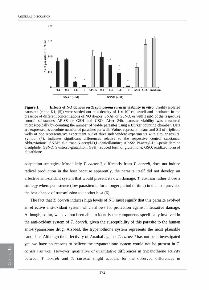
172
general discussion
ch
apt
er 1
0
2
Figure 1. Effects of NO donors on Trypanosoma carassii viability in vitro. Freshly isolated parasites (clone K1, (5)) were seeded out at a density of 1 x 10⁵ cells/well and incubated in the presence of different concentrations of NO donors, SNAP or GSNO, or with 1 mM of the respective control substances AP-SS or GSH and GSO. After 24h, parasite viability was measured microscopically by counting the number of viable parasites using a Bürker counting chamber. Data are expressed as absolute number of parasites per well. Values represent means and SD of triplicate wells of one representative experiment out of three independent experiments with similar results. Symbol (*), indicates significant differences relative to the respective control substance. Abbreviations: SNAP: S-nitroso-N-acetyl-D,L-penicillamine; AP-SS: N-acetyl-D,L-penicillamine disulphide; GSNO: S-nitroso-glutathion; GSH: reduced form of glutathione; GSO: oxidized form of glutathione.
adaptation strategies. Most likely T. carassii, differently from T. borreli, does not induce
radical production in the host because apparently, the parasite itself did not develop an
affective anti-oxidant system that would prevent its own damage. T. carassii rather chose a
strategy where persistence (low parasitemia for a longer period of time) in the host provides
the best chance of transmission to another host (6).
The fact that T. borreli induces high levels of NO must signify that this parasite evolved
an effective anti-oxidant system which allows for protection against nitrosative damage.
Although, so far, we have not been able to identify the components specifically involved in
the anti-oxidant system of T. borreli, given the susceptibility of this parasite to the human
anti-trypanosome drug, Arsobal, the trypanothione system represents the most plausible
candidate. Although the effectivity of Arsobal against T. carassii has not been investigated
yet, we have no reasons to believe the trypanothione system would not be present in T.
carassii as well. However, qualitative or quantitative differences in trypanothione activity
between T. borreli and T. carassii might account for the observed differences in
0
0.4
0.8
1.2
1.6
0.1 0.3 0.6 1 AP-SS 0.1 0.3 0.6 1 GSH GSO medium
SNAP (mM) GSNO (mM)
No
ofpa
rasi
tesx
10^6
/ml
* *
*
*
*
173
chapter 10
ch
apt
er 1
0
3
susceptibility to radicals between these two parasites. Physical barriers or additional anti-
oxidant enzymes present in T. borreli only, might allow this parasite to tolerate better the
high NO concentrations generated during infection (1, 4, 7).
Elevated nitric oxide production or serum nitrate/nitrite levels have also been associated
with natural trypanosome infections of human and cattle and with experimental infections
of mice. Whether NO was directly involved in parasitemia control, or in mediating
immunosuppression, was strictly dependent on the animal species under investigation.
Whereas experimental (non-homologous) murine infections with T. congolense clearly
point towards a role for NO in both parasitemia control and immunosuppression (8), natural
T. congolense infections of cattle indicates that NO is not involved in either of the two
aspects (9, 10). In fact, in contrast to the mouse model, the capacity of bovine macrophages
to produce NO is actually down-regulated in infected cattle. These apparently contrasting
findings underline the importance of homologous infection models for the evaluation and
extrapolation of experimental data to natural situations.
A question highly relevant to the understanding of the mechanisms leading to the high
production of NO in the T. borreli-carp model is: what triggers the production of NO?
Three possible mechanisms of NO induction which are not necessarily mutually exclusive
will be touched upon: 1) direct stimulation of pattern recognition receptors (PRRs) by
parasite components, 2) modification of host’s proteins that would act as ‘danger signals’
and 3) indirect stimulation by induction of host cytokines such as TNFα and IFNγ. The
latter mechanism will be more extensively dealt with later in this discussion.
The first mechanism is based on the fact that the innate immune system makes use of
PRRs such as the Toll-like receptors (TLRs) to sense conserved molecular structures of
invading pathogens (11) (12). TLRs are type I transmembrane proteins containing leucine-
rich repeat motifs important for the recognition of pathogen associated molecular patterns
(PAMPs). TLR2, at least in mammalian vertebrates, not only senses bacterial membrane
components but also glycosyl-phosphatidyl-inositol (GPI) anchors from parasites (13, 14).
Ongoing research in our laboratory is investigating the possibility that surface components
of T. borreli, possibly GPI-anchors, would signal via TLR2 to induce NO in carp
macrophages. TLR2 is one of the surface receptors implicated in macrophage activation
typically leading to increased NO production (15). Preliminary data suggest that, indeed,
supernatants from phosphatidylinositol-specific phospholipase C (PI-PLC)-treated parasites
can up-regulate TLR2 gene expression in carp macrophages (C.M.S. Ribeiro, personal
communication). Since also T. carassii has mucin-like GPI-anchored surface proteins (16),
174
general discussion
ch
apt
er 1
0
4
future experiments will have to point out whether GPI anchors from T. borreli have a
stronger capacity to stimulate PRR such as TLR2, leading to a higher production of NO in
carp macrophages.
The second mechanism by which parasites can skew the immune response of the host
towards an increased production of NO is the modification or degradation of self-derived
components. Modified host proteins or degradation products can then act as ‘alarmins’
delivering a ‘danger signal’ to host immune cells, such as macrophages. Transferrin (Tf)
cleavage products have been shown to induce NO production in goldfish (17) and carp (18)
macrophages. In fact, digestion of Tf by elastase results in a 31 amino acid peptide derived
from the N2 sub-domain of the Tf N-lobe, named TMAP. This fragment has been shown to
induce chemotaxis, nitric oxide and TNFα secretion in different macrophage populations
(19). It is well established that trypanosomes (T. brucei) need iron for growth and are
dependent on the availability of Tf-bound iron for their survival (20). Trypanosomes can
internalize Tf-bound iron via the flagellar pocket using specific Tf receptors (TfR) (21, 22).
Once internalized, the Tf-TfR complex is transported to the lysosome where iron is released
from Tf at acidic pH and Tf is proteolytically degraded. The resulting cleavage products are
released to the circulation while the iron remains parasite-associated (23). We recently
identified a T. borreli-specific cysteine proteinase and have shown that the activated
recombinant enzyme digests carp Tf at acidic pH (24). At this moment, it remains to be
proven whether TMAP-like peptides are present among the cleavage products most
probably released also by T. borreli, and whether these fragments contribute to the
induction of NO observed during infections.
We studied tissue tyrosine nitration as a marker of immunological activity resulting
from increased NO production, by using an anti-nitrotyrosine antibody in combination with
carp leukocyte sub-type specific monoclonal antibodies. This study revealed that not only
macrophages but also carp neutrophilic granulocytes strongly contributed to in vivo tissue
nitration, most likely through both, a peroxynitrite- and a myeloperoxidase-mediated
mechanism. In humans, neutrophilic granulocytes constitute the major type of blood
leukocytes, which does not seem to hold true for the blood of healthy carp. Although
human neutrophils have been assigned a critical role in innate immunity through their
phagocytosing capacity and release of various anti-microbial substances, they secrete lower
amounts of cytokines than CD14+ monocytes (25). Fish neutrophils have been reported to
possess a longer functional half-life than mammalian neutrophils (26), which may point at a
different functional role for neutrophils of fish. Certainly, in T. borreli-infected carp,
175
chapter 10
ch
apt
er 1
0
5
neutrophilic granulocytes contribute to the production of nitric oxide and to tissue-
associated pathology, possibly even to a larger extent than macrophages.
Regulation of nitric oxide production by cytokines: IFNγ and TNFα
As discussed above, it is likely that parasite components as well as modified host
proteins directly stimulate PRRs on innate immune cells and trigger NO production. In
addition, a modulating role for several cytokines, among which IFNγ and TNFα, should
certainly be considered as third mechanism of NO induction. The importance of IFNγ
signaling for the induction of TNFα and NO has been confirmed in numerous studies on
African trypanosomiasis of mammals (8, 27-33). Presentation of parasite antigens by MHC-
II can stimulate Th1 cells to secrete IFNγ that triggers the development of classically
activated macrophages, which in turn produce a second signal, namely TNFα. The
combination of IFNγ and TNFα signaling will then lead to the induction of iNOS
expression in these macrophages. During experimental infections of mice with T.
congolense, especially, the IFNγ-dependent NO production by classically activated
macrophages is fundamental for parasite killing. Despite the well-documented trypanotoxic
effects of NO in vitro (34), during experimental infections of mice with T. brucei, TNFα
more than NO seems to be involved in parasite control (27, 28). In fact, TNFα has been
shown to exert direct trypanotoxic effects against T. brucei, but not T. congolense (35, 36).
In our study, we reported an up-regulation of two isoforms of TNFα (TNFα1 and
TNFα2) in spleen of T. borreli-infected carp, but we also concluded that carp TNFα might
not be the major contributing factor to the production of high NO levels. Direct stimulation
of carp macrophages with TNFα in vitro did not result in iNOS up-regulation, nor in
increased NO production in macrophage culture supernatants. Furthermore, in vivo
inhibition of TNFα gene expression by administration of a TNFα inhibitor (PTX) also did
not influence iNOS gene expression. These findings excluded a direct effect of TNFα on
the production of NO and suggest that parasite components or signaling factors other than
TNFα should be responsible for the high induction of NO in T. borreli-infected carp.
In the mouse studies mentioned above, IFNγ-dependent activation of macrophages was
essential for NO production during trypanosome infections. In carp, only very recently two
IFNγ genes were reported and IFNγ2 was shown to share high structural and functional
similarity with IFNγ from mammalian vertebrates (37). IFNγ2 gene expression was induced
upon in vitro stimulation of carp leukocytes and mature thymocytes with the T cell mitogen
PHA. Furthermore, recombinant carp IFNγ2 alone, or in combination with LPS, was shown
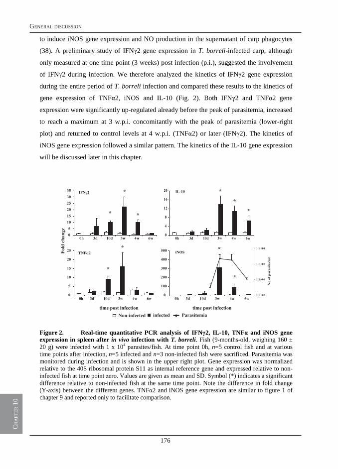
176
general discussion
ch
apt
er 1
0
6
to induce iNOS gene expression and NO production in the supernatant of carp phagocytes
(38). A preliminary study of IFNγ2 gene expression in T. borreli-infected carp, although
only measured at one time point (3 weeks) post infection (p.i.), suggested the involvement
of IFNγ2 during infection. We therefore analyzed the kinetics of IFNγ2 gene expression
during the entire period of T. borreli infection and compared these results to the kinetics of
gene expression of TNFα2, iNOS and IL-10 (Fig. 2). Both IFNγ2 and TNFα2 gene
expression were significantly up-regulated already before the peak of parasitemia, increased
to reach a maximum at 3 w.p.i. concomitantly with the peak of parasitemia (lower-right
plot) and returned to control levels at 4 w.p.i. (TNFα2) or later (IFNγ2). The kinetics of
iNOS gene expression followed a similar pattern. The kinetics of the IL-10 gene expression
will be discussed later in this chapter.
Figure 2. Real-time quantitative PCR analysis of IFNγ2, IL-10, TNFα and iNOS gene expression in spleen after in vivo infection with T. borreli. Fish (9-months-old, weighing 160 ± 20 g) were infected with 1 x 10⁴ parasites/fish. At time point 0h, n=5 control fish and at various time points after infection, n=5 infected and n=3 non-infected fish were sacrificed. Parasitemia was monitored during infection and is shown in the upper right plot. Gene expression was normalized relative to the 40S ribosomal protein S11 as internal reference gene and expressed relative to non-infected fish at time point zero. Values are given as mean and SD. Symbol (*) indicates a significant difference relative to non-infected fish at the same time point. Note the difference in fold change (Y-axis) between the different genes. TNFα2 and iNOS gene expression are similar to figure 1 of chapter 9 and reported only to facilitate comparison.
Non-infected infectedtime post infection time post infection
Foldchange
Parasitemia
0
4
8
12
16
20
0h 3d 10d 3w 4w 6w05101520253035
0h 3d 10d 3w 4w 6w
IFN 2 IL-10
*
*
*
**
*
TNF 2 iNOS
*
0
5
10
15
20
25
0h 3d 10d 3w 4w 6w0
100
200
300
400
500
0h 3d 10d 3w 4w 6w
*
*
*
Noofparasites/ml
1.E+08
1.E+07
1.E+06
1.E+05
177
chapter 10
ch
apt
er 1
0
7
Taken together, we hypothesize that upon infection with T. borreli, parasite antigens via
MHC-II stimulate T cells to secrete IFNγ which triggers a classical activation of
macrophages that, as a second signal, produce TNFα. Although TNFα does not seem to
drive the expression of iNOS, the high production of NO certainly does correlate with a
typical classical activation of macrophages. Furthermore, the ability of TNFα to activate
endothelial cells (EEC) cannot be disregarded. In fact, production of TNFα by activated
macrophages might stimulate EEC to secrete chemokines and promote leukocyte
extravasation to the tissue, thereby contributing to the inflammation observed in peripheral
organs. NO has been shown a potent vasodilatating agent in zebrafish (39), therefore high
levels of NO in the blood might favor the extravasation of T. borreli to the tissue. This
hypothesis is in accordance with the observation that T. borreli can penetrate peripheral
organs during the late stages of infection, whereas T. carassii, which does not induce NO
(1), is a ‘hematic’ parasite that is found exclusively in the blood circulation.
The present availability of recombinant carp TNFα as well as recombinant carp IFNγ,
and availability of specific antibodies against TNFα and IFNγ provide new tools that will
certainly contribute to a further elucidation of the role played by these two cytokines during
infection with T. borreli. This infection model, because of the typically high NO response,
seems extremely suitable for investigations on IFNγ-mediated activation of macrophages.
Regulation of nitric oxide production by cytokines: IL-10
In general, in a host-parasite relationship, both host and parasite are balancing their
impact on the immune response to not only guarantee survival of the host but also to
support persistence of the parasite. As discussed above, an early immune response driven
by IFNγ and by the potential trypanocidal molecules TNFα and NO is essential to limit
initial parasite replication. However, for a successful resolution of the infection and for
parasite clearance, the production of specific antibodies, limitation of immune-mediated
damage and,ultimately the generation of an anti-inflammatory immune response is essential
(40). Fundamental to this switch is the anti-inflammatory cytokine IL-10 (28, 30, 41, 42).
Studies with mammalian trypanosomes show that in the absence of a switch from an
inflammatory to an anti-inflammatory response, the host succumbs to infection owing to an
uncontrolled IFNγ production and inflammation-induced lesions (27, 28, 43).
The sequence for carp IL-10 has been described (44), and studies using the recombinant
protein are presently ongoing. Investigation of the kinetics of IL-10 gene expression during
T. borreli infection (Fig. 2) clearly shows a prolonged up-regulation of IL-10 in comparison
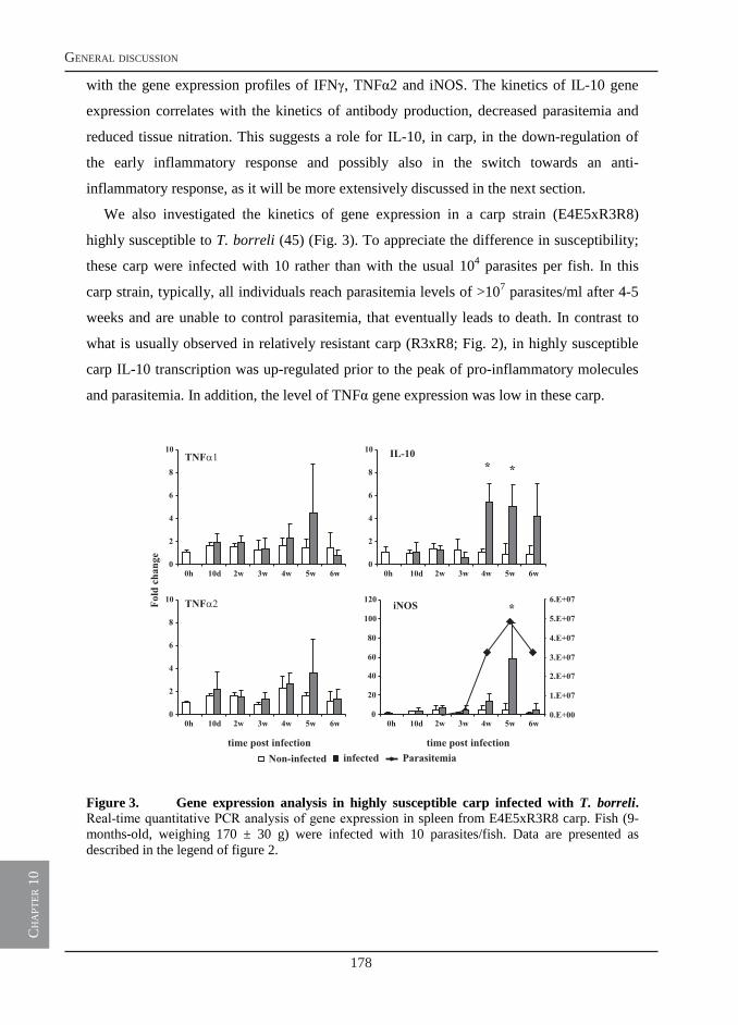
178
general discussion
ch
apt
er 1
0
8
with the gene expression profiles of IFNγ, TNFα2 and iNOS. The kinetics of IL-10 gene
expression correlates with the kinetics of antibody production, decreased parasitemia and
reduced tissue nitration. This suggests a role for IL-10, in carp, in the down-regulation of
the early inflammatory response and possibly also in the switch towards an anti-
inflammatory response, as it will be more extensively discussed in the next section.
We also investigated the kinetics of gene expression in a carp strain (E4E5xR3R8)
highly susceptible to T. borreli (45) (Fig. 3). To appreciate the difference in susceptibility;
these carp were infected with 10 rather than with the usual 104 parasites per fish. In this
carp strain, typically, all individuals reach parasitemia levels of >107 parasites/ml after 4-5
weeks and are unable to control parasitemia, that eventually leads to death. In contrast to
what is usually observed in relatively resistant carp (R3xR8; Fig. 2), in highly susceptible
carp IL-10 transcription was up-regulated prior to the peak of pro-inflammatory molecules
and parasitemia. In addition, the level of TNFα gene expression was low in these carp.
Figure 3. Gene expression analysis in highly susceptible carp infected with T. borreli. Real-time quantitative PCR analysis of gene expression in spleen from E4E5xR3R8 carp. Fish (9-months-old, weighing 170 ± 30 g) were infected with 10 parasites/fish. Data are presented as described in the legend of figure 2.
time post infection time post infection
Foldchange 0
2
4
6
8
10
0h 10d 2w 3w 4w 5w 6w
TNF
0
2
4
6
8
10
0h 10d 2w 3w 4w 5w 6w
TNF
Non-infected infected Parasitemia
0.E+00
1.E+07
2.E+07
3.E+07
4.E+07
5.E+07
6.E+07
0
20
40
60
80
100
120
0h 10d 2w 3w 4w 5w 6w
iNOS *
0
2
4
6
8
10
0h 10d 2w 3w 4w 5w 6w
IL-10**
179
chapter 10
ch
apt
er 1
0
9
In African trypanosomiasis, IL-10 has been shown to exert a detrimental effect by
mediating immunosuppression (46-48), but also to be beneficial by controlling excessive
lethal release of cytokines such as IFNγ and TNFα (49-51). Although we have no
conclusive evidence that IL-10 alone might be responsible for the high susceptibility of the
particular (E4E5xR3R8) carp strain, the difference in the kinetics of IL-10 gene expression
between relatively resistant and susceptible carp is striking. Our data suggests that an early
presence of IL-10 during infection with T. borreli could induce a state of
immunosuppression and, possibly, prevent the onset of an early inflammatory response
important for parasite control.
Kinetics of type-1 and type-2 immune responses during T. borreli infections
In mammals, type-1 inflammatory responses are characterized by the development of
(IFNγ-dependent-) classically activated macrophages and the subsequent production of
reactive oxygen species, NO, IL-12 and TNFα by these cells. Type-2 anti-inflammatory
responses, typically antagonize type-1 responses, and are driven by the development of (IL-
4/IL-13- or IL-10-dependent-) alternatively activated macrophages and by the production of
regulatory molecules such as TGFβ or IL-10 by these cells. The cytokine environment
created by these differentially activated macrophages will determine the progression
towards a cellular or a humoral response (52). Type-1 responses commonly are associated
with intracellular pathogens (bacteria or viruses), whereas type-2 responses are associated
with extracellular parasites (helminths or protoza). During infection, adaptation is an
essential element of the host-parasite relationship. Extreme modulation or
immunosuppression of the immune response by the parasite leads to unwanted death of the
host or to overreactions of the immune system. Uncontrolled type-1 immune reactions may
lead to an exacerbation of the inflammatory response, whereas uncontrolled type-2 immune
responses may lead to immunosuppression.
A good example of a strict requirement for a balanced type-1/type-2 immune response is
provided by experimental infection of mice with African trypanosomes (53-55). In these
infections, a type-1 cytokine environment and classical macrophage activation in the early
stage of infection is required, enabling mice to control the first peak of parasitemia.
Thereafter, a switch to a type-2 cytokine environment triggering alternative macrophage
activation is essential to enable B cell maturation for antibody production and
trypanotolerance in the chronic phase of infection.
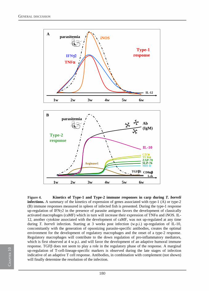
180
general discussion
ch
apt
er 1
0
10
Figure 4. Kinetics of Type-1 and Type-2 immune responses in carp during T. borreliinfections. A summary of the kinetics of expression of genes associated with type-1 (A) or type-2 (B) immune responses measured in spleen of infected fish is presented. During the type-1 response up-regulation of IFNγ2 in the presence of parasite antigens favors the development of classically activated macrophages (caMF) which in turn will increase their expression of TNFα and iNOS. IL-12, another cytokine associated with the development of caMF, was not up-regulated at any time during T. borreli infection. Starting at 3 weeks post infection (w.p.i.) up-regulation of IL-10, concomitantly with the generation of opsonizing parasite-specific antibodies, creates the optimal environment for the development of regulatory macrophages and the onset of a type-2 response. Regulatory macrophages will contribute to the down regulation of pro-inflammatory mediators, which is first observed at 4 w.p.i. and will favor the development of an adaptive humoral immune response. TGFβ does not seem to play a role in the regulatory phase of the response. A marginal up-regulation of T-cell-lineage-specific markers is observed during the late stages of infection indicative of an adaptive T cell response. Antibodies, in combination with complement (not shown) will finally determine the resolution of the infection.
1w 2w 3w 4w 5w 6w
iNOS
IFNγ2γ2γ2γ2TNFαααα
IL-12
parasitemia
Type-1 response
1w 2w 3w 4w 5w 6w
Ab(IgM)
IL-10
CD3εεεεTCRααααZAP-70SLP-76
CD8αβ αβ αβ αβ Lck
TGFββββ1
parasitemia
Arginase1MH-II
Type-2 response
B
A
Figure 4.
181
chapter 10
ch
apt
er 1
0
11
We have previously shown that carp macrophages can polarize into innate and
classically activated macrophages, based on the differential activity of iNOS and arginase,
respectively (2, 3). Innate activation can be induced by a microbial trigger alone (i.e. LPS,
parasite components), whereas classical activation is induced by a microbial trigger in
combination with IFNγ (56, 57). As summarized in Fig. 4A, TNFα expression and without
doubt NO production are typical of the early phase of T. borreli. Given the recently
described activity of goldfish (58) and carp IFNγ (38) to induce NO in macrophages in
vitro, and our present data on the early kinetics of IFNγ gene expression simultaneously
with the presence of parasite antigens in vivo, we believe a prototypical classical
macrophage activation occurs during T. borreli infection of carp.
Before we can analyze whether a type-2 anti-inflammatory response occurs during the
later stages of T. borreli infections, some recent views have to be considered. The name
originally given to macrophages that developed in the presence of IL-4 was ‘alternatively’
activated. However, this name implies that this would be the only other (alternative) way of
macrophage activation. More recently, a new classification based on three fundamental
macrophage activities important for maintaining homeostasis has been proposed. These
activities are host defense, wound healing and immune regulation (57). Classically
activated macrophages are associated with host defense and require IFNγ, as described
above. Wound healing macrophages develop in the presence of IL-4 and IL-13 and exhibit
arginase activity in response to tissue injury, or following an innate or an adaptive immune
response. Regulatory macrophages can arise during the late stages of adaptive immune
responses and are triggered by microbial agents and immune complexes (opsonized
pathogens) but also by prostaglandins and phagocytosis of apoptotic cells, and are
characterized but the production of high levels of the anti-inflammatory cytokine IL-10.
Two IL-4/13 loci have been described for teleost fish, although at this moment it is
unclear whether the teleost IL-4/13 genes are orthologous to either IL-4 or IL-13, or
whether these mammalian genes separated later in evolution (Ohtani et al, 2008:
Immunogenetics 60: 383-397). As shown in Fig. 4B, arginase gene expression was only
marginally up-regulated during infection and arginase activity in cyclicAMP-stimulated
head kidney leukocytes isolated at various time points after T. borreli infection was not
increased (2). Thus, based on the absence of increased arginase activity we conclude that
wound-healing macrophages do not play an important role in the resolution of T. borreli
infections. Identification of carp IL-4/IL-13 or identification of markers for wound-healing
macrophages, such as chitinase-like protein YM1 or resistin-like molecule-α (RELMα)
182
general discussion
ch
apt
er 1
0
12
(reviewed in (57)), should help to corroborate this hypothesis in the future. In contrast, our
data do support the involvement of regulatory macrophages during later stages of the
immune response to T. borreli, as shown by a profound up-regulation of IL-10, the
presence of opsonized parasites which might act as activating immune complexes and the
production of T. borreli-specific antibodies (Fig. 4B). Taken together, we conclude that a
type-2 anti-inflammatory immune response occurs during the last phase of T. borreli
infection, and that the down-regulation of the inflammatory response (IFNγ, TNFα, iNOS)
is driven by the production of IL-10 by regulatory macrophages.
Kinetics of the adaptive immune response to T. borreli
The main mechanism suggested to control infections of mammals with African
trypanosomes is the final production of parasite-specific antibodies, although most of the
actual mechanisms are still under debate. African trypanosomes, however, have developed
a very clever strategy particularly effective in evading the humoral immune response i.e.
antigenic variation of their variant surface glycoprotein (VSG) (59, 60). By periodically
changing their VSGs, trypanosome are initially not recognized by antibodies against VSG
epitopes displayed on the former surface coat. New variant antigenic types (VATs) carry on
the infection, whereas trypanosomes expressing the former coat are destroyed. Not only the
production, but also the isotype of VSG-specific antibodies is crucial. In T. brucei and T.
evansi infections opsonizing IgM, but in T. congolense infections opsonizing IgG is
required to reduce the number of circulating parasites. A clear role for complement-
mediated lysis could not be ascertained for trypanosome infections in mammals (61, 62). In
T. congolense infections, IgG also facilitates antibody-mediated phagocytosis of opsonized
parasites by Kupffer cells in the liver.
The humoral response to T. borreli is well documented. The production of T. borreli-
specific (IgM) antibodies (45, 63), T. borreli-unrelated antibodies indicative of polyclonal
B cell activation (45) and the presence of enlarged B cell areas in the spleen (7) have all
been observed. There is no evidence that antigenic variation might occur also in T. borreli
and therefore it cannot be a contributing factor to the large B cell proliferation. T. borreli
infections are not characterized by a chronic phase, and fish that survive infections are
protected upon re-infection. Classical complement activation plays a major role in
protection against T. borreli and the importance of antibody-dependent complement-
mediated lysis has been shown both in vitro (45) and in vivo (chapter 7). We have no
evidence that (opsonised) T. borreli would be cleared in large numbers by macrophages in
183
chapter 10
ch
apt
er 1
0
13
the carp liver although occasionally, evidence of phagocytosed parasites can be noted in
peripheral organs such as head kidney and liver (Fig. 5.) Previously, elimination of resident
macrophages from the peritoneal cavity and liver, by intraperitoneal injection of
clodronate-liposomes, was shown to render carp susceptible to opportunistic bacteria but
not to T. borreli (64). This suggests that phagocytosis of T. borreli in the liver might not
play a major role in parasite clearance. However, we do have clear indications that immune
regulation and activation does take place in liver of infected fish. Cytokines found to be up-
regulated in spleen, the organ of choice for most of the gene expression studies in this
thesis, were also highly up-regulated in liver. In addition, IL-1β gene expression did not
increase in spleen but did increase in liver (data not shown). Other genes including iNOS
(7), several complement factors (chapter 7) and α2-macroglobulin (65) were also up-
regulated in liver of infected carp. Possibly, liver macrophages isolated during the peak of
parasitemia and incubated in vitro with opsonized parasites could phagocytose, reduce
parasite motility or viability but also produce NO and maybe mediate antibody-dependent
cell-mediated cytotoxicity. Certainly, the immune regulation and activation noted in liver of
T. borreli-infect fish warrants a closer investigation of the liver in our infection model.
Figure 5. Phagocytosis of T. borreli by tissue macrophages. Electron microscopy picture of T. borreli phagocytosed by a macrophage in head kidney of infected carp. A) Black arrows indicate the presence of 4 parasites in the cytoplasm of the macrophage. B) Enlarged view of the area boxed in figure A. Open arrows indicate the position of the basal bodies (similar to centrioles) identifying the position of the two flagella of T. borreli. (Magnification: 4400x)
During infection with T. borreli several T-cell-related genes are up-regulated, especially
during the late phase of infection (Fig. 4B). We have also shown both a marked B and T
cell proliferation in the spleen of infected fish. In trypanosome infections of mammals, T
cell-dependent as well as T cell-independent mechanisms of B cell activation have been
reported. The mechanisms underlining the B and T cell proliferation during T. borreli
A B
184
general discussion
ch
apt
er 1
0
14
infections are still unknown. Possibly, mitogenic parasite antigens or the presence of
putative superantigens could be responsible for this pronounced lymphocyte activation,
although to date substantial evidence in favor of this hypothesis is not available. Owing to a
lack of suitable tools for their investigation, the functional T cell response to T. borreli is
still poorly characterized. Only very recently, a system was optimized where carp kidney
hematopoietic cells, co-cultured with a layer of feeder cells, proliferated and, after several
passages, differentiated in cells belonging to the T cell-lineage as assessed by gene
expression of GATA3, Lck and TCRβ (66). This system will certainly aid to the study of T.
borreli-specific T cells responses, at least in vitro.
African trypanosomiasis is characterised by a state of general immunosuppression, well
documented in T. brucei-infected mice and T. congolense-infected cattle (reviewed in (67)).
During T. congolense infections, immunosuppression was found to occur primarily in the
spleen and affects both humoral and cellular responses. Immunosuppression generally
increases with progression of infection and it appears that both suppressor macrophages as
well as antigen-specific suppressor T cells are responsible for the immunosuppressive
effects. Also during T. borreli infections, immunosuppression can be observed as the
inability to re-stimulate, ex vivo, cells isolated during the inflammatory phase of the
infection (data not shown). Whether this suppression is mediated primarily by NO,
macrophages, T cells, or by a combination of all will be the subject of further investigation.
No doubt, modulation of adaptive reactions by ensuring immunosuppression of several
components of the host humoral and cellular response, allows parasites to gain essential
time to survive in the individual host, ensuring propagation within the population.
Future perspectives
Zebrafish and carp: non-identical twins
As argued in the introduction to this thesis, high quality infection models of inbred
mouse strains have helped to pave the way for much of modern immunology. Although
comparative immunology has profited to a great extent from the recent genome initiatives,
advancement of fish immunology is still delayed by the use of several, phylogenetically
distant, fish species as well as by the lack of inbred fish line and a limited set of reagents.
Shared use of zebrafish as a common experimental animal besides selected fish species
would certainly bring a much-needed higher level of standardization between different
research laboratories. While zebrafish research initially centered on mutagenesis screens,
185
chapter 10
ch
apt
er 1
0
15
more recent years brought sophisticated reverse genetic methods such as morpholino
knock-down or targeting induced local lesions in genomes (TILLING, see also Table 1). In
addition, transgenic zebrafish have been developed and microarrays are now available to
characterize gene experession on a near genome-wide scale. This suggests a meaningful
interaction between zebrafish and aquacultural research should now be possible, although it
is good to realize that zebrafish, in fact, is less closely related to most fishes of aquacultural
interest (e.g. salmonids, sea bass, sea bream, cod, flatfishes) than those fish are among
themselves (68). The only exception is carp, which is the most commonly cultured food-
fish on the planet and is not only in the same Superorder but also in the same Family as
zebrafish. Where zebrafish offers the advantage of a well-recognized low-cost animal
model of vertebrate biology, the common carp, because of its large size, offers for example,
the advantage of large numbers of leukocytes (> 107) for in vitro assays. Combination of
the genetically tractable but small-sized zebrafish with the large-sized common carp would
create a very strong combination of animal models that could address disease-related
questions relevant to aquaculture such as vaccination, being based on increased
understanding of fundamental immune mechanisms in comparative immunology.
The zebrafish genome has been sequenced and its annotation is continuously improved,
gene microarrays and insertional mutants are commercially available, all of which are
strong, mostly technical, arguments in favor of using the zebrafish as a model for animal
physiology and development (Table 1). In addition, zebrafish is now gaining value as a
model system also for immunological questions, not only for fish but also for mammals,
particularly because of the ease of using embryonic stages to examine the development of
immunity. External fertilization and small, transparent embryos allow for easy and non-
invasive in vivo live-immaging observation up to the subcellular level. The translucency of
the zebrafish embryos has facilitated the detection of fluorescently tagged transgenes and
the monitoring of reporter gene activity. Similar to other teleosts, zebrafish are capable of
adaptive immune reactions but the adaptive immune response requires several weeks to
develop. This is often used as an argument to exclusively study innate immune responses in
zebrafish, during the initial few days after fertilization. It is important to keep in mind,
however, the limitation of studies in embryos only which creates an experimental and often
unnatural division between innate and acquired immune responses that studies in adults do
not suffer from. For that reason, the use of not only embryos but also adults in
immunological studies using zebrafish should be strongly encouraged.
186
general discussion
ch
apt
er 1
0
16
Table 1. Common techniques used in zebrafish research
Technique Description
FISH
Fluorescent in situ hybridization on whole-mount zebrafish embryos allows for simultaneous localization of gene expression patterns of two genes within the same embryo, permitting identification of colocalized expression within single cells
Microinjection of morpholino
oligomers, preventing mRNA
translation of targeted transcripts
Morpholinos are short oligomer sequences whose backbone is chemically modified, rendering them resistant to degradation by nucleases. This gene-specific knockdown antisense technology has proven to be a powerful tool for altering gene products and functions in zebrafish larvae. Knock-down is transient so the technique is not applicable to adult zebrafish.
Genetic screens
The method of choice for identifying genes with essential functions. Chemical mutagenesis can be used to generate loss-of-function phenotypes. Most mutants carry chemical (N-ethyl-N-nitrosourea; ENU)-induced mutations. Upon establishment of random mutant zebrafish lines, positional cloning can help to characterize the gene responsible for the stable phenotype.
TILLING
Targeting Induced Local Lesions IN Genomes (TILLING) is an important means by which mutations in specific genes of interest can be quickly identified and studied. The technique is effective to isolate mutants with defined mutations, and therefore can identify stable knock-out genotypes.
Injection of plasmid DNA Can generate stable transgenics. Can be used to image spatiotemporal expression of fluorescent transgenes such as GFP in various organs and tissues.
Microarrays Reflect a set of all mRNA transcripts actively being expressed at a given time-point following infection. Useful for transcriptome studies of infected animals.
One of the strongest observations made in this thesis was the crucial protective role for
membrane TNFα (mTNFα) against infection of carp with T. borreli. In mammals, TNFα is
first transported to the membrane and then cleaved by the TNFα converting enzyme TACE,
to release soluble TNFα (sTNFα). Studies in fish have concentrated on in vitro activities of
sTNFα and the presence and function of mTNFα was not considered so far. Interestingly, in
experimental infection of mice with Mycobacterium tuberculosis, mTNFα has been shown
sufficient to provide acute, but not long-term, control of infection (69, 70). In tuberculosis,
mycobacteria are phagocytosed by macrophages that are modified by persistent cytokine
signals (IFNγ) and that then recruit additional cells to form granulomas that eventually
contain the infection. Of particular interest with respect to the elucidation of the protective
mechanism mediated by mTNFα, is the natural infection model of zebrafish with M.
marinum, a model for tuberculosis (71, 72). Using live M. marinum-infected embryos, loss
of TNFα signaling was shown to cause increased mortality (73). TNFα was not required for
tuberculous granuloma formation, but maintained granuloma integrity indirectly by
restricting mycobacterial growth within macrophages and preventing their necrosis.
Although these studies were performed in zebrafish embryos, limiting conclusions to innate
187
chapter 10
ch
apt
er 1
0
17
immunity, a recent microarray study showed a large overlap of expression signatures
between infected embryos and adults, confirming the importance of innate immunity in
mycobacterial infection (74). The high applicability of the zebrafish-M. marinum model
and the fact that mTNFα can be sufficient to provide acute control of mycobacterial
infection in mice, suggests that mycobacterial infection of zebrafish can be a good model to
study mTNFα functions in fish.
We have shown that in carp, administration of a TACE-inhibitor in vivo, in the period
preceding the peak of parasitemia, was sufficient to confer full protection against T. borreli
infection. In vitro, we could also show that incubation of LPS-stimulated carp leukocytes
with TACE inhibitor resulted in a significant increase of surface-bound TNFα. However,
the cell types expressing mTNFα and the mechanisms underlining the protective effects
observed during T. borreli infection are unknown. Zebrafish macrophages express TNFα,
but Pu.1 morphant embryos lacking macrophages also express TNFα, indicating that other
cell types beside macrophages can produce TNFα (75). The use of transgenic zebrafish
expressing cell-lineage-specific fluorescent markers could help to elucidate the cell type(s)
expressing mTNFα. In addition, the use of antibodies specifically recognizing carp B cells,
granulocytes, macrophages, thrombocytes, immature thymocytes and mucosal T cells (76-
79) could be a complementary tool to study the cellular source of mTNFα. Combination of
the two animal model systems will certainly shed light on the role of mTNFα in fish.
For studies on mTNFα signaling, the M. marinum infection model of zebrafish certainly
should be informative. The kinetics of granuloma formation are well characterized (71) and
the importance of TNFα signaling through TNFα-receptor-1 (TNFR1) has already been
demonstrated in this model (73). However, the question whether TNFR1-signaling is
triggered by sTNFα or mTNFα has not been addressed. Application of a TACE inhibitor
during M. marinum infection of zebrafish should help to investigate whether, similar to
what has been observed in mice infected with M. tuberculosis (2, 3), mTNFα plays a role in
cell migration, granuloma formation and control of infection, especially during the early
phase of infection. In addition, a reverse genetic approach by administration of morpholinos
directed against TNFR1 or TNFR2 in combination with a TACE inhibitor will reveal
whether mTNFα signals preferentially via one TNFR in particular, or via both receptors.
Signaling by mTNFα through binding of the TNFR is often associated with activation of
NK (80, 81) and T cells (82) as well as macrophages (83) and the induction of cytotoxic
activities. The rapid clearance of parasites observed during T. borreli infections can only be
consistent with the involvement of cytotoxic mechanisms. In this respect, the zebrafish
188
general discussion
ch
apt
er 1
0
18
model could provide crucial information to clarify receptor-mediated signaling of mTNFα
and be instrumental for the development of future strategies such as production of anti-
TNFR antibodies and recombinant soluble TNFR, or application of specific inhibitors of
mTNFα or TNFR signaling with the aim to understand the mechanism of mTNFα-mediated
protection during T. borreli infection.
An additional signaling mechanism described for mTNFα in mammals is ‘reverse
signaling’ or ‘bidirectional signaling’ whereby the ligand acts as a receptor (84). In this
process, cellular signals are transduced by the membrane-bound ligand upon interaction
with a soluble or transmembrane receptor. Several members of the TNFα family, including
mTNFα, have been shown to deliver an intracellular signal through a casein kinase 1 (CKI)
motif (SX2S) present in the cytoplasmic domain. In mammals, reverse signaling by
members of the TNFα family described for B and T cells is generally of a stimulatory
nature (85, 86), whereas for monocytes mainly inhibitory effects were observed (87, 88),
indicating a differential role of this type of signal in adaptive and innate immunity. A
putative CKI motif (SX3S) could be identified in the cytoplasmic region of TNFα of several
fish species, including carp and zebrafish (personal observation, data not shown). In fish
TNFα, three but not two, intervening amino acids separate the two serine residues and at
least one acid residue (required for optimal CKI activity) is present upstream of the N-
terminal serine. At present, fourteen CK isoforms are annotated in the zebrafish database
(data not shown), therefore all components required for the investigation of ‘reverse
signaling’ by mTNFα are present, at least in zebrafish.
The drive to use zebrafish as animal model of vertebrate biology has primarily come
from a fundamental interest in the evolution of immunity and developmental biology,
however, the wish to address the use of adult zebrafish as animal model for studies in
aquatic biology and life sciences is increasing. State-of-the-art techniques developed at
technological zebrafish platforms, in combination with immunological research on closely
related but larger sized carp will stimulate the development of a sustainable,
environmentally and welfare-friendly aquaculture in the 21st century. It will do so by
stimulating fundamental but strategic research in aquatic biology and life sciences.
189
chapter 10
ch
apt
er 1
0
19
References 1. Saeij, J. P., W. B. Van Muiswinkel, A. Groeneveld, and G. F. Wiegertjes. 2002. Immune modulation
by fish kinetoplastid parasites: a role for nitric oxide. Parasitology 124:77-86. 2. Joerink, M., M. Forlenza, C. M. S. Ribeiro, B. J. de Vries, H. F. J. Savelkoul, and G. F. Wiegertjes.
2006. Differential macrophage polarisation during parasitic infections in common carp (Cyprinus carpio L.). Fish Shellfish Immunol 21:561-571.
3. Joerink, M., C. M. S. Ribeiro, R. J. M. Stet, T. Hermsen, H. F. J. Savelkoul, and G. F. Wiegertjes. 2006. Head kidney-derived macrophages of common carp (Cyprinus carpio L.) show plasticity and functional polarization upon differential stimulation. J Immunol 177:61-69.
4. Joerink, M., A. Groeneveld, B. Ducro, H. F. J. Savelkoul, and G. F. Wiegertjes. 2007. Mixed infection with Trypanoplasma borreli and Trypanosoma carassii induces protection: involvement of cross-reactive antibodies. Dev Comp Immunol 31:903-915.
5. Overath, P., J. Ruoff, Y. D. Stierhof, J. Haag, H. Tichy, I. Dykova, and J. Lom. 1998. Cultivation of bloodstream forms of Trypanosoma carassii, a common parasite of freshwater fish. Parasitol Res 84:343-347.
6. Wiegertjes, G. F., M. Forlenza, M. Joerink, and J. P. Scharsack. 2005. Parasite infections revisited. Dev Comp Immunol 29:749-758.
7. Forlenza, M., J. P. Scharsack, N. M. Kachamakova, A. J. Taverne-Thiele, J. H. W. M. Rombout, and G. F. Wiegertjes. 2008. Differential contribution of neutrophilic granulocytes and macrophages to nitrosative stress in a host-parasite animal model. Mol Immunol 45:3178-3189.
8. Magez, S., M. Radwanska, M. Drennan, L. Fick, T. N. Baral, F. Brombacher, and P. De Baetselier. 2006. Interferon-gamma and nitric oxide in combination with antibodies are key protective host immune factors during Trypanosoma congolense Tc13 Infections. J Infect Dis 193:1575-1583.
9. Taylor, K., V. Lutje, and B. Mertens. 1996. Nitric oxide synthesis is depressed in Bos indicus cattle infected with Trypanosoma congolense and Trypanosoma vivax and does not mediate T-cell suppression. Infect Immun 64:4115-4122.
10. Taylor, K. A. 1998. Immune responses of cattle to African trypanosomes: protective or pathogenic? Int J Parasitol 28:219-240.
11. Medzhitov, R. 2001. Toll-like receptors and innate immunity. Nat Rev Immunol 1:135-145. 12. Takeda, K., and S. Akira. 2005. Toll-like receptors in innate immunity. Int Immunol 17:1-14. 13. Campos, M. A., I. C. Almeida, O. Takeuchi, S. Akira, E. P. Valente, D. O. Procopio, L. R.
Travassos, J. A. Smith, D. T. Golenbock, and R. T. Gazzinelli. 2001. Activation of Toll-like receptor-2 by glycosylphosphatidylinositol anchors from a protozoan parasite. J Immunol 167:416-423.
14. Debierre-Grockiego, F., M. A. Campos, N. Azzouz, J. Schmidt, U. Bieker, M. G. Resende, D. S. Mansur, R. Weingart, R. R. Schmidt, D. T. Golenbock, R. T. Gazzinelli, and R. T. Schwarz. 2007. Activation of TLR2 and TLR4 by glycosylphosphatidylinositols derived from Toxoplasma gondii. J Immunol 179:1129-1137.
15. Benoit, M., B. Desnues, and J. L. Mege. 2008. Macrophage polarization in bacterial infections. J Immunol 181:3733-3739.
16. Lischke, A., C. Klein, Y. D. Stierhof, M. Hempel, A. Mehlert, I. C. Almeida, M. A. Ferguson, and P. Overath. 2000. Isolation and characterization of glycosylphosphatidylinositol-anchored, mucin-like surface glycoproteins from bloodstream forms of the freshwater-fish parasite Trypanosoma carassii. Biochem J 345 Pt 3:693-700.
17. Stafford, J. L., N. F. Neumann, and M. Belosevic. 2001. Products of proteolytic cleavage of transferrin induce nitric oxide response of goldfish macrophages. Dev Comp Immunol 25:101-115.
18. Jurecka, P., I. Irnazarow, J. L. Stafford, A. Ruszczyk, N. Taverne, M. Belosevic, H. F. J. Savelkoul, and G. F. Wiegertjes. 2009. The induction of nitric oxide response of carp macrophages by transferrin is influenced by the allelic diversity of the molecule. Fish Shellfish Immunol 26:632-638.
19. Haddad, G., and M. Belosevic. 2009. Transferrin-derived synthetic peptide induces highly conserved pro-inflammatory responses of macrophages. Mol Immunol 46:576-586.
20. Schell, D., N. K. Borowy, and P. Overath. 1991. Transferrin is a growth factor for the bloodstream form of Trypanosoma brucei. Parasitol Res 77:558-560.
21. Steverding, D. 2000. The transferrin receptor of Trypanosoma brucei. Parasitol Int 48:191-198. 22. Henri, G. A. M. v. L., K. Rudo, M. m. Rainer, E. Markus, R. Bas ter, and B. Piet. 2005.
Trypanosomes change their transferrin receptor expression to allow effective uptake of host transferrin. Mol Microbiol 58:151-165.
23. Grab, D. J., C. W. Wells, M. K. Shaw, P. Webster, and D. C. Russo. 1992. Endocytosed transferrin in African trypanosomes is delivered to lysosomes and may not be recycled. Eur J Cell Biol 59:398-404.
190
general discussion
ch
apt
er 1
0
20
24. Ruszczyk, A., M. Forlenza, M. Joerink, C. M. S. Ribeiro, P. Jurecka, and G. F. Wiegertjes. 2008. Trypanoplasma borreli cysteine proteinase activities support a conservation of function with respect to digestion of host proteins in common carp. Dev Comp Immunol 32:1348-1361.
25. Smedman, C., B. Gardlund, K. Nihlmark, P. Gille-Johnson, J. Andersson, and S. Paulie. 2009. ELISpot analysis of LPS-stimulated leukocytes: human granulocytes selectively secrete IL-8, MIP-1beta and TNF-alpha. J Immunol Methods 346:1-8.
26. Lamas, J., and A. E. Ellis. 1994. Atlantic salmon (Salmo salar) neutrophil responses to Aeromonas salmonicida. Fish Shellfish Immunol 4:201-219.
27. Antoine-Moussiaux, N., S. Magez, and D. Desmecht. 2008. Contributions of experimental mouse models to the understanding of African trypanosomiasis. Trends Parasitol 24:411-418.
28. Stijlemans, B., M. Guilliams, G. Raes, A. Beschin, S. Magez, and P. De Baetselier. 2007. African trypanosomosis: from immune escape and immunopathology to immune intervention. Vet Parasitol 148:3-13.
29. Magez, S., M. Radwanska, M. Drennan, L. Fick, T. N. Baral, N. Allie, M. Jacobs, S. Nedospasov, F. Brombacher, B. Ryffel, and P. De Baetselier. 2007. Tumor necrosis factor (TNF) receptor-1 (TNFp55) signal transduction and macrophage-derived soluble TNF are crucial for nitric oxide-mediated Trypanosoma congolense parasite killing. J Infect Dis 196:954-962.
30. Namangala, B., P. De Baetselier, and A. Beschin. 2009. Both Type-I and Type-II Responses Contribute to Murine Trypanotolerance. J Vet Med Sci 71:313-318.
31. Yoshihara, K., A. Morris, F. Iraqi, and J. Naessens. 2007. Cytokine mRNA profiles in bovine macrophages stimulated with Trypanosoma congolense. J Vet Med Sci 69:421-423.
32. Vincendeau, P., and B. Bouteille. 2006. Immunology and immunopathology of African trypanosomiasis. Anais da Academia Brasileira de Ciências 78:645-665.
33. MacLean, L., M. Odiit, and Jeremy M. Sternberg. 2001. nitric oxide and cytokine synthesis in human african trypanosomiasis. J Infect Dis 184:1086-1090.
34. Vincendeau, P., S. Daulouede, B. Veyret, M. L. Darde, B. Bouteille, and J. L. Lemesre. 1992. Nitric oxide-mediated cytostatic activity on Trypanosoma brucei gambiense and Trypanosoma brucei brucei. Exp Parasitol 75:353-360.
35. Lucas, R., S. Magez, R. De Leys, L. Fransen, J. P. Scheerlinck, M. Rampelberg, E. Sablon, and P. De Baetselier. 1994. Mapping the lectin-like activity of tumor necrosis factor. Science 263:814-817.
36. Magez, S., M. Geuskens, A. Beschin, H. del Favero, H. Verschueren, R. Lucas, E. Pays, and P. de Baetselier. 1997. Specific uptake of tumor necrosis factor-alpha is involved in growth control of Trypanosoma brucei. J Cell Biol 137:715-727.
37. Stolte, E. H., H. F. J. Savelkoul, G. Wiegertjes, G. Flik, and B. M. L. Verburg-van Kemenade. 2008. Differential expression of two interferon-gamma genes in common carp (Cyprinus carpio L.). Dev Comp Immunol 32:1467-1481.
38. Verburg-van Kemenade, B. M. L., E. H. Stolte, J. A. Arts, E. Tijhaar, G. F. Wiegertjes, G. Flik, and H. F. J. Savelkoul. 2009. A genuine T-lymphocyte associated role for IFNγ-2 in carp (Cyprinus carpio L.). In 11th Congress of the International Society of Developmental and Comparative Immunology, Prague, Czech Republic. p 71.
39. Fritsche, R., T. Schwerte, and B. Pelster. 2000. Nitric oxide and vascular reactivity in developing zebrafish, Danio rerio. Am J Physiol Regul Integr Comp Physiol 279:R2200-2207.
40. Belkaid, Y. 2007. Regulatory T cells and infection: a dangerous necessity. Nat Rev Immunol 7:875-888.
41. Noël, W., G. Hassanzadeh, G. Raes, B. Namangala, I. Daems, L. Brys, F. Brombacher, P. D. Baetselier, and A. Beschin. 2002. Infection stage-dependent modulation of macrophage activation in Trypanosoma congolense-resistant and -susceptible mice. Infect Immun 70:6180-6187.
42. Namangala, B., W. Noel, P. De Baetselier, L. Brys, and A. Beschin. 2001. Relative contribution of interferon-gamma and interleukin-10 to resistance to murine African trypanosomosis. J Infect Dis 183:1794-1800.
43. Magez, S., M. Radwanska, B. Stijlemans, H. V. Xong, E. Pays, and P. De Baetselier. 2001. A conserved flagellar pocket exposed high mannose moiety is used by African trypanosomes as a host cytokine binding molecule. J Biol Chem 276:33458-33464.
44. Ram, S., I. Daisuke, and S. Masahiro. 2003. Cloning, characterization and expression analysis of interleukin-10 from the common carp, Cyprinus carpio L. European Journal of Biochemistry 270:4647-4654.
45. Saeij, J. P., B. J. de Vries, and G. F. Wiegertjes. 2003. The immune response of carp to Trypanoplasma borreli: kinetics of immune gene expression and polyclonal lymphocyte activation. Dev Comp Immunol 27:859-874.
191
chapter 10
ch
apt
er 1
0
21
46. Uzonna, J. E., R. S. Kaushik, Y. Zhang, J. R. Gordon, and H. Tabel. 1998. Experimental murine Trypanosoma congolense infections. II. Role of splenic adherent CD3+Thy1.2+ TCR-alpha beta- gamma delta- CD4+8- and CD3+Thy1.2+ TCR-alpha beta- gamma delta- CD4-8- cells in the production of IL-4, IL-10, and IFN-gamma and in trypanosome-elicited immunosuppression. J Immunol 161:6189-6197.
47. Uzonna, J. E., R. S. Kaushik, J. R. Gordon, and H. Tabel. 1998. Immunoregulation in experimental murine Trypanosoma congolense infection: anti-IL-10 antibodies reverse trypanosome-mediated suppression of lymphocyte proliferation in vitro and moderately prolong the lifespan of genetically susceptible BALB/c mice. Parasite Immunol 20:293-302.
48. Shi, M. Q., G. J. Wei, and H. Tabel. 2007. Trypanosoma congolense infections: MHC class II-restricted immune responses mediate either protection or disease, depending on IL-10 function. Parasite Immunol 29:107-111.
49. Guilliams, M., G. Oldenhove, W. Noel, M. Herin, L. Brys, P. Loi, V. Flamand, M. Moser, P. De Baetselier, and A. Beschin. 2007. African trypanosomiasis: naturally occurring regulatory T cells favor trypanotolerance by limiting pathology associated with sustained type 1 inflammation. J Immunol 179:2748-2757.
50. Shi, M., G. Wei, W. Pan, and H. Tabel. 2006. Experimental African trypanosomiasis: a subset of pathogenic, IFN-gamma-producing, MHC class II-restricted CD4+ T cells mediates early mortality in highly susceptible mice. J Immunol 176:1724-1732.
51. Shi, M., W. Pan, and H. Tabel. 2003. Experimental African trypanosomiasis: IFN-gamma mediates early mortality. Eur J Immunol 33:108-118.
52. Martinez, F. O., A. Sica, A. Mantovani, and M. Locati. 2008. Macrophage activation and polarization. Front Biosci 13:453-461.
53. Baetselier, P. D., B. Namangala, W. Noël, L. Brys, E. Pays, and A. Beschin. 2001. Alternative versus classical macrophage activation during experimental African trypanosomosis. Int J Parasitol 31:574-586.
54. Noël, W., G. Raes, G. Hassanzadeh Ghassabeh, P. De Baetselier, and A. Beschin. 2004. Alternatively activated macrophages during parasite infections. Trends Parasitol 20:126-133.
55. Raes, G., A. Beschin, G. H. Ghassabeh, and P. De Baetselier. 2007. Alternatively activated macrophages in protozoan infections. Curr Opinion Immunol 19:454-459.
56. Martinez, F. O., L. Helming, and S. Gordon. 2009. Alternative activation of macrophages: an immunologic functional perspective. Annu Rev Immunol 27:451-483.
57. Mosser, D. M., and J. P. Edwards. 2008. Exploring the full spectrum of macrophage activation. Nat Rev Immunol 8:958-969.
58. Grayfer, L., and M. Belosevic. 2009. Molecular characterization, expression and functional analysis of goldfish (Carassius aurutus L.) interferon gamma. Dev Comp Immunol 33:235-246.
59. Antia, R., M. A. Nowak, and R. M. Anderson. 1996. Antigenic variation and the within-host dynamics of parasites. Proc Natl Acad Sci U S A 93:985-989.
60. Cross, G. A. 1996. Antigenic variation in trypanosomes: secrets surface slowly. Bioessays 18:283-291.
61. Baral, T. N., P. De Baetselier, F. Brombacher, and S. Magez. 2007. Control of Trypanosoma evansiinfection is IgM mediated and does not require a type I inflammatory response. J Infect Dis 195:1513-1520.
62. Macaskill, J. A., P. H. Holmes, D. D. Whitelaw, I. McConnell, F. W. Jennings, and G. M. Urquhart. 1980. Immunological clearance of 75Se-labelled Trypanosoma brucei in mice. II. Mechanisms in immune animals. Immunology 40:629-635.
63. Wiegertjes, G. F., A. Groeneveld, and W. B. van Muiswinkel. 1995. Genetic variation in susceptibility to Trypanoplasma borreli infection in common carp (Cyprinus carpio L.). Vet Immunol Immunopathol 47:153-161.
64. Saeij, J. P., A. Groeneveld, N. Van Rooijen, O. L. Haenen, and G. F. Wiegertjes. 2003. Minor effect of depletion of resident macrophages from peritoneal cavity on resistance of common carp Cyprinus carpio to blood flagellates. DAO 57:67-75.
65. Onara, D. F., M. Forlenza, S. F. Gonzalez, K. Ł. Rakus, A. Pilarczyk, I. Irnazarow, and G. F. Wiegertjes. 2008. Differential transcription of multiple forms of alpha-2-macroglobulin in carp (Cyprinus carpio) infected with parasites. Dev Comp Immunol 32:339-347.
66. Katakura, F., F. Takizawa, M. Yoshida, T. Yamaguchi, K. Araki, M. Tomana, M. Nakao, T. Moritomo, and T. Nakanishi. 2009. Co-culture of carp (Cyprinus carpio) kidney haematopoietic cells with feeder cells resulting in long-term proliferation of T-cell lineages. Veterinary Immunology and Immunopathology In Press, Corrected Proof.
192
general discussion
ch
apt
er 1
0
22
67. Tabel, H., G. Wei, and M. Shi. 2008. T cells and immunopathogenesis of experimental African trypanosomiasis. Immunol Rev 225:128-139.
68. Dahm, R., and R. Geisler. 2006. Learning from small fry: the zebrafish as a genetic model organism for aquaculture fish species. Mar Biotechnol (NY) 8:329-345.
69. Saunders, B. M., S. Tran, S. Ruuls, J. D. Sedgwick, H. Briscoe, and W. J. Britton. 2005. Transmembrane TNF is sufficient to initiate cell migration and granuloma formation and provide acute, but not long-term, control of Mycobacterium tuberculosis infection. J Immunol 174:4852-4859.
70. Fremond, C., N. Allie, I. Dambuza, S. I. Grivennikov, V. Yeremeev, V. F. Quesniaux, M. Jacobs, and B. Ryffel. 2005. Membrane TNF confers protection to acute mycobacterial infection. Respir Res 6:136.
71. Tobin, D. M., and L. Ramakrishnan. 2008. Comparative pathogenesis of Mycobacterium marinumand Mycobacterium tuberculosis. Cellular Microbiology 10:1027-1039.
72. Davis, J. M., and L. Ramakrishnan. 2009. The role of the granuloma in expansion and dissemination of early tuberculous infection. Cell 136:37-49.
73. Clay, H., H. E. Volkman, and L. Ramakrishnan. 2008. Tumor necrosis factor signaling mediates resistance to mycobacteria by inhibiting bacterial growth and macrophage death. Immunity 29:283-294.
74. van der Sar, A. M., H. P. Spaink, A. Zakrzewska, W. Bitter, and A. H. Meijer. 2009. Specificity of the zebrafish host transcriptome response to acute and chronic mycobacterial infection and the role of innate and adaptive immune components. Mol Immunol 46:2317-2332.
75. Clay, H., J. M. Davis, D. Beery, A. Huttenlocher, S. E. Lyons, and L. Ramakrishnan. 2007. Dichotomous role of the macrophage in early Mycobacterium marinum infection of the zebrafish. Cell Host Microbe 2:29-39.
76. Romano, N., J. J. Taverne-Thiele, J. C. Van Maanen, and J. H. M. W. Rombout. 1997. Leucocyte subpopulations in developing carp (Cyprinus carpio L.): immunocytochemical studies. Fish Shellfish Immunol 7:439-453.
77. Rombout, J. H. W. M., P. H. M. Joosten, M. Y. Engelsma, A. P. Vos, N. Taverne, and J. J. Taverne-Thiele. 1998. Indications for a distinct putative T cell population in mucosal tissue of carp (Cyprinus carpio L.). Dev Comp Immunol 22:63-77.
78. Secombes, C. J., J. J. van Groningen, and E. Egberts. 1983. Separation of lymphocyte subpopulations in carp Cyprinus carpio L. by monoclonal antibodies: immunohistochemical studies. Immunology 48:165-175.
79. van Diepen, J. C. E., G. T. M. Wagenaar, and J. H. W. M. Rombout. 1991. Immunocytochemical detection of membrane antigens of carp leucocytes using light and electron microscopy. Fish Shellfish Immunol 1:47-57.
80. Gersende, C., D. Yves, A. Jean-Pierre, M. Giovanni, H. Nathalie, B. Aline, M. Anthony, B. Jean-Yves, and J. Pascale. 1999. Human NK cells constitutively express membrane TNF-α (mTNFα) and present mTNFα-dependent cytotoxic activity. Eur J Immunol 29:3588-3595.
81. Xu, J., A. K. Chakrabarti, J. L. Tan, L. Ge, A. Gambotto, and N. L. Vujanovic. 2007. Essential role of the TNF-TNFR2 cognate interaction in mouse dendritic cell-natural killer cell crosstalk. Blood 109:3333-3341.
82. Georgopoulos, S., D. Plows, and G. Kollias. 1996. Transmembrane TNF is sufficient to induce localized tissue toxicity and chronic inflammatory arthritis in transgenic mice. J Inflamm 46:86-97.
83. Decker, T., M. L. Lohmann-Matthes, and G. E. Gifford. 1987. Cell-associated tumor necrosis factor (TNF) as a killing mechanism of activated cytotoxic macrophages. J Immunol 138:957-962.
84. Eissner, G., W. Kolch, and P. Scheurich. 2004. Ligands working as receptors: reverse signaling by members of the TNF superfamily enhance the plasticity of the immune system. Cytok Growth Factor Rev 15:353-366.
85. Harashima, S., T. Horiuchi, N. Hatta, C. Morita, M. Higuchi, T. Sawabe, H. Tsukamoto, T. Tahira, K. Hayashi, S. Fujita, and Y. Niho. 2001. Outside-to-inside signal through the membrane TNF-alpha induces E-selectin (CD62E) expression on activated human CD4+ T cells. J Immunol 166:130-136.
86. Suzuki, I., and P. J. Fink. 1998. Maximal proliferation of cytotoxic T lymphocytes requires reverse signaling through Fas ligand. J Exp Med 187:123-128.
87. Eissner, G., S. Kirchner, H. Lindner, W. Kolch, P. Janosch, M. Grell, P. Scheurich, R. Andreesen, and E. Holler. 2000. Reverse signaling through transmembrane TNF confers resistance to lipopolysaccharide in human monocytes and macrophages. J Immunol 164:6193-6198.
88. Lugering, A., M. Schmidt, N. Lugering, H. G. Pauels, W. Domschke, and T. Kucharzik. 2001. Infliximab induces apoptosis in monocytes from patients with chronic active Crohn's disease by using a caspase-dependent pathway. Gastroenterology 121:1145-1157.
193
summary
English summary
Parasitism is a form of symbiotic relationship in which the parasite benefits while the host is harmed. In general, all living organisms can be hosts and can house microparasites (viruses, bacteria, fungi and protozoan) or macroparasites (helminths, arthropods and other metazoan), depending on the size of the host. In this thesis, we have studied three fundamentally different but natural (homologous) infection models of common carp (Cyprinus carpio). These included macroparasitic infection with the ectoparasite Argulus japonicus (arthropod) and microparasitic infections with spring viraemia of carp virus (SVCV) or Trypanoplasma borreli (protozoa). The first aim of the research described in this thesis was to develop both molecular and cellular tools to be implemented in the characterization of the innate and adaptive immune response of carp to infections. The second aim of this thesis was to integrate molecular and cellular approaches to investigate the immune response of carp to infections, taking into account the nature of the pathogen. In the second part of this thesis, we focused on one model in particular, i.e. infections with the extracellular blood parasite T. borreli (Parabodonida; Kinetoplastida).
In this thesis, we first reviewed the arguments in favor of studying experimental infection models to help reveal basic mechanisms in immunology (chapter 1) and argued that parasite infections in particular deserve more attention as model systems in comparative immunology. Over the last decade tremendous advances have been made in the field of comparative immunology especially owing to the progress made with molecular techniques and to the implementation of molecular approaches otherwise exclusive to mammalian immunology. These advancements included open access to genome information on a number of fish species including two pufferfish species (Fugu rubripes, Tetraodon nigroviridis), three-spined stickleback (Gasterosteus aculeatus), medaka (Oryzias latipes) and zebrafish (Danio rerio), a close relative of carp, but also access to forward and reverse genetic approaches for studies on the development of immunity.
The combination of an increased amount of gene information and the development of a highly sensitive and accurate method to measure gene expression in real-time, has further pushed forward comparative immunology. Real Time-quantitative PCR (RT-qPCR) analysis has become the method of choice for quantitative and accurate measurement of mRNA expression levels. Indeed, RT-qPCR has been extensively used for the gene expression studies in this thesis and the most recent views on relative and absolute quantitation methods relevant for the analysis of gene expression data are discussed in chapter 2.
RT-qPCR was used to monitor the kinetics of immune gene expression during the immune response of carp to the macroparasite A. japonicus (chapter 3). Up-regulation of gene expression in the skin, of the chemokine CXCa, and to a lesser extent of the chemokine receptor CXCR1 and the cytokine tumor necrosis factor alpha (TNFα), proved to be a good indicator of parasite-induced skin damage and to correlate well with the increased number of neutrophilic granulocytes that migrated to the site of inflammation. In addition, we demonstrated that non-infected control skin samples isolated from infected fish should be considered for gene expression studies as suitable autologous controls.
Using a similar molecular approach, we proceeded with the gene expression analysis of selected immune genes during infection of carp with spring viraemia of carp virus (SVCV; Rhabdoviridae) (chapter 4). SVCV causes a severe infection of carp kept in aquaculture, with high mortalities especially at reduced water temperatures during spring. In this chapter, using a homology cloning approach, we reported the identification of cytotoxic T cell markers CD8α and CD8β, each present in duplicate copies in carp. RT-qPCR analysis pointed out the importance of interleukin-12, a cytokine with a crucial role
194
summary
in the development of an effective cytotoxic T cell (CTL) response. Naïve CD8+ T cells are activated by antigen (signal 1) and CD28 costimulation (signal 2) to undergo cell division, but programming for survival, effector function and memory requires a third signal that can be provided by IL-12 and/or type I interferons (IFN). The co-ordinated timing of expression of CD8 T cell markers and of IL-12 and type I IFN observed in chapter 4 provides evidence for an evolutionary conservation of function for these molecules to act as signal 3 cytokines during CTL activation in SVCV-infected carp.
Protection against SVC can be achieved by DNA vaccination, a prophylactic strategy that has proven particularly effective in fish. Although successful, DNA vaccination of carp against SVCV did not reach the high protection levels commonly observed for other rhabdoviruses. With this in mind, we proceeded with the characterization of the immune response of carp to DNA vaccination (chapter 5) using a recombinant plasmid encoding for the G protein of SVCV (SVCV-G). Common to all DNA vaccination studies is the early up-regulation of type I IFN-induced genes such as Mx (myxovirus resistance gene). Carp Mx1 cDNA was first identified in this study and indeed, carp Mx1 gene expression was up-regulated in fish that received the vaccine plasmid, but also in carp that received the empty plasmid only, possibly owing to the presence of stimulatory CpG motifs in the plasmid backbone. DNA vaccination against SVCV was successful but suboptimal, with a relative percent survival (RPS) of 66.7% and thus showed potential for further improvement. To this end, based on the results obtained in chapter 4, we report the construction of a recombinant plasmid encoding for a fused carp IL-12 heterodimer and discuss the possibility to co-administer carp IL-12 along with the SVCV-G DNA vaccine to serve as immunoadjuvant. All together, the data presented in chapter 4 and 5 provide a good example of how the homologous infection model with SVCV allows for the extrapolation of experimental data towards a practical implementation of prophylactic strategies such as DNA vaccination, in aquaculture.
The second aim of the research described in this thesis was to investigate the balance between the immune response of the host to infection with T. borreli, and evasion strategies of the parasite itself. Infections with T. borreli, are typically characterized by the extreme (>100 fold) up-regulation of inducible nitric oxide synthase (iNOS) gene expression and by the presence of elevated serum nitrite levels. Consequently, tyrosine nitration dramatically increased in tissues from parasite-infected fish (chapter 6). Tyrosine nitration is considered a hallmark for nitrosative stress caused by the release of reactive oxygen and nitrogen species by activated leukocytes at sites of inflammation. The combined use of an anti-nitrotyrosine antibody with carp leukocyte sub-type specific monoclonal antibodies revealed that carp neutrophilic granulocytes strongly contributed to in vivo tissue nitration most likely through both, a peroxynitrite- and a myeloperoxidase-mediated mechanism. Conversely, fish macrophages, by restricting the presence of radicals and enzymes to their intraphagosomal compartment, contributed to a much lesser extent to in vivo tissue nitration. By the use of antibodies directed against carp B cells we could also investigated the B cell response in tissue of infected fish. Evident was the presence of enlarged B cell areas in correspondence of nitrated areas and the marked B cell proliferation in the spleen of infected fish. Our results are in line with previous reports describing polyclonal B cell activation and the production of antibodies against T. borreli-specific and T. borreli-unrelated antigens during infections.
The effects of nitrosative stress on the parasite itself (chapter 6) were different. In vivo nitration of T. borreli was limited, if not absent, despite the presence of parasites in highly nitrated tissue areas. We noted that only peroxynitrite, but not nitric oxide (NO) exerted strong cytotoxicity on the parasite in vitro. Treatment with Arsobal, a human anti-trypanosome drug which directly interferes with the parasite-specific trypanothione anti-oxidant system, immediately decreased viability of T. borreli. Clearance of surface-bound antibodies is one evasion mechanism that allows trypanosomes
195
summary
to use their swimming movement to cause surface-bound antibodies to accumulate at the posterior end of the parasite, be internalized via the flagellar pocket and be degraded. Since high levels of NO lead to extensive tissue nitration in carp but did not seem to affect viability of T. borreli, we initially considered the high NO levels a parasite-driven evasion mechanism leading to immunosuppression. However, the induction of NO particularly during the early phase of T. borreli infections should be re-considered an effective part of the host immune response as demonstrated in chapter 7. We showed that at low NO concentrations T. borreli is able to rapidly remove surface-bound IgM. At moderate to high NO concentrations, however, surface-antibody clearance is hindered and IgM concentrations on the membrane of the parasites increase thereby favoring antibody-dependent complement-mediated parasite lysis.
In this thesis, we completed the missing information on the carp TCR complex by cloning TCR(α) and CD8(αβ) (chapter 4) as well as CD3(ε), Lck and ZAP-70 (chapter 8). The T cell receptor (TCR) complex consists of a single TCRαβ heterodimer associated with at least three CD3 dimers including γε, δε and ξξ and includes the CD4 or CD8 co-receptors. The protein tyrosine kinases Lck and ZAP-70 provide essential signals in a cascade of events which will finally lead to T cell signalling and activation. Despite the rapid advancements on genetic information, making available the sequence coding for most of the components of the TCR complex, the number of antibodies available for the identification of T cells in carp is limited. Commercially available antibodies directed against a highly conserved prolin-rich region in the cytoplasmic tail of hCD3ε, the N-terminal region of ZAP-70 and the C-terminal region of Lck were tested for their ability to specifically recognize carp T cells. The anti-hZAP-70 antibody in particular, recognized carp T cells in both immunohistochemistry and flow cytometry and a 70 kDa protein by Western blot analysis. Owing to the high conservation of the relevant epitope, the anti-hZAP-70 antibody will most likely be applicable as a pan-T cell marker also to other fish species. Two antibodies directed against selected antigenic epitopes present in the extracellular region of carp CD4 and CD8α were raised in rabbits. Immunohistological analysis, using a combination of the anti-hCD3ε, anti-hZAP-70, anti-cCD4 and anti-cCD8α antibodies revealed a conserved picture of the structure of carp thymus. Despite their suitability for immunohistochemistry, both anti-cCD4 and anti-cCD8 did not show immunoreactivity to live T cells in flow cytometry and new immunization strategies are required to obtain monoclonal antibodies specifically recognizing carp CD4+ and CD8+ T cells. The anti-hCD3ε and anti-hZAP-70 were further selected to study T cell distribution in the spleen of T. borreli-infected carp with the aim to complement the information obtained in chapter 6 on the B cell response to T. borreli. We showed the presence of not only enlarged B cell but also enlarged T cell areas in the spleen of infected fish. Double staining revealed that B and T cells lie intermingled to each other, with no clear distinction between a B or T cell zone, especially during infection. Despite the striking structural and functional similarity between the fish and mammalian thymus, the difference in cell distribution and organization in the spleen of fish was evident, leaving open a fundamental question concerning antigen presentation and the interaction between antigen presenting cells and cells of the adaptive immune system.
Upon RT-qPCR analysis, the up-regulation of both isoforms of carp TNFα (TNFα1 and TNFα2) in spleen of infected fish, drew our attention especially when considering the central role occupied by this cytokine in the immune system and more specifically in the immune response to trypanosome infections in mammals. Therefore, in the last part of this thesis (chapter 9), the biological activities of carp TNFα were studied both invitro and in vivo. Recombinant carp TNFα failed to directly stimulate typical receptor dependent activities of carp phagocytes such as the production of nitrogen and oxygen radicals. TNFα signaling, thus, could not explain the high NO induction observed in response to T. borreli. Recombinant carp TNFα did stimulate endothelial cells to increase expression of pro-inflammatory
196
summary
cytokines, chemokines and adhesion molecules and TNFα-induced mediators from these cells stimulated the production of radicals in carp phagocytes directly but also when phagocytes were primed with TNFα. In addition, our results showed an evolutionary conservation of the receptor-independent lectin-like activity of TNFα. The role of TNFα in vivo during T. borreli infection was examined using three fundamentally different but complementary approaches: i) inhibition of TNFα gene expression, ii) overexpression of TNFα and iii) inhibition of shedding of membrane-bound TNFα (mTNFα). Our results showed a strict requirement for a tight regulation of TNFα expression during parasitic infection, since depletion or excess of TNFα made an important difference to survival. We demonstrated a crucial protective role for mTNFα, with a yet unexploited function in fish. The work described in chapter 9 showed how a natural infection model has been instrumental to the understanding of the biological function of TNFα in carp. The availability of the T. borreli infection model proved fundamental for the in vivo approach which goes beyond the conventional in vitro investigation of biological activities of recombinant proteins. Chapter 10 integrated the progress made in the understanding of the protective immune response of carp to infections, with particular attention to the T. borreli infection model. Future perspectives concerning the exploitation and implementation of other animal models such as zebrafish, alongside the carp animal model, were discussed.
197
samenvatting
Samenvatting
Parasitisme is een vorm van symbiotische relatie waarbij de parasiet profiteert ten nadele van de gastheer. In principe kunnen alle levende organismen functioneren als gastheer. Afhankelijk van hun grootte zijn ze gastheer van microparasieten (virussen, bacteriën, schimmels en andere eencelligen) of van macroparasieten (wormen, geleedpotigen en andere meercelligen). In dit proefschrift hebben we drie fundamenteel verschillende, maar allemaal natuurlijke (homologe) infecties van de gewone karper (Cyprinus carpio) bestudeerd. Dit waren een macroparasitaire infectie met de huidparasiet Argulus japonicus (geleedpotige) en microparasitaire infecties met spring viraemia of carp virus (SVCV) en Trypanoplasma borreli (eencelligen). De eerste doelstelling van het onderzoek zoals beschreven in dit proefschrift was het ontwikkelen van zowel moleculaire als cellulaire methoden die gebruikt konden worden voor een karakterisering van het aangeboren en verworven afweersysteem van de karper. De tweede doelstelling van dit proefschrift was het integreren van deze moleculaire en cellulaire methoden in het immunologisch onderzoek naar de afweer tegen een ziekteverwekker, maar ook in het onderzoek naar effecten op de ziekteverwekker zelf. In het tweede deel van dit proefschrift hebben we ons met name gericht op het infectiemodel met de extracellulaire bloedparasiet T. borreli (Parabonida; Kinetoplastida).
In dit proefschrift hebben we eerst gekeken naar argumenten voor het gebruik van experimentele infecties als onderzoeksmodel voor studies aan fundamentele mechanismes binnen de immunologie. In hoofdstuk 1 hebben we beargumenteerd dat met name infecties met macroparasieten meer aandacht verdienen als experimenteel onderzoeksmodel in de vergelijkende immunologie. De tijdens het laatste decennium geboekte vooruitgang op het gebied van de vergelijkende immunologie is vooral te danken aan de progressie in een aantal moleculaire benaderingen die voorheen nog exclusief tot het terrein van de zoogdierimmunologie behoorden. Tot deze benaderingen behoren het verkrijgen van genoominformatie voor een aantal vissoorten waaronder twee kogelvis soorten (Fugu rubripes, Tetraodon nigroviridis), de driedoornige stekelbaars (Gasterosteus aculeatus), het rijstvisje (Oryzias latipes) én de nauw aan de karper verwante zebravis (Danio rerio), maar ook toegang tot ‘forward’ en ‘reverse’ genetische benaderingen voor studies aan de ontwikkeling van het afweersysteem. De vergaande genoominformatie in combinatie met de ontwikkeling van een zeer gevoelige methode voor het meten van genexpressie door middel van PCR in de tijd (‘real time quantitative PCR’), heeft de ontwikkeling van de discipline ’Vergelijkende immunologie’ sterk gestimuleerd. RT-qPCR analyse is de methode bij uitstek geworden voor accurate metingen van mRNA niveaus. In dit proefschrift is de RT-qPCR methode dan ook veelvuldig gebruikt voor genexpressie studies en de meest recente inzichten en de aan deze techniek gerelateerde rekenmethoden zijn bediscussieerd in hoofdstuk 2. In dit proefschrift is de RT-qPCR methode bijvoorbeeld gebruikt om de kinetiek in expressie van immuun genen te volgen tijdens de afweerrespons van de karper tegen de macroparasiet A. japonicus (hoofdstuk 3). Het bleek dat toename van genexpressie in de huid van met name het chemokine CXCa, en in mindere mate van de chemokine receptor CXCR1 en het cytokine tumor necrose factor alfa (TNFα), goede indicatoren waren van door de parasiet veroorzaakte huidbeschadiging. De toename in genexpressie kwam goed overeen met het toegenomen aantal neutrofiele granulocyten dat migreerde naar de plaats van ontsteking. Ook konden we aantonen dat huidmonsters, weliswaar genomen van geïnfecteerde vissen maar dan op niet-geïnfecteerde plekken op de huid, uitermate goed dienst kunnen doen als autologe controles.
Gebruik makend van een vergelijkende moleculaire aanpak zoals homologe klonering, zijn we verder gegaan met de genexpressie analyse van geselecteerde immuun genen tijdens infectie van de
198
samenvatting
karper met het virus SVCV (Rhabdovirideae) (hoofdstuk 4). SVCV is de oorzaak van een ernstige infectie van met name in de aquacultuur gehouden karpers met soms hoge sterfte, gerelateerd aan een lage watertemperatuur in de lente en wordt daarom in het Nederlands ook wel voorjaarsviremie genoemd. In dit hoofdstuk meldden we de identificatie van merkers voor de cytotoxische T cel: CD8α and CD8β, beiden als twee kopieën aanwezig in het transcriptoom van de karper. Onze RT-qPCR analyse liet het belang zien van interleukine-12, een cytokine met een cruciale rol in de ontwikkeling van een effectieve cytotoxische T cel (CTL) respons. Naïve CD8+ T cellen worden geactiveerd door antigeen (signaal 1) en costimulatie middels CD28 (signaal 2) om daarna tot celdeling over te gaan. Echter, de programmering van deze cellen voor overleving, effector functies en geheugenvorming heeft een derde signaal nodig, geleverd door IL-12 en/of een type I interferon (IFN) cytokine. Zoals beschreven in hoofdstuk 4 komen de genen voor CD8 en de genen voor IL-12 en type I IFN op hetzelfde tijdstip tot expressie. Dit kan als bewijs gezien worden voor een evolutionair geconserveerde rol van deze moleculen als signaal 3 cytokine tijdens de activatie van CTL in door SVCV geïnfecteerde karpers.
Bescherming tegen SVCV kan bereikt worden door middel van DNA vaccinatie, een profylactische strategie met bewezen effectiviteit in vissen. Hoewel succesvol, gaf DNA vaccinatie van de karper tegen SVCV niet het hoge beschermingsniveau dat normatiler gevonden wordt voor andere rhabdovirussen. Daarom hebben we de immuunresponsen van de karper na DNA vaccinatie in meer detail bestudeert (hoofdstuk 5), waarbij we gebruik gemaakt hebben van een recombinant plasmide dat codeert voor het G eiwit van SVCV (SVCV-G). Gebruikelijk voor alle DNA vaccinatie studies is de vroege opregulatie van type I IFN geïnduceerde genen zoals Mx (‘myxovirus resistance gene’). De sequentie van het cDNA voor Mx1 van de karper was nog niet eerder beschreven. De expressie van dit gen bleek inderdaad opgereguleerd in vissen behandeld met het vaccin plasmide, maar ook in karpers behandeld met het lege controle plasmide, mogelijk door de aanwezigheid van CpG motieven in de plasmide zelf. DNA vaccinatie tegen SVCV bleek succesvol maar suboptimaal met een relatief overlevingspercentage van 66.7% en biedt dus mogelijkheden voor verdere verbetering. Met name de resultaten beschreven in hoofdstuk 4 leidden tot de constructie van een recombinant plasmide dat codeert voor een IL-12 heterodimeer en in hoofdstuk 5 hebben we vervolgens de mogelijkheid bediscussieerd om karper IL-12 toe te dienen als immuno-adjuvant tegelijk met het SVCV-G DNA vaccin. Samengevat zijn de hoofdstukken 4 en 5 een goed voorbeeld van hoe belangrijk een homoloog infectiemodel zoals voorjaarsviremie kan zijn voor een extrapolatie van experimentele data naar een praktische toepassing van profylactische strategieën zoals DNA vaccinatie in de aquacultuur.
Het tweede doel van het onderzoek zoals beschreven in dit proefschrift was het bestuderen van de balans tussen het afweersysteem van de gastheer en de parasiet T. borreli, alsook ontwijkingstrategieën van de parasiet zelf. Infecties met T. borreli worden gekenmerkt door een extreme opregulatie (>100 keer) van het geïnduceerde stikstofmonoxide synthase (‘iNOS’) gen en door de aanwezigheid van verhoogde nitriet waarden in het serum van de karper. Als gevolg hiervan vindt er een dramatische verhoging plaats van tyrosine nitratie in het weefsel van vissen geïnfecteerd met de parasiet (hoofdstuk 6). Tyrosine nitratie kan worden beschouwd als een kenmerk van nitrosatieve stress, veroorzaakt door de afgifte van zuurstof en stikstof radicalen door geactiveerde leukocyten op de plaats van een ontsteking. Door een anti-nitrotyrosine antilichaam te combineren met monoklonale antilichamen specifiek voor bepaalde subtypes leukocyten konden we laten zien dat in de karper vooral neutrofiele granulocyten sterk bijdragen aan de nitratie in vivo, waarschijnlijk via een peroxynitriet- en een myeloperoxidase-gemedieerd mechanisme. Macrofagen daarentegen, beperken de aanwezigheid van radicalen en enzymen tot het fagosoom, waardoor zij een minder grote bijdrage leveren aan in vivo nitratie. Door gebruik te maken van antilichamen specifiek voor B lymfocyten van de karper konden we ook deze respons onderzoeken in het weefsel van geïnfecteerde vissen. Wat opviel was de proliferatie van B
199
samenvatting
cellen in de milt van geïnfecteerde vissen en de aanwezigheid van vergrote B cel gebieden binnen een genitreerde omgeving. Onze resultaten komen overeen met eerdere publicaties over een polyklonale B cel stimulatie en de productie van T. borreli-specifieke maar ook aspecifieke antilichamen gedurende infecties met T. borreli.
Het effect van nitrosatieve stress op de parasiet zelf (hoofdstuk 6) bleek verschillend van de hierboven beschreven effecten op karper leukocyten. In vivo bleef de nitratie van T. borreli zelf beperkt, of was zelfs afwezig, ondanks het feit dat de parasieten veelvuldig aanwezig waren in sterk genitreerde gebieden. In vitro zagen we dat alleen peroxynitriet, maar niet stikstofmonoxide (NO) een sterk cytotoxisch effect op de parasiet had. Behandeling met Arsobal, een humaan anti-trypanosoom geneesmiddel dat het anti-oxidant systeem van deze parasieten (trypanothion) verstoort, had onmiddellijk een negatief effect op de de levensvatbaarheid van T. borreli. Trypanosomen hebben een strategie waarmee ze het immuunsysteem kunnen ontwijken, waarbij ze oppervlakgebonden antilichamen verwijderen. Door gebruik te maken van hun zwembeweging hopen oppervlakgebonden antilichamen zich op aan het posterieure einde van de parasiet om te worden opgenomen in de zak waarin de flagel zich bevindt en daarna te worden afgebroken. Eerder zagen we dat hoge NO waardes leiden tot een sterke toename van nitratie van het weefsel, maar niet tot een verminderde levensvatbaarheid van de T. borreli parasiet zelf. Daarom beschouwden we in eerste instantie de hoge NO waardes als een parasietgestuurde ontwijkingstrategie die leidt tot onderdrukking van het afweersysteem. Echter, zoals aangetoond in hoofdstuk 7, zou met name de vroege NO inductie tijdens T. borreli infecties beschouwd moeten worden als een effectief deel van de immuunreacties van de gastheer. In hoofdstuk 7 lieten we zien dat bij lage NO concentraties T. borreli nog in staat is om in snel tempo oppervlakgebonden IgM te verwijderen. Maar bij middelmatig tot hoge NO concentraties wordt het verwijderen van oppervlakgebonden antilichamen verhinderd en neemt de IgM concentratie op het membraan van de parasiet toe, waardoor de antilichaam-afhankelijke complement-gemediëerde lysis van de parasiet wordt verhoogt.
In dit proefschrift hebben we de informatie die nog ontbrak aan het karper TCR complex aangevuld door de T cel receptor genen TCR(α) en CD8(αβ) (hoofdstuk 4) en CD3(ε), Lck en ZAP-70 te kloneren (hoofdstuk 8). Het TCR complex bestaat uit een enkele TCRαβ heterodimeer geassocieerd met minstens drie CD3 dimeren waaronder γε, δε en ξξ en omvat de CD4 en CD8 coreceptoren. De eiwit tyrosine kinases Lck en ZAP-70 geven belangrijke signalen door in de cascade van gebeurtenissen die uiteindelijk leiden tot T cel activatie. Ondanks de recente toename in de beschikbare genetische informatie voor wat betreft het merendeel van de componenten van het TCR complex, is het aantal beschikbare antilichamen voor de identificatie van T cellen in de karper nog steeds sterk gelimiteerd. Wij testten een aantal commercieel beschikbare antilichamen gericht tegen de sterk geconserveerde proline-rijke regio in het cytoplasmatische deel van hCD3ε, in de N-terminale regio van ZAP-70 en in de C-terminale regio van Lck op geschiktheid voor het herkennen van karper T cellen. Het anti-hZAP-70 antilichaam herkende karper T cellen in zowel immunohistochemie en flow cytometrie en reageerde met een eiwit van 70 kDa in Western blot analyse. Dankzij de sterke evolutionaire conservering van het relevante epitoop, zal het anti-hZAP-70 antilichaam hoogst waarschijnlijk ook toepasbaar zijn voor andere vissoorten als een algemene T-cel marker. Twee nieuwe antilichamen werden opgewekt in konijnen tegen geselecteerde antigen epitopen in het extracellulaire gebied van zowel karper CD4 en CD8α. Immunohistologische analyse, gebruikmakend van een combinatie van anti-hCD3ε, anti-hZAP-70, anti-cCD4 en anti-cCD8α antilichamen, liet een geconserveerd beeld zien van de thymus van de karper. Ondanks hun bruikbaarheid voor immunohistochemie, lieten de anti-cCD4 en anti-cCD8 konijnen antilichamen geen reactiviteit zien voor T cellen in de flow cytometer. Daarom zijn nieuwe immunisatie strategieën nodig om monoklonale antilichamen te verkrijgen die wel specifiek CD4+ en CD8+ T cellen van de karper kunnen herkennen. De anti-hCD3ε en anti-hZAP-70 werden gebruikt om
200
samenvatting
de distributie van T cellen te bestuderen in de milt van T. borreli geïnfecteerde karpers met als doel meer informatie te krijgen over de in hoofdstuk 6 gerapporteerde B cel respons tegen T. borreli. We konden laten zien dat in de milt van geïnfecteerde vissen niet alleen de B cel maar ook de T cel gebieden toenamen in grootte. Dubbelkleuring liet zien dat, met name tijdens infectie, de B en T cellen door elkaar lagen, zonder duidelijk onderscheid tussen een B en T cel zone. Ondanks het geconserveerde beeld voor de thymus van de karper, was het verschil in distributie en organisatie van de leukocyten in de milt opvallend. Dit vraagt om nader onderzoek met betrekking tot antigen presentatie en de interactie tussen antigen presenterende cellen en cellen van het verworven immuunsysteem van de karper.
De door RT-qPCR analyse waargenomen opregulatie van beide vormen van het cytokine tumor necrose factor alfa (TNFα1 en TNFα2) in de milt van geïnfecteerde karpers trok de aandacht, vooral omdat dit cytokine zo’n centrale rol inneemt binnen het immuunsysteem, met name in de afweer tegen trypanosoom infecties van zoogdieren. Daarom hebben we in het laatste deel van dit proefschrift (hoofdstuk 9) de biologische activiteiten van karper TNFα nog eens nader bestudeerd, zowel in vitro als in vivo. Recombinant karper TNFα kon niet de typisch receptorafhankelijke activiteit van karper fagocyten, zoals de productie van stikstof en zuurstof radicalen, stimuleren. Verhoogde concentraties van TNFα vormen dus geen verklaring voor de hoge concentraties aan stikstof radicalen tijdens infectie met T. borreli. Recombinant karper TNFα kon wel de genexpressie in endotheelcellen stimuleren van pro-inflammatoire cytokines, van chemokines en van adhesie moleculen. Als endotheelcellen gestimuleerd werden met TNFα dan kon het supernatant van deze cellen de productie van radicalen stimuleren in karper fagocyten, vooral als deze fagocyten vooraf gestimuleerd werden met TNFα. Tevens konden we een evolutionaire conservering laten zien van de receptoronafhankelijke ‘lectine’ activiteit van TNFα. De rol van TNFα in vivo tijdens T. borreli infectie werd bestudeerd middels drie fundamenteel verschillende, maar complementaire benaderingen: i) inhibitie van TNFα genexpressie, ii) overexpressie van TNFα en iii) inhibitie van ‘shedding’ van membraangebonden TNFα. In dit hoofdstuk konden we laten zien hoe belangrijk het is dat TNFα genexpressie nauwkeurig gereguleerd wordt tijdens een parasitaire infectie; de aanwezigheid van te weinig maar ook overmaat TNFα had een grote invloed op de overleving van de gastheer. Verder toonden we aan dat mTNFα een cruciaal beschermende rol heeft, met een nog niet in vissen benutte functie. Het werk in hoofdstuk 9 laat zien hoe belangrijk een natuurlijke infectie kan zijn voor het beter begrijpen van de biologische functie van TNFα in de karper. De beschikbaarheid over het T. borreli infectiemodel bleek essentieel voor de in vivo benadering, waarbij we verder konden gaan dan de conventionele benadering met in vitro onderzoek naar de biologische activiteit van recombinante eiwitten. In hoofdstuk 10 is getracht de opgedane kennis van de beschermende immuunresponsen tegen met name T. borreli te integreren tot één geheel. Als laatste zijn de toekomstperspectieven van andere diermodellen zoals de zebravis, naast die van de karper, bediscussieerd.

201
acknowledgments
Acknowledgments
Here we are! Time for the most important part ;-) I certainly have to thank many many people for having helped me in several ways to achieve one of the most important accomplishments of my life. Giuseppe, the Big Big Boss, if it wasn’t for you, if you wouldn’t have sent that e-mail long time ago, I would not have been able to fulfil a dream. THANK YOU! And not only for that, thank you also for all the genuine interest and for the support I received in all these years. I truly hope our collaboration will only get better and stronger. Huub, the Big Boss, you taught me sooooo much and for that I can never thank you enough. Already half your enthusiasm is enough to charge my battery for a month of work, then another meeting with you and another re-load. After every meeting I left with more, rather than fewer, experiments to do (Geert wasn’t always pleased with that) ;-). You said I was the next on the priority list, and indeed I was. You never missed an appointment (apart from when you missed a plane, while enjoying an extra beer) ;-) and especially over the last year your input and support has been really crucial to publish the one paper we are so proud of. Maybe you do not even remember, but very long time ago you once had a remark about my “diplomatic skills”. I can tell you, that made me think. I have tried, but still I am not sure I got all the answers about the subject, this ‘diplomacy thing’ is much more complicated than I thought. But thanks anyhow for making me think about it ;-) Thank you for having suggested one of the best books I have read over the last years (The survival of the sickest!) and also for all the fun we had in preparing sketches and movies, if you wouldn’t like science so much I would propose you for Hollywood! Geert, The Boss! Now that I finally managed to pronounce your name correctly, you only react when I call you Boss, gh! I actually do not even know where to start thanking you. These years have been incredible, magnificent! With ups and downs of course but I certainly can say I could not have wished a better Boss than you. Thank you for your endless time, even when you could barely stay awake ;-) thank you for being work-holic which meant that the last version of the paper could always be ended in at 11 p.m. or in the weekend and I would always get an answer, thank you for sharing the same passion for work and for supporting me in every initiative. Thank you also for not being as enthusiastic as I am (or Huub), somebody sometimes also had to say ‘No’. Thank you for being honest with me, although not always direct, I know that’s difficult for you ;-) Thank you for letting me help you with the organization of the Fish Workshop, certainly one of the best things that happened to me during the PhD period. It has been a great platform for the network, great team-work, a lot of fun, and most of all a lot of work, especially judging from your tired eyes at the end of the week. I can never say thank you enough! Without your help I could not have bought my house, another dream come true. Thank you, thank you, thank you, for this and much much more. Future in science is always a black box, but for sure I hope we can keep on working together for a long time, no matter where or how. Jörn, my first room-mate, apartment-mate and friend in Wageningen. I still remember when you told me you were ‘afraid’ that one day my ‘Mafia’ family would show up at the door because I was sharing the house with a guy ;-))) You see, nothing happened! Thank you for the help with the first encounter with the Dutch bureaucracy, for introducing me to the lab and to your favourite machine, the FACS, and for the endless discussions about controls, medium and recombinants. Most of all thank you for being my friend, for the great relaxing weekends with you, Astrid and now also Ella and of course for the exquisite fish dinners! Maaike, my second apartment-mate and my friend. It was difficult to let you go, and it still is sometimes, but maybe you and Remko might be back one day, you never know ;-) I want to thank you for being a wonderful person who could understand me without saying too much. You (and also Jörn) made me realize how important team-work is, how it works and how difficult it is to get. I really hope to see you back in The Netherlands soon. Joop, one of my lovely paranimfen, my room-mate and pool-mate for almost seven years! And you do not have one single white hair! Thank you for all the patience, especially in the first period when you said I was so irritating ;-) Thank you for the endless discussions about RQ, microarrays, buffers, calculations, statistics, …etc and for taking care of the beautiful lay-out of my thesis! Thank you for being my friend and because I can enter from the back door ;-) Prescilla, my other lovely
202
acknowledgments
paranimf, my room-mate, clay-mate and friend. After I met you, I realized that some direct, straightforward people finally do exist in The Netherlands ;-) and you know how much I appreciate that. Thank you for being one of my few listening ears and to share some of my frustrations, also outside work. Thank you for all the things you explained to me about human immunology, for many aspects it made the approach to fish immunology much easier. Thanks to you and Jeroen for the help with the painting of my house and for the nice chatting in the car going to the clay course. Also thank you for being one of the few that speaks Dutch to me! Trudi, for all these years we worked so much together, shared many students and it has been a great pleasure! We have also been paranimfen twice together and that has also been fun! Thank you so much for the enormous work done on the cell lines, antibodies and recombinants, it has not always been easy but certainly we learnt a lot. Thank you for all the suggestions, protocols, last minute orders and practical help on basically everything ;-) But most of all thank you because you helped me through some stressful situation by offering to be a mediator and by giving me very good advices. Anja, besides Prescilla you are the only other direct person I know! In the beginning I could also be a bit intimidated but now I know you have a heart bigger than the moon! Thank you for all the help, not only at work, it has been endless, during the week and sometimes also in the weekends ;-) Over the last two years we have worked so much together and I have enjoyed every moment of it, I have learned a lot about histology but also that there is somebody that can be ready with experiments even before I need the results ;-) I had my downs moments and you would be one of the few seeing it and discretely come to ask: “is it all OK?” and I could talk to you. Thank you for introducing me to the clay, I found another hobby. Thank you for all the nice dinners, barbeques, and excursions, I am sure there will be more of them! Nico, the sweetest man of the world, my third paranimf ;-) I do not think I know anybody else that can put me on a good mood as you do. Your smile is so contagious ;-) How many times did I call you in the weekend because of the CO2 incubator, for the FACS or for any kind of problem in the lab??? A million times at least! And you were always there and never complained, thank you so much. And also thank you for the best chocolate mousse ever! Chocolate is a joy forever, right? ;-) Jan, especially the last couple of years we worked more together and it has been a great pleasure. I have learnt a lot about histology and you started to use the RQ-PCR! Thank you for looking at all my slides, letting me use the antibodies, and for reviewing my papers. Are you really sure you want to retire? We still have so much work to do on T cells ;-) Yvonne, my new room-mate! Thank you for moving with me to our new, cosy room ;-) for the many discussions on “how do you do this and that…” on FACS, ELISA, Word, Xcel, medium, T cells, B cells, antibodies…I always learn a lot. I also had so much fun making the movie with you. Thank you also for letting me know about SQUINT! Every game is so much fun! Carla, thank you for the many discussions on almost every technique in the lab and for the help with the parasite isolation, with ‘Carla’s cells’ ;-) and for the first experiment with carp TNF on T. brucei! Thank you also for our southern-European discussions, I have learnt a lot from those too ;-) Hilda, thank you so much not only for the help with administration, bureaucracy, room booking, changing appointments with Huub and for reminding me that I had to take my days off, but most of all, thank you for the fun we had playing volleyball at the We-Day and with your team. Lidy, thank you for all your comments and fruitful discussions I am sure there is much more we can collaborate on in the future. Edwin, it is so nice to finally have in our group somebody who knows about proteins and recombinants! Thank you for sharing your protocols and many tricks and tips, but also for the help you are already providing with the production of antibodies and recombinants. Hopefully I can payback one day ;-) Marleen, you came not so long ago in our group, but it feels you have been with us since a very long time. I really want to sincerely thank you for the enormous work you did in the fish lab, it looks fantastic now, but most of all I want to thank you for the great help you gave me with the parasite infections, with the endless counting and bleeding. Fish that had to be transported, weighed, counted…you were always there to help. THANK YOU! I even have to admit that you can bleed better than I can ;-) A big big THANK YOU also goes to all my students!!!!! Marleen, Annemarie, Zbyszek, Indra, João, Mojgan, Dowty, Carlotta, Loes, Silvia, Jiabo, DJ, Graham, Anne, and Valentina. Most of you are finishing
203
acknowledgments
your PhD or already have a job and I am so proud. Some of you performed their first thesis with me and afterwards still decided to stay in science! What a responsibility! You have no idea how much I have learnt over the years, how much I have changed, just because working with each of you taught me something new. One thing I have also learnt: I have more patience than I thought ;-) Lieke, thank you so much for having translated my summary into Dutch! I know it was not a short summary but I checked it myself (gh!) and you did a great job ;-))). Ellen, thank you for helping with the painting even when your belly was already a bit in the way ;-) Thank you also to all other CBI members, old and new, Beja, Heidi, Mark Huising, Corine, Jamie, Ola, Kay, Nathalie, Tosca, Gerco, Hans, Jacobien, Erasto, Mark Bouwens, Danilo, Anders, Inge, Maria, Ruth, Virgil and Harry I really think we are a nice group especially now that the number of male (PhD) representatives has increased!!!! Nathalie, please do keep on trying to get us all to the cinema or out for a pizza now and then! Kris, my friend, after I met you I learnt that complaining is a waste of time! And that there is always a good reason to be happy and to smile. Thank you. I have learnt a lot about MH in fish but I am sure there is much more to it. Wim van Muiswinkel and Dr. Pilarczyk started a collaboration between Poland and The Netherlands, Geert and Ilgiz took over and maybe me and you can keep it going? I’ll see you soon in Gołysz, you can be sure about that. Ilgiz, thank you so much for all the tips about statistics, I still have a lot to learn though, so be sure I’ll bother you again in the future ;-) Thank you for being such a gentleman and for coping with my Italian temperament ;-) Thank you for your kindness and hospitality every time we came to Gołysz, for the very nice discussions about science and life and most of all for being the wise of the group ;-) Patrycja, already during our collaboration I found out that transferrin is much more interesting than I though and certainly after reading your thesis I learnt a lot more about it. Thank you also for your kindness and hospitality in Gołysz and for always being up for a beer ;-) Wim, thank you for being available for any kind of small and big question, for looking at my slides and teaching me about the spleen structure, for borrowing books and student reports, for making pictures, printing CDs and most of all for giving the most wonderful evening lectures which I ever had! Henk, thank you for the endless help with the microscope, editing pictures, emergency orders and computer tricks. Thank you for your kindness, for borrowing the digital video camera and for all the tips on movie making. I could promise that in the future I will not bother you again, but it would be a lie ;-) Thank you also to the staff of the ‘Haar Vissen’. Truus, thank you soooooo much for all the last moment arrangements for the fish, for finding a spare tank and for always being available and willing to help. Thank you also for your patience with my Dutch! Wian, you are a real gentleman! You always give me so many compliments for my Dutch that I almost start believing it! Thank you for all the help during energy black outs, when pumps exploded, snails proliferated, oxygen levels dropped and God knows what else! Aart, I can always smell your pipe in the weekends when you are around feeding the fish, or when you passed by the infection lab; the day you almost gave me a heart attack I must have had a cold ;-) Thank you for all the help and for the nice chatting. Sietze thank you for all your tips and for sharing your experience and Sander thank you too for always being so kind and for taking care of our fish. Adrie, no I did not forget about you! Although you left our group, we still meet at the Forum during the practicals and as always, your smile and kindness set me on a good mood! Thank you for the help when I just started at CBI with killing carps, making media, culturing cells and most of all for always having a smile even at 8 o’clock in the morning! Thank you also to the concierges! Harry, Chris, Olaf and Bertus, you are always so kind, willing to help and always up for a joke, thank you for running as soon as we have some black-outs in the lab, for copying, printing, repairing, building…especially with all these women walking around in the lab, is good to have some good guys like you ;-) Thank you to all PARITY members! Students as well as supervisors! Thank you to all those who visited our lab, Santiago, Peter, Neli and Dalia; besides the work, the struggle and the time it took, I am really happy about the results that came out of our collaborations. Peter, as you can see, your beloved parasite found a place on the front cover! I would not say it is as beautiful as ‘my’ T. borreli, but I have to admit it has its charm ;-) I really enjoyed working with you although only for a short period; do not forget to keep in touch! Victor, thank you so much for your hospitality, for your
204
acknowledgments
enthusiasm and for the great collaboration that has started. Thank you also for being so sportive when Spain won over Italy at the European Football Championship, that was a tough evening. I know you are now a full-time father but do not even consider skipping the Fish Workshop! Fran, thank you for your help with the TNF, it would have been nice to have you in our group but I am sure we will have plenty of occasions to collaborate and keep in touch. Niels, if I now like also viruses so much it is definitively thank to you!! The enthusiasm and dedication you show in your work, in your presentations and in all discussions we had, really made me think to the way I want to contribute to fish immunology. Together with you, Thomas and Dasa a very nice collaboration has started which I really hope will last for long. So I guess you won’t get rid of me too soon, ci vediamo presto ;-) Miki, it took some time but we did manage to write a paper together and I am so proud of it. Thank you for being open, for sharing your sequences and for your enthusiasm about our future collaborations. See you in Fukuoka in 2012! Stefan, thank you so much for the crucial help with the TNFα paper, one of the happiest days of my PhD was indeed when you told me that the trypanolysis assay was working!!! It was also nice to finally be able to talk ‘in parasite terms’ with somebody who knew about it and is in love with these little creatures as much as I am ;-) Talking and working with you has inspired me since the beginning and now, at the end of my PhD, I have so many ideas and I really hope I will get the chance to develop them; maybe in collaboration with you and maybe who knows, you might start liking fish trypanosomes as well ;-) Uwe and Bernd, thank you for your enthusiasm, for your support, for being open, willing to share and collaborate. Uwe, after all your explanations at the Fish Workshop I finally think I have understood the cytotoxicity test ;-) Bernd I really hope your new tricks will make a difference to make antibodies against T cells in fish, so I am really looking forward to work on it together ;-) Thank you to all product specialists of Westburg who greatly contributed to the set up of the RQ-PCR in our lab, and in particular to Monique and Albert, thank you for your patience for the billions of questions and for your great professionalism. Sjo, thank you for always being available for a quick question on EPC, endothelial cells, media… and for being willing to help whenever is possible. Who knows, maybe one day we can try again with IL-10 ;-)
Work would not be fun if you do not meet funny people! A special thank therefore goes to some (ex-)colleagues, who I can certainly call friends! The Danish delegation! Be sure that if they kick me out of The Netherlands I’ll come to Denmark!!! Michael, the macho-man. Thank you for the fruitful discussion especially about the Argulus experiment. A meeting or a conference without you is not the same, so I hope we will manage to keep the collaboration strong. Thank you also for all the fun, the exquisite dinners, dinner in the dark, dancing and for being one of the few who understands that also women like having muscles and driving fast cars ;-) Thomas, thank you for all your smiles, the fun, the dinners, the dances and for coping with Michael ;-) Although you left the ‘fish world’ I still know where to find you ;-). Jeannette, one of the most beautiful women I know! (I guess I can say that even if I am a woman, right?) ;-) You know I have a lot to thank you but I guess simple words would not be enough. Whenever you run out of stroopwafels just ring a bell and I’ll get them to you. Katja, every time I drink a glass of water you can be sure I think about you ;-) Thank you for the great after-IMAQUANIM-meetings drinks, chats and laughs ;-) Thank you for coming over to my house and for the moral support during the last period while writing my thesis, a nice energy-power e-mail was always welcome!
Of course I cannot forget to thank the many people who have directly or indirectly contributed to this thesis just by simply having supported me in different ways in my life outside work. Pim, my sbm, we know each other since so long and we are still learning about the other. You are an incredible man! I have learnt a lot and I have changed a lot since I met you, hopefully for the better ;-) Thank you for your (infinite) patience, for the many fights we have, and for the many advices (maybe I should have followed more of those). Thank you for sharing your music and your paintings with me, for letting me help with the Painted Dogs association and for turning every visit to a museum into a great, pleasant
205
acknowledgments
experience! CARPE DIEM! Vera, thank you for always making me feel comfortable already since the first time we met, for showing a genuine interest in me, for being such a good mediator between me and Joop ;-) for helping with the moving and the painting of my house and for all the nice dinners and birthday parties. I am sure there will be many more! Thank you to Gerti for your kindness and for the genuine interest you show every time we meet. I really appreciate that. One thing I know for sure, we both like straightforward people! Joke, thank you for the nice chats at the clay, for helping me with the Dutch and for always making me feel comfortable. I promise, I’ll do my best to keep Jan working for as long as possible such that you can still enjoy some free time at home ;-) Elisa, il mio piccolo angioletto custode e la mia seconda coscienza. Fortuna che ci sei tu che ogni tanto mi ricordi di riflettere. La cosa che rimpiango di piu’ e’ che non siamo piu’ vicine non solo per lavorare insieme ma anche perche’ cosi’ potevo romperti le scatole meglio ;-) Sono cosi’ orgogliosa di essere tua amica. Fortuna che dividiamo la stanza alle conferenze…Tantissime grazie a tutti I miei cipolli!!!! Quando mi chiedono cosa ti manca dell’Italia, non e’ il cibo o il tempo, ma siete voi!!! Zio Berny, Anto, Marco, Giorgia, Gabry, Meniu, zio Alfio e tutti gli altri. Lo so che siamo lontani ma ogni volta che ci vediamo sembra come ieri, nessuno potra’ mai prendere il vostro posto! Vi voglio un mondo di bene! Else, Nura, Yusra and the ‘Linde family’, the fact that I moved to the Netherlands was just a coincidence but it could not have been a better place since I knew I had a second family here! Else, finalmente ho l’occasione di ringraziarti per tutto cio’ che hai fatto per me in tutta la mia vita. Non voglio neanche immaginare come sarebbero andate tante cose se tu non fossi stata li’ per me. Sei stata una delle donne piu’ speciali della mia vita e sempre lo sarai. Adesso ci sono Nura e Yusra, e spero che un giorno io possa essere per loro la grande amica che tu sei stata, ed ancora sei, per me.Ovviamente un ringraziamento speciale va’ alla mia famiglia, si’ alla mia grandissima famiglia, ma soprattutto a mamma, Salvatore, Zia Margherita e Zia Emilia. Senza il vostro supporto questo grande sogno non si sarebbe realizzato. Ovviamente il sacrificio piu’ grande e’ stato sul tempo che ho potuto dedicarvi, non molto purtroppo o almeno non tanto quanto avrei voluto o avreste meritato. Ho dedicato la tesi a papa’, perche’ sono sicura che ne sarebbe stato orgoglioso, ma vorrei adesso dedicarla anche a tutti voi poiche’ questo e’ anche il frutto del vostro aiuto ma soprattutto del bene che mi volete. Vi voglio tanto tanto bene. And finally the best for the last, Susy, my love on four legs! Thank you for being the most honest and faithful friend I have! and for having travelled for over 1800 Km for being with me at such an important moment, Waf!
206
curriculum vitae
List of publications
M. Forlenza, S. Magez, J.P. Scharsack, A.H. Westphal, H.F.J. Savelkoul and G.F. Wiegertjes. 2009. Receptor-dependent and Lectin-like activity of carp TNFα. Journal of Immunology, 2009 183, 5319–5332.
M. Forlenza, M. Nakao, I. Wibowo, M. Joerink, J.A.J. Arts, H. F.J. Savelkoul and G.F. Wiegertjes 2009. Nitric oxide hinders antibody clearance from the surface of Trypanoplasma borreli and increases susceptibility to complement-mediated lysis. In Press in Molecular Immunology.
M. Forlenza, T. Kaiser, H.F.J. Savelkoul and G.F. Wiegertjes. 2009. The use of Real-Time quantitative PCR (RT-qPCR) for the analysis of cytokine mRNA levels. To be published in Cytokine Protocols (Methods in Molecular Biology), Marc De Ley (Editor), 2009. Humana Press Inc.
P. Jurecka, I. Irnazarow, A.H. Westphal, M. Forlenza, J.A.J. Arts, H.F. Savelkoul and G.F. Wiegertjes. 2009. Allelic discrimination, three-dimensional analysis and gene expression of multiple transferrin alleles of common carp (Cyprinus carpio L.). Fish Shellfish Immunol 26, 573-581.
K.Ł. Rakus, I. Irnazarow, M. Forlenza, R.J.M. Stet, H.F.J. Savelkoul and G.F. Wiegertjes. 2009. Classical crosses of common carp (Cyprinus carpio L.) show co-segregation of antibody response with major histocompatibility class II B genes. Fish Shellfish Immunol 26, 352-358.
M. Forlenza, J.D. de Carvalho Dias, T. Veselý, D. Pokorová, H.F.J. Savelkoul and G.F. Wiegertjes. 2008. Transcription of signal-3 cytokines, IL-12 and IFNaβ, coincides with the timing of CD8aβ up-regulation during viral infection of common carp (Cyprinus carpio L). Mol Immunol 45, 1531-1547.
M. Forlenza, J.P. Scharsack, N.M. Kachamakova, A.J. Taverne-Thiele, J.H.W.M. Rombout and G.F. Wiegertjes. 2008. Differential contribution of neutrophilic granulocytes and macrophages to nitrosative stress in a host-parasite animal model. Mol Immunol 45, 3178-3189.
M. Forlenza, P.D. Walker, B.J. de Vries, S.E. Wendelaar Bonga and G.F. Wiegertjes. 2008. Transcriptional analysis of the common carp (Cyprinus carpio L.) immune response to the fish louse Argulus japonicus Thiele (Crustacea: Branchiura). Fish Shellfish Immunol 25, 76-83.
207
curriculum vitae
D.F. Onara, M. Forlenza, S.F. Gonzalez, K.Ł. Rakus, A. Pilarczyk, I. Irnazarow and G.F. Wiegertjes. 2008. Differential transcription of multiple forms of alpha-2-macroglobulin in carp (Cyprinus carpio) infected with parasites. Dev Comp Immunol 32, 339-347.
A. Ruszczyk, M. Forlenza, M. Joerink, C.M.S. Ribeiro, P. Jurecka and G.F. Wiegertjes. 2008. Trypanoplasma borreli cysteine proteinase activities support a conservation of function with respect to digestion of host proteins in common carp. Dev Comp Immunol 32, 1348-1361.
A. Ruszczyk, M. Forlenza, H.F.J. Savelkoul and G.F. Wiegertjes. 2008. Molecular cloning and functional characterisation of a cathepsin L-like proteinase from the fish kinetoplastid parasite Trypanosoma carassii. Fish Shellfish Immunol 24, 205-214.
S.F. Gonzalez, M.O. Huising, R. Stakauskas, M. Forlenza, B.M.L. Verburg-van Kemenade, K. Buchmann, M.E. Nielsen and G.F. Wiegertjes. 2007. Real-time gene expression analysis in carp (Cyprinus carpio L.) skin: inflammatory responses to injury mimicking infection with ectoparasites. Dev Comp Immunol 31, 244-254.
M. Joerink, M. Forlenza, C.M.S. Ribeiro, B.J. de Vries, H.F.J. Savelkoul and G.F. Wiegertjes. 2006. Differential macrophage polarisation during parasitic infections in common carp (Cyprinus carpio L.). Fish Shellfish Immunol 21, 561-571.
F. Buonocore, M. Forlenza, E. Randelli, S. Benedetti, P. Bossu, S. Meloni, C.J. Secombes, M. Mazzini and G. Scapigliati. 2005. Biological activity of sea bass (Dicentrarchus labrax L.) recombinant interleukin-1β. Mar Biotechnol (NY) 7, 609-617.
G.F. Wiegertjes, M. Forlenza, M. Joerink and J.P. Scharsack. 2005. Parasite infections revisited. Dev Comp Immunol 29, 749-758.
F. Buonocore, M. Mazzini, M. Forlenza, E. Randelli, C.J. Secombes, J. Zou and G. Scapigliati. 2004. Expression in Escherchia coli and purification of sea bass (Dicentrarchus labrax) interleukin 1β, a possible immunoadjuvant in aquaculture. Mar Biotechnol (NY) 6, 53-59.
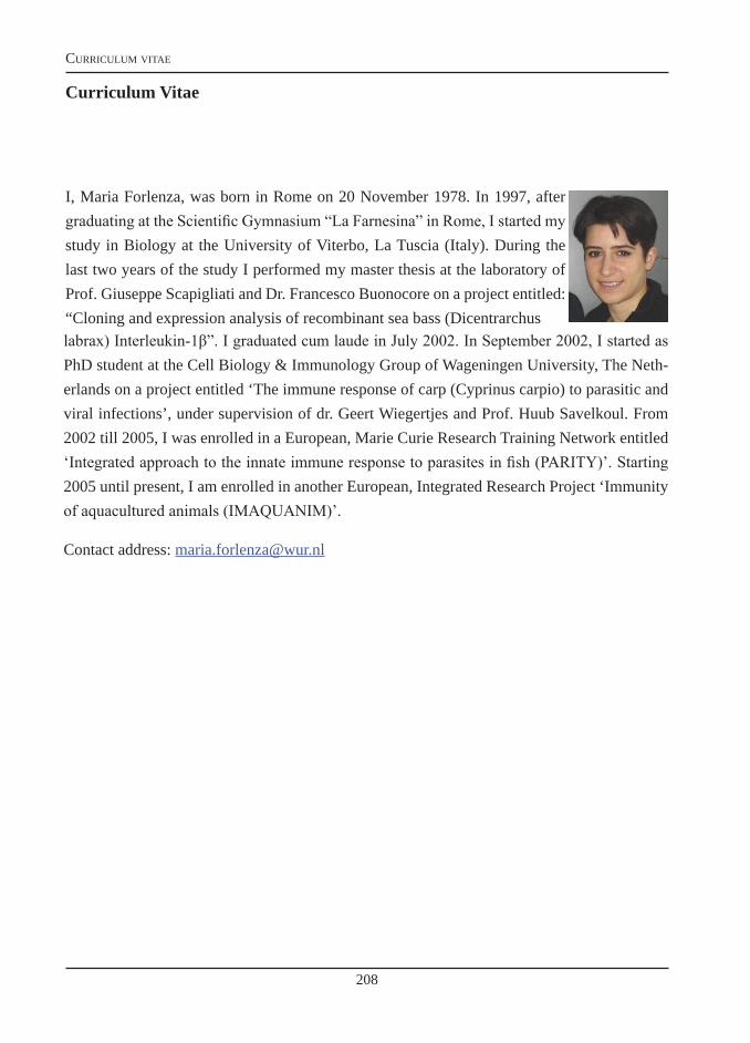
208
curriculum vitae
Curriculum Vitae
I, Maria Forlenza, was born in Rome on 20 November 1978. In 1997, after graduating at the Scientific Gymnasium “La Farnesina” in Rome, I started my study in Biology at the University of Viterbo, La Tuscia (Italy). During the last two years of the study I performed my master thesis at the laboratory of Prof. Giuseppe Scapigliati and Dr. Francesco Buonocore on a project entitled: “Cloning and expression analysis of recombinant sea bass (Dicentrarchuslabrax) Interleukin-1β”. I graduated cum laude in July 2002. In September 2002, I started as PhD student at the Cell Biology & Immunology Group of Wageningen University, The Neth-erlands on a project entitled ‘The immune response of carp (Cyprinus carpio) to parasitic and viral infections’, under supervision of dr. Geert Wiegertjes and Prof. Huub Savelkoul. From 2002 till 2005, I was enrolled in a European, Marie Curie Research Training Network entitled ‘Integrated approach to the innate immune response to parasites in fish (PARITY)’. Starting 2005 until present, I am enrolled in another European, Integrated Research Project ‘Immunity of aquacultured animals (IMAQUANIM)’.
Contact address: [email protected]
209
curriculum vitae
Training and Supervision PlanThe Basic PackageWIAS Introduction Course, Wageningen, The Netherlands 2003Horizon Debating course, Wageningen, The Netherlands 2003
Subtotal Basic Package 3 credits*Scientific ExposureInternational conferencesEAFP, Malta 2003ISDCI, St Andrews, Scotland, UK 2003First international symposium on quantitative PCR, Freising, Germany 2004EAFP, Copenhagen, Denmark 2005ISDCI, Charleston, USA, 2006NOFFI, Stirking, Scotland, UK 2007ISDCI, Prague, Czech Rep. 2009
Seminars and workshopsNVVI, Veldhoven, The Netherlands 2002WIAS seminar Trypanosomes: host-parasite interaction in the genomics era, Wageningen, The Netherlands 2002NVVI, Noordwijkerhout, The Netherlands 2003qPCR Seminar Experimental design and data analysis, Leusden, The Netherlands 2003NVVI, Maastricht, The Netherlands 2004First GeNeYousS Symposium, Utrecht, The Netherlands 2004NVVI, Soesterberg, The Netherlands 2005WIAS seminar Macrophage polarisation, Wageningen, The Netherlands 2006WIAS seminar Immune responses to viruses, Wageningen, The Netherlands 2006WIAS seminar Corticosteroid receptors in common carp, Wageningen, The Netherlands 2008WIAS seminar Multidisciplinary study of allergy, Wageningen, The Netherlands 2008WIAS seminar Fishing for Immunity, Wageningen, The Netherlands 2008
Poster PresentationsInnate immune responses to parasites in carp, Veldhoven, (NVVI) 2002Role of Nitric Oxide in the immune response to ‘tryps’ in carp, St. Andrews (ISDCI), 2003
Oral PresentationsFunctional characterization of carp leukocytes, Malta (EAFP) 2003(Anti)-inflammatory response of carp to parasite infections, Copenhagen (EAFP) 2005Experiences with Quantitative PCR for monitoring carp cytokines, Copenhagen (EAFP) 2005Cytokine expression profile after Typanosomes infection in carp. Westburg user’s meeting, Leusden 2005Interleukin-10: Let’s have a closer look at it, Charleston (ISDCI) 2006Differential contribution of neutrophilic granulocytes and macrophages, Prague (ISDCI), 2009TNFalpha: a double-edged sword, Prague (ISDCI) 2009
Subtotal Scientific Exposure 22 credits*
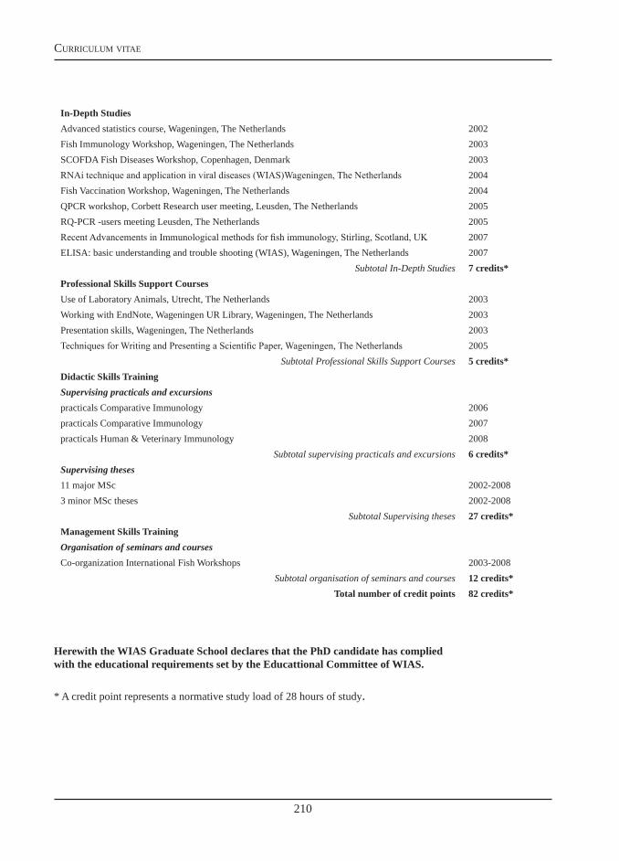
210
curriculum vitae
In-Depth StudiesAdvanced statistics course, Wageningen, The Netherlands 2002Fish Immunology Workshop, Wageningen, The Netherlands 2003SCOFDA Fish Diseases Workshop, Copenhagen, Denmark 2003RNAi technique and application in viral diseases (WIAS)Wageningen, The Netherlands 2004Fish Vaccination Workshop, Wageningen, The Netherlands 2004QPCR workshop, Corbett Research user meeting, Leusden, The Netherlands 2005RQ-PCR -users meeting Leusden, The Netherlands 2005Recent Advancements in Immunological methods for fish immunology, Stirling, Scotland, UK 2007ELISA: basic understanding and trouble shooting (WIAS), Wageningen, The Netherlands 2007
Subtotal In-Depth Studies 7 credits*Professional Skills Support CoursesUse of Laboratory Animals, Utrecht, The Netherlands 2003Working with EndNote, Wageningen UR Library, Wageningen, The Netherlands 2003Presentation skills, Wageningen, The Netherlands 2003Techniques for Writing and Presenting a Scientific Paper, Wageningen, The Netherlands 2005
Subtotal Professional Skills Support Courses 5 credits*Didactic Skills TrainingSupervising practicals and excursionspracticals Comparative Immunology 2006practicals Comparative Immunology 2007practicals Human & Veterinary Immunology 2008
Subtotal supervising practicals and excursions 6 credits*Supervising theses11 major MSc 2002-20083 minor MSc theses 2002-2008
Subtotal Supervising theses 27 credits*Management Skills TrainingOrganisation of seminars and coursesCo-organization International Fish Workshops 2003-2008
Subtotal organisation of seminars and courses 12 credits*Total number of credit points 82 credits*
Herewith the WIAS Graduate School declares that the PhD candidate has compliedwith the educational requirements set by the Educattional Committee of WIAS.
* A credit point represents a normative study load of 28 hours of study.

211
Printed by CPI, Wohrmann Print Service, Zutphen, The Netherlands.
The printing of this thesis was financially supported partly by Westburg, and by Eurogentec.
Cover: The picture of the parasite Argulus japanicus was kindly provided by Peter Walker.

212
Stupid questions do exist:
a stupid question is the one you ask without having thought about the possible answer yourself
Maria
Related Documents