SYMPOSIUM PAPER Annalisa Astolfi Simona Rolla Patrizia Nanni Elena Quaglino Carla De Giovanni Manuela Iezzi Piero Musiani Guido Forni Pier-Luigi Lollini Federica Cavallo Raffaele A. Calogero Immune prevention of mammary carcinogenesis in HER-2/neu transgenic mice: a microarray scenario Received: 4 August 2004 / Accepted: 1 October 2004 / Published online: 3 February 2005 Ó Springer-Verlag 2005 Abstract Neoplastic transformation is a multistep pro- cess in which gene products of specific regulatory path- ways are involved at each stage. Identification of these overexpressed or mutated gene products provides an unprecedented opportunity to address the immune sys- tem against defined antigens and eliminate transformed cells. Mice transgenic for these oncogenes (e.g. HER-2/ neu, a prototype of deregulated oncogenic protein kinase membrane receptors) are ideal experimental models for assessing the potential of active immuniza- tion. The demonstration that vaccines can cure HER-2/ neu transplantable tumors, prevent their onset and delay the progression of preneoplastic lesions in mice at risk suggests that efficient immunological inhibition of HER- 2/neu carcinogenesis can be achieved by specific vacci- nation. To further explore this issue, halting of tumor progression in the mammary glands of BALB-neuT mice with two immunization protocols in two laboratories has been studied independently by DNA microarray analysis. Combination of the two sets of results revealed a clear correlation between them when the tumor mass was titrated by transcription profiling. It was also clear that both protocols induced a strong, polyclonal anti- body response and halted tumor growth at a condition very similar to that at which the vaccination began. Differences in the expression profiles were mainly related to the expression levels of a few chemokines and T-cell- specific genes that may be in some way correlated with the efficacy of the vaccination. Last, combination of the expression data with the protection results indicated that chronic vaccination is needed to maintain an active IFN- c-mediated response in the mammary gland. Keywords HER-2/neu Tumor prevention Mammary carcinogenesis Cancer vaccines Microarray analysis Transcriptional profiling Introduction Neoplastic transformation is a multistep process in which gene products of specific regulatory pathways are involved at each stage. It may thus be supposed that a given stage can be influenced or even halted by inter- fering with its pathway/component. Identification of these distinct overexpressed or mutated gene products provides an unprecedented opportunity to address the immune system against defined antigens and eliminate transformed cells [10]. Mice transgenic for oncogenes are ideal experimental models for assessing the potential of active immunization [11, 23]. Those transgenic for the HER-2/neu oncogene are extensively studied because HER-2/neu is a prototype of the deregulated oncogenic protein kinase membrane receptors directly involved in cell carcinogenesis and markedly overexpressed by early neoplastic lesions. In addition, its amplification and consequent overexpression are associated with tumori- genesis in the breast, ovaries, brain and prostate, and an aggressive course [1, 13]. Vaccines can cure HER-2/neu transplantable tumors by preventing their onset and delaying the progression of preneoplastic alterations to invasive carcinomas [32]. This article is a symposium paper from the second international conference ‘‘Strategies for Immune Therapy’’, 29 February–3 March 2004, Wu¨ rzburg, Germany (summarized by G. Pawelec and C. Gouttefangeas) S. Rolla E. Quaglino G. Forni F. Cavallo (&) R. A. Calogero Department of Clinical and Biological Sciences, University of Turin, Orbassano, 10043, Italy E-mail: [email protected] Tel.: +39-011-6705419 Fax: +39-011-2365417 A. Astolfi P. Nanni C. De Giovanni P.-L. Lollini Cancer Research Section, Department of Experimental Pathology, University of Bologna, Bologna, 40126, Italy M. Iezzi P. Musiani Aging Research Center, CeSi, G. D’Annunzio University Foundation, Chieti, 66013, Italy Cancer Immunol Immunother (2005) 54: 599–610 DOI 10.1007/s00262-004-0635-4

Welcome message from author
This document is posted to help you gain knowledge. Please leave a comment to let me know what you think about it! Share it to your friends and learn new things together.
Transcript
SYMPOSIUM PAPER
Annalisa Astolfi Æ Simona Rolla Æ Patrizia Nanni
Elena Quaglino Æ Carla De Giovanni Æ Manuela Iezzi
Piero Musiani Æ Guido Forni Æ Pier-Luigi LolliniFederica Cavallo Æ Raffaele A. Calogero
Immune prevention of mammary carcinogenesis in HER-2/neu transgenicmice: a microarray scenario
Received: 4 August 2004 / Accepted: 1 October 2004 / Published online: 3 February 2005� Springer-Verlag 2005
Abstract Neoplastic transformation is a multistep pro-cess in which gene products of specific regulatory path-ways are involved at each stage. Identification of theseoverexpressed or mutated gene products provides anunprecedented opportunity to address the immune sys-tem against defined antigens and eliminate transformedcells. Mice transgenic for these oncogenes (e.g. HER-2/neu, a prototype of deregulated oncogenic proteinkinase membrane receptors) are ideal experimentalmodels for assessing the potential of active immuniza-tion. The demonstration that vaccines can cure HER-2/neu transplantable tumors, prevent their onset and delaythe progression of preneoplastic lesions in mice at risksuggests that efficient immunological inhibition of HER-2/neu carcinogenesis can be achieved by specific vacci-nation. To further explore this issue, halting of tumorprogression in the mammary glands of BALB-neuT micewith two immunization protocols in two laboratorieshas been studied independently by DNA microarrayanalysis. Combination of the two sets of results revealeda clear correlation between them when the tumor masswas titrated by transcription profiling. It was also clearthat both protocols induced a strong, polyclonal anti-
body response and halted tumor growth at a conditionvery similar to that at which the vaccination began.Differences in the expression profiles were mainly relatedto the expression levels of a few chemokines and T-cell-specific genes that may be in some way correlated withthe efficacy of the vaccination. Last, combination of theexpression data with the protection results indicated thatchronic vaccination is needed to maintain an active IFN-c-mediated response in the mammary gland.
Keywords HER-2/neu Æ Tumor prevention ÆMammary carcinogenesis Æ Cancer vaccines ÆMicroarray analysis Æ Transcriptional profiling
Introduction
Neoplastic transformation is a multistep process inwhich gene products of specific regulatory pathways areinvolved at each stage. It may thus be supposed that agiven stage can be influenced or even halted by inter-fering with its pathway/component. Identification ofthese distinct overexpressed or mutated gene productsprovides an unprecedented opportunity to address theimmune system against defined antigens and eliminatetransformed cells [10]. Mice transgenic for oncogenes areideal experimental models for assessing the potential ofactive immunization [11, 23]. Those transgenic for theHER-2/neu oncogene are extensively studied becauseHER-2/neu is a prototype of the deregulated oncogenicprotein kinase membrane receptors directly involved incell carcinogenesis and markedly overexpressed by earlyneoplastic lesions. In addition, its amplification andconsequent overexpression are associated with tumori-genesis in the breast, ovaries, brain and prostate, and anaggressive course [1, 13].
Vaccines can cure HER-2/neu transplantable tumorsby preventing their onset and delaying the progressionof preneoplastic alterations to invasive carcinomas [32].
This article is a symposium paper from the second internationalconference ‘‘Strategies for Immune Therapy’’, 29 February–3March 2004, Wurzburg, Germany (summarized by G. Pawelec andC. Gouttefangeas)
S. Rolla Æ E. Quaglino Æ G. Forni Æ F. Cavallo (&)R. A. CalogeroDepartment of Clinical and Biological Sciences,University of Turin, Orbassano, 10043, ItalyE-mail: [email protected].: +39-011-6705419Fax: +39-011-2365417
A. Astolfi Æ P. Nanni Æ C. De Giovanni Æ P.-L. LolliniCancer Research Section, Department of Experimental Pathology,University of Bologna, Bologna, 40126, Italy
M. Iezzi Æ P. MusianiAging Research Center, CeSi, G. D’AnnunzioUniversity Foundation, Chieti, 66013, Italy
Cancer Immunol Immunother (2005) 54: 599–610DOI 10.1007/s00262-004-0635-4

We have used HER-2/neu mammary carcinogenesis inBALB-neuT mice, one of the most aggressive models [3,32], to show that its efficient immunological inhibitioncan be achieved in this way by vaccination [21, 25–28].This protection always stems from the concurrence ofIFN-c release and specific antibodies against the onco-gene’s protein product p185neu. T-cell cytotoxicity, onthe other hand, appears of only marginal importance [4,5, 8, 21, 25–28] despite its proven association with theresponse against transplantable tumors. This paper de-scribes our further investigation of the role of antibodiesin this context through the employment of DNAmicroarray analysis to study the outcome of twoimmunization protocols independently in two labora-tories.
Combined allogeneic tumor cell vaccinations haltHER-2/neu carcinogenesis in BALB-neuT mice
Two vaccination protocols halt mammary carcinogene-sis in BALB-neuT mice (H-2d) [21, 26] transgenic for theactivated transforming rat HER-2/neu oncogene underthe transcriptional control of the mouse mammarytumor virus promoter [19]. These mice are one of themost aggressive models of mammary carcinogenesis,since p185neu is already overexpressed on the cell surfaceof the rudimentary mammary glands of 3-week-oldfemales. All ten glands inexorably develop invasive andmetastasizing carcinomas through a well-established andalmost synchronous progression [3, 5, 21, 26]. Lolliniet al. [21] observed that a combination of MHC allo-geneic (H-2q) tumor cells expressing p185neu and sys-
temic recombinant interleukin (IL-) 12 administered tohealthy, 6-week-old BALB-neuT mice reduced tumorincidence by 90–100% and more than doubled lifeexpectancy. Almost the same protection was providedby a single-cell vaccine composed of H-2q p185neu-po-sitive tumor cells gene-engineered to release IL-12 [8].Cavallo et al. [26] have shown that vaccination of 10-week-old mice bearing multiple in situ carcinomas withDNA plasmids coding for the extracellular and trans-membrane domain of p185neu, followed by a boost withp185neu-positive H-2q tumor cells gene-engineered tosecrete IFN-c, keeps 48% of mice tumor free until week32.
In both studies, cytokine release and antibody pro-duction were the most prominent immune mechanisms,whereas T-cell cytotoxicity was absent, and high serumtiters of anti-p185neu antibodies were found. Since theseantibodies were mainly IgG2a and to a lesser extentIgG2b, whereas IgG3 was a marginal component, vac-cination may be supposed to activate T cells producingIFN-c, the primary switch factor for IgG2a. The geneexpression patterns of the mammary glands of thesemice have now been investigated independently in thetwo laboratories [26; Astolfi et al., unpublished results]to determine the mechanisms through which protectionis provided. The results are compared in this paper.
Cross-study validation
Integration and cross-study validation of the geneexpression profiles were hampered by the use of distinctmicroarray platforms, sample sets, protocols, and ana-lytical approaches. Parmigiani et al. [24] have devised anintegrative correlation (IC) coefficient for quantificationof the extent to which independent studies can be reli-ably analyzed together for the systematic comparison ofmicroarray profiles. This approach has been used forcross-study validation of the data of Cavallo [26] andLollini [Astolfi, unpublished results]. All pairwise cor-relations (Pearson correlation coefficient) of geneexpression across samples within individual projects
Fig. 1 Cross-study validation of data generated by Quaglino et al.[26] and De Giovanni et al. [8; Astolfi, unpublished results] revealssignificant agreement of the gene expression patterns in these twoindependent studies. Cross-experiment consistency of expressionpatterns was evaluated with an integrative correlation (IC)coefficient that quantifies cross-study reproducibility withoutrelying on direct assimilation of expression measurements acrossexperiments. a Histogram of the empirical distribution of the gene-specific correlation of correlations. b Scatterplot of the pairwisecorrelation comparison within the probe sets in the two datasets
600
were calculated and the reproducibility of the results wasdefined without relying on direct comparison ofexpression across platforms. Overall reproducibility wassummarized by correlations of IC coefficients equal to0.907 and it was also evaluated gene by gene (Fig. 1).Both overall and gene-by-gene reproducibility revealedthe striking similarity of the two studies and hence al-lowed comparison of their data.
Titration of the tumor by transcriptional profiling
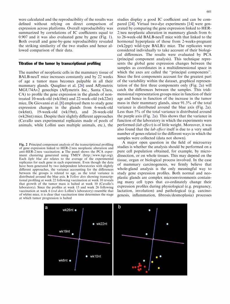
The number of neoplastic cells in the mammary tissue ofBALB-neuT mice increases constantly and by 22 weeksof age a tumor mass becomes palpable in all theirmammary glands. Quaglino et al. [26] used AffymetrixMGU74Av2 genechips (Affymetrix Inc., Santa Clara,CA) to profile the gene expression in the glands of non-treated 10-week-old (wk10nt) and 22-week-old (wk22nt)mice, De Giovanni et al. [8] employed them to study geneexpression changes in the glands from 6-week-old(wk6nt), 19-week-old (wk19nt), and 26-week-old(wk26nt) mice. Despite their slightly different approaches(Cavallo uses experimental replicates made of pools ofanimals, while Lollini uses multiple animals, etc.), the
studies display a good IC coefficient and can be com-pared [24]. Virtual two-dye experiments [14] were gen-erated by comparing the gene expression linked to HER-2/neu neoplastic alteration in mammary glands from 6-to 26-week-old BALB-neuT mice with that linked to thehormonal hyperplasia of those from 2-weeks-pregnant(wk2pgr) wild-type BALB/c mice. The replicates wereconsidered individually to take account of their biologi-cal differences. The results were evaluated by PCA(principal component analysis). This technique repre-sents the global gene expression changes between thesamples as coordinates in a multidimensional space inwhich the axes are called the ‘‘principal components’’.Since the first components account for the greatest partof the variability within the dataset, graphical represen-tation of the first three components only (Fig. 2a) willcatch the differences between the samples. This tridi-mensional representation groups mice in function of theirage and hence in function of the increase in the tumormass in their mammary glands, since 91.3% of the totalvariance is distributed around the blue axis (Fig. 2a).Less than 5% of the total variance is distributed aroundthe purple axis (Fig. 2a). This shows that the variance infunction of the laboratory in which the experiments wereperformed (lab effect) is of little weight. Moreover, it wasalso found that the lab effect itself is due to a very smallnumber of genes related to the different ways in which thesamples were collected (data not shown).
A major open question in the field of microarraystudies is whether the analysis should be performed on apure cell population obtained, for example, by micro-dissection, or on whole tissues. This may depend on thetissue, organ or biological process involved. In the caseof mammary carcinogenesis, we firmly believe thatwhole-gland analysis is the only meaningful way tostudy gene expression profiles. Both normal and neo-plastic glands are complex microenvironments contain-ing many cell types that co-ordinately change theirexpression profiles during physiological (e.g. pregnancy,lactation, involution) and pathological (e.g. carcino-genesis, inflammation, fibrosis/desmoplasia) processes
Fig. 2 Principal component analysis of the transcriptional profilingof gene expression linked to HER-2/neu neoplastic alteration andanti-HER-2/neu vaccination. a The panel shows the PCA exper-iment clustering generated using TMEV (http://www.tigr.org).Each light blue dot relates to the average of the experimentalreplicates for each gene in each experiment. Even though the datahave been generated by two independent laboratories with slightlydifferent approaches, the variance accounting for the differencesbetween the groups is related to age, as the total variance isdistributed around the blue axis. b Yellow dots showing transcrip-tional profiling at week 22 following vaccination at week 10 revealsthat growth of the tumor mass is halted at week 10 (Cavallo’slaboratory). Since the profiles at week 15 and week 26 followingvaccination at week 6 (red dots Lollini’s laboratory) resemble thatof wk6nt mice, it is clear that vaccination time determines the stageat which tumor progression is halted
601
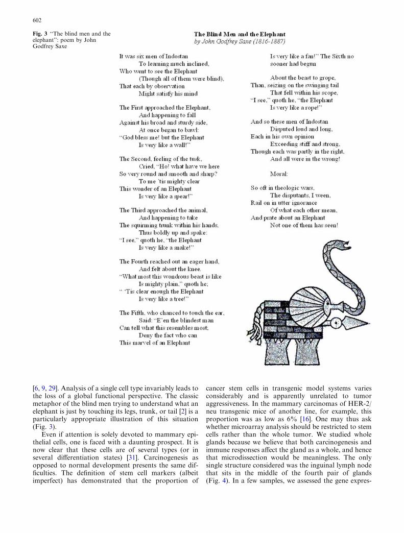
[6, 9, 29]. Analysis of a single cell type invariably leads tothe loss of a global functional perspective. The classicmetaphor of the blind men trying to understand what anelephant is just by touching its legs, trunk, or tail [2] is aparticularly appropriate illustration of this situation(Fig. 3).
Even if attention is solely devoted to mammary epi-thelial cells, one is faced with a daunting prospect. It isnow clear that these cells are of several types (or inseveral differentiation states) [31]. Carcinogenesis asopposed to normal development presents the same dif-ficulties. The definition of stem cell markers (albeitimperfect) has demonstrated that the proportion of
cancer stem cells in transgenic model systems variesconsiderably and is apparently unrelated to tumoraggressiveness. In the mammary carcinomas of HER-2/neu transgenic mice of another line, for example, thisproportion was as low as 6% [16]. One may thus askwhether microarray analysis should be restricted to stemcells rather than the whole tumor. We studied wholeglands because we believe that both carcinogenesis andimmune responses affect the gland as a whole, and hencethat microdissection would be meaningless. The onlysingle structure considered was the inguinal lymph nodethat sits in the middle of the fourth pair of glands(Fig. 4). In a few samples, we assessed the gene expres-
Fig. 3 ‘‘The blind men and theelephant’’: poem by JohnGodfrey Saxe
602
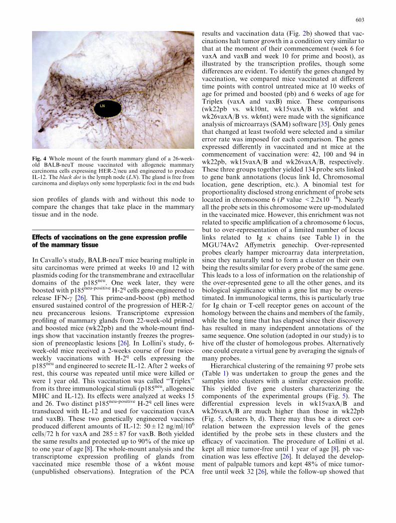
sion profiles of glands with and without this node tocompare the changes that take place in the mammarytissue and in the node.
Effects of vaccinations on the gene expression profileof the mammary tissue
In Cavallo’s study, BALB-neuT mice bearing multiple insitu carcinomas were primed at weeks 10 and 12 withplasmids coding for the transmembrane and extracellulardomains of the p185neu. One week later, they wereboosted with p185neu-positive H-2q cells gene-engineered torelease IFN-c [26]. This prime-and-boost (pb) methodensured sustained control of the progression of HER-2/neu precancerous lesions. Transcriptome expressionprofiling of mammary glands from 22-week-old primedand boosted mice (wk22pb) and the whole-mount find-ings show that vaccination instantly freezes the progres-sion of preneoplastic lesions [26]. In Lollini’s study, 6-week-old mice received a 2-weeks course of four twice-weekly vaccinations with H-2q cells expressing thep185neu and engineered to secrete IL-12. After 2 weeks ofrest, this course was repeated until mice were killed orwere 1 year old. This vaccination was called ‘‘Triplex’’from its three immunological stimuli (p185neu, allogeneicMHC and IL-12). Its effects were analyzed at weeks 15and 26. Two distinct p185neu-positive H-2q cell lines weretransduced with IL-12 and used for vaccination (vaxAand vaxB). These two genetically engineered vaccinesproduced different amounts of IL-12: 50±12 ng/ml/106
cells/72 h for vaxA and 285±87 for vaxB. Both yieldedthe same results and protected up to 90% of the mice upto one year of age [8]. The whole-mount analysis and thetranscriptome expression profiling of glands fromvaccinated mice resemble those of a wk6nt mouse(unpublished observations). Integration of the PCA
results and vaccination data (Fig. 2b) showed that vac-cinations halt tumor growth in a condition very similar tothat at the moment of their commencement (week 6 forvaxA and vaxB and week 10 for prime and boost), asillustrated by the transcription profiles, though somedifferences are evident. To identify the genes changed byvaccination, we compared mice vaccinated at differenttime points with control untreated mice at 10 weeks ofage for primed and boosted (pb) and 6 weeks of age forTriplex (vaxA and vaxB) mice. These comparisons(wk22pb vs. wk10nt, wk15vaxA/B vs. wk6nt andwk26vaxA/B vs. wk6nt) were made with the significanceanalysis of microarrays (SAM) software [35]. Only genesthat changed at least twofold were selected and a similarerror rate was imposed for each comparison. The genesexpressed differently in vaccinated and nt mice at thecommencement of vaccination were: 42, 100 and 94 inwk22pb, wk15vaxA/B and wk26vaxA/B, respectively.These three groups together yielded 134 probe sets linkedto gene bank annotations (locus link Id, Chromosomallocation, gene description, etc.). A binomial test forproportionality disclosed strong enrichment of probe setslocated in chromosome 6 (P value <2.2x10�16). Nearlyall the probe sets in this chromosome were up-modulatedin the vaccinated mice. However, this enrichment was notrelated to specific amplification of a chromosome 6 locus,but to over-representation of a limited number of locuslinks related to Ig j chains (see Table 1) in theMGU74Av2 Affymetrix genechip. Over-representedprobes clearly hamper microarray data interpretation,since they naturally tend to form a cluster on their ownbeing the results similar for every probe of the same gene.This leads to a loss of information on the relationship ofthe over-represented gene to all the other genes, and itsbiological significance within a gene list may be overes-timated. In immunological terms, this is particularly truefor Ig chain or T-cell receptor genes on account of thehomology between the chains and members of the family,while the long time that has elapsed since their discoveryhas resulted in many independent annotations of thesame sequence. One solution (adopted in our study) is tohive off the cluster of homologous probes. Alternativelyone could create a virtual gene by averaging the signals ofmany probes.
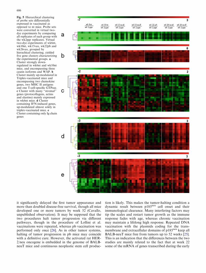
Hierarchical clustering of the remaining 97 probe sets(Table 1) was undertaken to group the genes and thesamples into clusters with a similar expression profile.This yielded five gene clusters characterizing thecomponents of the experimental groups (Fig. 5). Thedifferential expression levels in wk15vaxA/B andwk26vaxA/B are much higher than those in wk22pb(Fig. 5, clusters b, d). There may thus be a direct cor-relation between the expression levels of the genesidentified by the probe sets in these clusters and theefficacy of vaccination. The procedure of Lollini et al.kept all mice tumor-free until 1 year of age [8]. pb vac-cination was less effective [26]. It delayed the develop-ment of palpable tumors and kept 48% of mice tumor-free until week 32 [26], while the follow-up showed that
Fig. 4 Whole mount of the fourth mammary gland of a 26-week-old BALB-neuT mouse vaccinated with allogeneic mammarycarcinoma cells expressing HER-2/neu and engineered to produceIL-12. The black dot is the lymph node (LN). The gland is free fromcarcinoma and displays only some hyperplastic foci in the end buds
603
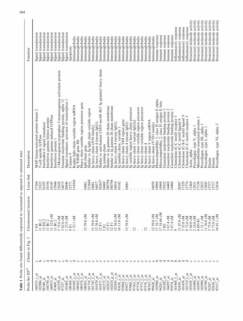
Table
1Probesets
founddifferentiallyexpressed
invaccinatedasopposedto
untreatedmice
ProbeSet
IDab
Cluster
inFig.5
Chromosomallocation
Locuslink
Description
Function
160353_i_at
c1E4
17164
MAPkinase-activatedprotein
kinase
2Signaltransduction
102906_at
b11B1.2
21822
T-cellspecificGTPase
Signaltransduction
98349_at
c13D2.2
16195
Interleukin
6signaltransducer
Signaltransduction
104750_at
d11B1.2
18356
Interferongammahitinosprotein
Signaltransduction
160933_at
d1132.0
cM16145
InterferongammainducedGTPase
Signaltransduction
97409_at
d11B1.2
15944
Interferonprotein
1Signaltransduction
102259_at
c575.0
cM22628
3-M
onooxgenase/tryptophan5-m
onooxgenase
activationprotein
Signaltransduction
97227_at
c582.0
cM14673
Guaninenucleotidebindingprotein,alpha12
Signaltransduction
101465_at
d125.9
cM20846
Signaltransducer,activatoroftranscription1
Signaltransduction
100368_at
c4E1
12371
Caspase
9Apoptosis
101845_s_at
d153.1
cM114564
Kappalightchain
variable
regionmRNA
Immunoglobulin
100362_f_at
e–
–Ig
V(H
)IIgeneH8
Immunoglobulin
100376_f_at
e–
–Heavychain
variable
regionprecursorgene
Immunoglobulin
100583_at
e1259.0
cM16061
IgH
chain
gene
Immunoglobulin
101718_f_at
e–
213684
Sim
ilarto
Iglightchain
variable
region
Immunoglobulin
101743_f_at
e1259.0
cM16061
Igheavychain
(J558family)
Immunoglobulin
101870_at
e1258.0
cM16017
Igheavychain
4(serum
IgG1)
Immunoglobulin
101871_f_at
e12F1
382657
Sim
ilarto
anti-humanCD19moAb4G7Ig
gamma1heavychain
Immunoglobulin
102372_at
e5E1
16069
Igjoiningchain
Immunoglobulin
102823_at
e12F1
380794
Sim
ilarto
Iggamma-2bchain
mem
brane
Immunoglobulin
102824_g_at
e12F1
380794
Sim
ilarto
Iggamma-2bchain
mem
brane
Immunoglobulin
102843_s_at
e1258.0
cM16014
Igheavychain
4(serum
IgG1)
Immunoglobulin
93638_s_at
e1613.0
cM16142
Iglambdachain,variable
1Im
munoglobulin
93904_f_at
e–
–Ig
heavychain
VDJregiongene
Immunoglobulin
93927_f_at
e1259.0
cM16061
Igheavychain
4(serum
IgG1)
Immunoglobulin
97008_f_at
e–
–Ig
variable
regionheavychain
precursor
Immunoglobulin
97563_f_at
e12
–Ig
heavychain
1(serum
IgG2a)
Immunoglobulin
97574_f_at
e–
–Ig
heavychain
variable
regionprecursor
Immunoglobulin
97575_f_at
e–
–Ig
heavychain
variable
regionprecursor
Immunoglobulin
97576_f_at
e12
–Ig
heavychain
4Im
munoglobulin
98765_f_at
e–
–Ig
heavychain
VregionmRNA
Immunoglobulin
99420_at
e1259.0
cM16059
Igheavychain
(V7183family)
Immunoglobulin
102904_at
b1718.7
cM14968
Histocompatibility2,class
IIantigen
Ealpha
Immuneresponse
94285_at
d1718.66cM
14969
Histocompatibility2,class
IIantigen
Ebeta
Immuneresponse
103202_at
d3H1
55932
Guanylate
nucleotidebindingprotein
3Im
muneresponse
104597_at
d367.4
cM14469
Guanylate
nucleotidebindingprotein
2Im
muneresponse
95974_at
d367.4
cM14468
Guanylate
nucleotidebindingprotein
1Im
muneresponse
161968_f_at
b9
–Chem
okine(C
-Cmotif)
receptor5
Inflammatory
response
92459_at
b1147.0
cM20307
Chem
okine(C
-Cmotif)
ligand8
Inflammatory
response
101436_at
d553.0
cM17329
Chem
okine(C
-X-C
motif)
ligand9
Inflammatory
response
102719_f_at
d972.0
cM12774
Chem
okine(C
-Cmotif)
receptor5
Inflammatory
response
101028_i_at
c264.0
cM11464
Actin,alpha
Structuralmolecule
activity
101080_at
c218.0
cM12831
Procollagen,typeV,alpha1
Structuralmolecule
activity
101095_at
c4D3-E1
17150
Microfibrillar-associatedprotein
2Structuralmolecule
activity
102990_at
c121.1
cM12825
Procollagen,typeIII,alpha1
Structuralmolecule
activity
103709_at
c1156.0
cM12842
Procollagen,typeI,alpha1
Structuralmolecule
activity
92207_at
c575.0
cM13717
Elastin
Structuralmolecule
activity
92836_at
c575.0
cM13717
Elastin
Structuralmolecule
activity
93517_at
c1041.1
cM12834
Procollagen,typeVI,alpha2
Structuralmolecule
activity
604
94305_at
c1156.0
cM12842
Procollagen,typeI,alpha1
Structuralmolecule
activity
99367_at
c2H1
13589
Microtubule-associatedprotein,RP/EB
family,mem
ber
1Structuralmolecule
activity
92559_at
d350.8
cM22329
Vascularcelladhesionhitinos1
Structuralmolecule
activity
100463_at
a544.93cM
12993
Casein
gamma
Transport
96030_at
a544.9
cM12990
Casein
alpha
Transport
99130_at
a544.91cM
12991
Casein
beta
Transport
102704_at
c186.0
cM11829
Aquaporin4
Transport
102708_at
a1312.0
cM12231
Butyrophilin,subfamily1,mem
ber
A1
Transport
160949_at
c14B
26430
Poly
(ADP-ribose)glycohydrolase
DNA
damageresponses
92833_at
b1051.0
cM15109
Histidineammonia
lyase
Biosynthesis
160617_at
c7B5
50794
Kruppel-likefactor13
Transcription
103926_at
c16A1
208643
Ribosomalprotein
L24
Translation
101139_r_at
c5E1
17830
Mucin10
Biological-process
unknown
102860_at
d12E
20715
Serine(orcysteine)
proteinase
inhibitor,cladeA,mem
ber
3G
Biological-process
unknown
93085_at
d1718.59cM
16912
Proteosomesubunit,betatype9(largemultifunctionalprotease
2)
Biological-process
unknown
102931_at
a110.5
cM22373
Whey
acidic
protein
(WAP)
Biological-process
unknown
93028_at
c769.03cM
14955
H19fetalliver
mRNA
Biological-process
unknown
103033_at
b1718.83cM
20567
Sex-lim
ited
protein
Biological-process
unknown
99126_at
cX
–InactiveX
specifictranscripts
Biological-process
unknown
99850_at
b15
–RIK
EN
cDNA
4432409D24gene
Biological-process
unknown
101518_at
c11B5
55978
RIK
EN
cDNA
0610009H04gene
Biological-process
unknown
103905_at
c9C
76459
RIK
EN
cDNA
2310047E01gene
Biological-process
unknown
100944_at
b19
–Expressed
sequenze
AW112010
Biological-process
unknown
103963_f_at
d18E1
60440
Expressed
sequenze
AW111922
Biological-process
unknown
96764_at
d18E1
60440
Expressed
sequence
AW111922
Biological-process
unknown
98410_at
d11B1.3
54396
Expressed
sequenze
AI481100
Biological-process
unknown
102721_at
e12F1
380795
EST
Biological-process
unknown
102722_g_at
e12F1
380795
EST
Biological-process
unknown
160173_at
c–
–EST
(acc:AI852838)
Biological-process
unknown
100880_at
d–
–EST
(acc:AA816121)
Biological-process
unknown
aExpressiondata
relatedto
thepresentprobesets
canberequestedto
theauthors
b34/37probesetsfoundover-representedin
chromosome6werelinked
toIg
klightchain
genes:21probesetswerelinked
tovariableregion(V
)28,3to
V8,1to
V1,1to
V20,1to
V21,
andtheothersweresimply
annotatedasIg
kgenes.Probesetsare
grouped
onthebasisoftheirputativefunction.Functionalgroupswereinferred
byprobesetsGeneOntology(http://
www.geneontology.org)annotationorbyGeneR
IF(http://w
ww.ncbi.nih.gov/entrez/query.fcgi?db=
gene)
annotation
605
it significantly delayed the first tumor appearance andmore than doubled disease-free survival, though all micedeveloped one or more tumors by week 52 (Cavallo,unpublished observation). It may be supposed that thetwo procedures halt tumor progression via differentpathways, though in the procedure of Lollini et al.vaccinations were repeated, whereas pb vaccination wasperformed only once [26]. As in other tumor systems,halting of tumor progression in pb mice may coincidewith a definitive cure. However, the activated rat HER-2/neu oncogene is embedded in the genome of BALB-neuT mice and continuous neoplastic stem cell produc-
tion is likely. This makes the tumor-halting condition adynamic result between p185neu cell onset and theirimmunological clearance. Many interfering factors maytip the scales and restart tumor growth as the immuneresponse fades with age, whereas chronic vaccinationmay maintain a lifelong high response. Repeated DNAvaccination with the plasmids coding for the trans-membrane and extracellular domains of p185neu keep allBALB-neuT mice free from tumors up to 52 weeks [25].This is an indication that the differences between the twostudies are mainly related to the fact that at week 22some of the mRNA of genes transcribed during the early
Fig. 5 Hierarchical clusteringof probe sets differentiallyexpressed in vaccinated asopposed to nt mice. Probe setswere converted in virtual two-dye experiments by comparingall replicates of each group withthe wk2pgr replicates. Virtualtwo-dye experiments of wk6nt,wk10nt, wk15vax, wk22pb andwk26vax, grouped byhierachical clustering, yieldedfive gene clusters characterizingthe experimental groups. aCluster strongly down-regulated in wk6nt and wk10ntmice, and encompassing threecasein isoforms and WAP. bCluster mainly up-modulated inTriplex-vaccinated mice andencompassing two chemokinegenes, two MHC II antigensand one T-cell-specific GTPase.c Cluster with many ‘‘stromal’’genes (protocollagens, actinsand elastins) mainly expressedin wk6nt mice. d Clustercontaining IFN-induced genes,up-modulated almost solely intriplex-vaccinated mice. eCluster containing only Ig chaingenes
606

phase of the immune response have already decayed,whereas they are constantly regenerated by repeatedvaccination. The five clusters are illustrated in Fig. 5.Cluster ‘‘a’’ groups genes strongly down-regulated in themammary glands of wk6nt and wk10nt mice. It ischaracterized by the presence of three isoforms of casein(a, b, c) and whey acidic protein (WAP), which arehighly induced in mammary glands from pregnant andvaccinated mice. Induction of genes related to latepregnancy and lactation in the glands of vaccinatedvirgin mice is an unexpected finding that emerged fromboth sets of data. Raw expression levels are shown inmore detail in Fig. 6, which also includes results fromadditional microarray experiments with non-transgenicmammary glands of BALB/c mice and with in vitrocultured N202.1A and N202.1E cell lines [22]. The re-sults clearly show that the induction of genes related tolate pregnancy and lactation was caused by vaccinationand unrelated to tumor growth. The WAP was onlyexpressed in the glands of pregnant mice and those in thetwo vaccination groups. Expression of casein genes wasdetectable in late progression samples (19–26 weeks ofage), but the levels induced by vaccination in tumor-freeglands were one order of magnitude higher than thosefound in tumors. The glands of vaccinated miceexpressed a signature normally induced by pregnancyand lactation, rather than by carcinoma growth andprogression. The strong IFN-c response induced byvaccination may well be involved in the induction of thisgroup of genes. In late pregnancy and lactation, WAPand caseins are induced by prolactin acting via Stat5[15]. The ubiquitous effects of IFN-c are mediated by
Stat1 activation, but in the breast and a few other tissuesIFN-c also activates Stat5 [34]. Induction of WAP andcasein c was higher in Triplex than in the pb mice, inparallel with the higher induction of all other IFN-c-sensitive genes. Prolactin-like activity triggered by IFN-cin the mammary gland is a possibility that deservesfurther investigation to determine the mechanism in-volved and its potential biological consequences.
Cluster ‘‘b’’ contains genes prevalently up-modulatedin vaccinated mice, particularly in wk15vaxA/vaxB andwk26vaxA/vaxB compared to wk22pb. Chemokinereceptor 5 (CCR5), chemokine ligand 8 (CCL8) and a T-cell-specific GTPase (TGTP) are enclosed in this cluster,together with MHC II antigens. Both CCR5 and CCL8are involved in inflammatory responses. Expression ofCCR5 has been detected in a promyeloblastic cell line,suggesting that it plays a role in granulocyte lineageproliferation and differentiation [30], while CCL8 re-cruits leukocytes to sites of inflammation and contrib-utes to tumor-associated leukocyte infiltration [37].
Cluster ‘‘c’’ mainly contains ‘‘stromal’’ genes such aselastin, collagen and IL-6. These genes are down-mod-ulated during tumor progression, as the epithelial com-ponent overcomes all other cell types in the mammarytissue. Their low expression in all the vaccinated samplessuggests remodeling of the mammary stroma due to ageor perhaps due to the immune response induced byvaccination. However, this remodeling has not yet beenestablished morphologically and further investigation ofthis discrepancy is needed.
Of the other genes in cluster ‘‘c’’, caspase-9 isup-modulated in vaccinated mice. It is processed by
Fig. 6 Level of expressionmeasured with microarrays ofWAP and alpha, beta andgamma casein in mammaryglands from non-transgenicBALB/c mice, pregnant mice,cell lines derived from tumors inHER-2/neu transgenic mice(1A-1E), mammary glands fromHER-2/neu transgenic nt miceat different time points, andfrom vaccinated mice (Triplex,Prime-boost). Both vaccinationsincrease the level of expressionof these genes, which arenormally related to pregnancyand lactation
607

caspase APAF1 [36] as one of the earliest steps in thecaspase activation cascade. Mapkapk2 kinase [33], alsopresent in this cluster, is involved in many cellular pro-cesses, including stress and inflammatory responses,nuclear export [18], gene expression regulation and cellproliferation [38]. Furthermore, the RAC1–MKK3–P38–Mapkapk2 pathway promotes urokinase plasmin-ogen activator mRNA stability in invasive breast cancercells [12].
Cluster ‘‘d’’ contains IFN-induced genes (Stat1, Ifi1,Ifi47, Igtp, Gbp1-3, CXCL9, PSMB9) that are only up-modulated strongly in vaxA and vaxB. Igtp is a Stat1-
dependent gene [7]. Its role in cell survival relies onactivation of PI3-K/Akt, inactivation of GSK-3 andsuppression of caspase-9 and caspase-3 [39]. The genesin this cluster are early-response genes that are probablyalready decayed in the pb experiments. The serum IFN-cburst occurs between day 1 and day 3 after vaccinationwith IL-12-transduced cells, and plasma levels return tothe baseline by the 4th day [8]. The IFN-c-mediatedresponse may be short-lived also in the mammary epi-thelium, particularly since these IFN-c-induced genesare much more up-regulated in wk15vaxA/B than inwk26vaxA/B mice. The 15th week, in fact, correspondsto the 2nd week of injection in the third course, whereasat 26 weeks mice are at the end of a 2-week pause beforethe sixth course. So we can conclude that chronic vac-cination is necessary to maintain an active IFN-c-med-iated response in the mammary gland, which is probablynecessary (but not sufficient) for long-term protectionfrom tumor onset, since only chronic boosting of thisresponse can prevent tumor development for a long time[8, 21, 25]. Cluster ‘‘d’’ also comprises VCAM1, which isa member of the Ig superfamily and encodes a cell sur-
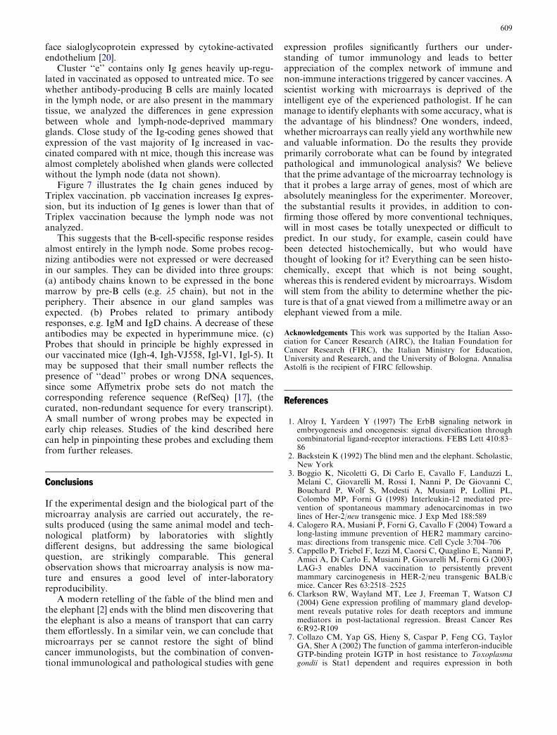
Fig. 7 Virtual two-dye experiments showing Ig chain genes inwk6nt, wk10nt, wk15vax, wk26vax and wk22pb with respect towk2pgn grouped by hierarchical clustering (Euclidean distance,average linkage). All the vaccinated mice show up-regulation ofimmunoglobulin genes with respect to nt mice. Triplex-vaccinatedmice (wk15vax, wk26vax) whose mammary glands were collectedwith the lymph node display a higher Ig gene induction thanwk22pb mice whose lymph node was not analyzed. This demon-strates that the B-cell response resides mainly in the lymph noderather than in the surrounding mammary tissue
608
face sialoglycoprotein expressed by cytokine-activatedendothelium [20].
Cluster ‘‘e’’ contains only Ig genes heavily up-regu-lated in vaccinated as opposed to untreated mice. To seewhether antibody-producing B cells are mainly locatedin the lymph node, or are also present in the mammarytissue, we analyzed the differences in gene expressionbetween whole and lymph-node-deprived mammaryglands. Close study of the Ig-coding genes showed thatexpression of the vast majority of Ig increased in vac-cinated compared with nt mice, though this increase wasalmost completely abolished when glands were collectedwithout the lymph node (data not shown).
Figure 7 illustrates the Ig chain genes induced byTriplex vaccination. pb vaccination increases Ig expres-sion, but its induction of Ig genes is lower than that ofTriplex vaccination because the lymph node was notanalyzed.
This suggests that the B-cell-specific response residesalmost entirely in the lymph node. Some probes recog-nizing antibodies were not expressed or were decreasedin our samples. They can be divided into three groups:(a) antibody chains known to be expressed in the bonemarrow by pre-B cells (e.g. k5 chain), but not in theperiphery. Their absence in our gland samples wasexpected. (b) Probes related to primary antibodyresponses, e.g. IgM and IgD chains. A decrease of theseantibodies may be expected in hyperimmune mice. (c)Probes that should in principle be highly expressed inour vaccinated mice (Igh-4, Igh-VJ558, Igl-V1, Igl-5). Itmay be supposed that their small number reflects thepresence of ‘‘dead’’ probes or wrong DNA sequences,since some Affymetrix probe sets do not match thecorresponding reference sequence (RefSeq) [17], (thecurated, non-redundant sequence for every transcript).A small number of wrong probes may be expected inearly chip releases. Studies of the kind described herecan help in pinpointing these probes and excluding themfrom further releases.
Conclusions
If the experimental design and the biological part of themicroarray analysis are carried out accurately, the re-sults produced (using the same animal model and tech-nological platform) by laboratories with slightlydifferent designs, but addressing the same biologicalquestion, are strikingly comparable. This generalobservation shows that microarray analysis is now ma-ture and ensures a good level of inter-laboratoryreproducibility.
A modern retelling of the fable of the blind men andthe elephant [2] ends with the blind men discovering thatthe elephant is also a means of transport that can carrythem effortlessly. In a similar vein, we can conclude thatmicroarrays per se cannot restore the sight of blindcancer immunologists, but the combination of conven-tional immunological and pathological studies with gene
expression profiles significantly furthers our under-standing of tumor immunology and leads to betterappreciation of the complex network of immune andnon-immune interactions triggered by cancer vaccines. Ascientist working with microarrays is deprived of theintelligent eye of the experienced pathologist. If he canmanage to identify elephants with some accuracy, what isthe advantage of his blindness? One wonders, indeed,whether microarrays can really yield any worthwhile newand valuable information. Do the results they provideprimarily corroborate what can be found by integratedpathological and immunological analysis? We believethat the prime advantage of the microarray technology isthat it probes a large array of genes, most of which areabsolutely meaningless for the experimenter. Moreover,the substantial results it provides, in addition to con-firming those offered by more conventional techniques,will in most cases be totally unexpected or difficult topredict. In our study, for example, casein could havebeen detected histochemically, but who would havethought of looking for it? Everything can be seen histo-chemically, except that which is not being sought,whereas this is rendered evident by microarrays. Wisdomwill stem from the ability to determine whether the pic-ture is that of a gnat viewed from a millimetre away or anelephant viewed from a mile.
Acknowledgements This work was supported by the Italian Asso-ciation for Cancer Research (AIRC), the Italian Foundation forCancer Research (FIRC), the Italian Ministry for Education,University and Research, and the University of Bologna. AnnalisaAstolfi is the recipient of FIRC fellowship.
References
1. Alroy I, Yardeen Y (1997) The ErbB signaling network inembryogenesis and oncogenesis: signal diversification throughcombinatorial ligand-receptor interactions. FEBS Lett 410:83–86
2. Backstein K (1992) The blind men and the elephant. Scholastic,New York
3. Boggio K, Nicoletti G, Di Carlo E, Cavallo F, Landuzzi L,Melani C, Giovarelli M, Rossi I, Nanni P, De Giovanni C,Bouchard P, Wolf S, Modesti A, Musiani P, Lollini PL,Colombo MP, Forni G (1998) Interleukin-12 mediated pre-vention of spontaneous mammary adenocarcinomas in twolines of Her-2/neu transgenic mice. J Exp Med 188:589
4. Calogero RA, Musiani P, Forni G, Cavallo F (2004) Toward along-lasting immune prevention of HER2 mammary carcino-mas: directions from transgenic mice. Cell Cycle 3:704–706
5. Cappello P, Triebel F, Iezzi M, Caorsi C, Quaglino E, Nanni P,Amici A, Di Carlo E, Musiani P, Giovarelli M, Forni G (2003)LAG-3 enables DNA vaccination to persistently preventmammary carcinogenesis in HER-2/neu transgenic BALB/cmice. Cancer Res 63:2518–2525
6. Clarkson RW, Wayland MT, Lee J, Freeman T, Watson CJ(2004) Gene expression profiling of mammary gland develop-ment reveals putative roles for death receptors and immunemediators in post-lactational regression. Breast Cancer Res6:R92-R109
7. Collazo CM, Yap GS, Hieny S, Caspar P, Feng CG, TaylorGA, Sher A (2002) The function of gamma interferon-inducibleGTP-binding protein IGTP in host resistance to Toxoplasmagondii is Stat1 dependent and requires expression in both
609

hematopoietic and nonhematopoietic cellular compartments.Infect Immun 70:6933–6939
8. De Giovanni C, Nicoletti G, Landuzzi L, Astolfi A, Croci S,Comes A, Ferrini S, Meazza R, Iezzi M, Di Carlo E, MusianiP, Cavallo F, Nanni P, Lollini PL (2004) Immunoprevention ofHER-2/neu transgenic mammary carcinoma through an inter-leukin 12-engineered allogeneic cell vaccine. Cancer Res64:4001–4009
9. Desai KV, Xiao N, Wang W, Gangi L, Greene J, Powell JI,Dickson R, Furth P, Hunter K, Kucherlapati R, Simon R, LiuET, Green JE (2002) Initiating oncogenic event determinesgene-expression patterns of human breast cancer models. ProcNatl Acad Sci U S A 99:6967–6972
10. Finn OJ (2003) Cancer vaccines: between the idea and thereality. Nat Rev Immunol 3:630–641
11. Finn OJ, Forni G (2002) Prophylactic cancer vaccines. CurrOpin Immunol 14:172–177
12. Han Q, Leng J, Bian D, Mahanivong C, Carpenter KA, PanZK, Han J, Huang S (2002) Rac1-MKK3-p38-MAPKAPK2pathway promotes urokinase plasminogen activator mRNAstability in invasive breast cancer cells. J Biol Chem 277:48379–48385
13. Hynes NE, Stern DF (1994) The biology of erbB-2/neu/HER-2and its role in cancer. Biochim Biophys Acta 1198:165–184
14. Kaminski N, Friedman N (2002) Practical approaches toanalyzing results of microarray experiments. Am J Respir CellMol Biol 27:125–132
15. Li S, Rosen JM (1995) Nuclear factor I and mammary glandfactor (STAT5) play a critical role in regulating rat whey acidicprotein gene expression in transgenic mice. Mol Cell Biol15:2063–2070
16. Li Y, Welm B, Podsypanina K, Huang S, Chamorro M, ZhangX, Rowlands T, Egeblad M, Cowin P, Werb Z, Tan LK, RosenJM, Varmus HE (2003) Evidence that transgenes encodingcomponents of the Wnt signaling pathway preferentially inducemammary cancers from progenitor cells. Proc Natl Acad Sci US A 100:15853–15858
17. Mecham BH, Wetmore DZ, Szallasi Z, Sadovsky Y, Kohane I,Mariani TJ (2004) Increased measurement accuracy for se-quence-verified microarray probes. Physiol Genomics 18:308–315
18. Meng W, Swenson LL, Fitzgibbon MJ, Hayakawa K, Ter HaarE, Behrens AE, Fulghum JR, Lippke JA (2002) Structure ofmitogen-activated protein kinase-activated protein (MAPKAP)kinase 2 suggests a bifunctional switch that couples kinaseactivation with nuclear export. J Biol Chem 277:37401–37405
19. Muller WJ, Sinn E, Wallance R, Pattengale PK, Leder P (1988)Single-step induction of mammary adenocarcinoma in trans-genicmice bearing the activated c-neu oncogene. Cell 54:105–115
20. Nakao S, Kuwano T, Ishibashi T, Kuwano M, Ono M (2003)Synergistic effect of TNF-alpha in soluble VCAM-1-inducedangiogenesis through alpha 4 integrins. J Immunol 170:5704–5711
21. Nanni P, Nicoletti G, De Giovanni C, Landuzzi L, Di Carlo E,Cavallo F, Pupa SM, Rossi I, Colombo MP, Ricci C, Astolfi A,Musiani P, Forni G, Lollini P-L (2001) Combined allogeneictumor cell vaccination and systemic IL-12 prevents mammarycarcinogenesis in HER-2/neu transgenic mice. J Exp Med194:1195–1205
22. Nanni P, Pupa SM, Nicoletti G, De Giovanni C, Landuzzi L,Rossi I, Astolfi A, Ricci C, De Vecchi R, Invernizzi AM, DiCarlo E, Musiani P, Forni G, Menard S, Lollini PL (2000)p185(neu) protein is required for tumor and anchorage-inde-pendent growth, not for cell proliferation of transgenic mam-mary carcinoma. Int J Cancer 87:186–194
23. Ostrand-Rosenberg S (2004) Animal models of tumor immu-nity, immunotherapy and cancer vaccines. Curr Opin Immunol14:172–177
24. Parmigiani G, Garrett-Mayer ES, Anbazhagan R, GabrielsonE (2004) A cross-study comparison of gene expression studiesfor the molecular classification of lung cancer. Clin Cancer Res10:2922–2927
25. Quaglino E, Iezzi M, Mastini C, Amici A, Pericle F, Di CarloE, Pupa SM, De Giovanni C, Spadaro M, Curcio C, Lollini PL,Musiani P, Forni G, Cavallo F (2004) Electroporated DNAvaccine clears away multifocal mammary carcinomas in Her-2/neu transgenic mice. Cancer Res 64:2858–2864
26. Quaglino E, Rolla S, Iezzi M, Spadaro M, Musiani P, DeGiovanni C, Lollini PL, Lanzardo S, Forni G, Sanges R, CrispiS, De Luca P, Calogero R, Cavallo F (2004) Concordantmorphologic and gene expression data show that a vaccinehalts HER-2/neu preneoplastic lesions. J Clin Invest 113:709–717
27. Rovero S, Amici A, Carlo ED, Bei R, Nanni P, Quaglino E,Porcedda P, Boggio K, Smorlesi A, Lollini PL, Landuzzi L,Colombo MP, Giovarelli M, Musiani P, Forni G (2000) DNAvaccination against rat her-2/Neu p185 more effectively inhibitscarcinogenesis than transplantable carcinomas in transgenicBALB/c mice. J Immunol 165:5133–5142
28. Rovero S, Boggio K, Di Carlo E, Amici A, Quaglino E, Por-cedda P, Musiani P, Forni G (2001) Insertion of the DNA forthe 163–171 peptide of IL1b enables a DNA vaccine encodingp185 (neu) to inhibit mammary carcinogenesis in Her-2/neutransgenic BALB/c mice. Gene Ther 8:447–452
29. Rudolph MC, McManaman JL, Hunter L, Phang T, NevilleMC (2003) Functional development of the mammary gland: useof expression profiling and trajectory clustering to revealchanges in gene expression during pregnancy, lactation, andinvolution. J Mammary Gland Biol Neoplasia 8:287–307
30. Samson M, Labbe O, Mollereau C, Vassart G, Parmentier M(1996) Molecular cloning and functional expression of a newhuman CC-chemokine receptor gene. Biochemistry 35:3362–3367
31. Smalley M, Ashworth A (2003) Stem cells and breast cancer: afield in transit. Nat Rev Cancer 3:832–844
32. Spadaro M, Lanzardo S, Curcio C, Forni G, Cavallo F (2004)Immunological inhibition of carcinogenesis. Cancer ImmunolImmunother 53:204–216
33. Stokoe D, Caudwell B, Cohen PTW, Cohen P (1993) Thesubstrate specificity and structure of mitogen-activated protein(MAP) kinase-activated protein kinase-2. Biochem J 296:843–849
34. Tourkine N, Schindler C, Larose M, Houdebine LM (1995)Activation of STAT factors by prolactin, interferon-gamma,growth hormones, and a tyrosine phosphatase inhibitor inrabbit primary mammary epithelial cells. J Biol Chem270:20952–20961
35. Tusher VG, Tibshirani R, Chu G (2001) Significance analysis ofmicroarrays applied to the ionizing radiation response. ProcNatl Acad Sci U S A 98:5116–5121
36. Twiddy D, Brown DG, Adrain C, Jukes R, Martin SJ,Cohen GM, MacFarlane M, Cain K (2004) Pro-apoptoticproteins released from the mitochondria regulate the proteincomposition and caspase-processing activity of the nativeApaf-1/caspase-9 apoptosome complex. J Biol Chem279:19665–19682
37. Van Coillie E, Fiten P, Nomiyama H, Sakaki Y, Miura R,Yoshie O, Van Damme J, Opdenakker G (1997) The humanMCP-2 gene (SCYA8): cloning, sequence analysis, tissueexpression, and assignment to the CC chemokine gene contigon chromosome 17q11.2. Genomics 40:323–331
38. Yannoni YM, Gaestel M, Lin LL (2004) P66(ShcA) interactswith MAPKAP kinase 2 and regulates its activity. FEBS Lett564:205–211
39. Zhang HM, Yuan J, Cheung P, Luo H, Yanagawa B, Chau D,Stephan-Tozy N, Wong BW, Zhang J, Wilson JE, McManusBM, Yang D (2003) Overexpression of interferon-gamma-inducible GTPase inhibits coxsackievirus B3-induced apoptosisthrough the activation of the phosphatidylinositol 3-kinase/Aktpathway and inhibition of viral replication. J Biol Chem278:33011–33019
610
Related Documents