|il||||!lllilllliriJIII!llllllllilll!l 2806097709 ‘ loU IMMUNE MECHANISMS IN ATOPIC ECZEMA AND THE IMPACT OF THERAPY A thesis submitted to the Faculty of Medicine, London University for the degree of Doctorate of Medicine, May 1999. by Dr Piu Banerjee MBBS, MRCP (Lond) MEDICAL LIBRARY ROYAL FREE HOSPITAL HAMPSTEAD

Welcome message from author
This document is posted to help you gain knowledge. Please leave a comment to let me know what you think about it! Share it to your friends and learn new things together.
Transcript
|il||||!lllilllliriJIII!llllllllilll!l2806097709
‘ loU
IMMUNE MECHANISMS IN ATOPIC
ECZEMA AND THE IMPACT OF
THERAPY
A thesis submitted to the Faculty of Medicine, London University for the degree
of
Doctorate of Medicine, May 1999.
by Dr Piu Banerjee MBBS, MRCP (Lond)
MEDICAL LIBRARY ROYAL FREE HOSPITAL HAMPSTEAD

ProQuest Number: 10609351
All rights reserved
INFORMATION TO ALL USERS The quality of this reproduction is dependent upon the quality of the copy submitted.
In the unlikely event that the author did not send a com plete manuscript and there are missing pages, these will be noted. Also, if material had to be removed,
a note will indicate the deletion.
uestProQuest 10609351
Published by ProQuest LLC(2017). Copyright of the Dissertation is held by the Author.
All rights reserved.This work is protected against unauthorized copying under Title 17, United States Code
Microform Edition © ProQuest LLC.
ProQuest LLC.789 East Eisenhower Parkway
P.O. Box 1346 Ann Arbor, Ml 48106- 1346

The author is a graduate of St Bartholomew's Hospital Medical College,
London.
The work described in this Thesis was performed in the Departments of
Dermatology and Immunology of the Royal Free Hospital and School of
Medicine, London.
Supervisors of research are Prof LW Poulter (Department of Immunology) and
Dr MHA Rustin (Department of Dermatology).
MEDICAL LIBRARY ROYAL FREE HOSPITAL HAMPSTEAD
2

V(o^

DEDICATED TO MY MOTHER
MEDICAL UBRARY ROYAL FREE HOSPITALHAMPSTEAD

ABSTRACT
Lesional skin in atopic eczema (AE) exhibits a T cell dominated cellular infiltrate
suggestive of a type IV hypersensitivity reaction. The T lymphocytes involved
appear to be predominantly T helper type 2 cells. Raised IgE levels and positive
prick test reactivity to aeroallergens are also commonly seen implicating
involvement of allergic immediate type hypersensitivity. The presence of IgE
receptors on antigen presenting cells may provide a link between these two
mechanisms. In AE, the low affinity IgE receptor,FcsRII or CD23, is found in
lesional skin to be largely expressed on Langerhans cells and dermal dendritic
cells, that is those cells capable of antigen presentation. This study investigates
the relevance of IgE receptors to the pathogenesis of AE in terms of cellular
distribution and the regulation of their expression. Relationships between CD23
expression and clinical severity are revealed in the context of local
immunological activity in the skin.
A cohort of patients with AE were recruited and treated with Chinese herbal
therapy (CHT) to test the relationship between immunological parameters and
disease severity. Firstly it was established that treatment with CHT in this
patient population resulted in an improvement in clinical disease. The
distribution of IgE receptors on immunocompetent cells in lesional skin in AE
and the emergence of these receptors during differentiation of cells of the
monocyte/macrophage series were investigated. The relationship between T
cell cytokine release and the level of expression of IgE receptors within the skinMEDICAL UBRARY
was determined.
HAMPSTEAD
4

Using a standard clinical scoring system, it was confirmed that CHT improved
the clinical status of patients in terms of reducing erythema and surface
damage and the area of skin involved. Sequential use of immunoperoxidase
and alkaline phosphatase-anti-alkaline-phosphatase immunohistological
methods were employed to reveal the distribution of CD23 on mature cells in
lesional skin. The results showed that as clinical severity was reduced, the
numbers of CD23+ Langerhans cells and the numbers of CD23+ macrophages
also decreased. Treatment also lead to a downregulation of the level of CD23
expressed by antigen presenting cells.
To determine whether the changing proportions of CD23+ macrophage subsets
in lesional skin resulted from dysfunction in monocyte differentiation, peripheral
blood monocytes were isolated by adherence and cultured for 7 days.
Immunocytochemical staining procedures were used to determine the relative
proportions of emerging macrophage subsets and the distribution of CD23.
Treatment did not affect the emergence of the RFD1+ inducer macrophage
phenotype during the culture period however, there was a significant increase
in the proportion of RFD7+ effector macrophages as compared to the situation
before treatment. Interestingly, monocytes from patients with AE differentiated
faster than those from normal controls but by day 7, the proportions of
differentiated macrophages were similar. At day 0, therapy reduced the
proportion of RFD1+ monocytes coexpressing CD23 however this difference
was lost after 7 days of culture. Notably, throughout the 7 day culture period,
larger proportions of monocytes from patients with AE were found to express
CD23 compared with normal controls.
5
To investigate aspects of T cell control of CD23 expression, methods of In situ
hybridisation were performed on lesional skin before and after clinical
improvement to identify the presence of mRNA for lnterleukin-2 and Interleukin-
4, (Th1 and Th2 type cytokines respectively). Following treatment, there was a
decrease in the level of expression of mRNA for IL-4 and no significant change
in the level of IL-2 mRNA.
This study reveals an aberrant expression of CD23 on antigen presenting cells
within lesional skin in AE and that treatment leading to clinical improvement
results in a downregulation of CD23 expression. Therapy also leads to a
decrease in Th2 activity as demonstrated by reduced IL-4 mRNA levels in the
lesions, suggesting that this may also be relevant to the pathogenesis of AE.
This research has thus revealed that CD23 expression by antigen presenting
cells and local TH2 like T cell activity, is modulated by a course of therapy that
improves clinical disease status.
6

ACKNOWLEDGEMENTS
I would like to firstly thank my supervisors Dr Malcolm Rustin, Consultant
Dermatologist and Professor Len Poulter, head of the Department of Clinical
Immunology at the Royal Free Hospital.
I would like to thank Dr Rustin who provided me with the opportunity to embark
on my research in the Departments of Dermatology and Immunology at the
Royal Free Hospital and for his invaluable support during my work towards this
MD.
I would like to express my sincere thanks to all my colleagues and friends in
the Department of Immunology at the Royal Free especially Dr Xia-Jun Xu and
Ms Aida Condez for their technical advice and support and also Dr Jenny
Craigen for her encouragement and help with the task of the formatting of my
final thesis.
I would like to thank my family especially my mother and my sister, Mou who
encouraged and supported me in the completion of this thesis.
Financial support from Phytopharm PLC is gratefully acknowledged.
I also appreciate the patience of my colleagues in the Departments of
Dermatology at St George’s and St Helier Hospitals during the preparation of
this thesis.
Finally I would like to thank Professor Poulter who has been an inspiring and
tremendously supportive supervisor. He has made my research years at the
Royal Free both challenging and enjoyable.
7

Contents
Abstract............................................................................................................................... 4
Acknowledgements........................................................................................................... 7
List of figures..................................................................................................................... 13
List of Tables.....................................................................................................................16
Abbreviations....................................................................................................................17
1. MAIN INTRODUCTION............................................................................................ 20
1.1 DEFINITION AND CLINICAL FEATURES OF ATOPIC
ECZEMA....................................................................................................20
1.2 AETIOLOGY AND PATHOGENESIS OF ATOPIC EC ZEM A 22
1.3 HISTOLOGY OF LESIONAL S K IN ..........................................................27
1.4 IMMUNOPATHOLOGY...............................................................................28
1.4.1 T C ells ....................................... 30
1.4.2 Langerhans Cells.......................................................................... 30
1.4.3 Eosinophils.....................................................................................31
1.4.4 Cytokines........................................................................................ 32
1.4.5 Adhesion Molecules.................................................................... 34
1.4.6 Th1 And Th2 Cells........................................................................35
1.4.7 Macrophages................................................................................. 37
1.5 LINK BETWEEN ALLERGY AND TYPE IV
HYPERSENSITIVITY............................................................................ 40
8
1.6 MANAGEMENT............................................................................................ 43
1.6.1 Behavioural.................................................................................... 43
1.6.2 Symptomatic..................................................................................43
1.6.3 Immunomodulation...................................................................... 44
1.6.3.1 Topical Therapy............................................................44
1.6.3.2 Systemic Therapy........................................................ 45
1.7 A IM S ...............................................................................................................51
2. A MODEL OF CLINICAL DISEASE MODIFICATION IN PATIENTS
WITH ATOPIC ECZEMA USING TREATMENT WITH CHINESE
HERBAL THERAPY........................................................................................... 53
2.1 INTRODUCTION..........................................................................................53
2.2 STUDY D E S IG N ..........................................................................................54
2.2.1 Patient Assessment..................................................................... 58
2.2.2 Statistics.........................................................................................59
2.3 RESULTS.......................................................................................................60
2.3.1 Objective Assessment Of Clinical Disease............................ 60
2.3.2 Patient Assessment Of Clinical Disease................................. 66
2.3.3 Adverse Events............................................................................ 66
2.4 DISCUSSION................................................................................................69
3. CHANGES IN CELL SURFACE CD23 EXPRESSION IN THE
BLOOD AND SKIN OF PATIENTS WITH ATOPIC ECZEMA
FOLLOWING TREATMENT WITH CHINESE HERBAL TH ER APY 72
3.1 INTRODUCTION.........................................................................................72
9

3.2 METHODS 75
3.2.1 Patients........................................................................................... 75
3.2.1.1 Baseline..........................................................................77
3.2.1.2 Treatment...................................................................... 77
3.2.1.3 Post Treatment Specimens.......................................78
3.2.2 Monocyte Culture From Peripheral Blood...............................78
3.2.3 Skin Biopsies.................................................................................79
3.2.4 Immunocytochemical Staining...................................................81
3.2.4.1 Immunoperoxidase Technique.................................. 83
3.2.4.2 Alkaline Phosphatase-Anti-Alkaline
Phosphatase Technique.............................................. 84
3.2.4.3 Immunofluorescence Technique...............................85
3.2.4.4 Immunoperoxidase/APAAP Double Staining 86
3.2.5 Analysis Of Stained Samples....................................................86
3.2.6 Statistics.........................................................................................87
3.3 RESULTS..................................................................................................... 88
3.3.1 Effect Of Treatment On Clinical Disease................................ 88
3.3.2 Effect Of Treatment On Monocyte Differentiation And
Comparison With Monocytes From Normal Controls 91
3.3.2.1 Proportions Of RFD1+ Peripheral Blood
Monocytes........................................................................91
3.3.2.2 Proportions Of RFD7+ Peripheral Blood
Monocytes........................................................................94
3.3.2.3 Macrophage Subset Phenotype................................96
10

3.3.3 Effect Of Therapy On CD23 Expression By
Differentiating Monocytes.........................................................98
3.3.4 Effect Of Therapy On T Cells And Antigen Presenting
Cells Within Lesional Skin ..................................................... 102
3.3.5 Effect Of Therapy On CD23 Expression By Antigen
Presenting Cells In Lesional S k in .........................................105
3.3.6 Effect of Therapy on Level of Expression of CD23
and HLA DR in Lesional Skin.................................................107
3.4 D ISCUSSION............................................................................................ 110
4. IL-2 AND IL-4 EXPRESSION IN LESIONAL SKIN OF PATIENTS
WITH ATOPIC ECZEMA AND MODULATION BY TREATMENT 116
4.1 INTRODUCTION........................................................................................ 116
4.2 M ETH O D S...................................................................................................118
4.2.1 Patients......................................................................................... 118
4.2.2 Biopsies........................................................................................ 120
4.2.3 Immunoperoxidase Technique................................................120
4.2.4 Cytokine Staining Using A Modified Alkaline
Phosphatase Technique.........................................................120
4.2.5 In Situ Hybridisation................................................................... 124
4.2.6 Quantification...............................................................................128
4.2.6.1 Analysis Of Stained Sections...................................128
4.2.6.2 Analysis Of In Situ Hybridisation Products:.......... 128
4.2.7 Statistics.......................................................................................128
4.3 RESULTS.....................................................................................................129
11

4.3.1 Effect Of Treatment On Clinical Scores...............................129
4.3.2 Effect Of Treatment On IL-4 mRNA In Lesional Skin
And Comparison With Normal Controls.............................132
4.3.3 Effect Of Treatment On IL-2 mRNA In Lesional Skin
And Comparison With Normal Controls............................ 135
4.3.4 Effect Of Treatment On Numbers Of T Lymphocytes
In Lesional S k in ...................................................................... 137
4.3.5 Effect Of Treatment On The Number Of Cells
Staining Positively For IL -4 ...................................................139
4.4 D ISCUSSION...........................................................................................141
5. GENERAL DISCUSSION.......................................................................................144
6. R EFER EN C ES........................................................................................................ 169
7. PUBLICATIONS/PRESENTATIONS ARISING FROM THIS TH E S IS ....... 195
7.1 PUBLICATIONS........................................................................................195
7.2 ABSTRACTS..............................................................................................197
7.3 ORAL PRESENTATIONS...................................................................... 200
7.4 POSTER PRESENTATIONS................................................................ 201
APPENDIX A .................................................................................................................... i
APPENDIX B.................................................................................................................. iii
APPENDIX C .................................................................................................................. vi
APPENDIX D .................................................................................................................vii
REFERENCES............................................................................................................. xi
12

List of Figures
Figure 2.1(A) The erythema scores for Group I patients, who received the
decoction, before and after 8 weeks of treatment with CHT.
62
Figure 2.1(B) The erythema scores for Group II patients, who received the
granules, before and after 8 weeks of treatment with CHT
......................................................................................................................63
Figure 2.2(A) The surface damage scores for Group I patients, who received
the decoction, before and after 8 weeks treatment with
C HT...............................................................................................................64
Figure 2.2(B) The surface damage scores for Group II patients, who received
the decoction, before and after 8 weeks treatment with
C H T...............................................................................................................65
Figure 3.1(A) The erythema scores for individual patients before and after 8
weeks of treatment with CHT................................................................. 89
Figure 3.1(B) The surface damage scores for individual patients before and
after 8 weeks of treatment with CHT....................................................90
Figure 3.2(A) The proportions of RFD1+ monocytes from patients with AE,
before and after 8 weeks treatment with CHT and from normal
controls........................................................................................................ 93
Figure 3.2(B) The proportions of RFD7+ monocytes from patients with AE,
before and after 8 weeks treatment with CHT and from normal
controls........................................................................................................ 95
13

Figure 3.3 The proportions of monocytes expressing RFD1 and RFD7 from
patients with AE, before and after 8 weeks treatment with CHT and
from normal controls................................................................................. 97
Figure 3.4(A) The proportions of RFD1+ monocytes coexpressing CD23 from
patients with AE, before and after 8 weeks treatment with CHT and
from normal controls............................................................................... 100
Figure 3.4(B)The proportions of RFD7+ monocytes coexpressing CD23 from
patients with AE, before and after 8 weeks treatment with CHT and
from normal controls............................................................................... 101
Figure 3.5 The numbers of immunocompetent cells in lesional skin from
patients with AE before and after 8 weeks treatment with CHT... 104
Figure 3.6 The number of antigen presenting cells expressing CD23 in lesional
skin from patients with AE before and after 8 weeks treatment with
CHT............................................................................................................. 106
Figure 3.7(A) The effect of efficacious treatment on the expression of CD23
and in lesional skin from patients with A E.........................................108
Figure 3.7(B) The effect of efficacious treatment on the expression of HLA DR
in lesional skin from patients with A E............................................109
Figure 4.1(A) The erythema scores from patients with AE before and after 8
weeks of treatment with CHT........................................................ 130
Figure 4.1(B) The surface damage scores from patients with AE before and
after 8 weeks of treatment with C HT..................................................131
Figure 4.2 The level of expression of mRNA for IL-4 and IL-2 in skin from
normal controls and in lesional skin of patients with AE before and
after 8 weeks of treatment with CHT.................................................. 133
14

Figure 4.3 The relative optical densities recorded of cell associated IL-4 mRNA
following in situ hybridisation in lesional skin of 5 patients with AE
before and after 8 weeks of treatment with C H T ............................ 134
Figure 4.4 The relative optical densities recorded of cell associated IL-2 mRNA
following in situ hybridisation in lesional skin of 5 patients with AE
before and after 8 weeks of treatment with C H T ............................ 136
Figure 4.5 The number of T lymphocytes in lesional skin of patients with AE
before and after 8 weeks of treatment with CHT.............................138
Figure 4.6 The number of cells expressing IL-4 in lesional skin of patients with
AE before and after 8 weeks of treatment with CHT.......................140
15
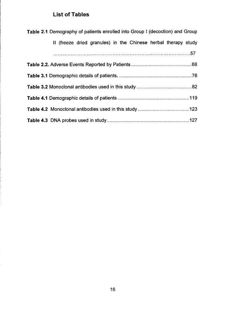
List of Tables
Table 2.1 Demography of patients enrolled into Group I (decoction) and Group
II (freeze dried granules) in the Chinese herbal therapy study
....................................................................................................................... 57
Table 2.2. Adverse Events Reported by Patients....................................................68
Table 3.1 Demographic details of patients................................................................76
Table 3.2 Monoclonal antibodies used in this study.............................................. 82
Table 4.1 Demographic details of patients.............................................................119
Table 4.2 Monoclonal antibodies used in this study............................................ 123
Table 4.3 DNA probes used in study....................................................................... 127
16

ABBREVIATIONS
AE atopic eczema
APAAP Alkaline phosphatase-anti-alkaline phosphatase
APC antigen presenting cells
BCIP 5-bromo-4-chloro-3 indolyl phosphate
BSA bovine serum albumin
CD cluster of differentiation
CHT Chinese herbal therapy
CLA cutaneous lymphocyte associated antigen
DAB 3,3 diaminobenzidine tetrahydrochloride
Der p Dermatophagoides pteronyssinus allergen
DNA deoxyribonucleic acid
ELAM-1 endothelial leucocyte adhesion molecule-1
FcsRI high affinity IgE receptor
FcsRII low affinity IgE receptor
FITC fluorescein isothiocyanate
HCL hydrochloric acid
HDM house dust mite
HLA human leucocyte antigen
ICAM-1 intercellular adhesion molecule-1
IFN Interferon
ig immunoglobulin
IL interleukin
LC Langerhans cells
17

LFA-1 lymphocyte function associated antigen-1
MgCb magnesium chloride
MoAb monoclonal antibody
mRNA messenger ribonucleic acid
Mw molecular weight
NaCI sodium chloride
NBT nitro blue tetrazolium
PB peripheral blood
PBM peripheral blood monocytes
PBS phosphate buffered saline
PBSM phosphate buffered saline containing magnesium chloride
RAST radioallergosorbent test
RNA ribonucleic acid
RNAse ribonuclease
ROD relative optical density
SSC saline sodium citrate
TBS Tris-buffered saline
Th1 T helper type 1 lymphocytes
Th2 T helper type 2 lymphocytes
TNF tumour necrosis factor
TNMT trinitro monotetrazolium
Tris Tris(hydroxymethyl) methylamine
TRITC tetramethylrhodamine isothiocyanate
18

CHAPTER 1
MAIN INTRODUCTION
19

1. MAIN INTRODUCTION
1.1 DEFINITION AND CLINICAL FEATURES OF ATOPIC
ECZEMA
Atopic eczema (AE) is an inflammatory skin disease presenting with severe
pruritis and classical features of dryness and erythema of the skin [Hanifin and
Rajka 1980]. Lesions commence as vesicles or papules and secondary to
scratching or itching, excoriations and lichenification develop. It has a chronic
and fluctuating course with an age of onset usually between 2 and 6 months
although the disease can start at any age [Hanifin and Rajka 1980]. During the
infantile phase, the face is often involved and as the child begins to crawl, the
extensor surfaces are commonly affected. The erythematous papules may
often become secondarily infected leading to exudation and crusting.
As the name implies, AE is a disease associated with dysfunction of
immunological mechanisms. The term atopy describes a state of hyperreactivity
to environmental allergens where the immunological mechanisms of affected
individuals "over respond", to a point where clinical signs or symptoms may be
manifest. Atopy was originally described by Coca and Cooke in 1923. The
immunological mechanisms involved are principally those associated with the
production of IgE class antibodies and the binding of these antibodies to mast
cells and basophils with the subsequent degranulation of these cells causing
the release of pharmacologically active factors that may promote the signs and
symptoms of disease. The commonest environmental agents that can promote
20
these reactions are house dust mite (the commonest allergens being Der p I
and Der p II, enzymes found in faecal pellets), pollens, animal dander and
some fungi [Thompson and Stewart 1993].
The clinical manifestations of atopic reactions range from hayfever (allergic
rhinitis) to life threatening systemic anaphylactic reactions that some people
experience after being sensitised to allergens such as antibiotics, most
commonly penicillin; or in food allergies such as in allergy to peanuts. As well
as these severe acute responses, atopic reactions are also associated with
chronic clinical conditions. The commonest of these are allergic asthma and
atopic eczema. Both these conditions may continue for years. In allergic
asthma, reversible episodes of bronchospasm are induced in the lungs by
exposure to environmental allergens. Interestingly, asthma may develop in
subjects who show no evidence of atopy [Poulter and Burke 1996], and such
observations led to research that has identified further underlying
immunoregulatory dysfunction promoting a chronic inflammatory reaction in the
bronchioles of asthmatics [Poulter et al. 1994]. It is now thought that a similar
underlying problem may be associated with AE (see below).
Although the association with allergen driven IgE responses and granulocyte
release of mediators still holds, it is clear that atopic reactivity to allergens is not
the sole cause of eczema. Atopic status (allergic type reactivity to
environmental allergen) can be demonstrated in vivo by "prick testing". Here a
solution of allergen is placed on the skin and a prick is made through this
solution into the skin thus introducing the allergen intradermally. Atopic subjects
21

will exhibit a classic acute wheal and flare reaction that is read 15 minutes after
testing. Using a panel of common environmental allergens, it is known that up
to 30% of the general population exhibit positive allergic responses. However
the vast majority of these people do not have eczema. Furthermore, these
artificially induced immediate type hypersensitivity reactions develop and
resolve within minutes whereas AE is recognised as a chronic inflammatory
condition. Such anomalies make it important to look more closely at the
pathogenic mechanisms at play in this disease.
1.2 AETIOLOGY AND PATHOGENESIS OF ATOPIC ECZEMA
There are many reports of immunological abnormalities associated with AE.
80% of patients with AE have raised IgE levels in the peripheral blood and this
is considered to be related to immediate allergic, or type I hypersensitivity
[Johansson 1969]. Indeed an immediate type hypersensitivity response can be
demonstrated in most patients with AE and they develop a wheal and flare
response 15-20 minutes after pricking the skin with allergens (see above).
"Prick" test positivity is only manifest to allergens that the subject has
encountered and become sensitised to. The immune reactivity is caused by a
specific IgE response to the allergen. The allergen specific IgE binds via Fc
receptors to mast cells and other basophils and following further exposure to
the allergen, (as occurs when prick testing is performed), there is cross-linking
of the bound IgE, leading to degranulation of the mast cells and the release of
vasoactive mediators. It is recognised however, that the clinical manifestations
of AE can occur in subjects with serum IgE levels within the normal range.
22

Furthermore levels of IgE may fluctuate with time and may not necessarily
correlate with disease severity [Jones et al. 1975].
The clinical severity of the disease including the area of skin affected also
varies with time although there appear to be no consistent precipitating factors
other than infection. Patients with AE are recognised as being more susceptible
to superficial bacterial skin infections [Brook et al. 1996, White and Noble
1986]. This may simply result from bacterial colonisation within damaged skin,
or may be a manifestation of an impaired immune defence system [Rogge and
Hanifin 1976].
There are further features of AE that suggest that the immune system in these
patients is abnormal. Attacks of herpes simplex may lead to a more widespread
eruption, eczema herpeticum (Kaposi's varicelliform eruption), suggesting an
increased susceptibility to the spreading of cutaneous Herpes in AE [Goodyear
et al. 1996, Rasanen et al. 1987]. This indicates a failure of defence
mechanisms to control the virus. There is undoubtedly depressed cell-mediated
immunity in AE [McGeady et al. 1975] with diminished tuberculin reactivity
[Uehara 1977]. This can be seen by the poor handling of viral, fungal and
bacterial infections of the skin in AE patients. However the incidence of viral
warts does not appear to be increased compared with non atopic individuals
[Williams et al. 1993]. Sensitivity to contact allergens such as
Dinitrochlorobenzene (DNCB) is also diminished in patients with extensive AE
but this reaction returns to normal with remission of the disease [Uehara and
Sawai 1989].
23

Regarding specific components of the immune defence system, cytotoxic
responses of natural killer cells and cytotoxic T cells have been reported to be
lowered in AE [Leung et al. 1983]. This may be the cause of the increased
incidence of disseminated Herpes simplex infections mentioned above.
Chemotaxis is impaired with slower migration of monocytes and neutrophils
[Rogge and Hanifin 1976] perhaps contributing to the frequent superficial
Staphylococcal infections [White and Noble 1986]. Indeed, Staphlococcus
aureus can be isolated from the involved skin in more than 90% of patients with
AE [Leyden et al. 1974].
Evidence of immunosuppression may appear paradoxical when chronic
inflammatory lesions persist. However many of the bacteria known to colonise
the AE lesions secrete exotoxins which act as superantigens and stimulate
macrophages to release pro-inflammatory cytokines, as well as activating large
numbers of T cells [Leung et al. 1995]. The T cells will secrete cytokines that
may induce tissue inflammation [Campbell and Kemp 1997]. Thus immune
dysfunction may work in a variety of ways and the development of the disease
may depend on a multitude of factors that can build one on the other over time.
It has been suggested that dairy products aggravate the disease [Agata et al.
1993, Chandra et al. 1989] but studies in children have produced conflicting
evidence as to whether egg and milk exclusion, for example, leads to clinical
improvement of the disease [Neild et al. 1986, Atherton et al. 1978, Webber et
al. 1989]. However one double-blind controlled crossover trial suggested that
24

dietary manipulation involving egg and milk avoidance is of benefit in reducing
disease activity in some children with AE [Atherton et al. 1978]. Another study
has shown that in families with a history of atopic disease, milk and egg
avoidance in mothers breastfeeding infants reduced the subsequent incidence
of AE in those children [Chandra et al. 1989]. Much work has investigated the
role of house dust mites (HDM) in the development of AE associated with
allergic reactivity to HDM related allergens Studies have shown that HDM are
present in higher quantities in the mattresses of patients with moderate to
severe AE [Beck and Korsgaard 1989, Colloff 1992], thus indicating that levels
of exposure to HDM may be related to disease severity. However, there is
conflicting evidence as to the benefit of eradication of the mites [Colloff et al.
1989, Tan et al. 1996]. A recent double blind placebo controlled study showed
that the use of vacuuming and gortex covered mattresses led to a marked
reduction in dust with an associated clinical improvement of the eczema [Tan et
al. 1996].
In addition to these clinical findings, prick test reactivity to house dust mite
antigen is usually positive in AE patients; there are often raised titres of IgE
complexes in the serum [Stone et al. 1973, Jones et al. 1975] and HDM antigen
specific T lymphocytes are present in lesional skin [Van der Heijden et al.
1991]. Despite the overwhelming evidence of the involvement of HDM in the
immunopathogenesis of AE, the relative importance of this as a precipitating
factor remains unclear.
25

There is increasing evidence that AE has a genetic component. Twin studies
have shown for example that the incidence of AE in both individuals is
significantly greater in monozygotic pairs, than in dizygotic twins where one
subject may develop AE while the other twin does not [Shultz Larsen et al.
1986]. The allergen-specific IgE-mediated hypersensitivity that is associated
with atopy has been shown to be transferred from an atopic bone marrow donor
to a non-atopic recipient [Agosti et al. 1988]. This was shown by the occurrence
of positive prick tests to aerollergens in previously negative subjects who were
recipients of allogenic bone marrow transplantation from atopic donors. One
study of seven atopic families revealed an autosomal dominant inheritance
linked to a DNA marker on chromosome 11 q [Cookson et al. 1989]. However a
further study of 95 families with AE refuted such an association [Coleman et al.
1993]. Linkage studies have shown an association between atopy and the beta
unit of the Fc epsilon Rl which has been localised to human chromosome 11 q
12-13 [Cox et al. 1998]. There have been further genetic linkages of AE with a
mast cell chymase variant [Mao et al. 1998]. Finally atopy has been associated
with a mutation in the alpha subunit of the interleukin 4 receptor [Hershey et al.
1997].
Another contributing factor to the development of atopic diseases including
eczema may be exposure to aeroallergens and dietary allergens in the neonatal
period [Holt et al. 1997]. In those genetically predisposed to AE, the immature
immune system of the young child appears to preferentially produce T
lymphocytes that secrete a cytokine profile that is associated with T helper type
2 (Th2) lymphocytes. Should this occur, the immune system appears to
26

maintain this tendency permanently thus possibly promoting atopic reactivity as
this T cell subset secretes IL-4 and IL-5 [Yabuhara et al. 1997 ].
The Th2 cytokines interleukin 4 and 5 promote IgE production and eosinophilia.
These features are both characteristic of atopy. During normal infancy,
exposure to environmental allergens is thought to result in those T cells that
respond to these allergens undergoing deletion or anergy and the subsequent
development of immunological tolerance [Prescott et al. 1997]. This process
may not occur in the atopic subject.
Thus the aetiology of AE is almost certainly multifactorial with genetic,
immunologic and environmental factors interacting to produce the disease
phenotype. With the emergence of new technology and epidemiological
studies, elucidation of the cellular and molecular interactions in AE and
therefore the exact pathogenesis may become an achievable goal.
1.3 HISTOLOGY OF LESIONAL SKIN
The histological features of lesional skin in AE change with time [McKee 1996].
In the acute lesion, there is marked intercellular oedema (spongiosis) which
may result in accumulation of enough fluid to result in intraepidermal vesicle
formation. There is also some epithelial thickening (acanthosis), vascular
dilatation in the dermis, and parakeratosis. In chronic lesions there is less
spongiosis with more marked epidermal proliferation with acanthosis and
elongation of the rete ridges leading to psoriasiform hyperplasia . There is also
27

prominent hyperkeratosis as well as some parakeratosis. Although these
features may be used to distinguish acute from chronic lesions, most
characteristics are common to both. However it is recognised that the
appearance of the lesions is not uniform and may vary, not only between
individuals, but also within individual patients when separate sites are
investigated.
In both acute and chronic lesions there is a dermal perivascular cellular infiltrate
consisting mainly of mononuclear cells [Zachary et al. 1985a]. The majority of
these are T lymphocytes [Braathen 1979]. There are also macrophages,
Langerhans cells and mast cells present; with only occasional neutrophils,
eosinophils, and basophils [Mihm 1976]. The preponderence of T lymphocytes
and antigen presenting cells in the dermis is suggestive of a cell-mediated
immune response within the lesion. Strikingly, the dermal infiltrate contains very
few plasma cells which suggests that raised IgE titres commonly seen in the
circulation are not a consequence of B cell activity in the skin.
1.4 IMMUNOPATHOLOGY
The immunopathology of AE presents us with an apparent paradox. On the one
hand raised IgE levels, circulating IgE-allergen complexes and positive prick
tests are all indicative of an antibody mediated response. On the other hand,
the inflammatory infiltrate within lesional skin exhibits a T cell dominated cellular
infiltrate suggestive of a chronic cell mediated response [Zachary et al. 1985a].
28

One is drawn to the conclusion that both types of immune response occur in AE
and both may therefore have an impact on the pathogenesis.
Such a situation is not unique to AE. There are other conditions where both
humoral and cell mediated responses are involved. In rheumatoid arthritis there
are circulating levels of autoantibodies while the synovial tissue within the joints
is predominantly infiltrated with macrophages and T lymphocytes, a T cell
mediated response.
In asthma, there is evidence of type IV mediated hypersensitivity with a T cell
infiltrate in the the bronchial wall [Poulter et al. 1994], yet allergic IgE responses
may promote acute bronchoconstriction following exposure to aeroallergens.
Indeed it has been suggested that it is a failure to downregulate the cell
mediated response within the bronchial walls rather than the antibody response
to aeroallergens that promotes the chronicity of the disease [Poulter et al.
1994].
There is little doubt that raised titres of circulating IgE occur in the majority of
cases of AE and are responsible for the atopic status of the patient. Of the five
classes of immunoglobulin found in the circulation, the IgE isotype is normally
present at the lowest titre contributing less than 0.1% of total immunoglobulin
[Ward et al. 1996] Normally expressed as International Units (IU), the normal
range is quoted as 0-120 lU/ml [Ward et al. 1996]. In patients with AE, this
figure may rise to >2000 lU/ml. Such changes are not unique to AE but reflect
atopic status in general and are seen in cases of asthma and rhinitis [Jones et
29

al. 1975]. As IgE is a cytophilic antibody, such high titres inevitably cause
attachment of the immunoglobulin to granulocytes via Fc receptors.
1.4.1 T Cells
Although the increase in serum IgE represents an abnormal antibody response,
the dermal perivascular infiltrate in the lesions of AE is predominantly
mononuclear with T lymphocytes, Langerhans cells and macrophages. The
major cell type in lesional skin is the T lymphocyte, the majority of which are T
helper lymphocytes expressing CD4, with a helpensuppressor ratio of 7:1
[Lever et al. 1987]. Many of these lymphocytes have undergone activation and
express CD25 (lnterleukin-2 receptor) and HLA DR (a class II major
histocompatability complex molecule). It is now recognised that T lymphocytes
may be functionally divided into Th1 and Th2 subsets [Del Prete et al. 1994],
the balance of which may be functionally important in AE, (see below).
1.4.2 Langerhans Cells
There are increased numbers of Langerhans cells (LC) in the dermis of AE
lesional skin [Zachary et al. 1985a]. These cells are identified under electron
microscopy by the presence of Birbeck granules, or at the light microscopic
level by expression of the antigen detected by MoAb CD1 [Ray and Schmitt
1988].
30

Langerhans cells are large dendritic cells normally found in the epidermis [Mihm
1976]. These cells differentiate from monocytes [Hanau et al. 1987] and are
recognised as part of the "dendritic cell" family. As LC constitutively express
surface class II MHC antigens (HLA-DR,DP,DQ) [Ruco et al. 1989], they are
considered to be the antigen presenting cells of the skin. During inflammatory
reactions, numbers of these cells migrate to the dermis where they are seen
interdigitating with the accumulations of T cells [Bos et al. 1986, Alegre et al.
1986].
Although dendritic cells of the dermis have been called "indeterminate cells",
these are now thought to be Langerhans cells involved in local immune
reactivity [Zachary et al. 1985b, Alegre et al. 1986]. In lesions of AE, a
proportion of these LC have been shown to have IgE bound to their surface
[Bieber et al. 1989a]; a situation not seen in clinically normal skin, even in
patients who exhibit raised serum IgE [Barker et al. 1988]. It would seem likely
therefore that LC activity contributes to the pathogenesis of AE.
1.4.3 Eosinophils
Eosinophils are recognised as being associated with allergy and Type I
hypersensitivity. Although there are normally only small numbers of eosinophils
present in AE lesions, deposits of eosinophil derived major basic protein have
been identified, probably as a result of eosinophil degranulation [Leiferman et
al. 1985]. In other allergic diseases such as asthma, eosinophils are thought to
play a major role [Busse and Sedgwick 1992]. Indeed circulating levels of
31
eosinophilic cationic protein have been found to correlate with disease severity
[Czech etal. 1992, Halmerbauer et al. 1997].
Several studies have demonstrated that raised levels of serum eosinophilic
cationic protein are present in patients with eczema [Miyasato et a l 1996,
Halmerbauer et a l 1997]. This must be released from eosinophil granules and
suggests that there is widespread activation of the eosinophil pool occurring in
AE [Czech et a l 1992, Uehara et a l 1990]. It is not uncommon for a systemic
eosinophilia to be present in patients with AE. Again however this phenomenon
appears to be an atopy related phenomenon as it may also be seen in subjects
with rhinitis and asthma [Griffin et a l 1991, Borres et a l 1995]. This
eosinophilia is also highly variable as many patients with AE have normal levels
of these cells in the circulation [Uehara et a l 1990]. It remains unclear what
may regulate or influence the eosinophil count but the cytokine lnterleukin-5
(released by a Th2 type T cell) may be a contributing factor, as this is
recognised as chemotactic for eosinophils and can also promote eosinophil
activation [Adachi and Alam 1998] (see below).
1.4.4 Cytokines
As stated above, T lymphocytes dominate in the lesions of AE. Within this
population are high numbers of allergen specific T lymphocytes [Van der
Heijden et a l 1991], clearly suggesting that the dermal T cell infiltrate
represents a recall reaction induced locally by immune reactivity to
aeroallergens. Activation of T cells within a cell-mediated response is in part
32

under the regulation of cytokines [Xu et al. 1996] and T cells contribute by being
cytokine producers [Romagnani 1992a]. Thus knowledge of changes in levels
of cytokines and information about their synthesis associated with the
inflammatory reactions in AE may offer clues as to the mechanisms underlying
the immunopathology.
Analysis of the cytokines produced by the housedust mite, Dermatophagoides
pteronyssinus (Der p.), specific T cells in the cellular infiltrate has revealed that
there are increased proportions of lnterleukin-4 (IL-4) producing cells in the
lesions, with relatively lower levels of Interferon-gamma (IFN-gamma)
production [Van der Heijden et al. 1991]. Similarly, T cells in the peripheral
blood of patients with AE have been shown to secrete higher amounts of IL-4
than IFN-gamma [Wierenga et al. 1990]. One of the important effects of IL-4 is
the induction of expression of the low affinity IgE receptor (CD23) on
Langerhans cells in the skin [Bieber et al. 1989b] and induction of similar
expression on T cells isolated from peripheral blood [Sakamoto et al. 1992].
It is these cytokines and other soluble mediators that regulate the recruitment of
the lymphocytes and monocytes from the peripheral blood thus orchestrating
the inflammatory response. As well as these interactions by cell surface
molecules and cytokines, adhesion molecules on endothelial cells are intimately
involved in lymphocyte trafficking.
33

1.4.5 Adhesion Molecules
The vascular endothelial cells and keratinocytes in AE express adhesion
molecules on the cell surface which are ligands for activated T lymphocytes
[Cooper 1994]. This ligand to ligand interaction enables the T lymphocytes to
migrate to the epidermis from the blood. Intercellular adhesion molecule-1
(ICAM-1) is expressed on keratinocytes and endothelial cells in the dermis of
inflammatory conditions such as psoriasis, AE, lichen planus and mycosis
fungoides and may regulate lymphocyte trafficking in the skin [Griffiths et al.
1995]. T lymphocytes also express the ligand lymphocyte function associated
antigen-1 (LFA-1) [Griffiths et al. 1989]. These ligands can attach to adhesion
molecules on endothelial cells lining blood vessels thus enabling T lymphocytes
to leave the vessels and infiltrate the skin [Lub et al. 1995].
Endothelial cells express endothelial leukocyte adhesion molecule-1 (EI_AM-1
or E selectin) which is a ligand for the cutaneous lymphocyte-associated
antigen (CLA) expressed by the effector T lymphocytes that infiltrate inflamed
skin [Santamaria et al. 1995]. The T lymphocytes that express these ligands
are those that have recently undergone activation, perhaps in the lymph nodes,
before becoming localised in the skin [Santamaria et al. 1995].
There are several cytokines that may induce the expression of ICAM-1 and
ELAM-1 on the endothelial cells [Thornhill and Haskard 1990] leading to
enhanced recruitment of leukocytes from the vasculature and extravasation and
infiltration into the inflammatory milieu in lesional skin. Tumour necrosis factor
34

(TNF), released by mast cells and macrophages can induce ELAM [Lidington et
al. 1998]. Interleukin-1 (IL-1), released by monocytes and keratinocytes can
induce ELAM-1 and ICAM-1 [Lidington et al. 1998]; and the T lymphocyte
derived cytokine, IL-4 potentiates ELAM-1 expression [Yao et al. 1996], As
described above, the cellular infiltrate in lesional skin in AE consists
predominantly of T lymphocytes and also macrophages. The interaction
between these cells and the release of cytokines by lymphocytes are all
important in the regulation of the immunopathogenesis of AE.
1.4.6 Th1 And Th2 Cells
There are increased numbers of allergen-specific IL-4 producing CD4+ T
lymphocytes in lesional skin of AE [Van der Heijden et al. 1991]. Furthermore
increased IL-4 production by T cells is associated with reduced IFN-gamma
production [Reinhold et al. 1991, Nakazawa et al. 1997]. Studies of the
cytokine profile of murine T cells have revealed that T helper lymphocytes can
be subdivided into type I (Th1) and type II (Th2), based on the repertoire of
cytokines they produce [Romagnani 1992a]. Th2 predominantly produce
cytokines IL-4, IL-5, IL-6 and IL-10 while Th1 predominantly produce IL-2 and
IFN-gamma [Romagnani 1991]. Th1 cells are recognised as the mediators of
delayed type hypersensitivity responses due to their ability to recruit monocytes
and macrophages at the site of intracellular infections [Romagnani 1992a].
They produce IFN-gamma which can modify macrophage function by inducing
the expression of Class II major histocompatibility complex cell surface antigen
and stimulate the synthesis of tumour necrosis factor (TNF-alpha) [Virelizier
35

and Arenzana-Seisdedos 1985]. Th2 cells on the other hand are the
synthesizers of IL-4 which has multiple effects on mononuclear cells, including
induction of B cell IgE production [Parronchi et al. 1990], induction of CD23
expression on monocytes, Langerhans cells and B lymphocytes [Bieber et al.
1989b]. It also inhibits T lymphocyte IFN-gamma production [Pene 1989]. Thus
the overall functional capacity of the T cell pool, particularly in inflammatory
infiltrates will reflect the relative balance of these two subsets.
In AE, it has been suggested that there is an imbalance of this Th1/Th2 ratio in
both the peripheral blood and in the lymphocytes infiltrating the skin [Bos et al.
1992], with a bias towards Th2 type IL-4 secreting T lymphocytes. This
observation provided the rationale for the clinical trials of recombinant IFN-
gamma treatment of AE [Renz et al. 1992] (see below).
The underlying cause of this imbalance remains unclear. However, it may be
that defective monocyte control contributes to this imbalance in the cytokines
secretion by T lymphocytes as there is abnormally increased cyclic adenosine
monophosphate(cAMP)-phosphodiesterase activity in monocytes in AE [Holden
1990]. This defect is associated with an increased monocyte prostaglandin E2
secretion which is inhibitory for T cell derived IFN-gamma. As aberrations in
LC and macrophage function have also been reported (see above), this
dysfunction of monocytes implicates this family of non lymphoid cells in the
overall pathogenesis of AE.
36

1.4.7 Macrophages
The monocyte/macrophage series is part of the bone marrow derived
mononuclear phagocyte system [Johnston 1988]. The functions of these cells
include: antigen processing and presentation [Unanue and Allen 1987];
secretion of bioactive products including IL-1 [Dinarello1985], tumour necrosis
factor [Beutler and Cerami 1987] complement components and bioactive lipids
such as leukotrienes [Nathan 1987]; and of course phagocytosis [Johnston
1988]. They also have pro-inflammatory activity in that macrophages are
recruited to sites of inflammation and release further chemotactic factors to
attract further cells.
Monocytes differentiate into macrophages as they enter the tissues.
Macrophages are a heterogeneous group of cells whose subsets can be
defined both functionally and phenotypically [Spiteri and Poulter 1991].
Previous extensive studies have developed the use of two antibodies to
subdivide macrophages further according to their phenotype. These are RFD1
and RFD7 monoclonal antibodies.
Originally described in 1986 [Poulter et al. 1986, Janossy et al. 1986], these
mouse anti human monoclonals have been used by many laboratories to
dissect the heterogeneity of macrophage function [Lenz et al. | 1993, Seldenrijk
et al. 1989, Teunissen et al. 1990, Zheng et al. 1995]. RFD1 is a mouse IgM
MoAb that recognises a membrane epitope associated with antigen
presentation. It is constitutively expressed on dendritic cells and on some B
37

lymphocytes [Poulter et al. 1986]. Macrophages and Langerhans cells (in the
epidermis) are RFD1-ve but can be induced to express this molecule on
activation [Teunissen et al. 1990]. RFD7 is a cytoplasmic 77kd glycoprotein
associated with mature phagocytes [Poulter et al. 1986]. It is not expressed by
dendritic cells or monocytes. Originally it was thought that the expression of
these molecules was mutually exclusive, however, first in the lung, and then in
other tissues a third subset of macrophages that expressed both RFD1 and
RFD7 positivity was identified [Spiteri etal. 1992b].
Of some significance, is the fact that these phenotypically distinct subsets have
been shown to exhibit functional heterogeneity. Cells with the phenotype
RFD1+RFD7- act predominantly as inducers of T cell activation, while those
with the phenotype RFD1-RFD7+ act as phagocytes [Poulter et al. 1996]. Cells
expressing positivity with both MoAbs (RFD1+RFD7+) exhibit T cell suppressive
activity [Spiteri et al. 1992b, Spiteri and Poulter 1991]. This is a dynamic group
of cells where one phenotype can change into another. Such plasticity can be
promoted by cytokines in the surrounding environment [Tormey et al. 1997].
Thus as with Th1 and Th2 T cell subsets, the overall functional capacity of the
macrophage pool is dependent on the relative balance, in this case, of at least
three subsets [Poulter et al. 1996 and Poulter et al. 1994].
Interestingly, LC within the dermis of AE lesions bear not only CD1 but also
express RFD-1 which is expressed only by antigen presenting cells [Zachary et
al. 1985b, Alegre et al. 1986]. Furthermore, RFD1+ antigen presenting cells not
expressing the CD1 LC marker are also present in relatively large numbers in
38

AE lesions [Bos et al. 1986]. All of these cells strongly express HLA DR. The
evidence thus clearly shows that within the cellular infiltrate of lesions of AE, not
only are there are activated T lymphocytes, but also activated antigen
presenting cells. Furthermore in AE, these cells may carry bound IgE
[Bruynzeel-Koomen et al. 1986, Leung et al. 1987], a situation not seen in
other dermatoses [Barker et al. 1988]. The reports of IgE present on the
surface of the antigen presenting cells implicate IgE in the cell-mediated
response exhibited within the skin in AE, and possibly suggest a link between
the T cell mediated reactions of the lesions and the manifestations of atopy in
these patients (see below).
Cells of the mononuclear phagocyte system exhibit a wide range of receptors
including receptors whose exact function is unclear. These include receptors for
insulin [Bar et al. 1977], transferrin [Spik and Montreuil 1983], thrombin
[Silverstein and Nachman 1987], and fibrinogen [Mosesson 1984]. Other
receptors which may be expressed on the surface include those for
complement components such as C3b [Law 1988], receptors for lipoproteins
[Fogelman et al. 1988], receptors for IgG and IgE [Ezekowitz and Gordon
1986, Gordon et al. 1989].
Receptors for IgE can be divided into two types, the high affinity (FcsRI) and
low affinity receptor (FcsRII or CD23) [Bieber 1992, Wang et al. 1992,
Sakamoto et al. 1990, Takigawa et al. 1991]. CD23 is expressed on peripheral
blood monocytes, eosinophils, platelets and lymphocytes (predominantly B
cells) [Kehry and Hudak 1989, Delespesse et al. 1992]. It has been shown that
39

CD23 in normal skin is expressed by RFD7+ macrophages and LC, but in AE
there is a switch to expression by the RFD1+ inducer cells [Buckley et al. 1992].
In both aeroallergen patch tests and lesional skin in AE, the proportion of LC
and other RFD1+ cells expressing CD23 is increased while the proportion of
RFD7+ macrophages expressing CD23 is reduced [Buckley et al. 1992]. The
emergence of CD23 on antigen presenting cells suggests that the redistribution
of CD23 within macrophage subsets may be important in the pathogenesis of
AE and that CD23 may be involved in antigen presentation.
1.5 LINK BETWEEN ALLERGY AND TYPE IV
HYPERSENSITIVITY
The firm conclusion to be drawn from studies of the immunopathology of AE to
date, is that the skin inflammation associated with this condition bears all the
hallmarks of a T cell driven Type IV hypersensitivity reaction. What then are the
relationships between this and the atopic status carried by patients suffering
from this disease? There is no doubt that AE patients are atopic as virtually all
exhibit prick test positivity to aeroallergens and have raised titres of IgE in the
circulation [Stone et al. 1973]. The reports of IgE bound to macrophages and
dendritic cells in the lesional infiltrates offer a possible clue to the
interrelationship between atopic reactivity and T cell mediated immunity.
IgE is a cytophilic antibody readily binding to mast cells, basophils, some
monocytes and macrophages [Sutton and Gould 1993]. This binding occurs via
receptors for the Fc region of the immunoglobulin. It is the binding of IgE to
40

these receptors that creates the state of "sensitivity" to allergens as cross
linking of the antigen binding sites of the IgE molecules by allergens,
precipitates the degranulation of mast cells producing the immediate type
hypersensitivity reactions characteristic of allergy. The low affinity receptors,
recognised by MoAb CD23, are thought to be less involved in these allergic
reactions [Sutton and Gould 1993]. However, initial studies of the distribution of
IgE receptors in AE lesional skin demonstrate that iM s expression of the low
affinity CD23 molecules thaf^is downregulated by treatmenlfand not FcsFtf/[Xu
etal. 1997].
Much work has been done investigating the possible role of IgE receptors in
presenting antigen to T cells. It is recognised that if the IgE bound to these
receptors on appropriate cells is complexed to antigen, this may promote T cell
stimulation [Van Der Heijden et al. 1995, Mudde et al. 1990b] (facilitated
antigen presentation). Although most studies of this mechanism have
concentrated on the role of FcsRI [Bieber 1997, Maurer and Stingl 1995], this
form of IgE complexed antigen presentation may occur if the IgE is bound to
CD23. Indeed in AE, CD23 has been shown to be expressed by antigen
presenting cells rather than macrophages [Buckley etal. 1992 ].
Evidence linking a delayed type hypersensitivity response to atopy in AE comes
from the study of patch test reactions in such patients. As well as exhibiting
prick test reactivity to aeroallergens, up to 40% of AE patients may exhibit a
delayed type reactivity if the specific allergen is applied as a "patch"
epicutaneously [Buckley et al. 1992, Clark and Adinoff 1989], normally
41

positioned on the upper back. These reactions peak at 48 hours and exhibit not
only signs of erythema and induration but also mononuclear cell infiltration in
the dermis [Buckley et al. 1992]. Immunohistological analysis of these reactions
revealed them to exhibit all the features of AE lesional skin, including the
aberrant expression of CD23 on antigen presenting cells [Buckley et al. 1993],
Importantly, when such reactions were directly compared to nickel induced
contact sensitivity reactions, it was the redistribution of low affinity IgE receptors
that distinguished the allergen patch test from the contact reaction [Buckley et
al. 1993].
The possible progression from allergic reactivity to T cell mediated
hypersensitivity via facilitated antigen presentation suggests therefore a link
between atopy and cell mediated immunity and an underlying complexity to the
pathogenesis of AE.
It goes without saying therefore that these complexities in the pathogenesis of
AE, introduced above, should be taken into account when considering the
treatment options. It is important in the development of effective and safe
therapy to target relevant abnormalities within the immunopathogenic
mechanisms observed. To decipher the parameters of immune dysregulation
relevant to the clinical situation, a model can be developed whereby peripheral
blood and lesional skin from patients with AE before and after effective
treatment is investigated. This approach provides the opportunity, using
immunocytochemical techniques, to understand the underlying pathogenic
mechanisms and their relevance to severity of clinical disease.
42

1.6 MANAGEMENT
There are several approaches to the management of AE that vary considerably,
ranging from manipulation of patient behavioural characteristics, to
immunomodulatory treatment. The immunomodulatory treatment can be topical
or systemic and the choice of treatment depends on severity of disease and
degree of disability that the patient experiences as a result of the disease.
1.6.1 Behavioural
An important part of the management of patients with AE, as in all chronic
diseases, is explanation, reassurance and discussion of the various treatment
options available. Patients are advised to avoid the application of substances
that dry or irritate the skin such as soaps and detergents. Advice is given to
avoid other aggravating factors such as food that is known to lead to worsening
of the disease in an individual. It is recommended that any attempts to follow
any dietary regime should be under the supervision of a dietician [Webber et al.
1989].
1.6.2 Symptomatic
Frequent and liberal application of emollients to the skin is recognised as
important in combatting dryness of the skin in AE. This is especially important
after bathing to retard evaporative water loss from the epidermis.
Antihistamines are often used as an adjunct to therapy. It is most common to
43

recommend those with a sedating effect as it is this central, sedative action that
appears to be of benefit, rather than peripheral Hi antagonism [Wahlgren et al.
1990]. Oral antibiotics are often required to treat exacerbations of eczema
caused by secondary bacterial infection. For eczema herpeticum, which can be
a complication of AE, either oral or intravenous acyclovir is used. Groups have
tested the value of alternative approaches with varying success. For example a
recent double blind placebo-controlled trial showed that the treatment of AE
with essential fatty acid supplements, evening primrose oil and fish oil did not
lead to clinical improvement [Berth-Jones and Graham-Brown 1993], although
some studies had previously reported some benefit from such treatment [Wright
and Burton 1982, Schalin-Karrila etal. 1987].
1.6.3 Immunomodulation
1.6.3.1 Topical Therapy
Topical corticosteroids are the mainstay of treatment of AE. Corticosteroids
inhibit the expression of many pro-inflammatory cytokines by binding to gene
transcription factors, upregulating the expression of cytokine inhibitory proteins,
and reducing the half-life and utility of cytokine mRNAs [Brattsand and Linden
1996]. The number of Langerhans cells with IgE bound to the surface is also
downregulated by treatment with topical corticosteroids [Bieber et al. 1989a].
This observation supports the role of IgE and the expression of its receptor on
antigen presenting cells as being of relevance to the immunopathogenesis .
The therapeutic range of corticosteroids used varies from mild to very potent.
Side effects include thinning of the skin leading to striae atrophicae and
44

telangiectasia. Long-term use of topical steroids on the eyelids has been
associated with the occurrence of glaucoma [Cubby 1976].
1.6.3.2 Systemic Therapy
1) Oral Corticosteroids:
Because of the failure of topical corticosteroids to control the disease in some
cases, short courses of oral corticosteroids may be prescribed. Long term use
is not considered advisable due to the side effects which include osteoporosis,
peptic ulceration, Cushing's syndrome, glaucoma, neuropsychiatric effects,
weight gain, impaired healing and easy bruising of the skin [Schimmer and
Parker 1996]. Corticosteroids modulate cytokine expression by a combination
of genomic mechanisms as described above. Administration of corticosteroids
has been shown to decrease high peripheral blood eosinophil counts and
decrease the survival rate of eosinophils by increasing apoptosis in patients
with AE [Matsukura et al. 1997]. Methylprednisolone has also been shown to
modulate circulating soluble interleukin 2 receptor levels [Sauer et al. 1993].
2) Ultraviolet Phototherapy:
It has been shown that treatment with psoralens and ultraviolet A (PUVA) is
often effective [Atherton et al. 1988, Larko 1996, Sheehan et al. 1993, Yoshiike
et al. 1993] in the treatment of AE. Although Broad band ultraviolet B was
initially thought to be less effective in the treatment of AE [Jekler and Larko
1991], a subsequent study has shown that narrow band ultraviolet B (TL-01)
therapy is as effective as PUVA in the treatment of chronic severe AE [George
et al. 1993]. A recent open multicentre trial showed that high dose UVA1
45

radiation is effective for severe exacerbations of AE [Krutmann et al. 1998].
However there can be long term problems involved in such therapies. For
example there is a dose-dependent increase in the risk of developing
squamous cell carcinoma of the skin following treatment with PUVA [Lindelof et
al. 1991, Bruynzeel et al. 1991]. There also appears to be an increased risk of
developing basal cell carcinomas [Bruynzeel et al. 1991]; and one study has
shown an increase in the incidence of respiratory cancer in males and females,
pancreatic cancer in males, and kidney and colonic cancer in females [Lindelof
etal. 1991].
3) Azathioprine:
Azathioprine is a purine antimetabolite. There have been no controlled studies
of azathioprine in the treatment of AE although it has been used as long-term
therapy for chronic AE [Younger et al. 1991]. This drug inhibits cell division, and
bone marrow suppression is a potential serious side effect [Min and Monaco
1991]. Other side effects include nausea, vomiting and diarrhoea [Diaso and
LoBuglio 1996]. Long term immunosuppression may be the reason why certain
tumours develop more commonly in patients treated with azathioprine such as
non-Hodgkin's lymphoma and squamous cell carcinoma [Taylor and Shuster
1992]. However, it is highly likely that this suppression of the immune system is
a necessary requirement for clinical improvement in AE.
4) Cyclosporin:
Cyclosporin belongs to the family of cyclic polypeptides derived from the
fungus Tolypocladum inflatum Gavis and is a potent immunosuppressant
46
[Diaso and LoBuglio 1996]. In the last few years cyclosporin has been used
increasingly to treat severe refractory AE [Sowden et al. 1991]. The
immunosuppressant effects of cyclosporin have already been established with
its use in transplantation to prevent rejection. Within lesional AE skin, treatment
with oral cyclosporin, leads to a decrease in the number of activated T cells
expressing the IL-2 receptor [Van Joost et al. 1992]. A double-blind crossover
study in adults showed that treatment with cyclosporin leads to an improvement
in clinical disease [Sowden et al. 1991] and also quality of life [Salek et al.
1993]. However the 8 weeks of treatment in this latter study, caused a rise in
the mean serum urea and creatinine, and a significant rise in bilirubin in some
patients. Thus the monitoring of blood pressure and renal and liver function
tests during treatment is essential. Other side-effects reported include
parasthesia, gastro-intestinal upset, gingival hyperplasia and hypertrichosis
[Diaso and LoBuglio 1996, Min and Monaco 1991]. There is also a reported
increase in the incidence of malignancies and lymphoproliferative disorders
following cyclosporin [Ryffel 1992, Gruber et al. 1994, Tanner and Alfieri
1996].
5) Interferon Gamma:
Subcutaneous injections of a recombinant form of this immunomodulatory drug
has been shown to be effective in the treatment of AE. A double-blind, placebo-
controlled trial in adults of daily subcutaneous recombinant interferon gamma
for 12 weeks showed significant clinical improvements [Hanifin etal. 1993]. This
study showed that side-effects of headaches, myalgias and chills were common
47
however transient granulocytopaenia occurred in some patients. Some patients
treated with interferon gamma also develop mild elevation of liver enzymes.
6) Tacrolimus:
Tacrolimus or FK506 is a macrolide antibiotic and inhibits T cell activation by
binding to cytosolic protein FKBP (FK506 binding protein) [Wiederrecht et al.
1993]. Oral tacrolimus has been used as an immunosuppressant for the
prevention of rejection in organ transplant patients and has also been
associated with an increased incidence of lymphoma in transplant recipients
[Ciancio et al. 1997]. More recently it has been found to be effective when used
topically for the treatment of AE [Ruzicka et al. 1997, Alaiti et al. 1998].
7) High Dose Intravenous Immune Globulin:
Administration of intravenous immune globulin on a regular basis has been
shown to reduce the need for systemic steroids in both asthma and AE
[Gelfand et al. 1996]. The mechanism of action is unclear although it has been
suggested that in AE, treatment with high dose intravenous immune globulin
leads to a downregulation in IL-4 release by T cells [Jolles et al 1999].
8)Thymopentin:
There have been several studies using subcutaneous injections of thymopentin
for periods of six to twelve weeks in the treatment of AE [Stiller et al. 1994,
Hsieh et al. 1992, Leung et al. 1990]. The long term benefits of this treatment
have not been clearly addressed in these studies. One of the mechanisms of
48

action may be the modulation of cytokine release, leading to a downregulation
of IL-4 release by T cells [Hsieh et al. 1992, Braga e t al. 1996].
9) Chinese Herbal Therapy:
For centuries, Chinese herbal therapy (CHT) has been used to treat eczema in
China and there are increasing numbers of herbalists that administer cocktails
of CHT in Britain. A proportion of patients who do not respond to conventional
therapy or develop complications do seek other less conventional forms of
treatment. Indeed many patients are interested in alternative therapies.
In an effort to develop a further effective yet safe form of treatment, two double
blind, placebo-controlled studies have been undertaken. These have shown
that a specific combination of 10 herbs is effective in the treatment of atopic
eczema, both in adults and in children [Sheehan and Atherton 1992, Sheehan
et al. 1992], The most common side effects experienced include nausea,
diarrhoea and abdominal bloating. Most side effects of CHT appear relatively
mild, however, it is recognised that hepatitis may be a complication of treatment
with CHT and there have been reports of liver damage amongst patients who
have received CHT from herbalists [Perharic et al. 1995]. However, results of
patients' liver function tests monitored during the above trials did not show any
persistent abnormalities. In one study there were two children reported to have
asymptomatic elevation of the liver enzyme, aspartate aminotransferase which
returned to normal within eight weeks of stopping the CHT [Sheehan and
Atherton 1994].
49

As part of the model of clinical disease variation for this thesis, a group of
patients with moderate to severe AE were treated with the 10 herb preparation
of CHT. This provided the opportunity to analyse samples of lesional skin and
peripheral blood from patients whose disease severity changed following
treatment. Thus changes in immunopathology that could be related to
variations in disease severity might be identified and potentially lead to a better
understanding of the immune mechanisms directly relevant to the
immunopathogenesis of AE.
50

1.7 AIMS
There is no doubt that a full understanding of the pathogenesis of AE should
provide a more rational approach to treatment and It is clear that many of the
treatments that are effective in AE modify the immune response in some way.
However the exact mechanism of action is not established in any of the
therapies. It is important to determine specific abnormalities in the chain of
pathological events within the immune system as these may provide targets for
a therapeutic approach. The area of immunopathogenesis requires further
investigation. In this study immunohistological techniques are used with
monoclonal antibodies to define cell phenotypes. mRNA probes are used to
define cytokine expression in situ.
Specific aims are:
1. To compare the decoction of Chinese herbal therapy with a freeze dried
granule preparation and characterise this as a model of efficacious therapy.
2. To investigate the effect of Chinese herbal therapy on monocyte
differentiation.
3. To determine whether local T cell derived cytokines in the skin influence the
pathogenesis.
51
CHAPTER 2
A MODEL OF CLINICAL DISEASE MODIFICATION IN
PATIENTS WITH ATOPIC ECZEMA USING TREATMENT
WITH CHINESE HERBAL THERAPY
52

2. A MODEL OF CLINICAL DISEASE MODIFICATION IN
PATIENTS WITH ATOPIC ECZEMA USING TREATMENT WITH
CHINESE HERBAL THERAPY
2.1 INTRODUCTION
An ideal way to investigate the relationship between specific immune
parameters and their relationship with disease severity is to investigate changes
within the skin and peripheral blood in association with changes in clinical
appearance. A dynamic model can be created whereby patients with moderate
to severe AE are treated with effective therapy so that their clinical disease
improves and becomes mild. Thus analysis of immunological parameters in
skin and peripheral blood using immunocytological analysis both before and
after such treatment provides the opportunity to develop associations between
changing disease status and changing immunopathology. The aim of this
approach is to identify those specific immunological alterations relevant to the
clinical signs and symptoms in AE.
A proportion of patients with AE do not respond to conventional therapy and
recently two double-blind placebo-controlled trials have shown that Chinese
herbal therapy (CHT) is an effective treatment of atopic eczema in both children
and adults [Sheehan and Atherton 1992; Sheehan et al. 1992].
In these trials of CHT, the patients were given sachets of 10 dried herbs which
were boiled daily for 90 minutes in 1 litre of water. This resulted in an
unpleasant smelling and tasting decoction which the patients had to drink
53

shortly after preparation each day. To produce a less time consuming, more
palatable and more conventional preparation of this effective treatment, a
freeze dried formulation of these herbs has been manufactured. Early studies
have shown this to be as effective as the previous formulation. At the Royal
Free Hospital, an open randomised study was designed involving administering
either the new freeze dried preparation or the previous herbal decoction
formulation of CHT, to patients with AE. This provided the opportunity to obtain
matched skin and peripheral blood samples from patients who received
treatment leading to a clinical improvement in disease.
The aims of the study were to determine the relative effectiveness of these two
preparations and patient compliance. This chapter describes this study and
reports the effect of these treatments on severity of AE.
2.2 STUDY DESIGN
68 patients with moderate to severe refractory AE, diagnosed according to
recognised criteria [Hanifin and Rajka 1980], were recruited. The study received
approval from the Royal Free Hospital Ethics Committee and all patients gave
written, informed consent to take part. Patients were defined as having
moderate to severe disease by assessing the extent and severity of clinical
disease, severity of pruritus and chronicity of the disease. The demographic
details of the patients are shown in Table 2.1. Patients included in the study
were aged 16 to 65 years with a mean age of 32 years, had normal baseline full
blood counts, liver function and renal function tests. Women of childbearing age
agreed to take appropriate contraceptive precautions during, and for 3 months
54

after the trial. The patients had longstanding AE with a mean age of onset of 3
to 5 years. All the patients had used topical steroids and emollients and many
had been prescribed systemic treatment, with 43 receiving courses of oral
steroids and 32 ultraviolet phototherapy. The numbers of patients receiving
such treatments were evenly distributed between the two groups.
Patients were excluded if the eczema was exudative or impetiginised or if they
had received oral corticosteroids, immunosuppressive therapy or ultraviolet
phototherapy within the previous 2 months. Patients were also excluded if they
had received systemic antibiotics within the previous 2 weeks, if they had any
serious concomitant illness or if they had participated in previous trials of CHT.
Women who were pregnant, intending to become pregnant or were
breastfeeding were excluded.
All patients were asked to maintain their current diet and topical therapy. In
particular, they were instructed not to alter the frequency or potency of topical
corticosteroid use during the eight week treatment period. All the patients had
been using topical steroids consistently for a period of at least 1 month prior to
starting treatment with CHT to maintain a stable baseline clinical appearance.
Patients were randomised using random number tables into two groups.
38 patients were randomised to Group I. They were placed on therapy using
the traditional method of preparing the herbs which involved boiling four
sachets in 1 litre of water for 90 minutes daily for 8 weeks. The herbs were:
Ledebouriella seseloides, Potentilla chinensis, Clematis armandii, Rehmannia
gfutinosa, Paeonia lactiflora, Lophatherum gracile, Dictamnus dasycarpus,
55

Tribulus terrestris, Glycyrrhiza glabra Schizonepeta tenuifolia. The herbs were
grown in mainland China and were prepared by methods described in the
Chinese Pharmacopoeia. All herbs were screened for microbial contaminants
and aflatoxin and for any heavy metal impurities such as lead, selenium and
chromium. Thin-layer chromatography was performed to "finger-print" the
individual herbs in each batch. The herbs were finely ground and packaged in
10 gram porous sachets. The sachets were prepared by Phytopharm Pic,
Godmanchester, Cambridgeshire, UK.
The second group of randomised patients, Group II, consisted of 30 patients.
They were asked to swallow the contents of 4 packets of granules daily for 8
weeks. The granules were swallowed with cold water or sprinkled on cold food
by patients. The 10 herbs used were identical to those used in previous trials
and underwent the same screening and identification procedure [Sheehan et al.
1992]. The granules were also prepared by the pharmaceutical company,
Phytopharm, by boiling batches of 100kg of herbs in 250litres of water to
produce a liquor which was then freeze dried. The powder was then
compressed into granules which were coated with a flavourless lacquer which
contained no active ingredients. Care was taken to ensure that exactly the
same quantity of each herb was present in both of the preparations.
56

Table 2.1 Demography of patients enrolled into Group I (decoction) and
Group II (freeze dried granules) in the Chinese herbal therapy study
Group I Group II
n 38 30
Mean age (Range) 32 years (19-65) 32 years (16-45)
Sex 20 male 18 male
18 female 12 female
Age of onset (Range) 3 years (0-28) 5 years (0-35)
Previous oral steroids 23 20
Previous UV
phototherapy
19 13
57

2.2.1 Patient Assessment
Patients were assessed clinically prior to starting treatment and after four and
eight weeks of treatment with either form of CHT. Quantitative assessments of
erythema and surface damage (i.e. papulation, vesiculation, scaling, excoriation
and lichenification) were made using a standardised scoring system (Heddle et
al. 1984) and the patients were assessed by the same person at each visit to
reduce variability between different assessors.
The body surface was divided into 20 roughly equal areas, 10 on the front and
10 on the back and within each area, a score of 0 to 3 was given for the
severity of erythema and severity of surface damage with 3 representing the
most severe. A score of 1 to 3 was then given for the percentage area involved
within each zone where 1 was <33%, 2 was 34% to 66% and 3 was >67%. A
score for each erythema and surface damage was calculated for each of the 20
zones. The sum of the severity score multiplied by the area score ( maximum
3x3 = 9) gave a score for each of the 20 areas assessed. The total score for
erythema or surface damage was the sum of the scores from the 20 zones, the
maximum being 180. The patients estimated the severity of itch and severity of
their eczema before starting treatment with CHT then after 8 weeks. A linear
analogue scale from 0 to 10 was used where 10 was the most severe.
Each time the patients were clinically evaluated, venesection was performed for
full blood count, urea and electrolytes, serum bilirubin, aspartate
aminotransferase, alanine transaminase, gamma glutamyl transferase, alkaline
phosphatase, albumin, calcium and phosphate. It was established at the start of
58

the study that patients who showed persistently abnormal full blood counts,
increased serum bilirubin or liver enzymes to above 1.5 times the normal range
or other complications should be withdrawn from the trial. Furthermore, patient
compliance was emphasized and it was established prior to treatment, that if on
questioning, failure to take the treatment on more than 5 days in a 4 week
period was reported, patients would be withdrawn. Requirement for systemic
corticosteroids or antibiotics during the 8 week treatment period was also
identified as a reason for withdrawal from the trial.
2.2.2 Statistics
The mean scores for both erythema and surface damage were calculated. The
mean scores were compared after 8 weeks of treatment with the baseline score
prior to commencing CHT using a paired t test. Similarly, the means for the
patient estimates of the severity of itch and severity of their disease were
compared after 8 weeks of treatment with baseline using the paired t test where
significance was recorded as p<0.05. The number of patients in each group
who complained about the palatability of the formulations of CHT was
compared using Chi-Squared.
59

2.3 RESULTS
Of the 68 patients recruited to the study, 38 patients were randomised to
prepare and consume the decoction (Group I) of which 32 completed the study.
30 patients received the freeze dried granules (Group 11} of whom 25 actually
completed the 8 week treatment period. Of the 6 patients who did not complete
the study in group I; 4 were lost to follow up, 1 withdrew after 18 days because
she was admitted to hospital with a supraventricular tachycardia complicating a
chest infection and 1 was unable to tolerate the taste. Of those patients whose
data was not analysed in Group II; 2 were lost to follow up, 1 developed
worsening of the atopic eczema, 1 withdrew due to persistent nausea and 1
withdrew due to severe diarrhoea.
2.3.1 Objective Assessment Of Clinical Disease
There was a significant decrease in mean scores for erythema and surface
damage in both groups (Figs. 2.1 and 2.2). At week 0 (just prior to starting
treatment with CHT), Group I patients had a mean score for erythema of 61 and
for surface damage 40. For Group II patients, the mean score for erythema was
57 and for surface damage 44. After 8 weeks of treatment, the mean score for
erythema in both groups was 25; and for surface damage, 15 in Group I, and
16 in Group II, (p< 0.0001 for erythema and p<0.0001 for surface damage in
Group I; and p<0.0009 for erythema and p<0.0004 for surface damage in
Group II). This data represents a 58% reduction in erythema scores and 61%
60

reduction in surface damage scores from baseline in Group I (Figs. 2.1A and
2.2A) and a 55% reduction in erythema and 63% reduction in surface damage
from baseline in Group II (Figs. 2.1 B and 2.2B).
61

CDOO
C /)COEQ)
UJ
200-i
175-
■150-
125-
100 -
75-
50-
25-
W eeks of treatment
Figure 2.1(A) The erythema scores for Group I patients, who received the
decoction, before and after 8 weeks of treatm ent with CHT. Values
represent an assessment of the degree and severity of the erythema in each
patient, based on an established clinical scoring system, (see methods).
62

200-i
175-
150-
125-
£ 100-
>»
75-
50-
25-
0 8
Weeks of treatm ent
Figure 2.1(B) The erythema scores for Group li patients, who received the
granules, before and after 8 weeks of treatm ent with CHT. Values represent
an assessment of the degree and severity o f the erythema in each patient,
based on an established clinical scoring system, (see methods).
63

<DL_ooif)a>U)roE
a>o£L_3
CO
140-.
130-
120 -
100 -
90-
80
70
60-
50-S%
40-
30-
2 0 -
0
0
80
Weeks of treatm ent
Figure 2.2(A) The surface damage scores for Group I patients, who
received the decoction, before and after 8 weeks treatm ent with CHT.
Values represent an assessment of the degree and severity of the surface
damage in each patient, based on an established clinical scoring system, (see
methods).
64

0i_OO
CO0D)O SECTJQ0o£3
CO
200-1
175-
150-
125-
100 -
75-
50-
25-
0 8W eeks of treatm ent
Figure 2.2(B). The surface damage scores for Group II patients, who
received the granules, before and after 8 weeks treatm ent with CHT.
Values represent an assessment o f the degree and severity of the surface
damage in each patient, based on an established clinical scoring system, (see
methods).
65

2.3.2 Patient Assessment Of Clinical Disease
There were reductions in the patient estimates of the severity of their eczema
and the severity of itch after 8 weeks of treatment as compared with baseline in
both treatment groups. For Group I, at week 0 the mean score for itch was 6.8
and mean score for eczema 6.6. At week 8, the mean score for itch was 5.7
and for eczema 5.3. Thus there was a decrease in the patients’ estimated
scores for both itch and severity of eczema with treatment. However on paired
analysis, this reduction was not significant for itch (p<0.1) but was significant for
eczema (p<0.003). For Group II patients who received the granules, the mean
score for itch was 6.3 at baseline and for eczema 6.0. After 8 weeks of
treatment, the scores were 4.9 for itch and 4.4 for eczema. The decrease in
scores for this group was highly significant with p<0.0003 for itch and p<0.004
for disease severity.
2.3.3 Adverse Events
Adverse events reported (Table 2.2) were gastrointestinal in nature and similar
in both groups. In Group I, 4 patients complained of diarrhoea, 5 of nausea, 5
of flatulence and 1 of a bloated abdomen. In Group II, 4 complained of
diarrhoea, 3 of nausea, 3 of flatulence and 2 of a bloated abdomen. Two of
those patients complaining of a bloated abdomen discontinued treatment. 25
patients from Group I complained about the taste of the decoction and 7 about
the lengthy preparation time. 9 patients from Group II complained about the
66

taste of the granules. When comparing the palatability of the two treatments ,
there was a significant preference for the granules with p<0.003.
The haematological and biochemical profiles remained normal in all but 1
patient who had received the freeze dried granules. This patient had normal
liver enzymes prior to starting treatment but on biochemical analysis after 8
weeks of treatment with CHT, the aspartate aminotransferase (AST) became
78 (normal range;5-40), alanine transaminase (ALT) 116 (normal range; 5-40)
and gamma glutamyl transaminase (gamma GT) 64 (normal range; 10-48) with
a normal bilirubin and alkaline phosphatase. Within three months of stopping
the CHT, the liver function tests had all returned to normal with the exception of
the gamma GT which was 56. The patient was taking carbamazepine for
longstanding petit mal epilepsy. She also admitted to taking a herbal
preparation called 'Kalms' sold in health shops for relieving stress a few days
prior to her appointment at week 8. She was extensively investigated and found
to have negative hepatitis A,B and C screen as well as a negative autoantibody
screen (including Anti-nuclear factor, Anti-mitochondrial antibody, Anti-smooth
muscle antibody), normal complement levels, normal immunoglobulin levels,
normal clotting screen and a carbamazepine level within the therapeutic range.
Interestingly, a repeated challenge with Kalms following withdrawal from the
trial lead to a further rise in liver enzymes.
67

Table 2.2. Adverse Events Reported by Patients
Adverse Events Number of Patients in
Group 1
Number of Patients in
Group II
Diarrhoea 4 4
Nausea 5 3
Flatulence 5 3
Bloated abdomen 1 2
68
2.4 DISCUSSION
The results clearly show that the two formulations of CHT caused a marked
improvement in the severity of the atopic eczema as assessed on the basis of
objective erythema and surface damage scores. Importantly, the patients
themselves recorded an improvement in disease severity and itch after
treatment with CHT. This was true for both the decoction and granules. The
results also showed no significant difference in efficacy between the two groups
of patients suggesting that the freeze dried preparation is as effective as the
original decoction. It was obvious however that the granules were more
palatable.
There has been some concern in the literature about the safety of CHT. The
Medical Toxicology Unit at Guys Hospital has reports of 11 patients developing
significantly raised liver function tests whilst on treatment with CHT from 1991
to 1993 [Perharic et al. 1995]. Often there have been no pre-treatment
measurements of liver function tests. In these reports, all the liver function tests
returned to normal within 6 weeks of stopping CHT except in one patient who
died, but there is some doubt as to the exact formulation of herbs that this
patient received [Perharic-Walton and Murray 1992; But 1993]. During this
present study one patient developed abnormal liver biochemistry results. She
had received the freeze dried granules. There are three possible explanations.
These changes could have resulted from a direct toxic effect of the herbs on
the liver or they may have arisen due to an interaction between the herbs and
carbamazepine. The third and most probable cause was the concomitant
69

administration of Kalms. Raised liver enzymes have been described in patients
who have taken herbal preparations for relieving stress [MacGregor et al.
1989]. Of these three cases, two patients developed liver failure following
ingestion of Kalms. Kalms contains valerian, hops, gentian, titanium dioxide and
sucrose. When our patient took a further three tablets of Kalms after the liver
function tests had returned to normal, (with the exception of a slightly elevated
gamma GT), she developed raised liver enzymes again with AST of 102, ALT
of 173 and gamma GT of 113. This strongly supports the suggestion that the
Kalms caused the abnormal liver function tests in this patient.
In this study, a specific formulation of Chinese herbal therapy was given to all
patients. Traditional Chinese herbalists often vary the combination of herbs that
they prescribe for individual patients which they claim is tailored to different
patient needs. The long term management of atopic eczema with CHT has
been investigated with studies in both adults and children where patients were
followed for 1 year [Sheehan and Atherton 1994; Sheehan et al. 1995].
Continued treatment with CHT has maintained an improvement in disease
activity and in many patients, the dose can be gradually reduced. However it is
important to monitor treatment closely and give rigorous attention to
concomitant medication.
70

CHAPTER THREE
CHANGES IN CELL SURFACE CD23 EXPRESSION IN
THE BLOOD AND SKIN OF PATIENTS WITH ATOPIC
ECZEMA FOLLOWING TREATMENT WITH CHINESE
HERBAL THERAPY
71

3. CHANGES IN CELL SURFACE CD23 EXPRESSION IN THE
BLOOD AND SKIN OF PATIENTS WITH ATOPIC ECZEMA
FOLLOWING TREATMENT WITH CHINESE HERBAL THERAPY
3.1 INTRODUCTION
The observed clinical improvement in atopic eczema (AE) following treatment
with Chinese Herbal Therapy (CHT) represents a model whereby the
relationship between immunopathology and clinical severity can be compared
in matched samples. As the immunopathology of AE has been shown to be a
complex mixture of a Type I Hypersensitivity response associated with raised
serum IgE levels and a Type IV response with a mononuclear cell infiltrate in
the skin, attention has focused on the IgE receptors as providing a possible link
between these two mechanisms that make up this apparent paradox [Bieber
1994, Van der Heijden et al. 1995 ] .
The distribution and level of expression of both the high affinity (FcsRI) and low
affinity (FcsRII) receptors for IgE on cells capable of antigen presentation may
contribute to the stimulation of T cell mediated mechanisms by binding of
IgE/allergen complexes [Bieber 1994; Van der Heijden et al. 1995]. This has
been shown for FcsRI by studies demonstrating facilitated antigen presentation
[Maurer and Stingl 1995]. With notable exceptions [Mudde et al. 1995a], less
evidence is available suggesting a role for the low affinity receptor. However[it
is)dramatic changes to the expression of this moiety that are seen to occur in
the lesions of atopic eczema [Buckley et al. 1992; Xu et al. 1997]. It has been
shown that FcsRI I in normal skin is expressed by both macrophages and
72

Langerhans cells [Gordon et al. 1989]. In atopic eczema, there is increased
expression of FcsRII on dermal dendritic cells [Leung et al. 1987]. In both
aeroallergen patch tests and lesional skin of eczema patients, the proportion of
Langerhans cells and dendritic cells expressing CD23 is increased while the
proportion of macrophages expressing CD23 is reduced [Buckley et al. 1992].
This work demonstrating the emergence of FcsRII on antigen presenting cells
suggests that the redistribution of CD23 within "macrophage subsets" may be
of importance in the pathogenesis of atopic eczema and that CD23 may have a
role in antigen presentation to T cells.
Tissue macrophages/dendritic cells are a heterogeneous group of cells which
are thought to develop from the blood monocyte as it enters the tissues. These
subsets have different functional characteristics [Spiteri and Poulter 1991].
They can be subdivided by phenotype using RFD1 and RFD7 monoclonal
antibodies [Spiteri et al. 1992b], It has been shown that cells with the
phenotype RFD1+RFD7- act predominantly as antigen presenting cells, those
with the phenotype RFD1-RFD7+ act as phagocytes and those cells which are
RFD1+RFD7+ act as suppressive macrophages [Spiteri and Poulter 1991;
Spiteri et al. 1992a; Hutter and Poulter 1992]. The low affinity IgE receptors
appear to be expressed predominantly by RFD1+ macrophages and
Langerhans cells within lesional skin of AE [Buckley et al. 1992]. However, it is
unclear as to whether this aberrant expression of CD23 on antigen presenting
cells is confined to the skin or occurs as a result of recruitment of monocytes
with increased expression of CD23 from the circulation. Recent work has
shown that in atopic eczema, a higher proportion of peripheral blood monocytes
73

(PBM) express the CD23 antigen as compared to normal controls [Nakamura et
a/. 1991; Buckley et at. 1995], yet the relevance of this to the cellular
interactions in eczematous lesions remains unknown. This study therefore
investigated whether increased CD23 expression was a local phenomenon in
lesional skin or was a result of systemic aberrations. The relevance of aberrant
CD23 expression on tissue macrophages and PBM was thus investigated.
Questions addressed by this study include: Are the changes within lesional skin
occurring after the monocytes have been recruited into the skin or do they
reflect recruitment of abnormal monocytes from the circulation? Further, using
CHT as a model of disease modification, can we link changes in CD23
expression to clinical improvement?
Thus the aim of this study was to determine whether immunological aberrations
in blood and skin are altered by effective treatment with CHT since previous
studies have demonstrated the clinical benefit of this treatment (see Chapter 2
and Sheehan and Atherton 1992; Sheehan etal. 1992).
The work investigated CD23 expression on antigen presenting cells locally in
lesional skin, systemically in terms of CD23 expression on peripheral blood
monocytes, and also during the differentiation of these cells into macrophages
in vitro. Furthermore, these parameters were studied in patients with moderate
eczema and subsequently (after treatment) with mild eczema.
74

3.2 METHODS
3.2.1 Patients
Eight patients with moderate to severe eczema and generalised skin
involvement were randomly selected from the group of patients who received
the granule preparation of CHT as described in Chapter 2. Demographic details
are presented in Table 3 .1. The median age of the patients was 30 and the
patients all had longstanding eczema with an age of onset of 0 to 9 years. The
patients’ eczema had not been adequately controlled with topical steroids and 5
of the patients had been treated with oral prednisolone previously and 2 with
ultraviolet phototherapy.
75

Table 3.1 Demographic details of patients who were treated with CHT in
this study.
Number of patients 8
Median age (range) 30 years (18-56)
Previous treatment
All systemic treatments were
discontinued at least 2 months prior to
this study so differences between
patients were not reflected in changes
in the parameters in this study .
All 8 had applied topical corticosteroids
5 had received oral corticosteroids
2 had undergone ultraviolet therapy
Median Age of onset of AE (range) 2 years (0-9)
76

3.2.1.1 Baseline
The clinical severity of the AE patients was assessed using a standard scoring
system [Sheehan and Atherton 1992, Sheehan et al. 1992] at baseline and
after the treatment period, (see Chapter 2 for details).
Peripheral blood (PB) was collected by venepuncture from the eight patients
with AE and from eight age-and sex-matched normal non-atopic volunteers, as
control samples. Those individuals in the control group had no history of atopic
diseases and were non reactive to prick testing with a battery of eight common
aeroallergens (house dust mite, grass, weed and tree pollen, cat, dog and
horse epithelium, mould spores). Skin biopsies were taken from lesional skin
from the eight AE patients.
3.2.1.2 Treatment
All eight AE patients then received treatment with Chinese herbal therapy for 8
weeks. These patients were selected from the group who had been randomised
to receive the freeze dried granule preparation and not the decoction in the
study described in Chapter two. It was thought at the start of the study that
compliance would be better amongst this group and it was important to have
consistency in the preparation of CHT used. During the 8 weeks, patients
continued to apply the topical steroids that they had been using for at least one
month prior to the study but were instructed not to vary the quantities to
77

maintain a stable state. However, any systemic therapy was discontinued at
least 8 weeks prior to the study.
3.2.1.3 Post Treatment Specimens
After 8 weeks of treatment with CHT, the clinical scores of the patients were
reassessed using the same scoring system as baseline. Peripheral blood was
taken and skin biopsies obtained from the eight AE patients. The 4 mm punch
biopsies were taken from an area of lesional skin that was adjacent to the
original area biopsied on the forearm.
3.2.2 Monocyte Culture From Peripheral Blood
15 ml Peripheral blood was collected in heparinised tubes, diluted 1:1 with
sterile phosphate buffered saline (PBS) and overlaid on to a density gradient;
5ml of 'Lymphoprep' (Nycomed, Oslo, Norway). This was centrifuged at 650g
for 15 minutes at room temperature. The mononuclear cells layered above the
Lymphoprep were removed using a pipette and washed twice with PBS. The
harvested cells were centrifuged between washes for 10 minutes at 650g. After
washing, the cells were suspended in 1 ml of PBS and a cell count and viability
test performed. Viability was determined using trypan blue exclusion in which
aliquots of cultured cells were introduced into a solution of 0.1% trypan blue in
PBS and proportions of dead cells (those that take up the blue stain) were
recorded after 30 seconds. All specimens showed a viability of >90%.
Culture medium was prepared by adding 1 ml of 200mM L-glutamine (GIBCO
Ltd.) and 1 ml of Streptomycin (100 micrograms/ml)/ penicillin (100
micrograms/ml) (GIBCO Ltd.) and 10ml of inactivated foetal calf serum to 88ml
78

RPMI 1640 medium (GIBCO Ltd.). The cells were suspended in the culture
medium at a concentration of 3-5x 10⁶ cells per ml. 1ml aliquots of this cell
suspension were placed in each well of a 24 well culture plate (Costar,
Cambridge, UK) and incubated at 37°C in an atmosphere of 5% carbon dioxide
in air for 2 hours. After this 2 hour period, all wells were aspirated vigorously
with a pastette to remove non adherent cells. Then 2ml of fresh supplemented
RPMI medium was added to each well and the plates were incubated for up to
7 days at 37°C in 5% carbon dioxide in air.
Immediately after adherence (Time 0) and after 2,5 and 7 days of culture, cells
were gently scraped from triplicate wells using a pastette. After scraping, the
cells were harvested by gentle aspiration using PBS at 4°C. Following
centrifugation at 260g, the resulting cell pellet was resuspended in PBS. The
viability was checked using trypan blue exclusion, and total cell counts
performed using a haemocytometer chamber. The concentration of the
monocytes was adjusted to 3-5x 10⁵ cells per ml and cytospins prepared using
50 microlitre aliquots in a Shandon cytofuge II and centrifuged at 80g.
Cytospins were air dried for 1 hour, fixed for 10 minutes in chloroform:acetone
(1:1), wrapped in cling film and stored at -20°C until immunocytochemical
staining was performed.
3.2.3 Skin Biopsies
All patients gave written consent for biopsy. This had received approval from
the Royal Free Ethics committee. At week 0 and after 8 weeks of treatment,
79
punch biopsies of lesional skin were taken under sterile conditions from the
volar aspect of the forearm using a 4mm punch biopsy (Stiefel Laboratory, UK)
and 1% xylocaine (Astra Pharmaceuticals Ltd., UK) as local anaesthetic. Skin
biopsy specimens were placed on "Cryoembed" medium (Bright Instrument
Company Ltd., UK) on a cork disc and rapidly frozen in isopentane cooled in a
bath of liquid nitrogen. Frozen specimens were stored for less than one month
in liquid nitrogen at -185°C until sectioning. 6 micron sections were cut in a
cryostat (Bright Instrument Company Ltd., UK) and mounted on poly-l-lysine
(Sigma Ltd., UK) coated slides. The microscope slides had been prepared in
advance by immersing them in poly-l-lysine for 1 minute then drying for 24
hours. Once collected on to the slides, the sections were air dried for 1 hour
then fixed in chlorofomracetone 1:1 for 10 minutes, wrapped with cling film and
stored at -20°C and used for immunocytochemical staining within two
months.
The tissue sections were removed from storage and allowed to equilibrate to
room temperature before removing the cling film prior to immunohistochemical
staining. The experiments were carried out in a moist chamber to prevent the
sections from drying. Because of the duration of treatment, it was not always
possible to investigate matched samples from the same patients
simultaneously. However control sections of tonsil tissue were always used
(see below) and skin samples were investigated in batches at least of four to
maintain consistency of staining. Sections of human palatine tonsil were
prepared as above to use as positive tissue controls during
80

immunocytochemical staining (see below) using the same antibodies as applied
to the test sections.
3.2.4 Immunocytochemical Staining
The monoclonal antibodies (MoAb) used for determining cell phenotype are
presented in Table 3.2. The appropriate dilutions and incubation periods were
determined using serial tonsil sections incubated with varying concentrations of
antibodies for different incubation times. Isotype controls for all MoAbs were
performed on tonsil sections using irrelevant antibodies of the same class to
exclude non specific staining.
81

Table 3.2
Monoclonal antibodies used in this study
NAME
(Source)
REACTIVITY MOLECULAR
W EIGHT OF
ANTIGEN
SUBCLASS
OF ANTIGEN
CD25
(Dako Ltd)
Interleukin 2 receptor (alpha chain) expressed on activated T lymphocytes
55 kd !gG2
CD45RO
(Dako Ltd)
Expressed on most thymocytes, primed or memory T cells, monocytes, NK ceils, some granulocytes, weakly on activated B cells
180 kd !gG2
CD1
(Dako Ltd)
Expressed on thymocytes and Langerhans cells
43-49 kd igG2
CD14
(Dako Ltd)
Expressed on monocytes 55 kd igGi
CD23
(Dako Ltd)
FceRII/low affinity IgE receptor 45 kd igGi
RFD1
(RFHSM)
Antigen expressed on antigen presenting cells
igM
RFD7
(RFHSM)
Antigen expressed by mature tissue macrophages
77 kd IgGi
RFDR1
(RFHSM)
Framework epitope on HLA DR molecule (MHCII antigen)
28-33 kd IgM
RFHSM: Roya Free Hospital School of Medicine
82

3.2.4.1 Immunoperoxidase Technique
The immunoperoxidase method was used to identify single antigens on cells
within tissue sections or cytospins. The samples were ringed with polysiloxane
to retain reagents and then incubated with normal rabbit serum diluted in PBS
(1:5) for 10 minutes at room temperature in a moist chamber. (This ensured
that non-specific binding sites were occupied before addition of the primary
antibody). The primary MoAb (diluted in PBS) was added to the sample which
was then incubated in a moist chamber for 45 minutes at room temperature.
The slides were then rinsed in PBS. The second layer peroxidase conjugated
rabbit anti-mouse immunoglobulin (Dako Ltd., UK code no. P161) was diluted in
PBS (1:100) and normal human serum added to a concentration of 4%. This
was then added to the slides and incubated for 45 minutes. The developer was
prepared by dissolving 10mg of 3,3-diaminobenzidine tetrahydrochloride (DAB)
(Sigma Ltd., UK) in 16.6ml of PBS then adding 166 microlitres of 1% hydrogen
peroxide just before use. This substrate solution was added to the sample for
10 minutes. The peroxidase catalyses the hydrolysis of hydrogen peroxide and
forms an oxidised product of DAB which appears as a brown compound
attached to the cells. After development, the slides were washed in running tap
water for 5 minutes then rinsed in distilled water before counterstaining in
Harris’s haematoxylin (BDH Ltd., UK)) for 2 minutes. After washing again for 5
minutes in running tap water, the samples were dehydrated by immersing for 1
minute once in 70% ethanol, twice in 90% ethanol, twice in absolute ethanol
and twice in 'Citroclear' (clearing agent for histology and cytology, HD supplies
UK) before finally mounting in DePeX (BDH Ltd., UK).
83

Cryostat sections of human tonsils were always studied in parallel to act as
positive controls for each of the MoAbs used. As a negative control, some skin
sections were incubated with the first layer (MoAb) replaced by the buffer
solution. The incubation was then continued with the usual second layer and
the development procedure.
3.2.4.2 Alkaline Phosphatase-Anti-Alkaline Phosphatase Technique
To demonstrate CD23 expression, the alkaline phosphatase-anti-alkaline
phosphatase method (APAAP) was used. The primary MoAb (CD23) was
diluted in Tris-buffered saline (TBS at pH 7.6) and added to the samples which
were incubated for 50 minutes at room temperature in a moist chamber. After
rinsing in TBS, the second layer of rabbit anti-mouse immunoglobulin (Dako
Ltd., UK code no.Z259) conjugated with alkaline phosphatase was added and
incubated for 45 minutes at room temperature in a moist chamber. The slides
were then washed in TBS before adding the third layer, an alkaline
phosphatase-mouse anti-alkaline phosphatase complex (APAAP; Dako Ltd.,
UK, code no. D651) for 45 minutes. The developer was prepared by dissolving
7.5mg of Fast red (Sigma Ltd., UK) and 3mg of Naphthol ASMX phosphate (the
substrate) (Sigma Ltd., UK) in 10ml of 0.05 Tris-HCL buffer (pH 9.0); 2.5 mg of
levamisole was also added to prevent endogenous alkaline phosphatase
activity. After the developer/substrate solution was added to the sample for 15
minutes, the sections/cytospins were rinsed in TBS, washed in running tap
water then counter stained with haematoxylin for 2 minutes. Finally, the slides
84

were mounted in PBS and glycerol 1 in 9. For the controls the first layer was
replaced by TBS.
Cryostat sections of human tonsils and cytospins of B cells were used as
positive controls. Skin sections incubated following the above procedure, with
buffer solutions replacing the MoAbs, were used as negative controls.
3.2.4.3 Immunofluorescence Technique
To identify those macrophages with the phenotype RFD1+RFD7+, the double
immunofluorescence method of staining was used. The two primary MoAbs
used were of different immunoglobulin class, that is RFD1 of IgM and RFD7 of
IgG class. A mixture of these reagents at appropriate dilutions was added to the
slides and incubated for 45 minutes at room temperature in a moist chamber.
The slides were then rinsed in PBS for 10 minutes. The second layer consisted
of a mixture of goat-anti mouse IgG conjugated to TRITC
(tetramethylrhodamine isothiocyanate) and goat-anti mouse IgM conjugated to
FITC (fluorescein isothiocyanate) (Southern Biotechnology Associates, Inc.,
USA) both diluted in PBS. This was applied to the slides for 45 minutes. This
second layer thus contained a mixture of anti IgG and IgM immunoglobulins
which would bind to RFD7 and RFD1 respectively. The samples were washed
in PBS, fixed in 4% Paraformaldehyde for 5 minutes before rinsing again in
PBS and finally mounting in PBS:glycerol 1:9. On subsequent analysis with a
fluorescence microscope, the RFD7/TRITC complex appeared red and the
RFD1/FITC complex green.
85

3.2.4.4 Immunoperoxidase/APAAP Double Staining
To visualise those cells expressing two antigens the immunoperoxidase method
was followed to the development stage then the indirect alkaline-phosphatase
anti-alkaline phosphatase method (APAAP) was used. The immunoperoxidase
method was used to identify either macrophages expressing RFD1 or RFD7
and the APAAP method to identify coexpression of CD23.
3.2.5 Analysis Of Stained Samples
Morphological parameters were assessed visually. The distribution and
frequency of phenotypically distinct cell types within tissue sections was
determined with a quantitative method using image analysis systems (Seescan
Ltd., Cambridge). Numbers of cells expressing specific markers were counted
in frame defined areas of sections. The analyser recorded the area and divided
the number of cells counted by the area of the section framed. All counts were
expressed as the number of cells per unit area of 1x10⁴pm2. Point counts were
made of cells that were positively stained in at least six random areas in
duplicate sections.
Immunofluorescence preparations were counted visually using a Zeiss
fluorescence microscope at a magnification of X40 with epi-illumination and
selective barriers filters for FITC and TRITC.
The level of expression of CD23 and HLA DR within the skin from five of the AE
patients was determined in a semi quantitative manner by measuring the
relative optical density of cytochemical reaction product using the image
86

analyser. Multiple areas within the epidermis and dermis were framed on the
computer using a circular mask (diameter 50 pm) and optical density related to
the area was measured for each specimen. Care was taken to ensure that all
peroxidase reactions for this analysis were performed at the same time with the
same reagents. No counter staining was used. At least 50 positively stained
areas were analysed. Each point analysed filled the frame at any time during
quantification. Initially, the optical density was recorded for the negative control
and this was taken as the threshold. The threshold value was then subtracted
from all subsequent test samples to take into account background staining.
3.2.6 Statistics
As no evidence of a normal distribution was established on immunohistology
data, significant differences between groups of patients before treatment was
determined using the non-parametric Mann Whitney test. Median numbers of
cells expressing single antigens within skin sections were calculated within
each group of samples. Proportions of peripheral blood monocytes expressing
RFD1 or RFD7 within a group of samples were determined and median values
calculated. Using double antigen determinants, the median proportion within
each of the populations also expressing CD23 was also documented. Data
from individual patients in groups was compared before and after treatment
using the non-parametric Wilcoxon matched pairs test. Significance was
determined as p<0.05.
The mean scores for both erythema and surface damage were calculated. The
mean scores were compared after 8 weeks of treatment with the baseline score
87

prior to commencing CHT using a paired t test. Significance was determined as
p<0.05.
3.3 RESULTS
3.3.1 Effect Of Treatment On Clinical Disease
There were significant decreases in the score for erythema (using clinical score
evaluation methods described in Chapter 2) in 6 out of 8 patients after
treatment with CHT with only a moderate increase in the erythema score in 2
patients. [Fig 3.1 A] Similarly there was a decrease in the surface damage
scores in almost all patients [Fig. 3.1B]. Using the paired t test a significant
decrease in the scores of the group was recorded for both surface damage and
erythema (p<0.05).
88

80-i
70-
60-
50-
30-
2 0 -
0Ji------------------------------------------------------------------------------ 10 8
W eeks of treatm ent
Figure 3.1(A) The erythema scores for individual patients before and after
8 weeks of treatm ent with CHT. Values represent an assessment of the
severity of the erythema based on an established clinical scoring system, (see
methods)
89

90-i
80-
70-
o 60-
40-
30-
20 -
10 -
0Ji-------------------------------------------------------------------------------10 8
Weeks of treatment
Figure 3.1(B) The surface damage scores for individual patients before
and after 8 weeks of treatm ent with CHT. Values represent an assessment
of the severity o f surface damage based on an established clinical scoring
system, (see methods)
90

3.3.2 Effect Of Treatment On Monocyte Differentiation And Comparison
With Monocytes From Normal Controls
The proportions of monocytes that expressed RFD1+ and RFD7+ were
quantified using immunoperoxidase over a 7 day culture period before and after
treatment and compared with normal controls.
3.3.2.1 Proportions Of RFD1+ Peripheral Blood Monocytes
At Time 0, peripheral blood monocytes (PBM) from patients with atopic eczema
contained a significantly higher proportion of RFD1+ cells (median 14%),
compared to normal controls (median 5% p<0.05) [Fig 3.2A]. The proportion of
PBM expressing RFD1 in AE patients (median 14%) did not significantly
change after treatment with CHT (median 10%), although the range of data in
samples taken after treatment was very large [Fig. 3.2A]. After 5 days of
culture, increased proportions of RFD1+ cells were seen in all samples [Fig.
3.2A].
The difference between the proportions of RFD1+ PBM in AE patients and
normal controls was maintained after 5 days of culture (p<0.01), but this
difference was lost after 7 days (p>0.05) [Fig. 3.2A]. In the group treated with
CHT, there was no marked difference in the proportion of RFD1+ PBM
compared with the untreated group at either 5 or 7 days of culture, however
there was a dramatic increase in variability of results, [Fig. 3.2A].
At Day 7, a median of 43% of monocytes from patients with AE before
treatment and 28% of monocytes from normal controls expressed RFD1
91

(p>0.05). There was no significant difference in the proportions of monocytes
expressing RFD1 before treatment (median 43% ) and after treatment with CHT
(median 44%) [Fig. 3.2A].
92

U Before CHT After CHT Normal
+ 50
</) 40
IDay 0 Day 5
D ays o f C u ltu re
Day 7
Figure 3.2(A) The proportions of RFD1+ monocytes from patients with AE,
before and after 8 weeks treatm ent with CHT, and from normal controls.
Data was obtained at harvest (Day 0) and after 5 and 7 days of culture. All
columns represent median proportions of cells staining positively using the
indirect immunoperoxidase method (see Text). Ranges are represented by the
vertical lines.
93

3.3.2.2 Proportions Of RFD7+ Peripheral Blood Monocytes
Similar analysis was performed on cells expressing the RFD7 antigen. At Time
0, there was no significant difference in the proportion of monocytes from AE
patients expressing the RFD7 antigen compared with normal controls: median
13% in AE and 10% in normals [Fig. 3.2B]. However, after treatment with CHT,
a significant increase in the proportion of monocytes expressing the RFD7
antigen was observed (median 27%).
Following culture for 5 and 7 days, the monocytes obtained from AE patients
both before and after treatment showed a more rapid increase in the
expression of RFD7 antigen compared to monocytes from normal controls [Fig.
3.2B]. At day 5, a median of 26% of monocytes harvested from AE patients
before treatment expressed RFD7, while a median of 39% of monocytes
harvested after treatment were RFD7+. In contrast only 11% of monocytes
from normal controls showed RFD1 positivity at day 5.
However, by day 7, no significant difference in the percentage of monocytes
expressing RFD7 was seen in either of the samples taken from patients with
AE, with a median of 35% expressing RFD7 before and 49% after treatment
(p>0.05). In the control cultures 28% of monocytes expressed RFD7 after 7
days of culture [Fig. 3.2B]. This was also not significantly different to the AE
groups.
94

I 1 Before CHT ^ After CHT Normal
■ ■Day 0 Day 5
D ays o f C u ltu re
Day 7
Figure 3.2 (B) The proportions of RFD7+ monocytes from patients with
AE, before and after 8 weeks treatm ent with CHT, and from normal
controls. Data was obtained at harvest (Day 0) and after 5 and 7 days of
culture. All columns represent median proportions of cells staining positively
using the indirect immunoperoxidase method (see Text). Ranges are
represented by the vertical lines.
95
3.3.2.3 Macrophage Subset Phenotype
Double immunofluorescence analysis revealed that 7 days of culture produced
a RFD1+/RFD7+ dominance in the control population (median 46%) compared
to <30% of cells expressing either RFD1 or RFD7 positivity [Fig. 3.3]. No such
dominance was seen in the differentiating subsets of monocytes obtained from
AE patients prior to treatment (median values: RFD1+=34%, RFD7+=33%,
RFD1+/RFD7+=32.5%). Monocytes obtained from AE patients post treatment
showed a dominance of RFD1+ cells (median 47%) compared to a median of
26% RFD7+ cells and 27% RFD1+/RFD7+. On analysis however none of these
differences reached statistical significance.
96

am Before CHT E222After CHT ■ ■ N o rm a l
50 -i — -------------------- — ----------
RFD1 RFD7 RFD1/RFD7
M acrophage Subsets
Figure 3.3 The proportions of monocytes expressing RFD1 and RFD7
from patients with AE, before and after 8 weeks treatm ent with CHT and
from normal controls. Data was obtained at Day 7 o f culture. The results
presented are median proportions of all positively staining cells using
immunofluorescence techniques for RFD1, RFD7 or cells that were positively
stained for both RFD1 and RFD7. No significant differences were found
between groups.
97
3.3.3 Effect Of Therapy On CD23 Expression By Differentiating Monocytes
Similar cultures were performed to investigate the effect of treatment on the
expression of CD23 on both RFD1+ and RFD7+ peripheral blood mononuclear
cells.
Following initial harvest (after 2 hours adherence) a median of 70% of RFD1 +
cells from samples obtained from patients with atopic eczema were seen to
express CD23 [Fig. 3.4A]. This percentage was significantly higher than that
seen in samples from normal controls (median 10%) (p<0.0001). The
proportion of RFD1 + cells expressing CD23 was significantly reduced by
therapy to a median of 42% (p<0.05).
As these cells were allowed to differentiate in culture, there continued to be a
marked difference between the proportions of RFD1+ cells expressing CD23 in
AE, both pre-treatment and post-treatment, compared with control samples that
were much lower. However the difference in the proportions of RFD1+ PBM
expressing CD23 between the pre-treatment and post-treatment samples was
gradually lost during the 7 day culture period [Fig. 3.4A]. At Day 7, the median
pre-treatment value was 80% and post-treatment 76%. Both however were
significantly raised compared to the normal controls (median 40%) (p<0.01 in
both cases) [Fig. 3.4A].
When the analysis above was repeated on the RFD7+ population within the
monocyte pool, the percentage of RFD7+ cells co-expressing CD23 in samples
98

from untreated patients was significantly higher than that in monocytes derived
from normal controls. However, there was no significant difference observed
between the treated and untreated AE groups at any time of culture.(Fig. 3.4B)
At day 0, after the initial harvest, a significantly higher proportion of RFD7+ cells
expressed CD23 with a median of 82.5% from patients with AE compared with
only 15% in normal controls (p<0.001). However there was no significant
difference observed when samples taken following treatment were analysed
(median 62.5%; p>0.05).
At Day 7 of culture, the untreated group had higher proportions of RFD7+ cells
expressing CD23 (median 84%) compared with normal samples (median 38%)
(p<0.05). By day 7 of culture, there remained no significant difference in the
proportions of RFD7+ cells coexpressing CD23 in the patients with AE before
treatment (median 84%) and after treatment with CHT (median 58%). Also, the
post treatment values were not significantly different to results from normal
controls (median 38%) (p>0.05).
99

100
CIZ] Before CHT £222 After CHT ■ ■ N orm a l
90-
Day 0 Day 2 Day 5 Day 7
D ays o f C u ltu re
Figure 3.4 (A) The proportions of RFD1+ monocytes coexpressing CD23
from patients with AE, before and after 8 weeks treatm ent with CHT and
from normal controls. Data was obtained at harvest (Day 0) and after 2,5 and
7 days of culture. The results presented for (A) and (B) are proportions of
monocytes that stained both for RFD1 with the immunoperoxidase method and
CD23 with alkaline phosphatase anti-alkaline phosphatase. The columns
represent median values and ranges are expressed as vertical lines for all data.
100

Before CHT \22Zft After CHT Normal
100
coC MQOo>c'</>(/>Q)i—
C LXoooV)
~oo+QLLa:
iDay 0 Day 2 Day 5
D ays o f C u ltu re
Day 7
Figure 3.4 (B) The proportions of RFD7+ monocytes coexpressing CD23
from patients with AE, before and after 8 weeks treatm ent with CHT and
from normal controls. Data was obtained at harvest (Day 0) and after 2,5 and
7 days of culture. The results presented for (A) and (B) are proportions of
monocytes that stained both for RFD1 with the immunoperoxidase method and
CD23 with alkaline phosphatase anti-alkaline phosphatase. The columns
represent median values and ranges are expressed as vertical lines for all data.
101

3.3.4 Effect Of Therapy On T Cells And Antigen Presenting Cells Within Lesional Skin
Once PBM differentiation and CD23 expression had been investigated, the cells
within sections of lesional skin were analysed. This analysis included an
investigation of numbers of T lymphocytes, antigen presenting cells and CD23
expression in lesional skin before and after treatment with CHT.
The median number of T lymphocytes within lesional skin that expressed
activation marker CD25 (the IL-2 receptor) at Time 0 was 1.7 cells per 104 pm2
and after treatment this was reduced to 0.86 cells per 104 pm2 (p<0.05). At
baseline there was a median of 2.97 CD45R0+ cells (Memory T cells) per 104
pm2 and after treatment these levels were reduced to 2.06 cells per 104 pm2
[Fig 3.5].
Similarly, the number of antigen presenting cells within lesional skin was
downregulated by treatment. At baseline, the median number of RFD1+ cells
was 2.16 cells per 104 pm2 and after treatment this was reduced to 1.17. The
median number of CD1+ cells (Langerhans cells) at baseline was 3.87 cells per
104 pm2 and after treatment this was downregulated to 2.17 . The number of
cells expressing RFDR1 (an epitope on HLA DR) was also reduced by
treatment with a median of 3.44 cells per 104 pm2 at baseline and 2.13 cells per
104 pm2 after CHT. [Fig. 3.5]
Thus, these results show that within lesional skin, treatment leads to significant
reductions in the number of T lymphocytes expressing the IL-2 receptor. In
102
addition the number of Langerhans cells and macrophages as well as HLA DR+
cells all decreased (p<0.05) clearly showing that the number of activated T cells
and antigen presenting cells within the inflammatory infiltrate in the skin had
been downregulated by treatment.
103

EA
<DQ .W"33O
<D-QE3
6-,
5
4-
3-
2 -
1 -
Before CHT
m
I
□ After CHT
CD45R0 CD25 CD1 RFD1 RFD7
AE Lesional SkinRFDR1
Figure 3.5 The numbers of cells in lesional skin from patients with AE before
and after 8 weeks treatment with CHT. Results are for numbers of cells using
immunoperoxidase methods expressing CD45RO, CD25, CD1, RFD1, RFDR1,
RFD7 and cells expressing CD23 using alkaline phosphatase anti-alkaline
phosphatase techniques. The values presented are median numbers of cells per
104 pm2 with ranges as vertical lines.
104

3.3.5 Effect Of Therapy On CD23 Expression By Antigen Presenting Cells In Lesional Skin
A significant reduction in the numbers of RFD1+ cells within lesional skin
expressing CD23 was observed after efficacious therapy with CHT (median 1.2
cells per 104 pm2 before treatment and 0.67 cells per 104 pm2 after treatment)
[Fig. 3.6], (p<0.01 paired analysis). Similar reductions were also seen in the
numbers of RFD7+ macrophages coexpressing CD23 (median 0.7 cells per 104
pm2 before and 0.5 cells per 104 pm2 after treatment) (p>0.05 paired analysis)
and CD1+ Langerhans cells expressing the low affinity IgE receptor (median
0.5 cells per 104 pm2 before and 0.2 cells per 104 pm2 after treatment p<0.05
paired analysis) [Fig 3.6].
105

3.0
2 .5 -
CZZ1 Before CHT
□ After CHT
2 . 0 -
a3o
1.5-
1 . 0 -
0.5 -
0 . 0 -RFD1/CD23 RFD7/CD23
AE Lesional Skin
CD1/CD23
Figure 3.6 The number of antigen presenting cells expressing CD23 in
lesional skin from patients with AE before and after 8 weeks treatment
with CHT. Results are for positively stained cells using immunoperoxidase
methods for RFD1, RFD7 and CD1 followed by alkaline phosphatase anti-
alkaline phosphatase stain to identify cells coexpressing CD23. The values
presented are median numbers of cells per 104 pm2 with ranges shown as
vertical lines.
106

3.3.6 Effect of Therapy on Level of Expression of CD23 and HLA DR in
Lesional Skin
Measurement of the relative optical density (ROD) of the reaction product on
the inflammatory cells of lesional skin from five of the AE patients before and
after treatment revealed a significant reduction in the relative level of
expression of CD23 on these cells in lesional skin (ROD 43 before treatment
and ROD 30 after treatment, p<0.05) (Wilcoxon matched pairs) [Fig. 3.7A],
Furthermore, treatment lead to a reduction in the level of expression of MHC
Class II antigens [HLA DR] on the inflammatory cells in the lesional skin (ROD
21.5 before and ROD 16.5 after CHT with p<.01) [Fig.3.7B].
107

65-i
55-
o>JS& sg sO <D'55 Q 8 « 45' 0 -5X Q.° O
COCMQO
35-
25J1------------------------------------------------------------------------------ 10 8
Weeks of treatment
Figure 3.7(A) The effect of efficacious treatment on the expression of
CD23 in lesional skin from patients with AE. Median relative optical density
values given for each patient before and after 8 weeks treatment with CHT.
108

24 n
23-
2 2 -
2 1 -
17-
16-
15-
i------------------------------------------------------------------------------ 10 8
Weeks of treatment
Figure 3.7 (B) The effect of efficacious treatment on the expression of
HLA DR in lesional skin from patients with AE. Median relative optical
density values given for each patient before and after 8 weeks treatment with
CHT.
109

3.4 DISCUSSION
This study shows that efficacious treatment of atopic eczema may be
associated with changes in the differentiation and function of cells of the
macrophage-monocyte lineage. Clearly monocytes from patients with AE
undergo aberrant monocyte differentiation in vitro compared with similar cells
obtained from normal non-atopic controls.
There is a more rapid expression of differentiation markers RFD1 and RFD7,
and an increased proportion of monocytes expressing CD23 in AE patients
compared with normals throughout the 7 day culture period. Treatment of AE
patients leads to a downregulation of CD23 expression by those monocytes
expressing RFD1 at day 0 of culture although this difference was not
maintained through the 7 day culture period. Although this suggests there may
be abnormal monocyte phenotype in AE compared with normals, this is only
significantly altered by treatment at day 0 of culture.
Notably, these changes are more evident in lesional skin where treatment
leads to marked decreases in CD23 expression by RFD1+ cells, RFD7+ cells
and Langerhans cells as well as causing decreased levels of expression of
CD23. Thus CHT appears to affect tissue bound tissue bound CD23 more than
monocytes. This suggests that downregulation of expression of CD23 by
mature antigen presenting cells in the skin is associated with clinical
improvement.
110

As might be expected, clinical improvement is associated with a decrease in
the inflammatory infiltrate with decreased activated T lymphocytes and antigen
presenting cells. Specifically, the number of CD23+ cells and the level of
expression of this receptor in the lesions is down regulated by therapy. The
aberrant CD23 expression and abnormal differentiation of circulating
monocytes obtained from AE patients although altered, remains abnormal even
when studied in samples taken after successful therapy. These data therefore
support the suggestion that clinical improvement is more associated with local
changes to the immune mechanisms in the skin rather than with systemic
effects on the circulating monocyte pool. Importantly, these results further
support the suggestion that CD23 expression may be associated with the
clinical severity of atopic eczema.
In atopy, it has been documented that there are higher numbers of monocytes,
macrophages, eosinophils, platelets and B lymphocytes expressing the low
affinity IgE receptor [Nakamura et al. 1991]. The role of this receptor and its
contribution to the immunopathogenesis of eczema is unclear. However, raised
levels of IgE in the peripheral blood of AE patients are considered related to
type I hypersensitivity and specific IgE can bind to antigen presenting cells via
these IgE receptors. This could result in antigen bound to this immunoglobulin
promoting T cell proliferation. Thus raised IgE and aberrant IgE receptor
expression in AE may link type I hypersensitivity with the changes in the skin
associated with type IV hypersensitivity. Increases in both high affinity (FceRI)
and low affinity (FceRII) IgE receptors have been demonstrated on Langerhans
cells in AE [Grabbe et al. 1993, Schmitt et al. 1990]. Thus both could contribute
111

to T cell stimulation in the skin via IgE bound antigen. However it is CD23
expression that is shown to be reduced by efficacious therapy (this study)
whereas the number of cells expressing FcsRI have been shown not to change,
[Xu et al. 1997]. This dichotomy could be taken to indicate a more significant
role for CD23 in the pathogenesis of AE.
I Klubal etal. 1997.
But see also Maurer and Stingl 1995,
This study shows that a larger proportion of monocytes from patients with
atopic eczema express CD23 compared with normals. One possible
explanation for this is the action of the cytokine Interleukin 4. Interleukin 4 (IL-4)
upregulates the expression of CD23 on PBM and macrophages [Bieber et al.
1989b] and IL-4 also induces the synthesis of IgE by B cells [Mudde et al.
1995a]. Interleukin 4 is a cytokine released by Th2 type cells which are found to
be present in increased numbers in AE [Van der Heijden et al. 1991] The local
presence of this cytokine could stimulate the expression of CD23 by
macrophages and is investigated in Chapter 4. CD23 is involved in B cell
growth and differentiation, and stimulates further IgE synthesis by B cells that
have been stimulated by II-4. Thus upregulation of CD23 may be considered
pro-inflammatory in patients with AE. The presence of CD23 on antigen
presenting cells in lesional skin from patients with atopic eczema is of
importance. Langerhans cells and dendritic cells within AE lesional skin are
known to express IgE molecules on their surface [Barker et al. 1988]. CD23
could thus have a role in antigen presentation in the following ways: Complexes
of allergen attached to IgE can bind to CD23 on antigen presenting cells and
lead to antigen specific T lymphocyte proliferation. This is known as facilitated
antigen presentation [Van Der Heijden et al. 1995]. Another role of CD23 may
112
be as the IgE receptor in IgE mediated allergen presentation by B cells [Mudde
etal. 1995a],
This study shows that not only is there a larger proportion of antigen presenting
cells expressing CD23 in lesional skin as well as increased expression of this
molecule, but also clinical improvement leads to a down regulation of
expression of this receptor. These results thus imply that CD23 expression is of
relevance to the immunopathogenesis of AE. As it is not possible to investigate
CD23 expression on RFD1+RFD7+ cells simultaneously (this triple phenotype
could not be achieved in immunohistology), the relevance of expression of
CD23 on "suppressive macrophages" is unknown. It is the raised expression of
CD23 on RFD1+ inductive cells that is however consistently associated with
increased inflammation.
It is well documented that the dermal mononuclear cell infiltrate in atopic
eczema shows a dominance of activated T lymphocytes [Zachary et al. 1985a].
Such activation may be promoted by antigen presenting cells as part of a
chronic type IV hypersensitivity reaction . Interestingly, comparison of patch test
reactions in AE patients and those with contact sensitivity (a recognised T cell
mediated reaction) showed similar immunopathology [Buckley et al. 1992]. The
only difference between these two reactions was a significant increase in CD23
expression on Langerhans cells and dermal dendritic cells in the atopic patch
test reactions, thus suggesting that this phenomenon may be allergy
associated. This present study shows no significant overall decrease in the
proportion of PBM expressing CD23 in matched samples from subjects
showing clinical improvement. This suggests that the increases of CD23 seen
113

in the samples of lesional skin represent a local response rather than a
systemic aberration leading to change in CD23 expression. Such results
promote the possibility that it is the local environment rather than systemic
factors that cause imbalance within mature macrophage populations and CD23
expression. As marked changes to monocyte differentiation have been shown
to be promoted by T cell derived cytokines [Tormey et al. 1997], further
experiments investigated these factors in lesional skin before and after effective
treatment.
114

CHAPTER 4
IL-2 AND IL-4 EXPRESSION IN LESIONAL SKIN OF
PATIENTS WITH ATOPIC ECZEMA AND MODULATION
BY TREATMENT
115

4. IL-2 AND IL-4 EXPRESSION IN LESIONAL SKIN OF
PATIENTS WITH ATOPIC ECZEMA AND MODULATION BY
TREATMENT
4.1 INTRODUCTION
The previous chapter has shown that in the present studies conducted in
patients with AE, immunologic changes identified within the skin rather than
changes in peripheral blood are related to clinical severity. To investigate the
cellular microenvironment within the skin and intercellular signalling between
lymphocytes and macrophages, cytokine expression was investigated using in
situ hybridisation and immunocytochemical techniques.
There are significant differences in the cytokine profiles released by different
populations of T lymphocytes. The T cell-mediated inflammatory infiltrate in the
lesions of AE has been found to contain CD4+ aeroallergen specific T cell
clones that in vitro produce IL-4 [van der Heijden et al. 1991]. Mouse T helper
or CD4+ lymphocytes have been subdivided into T helper type 1 and T helper
type 2 on the basis of the cytokines they secrete [Romagnani 1992a]. Th1 cells
produce predominantly IL-2, IL-12 and IFN-gamma whereas Th2 cells produce
IL-4, IL-5, IL-6 and IL-10, [Romagnani1991, Romagnani 1992a].
116

One of the relevant features of IL-4 is its ability to stimulate the cell surface
expression of the CD23 antigen as well as increased release of soluble CD23
[Bieber and Delespesse 1991, Delespesse et al. 1991]. The last chapter
revealed that there is aberrant expression of CD23 in AE both on circulating
monocytes and in lesional skin. An important finding has been that treatment
leading to clinical improvement resulted in a decrease in the numbers of cells
expressing CD23. This current chapter describes studies designed to
investigate whether the downregulation of CD23+ cells or CD23 expression is
associated with changes in IL-4 production locally within the skin.
This study uses in situ hybridisation to determine the level of expression of IL-2
mRNA and IL-4 mRNA in lesional skin from AE patients before and after
treatment with CHT. IL-4 levels are related to CD23 expression and represent a
marker cytokine for Th2 type cells. IL-2 was measured for comparison and as a
marker of Th1 type cells. The detection of levels of both cytokines was used to
determine whether any change was associated with 'real' change in expression
or rather a possible switch in the cytokine profile of local T cell populations. This
approach also provided the opportunity to investigate whether there is a
relationship between the local production of these cytokines by T cells and the
clinical severity of AE.
117

4.2 METHODS
4.2.1 Patients
Lesional skin biopsies were taken from 5 patients with moderate to severe
eczema with generalised skin involvement and from three normal volunteers.
The patients with AE were randomly recruited from the 'granules' group of
patients described in Chapter 2. Demographic details are presented in Table
4.I. The median age of the patients was 25 and the patients all had
longstanding eczema with an age of onset ranging from infancy to 9 years. All
the patients had eczema which had not been adequately controlled with topical
corticosteroids and 2 of the patients had been treated with oral prednisolone
previously and 2 with ultraviolet phototherapy. Skin biopsies were taken at
baseline and after 8 weeks of treatment with CHT. During the 8 weeks, the
patients continued to apply the topical steroids that they were using prior to the
study however any systemic therapy was discontinued at least 8 weeks prior to
the study. The clinical severity of the patients was assessed, at baseline and
after 8 weeks, using a standard scoring system (Heddle et al. 1984), as
described in Chapter 2.
118

Table 4.1 Demographic details of patients who were treated with CHT in
this study.
Number of patients 5
Median age (range) 25 years (18-56)
Previous treatment
All systemic treatments were
discontinued at least 2 months prior to
this study so differences between
patients were not reflected in changes
in the parameters in this study .
All 5 had applied topical corticosteroids
2 had received oral corticosteroids
2 had undergone ultraviolet therapy
Median Age of onset of onset (range) 2 years (0-9)
119
4.2.2 Biopsies
All subjects gave written consent for biopsy. At week 0 and after 8 weeks of
treatment, punch biopsies of skin were taken under sterile conditions from
eczematous lesions on the volar aspect of the forearm using a 4mm punch
biopsy (Stiefel Laboratory,UK) using 1% xylocaine (Astra Pharmaceuticals
Ltd.,UK) as the local anaesthetic. The same technique was used for the site
controlled biopsies from the non-atopic volunteers . The skin was stored and
sectioned as described in Chapter 3.
4.2.3 Immunoperoxidase Technique
The immunoperoxidase method was used for identifying the total number of T
lymphocytes using single antibody staining as described previously in Chapter
3.
4.2.4 Cytokine Staining Using A Modified Alkaline Phosphatase
Technique
The technique for staining cells that are secreting IL-4 used a mouse
monoclonal antibody requiring freshly cut tissue and special fixation techniques
before following a modified biotin streptavidin alkaline phosphatase technique.
Fresh 6 micron sections were cut using a cryostat then air dried for 1 hour
120

before ringing with polysiloxane. The sections were then fixed in a pre cooled
mixture of methanol:acetone 1:1 at -20°C for 10 minutes and then rinsed twice
for 30 seconds in PBS. The first layer was prepared by diluting the mouse anti
human antibody for IL-4 (Genzyme, UK) in 0.05% bovine serum albumin (BSA)
(Sigma Ltd, UK) in PBS at 4°C then 100^1 of this solution was added to each
section and incubated overnight in a moist, covered chamber for 16 hours at
4°C. The following day, the slides were washed in TBS (pH 7.6) for 2 minutes.
The biotinylated second layer was prepared as anti-mouse IgG (Vector Labs,
UK) in PBS-BSA, with 1:100 dilution. This was added to the sections which
were then incubated for 1 hour at room temperature in a moist chamber. The
sections were then rinsed in TBS for 2 minutes. Next, a solution of 1%
streptavidin-alkaline phosphatase (Vector Labs., UK) dissolved in PBS-BSA
was added to the samples and incubated at room temperature for 1 hour. The
substrate was prepared by dissolving 0.02g fast red (Sigma Ltd., UK) in a
solution of 0.01 g Naphthol ASBI phosphate (Sigma Ltd., UK) in 20ml Tris
buffered HCL (pH 8.2) and 400jul Dimethyl formamide (Sigma Ltd., UK); 500pl
of levamisole (Sigma Ltd., UK) was added to prevent endogenous alkaline
phosphatase activity. The substrate was added to the samples for 20-30
minutes. The slides were then washed in tap water for 2 minutes. Following
this, the samples were counter stained in Mayer's haematoxylin (Sigma Ltd.,
UK) for 3 minutes, washed in running tap water for 2 minutes then rinsed in
121

distilled water twice for two minutes. Finally, the slides were mounted in PBS
and glycerol 1:9.
The monoclonal antibodies (MoAb) used are presented in Table 4.2. The
appropriate dilutions and incubation periods were determined using serial tonsil
sections incubated with varying concentrations of antibodies for different
incubation times. Isotype controls for all MoAbs were performed on tonsil
sections using irrelevant antibodies to exclude non specific staining.
Cryostat sections of human tonsils were used as positive controls and for
negative controls instead of using the antibody for IL-4, the first layer was
replaced by the buffer solution.
122

Table 4.2 Monoclonal antibodies used in this study
NAME REACTIVITY SUBCLASS
anti-IL-4
(Genzyme, UK)
Recombinant antibody binds to IL-4
produced by thymocytes, subsets of T
lymphocytes, mast cells, basophils
lgG1
CD3 (RFHSM) Pan T lymphocyte marker igOi
RFHSM: Royal Free Hospital School of Medicine
123

4.2.5 In Situ Hybridisation
Until hybridisation with the probe was complete, autoclaved diethyl
pyrocarbonate treated water was used and all glassware had been baked for 12
hours at 180°C prior to use. Sterile gloves and pipettes were also used. These
precautions were all taken to reduce the risk of introducing contaminants
especially RNA and thus prevent non-specific interactions. Stored sections
were allowed to equilibrate to room temperature and were then fixed in 4%
paraformaldehyde (Sigma Ltd., UK) for 15 minutes just prior to commencing the
hybridisation procedure.
The slides were washed for 10 minutes with phosphate buffered saline (pH 7.4)
containing 5mM magnesium chloride (PBSM) (BDH Ltd., UK) then immersed
for 10 minutes in PBSM containing 0.25% Triton X-100 (Sigma Ltd., UK) and
0.25% Nondet P-40 (Sigma LTD.,UK) for 10 minutes, all at room temperature.
Then the slides were rinsed twice for 5 minutes in PBSM before dipping in 20%
acetic acid (Sigma Ltd., UK) at 4°C for 15 seconds. They were again washed in
PBSM only at 4°C. Slides were then incubated in 20% glycerol (Sigma Ltd., UK)
for 1 hour at room temperature. Following this, the slides were immersed twice
in saline sodium citrate (SSC) (3M NaCI, 0.3M trisodium citrate) (BDH Ltd., UK)
(pH 7.0) before probe hybridisation. The digoxigenin labelled oligonucleotides
to identify mRNA of IL-4 and IL-2, and the poly-dT probes (R&D Ltd., UK) were
124

dissolved in a hybridisation solution which contained 0.6M sodium chloride
(Sigma Ltd., UK), 30% formamide (Sigma-Aldrich Ltd., UK) and 150 microlitres
sonicated salmon sperm DNA. This was applied to the skin sections and
covered with a glass coverslip. Initial incubation was performed for 20 minutes
on a preheated tray at 70°C. The sections were then transferred to an incubator
for 16 hours at 37°C. The probes used are listed in Table 4.3.
The post hybridisation washes removed excess unbound probe and reduced
background signal by removing any loosely bound probes. Four 15 minute
washes in quadruple strength saline sodium citrate (SSC) were performed at
room temperature, during which time the coverslips were gently removed. The
slides were then washed in double strength SSC for 20 minutes at 60°C then
one fifth strength SSC for 20 minutes at 42°C. These processes were followed
by two washes at room temperature, the first in one tenth strength SSC for 5
minutes and the second in double strength SSC for 10 minutes. The slides
were then rinsed at room temperature for 10 minutes in 5% Triton X-100
dissolved in a solution of trinitro monotetrazolium (TNMT) (0.1M Tris HCL pH
7.5, 0.1M NaCI, 2mM MgCfe, 0.05M Triton X-100). All samples were then
immersed in 3% Bovine serum albumin (BSA) (Sigma Ltd., UK) dissolved in
TNMT for 1 hour at room temperature. Then again the slides were washed in
TNMT twice for 10 minutes at room temperature.
125

The next steps involved detection of the bound probe. The sections were
incubated overnight for 16 hours at room temperature with anti-digoxigenin
antibody (Boehringer, UK) dissolved in 1% BSA in TNMT. The slides were then
rinsed twice for 10 minutes in TNMT before rinsing twice for 5 minutes in 10ml
of 1mM magnesium chloride (BDH Ltd., UK) dissolved in Tris-HCL buffered
saline (NBT) (pH 9.6), all at room temperature. The developer, 5-bromo-4-
chloro-3 indolyl phosphate/ nitro blue tetrazolium (BCIP/NBT) (Sigma Ltd., UK)
dissolved in deionised water, was added to the sections and covered with a
coverslip overnight for 16 hours at room temperature. The following day the
slides were washed in deionised water and the coverslips were gently removed
during the wash. The slides were finally mounted in PBS/glycerol 1:1.
Negative controls were sections incubated with hybridisation solution only. In
order to demonstrate that the target nucleic acid was RNA, some tissue
sections were incubated with RNAse before application of the probe. The
RNAse degrades all mRNA in the sections therefore the resulting positive
staining would represent non-specific binding of the probe. For the positive
control, sections were incubated with digoxigenin labelled poly-dT
oligonucleotide (R&D Ltd., UK) which detects all mRNAs thus resulting in
positive staining. The probes used are listed in Table 4.3.
126

Table 4.3 DNA probes used in study
PROBE
(digoxigenin labelled)
REACTIVITY
IL-4 DNA probe
(R&D, UK)
Single stranded anti-sense oligonucleotide
probe complementary to mRNA for the IL-
4 gene
IL-2 DNA probe
(R&D, UK)
Single stranded anti-sense oligonucleotide
probe complementary to mRNA for the IL-
2 gene
poly-(dT) probe
(R&D, UK)
Single stranded oligonucleotide, identifies
total cellular mRNA
127
4.2.6 Quantification
4.2.6.1 Analysis Of Stained Sections
The positively staining cells in the epidermis and dermis were quantified using
an Olympus microscope at a magnification of X40 connected to a computerised
image analyzer to define and measure framed areas of section visualised on a
colour monitor. Point counts were made of cells that were positively stained in
at least six areas. The results were expressed as the mean number of cells per
unit area (104 jum2).
4.2.6.2 Analysis Of In Situ Hybridisation Products:
The optical density of the mRNA for IL-2 and IL-4 was measured semi-
quantitatively using a computerised image analyser (Seescan Ltd., UK).
Specific cells were defined and measured after first setting the threshold of
relative absorption, using the control preparation as zero. At least 50 cells were
counted and the median optical density calculated per section.
4.2.7 Statistics
The median values of optical densities and mean clinical scores were each
compared before and after treatment using Wilcoxon matched pairs and
128

Students' paired t test respectively. Median optical densities before treatment
were compared with normal controls using the Mann Whitney test. The
numbers of T cells and numbers of cells secreting IL-4 before and after
treatment were also compared using non parametric statistics. Significance was
defined as p<0.05.
The mean scores for both erythema and surface damage were calculated. The
mean scores were compared after 8 weeks of treatment with the baseline score
prior to commencing CHT using a paired t test. Significance was determined as
p<0.05.
4.3 RESULTS
4.3.1 Effect Of Treatment On Clinical Scores
The scores for erythema and surface damage in the five patients decreased
following treatment for 8 weeks with CHT resulting in a significant decrease in
clinical scores; p<0.05 using paired analysis. [Fig. 4.1 A and Fig. 4.1 B]
129

80-,
70-
60-
50-
30-
20 -
0 8
Weeks of treatment
Figure 4.1(A) The erythema scores from patients with AE before and after
8 weeks of treatment with CHT. Values represent an assessment of the
degree and severity of erythema based on an established clinical scoring
system, (see methods).
130
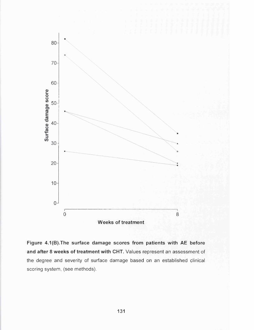
70-
60-
50-U)
40-
30-
20 -
i---------------------------------------------------------------- 10 8
Weeks of treatment
Figure 4.1(B).The surface damage scores from patients with AE before
and after 8 weeks of treatment with CHT. Values represent an assessment of
the degree and severity of surface damage based on an established clinical
scoring system, (see methods).
131

4.3.2 Effect Of Treatment On IL-4 mRNA In Lesional Skin And
Comparison With Normal Controls
The relative optical density (ROD) of the IL-4 mRNA in lesional skin from
patients with AE (median ROD 39) was significantly greater than that in the skin
of normal controls (median ROD 24.9, p<0.03). [Fig 4.2] After 8 weeks of
treatment with CHT, there was a marked down regulation of IL-4 mRNA
expression to a median relative optical density of 18.7 and this was significant
with p<0.01. [Fig 4.3]
132

60
I I Before CHT ^ After CHT
Normal
Interleukin 4 mRNA Interleukin 2 mRNA
Figure 4.2 The level of expression of mRNA for IL-4 and IL-2 in lesional skin of
patients with AE before and after 8 weeks of treatment with CHT and in skin
from normal controls. The columns represent median relative optical densities
recorded of formazan product of reaction of cells following in situ hybridisation.
Ranges are represented as vertical lines.
133

50-1
40-
>»21 a: o £ ^
T f C O
c -212 o. 30-
c JS
20 -
10Ji------------------------------------------------------------------------------10 8
Weeks of treatment
Figure 4.3 The relative optical densities recorded of reaction product
identifying cell associated IL-4 mRNA following in situ hybridisation in
lesional skin of 5 patients with AE before and after 8 weeks of treatment with
CHT.
134

4.3.3 Effect Of Treatment On IL-2 mRNA In Lesional Skin And Comparison
With Normal Controls
Analysis of levels of mRNA for IL-2 showed that in lesional skin from patients
before treatment, there was more IL-2 mRNA (median ROD 28.8) than in
normal skin (median relative optical density 17.2, p<0.05), although this
difference was not as marked as for IL-4 mRNA. [Fig 4.2] In 4 out of 5 patients,
there was a rise in levels of mRNA for IL-2 after treatment, however this did not
reach statistical significance because of the 1 sample in which there was a
decrease (median ROD 40.6, p>0.3). [Fig 4.4]
135

10J
0 8
Weeks of treatment
Figure 4.4 The relative optical densities recorded of reaction product of
cell associated IL-2 mRNA following in situ hybridisation in lesional skin
of 5 patients with AE before and after 8 weeks of treatm ent with CHT
136

4.3.4 Effect Of Treatment On Numbers Of T Lymphocytes In Lesional Skin
Numbers of lymphocytes that stained positively with CD3 (pan T cell marker)
were compared in lesional skin before and after treatment. Treatment lead to a
downregulation of numbers of T cells present with a median of 9.9 cells per 104
pm2 before treatment and 6.2 cells per 104 pm2 after treatment (p<0.04). [Fig
4.5]
137

2 -
0 8
Weeks of treatment
Figure 4.5 The number of T lymphocytes in lesional skin of patients with
AE before and after 8 weeks of treatment with CHT. Results are for
positively stained cells with immunoperoxidase method using the monoclonal
antibody CD3. The values represented are numbers of cells per 104 pm2
138

4.3.5 Effect Of Treatment On The Number Of Cells Staining Positively For
IL-4
As the level of expression for the mRNA for IL-4 decreased with treatment, it
was necessary to confirm that there was a downregulation in the number of
cells staining positively for IL-4. The median number of positively staining cells
before treatment was 4.0 cells per 104 pm2 and 2.7 cells per 104 pm2 after
treatment (p<0.001) [Fig 4.6]. This may, however be explained by an absolute
loss in the number of T cells. (See above)
139
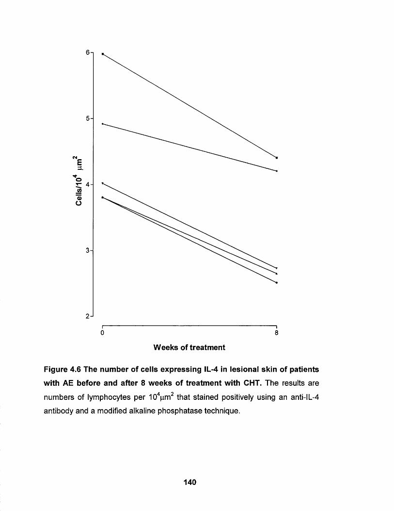
5
CM
o4-
3
0 8
Weeks of treatment
Figure 4.6 The number of cells expressing IL-4 in lesional skin of patients
with AE before and after 8 weeks of treatment with CHT. The results are
numbers of lymphocytes per 104pm2 that stained positively using an anti-IL-4
antibody and a modified alkaline phosphatase technique.
140

4.4 DISCUSSION
This study has shown that in AE lesions, there is increased expression of the
mRNA for the Th2 cytokine IL-4 and this is downregulated by treatment. This is
associated with improvement in clinical disease. Using in situ hybridisation
mRNA levels for the cytokine have been recorded which relate to the capacity
of the cells to secrete IL-4. Also, by using a MoAb for IL-4, actual numbers of
cells expressing IL-4 can be quantified. Treatment leading to a reduction in
disease severity downregulates both mRNA for IL-4 and numbers of cells
expressing IL-4. Not surprisingly, the results also show that an improvement in
clinical disease is associated with a decrease in the absolute numbers of
lymphocytes present in the inflammatory infiltrate within lesional skin. Thus, it is
not possible from these investigations to conclude whether the marked
decrease in IL-4 producing lymphocytes is due to a selective downregulation of
Th2 lymphocytes, decreased secretion of IL-4 from the lymphocytes or simply a
result of the decrease in total numbers of T cells. However, the downregulation
in IL-4 mRNA to near normal levels clearly suggests that this cytokine is more
relevant to disease severity than the Th1 cytokine IL-2 where the levels of
mRNA are not significantly affected by treatment, although there is no data of
numbers of cells that express IL-2. It may be relevant nevertheless that in 80%
of our subjects, treatment lead to an upregulation of IL-2 mRNA implying that
there may be a switch from Th2 to Th1 cells as clinical disease improves. This

also supports the hypothesis that it is predominantly Th2 cells that are
selectively downregulated as clinical severity is reduced.
142

CHAPTER 5
GENERAL DISCUSSION
143

5. GENERAL DISCUSSION
This study has quantified the level of disease severity and parameters of
immunopathology in samples of lesional skin from AE patients before and after
efficacious therapy, and thus it has been possible to dissect out aspects of
immune dysfunction that appear related to clinical severity of the disease.
Aberrations within the balance of immunocompetent cell populations in samples
of lesional skin from patients with AE have been identified. Culture studies have
shown that monocytes from patients with AE differentiate more rapidly
compared with similar cells from normal controls and increased numbers
express differentiation markers associated with the phenotype associated with
antigen presentation. Furthermore the study demonstrates a dysregulation in
the expression of the low affinity IgE receptor (CD23) on subsets of
macrophages. Using phenotypic markers it is shown that in AE antigen
presenting cells of the skin express these receptors; whereas in normal skin
tissue phagocytes contribute the major population of CD23+ cells. This
suggests that in AE, CD23 may be associated with antigen presentation.
Evidence is presented that these abnormalities are principally promoted by the
local environment rather than being the sequelae of a systemic immunologic
problem. The results also suggest that in this local environment, the balance of
144

cytokine production by T cells may promote aberrations in monocyte
differentiation and CD23 expression.
Eczema is a very common dermatological condition which is of great
importance both as a health care problem and economically, in that it is
associated with a considerable degree of morbidity which may affect work
habits. Despite the high incidence of the disease, the pathogenetic
mechanisms involved are poorly understood and the current therapeutic options
are not curative. The therapeutic approach ranges from the use of topical
emollients and allergen avoidance strategies, to the use of topical
corticosteroids and potent immunomodulatory drugs such as cyclosporin A
[Sowden et al. 1991] and Interferon gamma [Hanifin et a\. 1993]. Even
cytotoxic drugs such as azathioprine have been used [Younger et al. 1991].
Systemic treatment is limited by potentially serious side effects discussed in
Chapter one. Osteoporosis, hypertension and diabetes mellitus are examples
of the risks with long term steroid therapy [Schimmer and Parker 1996].
Cyclosporin has been shown to cause changes at the microscopic level in the
kidneys of all patients treated over a period of two years and arteriolar
hyalinosis and interstitial fibrosis are seen on renal biopsy [Zachariae et al.
1998]. Long term use of both cyclosporin and azathioprine have been
associated with lymphoproliferative and skin malignancies [Taylor and Shuster
145

1992, Jones et al. 1996, Sieber 1977]. Ultraviolet therapy increases the risk of
the subsequent development of squamous cell carcinomas of the skin
[Bruynzeel et al. 1991] and involves the inconvenience for patients of two to
three visits each week to a phototherapy unit during treatment. Gamma
interferon currently needs to be administered under controlled circumstances
subcutaneously within the hospital setting thus limiting its application.
Chinese herbal therapy has been used successfully by Chinese practitioners for
many centuries, without rigorous clinical trials. However, more recently a 10
herb preparation has been used in two double blind placebo controlled studies
in adults and children to treat moderate to severe recalcitrant AE [Sheehan et
al. 1992, Sheehan and Atherton 1992]. During these trials, the side effects were
generally mild and gastrointestinal in nature and the treatment was shown to be
effective. A major limitation of the original decoction was the daily time
consuming method involved in its preparation. This is markedly different from
conventional treatments, and continued compliance with the ritual of
preparation would be difficult for many patients. The initiation of a clinical trial of
CHT within the Department of Dermatology at Royal Free Hospital offered the
opportunity to biopsy AE patients before and after this potentially efficacious
therapy, thus determining which aspects of immunopathology may be
associated with clinical severity. This study compared the use of the original
146
decoction, with the associated drawback of laborious preparation time, with a
freeze dried granule preparation which is easily taken on a regular basis.
The current study was able to confirm the previous reports of efficacy [Sheehan
et al. 1992, Sheehan and Atherton 1992], and offered in addition convincing
evidence that the freeze dried granule preparation of the herbs was as effective
clinically as the original decoction. Perhaps more importantly it was able to
show that the granule preparation was better tolerated by the patients and
produced no greater side effects than the decoction. General compliance is
essential in any long term therapy. In asthma for example much effort is taken
up in monitoring compliance [Schmier and Leidy 1998]. With this study in
progress therefore, it provided an ideal opportunity to obtain a patient group
who clinically changed from a state of more to less severe eczema after
receiving Chinese herbal therapy.
As well as good compliance and the availability of 'before and after' samples,
attempts to relate immunopathological changes with clinical improvement
required a reproducible method of quantifying the clinical condition. Although
quantitative and semi-quantitative techniques are available for measuring
immunopathological parameters, clinical status measurement often relies
heavily on subjective judgement.
147

The clinical severity of the eczema in our patient group was assessed using a
scoring system which measures the area of skin involved, and the severity of
erythema and surface damage (papules, lichenification, vesicles); a system
described previously, [Heddle et al. 1984]. Although subjective, good
reproducibility was recorded, provided a full set of scores at all body sites was
taken into account [Xu 1998]. Other scoring systems also take into account
patients' symptoms such as pruritus leading to sleep disturbance [Costa et al.
1989, Sowden et al. 1991, Rajka and Langeland 1989]. These scoring systems
are also subjective in nature yet are well accepted by the medical and scientific
community [Finlay 1996]. Results reported here using this body scoring system
confirm previous reports of the clinical efficacy of CHT [Sheehan et al. 1992,
Sheehan and Atherton 1992, Sheehan and Atherton 1994, Sheehan et al.
1995].
Previous studies in this [Buckley et al. 1992, Buckley et al. 1993, Xu et al. 1997,
Zachary etal. 1985b] and other laboratories [Maurer and Stingl 1995, Mudde et
al. 1995a, Bruynzeel-Koomen eta l. 1986, Bieber 1997] have revealed specific
dysregulation in immunological parameters in AE. In particular aberrant
expression of CD23 [Buckley etal. 1992, Mudde etal. 1995a] and production of
IL-4 [ Van der Heijden et al. 1991, Mudde et al. 1992] have been implicated in
the pathogenesis. In order to identify whether the CD23 expression in the
148

lesions and the control of its expression by the Th2 cytokine, IL-4, are
components of the pathogenetic mechanisms leading to clinical disease, this
thesis used a dynamic clinical model. This study investigated changes in vivo
wherein patients with moderate to severe clinical disease were treated with
effective treatment resulting in clinical improvement. This provided the
opportunity to investigate specific parameters and their relationship to disease
severity. Ultimately, the future targets of immunotherapy may be identified by
revealing those immune parameters that are directly related to clinical disease
severity.
Chronicity of disease was also a consideration and in this study all patients had
disease which had not remitted for at least 1 year prior to starting treatment with
CHT. Clinical scores were assessed before and after 8 weeks treatment with
CHT. At these times, peripheral blood was collected and lesional skin biopsies
taken. There is a relationship between clinical severity and immunopathology
as demonstrated by the observed decrease in the numbers of T lymphocytes
and antigen presenting cells in the lesions when there is clinical improvement
after therapy. Work in this laboratory [Buckley et al. 1992] has also shown that
in both non lesional skin and lesional skin, histologically there is a mononuclear
cell infiltrate. There are however decreased numbers of T cells and antigen
presenting cells in non lesional compared with lesional skin suggesting that the
149

clinical involvement of the skin is reflected by an increased mononuclear
infiltrate on a microscopic level.
AE is deemed an allergic condition where the body's immune system responds
inappropriately to environmental allergens, for example cat or dog dander,
pollen, or house dust mite excrement. By definition therefore this clinical
condition is manifest only in atopic subjects. In such individuals, reactivity to
these environmental allergens generates raised serum IgE levels. Such raised
IgE titres [Stone et al. 1973] then predisposes atopies to immediate type
hypersensitivity reactions involving degranulation of mast cells and eosinophils.
Although AE and other clinical conditions such as hayfever and asthma are
associated with a state of atopy, this immune dysregulation alone is not the
cause of these clinical problems. Indeed, many subjects exhibiting atopic
reactivity do not exhibit AE [Jones et al. 1975]. Up to 40% of the population
show atopic reactivity if tested yet only 7 to 10% will exhibit clinical symptoms.
Furthermore cases of AE have been reported where circulating IgE levels may
be normal, while elevated levels of IgE are found in other conditions such as
lymphoma [Sundstrom et al. 1988], Job's syndrome [Donabedian and Gallin
1983], parasitic infection [Bell 1996] and hypergammaglobulinaemia [Jako et al.
1997] without any concurrent evidence of AE. Thus high IgE titres are not
pathognomonic of atopy and bear no relation as to whether eczema is present
or not. Other immunopathogenic features must therefore be present. The
150

development of the lesions in the skin cannot be based upon the inconsistently
raised levels of IgE in the peripheral blood or the type 1 response to
environmental allergens. Thus there is a need to investigate in more detail other
possible features of immune dysfunction within the skin and peripheral blood of
AE patients which may represent further targets for therapeutic intervention.
This study has confirmed the work of others, that within the skin of patients with
AE, a mononuclear cell infiltrate is found, dominated by T cells and antigen
presenting cells. The reaction exhibits the characteristics of a type IV delayed
hypersensitivity reaction [Zachary et al. 1985a]. Because of this, it is now
generally accepted that this disease is also associated with dysfunction within
the T cell mediated immune response network. However, the underlying
pathogenesis remains obscure. Macrophages and T cells are recognised as
crucial players in many other if not all chronic inflammatory diseases such as
sarcoidosis [Poulter 1990, du Bois 1990], rheumatoid arthritis [van den Berg
and van Lent 1996], ulcerative colitis [Allison et al. 1988] and asthma [Poulter
and Burke 1996 ].
The skin is a common site for immune dysregulation to be manifest as a clinical
problem. Other chronic skin conditions such as bullous pemphigoid, pemphigus
vulgaris and dermatitis herpetiformis are characterised by antibody or immune
complex deposition promoting inflammatory reactions [Jordon et al. 1985]. In
151

these cases underlying immunological defects are clear. For example in
pemphigus, circulating IgG autoantibodies directed against the keratinocyte
desmosomal proteins form local deposits resulting in loss of adhesion between
epidermal cells and intraepidermal splitting [Korman 1990]. Yet it is thought to
be both a loss in cell adhesion as result of activation of plasmin as well as
activation of the complement cascade by local autoantibody deposition that
results in the inflammatory process which destroys intercellular connections
within the epidermis. The circulating autoantibodies are also known to correlate
with the disease activity, yet we do not understand what promotes the formation
of the autoantibody.
In contact dermatitis it has been recognised for many years that the erythema
and induration of the skin are a result of a T cell mediated delayed type
hypersensitivity reaction promoted by hapten-protein conjugates forming after
contact with a variety of small molecular weight chemicals. In contrast, in AE,
the antigens promoting the T cell reaction cannot be identified within the skin
lesions which makes it difficult to understand what is promoting the T cell
reaction in AE.
It would be reasonable to suggest that an understanding of the links between
atopic reactivity and the chronic T cell reactions in the skin may unravel this
complex situation. With raised circulating IgE and IgE-allergen complexes, it is
152

reasonable to determine whether there is any increase in the IgE receptors that
can bind these complexes within the skin. To this end, this study has sought
evidence linking these two phenomena by investigating the distribution and
regulation of the expression of IgE receptors on antigen presenting cells in the
skin.
Aberrant T cell activation in AE lesions has been demonstrated previously with
an excess of activated T cells within the cellular infiltrate [Soter 1989, Bos et al.
1992]. There are increased numbers of dendritic cells and Langerhans cells;
cell types that have primary roles in antigen presentation although there is no
evidence of actual increased antigen presentation within the lesions [Zachary et
al. 1985a]. Eosinophils also accumulate in some but not all AE lesions
[Leiferman 1989, Soter 1989]. This would suggest that an increased
accumulation of eosinophils is not a prerequisite for the manifestation of this
disease. Interestingly the levels of eosinophilic cationic proteins in peripheral
blood has been reported to correlate with disease severity [Czech et al. 1992,
Halmerbauer et al. 1997]. Increased expression of IgE receptors has also been
demonstrated on dendritic cells within the lesional skin [Grabbe et al. 1993,
Schmitt et al. 1990].
This study has confirmed that there are increased numbers of dendritic cells,
with increased numbers of cells that express the low affinity IgE receptor in
153

lesional skin in AE. Also there are aberrations in local cytokine production.
These features describe an environment where aberrant CD23 expression on
antigen presenting cells could promote T cell activation via facilitated antigen
presentation [van der Heijden et al. 1995]. The increased expression of IL-4
within the lesions demonstrated in this study supports the suggestion that it is
local IL-4 that promotes CD23 expression on the dendritic cells, as IL-4
promotes CD23 expression on circulating B cells and monocytes [Alderson et
al. 1994, Delespesse et al. 1991].
Most previous work has been conducted on samples from existing lesions .
Such an approach fails to establish whether observed aspects of immune
dysfunction are causative of the clinical lesions or effects of the emergence of
pathology promoted by other abnormalities. This study has attempted to resolve
this problem by comparing the immunopathology in biopsies of lesional skin
sampled when the AE is recalcitrant to treatment, to matched samples taken
and investigated after clinical improvement has been achieved by efficacious
therapy. The approach rests with the premise that any move towards 'normal'
identified within an immunologic parameter that is associated with clinically
defined improvement is likely to have contributed to the original pathogenic
process promoting the disease.
154

Such an hypothesis is seen to hold in other allergic diseases. In eosinophilic
allergic aspergillosis (EAA), circulating precipitins to allergens may be present
in both symptomatic and asymptomatic patients [Johnson et al. 1989] which
suggests that the presence of these precipitins is related to the disease but not
necessarily to the disease severity. Lymphocytosis in the bronchoalveolar
lavage of EAA patients is present in symptomatic patients but declines when
treatment is given or patients are protected from exposure to allergens
[Johnson et al. 1989] suggesting that the T cells are relevant to active disease.
Study of the inflammatory reaction in the lung that promotes the restrictive
defect in this condition, consistently shows lymphocyte infiltration and activation
while no definite evidence for a pathogenic role for precipitins has emerged.
Immunohistological studies have focused attention on several aspects of
immune dysregulation found in AE. These include: the distribution of IgE
receptors [Maurer and Stingl 1995, Buckley et al. 1992, Mudde et al. 1995b,
Bieber 1997]; changes to the macrophage and dendritic cell populations in the
skin [Bos et al. 1986,]; and the local influence of cytokines derived from Th1
and Th2 T lymphocytes [van der Heijden et al. 1991, Bos et al. 1992, Mudde et
al. 1992].
Earlier studies in these laboratories identified aberrant expression of CD23 on
antigen presenting cells as being characteristic of AE lesions and distinguished
155

these from those of contact sensitivity reactions, despite the fact that both are
dominated by T cells [Buckley et al. 1992]. This work revealed that the main
difference between lesional skin in AE and a patch test with a contact allergen
is the switch in the expression of CD23 from effector macrophages to inducer
macrophages. This study has investigated further the relevance of this aberrant
CD23 expression to clinical disease severity.
From the results presented here it can be seen that there is a significant
reduction in numbers of CD23+ cells in all the different macrophage subsets
with treatment. Importantly, an overall decrease in the level of expression of
CD23 was also seen. It is of relevance to note there is also a decrease in both
the number of HLA DR+ cells and in the level of expression of this MHC class II
antigen which is expressed by antigen presenting cells. In the lesions of AE,
there are clearly increased numbers of antigen presenting cells, identified by
MoAb RFD1. The absolute numbers of RFD1+ cells and Langerhans cells
decrease in association with clinical improvement which suggests that there is
either a decrease in recruitment or an increase in removal of these cells from
the skin. This study has addressed the issue of whether there is aberrant
monocyte recruitment from the peripheral blood and subsequent differentiation
in vitro.
156

With the knowledge that some monocytes express CD23, the possibility
emerges that dysregulation of immune interactions within the lesions is a
consequence of recruitment of circulating monocytes that already exhibit
aberrant CD23 expression. Alternatively, the local environment within the skin
may exert a dominant influence on monocyte differentiation and the relative
expression of FcsRII on macrophage subsets as they mature. By directly testing
this possibility, the current work has confirmed that there is aberrant monocyte
differentiation in vitro when cells taken from patients with AE are compared with
normal non atopic subjects.
Macrophages can be divided into functionally distinct subsets which mature
from a common monocyte origin [Johnston 1988]. The RFD1+ macrophages
are inducer cells and associated predominantly with antigen presentation
[Poulter et al. 1986, Spiteri and Poulter 1991], the RFD7+ cells are phagocytic
effector cells [Spiteri and Poulter 1991, Spiteri et al. 1992b], and those
expressing both RFD1 and RFD7 exhibit a T cell suppressive function [Spiteri
et al. 1992a, Poulter and Burke 1996]. Over a 7 day culture period, monocytes
from atopic eczema patients exhibit a more rapid differentiation as measured by
early expression of the cell surface antigens, RFD1 and RFD7. However
efficacious treatment did not lead to any significant changes in this pattern of
differentiation.
157

There was however an increased proportion of monocytes expressing CD23 in
the AE patients compared with the normal controls throughout the culture
period. Furthermore, treatment lead to a downregulation of the proportion of4
RFD1+ monocytes that expressed CD23 during the first few days of culture
although this difference was lost by day 7. These results suggest that aberrant
monocyte differentiation in AE is present amongst those monocytes that exhibit
surface antigen RFD1 (a marker associated with antigen presentation) [Poulter
etal. 1986, Spiteri and Poulter 1991]. Notably, in cells from AE patients, RFD1 +
monocytes that express CD23 maintain higher proportions throughout the
culture period than equivalent cells from normal controls. Also, the proportion of
RFD1+/CD23+ cells is downregulated by efficacious treatment. These
observations support the suggestion that aberrant expression of CD23 on APCs
(RFD1+ cells) may contribute to pathogenic changes in AE.
There are however two types of IgE receptors: the high affinity (FcsRI) and low
affinity (FcsRII or CD23) [Bieber 1992, Wang et al. 1992, Sutton and Gould
1993, Maurer and Stingl 1995]. Of these, more is known of the function of the
high affinity receptor. FcsRI is expressed by Langerhans cells, mast cells and
other basophils in normal individuals [Ravetch and Kinet 1991, Sutton and
Gould 1993]. In atopic subjects, peripheral blood monocytes also express this
158

receptor [Maurer et al. 1994]. It is accepted that cross linking by allergen of IgE
molecules bound to mast cells and basophils via FceRI triggers cellular
degranulation releasing pro-inflammatory mediators; this being the basis of
immediate type hypersensitivity reactions [Ishizaka 1989]. It has been
suggested that FcsRI on antigen presenting cells has a role as an allergen-
focusing receptor in facilitated antigen presentation [Bieber 1997, Maurer and
Stingl 1995].
Circulating IgE-allergen complexes are detected in AE. The possibility exists
therefore that these IgE complexes if entering the skin can bind via Fc
receptors to APC within the skin, such as Langerhans cells and macrophages.
This may result in the subsequent presentation of antigen to T cells. It is known
that binding of IgE-allergen complexes enables T cell activation at antigen
concentrations that are 100-1000 fold lower than with non complexed antigens
[van der Heijden et al. 1995]. This process may thus form the link in
pathogenesis between raised IgE levels seen in most AE patients [Stone et al.
1973], and the T cell stimulation seen in the skin lesions. With notable
exception [Mudde et al. 1995a, van der Heijden et al. 1995], there has been
less systematic investigation of the role of CD23 in antigen presenting
mechanisms.
159
CD23 is an integral membrane protein and is a member of the calcium-
dependent lectin family of proteins [Delespesse et al. 1991]. Membrane bound
CD23 (mCD23) can be cleaved at the cell surface to form a soluble form of the
receptor (sCD23) which can also bind IgE [Delespesse et al. 1989]. Ligands,
other than IgE, have been reported for CD23 [Sutton and Gould 1993]. One
example is the CD21 molecule which is the receptor for the complement
component C3b [Fearon 1993], CD23 has also been implicated as having a
role in cell adhesion as its structure is similar to the selectin family of adhesion
molecules [Sutton and Gould 1993]. Another function of both forms of CD23 is
to promote B cell growth and differentiation [Delespesse et al. 1992]. In this
regard, sCD23 has been found to enhance IgE production by B cells thus
sCD23 may be contributing to the increased IgE seen in peripheral blood by
enhancing production of this immunoglobulin by B cells. Both forms of CD23
are thought to have a role in antigen presentation, as antigens can be
internalised by phagocytosis or endocytosis by these molecules [Sutton and
Gould 1993]. Interestingly there have been reports of increased levels of sCD23
in AE patients [Takigawa et al. 1991, Muller et al. 1991, Reddy et al. 1992,
Bujanowski-Weber et al. 1992]. CD23 is known to be expressed on a wide
variety of cells including lymphocytes, monocytes, eosinophils, platelets, natural
killer cells and Langerhans cells [Delespesse et al. 1991, Kehry and Hudak
1989]. In atopies, there is a well documented increase in the number of
160

peripheral blood mononuclear cells (PBM) that express CD23 [Nakamura et al.
1991].
Although most studies investigating facilitated antigen presentation have
concentrated on the binding of IgE-allergen complexes to FcsRI [Maurer and
Stingl 1995], there are reports of low affinity receptors being involved in similar
mechanisms of T cell stimulation [van der Heijden et al. 1995, Mudde et al.
1990a]. Although FcsRII may normally only play a minor role in facilitated
antigen presentation, this could be as a consequence of their normal
distribution within the macrophage populations being restricted to effector
phagocytes [Buckley et al. 1992], where they may act to promote opsonisation.
In AE, expression of FcsRII has been shown to switch to dendritic cells
expressing the phenotypic characteristics of APCs [Buckley et al. 1992], Under
these circumstances, the capacity of FcsRII to bind IgE-allergen complexes
and promote facilitated antigen presentation may be significantly enhanced.
Facilitated antigen presentation has been demonstrated in vitro to involve CD23
[van der Heijden et al. 1995]. In these studies, IgE-allergen complexes were
formed with Der p 2 (housedust mite antigen) and IgE by pre-incubation of Der
p 2 with atopic sera containing allergen-specific IgE. Epstein-Barr virus
transformed B cells, of which 60-80% expressed CD23, were used to bind
161

these complexes. When the IgE-allergen complexes were incubated with these
CD23 expressing B cells as well as T cells, this resulted in T cell proliferation.
Interestingly, when sera was used with radioallergosorbent tests (RAST)
showing lower Der-p concentrations, this resulted in reduced T cell proliferation
as compared with the use of sera giving higher IgE titres on RAST testing.
There is therefore some rationale for the view that T cell activation via FceRII
expressing dendritic cells binding IgE-allergen complexes, may form the basis
for the chronic cutaneous lesions of AE.
To support this hypothesis, evidence is needed that the local milieu within the
skin is such that it will promote and stimulate CD23 expression on dendritic
cells. Further, there needs to be evidence that IgE is bound to these potential
APCs. As far as this latter point is concerned there are several studies showing
the presence of IgE in lesions of AE [Bruynzeel-Koomen et al. 1986, Leung et
al. 1987], including reports that this immunoglobulin is present on the surface of
dendritic cells [Barker et al. 1988, Leung et al. 1987]. Regarding the local milieu
and CD23 expression; it is important to look to the work that has focused on the
dominance of the Th2-type T cells in these lesions [van der Heijden et al. 1991,
Mudde et al. 1992]. For T cells to interact with macrophages there must be
communication via soluble mediators released by these cells.
162

Th2-type T cells exhibit a cytokine repertoire that includes IL-4 [Del Prete et al.
1994]. This cytokine has been shown to function as a chemotactic factor for
basophils [Schleimer et al. 1992], and as a promoter of CD23 expression,
[Bieber et al. 1989b, Alderson et al. 1994, Delespesse et al. 1991]. Indeed, IL-4
has shown to induce CD23 on PBM [Dierks et al. 1994, Krauss et al. 1993,
Alderson et al. 1994] in a dose dependent manner. There is also evidence that
these latter functions may be linked.
Another role for CD23 in antigen presentation is in IgE mediated allergen
presentation by B cells [Kehry and Yamashita 1989, Mudde et al. 1995a]. IgE
bound via the IgE receptor on antigen specific B cells encounters
antigen/allergen thus resulting in activation of the B cell. The antigens are then
processed and presented to specific T cells in association with MHC II and if
the responding T cell is of the Th2 type, the B cell is induced to switch to
produce antigen specific IgE. Such IgE mediated antigen presentation could
thus contribute to the overproduction of IgE and the presence of increased
numbers of Th2 type cells, which in turn would lead to raised IL-4 production
[Romagnani 1991]. Thus a vicious circle may be initiated that drives the Th2
dominated T cell activation responsible for skin inflammation in AE.
The use of paired biopsies taken pre and post treatment where clinical
improvement occurred, makes it possible in the present study to link severity of
163

disease with raised expression of CD23. If local IL-4 production is responsible
for regulating CD23 expression, a fall in IL-4 post treatment would be predicted.
Using methods of in situ hybridisation, it has indeed been possible to
demonstrate an association between local IL-4 production, (as recorded by
levels of mRNA) and clinical severity. Of some interest was the further
observation that tissue levels of IL-2 mRNA were increased following therapy.
The cytokine IL-2 is produced by Th1-type rather than Th2-type T cells [Del
Prete et al. 1994]. This observation opens up two possibilities: either there is
positive stimulation of the Th1 subset with down regulation of Th2 cells; or that
CHT, (effective therapy), is associated with a shift of Th2 to Th1 cells i.e. with a
repertoire from IL-4 production to IL-2 production.
The results presented thus further our understanding on three points. Firstly, it
is confirmed that CHT is an effective therapeutic option in cases of atopic
eczema. This may be of particular importance in those cases where the
patients appear resistant to conventional therapy. Secondly evidence is
presented to support the hypothesis that CHT therapy promotes a modulation
of immunological mechanisms both directly and indirectly. Thirdly by identifying
concurrent changes in clinical status and immunopathology, evidence towards
the immune basis of AE emerges.
164

The cause and effect relationships between these phenomena remain obscure.
Together with existing knowledge however the results enable a working
hypothesis to be proposed. The lesions of AE are characterised by
macrophage subset imbalance, aberrant expression of IgE receptors and over
stimulation of Th2 type cells in the skin.
A hypothesis is proposed whereby:
1/ Aberrant monocyte differentiation leads to the expression of CD23 on
inductive cells.
2/ IgE-allergen complexes bind to CD23 on these inductive cells
3/ Inappropriate antigen presentation causes local stimulation of Th2 type cells
4/ Local production of IL-4 is induced
5/ This further promotes CD23 expression
Thus while exposure to allergen occurs, abnormal IgE titres exist and this
process will be self perpetuating.
It is important to emphasize that this hypothesis is speculative. However such a
pathogenic process fits the facts as currently understood. Further, this study
has revealed for the first time that a therapeutic agent which modulates
monocyte differentiation also causes reduced Th2 type activity and reduced
CD23 expression in vivo. There is of course no direct evidence that these
effects are related. Caution should also be exercised in interpreting the in vivo
165
data as changes to immunopathology are inevitably a dynamic process and this
interpretation is based on two time points where biopsies were taken.
Nevertheless there is a logic to the hypothesis proposed above as it satisfies
recent observations of dysregulation in IL-4 production [van der Heijden et al.
1991], T cell subsets [Romagnani 1992a] and IgE receptor expression [Grabbe
et al. 1993, Schmitt et al. 1990] in AE.
Recent observations of the role of apoptosis in regulating chronic inflammatory
reactions [Orteu et al. 1998] need to be considered. There would appear to be
a very real possibility that persistent stimulation of T lymphocytes within chronic
inflammatory conditions may not be due to continued stimulation but due to a
failure of reactive cells to be driven into programmed cell death. This possibility
requires further investigation in AE.
This thesis has confirmed that this ten herb extract of CHT is an effective
treatment for AE however it would be interesting to investigate whether these
herbs separately could be an effective treatment alone. Although in China,
polypharmacy has been in practice for centuries so it would be unlikely that the
herbs individually would be as effective clinically. Long term studies of both
clinical efficacy of CHT and importantly long term safety of treatment would be
essential.
166

Further study of immunopathology of sequential investigations of skin
immunopathology at several time points over a treatment period could
corroborate this work especially if deterioration in clinical disease was found to
show a change in immune parameters returning to that associated with more
severe disease. It would be interesting to repeat the studies with several
treatment modalities such as cyclosporin and azathioprine in addition to CHT.
There is no doubt that a full understanding of the pathogenesis of AE should be
the basis of the approach to treatment. It is clear that many of the treatmentst
that are effective in AE modify the immune response in some way. However theII exact mechanism of action is not established in any of the therapies. It is
| important to determine specific abnormalities in the chain of pathological events
I within the immune system as these may provide targets for a therapeutic
approach. The area of immunopathogenesis requires further investigation. In
this study immunohistological techniques were used with monoclonal antibodies
to define cell phenotypes and mRNA probes used to define cytokine expression
in situ.
This study investigates which of the abnormalities noted above are modulated
by treatment with CHT in a cohort of patients. This aims to help provide a link
between Type I Hypersensitivity (apparent in peripheral blood) and Type IV
167

hypersensitivity (mediated in the skin). The study investigates more closely the
increased presence of CD23+ antigen presenting cells in lesional skin in AE
and investigates further the increased Th2 lymphocyte activity in AE.
168

6. REFERENCES
Adachi T, Alam R. The mechanism of IL-5 signal transduction. Am J.Physiol. 1998; 275:C623-C633
Agata H, Kondo N, Fukutomi O, Shinoda S, Orii T. Effect of elimination diets on food-specific IgE antibodies and lymphocyte proliferative responses to food antigens in atopic dermatitis patients exhibiting sensitivity to food allergens. J.AIIergy Clin.Immunol. 1993; 91:668-679.
Agosti JM, Sprenger JD, Lum LG, Witherspoon RP, Fisher LD, Storb R, et al. Transfer of allergen-specific IgE-mediated hypersensitivity with allogeneic bone marrow transplantation. N.Engl.J.Med. 1988; 319:1623- 1628.
Alaiti S, Kang S, Fiedler VC, Ellis CN, Spurlin DV, Fader D, et al. Tacrolimus (FK506) ointment for atopic dermatitis: a phase I study in adults and children. J.Am Acad.Dermatol. 1998; 38:69-76.
Alderson MR, Armitage RJ, Tough TW, Ziegler SF. Synergistic effects of IL-4 and either GM-CSF or IL-3 on the induction of CD23 expression by human monocytes: regulatory effects of IFN-alpha and IFN-gamma. Cytokine 1994; 6:407-413.
Alegre VA, MacDonald DM, Poulter LW. The simultaneous presence of Langerhans' cell and interdigitating cell antigenic markers on inflammatory dendritic cells. Clin.Exp.lmmunol. 1986; 64:330-333.
Allison MC, Cornwall S, Poulter LW, Dhillon AP, Pounder RE. Macrophage heterogeneity in normal colonic mucosa and in inflammatory bowel disease. Gut 1988; 29:1531-1538.
169

Atherton DJ, Carabott F, Glover MT, Hawk JL. The role of psoralen photochemotherapy (PUVA) in the treatment of severe atopic eczema in adolescents. Br.J.Dermatol. 1988; 118:791-795.
Atherton DJ, Sewell M, Soothill JF, Wells RS, Chilvers CE. A double-blind controlled crossover trial of an antigen-avoidance diet in atopic eczema. Lancet 1978; 1:401-403.
Banerjee P, Xu XJ, Poulter LW, Rustin MH. Changes in CD23 expression of blood and skin in atopic eczema after Chinese herbal therapy. Clin. Exp. Allergy 1998; 28:306-314.
Bar RS, Kahn CR, Koren HS. Insulin inhibition of antibody-dependent cytoxicity and insulin receptors in macrophages. Nature 1977; 265:632- 635.
Barker JN, Alegre VA, MacDonald DM. Surface-bound immunoglobulin E on antigen-presenting cells in cutaneous tissue of atopic dermatitis. J.lnvest.Dermatol. 1988; 90:117-121.
Beck HI, Korsgaard J. Atopic dermatitis and house dust mites. Br.J.Dermatol. 1989; 120:245-251.
Bell RG. IgE, allergies and helminth parasites: a new perspective on an old conundrum. Immunology & Cell Biology 1996; 74:337-345.
Berth-Jones J, Graham-Brown RA. Placebo-controlled trial of essential fatty acid supplementation in atopic dermatitis [published erratum appears in Lancet 1993 Aug 28;342(8870):564]. Lancet 1993; 341:1557- 1560.
Beutler B, Cerami A. Cachectin: more than a tumor necrosis factor. N.Engl.J.Med. 1987; 316:379-385.
Bieber T. [IgE receptors in Langerhans cells. A link between the environment and the immune system?]. Hautarzt 1992; 43:753-762.
170

Bieber T. Fc epsilon Rl on human Langerhans cells: a receptor in search of new functions. Immunol.Today 1994; 15:52-53.
Bieber T. Fc epsilon Rl-expressing antigen-presenting cells: new players in the atopic game. Immunol.Today 1997; 18:311-313.
Bieber T, Dannenberg B, Prinz JC, Rieber EP, Stolz W, Braun-Falco O, et al. Occurrence of IgE-bearing epidermal Langerhans cells in atopic eczema: a study of the time course of the lesions and with regard to the IgE serum level. J.lnvest.Dermatol. 1989a; 93:215-219.
Bieber T, Delespesse G. Gamma-interferon promotes the release of IgE- binding factors (soluble CD23) by human epidermal Langerhans cells. J.lnvest.Dermatol. 1991; 97:600-603.
Bieber T, Rieger A, Neuchrist C, Prinz JC, Rieber EP, Boltz N, et al. Induction of Fc epsilon R2/CD23 on human epidermal Langerhans cells by human recombinant interleukin 4 and gamma interferon. J.Exp.Med. 1989b; 170:309-314.
Borres MP, Odelram H, Irander K, Kjellman Nl, Bjorksten B. Peripheral blood eosinophilia in infants at 3 months of age is associated with subsequent development of atopic disease in early childhood. J.AIIergy Clin.Immunol. 1995; 95:694-698.
Bos JD, van Gl, Krieg SR, Poulter LW. Different in situ distribution patterns of dendritic cells having Langerhans (T6+) and interdigitating (RFD1+) cell immunophenotype in psoriasis, atopic dermatitis, and other inflammatory dermatoses. J.lnvest.Dermatol. 1986; 87:358-361.
Bos JD, Wierenga EA, Sillevis SJ, van der Heijden FL, Kapsenberg, ML. Immune dysregulation in atopic eczema. Arch.Dermatol. 1992; 128:1509- 1512.
171

Braathen LR, Forre O, Natvig JB, Eeg-Larsen T. Predominance of T lymphocytes in the dermal infiltrate of atopic dermatitis. Br.J.Dermatol. 1979; 100:511-519.
Braga M, Gianotti L, Gentilini O, Fortis C, Consogno G, Di CV. Thymopentin modulates Th1 and Th2 cytokine response and host survival in experimental injury. J.Surg.Res. 1996; 62:197-200.
Brattsand R, Linden M. Cytokine modulation by glucocorticoids: mechanisms and actions in cellular studies. Aliment.Pharmacol.Ther. 1996; 10 Suppl 2:81-90.
Brook I, Frazier EH, Yeager JK. Microbiology of infected atopic dermatitis. International Journal of Dermatology 1996; 35:791-793.
Bruynzeel-Koomen C, van WD, Toonstra J, Berrens L, Bruynzeel, PL. The presence of IgE molecules on epidermal Langerhans cells in patients with atopic dermatitis. Arch.Dermatol.Res. 1986; 278:199-205.
Bruynzeel I, Bergman W, Hartevelt HM, Kenter CC, Van de Velde EA., Schothorst AA, et al. 'High single-dose' European PUVA regimen also causes an excess of non-melanoma skin cancer. Br.J.Dermatol. 1991; 124:49-55.
Buckley C, Ivison C, Poulter LW, Rustin MH. CD23/Fc epsilon R11 expression in contact sensitivity reactions: a comparison between aeroallergen patch test reactions in atopic dermatitis and the nickel patch test reaction in non-atopic individuals. Clin.Exp.Immunol. 1993; 91:357- 361.
Buckley C, Rustin MH, Ivison C, Poulter LW. Differentiation and CD23 expression of peripheral blood monocytes in patients with atopic dermatitis. Br.J.Dermatol. 1995; 133:757-763.
172

Buckley CC, Ivison C, Poulter LW, Rustin MH. Fc epsilon R11/CD23 receptor distribution in patch test reactions to aeroallergens in atopic dermatitis. J.lnvest.Dermatol. 1992; 99:184-188.
Bujanowski-Weber J, Knoller I, Wahn U, Konig W. Analysis of IgE-binding factors (sCD23) within newborn sera. lnt.Arch.Allergy Immunol. 1992; 99:127-132.
Busse WW, Sedgwick JB. Eosinophils in asthma. Ann.Allergy 1992; 68:286-290.
But PP. Need for correct identification of herbs in herbal poisoning. Lancet 1993; 341:637
Campbell DE, Kemp AS. Proliferation and production of interferon- gamma (IFN-gamma) and IL-4 in response to Staphylococcus aureus and staphylococcal superantigen in childhood atopic dermatitis. Clin.Exp.Immunol. 1997; 107:392-397.
Chandra RK, Puri S, Hamed A. Influence of maternal diet during lactation and use of formula feeds on development of atopic eczema in high risk infants [published erratum appears in BMJ 1989 Oct 7;299(6704):896]. BMJ 1989; 299:228-230.
Ciancio G, Siquijor AP, Burke GW, Roth D, Cirocco R, Esquenazi V, et al. Post-transplant lymphoproliferative disease in kidney transplant patients in the new immunosuppressive era. Clinical Transplantation 1997; 11:243-249.
Clark RA, Ad in off AD. Aeroallergen contact can exacerbate atopic dermatitis: patch tests as a diagnostic tool. J.Am Acad.Dermatol. 1989; 21:863-869.
Coleman R, Trembath RC, Harper Jl. Chromosome 11q13 and atopy underlying atopic eczema. Lancet 1993; 341:1121-1122.
173

Colloff MJ. Exposure to house dust mites in homes of people with atopic dermatitis. Br.J.Dermatol. 1992; 127:322-327.
Colloff MJ, Lever RS, McSharry C. A controlled trial of house dust mite eradication using natamycin in homes of patients with atopic dermatitis: effect on clinical status and mite populations. Br.J.Dermatol. 1989; 121:199-208.
Cookson WO, Sharp PA, Faux JA, Hopkin JM. Linkage between immunoglobulin E responses underlying asthma and rhinitis and chromosome 11 q. Lancet 1989; 1:1292-1295.
Cooper KD. Atopic dermatitis: recent trends in pathogenesis and therapy. J.lnvest.Dermatol. 1994; 102:128-137.
Costa C, Rilliet A, Nicolet M, Saurat JH. Scoring atopic dermatitis: the simpler the better?. Acta Dermato-Venereologica 1989; 69:41-45.
Cox HE, Moffatt MF, Faux JA, Walley AJ, Coleman R, Trembath RC, et al. Association of atopic dermatitis to the beta subunit of the high affinity immunoglobulin E receptor. Br.J.Dermatol. 1998; 138:182-187.
Cubey RB. Glaucoma following the application of corticosteroid to the skin of the eyelids. Br.J.Dermatol. 1976; 95:207-208.
Czech W, Krutmann J, Schopf E, Kapp A. Serum eosinophil cationic protein (ECP) is a sensitive measure for disease activity in atopic dermatitis. Br.J.Dermatol. 1992; 126:351-355.
de Fraisinette A, Schmitt D, Dezutter-Dambuyant C, Guyotat D, Zabot, MT, et al. Culture of putative Langerhans cell bone marrow precursors: characterization of their phenotype. Exp.Hematol. 1988; 16:764-768.
Del Prete G, Maggi E, Romagnani S. Human Th1 and Th2 cells: functional properties, mechanisms of regulation, and role in disease. Lab.lnvest. 1994; 70:299-306.
174

Delespesse G, Hofstetter H, Sarfati M. Low-affinity receptor for IgE (FcERII, CD23) and its soluble fragments. International Archives of Allergy & Applied Immunology 1989; 90 Suppl 1:41-44.
Delespesse G, Sarfati M, Wu CY, Fournier S, Letellier M. The low-affinity receptor for IgE. Immunological Reviews 1992; 125:77-97.
Delespesse G, Suter U, Mossalayi D, Bettier B, Sarfati M, Hofstetter, et al. Expression, structure, and function of the CD23 antigen. Advances in
Immunology 1991; 49:149-191.
Diaso R, LoBuglio AF. Goodman and Gilman's the Pharmacological Basis of Therapeutics, chapter 52, 9th edition ed. McGraw-Hill, 1996.
Dierks SE, Campbell KA, Studer EJ, Conrad DH. Molecular mechanisms of murine Fc epsilon RII/CD23 regulation. Molecular Immunology 1994; 31:1181-1189.
Dinarello CA. An update on human interleukin-1: from molecular biology to clinical relevance. J.CIin.Immunol. 1985; 5:287-297.
Donabedian H, Gallin Jl. The hyperimmunoglobulin E recurrent-infection (Job's) syndrome. A review of the NIH experience and the literature. Medicine 1983; 62:195-208.
Du Bois R. The alveolar macrophage in sarcoidosis. Sarcoidosis 1990; 7:15-18.
Ezekowitz RA, Gordon S. Interaction and regulation of macrophage receptors. Ciba Foundation Symposium 1986; 118:127-136.
Fearon DT. The CD19-CR2-TAPA-1 complex, CD45 and signaling by the antigen receptor of B lymphocytes. Current Opinion in Immunology 1993; 5:341-348.
175

Finlay AY. Measurement of disease activity and outcome in atopic dermatitis. Br.J.Dermatol. 1996; 135:509-515.
Fogelman AM, Van LB, Warden C, Haberland ME, Edwards PA. Macrophage lipoprotein receptors. Journal of Cell Science - Supplement 1988; 9:135-149.
Gelfand EW, Landwehr LP, Esterl B, Mazer B. Intravenous immune globulin: an alternative therapy in steroid-dependent allergic diseases. Clin.Exp.Immunol. 1996; 104 Suppl 1:61-66.
George SA, Bilsland DJ, Johnson BE, Ferguson J. IMarrow-band (TL-01) UVB air-conditioned phototherapy for chronic severe adult atopic dermatitis. Br.J.Dermatol. 1993; 128:49-56.
Gondo A, Saeki N, Tokuda Y. Challenge reactions in atopic dermatitis after percutaneous entry of mite antigen. Br.J.Dermatol. 1986; 115:485- 493.
Goodyear HM, McLeish P, Randall S, Buchan A, Skinner GR, Winther M, et al. Immunological studies of herpes simplex virus infection in children with atopic eczema. Br.J.Dermatol. 1996; 134:85-93.
Gordon J, Flores-Romo L, Cairns JA, Millsum MJ, Lane PJ, Johnson GD, et al. CD23: a multi-functional receptor/lymphokine?. Immunol.Today 1989; 10:153-157.
Grabbe J, Haas N, Hamann K, Kolde G, Hakimi J, Czarnetzki BM. Demonstration of the high-affinity IgE receptor on human Langerhans cells in normal and diseased skin. Br.J.Dermatol. 1993; 129:120-123.
Griffin E, Hakansson L, Formgren H, Jorgensen K, Peterson C, Venge P. Blood eosinophil number and activity in relation to lung function in patients with asthma and with eosinophilia. J.AIIergy Clin.Immunol. 1991; 87:548-557.
176
Griffiths CE, Railan D, Gallatin WM, Cooper KD. The ICAM-3/LFA-1 interaction is critical for epidermal Langerhans cell alloantigen presentation to CD4+ T cells. Br.J.Dermatol. 1995; 133:823-829.
Griffiths CE, Voorhees JJ, Nickoloff BJ. Characterization of intercellular adhesion molecule-1 and HLA-DR expression in normal and inflamed skin: modulation by recombinant gamma interferon and tumor necrosis factor. J.Am Acad.Dermatol. 1989; 20:617-629.
Gruber SA, Gillingham K, Sothern RB, Stephanian E, Matas AJ, Dunn DL. De novo cancer in cyclosporine-treated and non-cyclosporine-treated adult primary renal allograft recipients. Clinical Transplantation 1994; 8:388-395.
Halmerbauer G, Frischer T, Koller DY. Monitoring of disease activity by measurement of inflammatory markers in atopic dermatitis in childhood. Allergy: European Journal of Allergy & Clinical Immunology 1997; 52:765- 769.
Hanau D, Gothelf Y, Schmitt DA, Fabre M, Garaud JC, Gazit E, et al. Internalization by receptor-mediated endocytosis of T6 (CD1 "NA1/34") surface antigen in T6 positive human cord blood cells (Langerhans cell precursors?). Nouvelle Revue Francaise d Hematologie 1987; 29:279- 283.
Hanifin, J. M. and Rajka, G. Diagnostic features of atopic dermatitis. Acta Dermato-Venereologica suppl 92, 44-77.1980.
Hanifin JM, Schneider LC, Leung DY, Ellis CN, Jaffe HS, Izu AE, et al. Recombinant interferon gamma therapy for atopic dermatitis. J.Am Acad.Dermatol. 1993; 28:189-197.
Heddle RJ, Soothill JF, Bulpitt CJ, Atherton DJ. Combined oral and nasal beclomethasone diproprionate in children with atopic eczema: a
177

randomised controlled trial. British Medical Journal Clinical Research Ed 1984; .289:651-654.
Hershey GK, Friedrich MF, Esswein LA, Thomas ML, Chatila TA. The association of atopy with a gain-of-function mutation in the alpha subunit of the interleukin-4 receptor. N.Engl.J.Med. 1997; 337:1720-1725.
Holden CA. Atopic dermatitis: a defect of intracellular secondary messenger systems?. Clinical & Experimental Allergy 1990; 20:131-136.
Holt PG, Yabuhara A, Prescott S, Venaille T, Macaubas C, Holt BJ, et al. Allergen recognition in the origin of asthma. Ciba Foundation Symposium 1997; 206:35-49.
Hsieh KH, Shaio MF, Liao TN. Thymopentin treatment in severe atopic dermatitis-clinical and immunological evaluations. Archives of Disease in Childhood 1992; 67:1095-1102.
Hutter C, Poulter LW. The balance of macrophage subsets may be customised at mucosal surfaces. FEMS Microbiology Immunology 1992; 5:309-315.
Ishizaka K. Basic mechanisms of IgE-mediated hypersensitivity. Current Opinion in Immunology 1989; 1:625-629.
Jako JM, Gesztesi T, Kaszas I. IgE lambda monoclonal gammopathy and amyloidosis. lnt.Arch.Allergy Immunol. 1997; 112:415-421.
Janossy G, Bofill M, Poulter LW, Rawlings E, Burford GD, Navarrete C, et al. Separate ontogeny of two macrophage-like accessory cell populations in the human fetus. J.Immunol. 1986; 136:4354-4361.
Jekler J, Larko O. UVA solarium versus UVB phototherapy of atopic dermatitis: a paired-comparison study. Br.J.Dermatol. 1991; 125:569-572.
178

Johansson SG. IgE in allergic diseases. Proc. Royal Soc. Med.: 62:975- 976.
Johnson MA, Nemeth A, Condez A, Clarke SW, Poulter LW. Cell-mediated immunity in pigeon breeders' lung: the effect of removal from antigen exposure. Eur.Respir.J. 1989; 2:445-450.
Johnston RB, Jr. Current concepts: immunology. Monocytes and macrophages. N.Engl.J.Med. 1988; 318:747-752.
Jolles S, Hughes J, Rustin M. Intracellular interleukin-4 profiles during high-dose intravenous immunoglobulin treatment of therapy-resistant atopic dermatitis. J. Am. Acad. Dermatol. 1999; 40: 121-123.
Jones HE, Inouye JC, McGerity JL, Lewis CW. Atopic disease and serum immunoglobulin-E. Br.J.Dermatol. 1975; 92:17-25.
Jones M, Symmons D, Finn J, Wolfe F. Does exposure to immunosuppressive therapy increase the 10 year malignancy and mortality risks in rheumatoid arthritis? A matched cohort study. British Journal of Rheumatology 1996; 35:738-745.
Jordon RE, Kawana S, Fritz KA. Immunopathologic mechanisms in pemphigus and bullous pemphigoid. J.lnvest.Dermatol. 1985; 85:72s- 78s.
Kehry MR, Hudak SA. Characterization of B-cell populations bearing Fc epsilon receptor II. Cellular Immunology 1989; 118:504-515.
Kehry MR, Yamashita LC. Low-affinity IgE receptor (CD23) function on mouse B cells: role in IgE-dependent antigen focusing. Proceedings of the National Academy of Sciences of the United States of America 1989; 86:7556-7560.
Kondo N, Fukutomi O, Kameyama T, Nishida T, Li GP, Agata H, et al. Suppression of proliferative responses of lymphocytes to food antigens
179

by an anti-allergic drug, ketotifen fumarate, in patients with food-sensitive atopic dermatitis. Int.Arch.Allergy Immunol. 1994; 103:234-238.
Korman NJ. Pemphigus. Dermatol.Clin. 1990; 8:689-700.
Krauss S, Mayer E, Rank G, Rieber EP. Induction of the low affinity receptor for IgE (Fc epsilon RII/CD23) on human blood dendritic cells by interleukin-4. Advances in Experimental Medicine & Biology 1993; 329:231-236.
Krutmann J, Diepgen TL, Luger TA, Grabbe S, Meffert H, Sonnichsen N, et al. High-dose UVA1 therapy for atopic dermatitis: results of a multicenter trial. J.Am Acad.Dermatol. 1998; 38:589-593.
Larko O. Phototherapy of eczema. Photodermatology, Photoimmunology & Photomedicine 1996; 12:91-94.
Law SK. C3 receptors on macrophages. Journal of Cell Science - Supplement 1988; 9:67-97.
Leiferman KM. Eosinophils in atopic dermatitis. Allergy: European Journal of Allergy & Clinical Immunology 1989; 44 Suppl 9:20-26.
Leiferman KM, Ackerman SJ, Sampson HA, Haugen HS, Venencie PY, Gleich, et al. Dermal deposition of eosinophil-granule major basic protein in atopic dermatitis. Comparison with onchocerciasis. N.Engl.J.Med. 1985; 313:282-285.
Lenz A, Heine M, Schuler G, Romani N. Human and murine dermis contain dendritic cells. Isolation by means of a novel method and phenotypical and functional characterization. J.CIin.Invest. 1993; 92:2587-2596.
Leung DY, Hirsch RL, Schneider L, Moody C, Takaoka R, Li SH, et al. Thymopentin therapy reduces the clinical severity of atopic dermatitis. J.AIIergy Clin.Immunol. 1990; 85:927-933.
180

Leung DY, Schneeberger EE, Siraganian RP, Geha RS, Bhan AK. The presence of IgE on macrophages and dendritic cells infiltrating into the skin lesion of atopic dermatitis. Clin.Immunol.Immunopathol. 1987; 42:328-337.
Leung DY, Travers JB, Norris DA. The role of superantigens in skin disease. J.lnvest.Dermatol. 1995; 105:37S-42S.
Leung DY, Wood N, Dubey D, Rhodes AR, Geha RS. Cellular basis of defective cell-mediated lympholysis in atopic dermatitis. J.lmmunol. 1983; 130:1678-1682.
Lever R, Turbitt M, Sanderson A, MacKie R. Immunophenotyping of the cutaneous infiltrate and of the mononuclear cells in the peripheral blood in patients with atopic dermatitis. J.lnvest.Dermatol. 1987; 89:4-7.
Leyden JJ, Marples RR, Kligman AM. Staphylococcus aureus in the lesions of atopic dermatitis. Br.J.Dermatol. 1974; 90:525-530.
Lidington EA, McCormack AM, Yacoub MH, Rose ML. The effects of monocytes on the transendothelial migration of T lymphocytes. Immunology 1998; 94:221-227.
Lindelof B, Sigurgeirsson B, Tegner E, Larko O, Johannesson A, Berne, et al. PUVA and cancer: a large-scale epidemiological study. Lancet 1991; 338:91-93.
Lub M, van KY, Figdor CG. Ins and outs of LFA-1. Immunol.Today 1995; 16:479-483.
MacGregor FB, Abernethy VE, Dahabra S, Cobden I, Hayes PC. Hepatotoxicity of herbal remedies. BMJ 1989; 299:1156-1157.
Mao XQ, Shirakawa T, Enomoto T, Shimazu S, Dake Y, Kitano H, et al. Association between variants of mast cell chymase gene and serum IgE
181

levels in eczema [published erratum appears in Hum Hered 1998 Mar- Apr;48(2):91]. Human Heredity 1998; 48:38-41.
Matsukura M, Yamada H, Yudate T, Tezuka T, Chihara J. Steroid-induced changes of eosinophils in atopic dermatitis. lnt.Arch.Allergy Immunol. 1997; 114 Suppl 1:51-54.
Maurer D, Fiebiger E, Reininger B, Wolff-Winiski B, Jouvin MH, Kilgus, et al. Expression of functional high affinity immunoglobulin E receptors (Fc epsilon Rl) on monocytes of atopic individuals. J.Exp.Med. 1994; 179:745-750.
Maurer D, Stingl G. Immunoglobulin E-binding structures on antigen- presenting cells present in skin and blood. J.lnvest.Dermatol. 1995; 104:707-710.
McGeady SJ, Buckley RH. Depression of cell-mediated immunity in atopic eczema. J.AIIergy Clin.lmmunol. 1975; 56:393-406.
McKee PH. Pathology of the skin, pages 3.12-3.19, 2nd edition, Mosby- Wolfe, 1996.
Mihm MCJ, Soter NA, Dvorak HF, Austen KF. The structure of normal skin and the morphology of atopic eczema. J.lnvest.Dermatol. 1976; 67:305- 312.
Min Dl, Monaco AP. Complications associated with immunosuppressive therapy and their management. Pharmacotherapy 1991; 11:119S-125S.
Miyasato M, Tsuda S, Nakama T, Kato K, Kitamura N, Nagaji J, et al. Serum levels of eosinophil cationic protein reflect the state of in vitro degranulation of blood hypodense eosinophils in atopic dermatitis. Journal of Dermatology 1996; 23:382-388.
Mosesson MW. The role of fibronectin in monocyte/macrophage function. Progress in Clinical & Biological Research 1984; 154:155-175.
182

Mudde GC, Bheekha R, Bruijnzeel-Koomen CA. Consequences of lgE/CD23-mediated antigen presentation in allergy. Immunol.Today 1995a; 16:380-383.
Mudde GC, Bheekha R, Bruijnzeel-Koomen CA. IgE-mediated antigen presentation. Allergy 1995b; 50:193-199.
Mudde GC, Hansel TT, van Reijsen F, Osterhoff BF, Bruijnzeel K, CA. IgE: an immunoglobulin specialized in antigen capture? Immunol.Today 1990a; 11:440-443.
Mudde GC, van RF, Boland GJ, de GG, Bruijnzeel PL, Bruijnzeel-Koomen CA. Allergen presentation by epidermal Langerhans' cells from patients with atopic dermatitis is mediated by IgE. Immunology 1990b; 69:335- 341.
Mudde GC, van Reijsen F, Bruijnzeel-Koomen CA. IgE-positive Langerhans cells and Th2 allergen-specific T cells in atopic dermatitis. J.lnvest.Dermatol. 1992; 99:103S
Muller KM, Rocken M, Joel D, Bonnefoy JY, Saurat JH, Hauser C. Mononuclear cell-bound CD23 is elevated in both atopic dermatitis and psoriasis. Journal of Dermatological Science 1991; 2:125-133.
Nakamura K, Okubo Y, Minami M, Furue M, Ishibashi Y. Phenotypic analysis of CD23+ peripheral blood mononuclear cells in atopic dermatitis. Br.J.Dermatol. 1991; 125:543-547.
Nakazawa M, Sugi N, Kawaguchi H, Ishii N, Nakajima H, Minami M. Predominance of type 2 cytokine-producing CD4+ and CD8+ cells in patients with atopic dermatitis. J.AIIergy Clin.lmmunol. 1997; 99:673-682.
Nathan CF. Secretory products of macrophages. J.CIin.Invest. 1987; 79:319-326.
183

Neild VS, Marsden RA, Bailes JA, Bland JM. Egg and milk exclusion diets in atopic eczema. Br.J.Dermatol. 1986; 114:117-123.
Orteu CH, Poulter LW, Rustin MH, Sabin CA, Salmon M, Akbar AN. The role of apoptosis in the resolution of T cell-mediated cutaneous inflammation. J.lmmunol. 1998; 161:1619-1629.
Parronchi P, Tiri A, Macchia D, De CM, Biswas P, Simonelli C, et al. Noncognate contact-dependent B cell activation can promote IL-4- dependent in vitro human IgE synthesis. J.lmmunol. 1990; 144:2102- 2108.
Pene J. Regulatory role of cytokines and CD23 in the human IgE antibody synthesis. lnt.Arch.Allergy Immunol. 1989; 90:32-40.
Perharic-Walton L, Murray V. Toxicity of Chinese herbal remedies [letter]. Lancet 1992; 340:674
Perharic L, Shaw D, Leon C, De SP, Murray VS. Possible association of liver damage with the use of Chinese herbal medicine for skin disease. Vet.Hum.Toxicol. 1995; 37:562-566.
Poulter LW. Changes in lung macrophages during disease. FEMS Microbiology Immunology 1990; 2:327-331.
Poulter LW, Burke CM. Macrophages and allergic lung disease. Immunobiology 1996; 195:574-587.
Poulter LW, Campbell DA, Munro C, Janossy G. Discrimination of human macrophages and dendritic cells by means of monoclonal antibodies. Scandinavian Journal of Immunology 1986; 24:351-357.
Poulter LW, Janossy G, Power C, Sreenan S, Burke C. Immunological/physiological relationships in asthma: potential regulation by lung macrophages. Immunol.Today 1994; 15:258-261.
184

Prescott SL, Macaubes C, Yabuhara A, Venaille TJ, Holt BJ, Habre W, et al. Developing patterns of T cell memory to environmental allergens in the
first two years of life. lnt.Arch.Allergy Immunol. 1997; 113:75-79.
Rajka G, Langeland T. Grading of the severity of atopic dermatitis. Acta Dermato-Venereologica 1989; Supplementum. 144:13-14.
Rasanen L, Lehto M, Reunala T, Jansen C, Lehtinen M, Leinikki P. Langerhans cell- and T-lymphocyte functions in patients with atopic dermatitis with disseminated cutaneous herpes simplex virus infection. J.lnvest.Dermatol. 1987; 89:15-18.
Ravetch JV, Kinet JP. Fc receptors. Annual Review of Immunology 1991; 9:457-492.
Ray A, Schmitt D. Langerhans cell: CD1 antigens and the Birbeck granule. Pathol.Biol. 1988; 36:846-853.
Reddy MM, Weissman AM, Mazza DS, Meriney DK, Johns M, Carrabis S, et al. Circulating elevated levels of soluble CD23, interleukin-4, and CD20+CD23+ lymphocytes in atopic subjects with elevated serum IgE concentrations. Ann.Allergy 1992; 69:131-134.
Reinhard T, Sundmacher R. Perforating keratoplasty in endogenous eczema. An indication for systemic cyclosporin A--a retrospective study of 18 patients. Klinische Monatsblatter fur Augenheilkunde 1992; 201:159- 163.
Reinhold U, Kukel S, Goeden B, Neumann U, Kreysel HW. Functional characterization of skin-infiltrating lymphocytes in atopic dermatitis. Clin.Exp.Immunol. 1991; 86:444-448.
Renz H, Jujo K, Bradley KL, Domenico J, Gelfand EW, Leung DY. Enhanced IL-4 production and IL-4 receptor expression in atopic
185

dermatitis and their modulation by interferon-gamma. J.lnvest.Dermatol. 1992; 99:403-408.
Rogge JL, Hanifin JM. Immunodeficiencies in severe atopic dermatitis. Depressed chemotaxis and lymphocyte transformation. Arch.Dermatol. 1976; 112:1391-1396.
Romagnani S. Human Th1 and Th2 subsets: doubt no more. Immunol.Today 1991; 12:256-257.
Romagnani S. Human Th1 and Th2 subsets: regulation of differentiation and role in protection and immunopathology. Int.Arch.Allergy Immunol. 1992a; 98:279-285.
Romagnani S. Induction of Th1 and Th2 responses: a key role for the 'natural' immune response? Immunol.Today 1992b; 13:379-381.
Ruco LP, Uccini S, Baroni CD. The Langerhans' cells. Allergy: European Journal of Allergy & Clinical Immunology 1989; 44 Suppl 9:27-30.
Ruzicka T, Bieber T, Schopf E, Rubins A, Dobozy A, Bos JD, et al. A short-term trial of tacrolimus ointment for atopic dermatitis. European Tacrolimus Multicenter Atopic Dermatitis Study Group. N.Engl.J.Med. 1997; 337:816-821.
Ryffel B. The carcinogenicity of ciclosporin. Toxicology 1992; 73:1-22.
Sakamoto T, Nakayama F, Tamamori T, Takigawa M. Fc epsilon receptor II/CD23+ lymphocytes in atopic dermatitis. III. Aberrant control in the in vitro expression of Fc epsilon RII/CD23 on peripheral blood T cells in atopic dermatitis. Clin.Exp.Immunol. 1992; 87:87-93.
Sakamoto T, Takigawa M, Tamamori T, Horiguchi D, Yamada M. Fc epsilon receptor II/CD23 positive lymphocytes in atopic dermatitis: II. Infiltration of Fc epsilon R ll(+) T cells in the skin lesion. J.lnvest.Dermatol. 1990; 95:592-596.
186

Salek MS, Finlay AY, Luscombe DK, Allen BR, Berth-Jones J, Camp RD, et al. Cyclosporin greatly improves the quality of life of adults with severe atopic dermatitis. A randomized, double-blind, placebo-controlled trial. Br.J.Dermatol. 1993; 129:422-430.
Santamaria LF, Perez SM, Hauser C, Blaser K. Skin-homing T cells in human cutaneous allergic inflammation. Immunologic Research 1995; 14:317-324.
Santamaria LF, Perez SM, Hauser C, Blaser K. Allergen specificity and endothelial transmigration of T cells in allergic contact dermatitis and atopic dermatitis are associated with the cutaneous lymphocyte antigen. lnt.Arch.Allergy Immunol. 1995; 107:359-362.
Sauer J, Rupprecht M, Arzt E, Stalla GK, Rupprecht R. Glucocorticoids modulate soluble interleukin-2 receptor levels in vivo depending on the state of immune activation and the duration of glucocorticoid exposure. Immunopharmacology 1993; 25:269-276.
Schalin-Karrila M, Mattila L, Jansen CT, Uotila P. Evening primrose oil in the treatment of atopic eczema: effect on clinical status, plasma phospholipid fatty acids and circulating blood prostaglandins. Br.J.Dermatol. 1987; 117:11-19.
Schimmer BP, Parker KL. Goodman and Gilman's the Pharmacological Basis of Therapeutics, chapter 59, 9th edition ed. McGraw-Hill, 1996.
Schleimer RP, Sterbinsky SA, Kaiser J, Bickel CA, Klunk DA, Tomioka, et al. IL-4 induces adherence of human eosinophils and basophils but not neutrophils to endothelium. Association with expression of VCAM-1. J.lmmunol. 1992; 148:1086-1092.
Schmier JK, Leidy NK. The complexity of treatment adherence in adults with asthma: challenges and opportunities. J. Asthma. 1998; 35: 455-72.
187

Schmitt DA, Bieber T, Cazenave JP, Hanau D. Fc receptors of human Langerhans cells. J.lnvest.Dermatol. 1990; 94:15S-21S.
Schultz Larsen F, Holm NV, Henningsen K. Atopic dermatitis. A genetic- epidemiologic study in a population-based twin sample. J.Am Acad.Dermatol. 1986; 15:487-494.
Seldenrijk CA, Drexhage HA, Meuwissen SG, Pals ST, Meijer CJ. Dendritic cells and scavenger macrophages in chronic inflammatory bowel disease. Gut 1989; 30:486-491.
Sheehan MP, Atherton DJ. A controlled trial of traditional Chinese medicinal plants in widespread non-exudative atopic eczema. Br.J.Dermatol. 1992; 126:179-184.
Sheehan MP, Atherton DJ. One-year follow up of children treated with Chinese medicinal herbs for atopic eczema. Br.J.Dermatol. 1994; 130:488-493.
Sheehan MP, Atherton DJ, Norris P, Hawk J. Oral psoralen photochemotherapy in severe childhood atopic eczema: an update. Br.J.Dermatol. 1993; 129:431-436.
Sheehan MP, Rustin MH, Atherton DJ, Buckley C, Harris DW, Brostoff J, et al. Efficacy of traditional Chinese herbal therapy in adult atopic dermatitis [published erratum appears in Lancet 1992 Jul 18;340(8812):188]. Lancet 1992; 340:13-17.
Sheehan MP, Stevens H, Ostlere LS, Atherton DJ, Brostoff J, Rustin, et al. Follow-up of adult patients with atopic eczema treated with Chinese
herbal therapy for 1 year. Clin.Exp.Dermatol. 1995; 20:136-140.
Sieber SM. The action of antitumor agents: a double-edged sword?. Medical & Pediatric Oncology 1977; 3:123-131.
188

Silverstein RL, Nachman RL. Thrombospondin binds to monocytes- macrophages and mediates platelet-monocyte adhesion. J.CIin.Invest. 1987; 79:867-874.
Soter NA. Morphology of atopic eczema. Allergy: European Journal of Allergy & Clinical Immunology 1989; 44 Suppl 9:16-19.
Sowden JM, Berth-Jones J, Ross JS, Motley RJ, Marks R, Finlay AY, et al.Double-blind, controlled, crossover study of cyclosporin in adults with
severe refractory atopic dermatitis. Lancet 1991; 338:137-140.
Spik G, Montreuil J. The role of lactotransferrin in the molecular mechanisms of antibacterial defense. Bulletin Europeen de Physiopathologie Respiratoire 1983; 19:123-130.
Spiteri MA, Clarke SW, Poulter LW. Alveolar macrophages that suppress T-cell responses may be crucial to the pathogenetic outcome of pulmonary sarcoidosis. Eur.Respir.J. 1992a; 5:394-403.
Spiteri MA, Clarke SW, Poulter LW. Isolation of phenotypically and functionally distinct macrophage subpopulations from human bronchoalveolar lavage. Eur.Respir.J. 1992b; 5:717-726.
Spiteri MA, Poulter LW. Characterization of immune inducer and suppressor macrophages from the normal human lung. Clin.Exp.Immunol. 1991; 83:157-162.
Stiller MJ, Shupack JL, Kenny C, Jondreau L, Cohen DE, Soter NA. A double-blind, placebo-controlled clinical trial to evaluate the safety and efficacy of thymopentin as an adjunctive treatment in atopic dermatitis. J.Am Acad.Dermatol. 1994; 30:597-602.
Stone SP, Muller SA, Gleich GJ. IgE levels in atopic dermatitis. Arch.Dermatol. 1973; 108:806-811.
189

Sundstrom C, Nilsson K, Brenning G. IgE-producing low-grade non- Hodgkin lymphoma. European Journal of Haematology 1988; 41:388-395.
Sutton BJ, Gould HJ. The human IgE network. Nature 1993; 366:421- 428.
Takigawa M, Tamamori T, Horiguchi D, Sakamoto T, Yamada M, Yoshioka, et al. Fc epsilon receptor ll/CD23-positive lymphocytes in atopic dermatitis. I. The proportion of Fc epsilon RII+ lymphocytes correlates with the extent of skin lesion. Clin.Exp.lmmunol. 1991; 84:275-282.
Tan BB, Weald D, Strickland I, Friedmann PS. Double-blind controlled trial of effect of housedust-mite allergen avoidance on atopic dermatitis. Lancet 1996; 347:15-18.
Tanner JE, Alfieri C. Interactions involving cyclosporine A, interleukin-6, and Epstein-Barr virus lead to the promotion of B-cell lymphoproliferative disease. Leukemia & Lymphoma 1996; 21:379-390.
Taylor AE, Shuster S. Skin cancer after renal transplantation: the causal role of azathioprine. Acta Dermato-Venereologica 1992; 72:115-119.
Teunissen MB, Wormmeester J, Krieg SR, Peters PJ, Vogels IM, Kapsenberg ML, et al. Human epidermal Langerhans cells undergo profound morphologic and phenotypical changes during in vitro culture. J.lnvest.Dermatol. 1990; 94:166-173.
Thompson P, Stewart AC. Allergy, pages 1.1-1.14. Gower Publishing Ltd, 1993.
Thornhill MH, Haskard DO. IL-4 regulates endothelial cell activation by IL- 1, tumor necrosis factor, or IFN-gamma. J.lmmunol. 1990; 145:865-872.
Tormey VJ, Faul J, Leonard C, Burke CM, Dilmec A, Poulter LW. T-cell cytokines may control the balance of functionally distinct macrophage populations. Immunology 1997; 90:463-469.
190

Uehara M. Atopic dermatitis and tuberculin reactivitiy. Arch.Dermatol. 1977; 113:1226-1228.
Uehara M, Izukura R, Sawai T. Blood eosinophilia in atopic dermatitis. Clin.Exp.Dermatol. 1990; 15:264-266.
Uehara M, Sawai T. A longitudinal study of contact sensitivity in patients with atopic dermatitis. Arch.Dermatol. 1989; 125:366-368.
Unanue ER, Allen PM. The basis for the immunoregulatory role of macrophages and other accessory cells. Science 1987; 236:551-557.
van den Berg WB, van Lent P. The role of macrophages in chronic arthritis. Immunobiology 1996; 195:614-623.
van der Heijden FL, van NR, Kapsenberg ML. Relationship between facilitated allergen presentation and the presence of allergen-specific IgE in serum of atopic patients. Clin.Exp.Immunol. 1995; 99:289-293.
van der Heijden FL, Wierenga EA, Bos JD, Kapsenberg ML. High frequency of IL-4-producing CD4+ allergen-specific T lymphocytes in atopic dermatitis lesional skin. J.lnvest.Dermatol. 1991; 97:389-394.
van Joost T, Kozel MM, Tank B, Troost R, Prens EP. Cyclosporine in atopic dermatitis. Modulation in the expression of immunologic markers in lesional skin. J.Am Acad.Dermatol. 1992; 27:922-928.
Virelizier JL, Arenzana-Seisdedos F. Immunological functions of macrophages and their regulation by interferons. Medical Biology 1985; 63:149-159.
Wahlgren CF, Hagermark O, Bergstrom R. The antipruritic effect of a sedative and a non-sedative antihistamine in atopic dermatitis. Br.J.Dermatol. 1990; 122:545-551.
191

Wang B, Rieger A, Kilgus O, Ochiai K, Maurer D, Fodinger D, et al. Epidermal Langerhans cells from normal human skin bind monomeric IgE via Fc epsilon Rl. J.Exp.Med. 1992; 175:1353-1365.
Ward AM, Riches PG, Fifield R, Smith AM. Handbook of Clinical Immunochemistry. 5th ed 1996; pages 194-197; PRU Publications.
Webber SA, Graham-Brown RA, Hutchinson PE, Burns DA. Dietary manipulation in childhood atopic dermatitis. Br.J.Dermatol. 1989; 121:91- 98.
White Ml, Noble WC. Consequences of colonization and infection by Staphylococcus aureus in atopic dermatitis. Clin.Exp.Dermatol. 1986; 11:34-40.
Wiederrecht G, Lam E, Hung S, Martin M, Sigal N. The mechanism of action of FK-506 and cyclosporin A. Annals of the New York Academy of Sciences 1993; 696:9-19.
Wierenga EA, Snoek M, de GC, Chretien I, Bos JD, Jansen HM, et al. Evidence for compartmentalization of functional subsets of CD2+ T lymphocytes in atopic patients. J.lmmunol. 1990; 144:4651-4656.
Williams H, Pottier A, Strachan D. Are viral warts seen more commonly in children with eczema? Arch.Dermatol. 1993; 129:717-720.
Wright S, Burton JL. Oral evening-primrose-seed oil improves atopic eczema. Lancet 1982; 2:1120-1122.
Xu H, Dilulio NA, Fairchild RL. T cell populations primed by hapten sensitization in contact sensitivity are distinguished by polarized patterns of cytokine production: interferon gamma-producing (Tc1) effector CD8+ T cells and interleukin (II) 4/11-10-producing (Th2) negative regulatory CD4+ T cells. J.Exp.Med. 1996; 183:1001-1012.
192

Xu XJ. Immune Dysfunction in the Skin in Atopic Dermatitis and its Modulation by Chinese Herbal Therapy. Univ. London PhD thesis 1998; chap. 2.
Xu XJ, Banerjee P, Rustin MH, Poulter LW. Modulation by Chinese herbal therapy of immune mechanisms in the skin of patients with atopic eczema. Br.J.Dermatol. 1997; 136:54-59.
Yabuhara A, Macaubas C, Prescott SL, Venaille TJ, Holt BJ, Habre W, et al. Th2-polarized immunological memory to inhalant allergens in atopies is
established during infancy and early childhood. Clinical & Experimental Allergy 1997; 27:1261-1269.
Yao L, Pan J, Setiadi H, Patel KD, McEver RP. Interleukin 4 or oncostatin M induces a prolonged increase in P-selectin mRNA and protein in human endothelial cells. J.Exp.Med. 1996; 184:81-92.
Yoshiike T, Aikawa Y, Sindhvananda J, Ogawa H. A proposed guideline for psoralen photochemotherapy (PUVA) with atopic dermatitis: successful therapeutic effect on severe and intractable cases. Journal of Dermatological Science 1993; 5:50-53.
Younger IR, Harris DW, Colver GB. Azathioprine in dermatology. J.Am Acad.Dermatol. 1991; 25:281-286.
Zachariae H, Kragballe K, Hansen HE, Marcussen N, Olsen S. Renal biopsy in connection with long-term treatment of psoriasis with cyclosporine. Ugeskrift for Laeger 1998; 160:4534-4538.
Zachary CB, Allen MH, MacDonald DM. In situ quantification of T- lymphocyte subsets and Langerhans cells in the inflammatory infiltrate of atopic eczema. Br.J.Dermatol. 1985a; 112:149-156.
193

Zachary CB, Poulter LW, MacDonald DM. Cell-mediated immune responses in atopic dermatitis: the relevance of antigen-presenting cells. Br.J.Dermatol. 1985b; 113 Suppl 28:10-16.
Zheng L, Teschler H, Guzman J, Hubner K, Striz I, Costabel U. Alveolar macrophage TNF-alpha release and BAL cell phenotypes in sarcoidosis. Am J.Respir.Crit.Care Med. 1995; 152:1061-1066.
194

7. PUBLICATIONS/PRESENTATIONS ARISING FROM THIS
THESIS
7.1 PUBLICATIONS
1. Association of immunological changes with clinical efficacy in atopic eczema
patients treated with Traditional Chinese Herbal Therapy (Zemaphyte).
J. Int Archives of Allergy and Immunology 1996;109:243-249.
Latchman Y, Banerjee P, Poulter LW, Rustin M, Brostoff
2. Changes in CD23 expression of blood and skin in atopic eczema after
Chinese herbal therapy
Clinical and Experimental Allergy 1998;28:306-14.
Banerjee P, Xu X-J, Rustin MHA, Poulter LW.
3. Modulation of immune mechanisms in the skin of atopic eczema patients by
Chinese herbal therapy.
B rJ Dermatol 1997;136:54-59.
Xu X-J, Banerjee P, Rustin MHA, Poulter LW.
4. A new more palatable formulation of Chinese herbal therapy in the treatment
of atopic eczema in adults. (In press)
Clinical and Experimental Dermatology
Banerjee P, Rustin MHA.
195

5. Traditional Chinese herbal therapy (Zemaphyte) for atopic eczema induces
IL-10 and TNF-alpha production in vitro.
(submitted to J of Allergy and Clinical Immunology)
Latchman Y, Chow J, Banerjee P, Rustin M, Brostoff J.
7. Analysis of cytokines IL-2 and IL-4 expression in situ in lesional skin from
patients with atopic eczema and their relationship with disease severity, (in
preparation)
Banerjee P, Xu X-J, Condez A, Rustin MHA, Poulter LW.
8. Effects of Chinese herbal Therapy on cytokine mRNA Expression in the skin
of atopic dermatitis patients, (in press)
Clinical and Experimental Immunolo gy
Xu X-J, Banerjee P, Poulter LW, Rustin MHA.
9. Serum soluble CD30 reflects symptomatic disease activity in both atopic( eczema and
atopic asthma
(submitted to European J of Resp Medicine)
Leonard C, Banerjee P, Tormey VJ, Burke CM, Poulter LW.
196

7.2 ABSTRACTS
1. Efficacy of a new palatable formulation of Chinese herbal therapy as a
treatment of atopic eczema.
B rJ Dermatol 1994: 131 (suppl 44): 26.
Banerjee P , Rustin MHA.
2. Macrophage differentiation and cytokine production in atopic dermatitis.
J Invest Dermatol 1995; 104(4): 683.
Banerjee P , Xu X-J, Poulter LW, Rustin M.
3. Relationships between CD23 expression on non-lymphoid cells and disease
activity in atopic eczema.
J Invest Dermatol 1995; 105(3):516.
Banerjee P, Xu X-J, Poulter LW, Rustin MHA.
4. Clinical and immunological changes following successful treatment of atopic
eczema with a new formula of Chinese herbal therapy.
American Academy of Dermatology, New Orleans, February 1995.
Banerjee P , Xu X-J, Poulter LW, Latchman Y, Brostoff J, Rustin MHA.
5. T cell subset activity and circulating levels in patients with atopic eczema.
B rJ Dermatol 1996; 134(3): 570.
Banerjee P, Xu X-J, Poulter LW, Rustin MHA.
197

6. Relationships between Interleukin 4 production and CD23 expression in the
lesional skin of patients with atopic eczema.
J Invest Dermatol 1996; 107(3): 467.
Banerjee P, Xu X-J, Poulter LW, Rustin MHA.
7. Relationships between disease severity with Th2 activity and CD23 epression
in lesional skin of patients with atopic eczema.
B rJ Dermatol 1997; 136:54-59.
Banerjee P, Xu X-J, Rustin MHA, Poulter LW.
8. Immunohistochemical changes in the skin of patients with atopic eczema
treated with Chinese herbal therapy.
B rJ Dermatol 1994; 131(2): 442.
Xu X-J, Banerjee P, Poulter LW, Rustin MHA.
9. Effects of Chinese herbal therapy on cytokine mRNA expression in the skin of
atopic dermatitis patients.
The Second Asia Pacific Congress of Allergology and Clinical
Immunology.Taiwan, 18-22 November, 1995.
Xu X-J, Banerjee P, Rustin MHA, Poulter LW.
10. CD30 as a marker of Th2 activity in atopic individuals.
Irish Thoracic Society Annual Scientific Meeting, Belfast, November 1995.
Leonard C, Banerjee P, Tormey V, Faul J, Poulter LW, Burke CM.
198

11. Changes in cell surface markers with atopic eczema patients treated with
Zemaphyte - Traditional Chinese Herbs. International Meeting on Allergy and
Clinical Immunology, Sweden, June 1994.
Brostoff J, Latchman Y, Banerjee P, Rustin M.
199

7.3 ORAL PRESENTATIONS
1. The British Association of Dermatology; July 1994, LondonEfficacy of a New Palatable Formulation of Chinese Herbal Therapy as a
Treatment of Atopic Eczema.
2. The American Academy of Dermatology: February 1995. New Orleans
Clinical and Immunological Changes Following Successful Treatment of Atopic
Eczema with a New Palatable Formula of Chinese Herbal Therapy.
3. Eczema and Contact Dermatitis Conference for the Royal College of
Nurses; October 1995. LondonChinese Herbal Therapy in the Treatment of Atopic Eczema.
4. First European Atopic Dermatitis Workshop; Feb 1996. UtrechtDendritic cells in Atopic Dermatitis and their Expression of Low Affinity IgE
Receptors.
5. The British Society of Investigative Dermatology: March 1996, GlasgowT Cell Subset Activity and Circulating CD30 Levels in Patients with Atopic
Eczema.
6. The British Society of Investigative Dermatology: April 1997. BristolRelationships Between Disease Severity with Th2 Activity and CD23 Expression
in Lesional Skin of Patients with Atopic Eczema.
200
7 .4 P O S T E R P R E S E N T A T IO N S
1. The British Society o f Investigative Dermatology; September 1994, Bath
Immunohistochemical Changes in the Skin of Patients with Atopic Eczema
Treated with Chinese Herbal Therapy.
2. The Society o f Investigative Dermatology; May 1995, Chicago
Macrophage Differentiation and Cytokine Production in Atopic Dermatitis.
3. The European Society o f Dermatolocjical Research; September 1995,
Vienna
Relationships Between CD23 Expression on Non-Lymphoid cells and Disease
Activity in Atopic Eczema.
4. The European Society o f Dermatoloqical Research; September 1996,
Amsterdam
Relationships Between Interleukin 4 Production and CD23 expression in the
Lesional Skin of Patients with Atopic Eczema.
MEDICAL LIBRARYROYAL f p : : K X . T IIM* !">/•'rr ? ni ,-i{ o t y ,\ j
201

APPENDIX A
The following paragraphs should be read in the context of the Main Introduction within section 1.4 Immunopathology (page 31).
CUTANEOUS BASOPHIL HYPERSENSITIVITY
It has been documented that aqueous aeroallergen extract applied as a patch test to the mildly abraded skin on the back of patients with AE can induce an eczematous reaction (Mitchell et al. 1982) . Histologically these lesions consist of an inflammatory infiltrate with basophils, eosinophils , mononuclear cells and neutrophils and this is a form of cutaneous basophil hypersensitivity (CBH) (Dvorak 1976, Askenase 1977). Basophils have also been less frequently noted in contact hypersensitivity and in late phase reactions in human skin (Dvorak & Mihm 1972, Solley et al. 1976, deShazo et al. 1982). In guinea pigs CBH is considered to be lymphocyte dependent (Bast et al. 1971). In humans it has been shown that systemic plasma infusion or local intradermal serum injection from an atopic to a non atopic patient can transfer the ability to elicit the immediate and delayed type reactions in the skin to aeroallergens (Mitchell et al. 1984). In humans the presence of basophils in the patch test response can only be induced by local transfer of AE serum to non atopic recipients and not by systemic infusion of plasma prior to patch testing with the house dust mite antigen (Mitchell et al. 1984). However purified antibody to house dust mite (HDM ) antigen when transferred lead to eosinophil and not basophil recruitment at patch test sites. This suggests that basophil recruitment is not mediated by sensitisation of mast cells with IgE alone and perhaps this involves a combination of T cell cytokines as well as locally released mediators by basophils.
In chronic AE mast cells have been noted in the mononuclear infiltrate within the dermis (Mihm et al. 1976). Basophils and eosinophils are seen less frequently unlike in CBH. It has been suggested that basophil recruitment occurs early on in the formation of the AE lesions and that this is followed by an increase in the numbers of mast cells (Mihm et al. 1976). Aeroallergens applied to the skin can induce a delayed type response with the features of CBH. Mitchell et al. have also demonstrated that repeated application of high concentrations of allergen to the same site results in the basophils seen in the initial infiltrate being replaced by mast cells (Mitchell et al. 1986). In the latter study the five patients were known to have high serum IgE levels and IgE- allergen complexes as well as showing a positive patch test response with the HDM antigen. At day 2 of patch testing with the HDM antigen erythema, papules, induration and exudation was seen macroscopically and histologically the cellular infiltrate included basophils, eosinophils, mononuclear cells and neutrophils that are the features of CBH. The HDM antigen was reapplied every 2 days for 10 days in 4 patients. By day 10 the centre of the lesion had become paler and there was surrounding erythema and scaling. In 3 of the 4 patients the numbers of mast cells within the lesions expanded by 50% and there was a fall in the number of basophils. It was suggested that the reapplication of the HDM antigen to the same site mimicked the constant presence of HDM in the patients’ environment so was, more representative of

APPENDIX B
The following paragraphs should be read in the context of the study design and patient assessment in Chapter 2 (page 56).
The null hypothesis of the study (detailed in chapter 2) was that there was no difference between the two preparations of CHT used in the study; that is, both the granules and the decoction were equally effective. The primary measure of efficacy in this study was the percentage change in the clinical scores for erythema and surface damage. The power of the study was calculated to determine the numbers of patients that needed to be recruited to test the null hypothesis. Assuming a standard deviation of 50% and a two sided significance level of 5%, a sample size of 30 patients in each group is sufficient to detect a relevant difference of 30% with a power of 90% . The numbers of patients used in the clinical trial were thus adequate. For the current study a random selection of 5 to 8 patients were recruited to serve as a model of changing clinical status. The significance of the changes in each parameter demonstrates sufficient power.
Patients’ compliance was assessed by asking the patients at each visit whether they had missed any treatments and asking them to return all unused CHT treatment at the end of the trial. It was established at the start of the study that failure to take treatment on more than 5 days in a 4 week treatment period would lead to withdrawal from the study. Poor compliance did not lead to withdrawal of any patients from the study. Throughout the study all patients were asked to continue with the same topical corticosteroids that they had been using during the 1 month prior to the study. As no new topical corticosteroids were introduced during the study or in the month leading up to the study, any differences in clinical score during the study could not be attributed to the topical corticosteroids. This could have been supported further by giving patients diary cards to record details and quantities of topical treatments used during the study.
This was an open randomised parallel group study. An alternative design would have been to have a either a double or single blinded study and a crossover study. The difficulties involved in blinding both the patient and the observer is that both the appearance and packaging of the C H T preparations have obvious differences in that one is a large quantity of C H T sachets and the other is a smaller volume of CHT granules. One way to overcome this would have been to introduce placebo preparations for both the granules and decoction. However this would require larger patient numbers and recruitment is more difficult when patients are aware that they may receive a placebo. Another alternative is to blind the observer so only the patient is aware which of the two C HT preparations he has taken. Particularly the person who took the skin biopsy could have been blinded if patients from both groups had been used. If the study had been a crossover study, the duration would have been much longer thus there is an increased risk of patients dropping out of the trial. Another disadvantage of the crossover design is that the washout period between the treatments is of a variable length of time and it is difficult to accurately plan the length of time required for the clinical scores to reach
iii

APPENDIX C
The significance levels represented in the monocyte culture studies seem fair in Chapter 3 (page 91-101). No longitudinal analysis using A NO VA were required as multiple comparisons of the same data were not made simultaneously.
The advice of Dr Caroline Sabin in the Department of Epidemiology at the Royal Free Hospital NHS Trust is gratefully acknowledged.
vi

APPENDIX D
The following paragraphs should be read in the context of the Final Discussion and expand the discussion of the high (FcsRI) and low affinity IgE (FceRII) receptors (page 158).
The high affinity IgE receptor is a member of the immunoglobulin super family and on mast cells is present as a tetramer consisting of one a-chain, one chain and a gamma-chain homodimer (Ravetch & Kinet 1991). Normally only monocytes and Langerhans cells co-express the FcsRI a-chain (which binds IgE) and the gamma-chain (the signal- transducer) (Maurer & Stingl 1995). In AE it has been shown that there is increased expression of FceRI on dendritic cells in the epidermis of both lesional and non lesional skin (Wollenberg et al. 1996). Using flow cytometry these cells were shown to lack C D1a and the Birbeck granule, features of classical Langerhans cells (Wollenberg et al. 1996). It is not apparent whether these cells have a stimulatory function contributing to an amplification of the inflammatory reactions in the skin or have a downregulating function. It has been suggested that FcsRI is the predominant receptor for IgE in lesional skin in AE (Klubal et al. 1997). In this latter in vitro study by Klubal et a l , pre-incubation of acid treated AE skin with anti- FcsRI lead to lack of cellular uptake of IgE. The role of FcsRI on mast cells is clearly to bind IgE-allergen complexes and promote degranulation responsible for the type I hypersensitivity response. The role of FcsRI on monocytes and Langerhans cells is less clear. In vitro studies have shown that FcsRI on antigen presenting cells can function as an allergen focusing receptor resulting in 100-1000 fold amplification of autologous T-cell clones (Ravetch & Kinet 1991). This allergen specific T-cell clone proliferation was shown to be reduced by the addition of anti- FcsRI and not by anti- FcsRI I by one group (Ravetch & Kinet 1991) which suggests that FcsRI is the relevant IgE receptor involved antigen presentation.
There is increasing evidence to support the role of the low affinity IgE receptor or C D23 in AE. FcsRII is structurally very different from other Fc receptors including FcsRI in that structurally it is a type 2 integral membrane protein (Sutton & Gould 1993). The N terminal cytoplasmic tail can exist in either of two forms FcsRI la and FcsRIIb. FcsRI la is expressed on antigen activated B cells and mediates endocytosis by B cells. FcsRIIb is inducible by IL-4 on macrophages, monocytes, platelets, eosinophils, platelets, Langerhans cells, B and T cells, natural killer cells and is involved in IgE mediated phagocytosis by cells such as monocytes and eosinophils. Both types of FcsRII have a large C terminal extracellular region containing a C-type lectin which is present in the selectin family of adhesion molecules (Sutton & Gould 1993).
There is evidence showing increased numbers of peripheral blood mononuclear cells expressing C D23 and this is predominantly as a result of increased CD23 expression by B cells, monocytes and macrophages (Nakamura et al. 1991). It has even been shown that the number of circulating B cells expressing C D23 is related to disease severity in AE with >50% of

REFERENCES
Anonymous Severity scoring of atopic dermatitis: the SCORAD index. Consensus Report of the European Task Force on Atopic Dermatitis. Dermatology 1993; 186: 23-31.
Askenase PW Role of basophils, mast cells, and vasoamines inhypersensitivity reactions with a delayed time course. Progress in Allergy 1977; 23:199-320.
Bast RCJ, Simpson BA, Dvorak HF Heterogeneity of the cellular immune response. II. The role of adjuvant, lymphocyte stimulation in cutaneous basophil hypersensitivity. J.Exp.Med. 1971; 133: 202- 215.
Costa C, Rilliet A, Nicolet M, Saurat JH Scoring atopic dermatitis: thesimpler the better?. Acta Dermato-Venereologica 1989; 69: 41-45.
Delespesse G, Hofstetter H, Sarfati M Low-affinity receptor for IgE (FcERII, CD23) and its soluble fragments. International Archives of Allergy & Applied Immunology 1989; 90 Suppl 1: 41-44.
deShazo RD, Boehm TM, Kumar D, Galloway JA, et al Dermalhypersensitivity reactions to insulin: correlations of three patterns to their histopathology. J.Allergy Clin.ImmunoL 1982; 69: 229-237.
Dvorak HF Cutaneous basophil hypersensitivity. J.AIIergy Clin.ImmunoL 1976; 58: 229-240.
Dvorak HF, Mihm MC, Jr. Basophilic leukocytes in allergic contact dermatitis. J.Exp.Med. 1972; 135: 235-254.
Finlay AY Measurement of disease activity and outcome in atopic dermatitis. Br.J.Dermatol. 1996; 135: 509-515.
Finlay AY, Khan GK Dermatology Life Quality Index (DLQI)--a simplepractical measure for routine clinical use. Clin.Exp.Dermatol. 1994; 19: 210-216.
Finlay AY, Khan GK, Luscombe DK, Salek MS Validation of Sickness impact Profile and Psoriasis Disability Index in Psoriasis. Br.J.Dermatol. 1990; 123: 751-756.
Galli SJ, Dvorak AM What do mast cells have to do with delayed hypersensitivity?. Lab.lnvest. 1984; 50: 365-368.
Heddle RJ, Soothill JF, Bulpitt CJ, Atherton DJ Combined oral and nasal beclomethasone diproprionate in children with atopic eczema: a randomised controlled trial. British Medical Journal Clinical Research Ed 1984;. 289: 651-654.
x
Related Documents