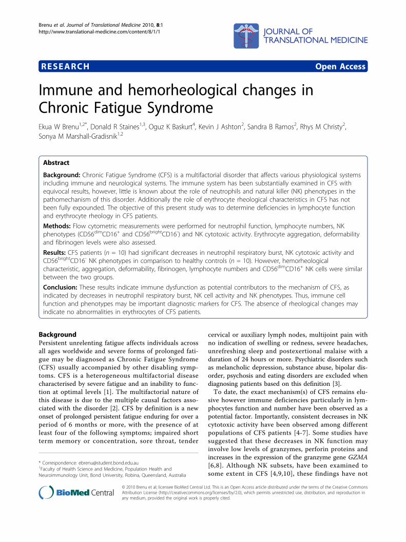
RESEARCH Open Access Immune and hemorheological changes in Chronic Fatigue Syndrome Ekua W Brenu 1,2* , Donald R Staines 1,3 , Oguz K Baskurt 4 , Kevin J Ashton 2 , Sandra B Ramos 2 , Rhys M Christy 2 , Sonya M Marshall-Gradisnik 1,2 Abstract Background: Chronic Fatigue Syndrome (CFS) is a multifactorial disorder that affects various physiological systems including immune and neurological systems. The immune system has been substantially examined in CFS with equivocal results, however, little is known about the role of neutrophils and natural killer (NK) phenotypes in the pathomechanism of this disorder. Additionally the role of erythrocyte rheological characteristics in CFS has not been fully expounded. The objective of this present study was to determine deficiencies in lymphocyte function and erythrocyte rheology in CFS patients. Methods: Flow cytometric measurements were performed for neutrophil function, lymphocyte numbers, NK phenotypes (CD56 dim CD16 + and CD56 bright CD16 - ) and NK cytotoxic activity. Erythrocyte aggregation, deformability and fibrinogen levels were also assessed. Results: CFS patients (n = 10) had significant decreases in neutrophil respiratory burst, NK cytotoxic activity and CD56 bright CD16 - NK phenotypes in comparison to healthy controls (n = 10). However, hemorheological characteristic, aggregation, deformability, fibrinogen, lymphocyte numbers and CD56 dim CD16 + NK cells were similar between the two groups. Conclusion: These results indicate immune dysfunction as potential contributors to the mechanism of CFS, as indicated by decreases in neutrophil respiratory burst, NK cell activity and NK phenotypes. Thus, immune cell function and phenotypes may be important diagnostic markers for CFS. The absence of rheological changes may indicate no abnormalities in erythrocytes of CFS patients. Background Persistent unrelenting fatigue affects individuals across all ages worldwide and severe forms of prolonged fati- gue may be diagnosed as Chronic Fatigue Syndrome (CFS) usually accompanied by other disabling symp- toms. CFS is a heterogeneous multifactorial disease characterised by severe fatigue and an inability to func- tion at optimal levels [1]. The multifactorial nature of this disease is due to the multiple causal factors asso- ciated with the disorder [2]. CFS by definition is a new onset of prolonged persistent fatigue enduring for over a period of 6 months or more, with the presence of at least four of the following symptoms; impaired short term memory or concentration, sore throat, tender cervical or auxiliary lymph nodes, multijoint pain with no indication of swelling or redness, severe headaches, unrefreshing sleep and postexertional malaise with a duration of 24 hours or more. Psychiatric disorders such as melancholic depression, substance abuse, bipolar dis- order, psychosis and eating disorders are excluded when diagnosing patients based on this definition [3]. To date, the exact mechanism(s) of CFS remains elu- sive however immune deficiencies particularly in lym- phocytes function and number have been observed as a potential factor. Importantly, consistent decreases in NK cytotoxic activity have been observed among different populations of CFS patients [4-7]. Some studies have suggested that these decreases in NK function may involve low levels of granzymes, perforin proteins and increases in the expression of the granzyme gene GZMA [6,8]. Although NK subsets, have been examined to some extent in CFS [4,9,10], these findings have not * Correspondence: [email protected] 1 Faculty of Health Science and Medicine, Population Health and Neuroimmunology Unit, Bond University, Robina, Queensland, Australia Brenu et al. Journal of Translational Medicine 2010, 8:1 http://www.translational-medicine.com/content/8/1/1 © 2010 Brenu et al; licensee BioMed Central Ltd. This is an Open Access article distributed under the terms of the Creative Commons Attribution License (http://creativecommons.org/licenses/by/2.0), which permits unrestricted use, distribution, and reproduction in any medium, provided the original work is properly cited.

Welcome message from author
This document is posted to help you gain knowledge. Please leave a comment to let me know what you think about it! Share it to your friends and learn new things together.
Transcript
RESEARCH Open Access
Immune and hemorheological changes inChronic Fatigue SyndromeEkua W Brenu1,2*, Donald R Staines1,3, Oguz K Baskurt4, Kevin J Ashton2, Sandra B Ramos2, Rhys M Christy2,Sonya M Marshall-Gradisnik1,2
Abstract
Background: Chronic Fatigue Syndrome (CFS) is a multifactorial disorder that affects various physiological systemsincluding immune and neurological systems. The immune system has been substantially examined in CFS withequivocal results, however, little is known about the role of neutrophils and natural killer (NK) phenotypes in thepathomechanism of this disorder. Additionally the role of erythrocyte rheological characteristics in CFS has notbeen fully expounded. The objective of this present study was to determine deficiencies in lymphocyte functionand erythrocyte rheology in CFS patients.
Methods: Flow cytometric measurements were performed for neutrophil function, lymphocyte numbers, NKphenotypes (CD56dimCD16+ and CD56brightCD16-) and NK cytotoxic activity. Erythrocyte aggregation, deformabilityand fibrinogen levels were also assessed.
Results: CFS patients (n = 10) had significant decreases in neutrophil respiratory burst, NK cytotoxic activity andCD56brightCD16- NK phenotypes in comparison to healthy controls (n = 10). However, hemorheologicalcharacteristic, aggregation, deformability, fibrinogen, lymphocyte numbers and CD56dimCD16+ NK cells were similarbetween the two groups.
Conclusion: These results indicate immune dysfunction as potential contributors to the mechanism of CFS, asindicated by decreases in neutrophil respiratory burst, NK cell activity and NK phenotypes. Thus, immune cellfunction and phenotypes may be important diagnostic markers for CFS. The absence of rheological changes mayindicate no abnormalities in erythrocytes of CFS patients.
BackgroundPersistent unrelenting fatigue affects individuals acrossall ages worldwide and severe forms of prolonged fati-gue may be diagnosed as Chronic Fatigue Syndrome(CFS) usually accompanied by other disabling symp-toms. CFS is a heterogeneous multifactorial diseasecharacterised by severe fatigue and an inability to func-tion at optimal levels [1]. The multifactorial nature ofthis disease is due to the multiple causal factors asso-ciated with the disorder [2]. CFS by definition is a newonset of prolonged persistent fatigue enduring for over aperiod of 6 months or more, with the presence of atleast four of the following symptoms; impaired shortterm memory or concentration, sore throat, tender
cervical or auxiliary lymph nodes, multijoint pain withno indication of swelling or redness, severe headaches,unrefreshing sleep and postexertional malaise with aduration of 24 hours or more. Psychiatric disorders suchas melancholic depression, substance abuse, bipolar dis-order, psychosis and eating disorders are excluded whendiagnosing patients based on this definition [3].To date, the exact mechanism(s) of CFS remains elu-
sive however immune deficiencies particularly in lym-phocytes function and number have been observed as apotential factor. Importantly, consistent decreases in NKcytotoxic activity have been observed among differentpopulations of CFS patients [4-7]. Some studies havesuggested that these decreases in NK function mayinvolve low levels of granzymes, perforin proteins andincreases in the expression of the granzyme gene GZMA[6,8]. Although NK subsets, have been examined tosome extent in CFS [4,9,10], these findings have not
* Correspondence: [email protected] of Health Science and Medicine, Population Health andNeuroimmunology Unit, Bond University, Robina, Queensland, Australia
Brenu et al. Journal of Translational Medicine 2010, 8:1http://www.translational-medicine.com/content/8/1/1
© 2010 Brenu et al; licensee BioMed Central Ltd. This is an Open Access article distributed under the terms of the Creative CommonsAttribution License (http://creativecommons.org/licenses/by/2.0), which permits unrestricted use, distribution, and reproduction inany medium, provided the original work is properly cited.

necessarily elucidated the role of CD56brightCD16negative(neg) NK and CD56dimCD16postive(pos) NK phenotypes inCFS. NK cells and their subsets are important inimmune regulation and pathogen lysis. CD56brightCD16-neg NK cells preferentially secrete high levels of cyto-kines and have limited cytotoxic function whileCD56dimCD16pos NK cells are mainly cytotoxic [11].Moreover, phagocytes such as neutrophils have receivedlittle attention, only one study has revealed that neutro-phils in CFS are more prone to apoptosis, this washeightened by the existence of large quantities ofTGFb1[12].The multifactorial and heterogeneous nature of CFS
suggests changes in other blood indicators, such as ery-throcytes. Some CFS patients demonstrate alterations inblood flow, erythrocyte rheology and erythrocyte mor-phology [13-17]. Abnormally shaped erythrocyte maypresent itself in the form of nondiscocytic, stomatocyticor cup formed erythrocyte [18]. Additionally, reductionsin erythrocyte width and mass, and changes in plateletaggregation have also been detected in some CFS patients[13,16]. Plasma proteins such as fibrinogen which influ-ence erythrocyte rheology are elevated in some CFScases, and this may be related to impaired coagulation[19] however, an association between erythrocyte aggre-gation and fibrinogen levels in CFS is not presentlyknown. Alterations in erythrocyte rheology may persistin CFS, these observations although indicative of indirectchanges in deformation and aggregation suggests theneed for further investigations to confirm the possiblelink between immune function and rheology in CFS.Hence, the objective of this study was to examine
immune function and rheological properties of periph-eral blood cells. This study investigated NK abnormal-ities in CFS to confirm those of other studies. NKphenotypes, NK cytotoxic activity, neutrophil function,lymphocyte numbers, fibrinogen levels and erythrocyterheology were measured in CFS patients. The CFS datawere compared to aged and sex matched group ofhealth volunteers.
Materials and methodsParticipantsThe present study was approved by Bond UniversityEthics Committee (RO852). Collection of venous bloodwas performed following consent from participants.Informed consent was prepared in accordance with theBond University Research Consultancy Service and pro-tocol. The CFS cohort comprised of 10 CFS patientsfrom a community based sample in New South Walesand Queensland, Australia and 10 healthy aged and sexmatched participants from a community local area. CFSpatients were chosen after completion of a questionnaireadapted from the CDC 1994 CFS case definition [3],
where the duration of CFS in our patient cohort wasmore than 5 years. Peripheral blood samples were ana-lysed for total lymphocytes, NK activity, NK phenotypes,neutrophil function, erythrocyte deformability, erythro-cyte aggregation and fibrinogen concentration.Lymphocytes assayPeripheral blood lymphocyte subsets were assessed usingfluorochrome-conjugated monoclonal antibodies fromthe Simultest IMK-Lymphocyte kit (BD Biosciences, SanJose, CA), specific for lymphocytes as previouslydescribed [20]. A fluorescence-activated cell sorting(FACS Calibur) flow cytometer (Becton DickinsonImmunocytometry Systems, San Jose, CA) was used todetermine lymphocyte subsets, CD3+/CD19 (B cells),CD3+ (T cell), CD3+/Cd4+ (T-helper cells), CD3+/CD8+ (T-cytotoxic, T suppressor), CD3-/CD16+/CD56+(Natural Killer cells).Assessment of NK lymphocyte activityNK cytotoxicity was performed as previously described[21]. Briefly, NK cells were isolated from whole bloodvia density gradient centrifugation using ficoll-Hypaque(GE Healthcare). NK cells were labelled with 0.4% PKH-26 (Sigma, St Louis, MO). NK cells were resuspended ata final concentration of 5 × 10⁶cells/mL. The K562 cellline was used as the target cells at a concentration of 1× 10⁵cells/mL. K562 cells were cultured with NK cellsin RPMI-1640 culture media (Invitrogen, Carlsbad, CA)for 4 hours in 37°C incubator with 5% CO2, at an effec-tor (NK) to target (K562) ratio of 25:1 with a controlsample containing only K562 cells. Apoptosis was mea-sured via flow cytometry, using Annexin V-FITC conju-gated mAB and 7-AAD reagent (BD Pharmingen, SanDiego, CA) according to the manufacturer’s instructions.Percent lysis of K562 cells were calculated as previouslydescribed [21].Quantification of NK phenotypesTo assess the levels of NK phenotypes in CFS patientsand healthy controls, NK lymphocytes were isolatedfrom whole blood according to manufacturer’s instruc-tions using RosetteSep Human Natural Killer cellEnrichment Cocktail (StemCell Technologies, Vancou-ver, BC), containing micro-beads that negatively selectfor only NK cells and ficoll-hypaque density centrifuga-tion. Samples were washed twice with PBS and labelledwith mAB CD56-FITC (BD Bioscience, San Jose, CA)and CD16-PE (BD Bioscience, San Jose, CA) accordingto manufacturer’s specifications and analysed on flowcytometer.Neutrophil function testImmune response to pathogens was measured in granu-locytes from lithium heparinised blood where phagocyteactivity and respiratory burst was examined using thePhagotest and Phagoburst kit (Orpegen Pharma GmbH,Heidelberg, Germany) respectively as specified by the
Brenu et al. Journal of Translational Medicine 2010, 8:1http://www.translational-medicine.com/content/8/1/1
Page 2 of 10
manufacturer. In summary, to determine phagocytosis,blood samples were mixed with FITC-labelled opsonisedE.coli and incubated for 10 minutes in 37°C water bathor on ice at 0°C. Quenching solution was added toremove the FITC from the E.coli. Intracellular oxidationwas performed by incubating heparinised whole bloodin phorbol 12-myristate 13-acetate (PMA) for 10 min-utes at 37°C. Dihydrohodamine (DHR) was then addedto the samples followed by an incubation period of 10minutes at 37°C. DHR was used as it is an indicator ofneutrophil respiratory burst [22]. Samples were analysedon the flow cytometer.Measurement of erythrocyte aggregation and fibrinogenconcentrationErythrocyte aggregation was performed using the Myr-enne aggregometer (Myrenne GmbH, Roetgen, Germany)in autologous plasma and 3% dextran solution (70 kDa;Sigma, St. Louis, MO) as previously described [23,24].This method generates two distinct measures of erythro-cyte aggregation at stasis (M0) and at a low shear (M1)after a shear rate of 600 s-1. Erythrocyte aggregationindices were determined at hematocrit of 40% at roomtemperature. Fibrinogen analysis was determined usingblood mixed with sodium citrate solution. Samples werecentrifuged at 1200 rcf for 10 minutes, platelet-poorplasma was collected and stored at -80°C for later analy-sis. Plasma fibrinogen was assessed by the CLAUSSmethod [25] using a STA-Compact analyser (Diagnostica
Stago, Asnieres, France) where the intra-assay coefficientof variation was 2.64% and the inter-assay coefficient ofvariation was 2.82%.Erythrocyte deformability measurementDeformability of erythrocyte was performed as previouslydescribed [26]. Blood samples were mixed with 0.99%RheoScan-D reagent (Incyto, Korea) and analysed on theRheoScan-D ektacytometer (Sewon Meditech, Korea).The elongation index was measured between shear stres-ses of 0.5 to 20 Pa. Shear stress for half-maximal defor-mation (SS1/2) and the maximum elongation index(EImax) was deduced using Lineweaver-Burk analysis.Measurements were carried out within 6 hours of bloodcollection and performed at room temperature (25°C).Statistical analysisStatistical significance between the two subject groupswas determined for all data using the independent sam-ple t test. The data are represented as mean ± standarderror of the mean (SEM).
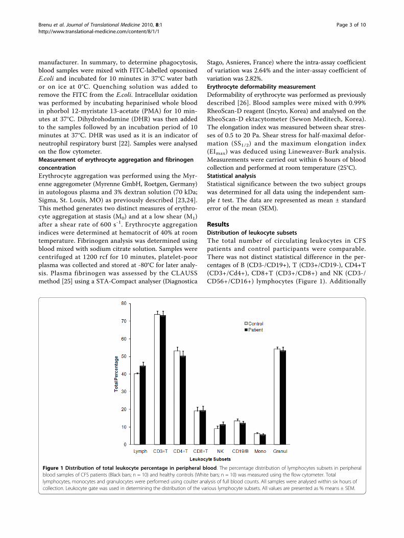
ResultsDistribution of leukocyte subsetsThe total number of circulating leukocytes in CFSpatients and control participants were comparable.There was not distinct statistical difference in the per-centages of B (CD3-/CD19+), T (CD3+/CD19-), CD4+T(CD3+/Cd4+), CD8+T (CD3+/CD8+) and NK (CD3-/CD56+/CD16+) lymphocytes (Figure 1). Additionally
Figure 1 Distribution of total leukocyte percentage in peripheral blood. The percentage distribution of lymphocytes subsets in peripheralblood samples of CFS patients (Black bars; n = 10) and healthy controls (White bars; n = 10) was measured using the flow cytometer. Totallymphocytes, monocytes and granulocytes were performed using coulter analysis of full blood counts. All samples were analysed within six hours ofcollection. Leukocyte gate was used in determining the distribution of the various lymphocyte subsets. All values are presented as % means ± SEM.
Brenu et al. Journal of Translational Medicine 2010, 8:1http://www.translational-medicine.com/content/8/1/1
Page 3 of 10
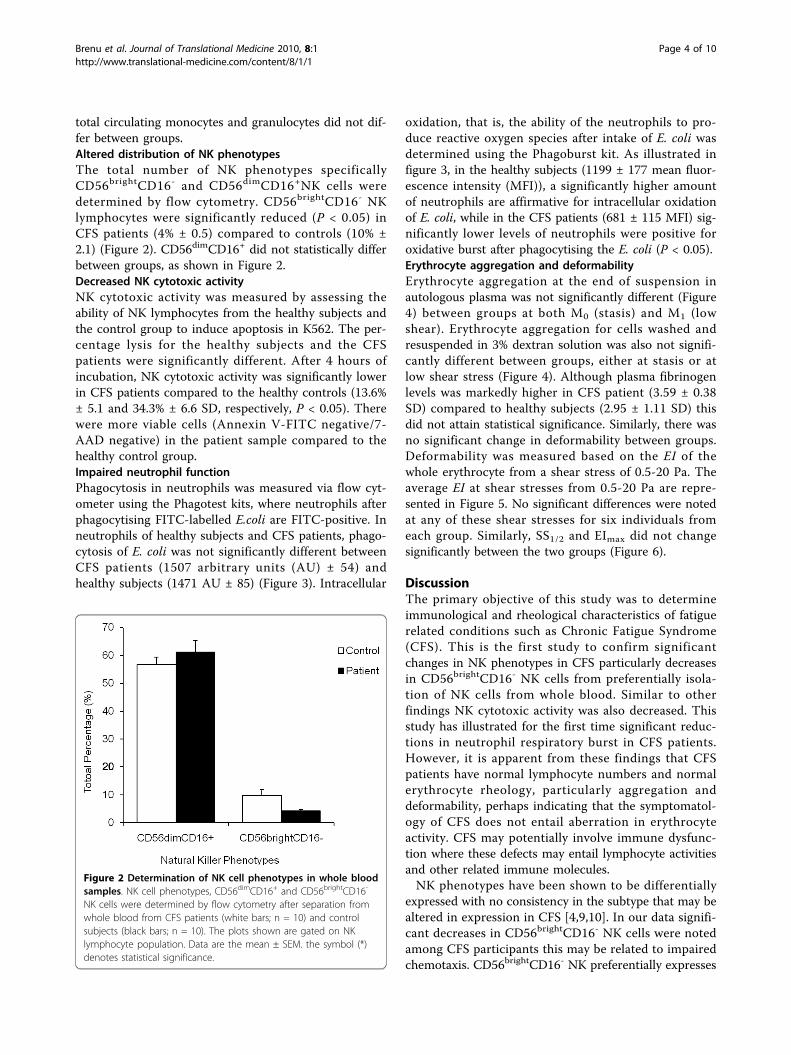
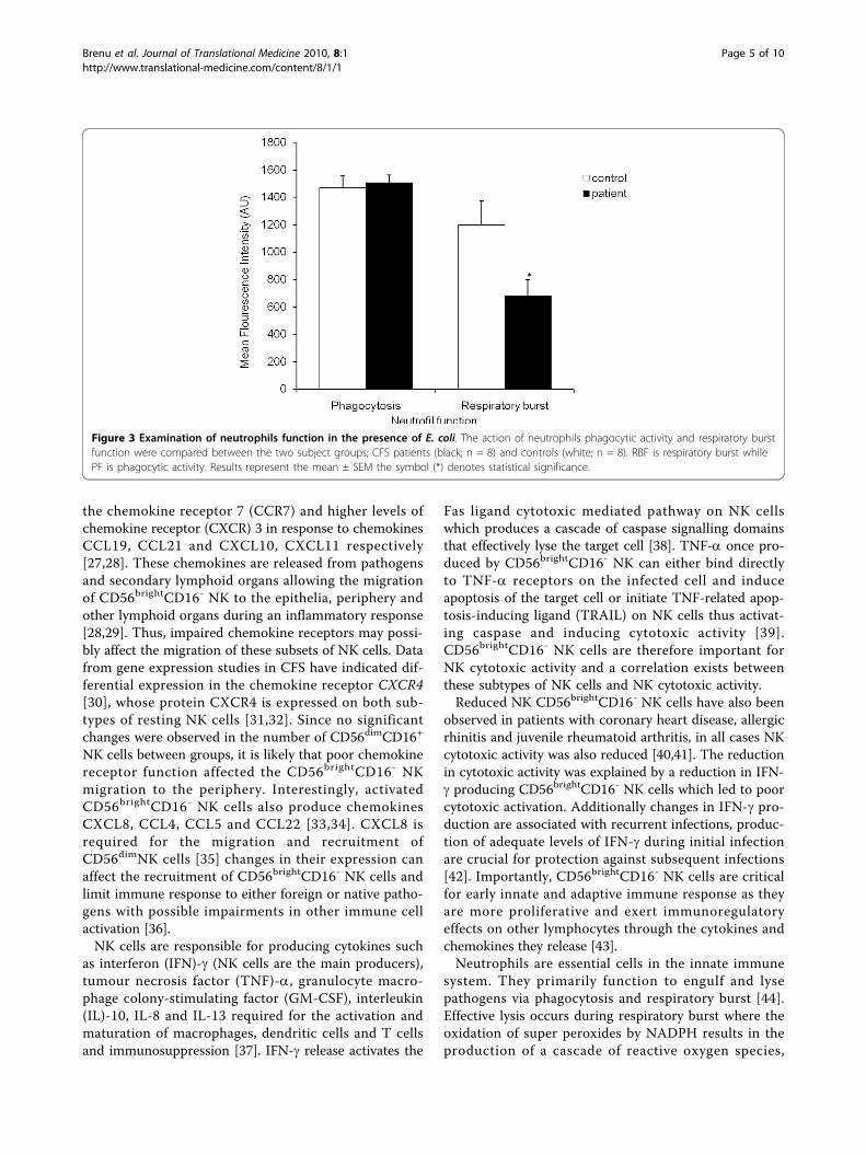
total circulating monocytes and granulocytes did not dif-fer between groups.Altered distribution of NK phenotypesThe total number of NK phenotypes specificallyCD56brightCD16- and CD56dimCD16+NK cells weredetermined by flow cytometry. CD56brightCD16- NKlymphocytes were significantly reduced (P < 0.05) inCFS patients (4% ± 0.5) compared to controls (10% ±2.1) (Figure 2). CD56dimCD16+ did not statistically differbetween groups, as shown in Figure 2.Decreased NK cytotoxic activityNK cytotoxic activity was measured by assessing theability of NK lymphocytes from the healthy subjects andthe control group to induce apoptosis in K562. The per-centage lysis for the healthy subjects and the CFSpatients were significantly different. After 4 hours ofincubation, NK cytotoxic activity was significantly lowerin CFS patients compared to the healthy controls (13.6%± 5.1 and 34.3% ± 6.6 SD, respectively, P < 0.05). Therewere more viable cells (Annexin V-FITC negative/7-AAD negative) in the patient sample compared to thehealthy control group.Impaired neutrophil functionPhagocytosis in neutrophils was measured via flow cyt-ometer using the Phagotest kits, where neutrophils afterphagocytising FITC-labelled E.coli are FITC-positive. Inneutrophils of healthy subjects and CFS patients, phago-cytosis of E. coli was not significantly different betweenCFS patients (1507 arbitrary units (AU) ± 54) andhealthy subjects (1471 AU ± 85) (Figure 3). Intracellular
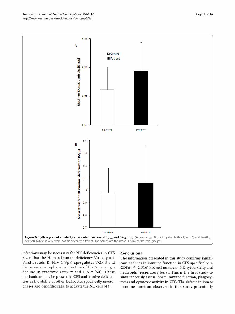
oxidation, that is, the ability of the neutrophils to pro-duce reactive oxygen species after intake of E. coli wasdetermined using the Phagoburst kit. As illustrated infigure 3, in the healthy subjects (1199 ± 177 mean fluor-escence intensity (MFI)), a significantly higher amountof neutrophils are affirmative for intracellular oxidationof E. coli, while in the CFS patients (681 ± 115 MFI) sig-nificantly lower levels of neutrophils were positive foroxidative burst after phagocytising the E. coli (P < 0.05).Erythrocyte aggregation and deformabilityErythrocyte aggregation at the end of suspension inautologous plasma was not significantly different (Figure4) between groups at both M0 (stasis) and M1 (lowshear). Erythrocyte aggregation for cells washed andresuspended in 3% dextran solution was also not signifi-cantly different between groups, either at stasis or atlow shear stress (Figure 4). Although plasma fibrinogenlevels was markedly higher in CFS patient (3.59 ± 0.38SD) compared to healthy subjects (2.95 ± 1.11 SD) thisdid not attain statistical significance. Similarly, there wasno significant change in deformability between groups.Deformability was measured based on the EI of thewhole erythrocyte from a shear stress of 0.5-20 Pa. Theaverage EI at shear stresses from 0.5-20 Pa are repre-sented in Figure 5. No significant differences were notedat any of these shear stresses for six individuals fromeach group. Similarly, SS1/2 and EImax did not changesignificantly between the two groups (Figure 6).
DiscussionThe primary objective of this study was to determineimmunological and rheological characteristics of fatiguerelated conditions such as Chronic Fatigue Syndrome(CFS). This is the first study to confirm significantchanges in NK phenotypes in CFS particularly decreasesin CD56brightCD16- NK cells from preferentially isola-tion of NK cells from whole blood. Similar to otherfindings NK cytotoxic activity was also decreased. Thisstudy has illustrated for the first time significant reduc-tions in neutrophil respiratory burst in CFS patients.However, it is apparent from these findings that CFSpatients have normal lymphocyte numbers and normalerythrocyte rheology, particularly aggregation anddeformability, perhaps indicating that the symptomatol-ogy of CFS does not entail aberration in erythrocyteactivity. CFS may potentially involve immune dysfunc-tion where these defects may entail lymphocyte activitiesand other related immune molecules.NK phenotypes have been shown to be differentially
expressed with no consistency in the subtype that may bealtered in expression in CFS [4,9,10]. In our data signifi-cant decreases in CD56brightCD16- NK cells were notedamong CFS participants this may be related to impairedchemotaxis. CD56brightCD16- NK preferentially expresses
Figure 2 Determination of NK cell phenotypes in whole bloodsamples. NK cell phenotypes, CD56dimCD16+ and CD56brightCD16-
NK cells were determined by flow cytometry after separation fromwhole blood from CFS patients (white bars; n = 10) and controlsubjects (black bars; n = 10). The plots shown are gated on NKlymphocyte population. Data are the mean ± SEM. the symbol (*)denotes statistical significance.
Brenu et al. Journal of Translational Medicine 2010, 8:1http://www.translational-medicine.com/content/8/1/1
Page 4 of 10
the chemokine receptor 7 (CCR7) and higher levels ofchemokine receptor (CXCR) 3 in response to chemokinesCCL19, CCL21 and CXCL10, CXCL11 respectively[27,28]. These chemokines are released from pathogensand secondary lymphoid organs allowing the migrationof CD56brightCD16- NK to the epithelia, periphery andother lymphoid organs during an inflammatory response[28,29]. Thus, impaired chemokine receptors may possi-bly affect the migration of these subsets of NK cells. Datafrom gene expression studies in CFS have indicated dif-ferential expression in the chemokine receptor CXCR4[30], whose protein CXCR4 is expressed on both sub-types of resting NK cells [31,32]. Since no significantchanges were observed in the number of CD56dimCD16+
NK cells between groups, it is likely that poor chemokinereceptor function affected the CD56brightCD16- NKmigration to the periphery. Interestingly, activatedCD56brightCD16- NK cells also produce chemokinesCXCL8, CCL4, CCL5 and CCL22 [33,34]. CXCL8 isrequired for the migration and recruitment ofCD56dimNK cells [35] changes in their expression canaffect the recruitment of CD56brightCD16- NK cells andlimit immune response to either foreign or native patho-gens with possible impairments in other immune cellactivation [36].NK cells are responsible for producing cytokines such
as interferon (IFN)-g (NK cells are the main producers),tumour necrosis factor (TNF)-a, granulocyte macro-phage colony-stimulating factor (GM-CSF), interleukin(IL)-10, IL-8 and IL-13 required for the activation andmaturation of macrophages, dendritic cells and T cellsand immunosuppression [37]. IFN-g release activates the
Fas ligand cytotoxic mediated pathway on NK cellswhich produces a cascade of caspase signalling domainsthat effectively lyse the target cell [38]. TNF-a once pro-duced by CD56brightCD16- NK can either bind directlyto TNF-a receptors on the infected cell and induceapoptosis of the target cell or initiate TNF-related apop-tosis-inducing ligand (TRAIL) on NK cells thus activat-ing caspase and inducing cytotoxic activity [39].CD56brightCD16- NK cells are therefore important forNK cytotoxic activity and a correlation exists betweenthese subtypes of NK cells and NK cytotoxic activity.Reduced NK CD56brightCD16- NK cells have also been
observed in patients with coronary heart disease, allergicrhinitis and juvenile rheumatoid arthritis, in all cases NKcytotoxic activity was also reduced [40,41]. The reductionin cytotoxic activity was explained by a reduction in IFN-g producing CD56brightCD16- NK cells which led to poorcytotoxic activation. Additionally changes in IFN-g pro-duction are associated with recurrent infections, produc-tion of adequate levels of IFN-g during initial infectionare crucial for protection against subsequent infections[42]. Importantly, CD56brightCD16- NK cells are criticalfor early innate and adaptive immune response as theyare more proliferative and exert immunoregulatoryeffects on other lymphocytes through the cytokines andchemokines they release [43].Neutrophils are essential cells in the innate immune
system. They primarily function to engulf and lysepathogens via phagocytosis and respiratory burst [44].Effective lysis occurs during respiratory burst where theoxidation of super peroxides by NADPH results in theproduction of a cascade of reactive oxygen species,
Figure 3 Examination of neutrophils function in the presence of E. coli. The action of neutrophils phagocytic activity and respiratory burstfunction were compared between the two subject groups; CFS patients (black; n = 8) and controls (white; n = 8). RBF is respiratory burst whilePF is phagocytic activity. Results represent the mean ± SEM the symbol (*) denotes statistical significance.
Brenu et al. Journal of Translational Medicine 2010, 8:1http://www.translational-medicine.com/content/8/1/1
Page 5 of 10

which cumulatively eliminate the pathogen. Decreases inneutrophil function are indicative of impaired immunefunction in CFS. Only one study to date has demon-strated that neutrophils in CFS patients are highly apop-totic with an increase in TGF-b and TNFR1 [12].Delayed or limited apoptosis correlates with an increasein respiratory burst [45], thus a situation wheredecreases in respiratory burst persist may likely be anindicator of elevations in apoptotic neutrophils. Thispotentially increases the life of bacteria and other
pathogens in the body as they are not efficiently lysedowing to limited intracellular oxidative processes.Diminishing levels of CD56brightCD16- NK cells maylimit the production of TNFs, cytokines required foractivation of respiratory burst in neutrophils. TNF-aand GM-CSF, produced by CD56brightCD16- NK, areimportant for the induction of superperoxide thus prim-ing the neutrophils for respiratory burst [46].Decreases in NK cytotoxic activity have been consis-
tently reported in previous studies [4,6]. Decrease in NK
Figure 4 Assessment of erythrocyte aggregation in autologous plasma (A) and dextran solution (B). Peripheral blood samples from CFSpatients (black; n = 10) and healthy controls (white; n = 10) assessed on measures of aggregation at stasis (M0) and at low shear rate (M1). Sampleswere measured after adjustment of hematocrit to 40% (A) following which they were washed and suspended in 3% dextran solution with ahematocrit 40% hematocrit adjustmnent (B). Samples were analysed within 12 hours of blood collection. Results are represented as mean ± SEM.
Brenu et al. Journal of Translational Medicine 2010, 8:1http://www.translational-medicine.com/content/8/1/1
Page 6 of 10

activity may be correlated with decreases in perforin andgranzyme production [6] and changes in granzyme gene(GZMA) expression [8]. These deficiencies in NK activ-ity may increase viral load in CFS, incidentally a recentstudy observed increases in xenotropic murine leukemiavirus-related virus (XMRV) in peripheral blood samplesof CFS patients [47]. These viruses may potentially alteraspects of the immune response such as cytotoxic activ-ity thus promoting their survival in particular immunecells. NK cells and neutrophil deficiencies in CFS maybe related to the presence of autoantibodies. Some ofthese autoantibodies are specific for proteins that mayinteract with immune cells have been detected in serumsamples in CFS patients [48-50], however, these autoan-tibodies are yet to be detected against specific receptorsexpressed on immune cells or cellular lytic pathways.There was no change in erythrocyte deformability or
aggregation between groups, although other studies haveconfirmed changes in erythrocyte shape in CFS patients,particularly an increase in stomatocytes or lepotocytes[15,51]. Equally, the Lineweaver-Burk analysis did notindicate statistical significance between the two groups.The most likely consequence of these observations isthe heterogeneity of CFS. Nonetheless, observable rheo-logical changes are perhaps associated with the acutephase of CFS while these maybe absent during thechronic stages of the disorder [52]. Notably all CFS par-ticipants in this study were in the chronic phase. Thus,erythrocyte deformability and aggregation may not bedistinct markers for CFS.
Given the paucity in CD56brightCD16- NK cells amongCFS patients in this study and their role in immunore-gulation and activation, reduced CD56brightCD16- NKcell numbers may be important in the pathomechanismof CFS, a disorder shown to be characterised bydecreases in NK cytotoxic activity. Although changes inNK cell makers have been previously reported, amechanism underlying diminishing NK cell markers andphenotypes has not yet been established. This mechan-ism may also involve changes at the genomic levelwhich results in deficient cytokine and chemokinereceptor expression. For example, alterations in RNAexpression levels for CD56brightCD16- NK receptors hasbeen demonstrated in patients with Autism SpectrumDisorder where cytotoxic activity and NK cell numberswere also decreased when NK cells were stimulated by apathogen [53]. Exposure to pathogens in the presence ofdifferential expression of certain NK cytokine and che-mokine receptor genes may trigger a decline inCD56brightCD16- NK cells and NK cytotoxicity in CFS.However, the heterogeneity and multifactorial nature of
CFS suggests variations in molecular changes and cellularmechanisms among patients. Certain cytokines increasecytotoxic ability (IL-2) and IFN-g production (IL-12 andIL-18) of CD56brightCD16- NK [36], therefore a possiblemechanism limiting the production of these cytokinesand may adversely alter the role of CD56brightCD16- NKduring pathogen invasion and lysis. High levels of TFG-balso cause an increase in neutrophil apoptosis and thisoccurs in some cases of CFS [11]. Finally viral-specific
Figure 5 Assessment of erythrocyte deformability in CFS. Peripheral blood samples from CFS patients (black; n = 6) and healthy controls(white; n = 6) were assessed. Deformability was assessed at shear stresses from 0.5-20 Pa. The mean ± SEM are represented on the graph
Brenu et al. Journal of Translational Medicine 2010, 8:1http://www.translational-medicine.com/content/8/1/1
Page 7 of 10
infections may be necessary for NK deficiencies in CFSgiven that the Human Immunodeficiency Virus type 1Viral Protein R (HIV-1 Vpr) upregulates TGF-b anddecreases macrophage production of IL-12 causing adecline in cytotoxic activity and IFN-g [54]. Thesemechanisms may be present in CFS and involve deficien-cies in the ability of other leukocytes specifically macro-phages and dendritic cells, to activate the NK cells [43].
ConclusionsThe information presented in this study confirms signifi-cant declines in immune function in CFS specifically inCD56brightCD16- NK cell numbers, NK cytotoxicity andneutrophil respiratory burst. This is the first study tosimultaneously assess innate immune function, phagocy-tosis and cytotoxic activity in CFS. The defects in innateimmune function observed in this study potentially
Figure 6 Erythrocyte deformability after determination of EImax and SS1/2. EImax (A) and SS1/2 (B) of CFS patients (black; n = 6) and healthycontrols (white; n = 6) were not significantly different. The values are the mean ± SEM of the two groups.
Brenu et al. Journal of Translational Medicine 2010, 8:1http://www.translational-medicine.com/content/8/1/1
Page 8 of 10
suggests an altered adaptive immune response in CFSand these may be important in understanding the patho-mechanism of CFS. Further studies are however requiredto determine cytokine and chemokine expression in CFSpatients. Neutrophil apoptosis in relation to respiratoryburst, cytotoxic activity in CD8 T cells, perforin andgranzyme production and CD4+T cell cytokine secretionin CFS patients are potential topics for future investiga-tions. These studies will allow a comprehensive analysisof the overall immune function in CFS patients.
Conflict of interest statementThe authors declare that they have no competinginterests.
Authors’ contributions to the paperEWB assessed and recruited patients and controls forstudy, performed NK cytotoxic activity, NK phenotypeanalysis and erythrocyte experimental assessments, allstatistical analysis and wrote the manuscript. SBR per-formed the IMK lymphocyte and full blood count test.RMC performed neutrophil function analysis. DRS pro-vided the patient cohort and reviewed the manuscript.KJA second principal investigator advised on methodol-ogy and reviewed the paper. OKB provided the metho-dology for erythrocyte aggregation and deformability.SMM-G primary principal investigator advised on meth-odology and reviewed the manuscript. Authors read andapproved the manuscript.
AcknowledgementsThis study was supported by Bond University Research fund.
Author details1Faculty of Health Science and Medicine, Population Health andNeuroimmunology Unit, Bond University, Robina, Queensland, Australia.2Faculty of Health Science and Medicine, Bond University, Robina,Queensland, Australia. 3Queensland Health, Gold Coast Population HealthUnit, Southport, Gold Coast, Queensland, Australia. 4Department ofPhysiology, Akdeniz University Faculty of Medicine, Antalya, Turkey.
Received: 26 June 2009Accepted: 11 January 2010 Published: 11 January 2010
References1. Afari N, Buchwald D: Chronic fatigue syndrome: a review. Am J Psychiatry
2003, 160:221-236.2. Salit IE: Precipitating factors for the chronic fatigue syndrome. J Psychiatr
Res 1997, 31:59-65.3. Fukuda K, Straus SE, Hickie I, Sharpe MC, Dobbins JG, Komaroff A: The
chronic fatigue syndrome: A comprehensive approach to its definitionand study. Ann Intern Med 1994, 121:953-959.
4. Klimas N, Salvato F, Morgain R, Fletcher MA: Immunologic abnormalities inchronic fatigue syndrome. J Clin Microbiol 1990, 28:1403-1410.
5. Ojo-Amaise EA, Conley EJ, Peters JB: Decreased natural killer cell activity isassociated with severity of chronic fatigue syndrome. Clin Infect Dis 1994,18(Suppl 1):157-159.
6. Maher KJ, Klimas NG, Fletcher MA: Chronic fatigue syndrome is associatedwith diminished intracellular perforin. Clin Exp Immunol 2005, 142:505-511.
7. Masuda A, Nozoe SI, Matsuyama T, Tanaka H: Psychobehavioral andimmunological characteristics of adult people with chronic fatigue andpatients with chronic fatigue syndrome. Psychosom Med 1994, 56:512-518.
8. Saiki T, Kawai T, Morita K, Ohta M, Saito T, Rokutan K, Ban N: Identificationof marker genes for differential diagnosis of chronic fatigue syndrome.Mol Med 2008, 14:599-607.
9. Tirelli U, Marotta G, Improta S, Pinto A: Immunological abnormalities inpatients with chronic fatigue syndrome. Scand J Immunol 1994, 40:601-608.
10. Robertson MJ, Schacterle RS, Mackin GA, Wilson SN, Bloomingdale , Ritz J,Komaroff AL: Lymphocyte subset differences in patients with chronicfatigue syndrome, multiple sclerosis and major depression. Clin ExpImmunol 2005, 141:326-332.
11. Vivier E, Tomasello E, Baratin M, Walzer T, Uqolini S: Functions of naturalkiller cells. Nat Immunol 2008, 9:503-10.
12. Bryceson YT, March ME, Ljunggren HG, Long EO: Activation, coactivationand costimulation of resting human natural killer cells. Immunol Rev2006, 214:73-91.
13. Kennedy G, Spence V, Underwood C, Belch JJF: Increased neutrophilapoptosis in chronic fatigue syndrome. J Clin Pathol 2004, 57:891-893.
14. Streeten DHP, Bell DS: Circulating blood volume in Chronic FatigueSyndrome. J Chronic Fatigue Syndrome 1998, 4:3-11.
15. Yoshiuchi K, Farkas J, Natelson BH: Patients with chronic fatigue syndromehave reduced absolute cortical blood flow. Clin Physiol Funct Imaging2006, 26:83-86.
16. Richards RS, Roberts TK, McGregor NR, Dunstan RH, Butt HL: Bloodparameters indicative of oxidative stress are associated with symptomexpression in chronic fatigue syndrome. Redox Rep 2000, 5:35-41.
17. Niblett SH, King KE, Dunstan RH, Clift-Bligh P, Hoskin AL, Roberts TK,Fulcher GR, McGregor NR, Dunsmore JC, Butt HL, Klineberg I, Rothkirch TB:Hematologic and urinary excretion anomalies in patients with chronicfatigue syndrome. Exp Biol Med 2007, 232:1041-1049.
18. Simpson LO, Shand BI, Olds RJ: Blood rheology and myalgicencephalomyelitis: a pilot study. Pathol 1986, 18:190-192.
19. Simpson LO, O’Neill DJ: Red blood cell shape, symptoms and reportedlyhelpful treatments in Americans with Chronic Disorder. J Orthomol Med2001, 16:157-165.
20. Kennedy G, Norris G, Spence V, McLaren M, Belch JJF: Is Chronic FatigueSyndrome Associated with Platelet activation?. Blood Coagul Fibrinolysis2006, 17:89-92.
21. Berg D, Berg LH, Couvaras J: Is CFS/FMS due to an undefinedhypercoagulable state brought on by immune activation of coagulation?Does adding anticoagulant therapy improve CFS/FMS Patientsymptoms?. AACFS Proceedings: Cambridge 1998, 62.
22. Jentsch-Ullrich K, Koenigsmann M, Mohren M, Franke A: Lymphocytessubsets’ reference ranges in an age-and gender-balanced population100 healthy adults-a monocentric German study. Clin Immunol 2005,116:192-7.
23. Aubry JP, Blaeck A, Lecoanet-Henchoz S, Jeannin P, Herbault N, Caron G,Moine V, Bonnefoy JY: Annexin V used in measuring apoptosis in theearly events of cellular cytotoxicity. Cytometry 1999, 37:197-204.
24. Rothe G, Oser A, Valet G: Dihydrohodamine 123: a new flow cytometricindicator for respiratory burst activity in neutrophil granulcoytes.Naturwissenschaften 1988, 75:354-5.
25. Vaya A, Falco C, Fernandez P, Contreras T, Valls M, Aznar J: Erythrocyteaggregation determined with the Myrenne aggregometer at two modes(M0, M1) and at two times (5 and 10 sec). Clin Hemorheol Microcirc 2003,29:119-127.
26. Baskurt OK, Temiz A, Meiselman HJ: Red blood cell aggregation inexperimental sepsis. J Lab Clin 1997, 130:183-109.
27. Clauss A: Gerinnung-physiologische schnell-method zur bestimung desfibrinogens. Acta Haematol 1957, 17:237-246.
28. Shin S, Ku Y, Park MS, Suh JS: Slit-flow ektacytometry: laser diffraction in arheometer. Cytometry Part B: Clin Cytom 2005, 65:6-13.
29. Kim CH, Pelus LM, Appelbaum E, Johanson K, Anzai N, Broxmeyer HE: CCR7ligands, SLC/6Ckine/Exodus2/TCA4 and CK-11/MIP-3/ELC, arechemoattractants for CD56+CD16- NK cells and late stage lymphoidprogenitors. Cell Immunol 1999, 193:226-235.
30. Wald O, Weiss ID, Wald H, Shoham H, Bar-Shavit Y, Beider K, Galun E,Weiss L, Flaishon L, Shachar I, Nagler A, Lu B, Gerard C, Gao J-L, Mishan E,Farber J, Peled A: IFN-gamma acts on T cells to induce NK cell
Brenu et al. Journal of Translational Medicine 2010, 8:1http://www.translational-medicine.com/content/8/1/1
Page 9 of 10
mobilization and accumulation in target organs. J Immunol 2006,176:4716-29.
31. Kerr JR, Petty R, Burke B, Gough J, Fear D, Sinclair LI, Mattey Dl, Richards SC,Montgomery J, Baldwin DA, Kellam P, Harrison TJ, Griffin GE, Main J,Enlander D, Nutt DJ, Holgate ST: Gene expression subtypes in patientswith chronic fatigue syndrome/myalgic encephalomyelitis. J Infect Dis2008, 197:1171-1184.
32. Inngjerdingen M, Damaj B, Maghaz AA: Expression and regulation ofchemokine receptors in human natural killer cells. Blood 2001, 97:367-75.
33. Taub DD, Sayers TJ, Carter CR, Ortaldo JR: Alpha and beta chemokinesinduce NK cell migration and enhance NK-mediated cytolysis. J Immunol1995, 155:3877-88.
34. Nieto M, Navarro F, Perez-Villar JJ, de Pozo MA, Gonzalez-Amaro R,Mellado M, Frade JMR, Martinez-A C, Lopez-Botet M, Sanchez-Madrid F:Roles of chemokines and receptor polarization in NK-target cellinteractions. J Immunol 1998, 161:3330-9.
35. Hedrick JA, Zlotnik A: Identification and characterization of a novel betachemokine containing six conserved cysteines. J Immunol 1997,159:1589-1593.
36. Robertson MJ: Role of chemokines in the biology of natural killer cells. JLeukoc Biol 2002, 71:173-183.
37. Fehniger TA, Cooper MA, Nuovo GJ, Facchetti F, Colonna M, Caligiuri MA:CD56bright natural killer cells are present in human lymph nodes andare activated by T cell-derived IL-2: a potential new link betweenadaptive and innate immunity. Blood 2003, 101:3052-3057.
38. Cooper MA, Fehniger TA, Turner SC, Chen KS, Ghaheri BA, Ghayur T,Carson We, Caligiur MA: Human natural killer cells; a unique innateimmunoregulatory role for the CD56bright subset. Blood 2001, 97:3146-3151.
39. Cretney E, Takeda K, Yagita H, Glaccum M, Peschon JJ, Smyth MJ: Increasedsusceptibility to tumor initiation and metastasis in TNF-relatedapoptosis-inducing ligand-deficient mice. J Immunol 2002, 168:1356-1361.
40. Takeda K, Smyth MJ, Cretney E, Hayakawa Y, Yamaguchi N, Yagita H,Okumura K: Involvement of tumor necrosis factor-related apoptosis-inducing ligand in NK cell-mediated and IFN-gamma-dependentsuppression of subcutaneous tumor growth. Cell Immunol 2001, 214:194-200.
41. Scordamaglia F, Balsamo M, Scordamaglia A, Moretta A, Cristina MM,Canonica GW, Moretta L: Perturbations of natural killer cell regulatoryfunctions in respiratory allergic diseases. J Allergy Clin Immunol 2008,121:479-485.
42. Villanueva J, Lee S, Giannini EH, Graham TB, Passo MH, Filipovich A,Grom AA: Natural killer cell dysfunction is a distinguishing feature ofsystemic onset juvenile rheumatoid arthritis and macrophage activationsyndrome. Arthritis Res Ther 2005, 7:R30-37.
43. Lee Y-M, Miyahara N, Takeda K, Prprich J, Oh A, Balhorn A, Joetham A,Gelfand EW, Darkhama A: IFN-g production during initial infectiondetermines the outcome of reinfection with respiratory syncytial virus.Am J Respir Crit Care Med 2008, 177:208-218.
44. Dalbeth N, Grundle R, Davies RJO, Lee YCG, McMichael AJ, Callan MFC:CD56bright NK cells are enriched at inflammatory sites and can engagewith monocytes in reciprocal programme of activation. J Immunol 2004,173:6418-6426.
45. Robinson JM, Ohira T, Badwey JA: Regulation of NADPH-oxidase complexof phagocytic leukocytes. Recent insights from structural biology,molecular genetics and microscopy. Histochem Cell Biol 2004, 122:293-304.
46. Sakka NE, Galley HF, Sharaki O, Helmy M, Marzouk S, Azmy S, Sedrak M,Webster NR: Delayed neutrophil apoptosis in patients with multipleorgan dysfunction syndrome. Crit Care Shock 2006, 9:9-15.
47. Lombardi VC, Ruscetti FW, Gupta JD, Pfost MA, Hagen KS, Peterson DL,Ruscetti SK, Bagni RK, Petrow-Sadowski C, Gold B, Dean M, Silverman RH,Mikovits JA: Detection of an Infectious Retrovirus, XMRV, in Blood Cellsof Patients with Chronic Fatigue Syndrome. Science 2009, 326:585-589.
48. Nancy AL, Shoenfeld Y: Chronic fatigue syndrome with autoantibodies–the result of an augmented adjuvant effect of hepatitis-B vaccine andsilicone implant. Autoimmun Rev 2008, 8:52-55.
49. Buskila D, Atzeni F, Sarzi-Puttini P: Etiology of fibromyalgia: the possiblerole of infection and vaccination. Autoimmun Rev 2008, 8:41-43.
50. Ortega-Hernandez OD, Cuccia MC, Bozzini S, Bassi N, Moscavitdh S, Diaz-Gallo LM, Blank M, Agmon-Levin N, Shoenfeld Y: Autoantibodies,polymorphisms in the serotonin pathway, and human leukocyte antigen
glass II alleles in chronic fatige syndrome. Ann NY Acad Sci 2009,1173:589-599.
51. Suzuki K, Hino M, Hato F, Tatsumi N, Kitagawa S: Cytokine-specificactivation of distinct mitogen-activated protein kinase subtype cascadesin human neutrophils stimulated by granulocyte colony-stimulatingfactor, granulocyte-macrophage colony stimulating factor, and tumornecrosis factor-a. Blood 1999, 93:341-349.
52. Richards R, Wang L, Jelink H: Erythrocyte oxidative damage in chronicfatigue syndrome. Arch Med Res 2007, 38:94-98.
53. Enstrom AM, Onore CE, Gregg JP, Hansen RL, Pessah IN, Hertz-Picciotto I,Water van de JA, Sharp FR, Ashwood P: Altered gene expression andfunction of peripheral blood natural killer cells in children with autism.Brain Behav Immun 2009, 23:124-43.
54. Majumder B, Venkatachari NJ, O’Leary S, Ayyavoo V: Infection with Vpr-positive human immunodeficiency virus type 1 impairs NK cell functionindirectly through cytokine dysregulation of infected target cells. J Virol2008, 82:7189-7200.
doi:10.1186/1479-5876-8-1Cite this article as: Brenu et al.: Immune and hemorheological changesin Chronic Fatigue Syndrome. Journal of Translational Medicine 2010 8:1.
Submit your next manuscript to BioMed Centraland take full advantage of:
• Convenient online submission
• Thorough peer review
• No space constraints or color figure charges
• Immediate publication on acceptance
• Inclusion in PubMed, CAS, Scopus and Google Scholar
• Research which is freely available for redistribution
Submit your manuscript at www.biomedcentral.com/submit
Brenu et al. Journal of Translational Medicine 2010, 8:1http://www.translational-medicine.com/content/8/1/1
Page 10 of 10
Related Documents