Accepted Article This article has been accepted for publication and undergone full peer review but has not been through the copyediting, typesetting, pagination and proofreading process which may lead to differences between this version and the Version of Record. Please cite this article as an 'Accepted Article', doi: 10.1111/imm.12438 This article is protected by copyright. All rights reserved. Received Date : 08-Apr-2014 Revised Date : 15-Dec-2014 Accepted Date : 23-Dec-2014 Article type : Original Article STAT-1 plays a critical role in control of Trypanosoma cruzi infection Manjusha M. Kulkarni 2,3# , Sanjay Varikuti 4# , Cesar Terrazas 4 , Jennifer L. Kimble 4 , Abhay Satoskar 2,3,4* and Bradford S. McGwire 1,2,3 * 1 Division of Infectious Diseases, 2 Center for Microbial Interface Biology, 3 Department of Microbial Infection and Immunity, and Department of Pathology 4 The Ohio State University Medical Center, Columbus, Ohio, USA Running title: STAT-1 controls T. cruzi infection Key words: STAT-1, Trypanosoma cruzi, infection, immune response

Welcome message from author
This document is posted to help you gain knowledge. Please leave a comment to let me know what you think about it! Share it to your friends and learn new things together.
Transcript

Acc
epte
d A
rtic
le
This article has been accepted for publication and undergone full peer review but has not been through the copyediting, typesetting, pagination and proofreading process which may lead to differences between this version and the Version of Record. Please cite this article as an 'Accepted Article', doi: 10.1111/imm.12438
This article is protected by copyright. All rights reserved.
Received Date : 08-Apr-2014
Revised Date : 15-Dec-2014
Accepted Date : 23-Dec-2014
Article type : Original Article
STAT-1 plays a critical role in control of
Trypanosoma cruzi infection
Manjusha M. Kulkarni 2,3#, Sanjay Varikuti 4#, Cesar Terrazas 4, Jennifer L. Kimble 4, Abhay Satoskar
2,3,4* and Bradford S. McGwire 1,2,3 *
1Division of Infectious Diseases, 2Center for Microbial Interface Biology, 3Department of Microbial
Infection and Immunity, and Department of Pathology 4
The Ohio State University Medical Center, Columbus, Ohio, USA
Running title: STAT-1 controls T. cruzi infection
Key words: STAT-1, Trypanosoma cruzi, infection, immune response
Acc
epte
d A
rtic
le
This article is protected by copyright. All rights reserved.
Abbreviations: CpG, cytosine-phosphate-guanine; DMEM, Dulbecco’s modified-Eagles medium;
ELISA, enzyme-linked immunosorbent assay; IL, interleukin; IFN-γ, interferon gamma; KO, knock
out; STAT, signal transducer and activator of transcription; Th, T-cell helper; LPS,
lipopolysaccharide; WT, wild-type; TNF, tumor necrosis factor
# Authors contributed equally and share first authorship
*Corresponding authors: Bradford S. McGwire, MD, PhD, Doan Hall N1145, 410 W. 10th Ave.
Columbus, Ohio 43210. Email: [email protected]; Tel: 614-292-3226; Fax: 614-292-9616;
Abhay Satoskar, MD, PhD, M417 Starling-Loving Hall, 320 10th Ave. Columbus, Ohio 43210. Email:
[email protected]; Tel. 614-366-3417; Fax. (614) 292-7072.
ABSTRACT
The control of Trypanosoma cruzi infection is related to IFN-γ activation leading to intracellular
clearance of parasites. The transcription factor STAT (signal transducer and activator of
transcription) -1 is a key mediator of IFN-γ intracellular signaling and knock-out of this protein
leads to susceptibility to several intracellular microbes. To determine the role of STAT-1 in host
susceptibility to T. cruzi infection we compared the survival, parasite loads and balance of IFN-
γ and IL-10 responses between WT and STAT-1KO mice. We found that the lack of STAT-1 resulted
in a more robust infection, leading to higher levels of blood and tissue parasites and markedly
reduced survival. In addition, infected STAT-1KO mice had higher systemic levels of both IFN-γ and

Acc
epte
d A
rtic
le
This article is protected by copyright. All rights reserved.
IL-10 suggesting that the absence of STAT-1 leads to a disequilibrium of pro- and anti-
inflammatory cytokines. Analysis of spleen cells indicates that CD4, CD8 generate IFN-γ and NK
cells express IL-13 in STAT-1KO animals. The production of IL-17 is particularly enhanced in the
absence STAT-1 expression yet did not reduce mortality. Overall these results indicate that STAT-1
is important for the control of T. cruzi infection in mice.
INTRODUCTION
The parasitic protozoan Trypanosoma cruzi has a complex life cycle wherein it alternately infects
insect and mammalian hosts. Infection of mammals is initiated by deposition of infective
metacyclic trypomastigotes present in insect urine and feces into the bite wound. Parasites then
infect both non-phagocytic and phagocytic cells causing acute infection. Internalized parasites
transform into intracellular amastigotes where they replicate and thereafter transform into
motile trypomastigotes and exit infected host cells. Dissemination of these parasites occurs
throughout the body in the blood and lymphatic systems lead to infection of multiple cell types.
Chronic infection leads to disease in the cardiovascular (cardiomyopathy) and gastrointestinal
(megaorgan syndromes) pathology in about 30% of infected patients. Host control of T. cruzi
infection is not well understood but probably involves the expression of interferon-gamma (IFN-γ)
and its action on target cells which initiate the production of anti-parasitic nitric oxide in both
non-phagocytic and phagocytic cells 1.
The transduction of signals from engagement of IFN-γ receptors is mediated by JAK (Janus Kinase)
phosphorylation of STAT-1 (signal transducers and activators of transcription) which translocates
to the nucleus to act on the target promoters leading to downstream gene activation which is

Acc
epte
d A
rtic
le
This article is protected by copyright. All rights reserved.
important for the production pro-inflammatory responses responsible for microbial control 2-5.
Studies using STAT-1 knockout mice have shown that they are more susceptible to viral infections
and other microbial agents such as Listeria monocytogenes and intracellular protozoa Leishmania
and Toxoplasma 6-8. The microbial susceptibility in these mice are thought to be due to impaired
IFN-γ responsiveness leading to defective nitric oxide production and impaired T-cell responses.
STAT-1 expression may also be important for the regulation of anti-inflammatory IL-10 during
microbial infection9.
Although IFN-γ probably plays a central role in controlling T. cruzi infection as does the
development of a Th1 response, the roles of the balance in Th1/Th2 responses in the outcome of
infection are unknown10-12. The role of STAT-1 in control of T. cruzi infection has not previously
been reported although IL-17, which undergoes STAT-1-dependent regulation13, has recently been
reported to be important for controlling the inflammation and mortality associated with T. cruzi
infection 14, 15. Here we show that STAT-1KO leads to enhanced susceptibility to T. cruzi infection
and dissemination of parasites and death. The elevated levels of systemic IFN-γ and IL-10 in
infected STAT-1KO mice suggest dysregulation of opposing pro- and anti-inflammatory cytokine
production in the absence STAT-1. IL-17 is elevated in the absence of STAT-1 which coincides with
the neutrophilic inflammation of the spleen and heart pointing to reciprocal regulation of IFN-γ
and IL-17 in the control of T. cruzi infection.
MATERIALS AND METHODS
Parasites- Trypanosoma cruzi strains (Brazil) were routinely cultivated as epimastigotes in liver-
digested neutralized-tryptone medium supplemented with 10% heat inactivated fetal bovine

Acc
epte
d A
rtic
le
This article is protected by copyright. All rights reserved.
serum (HIFBS) and 20 μg ml-1 hemin. Late stationary phase cells were used to infect the HCF9 line
of rat heart myoblasts which were routinely grown in DMEM supplemented with 10% heat-
inactivated bovine serum. Tissue culture trypomastigotes emerging from these infected cultures
were collected, enumerated and diluted to the appropriate density prior to use in infection
studies.
Mouse infection- Six- to 8-wk-old STAT-1−/− female Balb/c mice were generated as described
previously 8. Wild-type (WT) female Balb/c mice were purchased from The Jackson Laboratory
(Bar Harbor, ME, USA). All mice were kept in specific pathogen-free facilities at Ohio State
University according to institutional guidelines. Mice were infected intraperitoneally with 105
tissue-culture trypomastigotes in 100 μL of DMEM. Mice were observed up to 30 days post-
infection. Mice that were determined to be significantly ill but had not yet succumbed to infection
were subjected to euthanasia and processed for analysis. Tissue from animals was processed for
parasite outgrowth explantation by mincing of tissue and placing in LDNT and incubating cultures
at 25º C and observed daily by light microscopy. In cultures where motile parasites first appeared,
the average number of parasites in at least 10 high-powered fields was counted. For
histopathologic analysis organs were fixed in formalin prior to sectioning and staining.
In vivo antibody injections -WT and STAT-1KO mice infected with T. cruzi were treated with either
200μg rat anti IL-10 monoclonal antibody (BioXcell, West Lebanon, NH) or rat IgG isotype control
(Sigma Aldrich) at day 1, 3, 5, 8 and 12 post-infection via intra-peritoneal route.
Acc
epte
d A
rtic
le
This article is protected by copyright. All rights reserved.
Serum and intracellular cytokine analysis- Serum was collected from animals at indicated times
post-infection and the levels of TNF-α, IFN-γ, IL-4, IL-6, IL-10, IL-12 and IL-13 were measured by
ELISA. All reagents for cytokine ELISA were purchased from BioLegend Inc. (San Diego, CA, USA).
For intracellular staining, cells were stimulated with 50ng/ml PMA, 1ug/ml of ionomycin and
stained for extracellular markers, fixed with 2% para-formaldehyde, permeabilized, and stained
with anti-IFN-¥ and IL-13 antibodies( Biolegend). Cells were acquired through Fluorescence
Activated Cell Sorter (FACS, BD biosciences, San Jose, CA, USA). Analysis was performed with
FlowJo software (Tree Star Inc, Ashland, OR, USA).
Statistical analysis-An unpaired Student’s t test was used to determine the statistical significance
of differences observed in the cytokine profiles. The significance of differences in survival
determined using the Kaplan-Meier method. Values of P < 0.05 were considered significant.
RESULTS
STAT-1KO mice are more susceptible to T. cruzi infection than wild-type animals. Three
independent infection experiments (4-6 mice in each group) were performed to compare the
differences in survival of wild-type (WT) and STAT-1KO mice infected with Brazil strain T. cruzi. In
each experiment the 100% of the STAT-1KO mice became sick (developed ruffled fur, slow
mobility, huddled and had decreased food and water intake) or died between 13-18 days post-
infection (Fig 1). Whereas the infected WT control mice remained healthy throughout the 30 day
observation period. Infected STAT-1KO and matched WT mice were sacrificed at 15 days post-
infection and tissue was analyzed histopathologically and by parasite outgrowth assays. Analysis
of cardiac tissue from STAT-1KO mice showed that it was heavily infected with parasites in nests of

Acc
epte
d A
rtic
le
This article is protected by copyright. All rights reserved.
pseudocysts (Fig. 2). This was typical, being found in 75% of the analyzed hearts. The pseudocysts
were found in different areas of the myocardium and in some areas we found pseudocysts also
infiltrated with predominantly neutrophils (see Fig. 2, panel B). We did not find evidence of
infiltrating parasites or pseudocysts in WT mice at 15 days post-infection. The spleens of all
parasite-infected STAT-1KO mice were between 3-5 times the weights of those of WT mice.
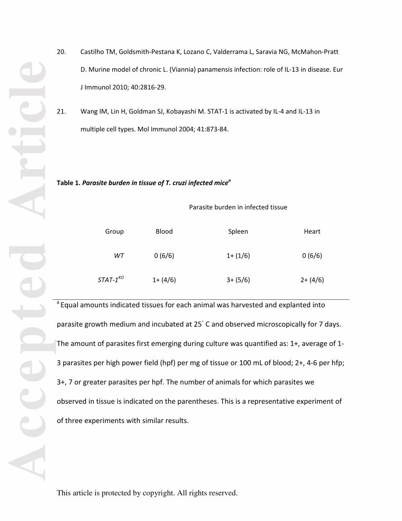
Parasite growth explant analysis of blood, heart and splenic tissue showed that all tissues were
heavily parasitized as compared to infected WT animals (Table 1). Parasites grew from the blood
of the majority of animals from the STAT-1KO group but none from the WT animals. Similarly,
parasites grew from cardiac and spleen tissue of the majority of the STAT-1KO mice but not the WT
groups.
Systemic IL-10 and IFN-γ dysregulation occurs in T. cruzi-infected STAT-1KO mice. Since STAT-1
expression is important for the transduction of IFN-γ signals during inflammation which are
important for macrophage activation and intracellular killing, we measured systemic IFN-γ levels
in the serum of infected animals at days 5, 10 and 15 (Fig. 3) Interestingly, we found that the
levels of IFN-γ in infected STAT-1KO mice were ~100-1000-fold higher on days 10- and 15-post-
infection compared with WT mice. This suggests that during uncontrolled infection there is hyper-
IFN-γ response that is ineffective at inducing intracellular killing in mice with absent STAT-1
expression. The systemic levels of IL-4 and IL-12 were not statistically different between groups,
and we found no detectable TNF-α or IL-6 in either group. The levels of the anti-inflammatory
cytokine IL-10, were approximately 3- to 9-fold higher at 10-15 days post-infection, respectively,
compared to WT animals. IL-10 blockade has been shown to protect mice from T. cruzi infection,
presumably increasing IFN-γ mediated signaling16. We tested whether neutralization of IL-10 could

Acc
epte
d A
rtic
le
This article is protected by copyright. All rights reserved.
alter the diminished survival of parasite infected STAT-1KO mice. Mice were treated with daily
administrations of IL-10 antibody, or isotype control antibody, through the course of parasite
infection. Despite IL-10 neutralization, the infected STAT-1KO mice had decreased overall survival
relative to infected WT mice. Thus neutralization of IL-10 using antibody therapy had no effect on
changes in survival.
CD4 and CD8 cells contribute to IFN-γ and IL-13 production in infected STAT-1 KO mice. The lack of
STAT-1 expression leads to IFN-γ hyper-production, thus we wanted to determine which cells are
responsible for this. Intracellular cytokine analysis flow cytometric analysis revealed that CD4 and
CD8 cells both contributed to the production of IFN-γ in STAT-1KO mice (Fig 4) whereas NK cells did
not appear to contribute to the increase of production of IFN-γ in the STAT-1KO over that of the
WT mice (Fig. 4). IL-13 expression appeared to be variably expressed in splenic at different times
post-infection in NK, CD4 and CD8 cells in both WT and STAT-1KO mice.
Antigen-specific IL-17 is robust early in infection but diminishes in STAT-1KO infected mice. IL-17
production is increased in deficient IFN-γ signaling secondary to STAT-1 ablation 13. In addition, IL-
17 expression is important in reducing mortality and inflammation during T. cruzi infection 14, 15.
Thus, we compared the IL-17 responses in splenic cells in T. cruzi-infected WT and STAT-1KO
animals and found that the STAT-1KO mice had increased levels of Ag-induced IL-17 expression
over those of WT mice, especially prevalent at days 5 and 10 post-infection. CD3-induction of IL-
17 was present in both WT and STAT-1KO animals indicating that they were both able to respond

Acc
epte
d A
rtic
le
This article is protected by copyright. All rights reserved.
to TCR-engagement, but the day 5 and 10 post-infected STAT-1KO animals had especially robust
induction of IL-17 in response to CD3.
DISCUSSION
These results indicate that the expression of STAT-1 is important to the control of T. cruzi
replication and dissemination in the infected mice. The uncontrolled replication and
dissemination of parasites in the knock-out strain leads to death in the acute stages of infection
and dissemination includes deposition of parasites within the myocardium, a chief site of
pathology in T. cruzi infection. The heavy parasitemia in these animals is evidenced by both the
finding of viable parasites in the blood of most infected animals as well as the heavy deposition in
the spleens.
It has been shown that the induction of IL-10 by LPS or E. coli infection occurs independent of
STAT-1 expression, and that subsequent repression of IL-10 are dependent on STAT-19. Similarly
the production of IL-10 by dendritic cells exposed to LPS or CpG is suppressed by IFN-γ in a STAT-1
dependent manner17. The presence of IL-10 has been shown to promote T. cruzi infection in the
C57BL/6 mouse strain and that depletion of IL-10 using IL-10 antibodies is protective 16, 18. This it
thought to be related to the IL-10 mediated interference of IFN-γ macrophage activation. In the
absence of STAT-1, IFN-γ mediated activation cannot occur, and the elevation of IL-10 does not
function as an anti-inflammatory cytokine in the absence of STAT-1. The elevation of IFN-γ and IL-
10 may be a reflection of exaggerated Th1 and Th2 responses during infection as a result of the

Acc
epte
d A
rtic
le
This article is protected by copyright. All rights reserved.
absence of STAT-1. We tested the role of IL-10 as reason for diminished survival in STAT-1KO mice
using anti-IL-10 administration throughout the infection. Neutralization of IL-10 in these animals
did not alter the diminished survival. Thus IL-10 expression, in the absence of STAT-1 expression,
does not appear to be responsible for our observations.
The role of IL-13 in T. cruzi infection in mice is not well described. This IL-4-like cytokine is
implicated in allergic responses and parasitic nematode expulsion and is produced by Th-2 cells,
basophils and eosinophils. IL-13 expression is important for the development of murine
cutaneous disease by several Leishmania species where it plays a role in parasite persistence
during Th2 responses19, 20. Our data indicates that it is produced systemically in the early phase of
T. cruzi infection in wild-type mice, but is ablated in the absence of STAT-1 expression. The effects
of IL-13 and IL-4 are mediated through the activation of both STAT-1 and STAT-6 in a variety of cell
types 21 yet we do not understand the relationship between the lack of STAT-1 and the lack of
systemic IL-13 provoked during parasite infection. Perhaps the absence of STAT-1 affects the
production of IL-13 by cells in response to parasite exposure. We found that CD4+, CD8+ and NK
cells were responsible for IL-13 production in the spleen of both WT and STAT-1KO presenting
higher IL-13 production in STAT-1KO mice at day 10. Despite this, the systemic levels of IL-13 were
higher in WT animals, suggesting that IL-13 is produced predominantly from a non-splenic source
in these animals, or, less likely, that in the absence of STAT-1 IL-13 secretion is inhibited despite
the presence of demonstrable intracellular protein.
IL-17 expression is important for the control of T. cruzi infection and is thought to be
predominantly generated by B cells in response to exposure by parasite trans-sialidase 14,

Acc
epte
d A
rtic
le
This article is protected by copyright. All rights reserved.
although it is classically secreted by Th17 helper cells. Although not entirely elucidated, IL-
17 is important for host resistant to T. cruzi infection. However, IL-17 production can be
down-regulated by STAT-1 13. This can explain the higher levels of IL-17 found in STAT-1KO
infected mice, and also the neutrophilic inflammation in the heart and spleen.
Interestingly, during T. cruzi infection, IL-17RA is important for recruitment of IL-10
producing neutrophils which in turn suppresses inflammation and reduces mortality15.
Our results found that splenic Ag-driven IL-17 was up-regulated in infected STAT-1KO
animals as early as day 5 post-infection and remained substantially elevated through day
10, whereas WT animals had nearly undetectable IL-17 responses. We measured the
abundance of Ly6G+, Ly6c+ neutrophils in the spleens of uninfected and T. cruzi-infected
WT and STAT-1KO animals and found that at baseline, STAT-1KO animals had ~2-fold more
resident neutrophils than WT mice, and upon infection both increased 2-3 fold above this
baseline with the number of neutrophils in the STAT-1KO animals being 1.4-fold higher
than in WT animals. This supports the idea that IL-17 drives neutrophilic response;
however we found this neutrophilic response was only robust in the absence of STAT-1
expression yet ineffective at controlling parasite load and preventing premature death
relative to WT animals. Further experiments are necessary to elucidate the phenotype of
neutrophils recruited in STAT-1KO mice.
Overall this study demonstrates that STAT-1 is critical for the control of T. cruzi infection and also
suggests that STAT-1 plays a role in the maintenance of cytokine systemic levels, particularly IFN-
γ, IL-10 and IL-13, during infection. The absence of STAT-1 has pleotrophic effects causing the
production of and response to multiple cytokines. Further detailed studies are necessary to

Acc
epte
d A
rtic
le
This article is protected by copyright. All rights reserved.
understand the cell-type and organ-specific origin of these effects with respect to T. cruzi
infection.
ACKNOWLEDGEMENTS: MK, SV, CT, JLK performed experiments and analyzed data; AS, designed
experiments, analyzed data and edited manuscript; BM, designed experiments, analyzed data,
wrote and edited manuscript. The work in this paper was supported by grants from the American
Heart Association (SDG #0735241N to BSM). Work in the Satoskar lab is supported by grants from
the National Institutes of Health.
Disclosures: The authors have no conflicts of interest to report.
REFERENCES
1. Abrahamsohn IA. Cytokines in innate and acquired immunity to Trypanosoma cruzi
infection. Braz J Med Biol Res 1998; 31:117-21.
2. O'Shea JJ, Gadina M, Schreiber RD. Cytokine signaling in 2002: new surprises in the
Jak/Stat pathway. Cell 2002; 109 Suppl:S121-31.
3. Ramana CV, Gil MP, Schreiber RD, Stark GR. Stat1-dependent and -independent pathways
in IFN-gamma-dependent signaling. Trends Immunol 2002; 23:96-101.
4. Durbin JE, Hackenmiller R, Simon MC, Levy DE. Targeted disruption of the mouse Stat1
gene results in compromised innate immunity to viral disease. Cell 1996; 84:443-50.

Acc
epte
d A
rtic
le
This article is protected by copyright. All rights reserved.
5. Meraz MA, White JM, Sheehan KC, Bach EA, Rodig SJ, Dighe AS, et al. Targeted disruption
of the Stat1 gene in mice reveals unexpected physiologic specificity in the JAK-STAT
signaling pathway. Cell 1996; 84:431-42.
6. Lieberman LA, Banica M, Reiner SL, Hunter CA. STAT1 plays a critical role in the regulation
of antimicrobial effector mechanisms, but not in the development of Th1-type responses
during toxoplasmosis. J Immunol 2004; 172:457-63.
7. Ray M, Gam AA, Boykins RA, Kenney RT. Inhibition of interferon-gamma signaling by
Leishmania donovani. J Infect Dis 2000; 181:1121-8.
8. Rosas LE, Snider HM, Barbi J, Satoskar AA, Lugo-Villarino G, Keiser T, et al. Cutting edge:
STAT1 and T-bet play distinct roles in determining outcome of visceral leishmaniasis
caused by Leishmania donovani. J Immunol 2006; 177:22-5.
9. VanDeusen JB, Shah MH, Becknell B, Blaser BW, Ferketich AK, Nuovo GJ, et al. STAT-1-
mediated repression of monocyte interleukin-10 gene expression in vivo. Eur J Immunol
2006; 36:623-30.
10. Tarleton RL, Grusby MJ, Zhang L. Increased susceptibility of Stat4-deficient and enhanced
resistance in Stat6-deficient mice to infection with Trypanosoma cruzi. J Immunol 2000;
165:1520-5.
11. Kumar S, Tarleton RL. Antigen-specific Th1 but not Th2 cells provide protection from
lethal Trypanosoma cruzi infection in mice. J Immunol 2001; 166:4596-603.
12. Cuervo H, Pineda MA, Aoki MP, Gea S, Fresno M, Girones N. Inducible nitric oxide
synthase and arginase expression in heart tissue during acute Trypanosoma cruzi

Acc
epte
d A
rtic
le
This article is protected by copyright. All rights reserved.
infection in mice: arginase I is expressed in infiltrating CD68+ macrophages. J Infect Dis
2008; 197:1772-82.
13. Villarino AV, Gallo E, Abbas AK. STAT1-activating cytokines limit Th17 responses through
both T-bet-dependent and -independent mechanisms. J Immunol 2010; 185:6461-71.
14. Bermejo DA, Jackson SW, Gorosito-Serran M, Acosta-Rodriguez EV, Amezcua-Vesely MC,
Sather BD, et al. Trypanosoma cruzi trans-sialidase initiates a program independent of the
transcription factors RORgammat and Ahr that leads to IL-17 production by activated B
cells. Nat Immunol 2013; 14:514-22.
15. Tosello Boari J, Amezcua Vesely MC, Bermejo DA, Ramello MC, Montes CL, Cejas H, et al.
IL-17RA signaling reduces inflammation and mortality during Trypanosoma cruzi infection
by recruiting suppressive IL-10-producing neutrophils. PLoS Pathog 2012; 8:e1002658.
16. Reed SG, Brownell CE, Russo DM, Silva JS, Grabstein KH, Morrissey PJ. IL-10 mediates
susceptibility to Trypanosoma cruzi infection. J Immunol 1994; 153:3135-40.
17. Flores RR, Diggs KA, Tait LM, Morel PA. IFN-gamma negatively regulates CpG-induced IL-
10 in bone marrow-derived dendritic cells. J Immunol 2007; 178:211-8.
18. Silva JS, Morrissey PJ, Grabstein KH, Mohler KM, Anderson D, Reed SG. Interleukin 10 and
interferon gamma regulation of experimental Trypanosoma cruzi infection. J Exp Med
1992; 175:169-74.
19. Hurdayal R, Brombacher F. The role of IL-4 and IL-13 in cutaneous Leishmaniasis. Immunol
Lett 2014.
Acc
epte
d A
rtic
le
This article is protected by copyright. All rights reserved.
20. Castilho TM, Goldsmith-Pestana K, Lozano C, Valderrama L, Saravia NG, McMahon-Pratt
D. Murine model of chronic L. (Viannia) panamensis infection: role of IL-13 in disease. Eur
J Immunol 2010; 40:2816-29.
21. Wang IM, Lin H, Goldman SJ, Kobayashi M. STAT-1 is activated by IL-4 and IL-13 in
multiple cell types. Mol Immunol 2004; 41:873-84.
Table 1. Parasite burden in tissue of T. cruzi infected micea
Parasite burden in infected tissue
Group Blood Spleen Heart
WT 0 (6/6) 1+ (1/6) 0 (6/6)
STAT-1KO 1+ (4/6) 3+ (5/6) 2+ (4/6)
a Equal amounts indicated tissues for each animal was harvested and explanted into
parasite growth medium and incubated at 25◦ C and observed microscopically for 7 days.
The amount of parasites first emerging during culture was quantified as: 1+, average of 1-
3 parasites per high power field (hpf) per mg of tissue or 100 mL of blood; 2+, 4-6 per hfp;
3+, 7 or greater parasites per hpf. The number of animals for which parasites we
observed in tissue is indicated on the parentheses. This is a representative experiment of
of three experiments with similar results.

Acc
epte
d A
rtic
le
This article is protected by copyright. All rights reserved.
FIGURE LEGENDS
Fig .1. Kaplan-Meier survival plot of T. cruzi-infected WT and STAT-1KO mice. Mice were infected
intraperitoneally with 10,000 Brazil strain tissue-culture derived trypomastigotes of T. cruzi.
Survival of animals was determined over the course of 30 days post-infection. STAT-1KO animals
succumbed to infection between 13-18 days post infection whereas WT animals survived. Blood
and tissue from mice was analyzed histopathologically, by parasite outgrowth explantation and
cytokine analysis (see Figs 2, 3 and Table 1). This shows a representative experiment of 3
experiments with similar results.
Fig. 2. Histopathologic analysis of heart tissue from infected mice. STAT-1 (panels a-c) and WT
(panel d) mice infected with Brazil strain T. cruzi tissue culture trypomastigotes were sacrificed at
15 days post-infection. Cardiac tissue from STAT-1 mice showed numerous areas of parasite
pseudocyst formation (indicated by arrows) with areas of associated inflammation, particularly of
neutrophils (see panel b), whereas tissue from WT mice did not show evidence of disease. 100X
(panel a) and 40x (panels b-c) magnification are shown.
Fig. 3. Measurement of serum cytokines from T. cruzi-infected WT and STAT-1KO mice. The
serum obtained from the indicated infected mice from post-infection day 5, 10 and 15 were
analyzed by ELISA using cytokine specific antisera. The mean values ±SE (n= 3-4 animals per
group) are shown. * P values are ≤ 0.05 compared to the corresponding values in the opposing
group.
Fig. 4. Analysis of splenic cells subsets for production of IFN-γ and IL-13. Spleen cells harvested
from parasite-infected WT and STAT-1KO animals were analyzed by FACS for co-expression of
CD4-, CD8- and NK-associated surface markers and intracellular staining of the indicated cytokines

Acc
epte
d A
rtic
le
This article is protected by cop
(n= 4-5 animals per group were
y-axis. * P values are ≤ 0.05 com
Fig. 5. Analysis of IL-17 and neu
from spleens of parasite infected
indicated times post-infection; b
and Ly6c/Ly6c (lower) from WT
0.05 compared to the correspon
pyright. All rights reserved.
analyzed). The % of the total population of cells are
mpared to the corresponding values in the opposing
trophils in spleens of infected animals. a) The IL-17
d animals in response to T. cruzi antigen (Ag) and CD
b) FACS analysis of spleen cells for expression of CD1
and STATKO animals at 15 days post-infection. *** P
nding values in the opposing group.
e shown on the
group.
7 production
D3 from
11b (upper)
values are ≤

Acc
epte
d A
rtic
le
This article is protected by coppyright. All rights reserved.

Acc
epte
d A
rtic
le
This article is protected by coppyright. All rights reserved.

Acc
epte
d A
rtic
le
This article is protected by coppyright. All rights reserved.

Acc
epte
d A
rtic
le
This article is protected by coppyright. All rights reserved.