IMGT unique numbering for immunoglobulin and T cell receptor constant domains and Ig superfamily C-like domains Marie-Paule Lefranc * , Christelle Pommie ´, Quentin Kaas, Elodie Duprat, Nathalie Bosc, Delphine Guiraudou, Christelle Jean, Manuel Ruiz, Isabelle Da Pie ´dade, Mathieu Rouard, Elodie Foulquier, Vale ´rie Thouvenin, Ge ´rard Lefranc IMGT, the International ImMunoGeneTics Information System w , LIGM, Laboratoire d’ImmunoGe ´ne ´tique Mole ´culaire, Universite ´ Montpellier II, UPR CNRS 1142, IGH, 141 rue de la Cardonille, 34396 Montpellier cedex 5, France Received 19 May 2004; accepted 16 July 2004 Available online 1 September 2004 Abstract IMGT, the international ImMunoGeneTics information system w (http://imgt.cines.fr) provides a common access to expertly annotated data on the genome, proteome, genetics and structure of immunoglobulins (IG), T cell receptors (TR), major histocompatibility complex (MHC), and related proteins of the immune system (RPI) of human and other vertebrates. The NUMEROTATION concept of IMGT-ONTOLOGY has allowed to define a unique numbering for the variable domains (V-DOMAINs) and for the V-LIKE-DOMAINs. In this paper, this standardized characterization is extended to the constant domains (C-DOMAINs), and to the C-LIKE-DOMAINs, leading, for the first time, to their standardized description of mutations, allelic polymorphisms, two-dimensional (2D) representations and tridimensional (3D) structures. The IMGT unique numbering is, therefore, highly valuable for the comparative, structural or evolutionary studies of the immunoglobulin superfamily (IgSF) domains, V-DOMAINs and C-DOMAINs of IG and TR in vertebrates, and V-LIKE-DOMAINs and C-LIKE-DOMAINs of proteins other than IG and TR, in any species. q 2004 Elsevier Ltd. All rights reserved. Keywords: IMGT; Immunoglobulin; T cell receptor; Constant domain; Immunoglobulin superfamily; V-set; C-set; Colliers de Perles 1. Introduction IMGT, the international ImMunoGeneTics infor- mation system w (http://imgt.cines.fr) [1] is a high quality integrated knowledge resource specialized in immunoglobulins (IG), T cell receptors (TR), major 0145-305X/$ - see front matter q 2004 Elsevier Ltd. All rights reserved. doi:10.1016/j.dci.2004.07.003 Developmental and Comparative Immunology 29 (2005) 185–203 www.elsevier.com/locate/devcompimm Abbreviations: 2D, two-dimensional; 3D, tridimensional; IG, immunoglobulin; IgSF, immunoglobulin superfamily; MHC, major histocompatibility complex; RPI, related proteins of the immune system; TR, T cell receptor. * Corresponding author. Tel.: C33-4-9961-9965; fax: C33-4- 9961-9901. E-mail address: [email protected] (M.-P. Lefranc).

Welcome message from author
This document is posted to help you gain knowledge. Please leave a comment to let me know what you think about it! Share it to your friends and learn new things together.
Transcript

IMGT unique numbering for immunoglobulin and T cell receptor
constant domains and Ig superfamily C-like domains
Marie-Paule Lefranc*, Christelle Pommie, Quentin Kaas, Elodie Duprat,Nathalie Bosc, Delphine Guiraudou, Christelle Jean, Manuel Ruiz,
Isabelle Da Piedade, Mathieu Rouard, Elodie Foulquier,Valerie Thouvenin, Gerard Lefranc
IMGT, the International ImMunoGeneTics Information Systemw, LIGM, Laboratoire d’ImmunoGenetique Moleculaire,
Universite Montpellier II, UPR CNRS 1142, IGH, 141 rue de la Cardonille,
34396 Montpellier cedex 5, France
Received 19 May 2004; accepted 16 July 2004
Available online 1 September 2004
Abstract
IMGT, the international ImMunoGeneTics information systemw (http://imgt.cines.fr) provides a common access to expertly
annotated data on the genome, proteome, genetics and structure of immunoglobulins (IG), T cell receptors (TR), major
histocompatibility complex (MHC), and related proteins of the immune system (RPI) of human and other vertebrates. The
NUMEROTATION concept of IMGT-ONTOLOGY has allowed to define a unique numbering for the variable domains
(V-DOMAINs) and for the V-LIKE-DOMAINs. In this paper, this standardized characterization is extended to the constant
domains (C-DOMAINs), and to the C-LIKE-DOMAINs, leading, for the first time, to their standardized description of
mutations, allelic polymorphisms, two-dimensional (2D) representations and tridimensional (3D) structures. The IMGT unique
numbering is, therefore, highly valuable for the comparative, structural or evolutionary studies of the immunoglobulin
superfamily (IgSF) domains, V-DOMAINs and C-DOMAINs of IG and TR in vertebrates, and V-LIKE-DOMAINs and
C-LIKE-DOMAINs of proteins other than IG and TR, in any species.
q 2004 Elsevier Ltd. All rights reserved.
Keywords: IMGT; Immunoglobulin; T cell receptor; Constant domain; Immunoglobulin superfamily; V-set; C-set; Colliers de Perles
0145-305X/$ - see front matter q 2004 Elsevier Ltd. All rights reserved.
doi:10.1016/j.dci.2004.07.003
Abbreviations: 2D, two-dimensional; 3D, tridimensional; IG,
immunoglobulin; IgSF, immunoglobulin superfamily; MHC, major
histocompatibility complex; RPI, related proteins of the immune
system; TR, T cell receptor.
* Corresponding author. Tel.: C33-4-9961-9965; fax: C33-4-
9961-9901.
E-mail address: [email protected] (M.-P. Lefranc).
1. Introduction
IMGT, the international ImMunoGeneTics infor-
mation systemw (http://imgt.cines.fr) [1] is a high
quality integrated knowledge resource specialized in
immunoglobulins (IG), T cell receptors (TR), major
Developmental and Comparative Immunology 29 (2005) 185–203
www.elsevier.com/locate/devcompimm
M.-P. Lefranc et al. / Developmental and Comparative Immunology 29 (2005) 185–203186
histocompatibility complex (MHC), and related
proteins of the immune system (RPI) of human
and other vertebrates [1–14]. IMGT provides a
common access to expertly annotated data on the
genome, proteome, genetics and structure of the IG,
TR, MHC, and RPI, based on the IMGT Scientific
chart rules and on the IMGT-ONTOLOGY concepts
[15]. More particularly, the IMGT unique number-
ing [16–18], based on the NUMEROTATION
concept of IMGT-ONTOLOGY, has been set up to
provide a standardized description of mutations,
allelic polymorphisms, two-dimensional (2D) and
tridimensional (3D) structure representations of the
IG and TR variable domains (V-DOMAINs),
whatever the antigen receptor, the chain type or
the species [18]. The IMGT unique numbering for
V-DOMAINs is used in all the IMGT components
[3–8]: databases (IMGT/LIGM-DB [19], IMGT/
PRIMER-DB [20], IMGT/GENE-DB [21], IMGT/
3Dstructure-DB [22]), tools for sequence and
structure analysis (IMGT/V-QUEST [23], IMGT/
JunctionAnalysis [24], IMGT/Allele-Align, IMGT/
PhyloGene [25], IMGT/StructuralQuery [22]), and
Web resources (‘IMGT Protein displays’ [26,27],
‘IMGT Colliers de Perles’ 2D representations [28],
and ‘IMGT Alignments of Alleles’ [29,30]; see
IMGT Repertoire, http://imgt.cines.fr). Interestingly,
the IMGT unique numbering for V-DOMAIN was
fully applicable to the V-LIKE-DOMAINs of
proteins other than IG and TR [18], although their
genomic structure (V-LIKE-DOMAINs are often
encoded by one exon) is different from that of the
IG and TR V-DOMAINs (encoded by the
rearranged V–(D–)J genes).
In this paper, the standardized IMGT unique
numbering is extended to the IG and TR constant
domains (C-DOMAINs) of the IG and TR of all jawed
vertebrates, and to the C-LIKE-DOMAINs of proteins
other than IG and TR of any species. The IMGT
unique numbering represents therefore a major step
forward for the comparative analysis and for the 2D
and 3D structure and evolution studies of the
immunoglobulin superfamily (IgSF) domains,
V-DOMAINs and C-DOMAINs of IG and TR in
vertebrates, and V-LIKE-DOMAINs and C-LIKE-
DOMAINs of proteins other than IG and TR, in any
species.
2. C-DOMAIN definition and relations
with C-REGION
The C-DOMAIN of an IG or TR chain is a 3D
structural unit comprising about 100 amino acids in
seven antiparallel beta strands, on two sheets
[31–33]. The seven strands of the C-DOMAIN,
designated as A, B, C, D, E, F and G, have a
topology and 3D structure similar to that of a
V-DOMAIN without its C 0 and C 00 strands. Indeed,
the C-DOMAIN beta sandwich fold is built up
from the seven beta strands arranged so that four
strands form one beta sheet, and three strands form
a second sheet [31–33]. Depending from the
topology of the D strand, which can be in one or
the other sheet, the beta sandwich comprises ABE
and GFCD, or ABED and GFC. ABE and GFC are
closely packed against each other and joined
together by a disulfide bridge from strand B in
the first sheet with strand F in the second sheet
[31], conserved in both the C-DOMAINs and
V-DOMAINs. Amino acids with conserved phy-
sico-chemical characteristics form and stabilize the
framework by packing the beta sheets through
hydrophobic interactions giving a hydrophobic core
[31,34].
Whereas the C-DOMAIN is a structural unit of an
IG or TR chain, the C-REGION represents the part of
an IG or TR chain encoded by the C-GENE [29,30].
Depending on the IG or TR chain type, the
C-DOMAIN may correspond to a complete
C-REGION, or to most of the C-REGION, or to
only part of the C-REGION (if the C-REGION
comprises several C-DOMAINs). As respective
examples, (i) the domains C-KAPPA, C-LAMBDA
and C-IOTA correspond to the complete C-REGION
of an IG kappa, lambda, and iota chains, respectively
(if the IG light chain type is not specified, the domain
is designated as CL); (ii) the domains C-ALPHA,
C-BETA, C-GAMMA and C-DELTA correspond to
most (but not to the entirety) of the C-REGION of the
TR alpha, beta, gamma and delta chains, respectively
(indeed, the TR chains are transmembrane proteins
whose C-REGION encoded by the C-GENE
comprises, in addition to the C-DOMAIN, the
CONNECTING-REGION, the TRANSMEMBRANE-
REGION, and the INTRACYTOPLASMIC-REGION)
[29,30]; (iii) the domains CH1, CH2, CH3 and,

M.-P. Lefranc et al. / Developmental and Comparative Immunology 29 (2005) 185–203 187
if present, CH4, correspond to only part of the
C-REGION of the IG heavy chains (e.g. the domains
CH1, CH2 and CH3 of the human IGHG1 represent
98, 110 and 105 amino acids, respectively, on a total
of 399 or 330 amino acids for the complete
C-REGION of a membrane gamma 1 chain or that
of a secreted gamma 1 chain, respectively [29])
(Fig. 1). It is worth to note that these relations between
Fig. 1. Correspondence between exons, domains and regions. (A) Exons of
exons are in base pairs. Introns are not at scale (see Ref. [29] for a repr
membrane and secreted IG gamma 1 heavy chain shown as examples. Len
CH3 exon (320 nucleotides) encodes 107 amino acids (105 amino acids o
secreted gamma 1 chain). Exon M1 (131 nucleotides) encodes 44 amino a
the 27 amino acids of the TRANSMEMBRANE-REGION), exon M2 (8
TRANSMEMBRANE-REGION and the 26 amino acids of the INTRACY
and Protein displays in IMGT Repertoire, http://imgt.cines.fr and [29]).
C-DOMAIN and C-REGION are quite different from
those between V-DOMAIN and V-REGION, since
the V-DOMAIN of an IG or TR chain results from the
junction of two or three different regions: V and J (V–
J-REGION of the IG light chains, and of the TR alpha
and gamma chains), or V, D and J (V–D–J-REGION
of the IG heavy chains, and of the TR beta and delta
chains) [29,30] (Fig. 1).
the Homo sapiens IGHG1 gene shown as an example. Length of the
esentation at scale). (B) Domains and regions of a Homo sapiens
gths of the domains and regions are in number of amino acids. The
f the CH3 domain and 2 amino acids of CH-S, only present in the
cids (the 18 amino acids of the CONNECTING-REGION and 26 of
1 nucleotides) encodes 27 amino acids (the last amino acid of the
TOPLASMIC-REGION). (Human IGHC ‘Alignments of Alleles’
M.-P. Lefranc et al. / Developmental and Comparative Immunology 29 (2005) 185–203188
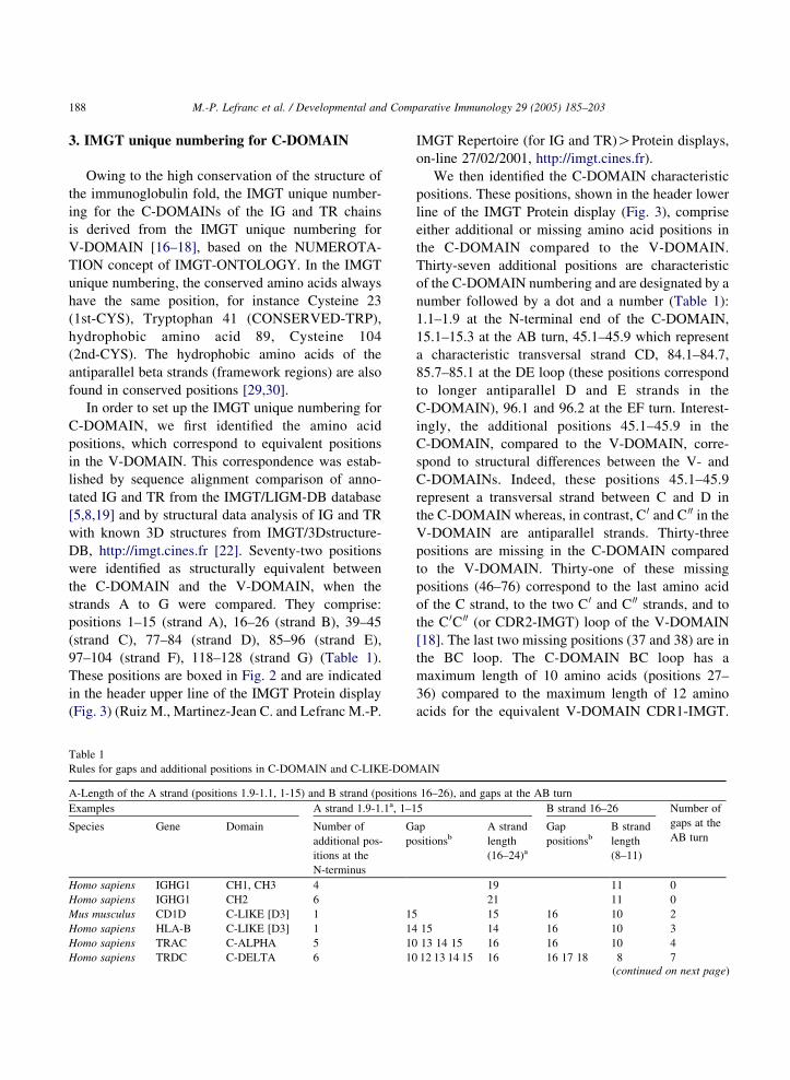
3. IMGT unique numbering for C-DOMAIN
Owing to the high conservation of the structure of
the immunoglobulin fold, the IMGT unique number-
ing for the C-DOMAINs of the IG and TR chains
is derived from the IMGT unique numbering for
V-DOMAIN [16–18], based on the NUMEROTA-
TION concept of IMGT-ONTOLOGY. In the IMGT
unique numbering, the conserved amino acids always
have the same position, for instance Cysteine 23
(1st-CYS), Tryptophan 41 (CONSERVED-TRP),
hydrophobic amino acid 89, Cysteine 104
(2nd-CYS). The hydrophobic amino acids of the
antiparallel beta strands (framework regions) are also
found in conserved positions [29,30].
In order to set up the IMGT unique numbering for
C-DOMAIN, we first identified the amino acid
positions, which correspond to equivalent positions
in the V-DOMAIN. This correspondence was estab-
lished by sequence alignment comparison of anno-
tated IG and TR from the IMGT/LIGM-DB database
[5,8,19] and by structural data analysis of IG and TR
with known 3D structures from IMGT/3Dstructure-
DB, http://imgt.cines.fr [22]. Seventy-two positions
were identified as structurally equivalent between
the C-DOMAIN and the V-DOMAIN, when the
strands A to G were compared. They comprise:
positions 1–15 (strand A), 16–26 (strand B), 39–45
(strand C), 77–84 (strand D), 85–96 (strand E),
97–104 (strand F), 118–128 (strand G) (Table 1).
These positions are boxed in Fig. 2 and are indicated
in the header upper line of the IMGT Protein display
(Fig. 3) (Ruiz M., Martinez-Jean C. and Lefranc M.-P.
Table 1
Rules for gaps and additional positions in C-DOMAIN and C-LIKE-DOM
A-Length of the A strand (positions 1.9-1.1, 1-15) and B strand (positions
Examples A strand 1.9-1.1a, 1–1
Species Gene Domain Number of
additional pos-
itions at the
N-terminus
G
po
Homo sapiens IGHG1 CH1, CH3 4
Homo sapiens IGHG1 CH2 6
Mus musculus CD1D C-LIKE [D3] 1 15
Homo sapiens HLA-B C-LIKE [D3] 1 14
Homo sapiens TRAC C-ALPHA 5 10
Homo sapiens TRDC C-DELTA 6 10
IMGT Repertoire (for IG and TR)OProtein displays,
on-line 27/02/2001, http://imgt.cines.fr).
We then identified the C-DOMAIN characteristic
positions. These positions, shown in the header lower
line of the IMGT Protein display (Fig. 3), comprise
either additional or missing amino acid positions in
the C-DOMAIN compared to the V-DOMAIN.
Thirty-seven additional positions are characteristic
of the C-DOMAIN numbering and are designated by a
number followed by a dot and a number (Table 1):
1.1–1.9 at the N-terminal end of the C-DOMAIN,
15.1–15.3 at the AB turn, 45.1–45.9 which represent
a characteristic transversal strand CD, 84.1–84.7,
85.7–85.1 at the DE loop (these positions correspond
to longer antiparallel D and E strands in the
C-DOMAIN), 96.1 and 96.2 at the EF turn. Interest-
ingly, the additional positions 45.1–45.9 in the
C-DOMAIN, compared to the V-DOMAIN, corre-
spond to structural differences between the V- and
C-DOMAINs. Indeed, these positions 45.1–45.9
represent a transversal strand between C and D in
the C-DOMAIN whereas, in contrast, C 0 and C 00 in the
V-DOMAIN are antiparallel strands. Thirty-three
positions are missing in the C-DOMAIN compared
to the V-DOMAIN. Thirty-one of these missing
positions (46–76) correspond to the last amino acid
of the C strand, to the two C 0 and C 00 strands, and to
the C 0C 00 (or CDR2-IMGT) loop of the V-DOMAIN
[18]. The last two missing positions (37 and 38) are in
the BC loop. The C-DOMAIN BC loop has a
maximum length of 10 amino acids (positions 27–
36) compared to the maximum length of 12 amino
acids for the equivalent V-DOMAIN CDR1-IMGT.
AIN
16–26), and gaps at the AB turn
5 B strand 16–26 Number of
gaps at the
AB turnap
sitionsb
A strand
length
(16–24)a
Gap
positionsb
B strand
length
(8–11)
19 11 0
21 11 0
15 16 10 2
15 14 16 10 3
13 14 15 16 16 10 4
12 13 14 15 16 16 17 18 8 7(continued on next page)

Table 1 (continued)
B-Length of the AB turn (positions 15.1–15.3)
Examples AB turn 15.1–15.3
Species Gene Domain Additional positionsc AB turn length (0–3)c
Homo sapiens IGHG1 CH1, CH3 0
Homo sapiens TRBC2 C-BETA2 15.1 1
Homo sapiens CD4 C-LIKE [D2] 15.1 1
Homo sapiens IGHG1 CH2 15.1 15.2 2
C-Length of the BC loop (positions 27–36)
Examples BC loop 27–36
Species Gene Domain Number of gaps Gap positionsd BC loop length
(6–10)e
Homo sapiens IGHG1 CH2 0 10
Homo sapiens IGHE CH3 0 10
Mus musculus IGHE CH3 1 32 9
Homo sapiens IGHG1 CH1, CH3 2 31 32 8
Homo sapiens TRAC C-ALPHA 3 31 32 33 7
Mus musculus IGHD CH1 4 30 31 32 33 6
D-Length of the C strand (positions 39–45) and CD transversal strand (positions 45.1–45.9)
Examples C strand 39–45 CD transversal strandg 45.1–45.9
Species Gene Domain C strand length (7)g Additional
positionsf
CD length
(0–9)f
Homo sapiens TRAC C-ALPHA 7 0
Homo sapiens CD4 C-LIKE [D4] 7 0
Homo sapiens TRDC C-DELTA 7 45.1 1
Homo sapiens FCGR1A C-LIKE [D1] 7 45.1,45.2 2
Homo sapiens FCGR1A C-LIKE [D2] 7 45.1–45.3 3
Homo sapiens IGHG1 CH1 7 45.1–45.3 3
Homo sapiens IGHG1 CH2, CH3 7 45.1–45.4 4
Homo sapiens TRBC2 C-BETA2 7 45.1–45.5 5
Homo sapiens TRGC1 C-GAMMA1 7 45.1–45.5 5
Homo sapiens HLA-B C-LIKE [D3] 7 45.1–45.5 5
Homo sapiens IGHA1 CH3 7 45.1–45.6 6
Sus crofa IGHE CH3 7 45.1–45.7 7
Anarhichas minor IGHC1S1 C-IOTA 7 45.1–45.9 9
Seriola quinqueradiata IGIC1S22 C-IOTA 7 45.1–45.9 9
Siniperca chuatsi IGIC1S3 C-IOTA 7 45.1–45.9 9
Siniperca chuatsi IGIC1S5 C-IOTA 7 45.1–45.9 9
E-Length of the D strand (positions 77–84) and E strand (positions 85–96), and gaps at the DE
Examples D strand 77–84h E strand 85–96 Number of
gaps at the
DE turnSpecies Gene Domain Gap positionsi D strand length
(5–8)h
Gap positions E strand length
(8–12)j
Homo sapiens IGHG1 CH2, CH3 8 12 0
Homo sapiens FCGR1A C-LIKE [D2] 83 84 6 85 86 10 4
Homo sapiens FCGR1A C-LIKE [Dl] 82 83 84 5 85 86 10 5
F-Length of the DE turn (positions 84.1–84.7, 85.7–85.1)
Examples DE turn 84.1–84.7, 85.7–85.1
Species Gene Domain Additional positionsk DE turn length (6–14)k
Meleagris gallopavo Telokin C-LIKE [D] 84.1 1
Homo sapiens CD4 C-LIKE [D4] 84.1 1
Homo sapiens CD3E C-LIKE [D] 84.1, 84.2, 85.1, 85.2 4
(continued on next page)
M.-P. Lefranc et al. / Developmental and Comparative Immunology 29 (2005) 185–203 189

Table 1 (continued)
Examples DE turn 84.1–84.7, 85.7–85.1
Species Gene Domain Additional positionsk DE turn length (6–14)k
Homo sapiens ICAM1 C-LIKE [D1] 84.1–84.3, 85.1, 85.2 5
Mus musculus IGHE CH1 84.1–84.3, 85.1–85.3 6
Homo sapiens TRDC C-DELTA 84.1–84.4, 85.1–85.4 8
Homo sapiens TRGC1 C-GAMMA1 84.1–84.4, 85.1–85.4 8
Homo sapiens IGHG1 CH1, CH2, CH3 84.1–84.4, 85.1–85.4 8
Mus musculus TRBC1 C-BETA1 84.1–84.5, 85.1–85.4 9
Canis familiaris IGHA CH3 84.1–84.5, 85.1–85.5 10
Homo sapiens IGHA1 CH3 84.1–84.6, 85.1–85.5 11
Homo sapiens IGHM CH2 84.1–84.6, 85.1–85.6 12
Homo sapiens TRBC2 C-BETA2 84.1–84.7, 85.1–85.6 13
Homo sapiens TRAC C-ALPHA 84.1–84.7, 85.1–85.7 14
G-Length of the E strand (positions 85–96) and F strand (positions 97–104), and gaps at the EF turn
Examples E strand 85–96 F strand 97–104 Number of
gaps at the
EF turnSpecies Gene Domain Gap positions E strand length
(8–12)l
Gap positions F strand length
(4–8)m
Homo sapiens IGHG1 CH2, CH3 12 8 0
Homo sapiens FCGR2A C-LIKE [D1] 85 86 96 9 8 1
Homo sapiens IGHG1 CH1 96 11 8 1
Bos taurus IGHG1 CH1 96 11 97 7 2
Homo sapiens TRGC1 C-GAMMA1 96 11 97 7 2
Homo sapiens HLA-B C-LIKE [D3] 95 96 10 97 7 3
Rattus norvegicus IGHG2B CH1 91 92n 10 97 7 3
Homo sapiens TRDC C-DELTA 95 96 10 97 98 6 4
Oryctolagus cuniculus IGHG CH1 95 96 10 97 98 6 4
Homo sapiens TRAC C-ALPHA 93 94 95 96 8 97 98 99 100 4 8
H-Length of the EF turn (positions 96.1–96.2)
Examples EF turn 96.1–96.2
Species Gene Domain Additional positionso EF turn length (0.2)o
Homo sapiens IGHG1 CH1, CH2, CH3 0
Homo sapiens TRBC2 C-BETA2 96.1 1
Homo sapiens IGHM CH1 96.1 96.2 2
I-Length of the FG loop (positions 105–117) (except for the C-BETA domains)
Examples FG loop 105–117
Species Gene Domain Number of gaps Gap positions FG loop length (7–13)p
Homo sapiens IGHE CH1 0 13
Mus musculus TRAC C-ALPHA 1 111 12
Homo sapiens IGHG1 CH3 1 111 12
Homo sapiens IGHG1 CH1, CH2 2 111 112 11
Homo sapiens FCGR2A C-LIKE [D2] 3 110 111 112 10
Ovis aries IGHA CH2 4 110 111 112 113 9
Homo sapiens FCGR2A C-LIKE [D1] 6 109 110 111 112
113 114
7
J-Length of the FG loop of the C-BETA domains (positions 105–111.6, 112.6–117)
Examples FG loop 105–111.6, 112.6–117
Species Gene Domain Number of
additional positions
Additional
positionsq
FG loop length (25)q
Homo sapiens TRBC2 C-BETA2 12 111.1–111.6,
112.1–112.6
25
(continued on next page)
M.-P. Lefranc et al. / Developmental and Comparative Immunology 29 (2005) 185–203190

K-Length of the G strand (positions 118–128)
Examples G strand positions 118–128
Species Gene Domain C-terminal
positions
G strand length
(4–11)r
Homo sapiens IGHG1 CH1 121 4
Homo sapiens HLA-A C-LIKE [D3] 121 4
Mus musculus IGHE CH1 122 5
Homo sapiens IGHG1 CH2, CH3 125 8
Mus musculus IGHE CH2, CH3, CH4 125 8
Mus musculus TRBC1 C-BETA1 125 8
Homo sapiens IGKC C-KAPPA 126 9
Homo sapiens FCGR2A C-LIKE [D2] 126 9
Homo sapiens IGLC1 C-LAMBDA1 127 10
Homo sapiens FCGR1A C-LIKE [D1] 127 10
Homo sapiens FCGR2A C-LIKE[D1] 127 10
Rules are described by comparison to the IMGT unique numbering for V-DOMAIN and V-LIKE-DOMAIN [18]. Gaps and additional positions
were confirmed for proteins with known 3D structures (PDB codes in IMGT RepertoireOProtein displays, http://imgt.cines.fr). Strand, turn and
loop lengths are shown, in number of amino acids between parentheses, in the table headers.a Up to nine additional positions (numbered 1.1–1.9, starting from position next to 1 towards the N-terminal end) may be found at the
N-terminal end of the A strand. The maximal length of the A strand is 24 amino acids.b Gap positions are based on 3D structures, and if 3D structures are not known, gaps are equally distributed on strands A and B with, for an odd
number, one more gap on strand A.c C-DOMAIN and C-LIKE-DOMAIN may have additional amino acids (potentially 3) at the AB turn, which define the AB turn length.d Gap positions start with 32, then 31, 33, 30.e The maximum length of the BC loop is 10 amino acids. If the number of amino acids is odd, there is one more amino acid position on the left.
There are no positions 37 and 38 in C-DOMAINs and C-LIKE-DOMAINs.f The maximum length of the C strand is seven amino acids. There is no position 46 in C-DOMAINs and C-LIKE-DOMAINs.g The CD transversal strand is characteristic of the C-DOMAIN and C-LIKE-DOMAIN. The maximum length of the CD strand is 9 amino
acids, found in Teleostei IGIC (available online in IMGT Repertoire http://imgt.cines.fr): IGIC1S1 (AF137397) gene of Spotted wolffish
(Anarhichas minor), IGIC1S22 (AB062662) gene of Five-ray yellowtail (Seriola quinqueradiata), IGIC1S3 (AF454470) and IGIC1S5
(AY013294) genes of Chinese perch (Siniperca chuatsi). However, since this length is exceptional, usual IMGT Collier de Perles only display
seven positions. Amino acid positions are added from left to right in sequence alignments.h The maximal length of the D strand is eight amino acids. There are no positions 75 and 76 in C-DOMAINs and C-LIKE-DOMAINs.i Gap positions are based on 3D structures, and if 3D structures are not known, gaps at the DE turn are equally distributed on strands D and E
with, for an odd number, one more gap on strand D.j The maximum length of the E strand is 12 amino acids. Note that the E strand length also depends from gaps found at the EF turn, which is
reflected by E strand lengths of eight and nine amino acids, as described in Table 1G.k Most of the C-DOMAINs and C-LIKE-DOMAINs have additional amino acids (potentially 14) at the DE turn which extend the D and E
antiparallel beta strands. The number of additional positions defines the DE turn length. The numbering of the additional positions starts from
positions next to 84 and 85, respectively, towards the top of the DE turn. If the number of additional amino acids is odd, there is one more
position on the left.l The maximal length of the E strand is 12 amino acids. Note that the E strand length also depends from gaps found at the DE turn (gap
positions 85, 86 shown in italics, for the Homo sapiens FCGR2A) as described in Table 1E.m The maximal length of the F strand is eight amino acids. Gap positions are based on 3D structures, but if 3D structures are not known, gaps
are based on sequence alignments.n Gaps were assigned by sequence alignment with the CH1 of the IGHG2A and IGHG2C.o C-DOMAIN and C-LIKE-DOMAIN may have additional positions (potentially 2) at the EF turn, which define the EF length.p Except for the C-BETA domains (TRBC sequences) described in Table 1J, the maximal length of the FG loop is 13 amino acids. For an odd
number of gaps, there is one more gap on the left (starting with position 111).q C-BETA domains (TRBC sequences) have an insertion of 12 positions between 111 and 112. The length of the FG loop in C-BETA domains
is 25 amino acids. The numbering of the additional positions starts from positions next to 111 and 112, respectively, towards the top of the FG
loop.r If longer G strands are found, positions will be numbered consecutively.
M.-P. Lefranc et al. / Developmental and Comparative Immunology 29 (2005) 185–203 191
Fig
.2.C
orr
esponden
cebet
wee
nth
eC
-DO
MA
INan
dV
-DO
MA
INIM
GT
uniq
ue
num
ber
ing.A
min
oac
idse
quen
ceof
anH
om
osa
pie
ns
IGL
V2
-23
-IG
LJ2
V-D
OM
AIN
and
of
the
H.s
ap
ien
sIG
LC
1C
-DO
MA
INar
esh
ow
nas
exam
ple
s.T
he
up
per
lin
ein
dic
ates
the
bet
ast
ran
ds
A,B
,C,C
0 ,C
00,D
,E,F
and
Gw
ith
anh
ori
zon
talar
row
.C0an
dC
00,o
nly
fou
nd
inth
e
V-D
OM
AIN
,ar
esh
ow
nw
ith
das
hed
arro
ws.
AB
,B
C,C
D,C
0 C00,D
E,E
Fan
dF
Gco
rres
po
nd
toth
etu
rns
and
loo
ps
bet
wee
nth
esa
nd
wic
hfo
ldb
eta
stra
nds.
AB
turn
,B
Clo
op
,D
E
and
EF
turn
s,an
dF
Glo
op
are
fou
nd
inb
oth
V-D
OM
AIN
and
C-D
OM
AIN
.C
0 C00
loop
isonly
found
inV
-DO
MA
IN,
wher
eas
CD
tran
sver
sal
stra
nd
isch
arac
teri
stic
of
the
C-D
OM
AIN
.T
he
IMG
Tu
niq
ue
nu
mb
erin
gis
sho
wn
on
lin
es2
and
3w
ith
add
itio
nal
po
siti
on
sfo
un
din
C-D
OM
AIN
on
lin
e3
.C
on
serv
edp
osi
tio
ns
23
(1st
-CY
S)
and
10
4
(2n
d-C
YS
)ar
ein
mag
enta
.Con
serv
edp
osi
tio
ns
41
(CO
NS
ER
VE
D-T
RP
),8
9(h
yd
roph
ob
ic)
and
12
1(h
yd
roph
ob
icin
C-D
OM
AIN
)ar
ein
blu
e.B
ox
esin
dic
ates
equ
ival
entp
osi
tio
ns
inb
oth
the
V-D
OM
AIN
and
C-D
OM
AIN
.A
min
oac
ids
atad
dit
ion
alp
osi
tio
ns
inth
eC
-DO
MA
INse
qu
ence
are
sho
wn
inb
old
.A
sp
osi
tio
n4
5.1
of
C-D
OM
AIN
isn
ot
equ
ival
ent
to
posi
tion
46
inV
-DO
MA
IN,
both
posi
tions
45.1
and
46
are
show
nin
this
figure
.A
sa
V-D
OM
AIN
sequen
cere
sult
sfr
om
the
rear
rangem
ent
of
aV
–J-
or
V–D
–J-
RE
GIO
N,
the
seq
uen
ceo
fth
eg
erm
lin
eV
-RE
GIO
Nan
dth
ato
fth
eg
erm
lin
eJ-
RE
GIO
N(i
ng
reen
)ta
ken
asex
amp
les
are
sho
wn
on
the
sam
eli
ne,
toin
dic
ate
the
con
trib
uti
on
of
each
reg
ion
toth
e
V-D
OM
AIN
.N
ote
that
ina
‘tru
e’V
–J
rear
ran
gem
ent,
the
gap
sw
ou
ldb
ep
lace
dat
the
top
of
the
CD
R3
-IM
GT
loo
p[1
8],
asfo
rth
eC
-DO
MA
INF
Glo
op
.T
he
lin
esb
elo
wth
e
sequen
ces
indic
ate
the
V-D
OM
AIN
FR
-IM
GT
and
CD
R-I
MG
Tan
dth
eir
del
imit
atio
ns.
M.-P. Lefranc et al. / Developmental and Comparative Immunology 29 (2005) 185–203192
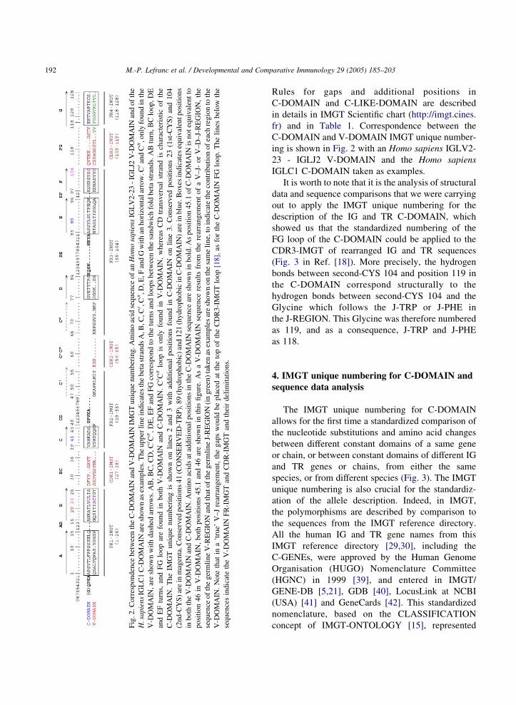
Rules for gaps and additional positions in
C-DOMAIN and C-LIKE-DOMAIN are described
in details in IMGT Scientific chart (http://imgt.cines.
fr) and in Table 1. Correspondence between the
C-DOMAIN and V-DOMAIN IMGT unique number-
ing is shown in Fig. 2 with an Homo sapiens IGLV2-
23 - IGLJ2 V-DOMAIN and the Homo sapiens
IGLC1 C-DOMAIN taken as examples.
It is worth to note that it is the analysis of structural
data and sequence comparisons that we were carrying
out to apply the IMGT unique numbering for the
description of the IG and TR C-DOMAIN, which
showed us that the standardized numbering of the
FG loop of the C-DOMAIN could be applied to the
CDR3-IMGT of rearranged IG and TR sequences
(Fig. 3 in Ref. [18]). More precisely, the hydrogen
bonds between second-CYS 104 and position 119 in
the C-DOMAIN correspond structurally to the
hydrogen bonds between second-CYS 104 and the
Glycine which follows the J-TRP or J-PHE in
the J-REGION. This Glycine was therefore numbered
as 119, and as a consequence, J-TRP and J-PHE
as 118.
4. IMGT unique numbering for C-DOMAIN and
sequence data analysis
The IMGT unique numbering for C-DOMAIN
allows for the first time a standardized comparison of
the nucleotide substitutions and amino acid changes
between different constant domains of a same gene
or chain, or between constant domains of different IG
and TR genes or chains, from either the same
species, or from different species (Fig. 3). The IMGT
unique numbering is also crucial for the standardiz-
ation of the allele description. Indeed, in IMGT,
the polymorphisms are described by comparison to
the sequences from the IMGT reference directory.
All the human IG and TR gene names from this
IMGT reference directory [29,30], including the
C-GENEs, were approved by the Human Genome
Organisation (HUGO) Nomenclature Committee
(HGNC) in 1999 [39], and entered in IMGT/
GENE-DB [5,21], GDB [40], LocusLink at NCBI
(USA) [41] and GeneCards [42]. This standardized
nomenclature, based on the CLASSIFICATION
concept of IMGT-ONTOLOGY [15], represented

M.-P. Lefranc et al. / Developmental and Comparative Immunology 29 (2005) 185–203 193
a major step in the setting up of the ‘Tables of
alleles’ and ‘Alignments of alleles’ of the IG and TR
genes (http://imgt.cines.fr). V-GENE polymorphisms
in IMGT [29,30] have been described from the start
according to the IMGT unique numbering for
V-REGION set up in 1997 [16–18]. In contrast,
C-GENE polymorphisms were initially described
according to the exon numbering [29,30] and
sequence comparison between exons of different
lengths was not easy. The implementation of the
IMGT unique numbering for the C-DOMAIN
represents therefore a new major step in the setting
up of standardized ‘Alignments of alleles’ whatever
the receptor, the chain, or the species (IMGT
Repertoire, http://imgt.cines.fr) (Fig. 3). Owing to
that standardization, the sequence polymorphisms of
any C-DOMAIN of any IG or TR can very easily be
compared and analysed.
5. IMGT unique numbering for C-DOMAIN and
structural data comparison
Beyond sequence data comparison, the IMGT
unique numbering for C-DOMAIN provides infor-
mation on the strand and loop lengths (Table 2) and
allows standardized IMGT Protein displays (Fig. 3)
and IMGT Colliers de Perles (Fig. 4) for the IG and
TR C-DOMAINs of any chain type from any species.
Practically, structural data comparison of strand or
loop of the same length can be done directly using the
IMGT unique numbering. For example, all codons (or
amino acids) at position 28 can be compared between
domains with a BC loop of a given length. This
standardization allows the structural characterization
of a position inside a domain, and the statistical
analysis of amino acid properties, position per
position, between domains, as this has been demon-
strated for the V-DOMAIN [34]. Fig. 4 shows the
IMGT Colliers de Perles for the Homo sapiens IGHG1
CH1, C-KAPPA, C-LAMBDA1 and Mus musculus
C-BETA1 domains, as examples.
As soon as the first IMGT Collier de Perles was set
up on the Web site in December 1997, the enormous
potential of the IMGT unique numbering as a means
to control data coherence was obvious. For new
sequences, for which no 3D structures are available,
the IMGT Colliers de Perles allow to precisely delimit
the strands and loops and give information on the
topological organization of the domain. The IMGT
unique numbering is also used in more sophisticated
representations of the IMGT Colliers de Perles on two
layers (Fig. 4) which allow, when 3D structures are
available, the visualisation of the hydrogen bonds
between amino acids belonging to beta strands from
the same sheet or from different sheets (IMGT/
3Dstructure-DB, http://imgt.cines.fr) [22].
6. IMGT unique numbering for C-LIKE-DOMAIN
A C-LIKE-DOMAIN is a domain of similar
structure to a C-DOMAIN, found in chains other
than IG and TR [43–52]. The IMGT unique number-
ing for the C-LIKE-DOMAIN follows exactly the
same rules as those of the C-DOMAIN (Table 1).
Strand and loop lengths of 40 examples of C-LIKE-
DOMAINs are given in Table 2. The IMGT Protein
display of the corresponding C-LIKE-DOMAIN
sequences are shown in Fig. 3. The IMGT Colliers
de Perles of four representative C-LIKE-DOMAINs
(Homo sapiens HLA-B [D3], B2M [D], FCGR2A
[D1], and Meleagris gallopavo telokin [D]) are
represented in Fig. 5. Detailed IMGT Alignment of
alleles of Homo sapiens FCGR3B and IMGT Colliers
de Perles of [D1] and [D2] of the FCGR3B*02 allele
[53] further highlight the importance of the IMGT
unique numbering standardization for the polymorph-
ism and structure analysis and comparison of the
C-LIKE-DOMAINs.
The IMGT unique numbering provides, for the first
time, a standardized approach to analyse the
sequences and structures of any domain belonging
to the C-set of the IgSF [32] (the C-set comprises the
IMGT C-DOMAINs and C-LIKE-DOMAINs). Three
features are worth noting: (i) In IMGT, any
C-DOMAIN or C-LIKE-DOMAIN is characterized
by its strand and loop lengths (Table 2). Examples are
shown in Figs. 3–5. This first feature of the IMGT
standardization based on the IMGT unique numbering
shows that the distinction between the C1, C2, I1 and
I2 types found in the literature and in the databases to
describe the IgSF C-set domains [32,33,54–56] is
unapplicable when dealing with sequences for which
no structural data are known. Indeed, the four domain

Fig. 3. IMGT Protein display of examples of C-DOMAINs (IG and TR) and C-LIKE-DOMAINs (proteins other than IG and TR). The protein display is according to the IMGT
unique numbering for C-DOMAIN and C-LIKE-DOMAIN, based on the NUMEROTATION concept of IMGT-ONTOLOGY [15]. Sandwich fold beta strands are shown by
horizontal arrows. Dots indicate missing amino acids according to the IMGT unique numbering. Amino acids resulting from a splicing with a preceding exon are shown between
parentheses (for Homo sapiens FCGR3B [D2], the information is from M90745, for H. sapiens HLA-DMA [D2] from NT_007592, for H. sapiens CD3E [D] from NT_033899, for
H. sapiens CD4 [D2] and [D4] from NT_009759, for H. sapiens CEACAM5 [D3], [D4] and [D5] from NT_011109, and for Mus musculus H2-Aa [D2] and H2-Ab [D2] from
NT_039649). Putative N-glycosylation sites (N-X-S/T) are underlined. (1) Accession numbers are from IMGT/LIGM-DB (http://imgt.cines.fr) [5,19] for IG and TR and from
EMBL/GenBank/DDBJ [35–37] for proteins other than IG and TR; Telokin identifier is from PDB [38] and IMGT/3Dstructure-DB [22]. (2) Molecule type. c: cDNA; g: genomic
DNA; p: protein. (3) Gene names (symbols) for IG and TR are according to the IMGT Nomenclature committee (IMGT-NC) [29,30] and the HUGO Nomenclature Committee
(HGNC) [39]. Full gene designations are the following: IGHG1: Immunoglobulin heavy constant gamma 1; IGHG4: Immunoglobulin heavy constant gamma 4; TRAC: T cell
M.-P
.L
efran
cet
al.
/D
evelop
men
tal
an
dC
om
pa
rative
Imm
un
olo
gy
29
(20
05
)1
85
–2
03
19
4
typ
esC
1,
C2
(alsok
no
wn
asH
[57
]),I1
and
I2,
are
based
on
structu
rald
ifferences
[55
,58
].C
1an
dC
2
differ
by
the
locatio
no
fth
efo
urth
strand
D,
wh
ichis
inth
eA
BE
sheet
for
C1
,an
din
the
GF
Csh
eetfo
rC
2
(the
Dstran
dis
then
desig
nated
asC
0b
yso
me
auth
ors)
(Fig
.6
).T
hu
sC
1co
ntain
sA
BE
Dan
dG
FC
sheets,
wh
ereasC
2is
characterized
by
AB
Ean
d
GF
CD
(or
GF
CC
0)sh
eets(T
able
3).
Th
eC
1ty
pe
initially
defi
ned
by
the
top
olo
gy
ob
served
inth
eIG
con
stant
do
main
was
describ
edas
bein
gsh
aredb
yth
e
TR
con
stant
do
main
,an
dth
eC
-like
do
main
of
MH
C
classI,
MH
Cclass
IIan
dB
2M
,an
dth
ou
gh
tto
be
a
and
asC
1,it
lacks
the
V-D
OM
AIN
C0an
dC
00strand
s.
Th
isI
typ
ew
aslater
div
ided
inI1
and
I2[5
8]
wh
ich
differ
by
the
locatio
no
fth
efo
urth
strand
,w
hich
isin
the
AB
Esh
eetfo
rI1
,an
din
the
A0G
FC
sheet
for
I2.
Th
us
I1co
ntain
sA
BE
Dan
dA
0GF
Csh
eets,w
hereas
I2is
ch
ara
cte
rized
by
AB
Ean
dA
0GF
CD
(or
A0G
FC
C00)
sheets
(I1an
dI2
ared
escribed
asI
and
Ety
pes
in[6
1]).
Inth
eab
sence
of
3D
structu
res,a
classificatio
no
fth
eco
nstan
td
om
ains
based
on
such
structu
rald
ifferences
cann
ot
be
taken
into
accou
nt,
and
the
IMG
Td
escriptio
nb
asedo
nstran
dan
dlo
op
len
gth
san
dIM
GT
Co
lliers
de
Perle
sis
mo
re
pertin
en
t.(ii)
Ase
co
nd
featu
reo
fth
eIM
GT
stand
ardizatio
nis
the
com
pariso
no
fcD
NA
and
/or
amin
oacid
sequ
ences
with
gen
om
icseq
uen
ces,an
d
the
iden
tificatio
no
fth
esp
licing
sites,to
delim
it
pre
cise
lyth
elim
itso
fth
eC
-LIK
E-D
OM
AIN
,
receptor alpha constant; TRBC2: T cell receptor beta constant 2; TRGC1: T cell receptor gamma constant 1; TRDC: T cell receptor delta constant; IGLL1: Immunoglobulin lambda-
like polypeptide 1; PTCRA: pre T-cell antigen receptor alpha; HLA-B: Major histocompatibility complex, class I, B; HLA-DMA: MHC class II, DM alpha; HLA-DMB: MHC class
II, DM beta; B2M: Beta-2-microglobulin; CD1A: CD1A antigen, a polypeptide; CD2: CD2 antigen (p50), sheep red blood cell receptor; CD3E: CD3E antigen, epsilon polypeptide
(TiT3 complex); CD4: CD4 antigen (p55); CEACAM5: Carcinoembryonic antigen-related cell adhesion molecule 5; ICAM1: intercellular adhesion molecule 1 (CD54), human
rhinovirus receptor; ICAM2: intercellular adhesion molecule 2 t of IgG, high affinity Ia, receptor for (CD64);
FCGR2A: Fc fragment of IgG, low affinity IIa, receptor for (CD 32); FCGR3B: Fc fragment of IgG, low affinity
IIIb, receptor for (CD16); FCER1A: Fc fragment of IgE, h tibility 2, class II antigen A, alpha; H2-Ab:
histocompatibility 2, class II antigen A, beta; H2-K: histocomp n name. The C-DOMAINs are designated with
the IMGT labels (IMGT Scientific chart, http://imgt.cines.fr) T ts with a number, corresponding to the position
of the domain from the N-terminal end of the protein, and relat of type I, that is with the N-terminal end being
extracellular. There is no number if there is a unique C-LIKE replace A in PDB and IMGT/3Dstructure-DB
entries 1tcr_A). (6) B2M [D] is encoded by EX2 and is precede he EX1 end in Homo sapiens or Mus musculus,
respectively (EX1 encodes 23 amino acids, including the L-RE in Homo sapiens, K in Mus musculus) and the
first EX2 codon (encoding T in both species). The proteolytic EX3 and is preceded at the N-terminal end by
seven amino acids (A)SADSQA encoded by EX2. The proteol and C-terminal ends of Telokin [D] need to be
confirmed by genomic sequence. Amino acid one-letter abbrev Glu), glutamic acid; F (Phe), phenylalanine; G
(Gly), glycine; H (His), histidine; I (Ileu), isoleucine; K (Lys), P (Pro), proline; Q (Gln), glutamine; R (Arg),
arginine; S (Ser), serine; T (Thr), threonine; V (Val), valine;
M.-P
.L
efran
cet
al.
/D
evelop
men
tal
an
dC
om
pa
rative
Imm
un
olo
gy
29
(20
05
)1
85
–2
03
19
5
characteristic
of
the
imm
un
orecep
tor
of
the
adap
tative
imm
un
eresp
on
se.In
the
literature,
the
assign
men
to
f
C-set
do
main
sto
C1
or
C2
iso
ftend
on
eb
y
extrap
olatio
n,
with
ou
tav
ailable
3D
structu
res.M
ore-
ov
er,th
e3
Dstru
cture
of
the
2C
Tcell
recepto
r
C-A
LP
HA
do
main
(PD
Ban
dIM
GT
/3D
structu
re-DB
:
1tcr_
A)
disp
lays
aq
uite
differen
tco
nfo
rmatio
nth
an
that
of
the
C1
typ
ew
itha
beta
sheet
con
tainin
gth
e
GA
BE
Dstran
ds
[59
].T
herefo
re,th
ed
istinctio
n
betw
eenC
1an
dC
2is
no
tso
straigh
tforw
ardan
das
mo
re3
Dstru
ctures
beco
me
availab
le,m
ore
inter-
med
iateo
rv
ariant
structu
resw
illb
ed
escribed
.
Th
estru
ctural
analy
siso
ftelo
kin
,th
eC
-termin
al
do
main
of
my
osin
ligh
tch
aink
inase
[60
]rev
ealeda
new
do
main
typ
ed
esign
atedas
‘I’[5
5],
wh
ichw
as
defi
ned
asan
interm
ediate
betw
eenth
eV
and
C1
typ
esb
ecause,
asfreq
uen
tlyfo
un
din
the
Vd
om
ain
typ
e,th
eA
strand
,in
itsseco
nd
part,
islo
catedo
nth
e
GF
Csh
eet(an
dth
erefore
desig
nated
asA
0stran
d),
; VCAM1: vascular cell adhesion molecule 1; FCGR1A: Fc fragmen
32); FCGR2B: Fc fragment of IgG, low affinity IIb, receptor for (CD
igh affinity I, receptor for; alpha polypeptide; H2-Aa: histocompa
atibility 2, K region; CD1D: CD1D antigen, d polypeptide. (4) Domai
he C-LIKE-DOMAINs are designated by the letter D between bracke
ive to the other domains. Membrane proteins quoted in this figure are
domain in the chain. (5) Q at position 6 is according to M64239 (and
d at the N-terminal end by SGLEGIQR or TGLYAIQK encoded by t
GION). The splicing site is between the last EX1 codon (encoding R
cleavage site of the L-REGION is not known. (7) [D1] is encoded by
ytic cleavage site of the L-REGION is not known. (8) The N-terminal
iation: A (Ala), alanine; C (Cys), cysteine; D (Asp), aspartic acid; E (
lysine; L (Leu), leucine; M (Met), methionine; N (Asn), asparagine;
W (Trp), tryptophan; Y (Tyr), tyrosine.

Table 2
Strand and loop lengths of examples of C-DOMAINs and C-LIKE-DOMAINS
Species Gene name PDB code Domain A AB B BC C CD D DE E EF F FG G Total
1.9–1.1
1–15
(6–23)
15.1–15.3
(0–2)
16–26
(7–11)
27–36
(4–10)
39–45
(7)
45.1–45.7
(0–7)
77–84
(4–8)
84.1–84.7
85.7–86.1
(0–14)
85–96
(8–12)
96.1–96.2
(0–2)
97–104
(4–8)
105–117
111.1–111.6,
112–1–112.6
(7–25)
118–128
(4–10)
C-DOMAINS
Homo
sapiens
IGHG1 1hzh_H CH1 C4 19 11 8 7 3 8 8 11 8 11 4 98
IGHG1 1hzh_H CH2 C6 21 2 11 10 7 4 8 8 12 8 11 8 110
IGHG4 1adq_A CH3 C4 19 11 8 7 4 8 8 12 8 12 8 105
IGKC 1dfb_L C–KAPPA C4 19 11 8 7 5 8 9 12 8 11 9 107
IGLC1 1a8j_L C–LAMBDA1 C5 20 11 8 7 5 8 8 12 8 9 10 106
TRAC 1qrn_D C–ALPHA C5,K 4 16 10 7 7 7 14 8 4 11 7 91
TRBC2 1qm_E C–BETA2 C7 22 1 11 8 7 5 8 13 12 1 8 25 8 129
TRGC1 1hxm_B C–GAMMA1 C8 23 1 11 8 7 5 7 8 11 7 13 9 110
TRDC 1hxm_A C–DELTA C6,K 5 16 8 8 7 1 8 8 10 6 12 9 93
Mus mus-
culus
TRAC 1tcr_A C–ALPHA C5,K 4 16 10 7 7 7 14 8 4 12 2 87
TRBC1 1tcr_B C–BETA1 C7 22 1 11 8 7 5 8 9 12 1 8 25 8 125
C-LIKE-DOMAINs
Homo
sapiens
IGLL1 [D] C5 20 11 8 7 5 8 8 12 8 9 10 106
PTCRA [D] C5 20 11 9 7 5 8 8 12 8 12 7 107
HLA–B 1a1m–A [D3] C1,K 2 14 10 8 7 5 8 8 10 7 11 4 92
HLA–DMA 1hdm_A [D2] C1,K 1 15 11 8 7 4 8 8 10 7 11 4 93
HLA–DMB 1hdm_B [D2] C1,K l 15 11 8 7 5 8 8 10 7 11 4 94
B2M 1lds_A [D] K 1 14 11 8 7 4 8 8 10 7 11 4 92
CD1A 1onq_A [D3] C1,K 1 15 10 8 7 4 8 8 10 7 12 4 93
CD2 1hnf [D2] C1,K 4 12 9 5 7 4 4 9 8 9 9 76
CD3E [D] C3 18 11 4 7 4 8 4 11 8 13 6 94
CD4 1wio_A [D2] K 5 10 1 11 6 7 2 6 9 8 11 7 78
CD4 1wio_A [D4] K 5,K 4 6 7 8 7 8 1 9 6 10 5 67
Homo
sapiens
CEACAM5 [D3] C2 17 11 6 7 6 10 8 13 7 85
CEACAM5 [D4] C2 17 1 11 5 7 5 8 12 8 13 6 93
CEACAM5 [D5] C2 17 11 6 7 6 10 8 13 7 85
ICAM1 1d3l_A [D1] C3,K l 17 1 11 6 7 7 5 10 6 9 9 88
ICAM2 1zxq [D1] C5,K l 19 1 11 6 7 7 4 10 6 9 9 89
VCAMI 1vsc_A [D1] C2 17 1 11 6 7 4 6 4 11 7 9 9 92
ICAM1 1d3l_A [D2] C2 17 11 7 7 4 8 12 2 8 16 10 102
ICAM2 1zxq [D2] C2 17 11 7 7 4 8 7 12 8 16 10 107
VCAM1 1vsc_A [D2] C1 16 11 7 7 4 8 9 12 8 16 9 107
FCGR1A [D1] C2 17 2 11 7 7 2 5 10 8 7 10 86
FCGR2A 1fcg_A [D1] C2 17 2 11 7 7 2 6 9 8 7 10 86
FCGR2B 2fcb_A [D1] C2 17 2 11 7 7 2 6 9 8 7 10 86
FCGR3B 1e4k_C [D1] C2 17 2 11 7 7 2 5 10 8 7 10 86
FCER1A 1f6a_A [D1] C3 18 11 7 7 2 5 10 8 7 10 85
FCGR1A [D2] K 1 14 11 7 7 3 6 10 8 9 10 85
FCGR2A 1fcg_A [D2] K 1 14 11 7 7 3 6 10 8 10 9 85
FCGR2B 2fcb_A [D2] K 1 14 11 7 7 3 6 10 8 10 9 85
FCGR3B le4k_C [D2] K 1 14 11 7 7 3 6 10 8 10 10 86
FCER1A 1f6a_A [D2] K 1 14 11 7 7 3 6 10 8 10 10 86
KIR2DL2 1efx_D [D1] C2 17 1 11 5 7 5 8 3 12 8 14 9 100
KIR2DL2 1efx_D [D2] C2 17 1 11 5 7 5 8 3 11 7 14 9 98
M.-P
.L
efran
cet
al.
/D
evelop
men
tal
an
dC
om
pa
rative
Imm
un
olo
gy
29
(20
05
)1
85
–2
03
19
6

Mu
smu
scu
-
lus
IGL
L1
[D]
C5
20
11
87
58
81
28
91
010
6
PT
CR
A[D
]C
52
01
19
75
88
12
81
27
10
7
H2
–A
a1
d9k
_C
[D2
]C
1,K
11
51
18
75
88
10
71
14
94
H2
–A
bId
9k
_D
[D2
]C
1,K
11
51
18
75
88
10
71
14
94
H2
–K
2v
aa_
A[D
3]
C1
,K2
14
10
87
58
81
07
11
492
B2
M2
vaa
_B
[D]
K1
14
11
87
48
81
07
11
492
CD
1D
1cd
1_A
[D3
]C
1,K
11
51
08
74
88
10
71
24
93
Mel
eag
ris
ga
llo
pa
vo
Tel
ok
in1
fhg
_A
[D]
15
a1
16
77
81
12
89
9a
93
Th
era
ng
eo
fle
ng
ths
ob
serv
edin
the
sele
cted
exam
ple
so
fC
-DO
MA
INs
and
C-L
IKE
-DO
MA
INs
are
show
nb
etw
een
par
enth
eses
inth
eh
ead
er.
Th
eto
tal
nu
mb
erof
amin
oac
ids
of
each
do
mai
nis
ind
icat
edin
the
colu
mn
‘To
tal’
.a
Th
eN
-ter
min
alan
dC
-ter
min
alen
ds
of
telo
kin
[D]
nee
dto
be
con
firm
edb
yg
eno
mic
seq
uen
ce.
M.-P. Lefranc et al. / Developmental and Comparative Immunology 29 (2005) 185–203 197
a C-LIKE-DOMAIN being frequently encoded by a
unique exon, as this is the case for the C-DOMAIN
(Fig. 1). This IMGT standardization for the domain
delimitations explains the discrepancies observed
with the generalist Swiss-Prot database which does
not take into account this criteria. (iii) At last, a third
feature is the C-LIKE-DOMAIN IMGT Collier de
Perles, which, in the absence of available 3D
structures, is particularly useful to compare domains
of very diverse families, and to characterize them by
their strand and loop lengths.
7. Conclusion
The IMGT unique numbering allows, for the first
time, to compare any C-DOMAIN of IG and TR and
C-LIKE-DOMAIN of proteins other than IG or TR,
between them, and to any V-DOMAIN and V-LIKE-
DOMAIN [18], that is to compare any domain
belonging to the IgSF C-set or V-set. Sequences and
3D structures can be analysed whatever the domain
(C-DOMAIN, C-LIKE-DOMAIN, V-DOMAIN,
V-LIKE-DOMAIN), the receptor (IG, TR, or more
generally IgSF), the chain type (heavy or light for IG;
alpha, beta, gamma or delta for TR; or more generally
IgSF chain), or the species. The IMGT unique
numbering has many advantages. The strand and
loop lengths (the number of codons or amino acids,
that is the number of occupied positions) become
crucial information, which characterizes the domains
(V-DOMAINs, V-LIKE-DOMAINs, C-DOMAINs
and C-LIKE-DOMAINs). The IMGT unique number-
ing allows standardized representations of nucleotide
and amino acid sequences in IMGT Repertoire (http://
imgt.cines.fr): Tables of strand and loop lengths,
Tables of alleles, Alignments of alleles, Protein
displays, IMGT Colliers de Perles, 3D structures.
The IMGT unique numbering is applied through all
the components (databases, tools and Web resources)
of the IMGT information systemw (http://imgt.cines.
fr) [5], for the standardized label annotations [62] and
database queries [19–22]. The IMGT unique number-
ing represents, therefore, a major step forward in the
analysis and comparison of the sequence evolution
and structure of the IgSF domains.

Fig. 4. IMGT Collier de Perles of C-DOMAINs. (A) on one layer (B) on two layers. CH1 of Homo sapiens IGHG1 (IMGT/LIGM-DB: J00228);
C-KAPPA of H. sapiens IGKC (IMGT/LIGM-DB: J00241); C-LAMBDA1 of H. sapiens IGLC1 (IMGT/LIGM-DB: X51755); C-BETA1 of
Mus musculus TRBC1 (IMGT/LIGM-DB: X02384). The first amino acids (A1.4, R1.4, G1.5 and E1.7) are encoded by a codon which results
from the splicing with an IGHJ, IGKJ, IGLJ and TRBJ, respectively [29,30]. Amino acids are shown in the one-letter abbreviation. Positions at
which hydrophobic amino acids (hydropathy index with positive value: I, V, L, F, C, M, A) and Tryptophan (W) are found in more than 50% of
analysed IG and TR sequences are shown in blue. All Proline (P) are shown in yellow. The positions 26, 39, 104 and 118 are shown in squares by
h l i h h di i i i h O A i i 45 d hi h d li i h h i i l ‘C ’ d f
M.-P. Lefranc et al. / Developmental and Comparative Immunology 29 (2005) 185–203198

Fig. 4 (continued)
homology with the corresponding positions in the V-DOMAINs. Positions 45 and 77 which delimit the characteristic transversal ‘CD’ strand of
the C-DOMAINs are also shown in squares. Hatched circles correspond to missing positions according to the IMGT unique numbering for
C-DOMAINs. Arrows indicate the direction of the beta strands (IMGT Repertoire, http://imgt.cines.fr).
3
M.-P. Lefranc et al. / Developmental and Comparative Immunology 29 (2005) 185–203 199
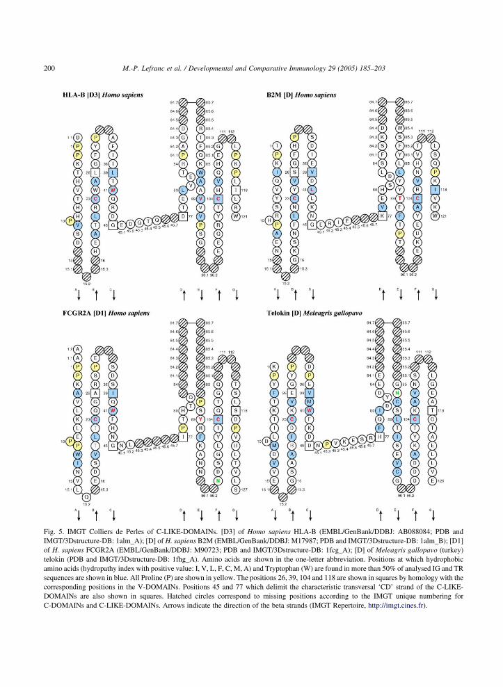
Fig. 5. IMGT Colliers de Perles of C-LIKE-DOMAINs. [D3] of Homo sapiens HLA-B (EMBL/GenBank/DDBJ: AB088084; PDB and
IMGT/3Dstructure-DB: 1alm_A); [D] of H. sapiens B2M (EMBL/GenBank/DDBJ: M17987; PDB and IMGT/3Dstructure-DB: 1alm_B); [D1]
of H. sapiens FCGR2A (EMBL/GenBank/DDBJ: M90723; PDB and IMGT/3Dstructure-DB: 1fcg_A); [D] of Meleagris gallopavo (turkey)
telokin (PDB and IMGT/3Dstructure-DB: 1fhg_A). Amino acids are shown in the one-letter abbreviation. Positions at which hydrophobic
amino acids (hydropathy index with positive value: I, V, L, F, C, M, A) and Tryptophan (W) are found in more than 50% of analysed IG and TR
sequences are shown in blue. All Proline (P) are shown in yellow. The positions 26, 39, 104 and 118 are shown in squares by homology with the
corresponding positions in the V-DOMAINs. Positions 45 and 77 which delimit the characteristic transversal ‘CD’ strand of the C-LIKE-
DOMAINs are also shown in squares. Hatched circles correspond to missing positions according to the IMGT unique numbering for
C-DOMAINs and C-LIKE-DOMAINs. Arrows indicate the direction of the beta strands (IMGT Repertoire, http://imgt.cines.fr).
M.-P. Lefranc et al. / Developmental and Comparative Immunology 29 (2005) 185–203200

Fig. 6. Schematic representations of the C-DOMAIN and C-LIKE-DOMAIN, and of the V-DOMAIN and V-LIKE-DOMAIN. A double-headed
arrow shows that the D strand can be localized in sheet 1 (on the back) or sheet 2 (on the front) depending from the length of the CD transversal
strand. The second part of the A strand can be located in sheet 2 and is then designated as A 0. This feature, described as ‘strand A switching’ in
the literature, is not shown in IMGT Colliers de Perles, as this can be determined or verified only if 3D structures are available.
Table 3
Correspondence between the IMGT classification of the IgSF domains (C-DOMAIN, C-LIKE-DOMAIN, V-DOMAIN and V-LIKE-
DOMAIN) and the diverse designations found in the literature
IMGT IgSF
domains
C-DOMAIN
(for IG and
TR)
C-LIKE-DOMAIN (for proteins other than IG and TR) V-DOMAIN
(for IG and
TR)
V-LIKE-
DOMAIN (for
proteins other
than IG and
TR)
Literature C1a [32] C1a [32] C2 [32] or
H [57]
I1 [58] or
I [55,61]
I2 [58] or
E [61]
V [32] V [32]
Sheet 1 ABED ABED ABE ABED ABE ABED ABED
Sheet 2 GFC GFC GFCD (or
GFCC 0)
A 0GFC A 0GFCD (or
A 0GFCC 0)
A 0GFCC0C 00 A 0GFCC 0C 00
a In the literature C1 is used for the IG and TR C-DOMAINs and for the MHC C-like domains. The diverse designations found in the literature
for the C-set domain (C1, C2, Il, 12) are based on strand localisation in sheets 1 and 2. These designations are not used for the assignment of
IMGT C-DOMAIN and C-LIKE-DOMAIN sequences, for the following reasons: (i) they are frequently extrapolated to sequences for which no
3D structures are available, therefore leading to misinterpretation, (ii) the designation ‘strand C 0’ used in the literature for the C2 (or H) and I2
(or E) domain types is not the equivalent of strand C 0 of the V-DOMAINs and V-LIKE-DOMAINs, (iii) the distinction made in the literature
between C1 (as found in proteins of the immune adaptative response) and C2 (as found in other proteins) may suffer exceptions, (iv) as more 3D
structures become available, and given the heterogeneity of the loop and strand lengths for C-DOMAINs and C-LIKE-DOMAINs, more
intermediate or variant structures may be described (as shown by the 2C T cell receptor 3D structure IMGT/3Dstructure-DB: 1tcr_A).
M.-P. Lefranc et al. / Developmental and Comparative Immunology 29 (2005) 185–203 201
Acknowledgements
We are grateful to Geraldine Folch, Chantal
Ginestoux, Veronique Giudicelli, Joumana Jabado-
Michaloud, Celine Protat, Dominique Scaviner and
Denys Chaume for their helpful discussions. We
thank Laurent Douchy and Bertrand Monnier for
contribution to the tables, and Nora Bonnet-Saidali
and Anita Gomez for editorial work. IMGT is
funded by the European Union’s 5th PCRDT
(QLG2-2000-01287) program, the Centre National
de la Recherche Scientifique (CNRS), and the
Ministere de l’Education Nationale et de de la
Recherche.
M.-P. Lefranc et al. / Developmental and Comparative Immunology 29 (2005) 185–203202
References
[1] Lefranc M-P. IMGT, the international ImMunoGeneTics
database. Nucl Acids Res 2003;31:307–10.
[2] Warr GW, Clem LW, Soderhall K. The international
ImMunoGeneTics database IMGT. Dev Comp Immunol
2003;27:1.
[3] Lefranc M-P. IMGT, the international ImMunoGeneTics
database: a high-quality information system for comparative
immunogenetics and immunology. Dev Comp Immunol 2002;
26:697–705.
[4] Lefranc M-P. IMGT-ONTOLOGY databases, tools and web
resources for immunogenetics and immunoinformatics. Mol
Immunol 2004;40:647–59.
[5] Lefranc M-P, Giudicelli V, Ginestoux C, Bosc N, Folch G,
Guiraudou D, Jabado-Michaloud J, Magris S, Scaviner D,
Thouvenin V, Combres K, Girod D, Jeanjean S, Protat C,
Yousfi Monod M, Duprat E, Kaas Q, Pommie C, Chaume D,
Lefranc G, IMGT-ONTOLOGY for Immunogenetics
and Immunoinformatics. In Silico Biology 2003;4,0004.
http://www.bioinfo.de/isb/2003/04/0004/. In Silico Biology
2004;4: 17–29.
[6] Lefranc M-P. IMGT databases, web resources and tools for
immunoglobulin and T cell receptor sequence analysis.
Leukemia 2003;17:260–6 http://imgt.cines.fr.
[7] Lefranc M-P. IMGT, the international ImMunoGeneTics
information systemw. http://imgt.cines.fr. In: Bock G,
Goode J, editors. Immunoinformatics: bioinformatics strat-
egies for better understanding of immune function. Novartis
Foundation Symposium 254. Chichester, UK: Wiley; 2003.
p.126–36 [discussion p. 136–42, 216–22, 250–52].
[8] Lefranc M-P. IMGT, the international ImMunoGeneTics
information systemw. http://imgt.cines.fr. In: B.K.C. Lo
Antibody engineering: methods and protocols. Methods in
Molecular 248 Biology, 2nd ed. Totowa, NJ: Humana press;
2003;248:27–49 [chapter 3].
[9] Giudicelli V, Chaume D, Bodmer J, Muller W, Busin C,
Marsh S, Bontrop R, Lemaitre M, Malik A, Lefranc M-P.
IMGT, the international ImMunoGeneTics database. Nucl
Acids Res 1997;25:206–11.
[10] Lefranc M-P, Giudicelli V, Busin C, Bodmer J, Muller W,
Bontrop R, Lemaitre M, Malik A, Chaume D. IMGT, the
International ImMunoGeneTics database. Nucl Acids Res
1998;26:297–303.
[11] Lefranc M-P, Giudicelli V, Ginestoux C, Bodmer J, Muller W,
Bontrop R, Lemaitre M, Malik A, Barbie V, Chaume D.
IMGT, the international ImMunoGeneTics database. Nucl
Acids Res 1999;27:209–12.
[12] Ruiz M, Giudicelli V, Ginestoux C, Stoehr P, Robinson J,
Bodmer J, Marsh SG, Bontrop R, Lemaitre M, Lefranc G,
Chaume D, Lefranc M-P. IMGT, the international ImMuno-
GeneTics database. Nucl Acids Res 2000;28:219–21.
[13] Lefranc M-P. IMGT ImMunoGeneTics Database. Inter-
national BIOforum 2000;4:98–100.
[14] Lefranc M-P. IMGT, the international ImMunoGeneTics
database. Nucl Acids Res 2001;29:207–9.
[15] Giudicelli V, Lefranc M-P. Ontology for immunogenetics: the
IMGT-ONTOLOGY. Bioinformatics 1999;15:1047–54.
[16] Lefranc M-P. Unique database numbering system for
immunogenetic analysis. Immunol Today 1997;18:509.
[17] Lefranc M-P. The IGMT unique numbering for immunoglo-
bulins. T cell receptors and Ig-like domains. The Immunol-
ogist 1999;7:132–6.
[18] Lefranc M-P, Pommie C, Ruiz M, Giudicelli V, Foulquier E,
Truong L, Thouvenin-Contet V, Lefranc G. IMGT unique
numbering for immunoglobulin and T cell receptor variable
domains and Ig superfamily V-like domains. Dev Comp
Immunol 2003;27:55–77.
[19] Chaume D, Giudicelli V, Lefranc M-P. IMGT/LIGM-DB. In:
The molecular biology database collection. Nucl Acids Res
2004;32 http://www3.oup.co.uk/nar/database/summary/504.
[20] Folch G, Bertrand J, Lemaitre M, Lefranc M-P. IMGT/PRI-
MER-DB. In: The molecular biology database collection.
Nucl Acids Res 2004;32 http://www3.oup.co.uk/nar/database/
summary/505.
[21] Giudicelli V, Lefranc M-P. IMGT/GENE-DB. In: The
molecular biology database collection. Nucl Acids Res
2004;32. http://www3.oup.co.uk/nar/database/summary/503.
[22] Kaas Q, Ruiz M, Lefranc M-P. IMGT/3Dstructure-DB and
IMGT/StructuralQuery, a database and a tool for immunoglo-
bulin, T cell receptor and MHC structural data. Nucl Acids Res
2004;32:D208–D210.
[23] Giudicelli V, Chaume D, Lefranc M-P. IMGT/V-QUEST, an
integrated software for immunoglobulin and T cell receptor
V–J and V–D–J rearrangement analysis. Nucl Acids Res 2004;
32:W435–W440.
[24] Yousfi Monod M, Giudicelli V, Chaume D, Lefranc MP.
IMGT/JunctionAnalysis: the first tool for the analysis of the
immunoglobulin and T cell receptor complex V–J and V–D–J
JUNCTIONs. Bioinformatics 2004;20:I379–I385.
[25] Elemento O, Lefranc M-P. IMGT/PhyloGene: an online
software package for phylogenetic analysis of immunoglobu-
lin and T cell receptor genes. Dev Comp Immunol 2003;27:
763–79.
[26] Scaviner D, Barbie V, Ruiz M, Lefranc M-P. Protein displays
of the human immunoglobulin heavy, kappa and lambda
variable and joining regions. Exp Clin Immunogenet 1999;16:
234–40.
[27] Folch G, Scaviner D, Contet V, Lefranc M-P. Protein displays
of the human T cell receptor alpha, beta, gamma and delta
variable and joining regions. Exp Clin Immunogenet 2000;17:
205–15.
[28] Ruiz M, Lefranc M-P. IMGT gene identification and Colliers
de Perles of human immunoglobulins with known 3D
structures. Immunogenetics 2002;53:857–83.
[29] Lefranc M-P, Lefranc G. The immunoglobulin FactsBook.
London, UK: Academic Press; 2001, pp. 458.
[30] Lefranc M-P, Lefranc G. The T cell receptor FactsBook.
London, UK: Academic Press; 2001, pp. 398.
[31] Lesk AM, Chothia C. Evolution of proteins formed by beta-
sheets II. The core of the immunoglobulin domains. J Mol Biol
1982;160:325–42.

M.-P. Lefranc et al. / Developmental and Comparative Immunology 29 (2005) 185–203 203
[32] Williams AF, Barclay AM. The immunoglobulin family:
domains for cell surface recognition. Annu Rev Immunol
1988;6:381–405.
[33] Bork P, Holm L, Sander C. The immunoglobulin fold.
Structural classification, sequence patterns and common
core. J Mol Biol 1994;242:309–20.
[34] Pommie C, Levadoux S, Sabatier R, Lefranc G, Lefranc M-P.
IMGT standardized criteria for statistical analysis of immu-
noglobulin V-REGION amino acid properties. J Mol Recogn
2004;17:17–32.
[35] Kulikova T, Aldebert P, Althorpe N, Baker W, Bates K,
Browne P, van den Broek A, Cochrane G, Duggan K,
Eberhardt R, Faruque N, Garcia-Pastor M, Harte N, Kanz C,
Leinonen R, Lin Q, Lombard V, Lopez R, Mancuso R,
McHale M, Nardone F, Silventoinen V, Stoehr P, Stoesser G,
Tuli MA, Tzouvara K, Vaughan R, Wu D, Zhu W, Apweiler R,
The EMBL. Nucleotide sequence database. Nucl Acids Res
2004;32:D27–D30.
[36] Benson DA, Karsch-Mizrachi I, Lipman DJ, Ostell J,
Wheeler DL. GenBank: update. Nucl Acids Res 2004;32:
D23–D26.
[37] Miyazaki S, Sugawara H, Ikeo K, Gojobori T, Tateno Y.
DDBJ in the stream of various biological data. Nucl Acids Res
2004;32:D31–D34.
[38] Berman HM, Westbrook J, Feng Z, Gilliland G, Bhat TN,
Weissig H, Shindyalov IN, Bourne PE. The Protein Data
Bank. Nucl Acids Res 2000;28:235–42.
[39] Wain HM, Bruford EA, Lovering RC, Lush MJ, Wright MW,
Povey S. Guidelines for human gene nomenclature. Genomics
2002;79:464–70.
[40] Letovsky SI, Cottingham RW, Porter CJ, Li PW. GDB: the
human genome database. Nucl Acids Res 1998;26:94–9.
[41] Pruitt KD, Maglott DR. RefSeq and LocusLink: NCBI gene-
centered resources. Nucl Acids Res 2001;29:137–40.
[42] Rebhan M, Chalifa-Caspi V, Prilusky J, Lancet D. GeneCards:
integrating information about genes, proteins and diseases.
Trends Genet 1997;13:163.
[43] Garrett TPJ, Wang J, Yan Y, Liu J, Harrison SC. Refinement
and analysis of the structure of the first two domains of human
CD4. J Mol Biol 1993;234:763–78.
[44] Bajorath J, Peach RJ, Linsley PS. Immunoglobulin fold
characteristics of B7-1 (CD80) and B7-2 (CD86). Protein Sci
1994;3:2148–50.
[45] Huang Z, Li S, Korngold R. Immunoglobulin superfamily
proteins: structure, mechanisms, and drug discovery. Biopo-
lymers 1997;43:367–82.
[46] Samaridis J, Colonna M. Cloning of novel immunoglobulin
superfamily receptors expressed on human myeloid and
lymphoid cells: structural evidence for new stimulatory and
inhibitory pathways. Eur J Immunol 1997;27:660–5.
[47] Chretien I, Marcuz A, Courtet M, Katevuo K, Vainio O,
Heath JK, White SJ, Du Pasquier L. CTX, a Xenopus
thymocyte receptor, defines a molecular family conserved
throughout vertebrates. Eur J Immunol 1998;28:4094–104.
[48] Halaby DM, Mornon JP. The immunoglobulin superfamily: an
insight on its tissular, species, and functional diversity. J Mol
Evol 1998;46:389–400.
[49] Ioerger TR, Du C, Linthicum DS. Conservation of cys–cys trp
structural triads and their geometry in the protein domains of
immunoglobulin superfamily members. Mol Immunol 1999;
36:373–86.
[50] Davis RS, Dennis Jr G, Odom MR, Gibson AW, Kimberly RP,
Burrows PD, Cooper MD. Fc receptor homologs: newest
members of a remarkably diverse Fc receptor gene family.
Immunol Rev 2002;190:123–36.
[51] Guethlein LA, Flodin LR, Adams EJ, Parham P. NK cell
receptors of the orangutan (Pongo pygmaeus): a pivotal
species for tracking the coevolution of killer cell Ig-like
receptors with MHC-C. J Immunol 2002;169:220–9.
[52] Guselnikov SV, Ershova SA, Mechetina LV, Najakshin AM,
Volkova OY, Alabyev BY, Taranin AV. A family of highly
diverse human and mouse genes structurally links leukocyte
FcR, gp42 and PECAM-1. Immunogenetics 2002;54:87–95.
[53] Bertrand G, Duprat E, Lefranc M-P, Marti J, Coste J. Human
FCGR3B*02 (HNA-1b, NA2) cDNAs and IMGT standardized
description of FCGR3B alleles. Tissue Antigens 2004;64:
119–31.
[54] Jones EY. The immunoglobulin superfamily. Curr Opin Struct
Biol 1993;3:846–52.
[55] Harpaz Y, Chothia C. Many of the immunoglobulin super-
family domains in cell adhesion molecules and surface
receptors belong to a new structural set which is close to
that containing variable domains. J Mol Biol 1994;238:
528–39.
[56] Smith DK, Xue H. Sequence profiles of immunoglobulin and
immunoglobulin-like domains. J Mol Biol 1997;274:530–45.
[57] Hunkapiller T, Hood L. Diversity of the immunoglobulin gene
superfamily. Adv Immunol 1989;44:1–63.
[58] Casasnovas JM, Stehle T, Liu JH, Wang JH, Springer TA. A
dimeric crystal structure for the N-terminal two domains of
intercellular adhesion molecule-1. Proc Natl Acad Sci USA
1998;95:4134–9.
[59] Garcia KC, Degano M, Stanfield RL, Brunmark A,
Jackson MR, Peterson PA, Teyton L, Wilson IA. An alphabeta
T cell receptor structure at 2.5 A and its orientation in the
TCR-MHC complex. Science 1996;274:209–19.
[60] Holden HM, Ito M, Hartshorne DJ, Rayment I. X-ray structure
determination of telokin, the C-terminal domain of myosin
light chain kinase, at 2.8 A resolution. J Mol Biol 1992;227:
840–51.
[61] Sun P, Boyington J. Overview of protein folds in the immune
system In: Curr protocols immunology. New York, NY:
Wiley; 2000, A.1N.1–A.1N.45.
[62] Giudicelli V, Protat C, Lefranc M-P. The IMGT strategy for
the automatic annotation of IG and TR cDNA sequences:
IMGT/Automat. In: Proceeding of the European Conference
on Computational Biology (ECCB’2003), Paris, France; 2003
INRIA (DISC, Spid) DKB-31,103–104. http://www.inra.fr/
eccb2003/posters/pdf/Annot_Giudicelli_20030528_160703.
pdf.
Related Documents