Imaging studies of nervous system Dr. Abubakr 26.5.2008

Imaging studies of nervous system Dr. Abubakr 26.5.2008.
Dec 15, 2015
Welcome message from author
This document is posted to help you gain knowledge. Please leave a comment to let me know what you think about it! Share it to your friends and learn new things together.
Transcript

Imaging studies of nervous system
Dr. Abubakr26.5.2008
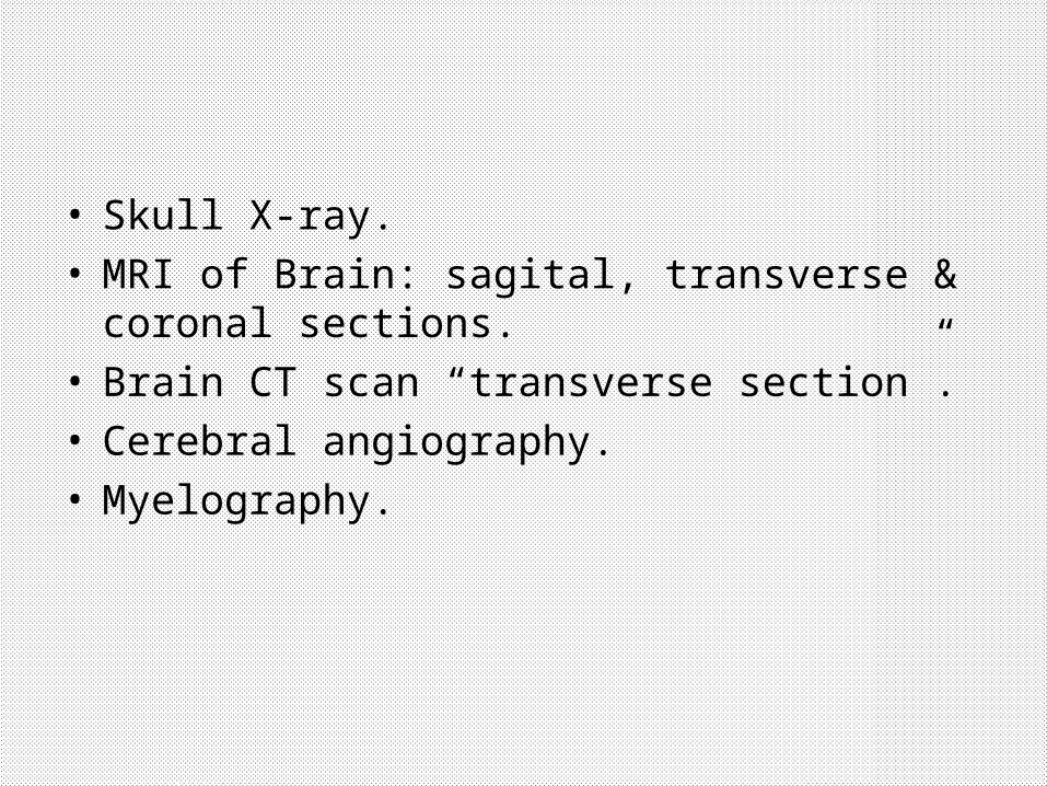
• Skull X-ray.• MRI of Brain: sagital, transverse & coronal
sections.• Brain CT scan “transverse section”.• Cerebral angiography.• Myelography.

Skull X-ray.
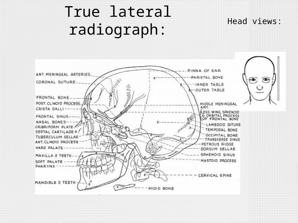
True lateral radiograph:
Head views:


Posterior-anterior radiograph:
Head views:


Head views:Occipitomental radiograph:

Head views:Occipitomental radiograph:

MRI of Brain: sagital section
• The frontal air sinus (1).
• sphenoidal air sinus (2).
• The nasal cavity (3).
• nasopharynx (4).• foramen magnum
(5). • odontoid process
(6).

• optic chiasma (1). • pituitary gland (2).• cerebral aqueduct (often
called the aqueduct of Sylvius) (3)
• fourth ventricle (4) • tentorium cerebelli (5) divides
the cranial cavity within the skull into a supratentorial compartment and an infratentorial compartment.
• The paired mamillary bodies(6) lie in the floor of the third ventricle. These structures form part of the limbic system of the brain.
MRI of Brain: sagital section
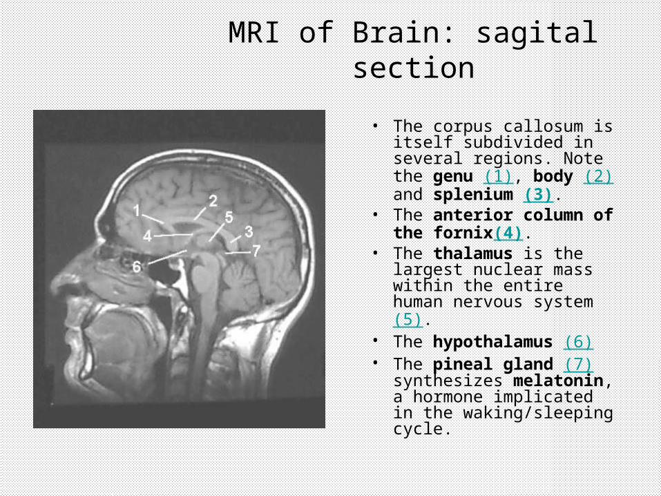
• The corpus callosum is itself subdivided in several regions. Note the genu (1), body (2) and splenium (3).
• The anterior column of the fornix(4).
• The thalamus is the largest nuclear mass within the entire human nervous system (5).
• The hypothalamus (6) • The pineal gland (7)
synthesizes melatonin, a hormone implicated in the waking/sleeping cycle.
MRI of Brain: sagital section

transverse section of the brain
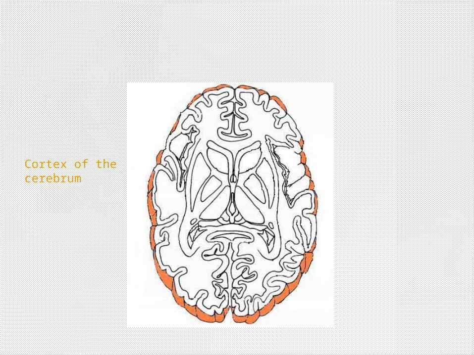
Cortex of the cerebrum
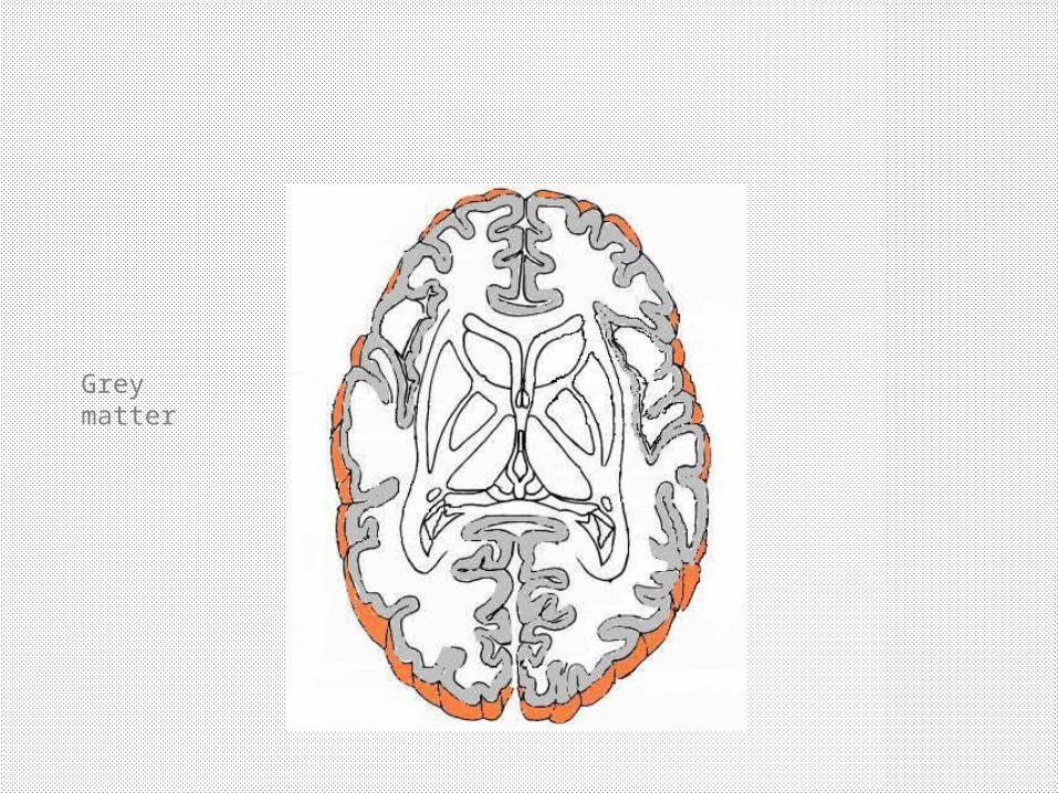
Grey matter

Basal nuclei & thalamus

ventricles

Internal Internal capsulecapsule

External External capsulecapsule
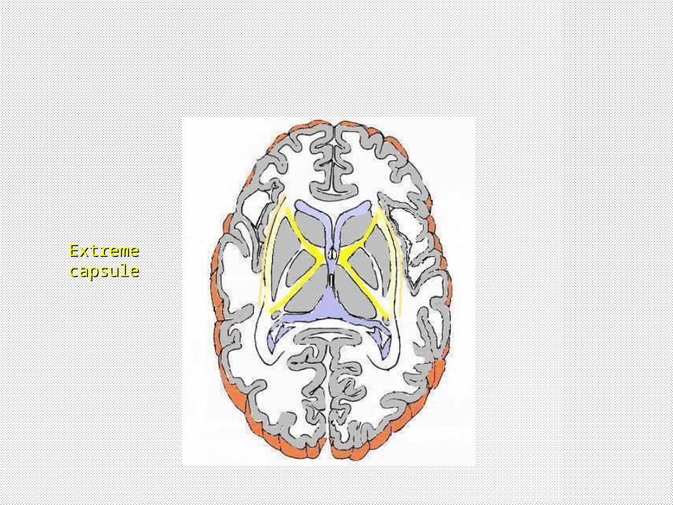
Extreme Extreme capsulecapsule

1. Lateral Ventricle. 2. Third ventricle. 3. Caudate Nucleus. 4. a: Anterior Limb of Internal
Capsule.b: Genu of Internal Capsule.c: Posterior limb of Internal Capsule.
5. Lentiform Nucleus. 6. Insula. 7. Insular artery. 8. Thalamus. 9. a. Gray Matter.
b. White Matter.
MRI of Brain: transverse section

1. Longitudinal fissure.2. cingulate sulcus.3. cingulated gyrus.4. corpus callosum.5. lateral ventricle.6. third ventricle.7. cerebral aqueduct.8. midbrain.9. cerebellar hemisphere.10. fourth ventricle cavity.11. temporal lobe.12. lateral sulcus.
MRI of Brain: coranal section


1. Frontal Lobe. 2. Parietal Lobe. 3. Occipital Lobe. 4. Septum Pellucidum. 5. a. Rostrum of Corpus
Callosum.b. Body of Corpus Callosum.c. Splenium of Corpus Callosum.
6. Pituitry. 7. Sphenoid air sinus. 8. Nasopharynx. 9. Frontal Air sinus. 10. Pons. 11. Medulla Oblongata. 12. Cerebellum. 13. Spinal Cord. 14. Fourth ventricle. 15. Sinus Confluence.

Brain CT scan “transverse section”.
• The dark spaces within the bones represent the air sinuses. The frontal air sinus (A).
• The nasal cavity (B).• In the lateral walls of the
nasal cavity are numerous small dark areas representing the ethmoid air cells (C).
• In the mid-line and at the posterior to the nasal cavity is the sphenoid air sinus (D).

• The orbit (A)
• The eye ball(C).
• Optic nerve (D)
• Optic canal (E) or foramen.
• The medial rectus muscle (B).
Brain CT scan “transverse section”.

Use this image to study the bony features of the skull.
• frontal bone(A). • the zygoma(B). The
temporal bone has a thin squamous component (C) and a much thicker petrous part (D).
• Mastoid air cells (E).• The pinna (F).• The occipital bone (G).
Brain CT scan “transverse section”.

1. Frontal lobe.2. Longitudinal fissure.3. Septum pellucidum.4. Anterior horn of lateral
ventricle.5. Caudate nucleus.6. Third ventricle.7. Posterior horn of tateral
ventricle & choroid plexus.
8. falx cerebri.9. Internal occipital
protuberance.10. Occipital lobe.11. thalamus12. White matter.
Brain CT scan “transverse section”.

Cerebral angiography
Abbreviations:
ACI - internal carotid artery ACA - anterior cerebral artery ACM - middle cerebral artery
BA - basilar artery VA - vertebral artery PCA - posterior cerebral artery PICA - posterior inferior cerebellar artery
Carotid angiogramAnterior view
Carotid angiogramLateral View
Vertebral angiogramAnterior view
Vertebral angiogramLateral View

Myelography

Some Abnormalities:• Space occupying lesions: e.g. tumor, AV malformation,
abscess…

Some Abnormalities:
• prolapsed Intervertebral disc

Some Abnormalities
• Epidural hemorrhage:

Some Abnormalities
• Subdural hematoma:

Some Abnormalities
•Hydrocephalus

Thanks
Related Documents