Imaging and Treatment of Chronic Midportion Achilles Tendinopathy Robert-Jan de Vos

Welcome message from author
This document is posted to help you gain knowledge. Please leave a comment to let me know what you think about it! Share it to your friends and learn new things together.
Transcript

Imaging and Treatment of Chronic Midportion Achilles Tendinopathy
Robert-Jan de Vos

ISBN: 978-90-8559-053-8
Cover by: Robert-Jan de Vos, Evelien Kerkhof and Remko Verhagen
Layout and printing: Optima Grafische Communicatie, Rotterdam, the Netherlands
© R.J. de Vos, the Netherlands, 2010. All rights reserved. No part of this thesis may be reproduced or transmitted in any form or by any means, without prior written permis-sion by the author

Imaging and Treatment of Chronic Midportion Achilles Tendinopathy
Beeldvorming en behandeling van chronische midportion Achilles tendinopathie
Proefschrift
ter verkrijging van de graad van doctor aan deErasmus Universiteit Rotterdam
op gezag van derector magnificus
Prof.dr. H.G. Schmidt
en volgens het besluit van het College voor Promoties
De openbare verdediging zal plaatsvinden opwoensdag 22 September 2010 om 15.30 uur
Door
Robert Johannes de VosGeboren te Gouda

PRoMoTIeCoMMIssIe
Promotoren
Prof.dr.ir. H. WeinansProf.dr. J.A.N. Verhaar
overige leden
Prof.dr. B.W. KoesProf.dr. R.L. DiercksProf.dr. C.N. van Dijk
Copromotoren
dr. J.L. Toldr. H.T.M. van Schie

Contents
Chapter 1 General Introduction 7
Chapter 2 Interobserver reliability of neovascularisation score using Power Doppler ultrasonography in midportion Achilles tendinopathy
23
Chapter 3 The value of Power Doppler ultrasonography in Achilles tendinopathy – a prospective study
35
Chapter 4 Ultrasonographic Tissue Characterisation of human Achilles tendons: quantification of tendon structure through a novel non-invasive approach
49
Chapter 5 Tendon structure is not related to clinical outcome following eccentric exercises in chronic midportion Achilles tendinopathy
63
Chapter 6 Effects of platelet-rich plasma on ultrasonographic findings in chronic midportion Achilles tendinopathy
79
Chapter 7 The additional value of a night splint to eccentric exercises in chronic midportion Achilles tendinopathy – a randomised controlled trial
95
Chapter 8 Autologous growth factor injections in chronic tendinopathy – a systematic review
111
Chapter 9 Platelet-rich plasma injection for chronic Achilles tendinopathy – a randomised controlled trial
129
Chapter 10 General Discussion 145
Chapter 11 Summary 161
Appendices Nederlandse samenvatting 171
Dankwoord 179
Curriculum Vitae 183
PhD Portfolio Summary 185
List of publications 189
VISA-A questionnaire 193


Chapter 1General Introduction


General Introduction 9
PRefACe
According to the Greek mythology Achilles, the legendary warrior from Iliad, was in-vulnerable except for his heel. Since he died due to an arrow that pierced his Achilles tendon, the “Achilles’ heel” symbolises a person’s principal weakness. Nowadays, weak-ness of the Achilles tendon is increasingly gaining attention to medical specialists.
Achilles tendon disorders are a common entity in middle-aged active people, but can also affect the sedentary individuals.1-3 With increasing sports participation in the general population, the number of overuse injuries has increased.1 Tendon disorders comprise 30-50% of all sports-related injuries 3 and there is a lifetime risk of 52% in elite long-distance runners of suffering from an Achilles tendon injury.4 Despite the high prevalence there is still a lack of knowledge about the aetiology and pathogenesis of these injuries.
The terminology used to describe chronic tendon disorders has changed in the past few decades.5 For many years this condition was persistently defined as “tendinitis”, de-noting an inflammation of the tendon. Several authors proposed abandoning this term, as there were no signs of inflammation in chronic painful tendons analysed after biopsy or with microdialysis.5-8 To redress this confusing terminology, the term “tendinopathy” was introduced to describe the clinical condition. Histopathological studies showed that tendinopathy is frequently characterised by degeneration of the tendon tissue, also referred to as “tendinosis”.7 The term tendinosis is based on histopathological character-istics and should only be used after histopathological confirmation.3 The treatment of tendinopathy has been challenging in sports medicine and orthopaedics and therefore it is becoming a major problem in this field. Below an overview will be given on several aspects of chronic Achilles tendon disorders, with a special focus on the imaging and treatment modalities.
AnAToMy of The AChIlles Tendon
The Achilles tendon is the strongest and largest tendon in the human body, which con-nects the calf muscles with the calcaneal bone.9 The midportion of the tendon shows a spiral rotation around its axis that works as an elastic coil functioning as an energy-storing tendon to improve the efficiency of locomotion.9,10 During running the Achilles tendon is subjected to tensile loads up to twelve times the body weight.11 These forces are transmitted through the tendon collagen bundles, which are hierarchically arranged and well ordered predominantly in a longitudinal direction (Figure 1.1).12 The densely packed collagen, mainly type I fibres, gives the tendon its typical white glistening ap-pearance.13 Tendon collagen is the main extracellular matrix (ECM) component and it is

10 Chapter 1
produced by sparsely distributed tenocytes.2,7 The tenocytes, or specialised fibroblasts, normally have a flat phenotype. The major non-collagenous ECM constituents are water and proteoglycans which are core proteins that link the glycosaminoglycans (GAGs) to one large glycosaminoglycan chain.14 The GAGs contain large amounts of bonded negatively charged sulphate ions and thereby extract also free positive charged ions (sodium) from the environment. The subsequent resulting high concentrations of ions attract water in order to balance the internal and external ion concentrations.15 As long as the collagen fibres are well-organised and intact, the matrix does not expand due to the water attraction. The collagen fibres are exposed to an internal stress that balances the water impression. The collagen fibres in the ECM of healthy tendons are in a state of dynamic equilibrium between synthesis and degradation regulated by matrix metal-loproteinases and their inhibitors.7,16
Small blood vessels run throughout the Achilles tendon in the longitudinal plane. The blood supply of the Achilles tendon is physiologically the lowest in the midportion, 2-7 cm proximal from the insertion of the tendon on the calcaneal bone.13,14 This area is therefore defined as the “watershed region”, although this limited blood supply should be sufficient for the metabolic needs of the tendon.17 The longitudinally arranged blood vessels are accompanied by nerves and it is known that the body of the tendon is poorly innervated.
figure 1.1 – The hierarchical structure of a tendon. Collagen forms microfibrils, fibrils, and fibres. A group of fibres constitutes a fascicle. The fascicles unite to form bundles, and are surrounded by the endotenon. The endotenon is a mesh of connective tissue that holds the bundles together and allows some movement of the bundles relative to each other. It carries the longitudinally running blood vessels and nerves. A fine connective tissue sheath, the epitenon, is continuous throughout its inner surface with the endotenon and surrounds the whole tendon. The paratenon is a thin membrane surrounding the epitenon and functions as an elastic sleeve permitting free movement of the tendon against adjacent structures. The peritendon is a general descriptor that consists of the epitenon and the paratenon (modified from Kirkendall & Garret, 1997).

General Introduction 11
There are several structures surrounding the Achilles tendon. The tendon is enclosed by a thin membrane that is frequently referred to as paratenon.9 The paratenon is vascu-lar and better innervated than the Achilles tendon itself. It functions as an elastic sleeve which permits free movement of the tendon against adjacent structures (Figure 1.2). Other clinically relevant structures are a superficial and retrocalcaneal bursa near the insertion of the tendon, the posterior ankle structures, the sural nerve and the medial tendons of the foot and toes.13
hIsToPAThology
Based on histopathological examination, changes of the Achilles tendon can be divided into intratendinous and peritendinous disorders, which can coexist.18
Although tendinosis is a general term for intratendinous degeneration, on histological assessment tendinosis encompasses a wide range of histological degenerative entities not only affecting collagen, but also tenocytes and other non-collagenous matrix com-ponents.19 Macroscopically, degenerative tendon has a soft appearance with a greyish white colour (Figure 1.3).3,5 There are a few histopathological signs that are described in literature.20 Most remarkably is the loss of a well-organised tendon tissue structure. The tendon bundles may show an increased crimping, but separation and complete ruptur-
figure 1.2 – Macroscopic view on an Achilles tendon during surgery. The peritendon, which envelopes the tendon, has been excised and stripped (with permission from Prof. N. Maffulli).

12 Chapter 1
ing of the tendon bundles may also be evident. Some advocate using the term partial tear when there is a large area of discontinuous bundles, but these are probably a severe result of degenerative origin.1 Another feature of tendon degeneration is the diversity in cellular density with areas of hypercellularity and hypocellularity and the tenocyte nuclei may become more rounded. The GAG concentration may be increased and due to the degradation of collagen the tendon attracts high amounts of water and swells. Another obvious feature is an increased vascularisation. The blood vessels are thought to be newly formed and are characterised by a tortuous phenotype and a small lumen. The functional relevance of these blood vessels is questionable as it is thought to be a failed healing response.14
An apparent macroscopical peritendinous change is thickening of the paratenon.1-3,5,18 In addition adhesions may be present. These adhesions can result in contractions around the tendon due to proliferation of connective tissue. This can lead to tendon constriction and increased friction with the surrounding structures.18
Although the histological changes in tendon tissue are referred to as abnormal, from post-mortem studies it is known that these observations are present in Achilles tendons in more than 50% of previously asymptomatic older individuals.21
figure 1.3 – Macroscopic view on an incised degenerative Achilles tendon during surgery. Note the greyish colour and loss of white glistening appearance (with permission from M.P. Heijboer).

General Introduction 13
ClInICAl feATuRes
Tendinopathy is a clinical diagnosis characterised by pain, swelling and impaired load bearing capacity.5 Most patients are middle-aged, between 30 and 60 years old, and participate in sports activity,1-3 although, tendinopathy is also commonly diagnosed in the sedentary.1,2,22,23
On history, pain is typically felt in the tendon midportion. A common training error that is associated with tendinopathy is a rapid increase in activity.24 The phrase “Too much, too soon” is frequently heard in the patient’s history.14 Initially pain is only present during the warming-up period or after activity. Rest may initially decrease the symptoms, but frequently they will return with an increase in activity.13 In a later stage the tendon may become painful during rest and activities of daily living.25
There are no specific time criteria to classify tendinopathy as acute or chronic. Some define “chronic” as more than six weeks, but others advocate a cut-off of 12 weeks. These arbitrary distinctions are not based on histopathological features.18 Another important symptom is morning stiffness, which is also thought to be a good indicator of symp-tomatic recovery.13,24 A validated outcome measure to assess the clinical severity of decreased activity and symptoms in Achilles tendinopathy is the Victorian Institute of Sports Assessment-Achilles (VISA-A) questionnaire (see Appendix).26
On clinical examination the swelling in the tendon midportion may be obvious. Achilles tendon pain is usually localised to the tendon itself and does not refer to other regions, however a few immediate surrounding structures may be relevant for clinical examination.13 It is important to distinguish insertional disorders from midportion ten-dinopathy because of the different underlying pathologies and treatment approach.22 An attempt can be made to differentiate between tendon and paratenon lesions with clinical examination. In cases of paratendinopathy, crepitations may be felt on palpa-tion during movement. Additionally, when the ankle is dorsiflexed the area of swelling and pain remains fixed, whereas in cases of intratendinous pathology the area of focal tenderness moves when the ankle is dorsiflexed.27 Even in experienced hands, however, examiners may have problems reproducing these simple tests. Maffulli et al.28 stated that in the presence of swelling with pain on palpation, which decreases after stretch-ing, there is a high probability that histology will show features of tendinosis.
AddITIonAl dIAgnosTICs
There are several modalities available for imaging of the Achilles tendon. Radiographic imaging is not the prime imaging method to evaluate the midportion of the Achilles tendon, while ultrasonography (US) and Magnetic Resonance Imaging (MRI) are the ad-

14 Chapter 1
ditional diagnostics of first choice in the evaluation of Achilles tendinopathy because of their excellent depiction of soft tissue.29 US provides several benefits in comparison with MRI. It is readily accessible, quick and patient friendly with the possibility of interaction with the patient. Moreover, the addition of Power Doppler can be helpful in examining the blood flow within and around the tendon. The major ultrasonographic findings in chronic midportion Achilles tendinopathy are tendon thickening, hypoechoic areas, disorganised tendon tissue structure and increased Power Doppler flow.27,29
The presence of blood flow in symptomatic tendons detected with Power Doppler ultrasonography (PDU) was first described by Newman et al.30 in 1994, who reported an increased Doppler flow in patients with tendinopathy (Figure 1.4). Öhberg et al.31 performed a case-control study and measured blood flow with Colour Doppler ultra-sonography (CDU) in patients with chronic midportion Achilles tendinopathy. These authors described the increased blood flow as “neovascularisation”, referring to the for-mation of new blood vessels. All 28 painful tendons showed neovascularisation and the 20 asymptomatic control tendons did not. Subsequently, several researchers examined the relationship between neovascularisation and clinical symptoms with inconsistent results. There was a significant difference in the Visual Analogue Scale (VAS) pain score when a group of patients with Achilles tendinopathy without neovascularisation was compared to a group with neovascularisation before the start of treatment.32 Others re-ported that presence of neovascularisation in symptomatic tendons was not correlated with the VISA-A score 33, however more recently researchers have found that patients with tendon neovascularisation had a worse VISA-A score.34 The predictive value of neovascularisation has been studied once, but only a VAS and not a functional score was used in this study. These authors reported that the presence of neovascularisation at baseline had no role in outcome prediction after three and six months.32 There is still discussion about the presence and significance of neovascularisation in chronic tendinopathy. Despite the many published studies, no studies have been performed to test the reliability and reproducibility of the methods to quantify neovascularisation
figure 1.4 – An increased neovascularisation within the thickened Achilles tendon, observed with Power Doppler ultrasonography. The severity of neovascularisation is frequently evaluated in an easy to use five-grade scale (0 to 4+). Figure 1.4A shows a lower degree of neovascularisation, which was defined as grade 2 during the dynamic examination. Figure 1.4B is an example of the highest degree in neovascularisation, scored as grade 4.

General Introduction 15
with PDU or CDU. In the studies on neovascularisation in Achilles tendinopathy, several neovascularisation scoring systems are used but none of these have been validated, nor is the reproducibility reported.
The Achilles tendon, with its superficial location, lends itself for examination with grey scale US.35 By producing a sound wave, receiving echoes, and interpreting those echoes, an ultrasonographic image can be obtained. The sound wave is partially reflected from the layers between different tissue types, and the ability to generate echoes is called echogenicity. On grey scale US, the echogenicity of tendons is based on the density and arrangement of the collagenous matrix. Normal tendons are characterised by a regular echo pattern 35,36 due to well-organised tendon bundles. Disorganisation of tendon tissue leads to a loss of the echogenicity, referred to as hypoechoic areas or irregular tendon structure which are frequently described in cases of tendinosis.29 Some research-ers have focussed on the relationship between the clinical severity of tendinopathy and the ultrasonographic disorders with conflicting results.37-39 The main problem with US is that it is an operator-dependent technique; transducer handling and machine settings can influence size and appearance of a hypoechoic lesion. The interobserver reliability of ultrasonographic assessment of tendon structure has not been studied. It is also very hard to quantify the observed abnormalities through a qualitative assessment, making changes over time difficult to compare.
MRI provides good quality images of the Achilles tendon due to the three-dimensional view and the excellent soft-tissue contrast imaging.29 The MRI appearance of a normal healthy tendon is dark with compact and parallel arrangement of the collagen with low intrinsic water contents, whereas pathologic conditions of the tendon are well recog-nised through an abnormal increase in water signal.40 The interobserver agreement of MRI findings was found to be good, but there may be an overlap of findings in symptom-atic and asymptomatic tendons.41,42 In one study, a lesser extent of signal abnormalities was associated with better clinical outcome at follow-up.39 However, due to the high costs and limited availability MRI is a less used method.24
TReATMenT oPTIons
The treatment of tendinopathy has changed considerably in recent years due to increas-ing knowledge about the underlying pathology. Nonetheless, the treatment of choice can vary from country to country, from clinic to clinic and from clinician to clinician.13
Decades ago, rest was recommended initially due to the lack of a reasonable alter-native.25 Rest can have an effect on symptoms, but it appeared to affect tendon tissue negatively by reducing the collagen amount.43 Later on, non-steroidal anti-inflammatory agents were prescribed.44 However, due to the shift from the “inflammatory” to “degen-

16 Chapter 1
erative” theory in chronic tendon disorders, these medications have fallen out of favour. The role of local anti-inflammatory corticosteroid injections in the management of Achilles tendinopathy has been widely debated. The effects of corticosteroids in Achilles tendinopathy have only been described in anecdotal reports or based on expert opin-ion, but no large studies with long-term follow-up have been performed to examine the role of corticosteroid injections. Injections of corticosteroids have been reported to be associated with spontaneous rupture of the Achilles tendon 3,13,24 and nowadays these injections are discouraged for the treatment of Achilles tendinopathy.
In 1986, Stanish et al.45 developed an exercise program for chronic Achilles tendon pain with the emphasis on increasing speed and number of repetitions with pain being used to guide the intensity of the exercises. Though it was reported that these eccentric exercises led to an improvement in many patients, these exercises only gained atten-tion many years later. Alfredson et al.46 reported that ignoring pain, increasing load and performing the exercises slowly during the program is thought to provide better results. For 12 weeks, 180 repetitions of eccentric exercises were performed daily, by 15 athletes with chronic midportion Achilles tendinopathy. The results were excellent in all athletes. In subsequent studies comparable results were reported, with a patient satisfaction around 80-90%.47,48 The working mechanisms of eccentric exercises are hypothetical and without scientific evidence.3 Some of the hypotheses proposed by Alfredson are based on the repetitive dorsal flexion. On the one hand this could destroy the neovas-cularisation and the accompanying nerves and on the other hand a lengthening of the musculotendinous junction with less strain on the Achilles tendon could be induced.3 A recent systematic review on the clinical effects of eccentric exercise therapy in patients with Achilles tendinopathy showed that the effects on pain are promising.49 The authors stated however, that the major weakness of the included studies is that only pain was used as outcome. Besides pain, the functional status should be recorded to determine the magnitude of the effect, and they suggested the use of the validated VISA-A score.
Based on the promising results, eccentric exercise therapy is increasingly prescribed for patients with chronic midportion Achilles tendinopathy. When there is a failure after an eccentric exercise program, there are many proposed conservative treatment op-tions. With the increasing knowledge on basic science in tendinopathy, new treatment modalities are developing.16
Splints are a common treatment of plantar fasciopathy, which may have a mechanism of action comparable to eccentric exercise therapy; with a continued tendon stretch. The clinical effects of splinting with passive dorsiflexion have been studied twice in patients with Achilles tendinopathy. One research group found benefit using splinting alone in a case series 50, but there was no benefit of a night splint when compared to eccentric exercises in a randomised clinical trial.51 However these studies used pain or less specific functional questionnaires as the primary outcome measures.

General Introduction 17
Other recently introduced therapies with promising results are sclerosing injections with polidocanol to eliminate the neovascularisation 52, the administration of topical glyceryl trinitrate patches to increase collagen production 53, the application of extra-corporeal shockwave therapy to induce a healing response 54 and very recently research groups have been focussing on injecting autologous platelets which may secrete growth factors with regenerative effects.13
It would seem that conservative management provides generally satisfactory results. However, in an eight-year follow-up study it was found that 29% of the 83 patients who were treated conservatively, needed surgery.55 Several types of surgery are nor-mally used.3,18,23,24,56 The most important are incision of the paratenon, excision of the intratendinous macroscopic lesions and multiple longitudinal incisions which can be performed to induce tendon repair processes. Results of surgery are estimated to reach good subjective patient satisfaction in 75-100%.18,24 However, Maffulli et al.23 reported that these success rates are not that high in routine non-specialised clinical practice. Moreover a recent paper reported that studies with a higher methodological score were associated with lower success rates.57
In summary, the treatment of chronic midportion Achilles tendinopathy is difficult with a relatively high percentage of non-responders. There are many proposed treat-ment options, of which an eccentric exercise program is currently the first treatment of choice. Despite the good results on pain scores after eccentric exercises, there are limited data describing functional outcomes. Many new therapies have been develop-ing with promising preliminary results.
AIMs And ouTlIne of ThIs ThesIs
The present thesis is a clinical approach to evaluate the role of ultrasonographic imaging and new conservative treatment options in patients with chronic midportion Achilles tendinopathy.
We were at first interested in whether the determination of a neovascularisation score, measured with PDU, is reliable and of value in the clinical practice. The interobserver reliability of an easy to use five-grade scale was determined (chapter 2). The amount of neovascularisation was also compared with the clinical severity of symptoms at a single point in time.
The predictive value of the neovascularisation score on treatment outcome was evaluated in a follow-up study. In chapter 3 it was hypothesised that presence of neo-vascularisation resulted in a worse treatment outcome after eccentric exercise therapy.
The arrangement of tendon tissue structure may be of relevance because symptomatic tendons frequently show signs of degeneration with a disorganised tendon structure.

18 Chapter 1
In chapter 4 we introduced a novel ultrasonographic technique in human tendons, Ultrasonographic Tissue Characterisation (UTC), which can quantify tendon structure in equine tendons. In these veterinary studies the quantified and categorised tendon structure was related to the histomorphology of equine tendons as a reference test. In this study on human Achilles tendons, we also determined the interobserver reliability of evaluating the obtained images.
In literature, there has been a lot of discussion about the relationship between tendon structure and symptoms. We depict this relationship in chapter 5 for the first time with quantification of the amount of disorganised tendon tissue. Furthermore, the degree of tendon structure disorganisation was evaluated as a predictive factor for clinical improvement when measured before the start of eccentric exercise therapy.
Eccentric exercises are thought to have some influence on increasing tendon collagen type I, but more improved regenerative effects may be achieved with the administra-tion of platelet-rich plasma (PRP), which results in increased amounts of growth factors after degranulation of the platelets. It is suggested that PRP leads to tendon collagen regeneration and increased angiogenesis. The aim of chapter 6 was to study the in vivo effects of PRP in tendinopathy with use of ultrasonographic techniques (UTC and CDU) on tendon structure disorganisation and neovascularisation.
The increasing scientific interest in tendon disorders has led to rapid developments of novel therapies for chronic tendinopathies in the last decades. Because plantar fasciopathy is commonly treated with splinting devices, we hypothesised that it could be beneficial in the treatment of Achilles tendon disorders as well. The use of a night splint is fairly common in the Netherlands. Therefore, a prospective randomised clinical trial was performed in which one group performed an eccentric exercise program and another group performed the same exercises in combination with use of a night splint (chapter 7).
In the present literature there is increasing interest in the use of growth factor injec-tions for chronic tendon disorders. These growth factors can be derived from autologous products through injecting whole blood or PRP. In chapter 8, a systematic review of the literature is reported on the clinical results of autologous growth factor injections.
The use of platelet-rich plasma is gaining popularity due to promising results from laboratory studies and small clinical trials and is being heavily marketed. The conclu-sions and suggestions in recent reviews have led to a widely increased use of PRP in the clinical setting. We performed a double-blind placebo-controlled randomised clinical trial on the effects of a PRP injection in the management of chronic midportion Achilles tendinopathy, which is described in chapter 9.
General Introduction 19
RefeRenCes
1. Maffulli N, Wong J, Almekinders LC. Types and epidemiology of tendinopathy. Clin Sports Med, 2003; 22(4): 675-92.
2. Schepsis AA, Jones H, Haas AL. Achilles tendon disorders in athletes. Am J Sports Med, 2002; 30(2): 287-305.
3. Alfredson H. Chronic midportion Achilles tendinopathy: an update on research and treatment. Clin Sports Med, 2003; 22(4): 727-41.
4. Kujala UM, Sarna S, Kaprio J. Cumulative incidence of Achilles tendon rupture and tendinopathy in male former elite athletes. Clin J Sport Med, 2005; 15: 133-35.
5. Maffulli N, Khan KM, Puddu G. Overuse tendon conditions: time to change a confusing terminol-ogy. Arthroscopy, 1998; 14(8): 840-3.
6. Khan KM, Cook JL, Kannus P, Maffulli N, Bonar SF. Time to abandon the “tendinitis” myth. BMJ, 2002; 16;324(7338): 626-7.
7. Khan KM, Cook JL, Bonar F, Harcourt P, Astrom M. Histopathology of common tendinopathies. Update and implications for clinical management. Sports Med, 1999; 27(6): 393-408.
8. Alfredson H, Thorsen K, Lorentzon R. In situ microdialysis in tendon tissue: high levels of glu-tamate, but not prostaglandin E2 in chronic Achilles tendon pain. Knee Surg Sports Traumatol Arthrosc, 1999; 7(6): 378-81.
9. O’Brien M. The anatomy of the Achilles tendon. Foot Ankle Clin, 2005; 10(2): 225-38. 10. Lichtwark GA, Wilson AM. In vivo mechanical properties of the human Achilles tendon during
one-legged hopping. J Exp Biol, 2005; 208(Pt24): 4715-25. 11. Komi PV. Relevance of in vivo force measurements to human biomechanics. J Biomech, 1990; 23
(Suppl 1): 23-34. 12. Jozsa LG, Kannus P. Human Tendons; anatomy, physiology and pathology. Human Kinetics; Cham-
paign, IL, United States of America: 1997. 13. Woo SL, Renstrőm PA, Arnoczky SP. Tendinopathy in athletes, the encyclopaedia of sports medi-
cine, volume XII. Blackwell Publishing; Hong Kong, China: 2007. 14. Cook JL, Khan KM, Purdam C. Achilles tendinopathy. Man Ther, 2002; 7(3): 121-30. 15. Wang JH. Mechanobiology of tendon. J Biomech, 2006; 39(9): 1563-82. 16. Riley G. The pathogenesis of tendinopathy. A molecular perspective. Rheumatology (Oxford),
2004; 43(2): 131-42. 17. Aström M, Westlin N. Blood flow in chronic Achilles tendinopathy. Clin Orthop Relat Res, 1994;
(308): 166-72. 18. Paavola M, Kannus P, Järvinen TA, Khan K, Józsa L, Järvinen M. Achilles tendinopathy. J Bone Joint
Surg Am, 2002; 84-A(11): 2062-76. 19. Leadbetter WB. Cell-matrix response in tendon injury. Clin Sports Med, 1992; 11(3): 533-78. 20. Aström M, Rausing A. Chronic Achilles tendinopathy. A survey of surgical and histopathologic
findings. Clin Orthop Relat Res, 1995; (316): 151-64. 21. Kannus P, Józsa L. Histopathological changes preceding spontaneous rupture of a tendon. A
controlled study of 891 patients. J Bone Joint Surg Am, 1991; 73(10): 1507-25. 22. Brukner P, Khan K. Clinical Sports Medicine; 2nd edition. McGraw Hill; Sydney: 2001. 23. Maffulli N, Sharma P, Luscombe KL. Achilles tendinopathy: aetiology and management. J R Soc
Med, 2004; 97(10): 472-6. 24. Vora AM, Myerson MS, Oliva F, Maffulli N. Tendinopathy of the main body of the Achilles tendon.
Foot Ankle Clin, 2005; 10(2): 293-308.

20 Chapter 1
25. Burry HC. Late effects of neglected soft tissue injury. Proc R Soc Med, 1969; 62(9): 930-2. 26. Robinson JM, Cook JL, Purdam C, Visentini PJ, Ross J, Maffulli N, Taunton JE, Khan KM. The VISA-A
questionnaire: a valid and reliable index of the clinical severity of Achilles tendinopathy. Br J Sports Med, 2001; 35(5): 335-41.
27. Paavola M, Järvinen TA. Paratendinopathy. Foot Ankle Clin, 2005; 10(2): 279-92. 28. Maffulli N, Kenward MG, Testa V, Capasso G, Regine R, King JB. Clinical diagnosis of Achilles tendi-
nopathy with tendinosis. Clin J Sport Med, 2003; 13(1): 11-5. 29. Bleakney RR, White LM. Imaging of the Achilles tendon. Foot Ankle Clin, 2005; 10: 239-54. 30. Newman JS, Adler RS, Bude RO, Rubin JM. Detection of soft-tissue hyperemia: value of power
Doppler sonography. AJR Am J Roentgenol, 1994; 163(2): 385-9. 31. Ohberg L, Lorentzon R, Alfredson H. Neovascularisation in Achilles tendons with painful tendi-
nosis but not in normal tendons: an ultrasonographic investigation. Knee Surg Sports Traumatol Arthrosc, 2001; 9(4): 233-8.
32. Zanetti M, Metzdorf A, Kundert HP, Zollinger H, Vienne P, Seifert B, Hodler J. Achilles tendons: clini-cal relevance of neovascularisation diagnosed with power Doppler US. Radiology, 2003; 227(2): 556-60.
33. Peers KH, Brys PP, Lysens RJ. Correlation between power Doppler ultrasonography and clinical severity in Achilles tendinopathy. Int Orthop, 2003; 27(3): 180-3.
34. Reiter M, Ulreich N, Dirisamer A, Tscholakoff D, Bucek RA. Colour and power Doppler sonography in symptomatic Achilles tendon disease. Int J Sports Med, 2004; 25(4): 301-5.
35. Martinoli CM, Derchi LE, Pastorino C, Bertolotto M, Silvestri E. Analysis of echotexture of tendons with US. Radiology, 1993; 186: 839-43.
36. van Schie HT, Bakker EM. Structure-related echoes in ultrasonographic images of equine superfi-cial digital flexor tendons Am J Vet Res, 2000; 61: 202-9.
37. Öhberg L, Lorentzon R, Alfredson H. Eccentric training in patients with chronic Achilles tendino-sis: normalised tendon structure and decreased thickness at follow up. Br J Sports Med, 2004; 38: 8-11.
38. Archambault JM, Wiley JP, Bray RC, Verhoef M, Wiseman DA, Elliott PD. Can sonography predict the outcome in patients with achillodynia? J Clin Ultrasound, 1998; 26(7): 335-9.
39. Khan KM, Forster BB, Robinson J, Cheong Y, Louis L, Maclean L, Taunton JE. Are ultrasound and magnetic resonance imaging of value in assessment of Achilles tendon disorders? A two year prospective study. Br J Sports Med, 2003; 37(2): 149-53.
40. Shalabi A. Magnetic resonance imaging in chronic Achilles tendinopathy. Acta Radiol Suppl (Stockholm), 2004; (432): 1-45.
41. Shalabi A, Movin T, Kristoffersen-Wiberg M, Aspelin P, Svensson L. Reliability in the assessment of tendon volume and intratendinous signal of the Achilles tendon on MRI: a methodological description. Knee Surg Sports Traumatol Arthrosc, 2005; 13(6): 492-8.
42. Haims AH, Schweitzer ME, Patel RS, Hecht P, Wapner KL. MR imaging of the Achilles tendon: overlap of findings in symptomatic and asymptomatic individuals. Skeletal Radiol, 2000; 29(11): 640-5.
43. Kannus P, Józsa L, Natri A, Järvinen M. Effects of training, immobilization and remobilization on tendons. Scand J Med Sci Sports, 1997; 7(2): 67-71.
44. Contompasis JP. The management of heel pain in the athlete. Clin Podiatr Med Surg, 1986; 3(4): 705-11.
45. Stanish WD, Rubinovich RM, Curwin S. Eccentric exercise in chronic tendinitis. Clin Orthop Relat Res, 1986; (208): 65-8.
General Introduction 21
46. Alfredson H, Pietilä T, Jonsson P, Lorentzon R. Heavy-load eccentric calf muscle training for the treatment of chronic Achilles tendinosis. Am J Sports Med, 1998; 26(3): 360-6.
47. Mafi N, Lorentzon R and Alfredson H. Superior short-term results with eccentric calf muscle train-ing compared to concentric training in a randomised prospective multicentre study on patients with chronic Achilles tendinosis. Knee Surg Sports Traumatol Arthrosc, 2001; 9(1): 42-7.
48. Fahlstrom M, Jonsson P, Lorentzon R, Alfredson H. Chronic Achilles tendon pain treated with ec-centric calf-muscle training. Knee Surg Sports Traumatol Arthrosc, 2003; 11(5): 327-33.
49. Kingma JJ, de Knikker R, Wittink HW, Takken T. Eccentric overload training in patients with a chronic Achilles tendinopathy: a systematic review. Br J Sports Med, 2006; 41(6): e3
50. Dijkstra HJ, Van Enst GC. The therapeutic value of a G-brace in the management of chronic achil-les tendinosis: a pilot study. Geneeskunde en Sport, 2003; 36: 137-40.
51. Roos EM, Engström M, Lagerquist A, Söderberg B. Clinical improvement after 6 weeks of eccentric exercise in patients with mid-portion Achilles tendinopathy - a randomised trial with 1-year follow-up. Scand J Med Sci Sports, 2004; 14(5): 286-95.
52. Öhberg L, Alfredson H. Ultrasound guided sclerosis of neovessels in painful chronic Achilles tendinosis: pilot study of a new treatment. Br J Sports Med, 2002; 36: 173-7.
53. Paoloni JA, Appleyard RC, Nelson J, Murrell GA. Topical glyceryl trinitrate treatment of chronic noninsertional achilles tendinopathy. A randomised, double-blind, placebo-controlled trial. J Bone Joint Surg Am, 2004; 86-A: 916-22.
54. Rompe JD, Nafe B, Furia JP, Maffulli N. Eccentric loading, shock-wave treatment, or a wait-and-see policy for tendinopathy of the main body of tendo Achillis: a randomized controlled trial. Am J Sports Med, 2007; 35(3): 374-83.
55. Paavola M, Kannus P, Paakkala T, Pasanen M, Järvinen M. Long-term prognosis of patients with Achilles tendinopathy. An observational 8-year follow-up study. Am J Sports Med, 2000; 28(5): 634-42.
56. Kader D, Saxena A, Movin T, Maffulli N. Achilles tendinopathy: some aspects of basic science and clinical management. Br J Sports Med, 2002; 36(4): 239-49.
57. Tallon C, Coleman BD, Khan KM, Maffulli N. Outcome of surgery for chronic Achilles tendinopathy. A critical review. Am J Sports Med, 2001; 29(3): 315-20.

Chapter 2Interobserver reliability of neovascularisation
score using Power Doppler ultrasonography in midportion Achilles tendinopathy
P.M. SengkerijR.J. de Vos
A. WeirB.J.G. van Weelde
J.L. Tol
Department of Sports Medicine and Radiology
The Hague Medical Centre Antoniushove
Leidschendam, the Netherlands
Am J Sports Med 2009; 37(8): 1627-1631

24 Chapter 2
AbsTRACT
Background - Power Doppler ultrasonography is widely used to examine neovasculari-sation in midportion Achilles tendinopathy. The reliability of the grading of the amount of neovascularisation has not been examined previously.
Hypothesis - Power Doppler ultrasonography can be performed with a high interob-server reliability to determine the neovascularisation score in patients with midportion Achilles tendinopathy.
Study design - Case control study (diagnosis); Level of evidence, 4.
Methods - Thirty-three symptomatic and 17 asymptomatic Achilles tendons from 25 consecutive patients were included for ultrasound examination. Victorian Institute of Sport Assessment-Achilles score was used to assess the severity of the Achilles tendi-nopathy. Each tendon was scored twice by different radiologists using the modified Öhberg score for neovascularisation.
Results - The intraclass correlation coefficient for interobserver reliability was 0.85. Neo-vascularisation was observed in 70% (23/33) of the symptomatic tendons and in 29% (5/17) of the asymptomatic tendons. The Spearman correlation coefficient between the Victorian Institute of Sport Assessment-Achilles score and the degree of neovascularisa-tion was -0.16 (p = 0.10).
Conclusion - An excellent interobserver reliability was found for determining the degree of neovascularisation on Power Doppler ultrasonography examination. Neovessels were present in a majority of symptomatic tendons. The severity of symptoms was not cor-related with the neovascularisation score.
Clinical relevance - Power Doppler ultrasonography is widely used to evaluate tendi-nopathy without knowledge of the difference in observations between several testers. Interobserver reliability of the evaluation of the degree of neovascularisation in chronic midportion Achilles tendinopathy is excellent.
Interobserver reliability of neovascularisation score 25
InTRoduCTIon
Midportion Achilles tendinopathy is a commonly used term for noninsertional chronic pain in the Achilles tendon.1,6,7,12 In long-standing midportion Achilles tendinopathy, structural changes within the tendon can be detected using ultrasound imaging. Ultrasonography is now widely used and is relatively safe and inexpensive, provides fast results, and is tolerable for most patients. However, it is frequently described as an operator-dependent imaging technique.2
Neovascularisation throughout the tendon, detected with Power Doppler ultrasonog-raphy (PDU), has been observed frequently in the last decade in Achilles tendinopathy.1,2 A neovascularisation score determined with PDU is widely used to assess the severity of Achilles tendinopathy. In some studies neovascularisation was observed in all symp-tomatic tendons 11,14, whereas others reported neovessels in 47% to 88% of symptomatic tendons.6,9,15,16,20 The significance of these neovessels in chronic midportion Achilles ten-dinopathy is a topic of debate. Many studies have examined the relationship between neovascularisation and pain or discomfort, with conflicting results.6,9 Some studies showed a relationship between the degree of neovascularisation and symptoms 11,13,16, whereas others failed to find a relationship.3,15,17 The reasons for these differences are unknown. One problem when comparing research results, is the lack of a uniform man-ner of assessing the degree of neovascularisation.
Varying methods have been described to assess the degree of neovascularisation. Some studies described only the presence or absence of neovessels 14,20, others used a scoring system ranging from 0 to 4+ 11,13, and others used a surface area measure-ment.3,4,15
De Vos et al.6 suggested that physical activity, patient positioning, and ultrasound examination should be standardised to improve the reliability of assessing the degree of neovascularisation. These investigators recommended 24-hour abstinence from heavy-load eccentric training, sporting activity, or physical exertion before examination. During PDU, plantar flexion of the ankle should be standardised and the probe pressure should be minimal to avoid obliteration of the vessels. By using these standardised conditions as well as the modified Öhberg score, investigators can focus on the reproducibility and the interobserver and intra-observer reliability of the neovascularisation score.6 No pre-vious studies have examined the reliability of PDU measurement of neovascularisation in Achilles tendinopathy.
The first aim of this study was to evaluate the interobserver reliability of the ultraso-nographic degree of neovascularisation using the modified Öhberg score.6 The second aim was to observe the prevalence of neovessels in symptomatic and asymptomatic tendons. The third aim was to evaluate the correlation between the degree of neovascu-larisation and the Victorian Institute of Sports Assessment-Achilles (VISA-A) score.
26 Chapter 2
MeThods
Patients
Patients were recruited from the sports medicine department of a large district hospital. A clinical diagnosis of Achilles tendinopathy was made when patients were evaluated for palpation pain on the Achilles tendon 2 to 7 cm proximal to the insertion on the calcaneus and tenderness of the Achilles tendon during and after sporting activities. Inclusion and exclusion criteria are listed in Table 2.1.
Table 2.1 – Inclusion and exclusion criteria.
Inclusion Criteria exclusion Criteria
Age 18-70 years Achilles tendon insertional disorder
Symptoms more than two months Complete tendon rupture
Active participation in sports Systemic disorders
Clinical diagnosis of midportiontendinopathy
All patients were assessed by an experienced sports medicine physician to ensure correct inclusion. Patients were informed about the aim and background of the study and gave their written consent. All patients were then examined by a single researcher using a standard protocol. The VISA-A questionnaire was completed with minimal re-searcher assistance. A folder with information about the study was given to the patient. Patients were instructed to abstain from heavy physical or sporting activity 24 hours before the examination. The study protocol was approved by the regional medical ethics committee.
study design
The study design was a case control study. All consecutive patients with midportion Achilles tendinopathy who met the inclusion criteria and consented to participate underwent PDU examination of both Achilles tendons. Eight radiologists participated in the study.
ultrasonography examination
Power Doppler ultrasonography was performed using a linear high-frequency 8 to 13 MHz transducer (Elegra, Siemens System, Erlangen, Germany). A pulse repetition fre-quency of 868 Hz was used for maximal sensitivity of Power Doppler. Patients were ex-amined by two of the eight radiologists. All radiologists were trained in musculoskeletal ultrasound. They had participated in previous studies concerning neovascularisation score in Achilles tendinopathy. Before this study began, all participating radiologists were instructed in determining the degree of neovascularisation using the modified

Interobserver reliability of neovascularisation score 27
Öhberg score.14 The radiologists were blinded to the clinical status of the tendons. Both radiologists, one after another, independently scored the degree of neovascularisation in both tendons of a patient. Both radiologists used the same ultrasound machine in the same room. Patients lay prone on the examination table during the short period between the two measurements. For the measurements, patients lay prone with a cushion-roll under their ankles. The ankle was placed in neutral position. To avoid obliteration of the vessels, the pressure of the probe was kept to a minimum. The tendons were examined in longitudinal and transverse planes as described by Öhberg et al.13,14, Peers et al.15, Reiter et al.16 and de Vos et al.6 The location and number of neovessels observed with PDU were scored using the modified Öhberg score.6,14 This score was recorded as 0 (no vessels visible), 1+ (one vessel, mostly anterior to the tendon), 2+ (one or two vessels throughout the tendon), 3+ (three vessels throughout the tendon), or 4+ (more than three vessels throughout the tendon). The images were stored to disk.
data analysis
Interobserver reliability was calculated with a one-way random model. The intraclass correlation coefficient (ICC) was used to evaluate the correlation between the observa-tions of the radiologists in symptomatic Achilles tendons. To evaluate the ICC, one score per observer per tendon was computed. According to Fleiss 8, the reliability is excellent if ICC is higher than 0.75, fair to good if ICC is 0.75 to 0.4 and poor if ICC is less than 0.4. The Spearman correlation coefficient analysis was used to examine the correlation between the VISA-A score and the mean neovascularisation score of the two measurements. The Spearman correlation (positive or negative) is interpreted as no association when 0.0, weak when 0.2, moderate when 0.5, strong when 0.8, and perfect when 1.00.21 Statistical significance was assumed when P values were less than 0.05. SPSS version 15.0.0 statisti-cal software (SPSS Science Inc, Chicago, Illinois, USA) was used to perform the statistical analysis.
ResulTs
Patients
Twenty-five patients (50 Achilles tendons) were included. One patient was excluded. There were 16 male and nine female patients. Twelve patients were active long-distance runners, five patients visited fitness centres to do spinning and aerobics, three patients played competitive soccer, three patients were active hikers, and two patients were students at an academy of sports. The mean age was 44.4 years (range, 18-63), mean duration of symptoms was 36.7 weeks (range, 8-120), and mean body mass index was 24.9 kg/m² (range, 19.3-28.9). Of the 50 tendons, 33 were symptomatic and 17 were
28 Chapter 2
asymptomatic. In six patients symptoms were located in the left Achilles tendon, 11 patients had symptoms in the right tendon, and eight patients had bilateral complaints. Ten patients had had to stop their sporting activities, and 11 patients had had to reduce their activities. Four patients had been able to continue their sporting activities.
Interobserver reliability of the neovascularisation score
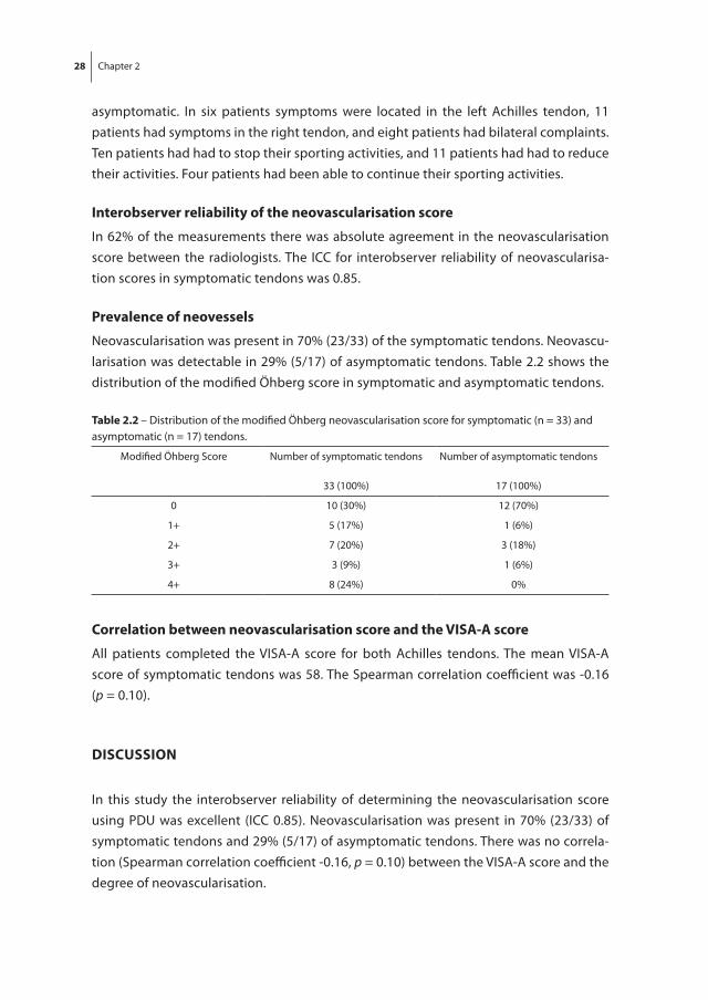
In 62% of the measurements there was absolute agreement in the neovascularisation score between the radiologists. The ICC for interobserver reliability of neovascularisa-tion scores in symptomatic tendons was 0.85.
Prevalence of neovessels
Neovascularisation was present in 70% (23/33) of the symptomatic tendons. Neovascu-larisation was detectable in 29% (5/17) of asymptomatic tendons. Table 2.2 shows the distribution of the modified Öhberg score in symptomatic and asymptomatic tendons.
Table 2.2 – Distribution of the modified Öhberg neovascularisation score for symptomatic (n = 33) and asymptomatic (n = 17) tendons.
Modified Öhberg Score Number of symptomatic tendons
33 (100%)
Number of asymptomatic tendons
17 (100%)
0 10 (30%) 12 (70%)
1+ 5 (17%) 1 (6%)
2+ 7 (20%) 3 (18%)
3+ 3 (9%) 1 (6%)
4+ 8 (24%) 0%
Correlation between neovascularisation score and the VIsA-A score
All patients completed the VISA-A score for both Achilles tendons. The mean VISA-A score of symptomatic tendons was 58. The Spearman correlation coefficient was -0.16 (p = 0.10).
dIsCussIon
In this study the interobserver reliability of determining the neovascularisation score using PDU was excellent (ICC 0.85). Neovascularisation was present in 70% (23/33) of symptomatic tendons and 29% (5/17) of asymptomatic tendons. There was no correla-tion (Spearman correlation coefficient -0.16, p = 0.10) between the VISA-A score and the degree of neovascularisation.
Interobserver reliability of neovascularisation score 29
This study shows that interobserver reliability when quantifying neovascularisation with PDU in symptomatic Achilles tendons, using the modified Öhberg score, was excel-lent. This means that different radiologists examining the same tendon had excellent agreement between their findings. There are no other studies in which the reliability of measuring the neovascularisation in Achilles tendinopathy has been examined.
The reliability of ultrasound assessment for other musculoskeletal disorders was studied by Szkudlarek et al.19, who evaluated the interobserver agreement of ultrasono-graphic assessment of finger and toe joints in patients with active rheumatoid arthritis. In one assessment these investigators evaluated the agreement of Power Doppler signal in the joints. Two observers examined flow and vessels with PDU, using a four-grade measuring system, in 150 small joints of 30 patients. They found exact agreement between observers in 87% of cases. An ICC of 0.72 was found, which represents a fair to good reliability.8 Cook et al.5 found high reproducibility of the measurement of vascularity within patellar tendons using Colour Doppler ultrasonography (CDU). Two observers estimated tendon vascularity in millimetres in 74 tendons during ultrasound examination and from stored films. The correlation between examiners was excellent for the vessel lengths measured from the digital images.
The prevalence of neovascularisation in symptomatic Achilles tendons (70%) in this study is within the range reported by other investigators. Peers et al.15, Reiter et al.16, Zanetti et al.20, de Vos et al.6 and Leung and Griffith 10 reported the presence of neoves-sels in 47% to 88% of the symptomatic tendons. On the contrary, Öhberg et al.13,14 and Lind et al.11 found neovascularisation in all symptomatic tendons. Besides examining the prevalence of neovascularisation in symptomatic tendons, we also examined neovessels in asymptomatic tendons; 29% (5/17) of these asymptomatic tendons had a degree of neovascularisation.
Boesen et al.3 reported that in 30% (6/20) of asymptomatic tendons of untrained sub-jects, CDU activity was measured in the tendon. Those authors found that prior physical activity can influence the degree of neovascularisation. It is questionable whether neovascularisation in asymptomatic tendons is a physiological phenomenon or if this represents a prepathologic stage.
The differing results may be explained, in part, by the findings of Richards et al.17 who reported that PDU is preferred over CDU for examination of vessels because PDU shows a greater number and extent of vessels and more contiguous vessels than does CDU. Boesen et al.3 and Öhberg et al.14 used CDU to examine neovascularisation in the Achilles tendons. In this study we found a lower prevalence of neovessels than Öhberg et al.14 did, despite the use of PDU. No explanation for this difference could be found. Although variations in neovascularisation score might be attributable to machine set-tings, differences in the prevalence of neovessels also might be explained by the use of standardised conditions, as mentioned recently by de Vos et al.6

30 Chapter 2
We found no correlation between the degree of neovascularisation and the symptoms. To examine the relationship of symptomatic Achilles tendinopathy and neovascularisa-tion score, we used the VISA-A questionnaire, which provides a valid and reliable index of the severity of Achilles tendinopathy.18 De Vos et al.6 found no correlation between the neovascularisation score and the VISA-A score at baseline in their study. On the contrary, Peers et al.15 found a negative correlation between PDU and a functional test and pain score when using one item (jumping capability) of the VISA-A questionnaire. Reiter et al.16 found significantly worse VISA-A scores in patients with blood flow in the Achilles tendon than in symptomatic patients without flow seen on PDU. Zanetti et al.20 found higher visual analogue scale scores in patients with tendons showing neovascularisa-tion at the initial evaluation. In 2002, Öhberg and Alfredson 13 reported that all patients who were unsatisfied after sclerosing treatment had neovascularisation on CDU. Similar results were found by Lind et al.11 in 2006.
Although analysing Achilles tendons with PDU is reliable, the conflicting results about the presence of neovessels and correlation between the degree of neovascularisation and pain or discomfort do not help to clarify the significance of neovessels in Achilles tendinopathy.9
One of the limitations of this study is that standardisation of physical activity before the examination was not validated. Boesen et al.3 reported an increase of neovascu-larisation directly after exercise. It is unclear whether the increase in the detectability of neovessels lasts longer than 24 hours after exercise. Therefore, there are indications that neovascularisation is influenced during a treatment period. This may have influenced the prevalence of neovascularisation in this study group. However, the results of the interobserver reliability are independent of treatment period, because measurements were performed at the same moment.
Another limitation is the use of the VISA-A score to index asymptomatic tendons. In this study, the VISA-A score for asymptomatic tendons was probably lower because of a “negative” influence on functional activity of the contralateral symptomatic tendon in one subject.
In cases of severe pain it is not always possible to blind the radiologist as to whether a tendon is symptomatic, because application of probe pressure elicits a pain response from the patient. As a result, radiologists may examine these symptomatic tendons more thoroughly to find any neovascularisation. Another limitation is that because of previous studies in this hospital, the radiologists had considerable experience in using PDU to assess neovascularisation.
More studies are needed that examine intra-observer reliability, test-retest character-istics, and validity of the assessment of neovascularisation with PDU using the modified Öhberg score. Once these factors have been determined, larger studies will be needed

Interobserver reliability of neovascularisation score 31
to determine the clinical significance of neovascularisation in cases of Achilles tendi-nopathy.
ConClusIon
This is the first study to evaluate interobserver reliability in assessing the sonographic degree of neovascularisation in Achilles tendinopathy. An excellent interobserver reli-ability (ICC 0.85) was found. No correlation was found between VISA-A score and neo-vascularisation score. Further studies examining intra-observer reliability and test-retest characteristics are needed.

32 Chapter 2
RefeRenCes
1. Alfredson H. Chronic midportion Achilles tendinopathy: an update on research and treatment. Clin Sports Med. 2003; 22: 727-41.
2. Bleakney RR, White LM. Imaging of the Achilles tendon. Foot Ankle Clin. 2005; 10: 239-54. 3. Boesen MI, Koenig MJ, Torp-Pedersen S, Bliddal H, Langberg H. Tendinopathy and Doppler activ-
ity: the vascular response of the Achilles tendon to exercise. Scand J Med Sci Sports. 2006; 16: 463-9.
4. Boesen MI, Torp-Pedersen S, Koenig MJ, et al. Ultrasound guided electrocoagulation in patients with chronic non-insertional Achilles tendinopathy: a pilot study. Br J Sports Med. 2006; 40: 761-6.
5. Cook JL, Ptazsnik R, Kiss ZS, Malliaras P, Morris ME, De Luca J. High reproducibility of patellar tendon vascularity assessed by colour Doppler ultrasonography: a reliable measurement tool for quantifying tendon pathology. Br J Sports Med. 2005; 39: 700-3.
6. de Vos RJ, Weir A, Cobben LP, Tol JL. The value of power Doppler ultrasonography in Achilles tendinopathy: a prospective study. Am J Sports Med. 2007; 35: 1696-1701.
7. de Vos RJ, Weir A, Visser RJ, de Winter T, Tol JL. The additional value of a night splint to eccentric exercises in chronic midportion Achilles tendinopathy: a randomised controlled trial. Br J Sports Med. 2007; 41: e5.
8. Fleiss JL. The Design and Analysis of Clinical Experiments. New York, NY: John Wiley; 1986. 9. Knobloch K, de Vos RJ, Weir A, Tol JL. The use of a neovascularisation score to predict clinical
severity in Achilles tendinopathy. Am J Sports Med. 2008; 36: 395-7. 10. Leung JL, Griffith JF. Sonography of chronic Achilles tendinopathy: a case-control study. J Clin
Ultrasound. 2008; 36: 27-32. 11. Lind B, Öhberg L, Alfredson H. Sclerosing polidocanol injections in midportion Achilles tendi-
nosis: remaining good clinical results and decreased tendon thickness at 2-year follow-up. Knee Surg Sports Traumatol Arthrosc. 2006; 14: 1327-32.
12. Maffulli N, Wong J, Almekinders LC. Types and epidemiology of tendinopathy. Clin Sports Med. 2003; 22: 675-92.
13. Öhberg L, Alfredson H. Ultrasound guided sclerosis of neovessels in painful chronic Achilles tendinosis: pilot study of a new treatment. Br J Sports Med. 2002; 36: 173-7.
14. Öhberg L, Lorentzon R, Alfredson H. Neovascularisation in Achilles tendons with painful tendi-nosis but not in normal tendons: an ultrasonographic investigation. Knee Surg Sports Traumatol Arthrosc. 2001; 9: 233-8.
15. Peers KH, Brys PP, Lysens RJ. Correlation between power Doppler ultrasonography and clinical severity in Achilles tendinopathy. Int Orthop. 2003; 27: 180-3.
16. Reiter M, Ulreich N, Dirisamer A, Tscholakoff D, Bucek RA. Colour and power Doppler sonography in symptomatic Achilles tendon disease. Int J Sports Med. 2004; 25: 301-5.
17. Richards PJ, Win T, Jones PW. The distribution of microvascular response in Achilles tendinopathy assessed by colour and power Doppler. Skeletal Radiol. 2005; 34: 336-42.
18. Robinson JM, Cook JL, Purdam C, et al. The VISA-A questionnaire: a valid and reliable index of the clinical severity of Achilles tendinopathy. Br J Sports Med. 2001; 35: 335-41.
19. Szkudlarek M, Court-Payen M, Jacobsen S, Klarlund M, Thomsen HS, Ostergaard M. Interob-server agreement in ultrasonography of the finger and toe joints in rheumatoid arthritis. Arthritis Rheum. 2003; 48: 955-62.
20. Zanetti M, Metzdorf A, Kundert HP, et al. Achilles tendons: clinical relevance of neovascularisation diagnosed with power Doppler US. Radiology. 2003; 227: 556-60.

Interobserver reliability of neovascularisation score 33
21. Zou KH, Tuncali K, Silverman SG. Correlation and simple linear regression. Radiology. 2003; 227: 617-622.


Chapter 3The value of Power Doppler ultrasonography in
Achilles tendinopathy – a prospective study
R.J. de VosA. Weir
L.P.J. CobbenJ.L. Tol
Department of Sports Medicine and Radiology
The Hague Medical Centre Antoniushove
Leidschendam, the Netherlands
Am J Sports Med 2007; 35(10): 1696-1701

36 Chapter 3
AbsTRACT
Background - Neovascularisation, detected with Power Doppler ultrasonography (PDU), is thought by some to play a central role in pathogenesis of Achilles tendinopathy.
Hypothesis - Power Doppler ultrasonography neovascularisation score is correlated with clinical severity at baseline and after conservative treatment.
Study design - Cohort study (prognosis); Level of evidence, 2.
Methods - Seventy tendons from 58 patients with chronic midportion Achilles tendi-nopathy were included, and 63 symptomatic tendons were analysed. All patients were prescribed a 12-week heavy-load eccentric training program and evaluated with PDU at baseline and 12 weeks. Patient satisfaction, Victorian Institute of Sports Assessment-Achilles (VISA-A) score, and mean visual analogue scale (VAS) score were correlated with degree of neovascularisation (five-grade scale).
Results - Of the 63 symptomatic tendons, baseline neovascularisation scores were 23 grade 0 (37% no neovessels), 18 grade 1, 8 grade 2, 8 grade 3, and 6 grade 4 (63% neovas-cularisation grades 1-4). At baseline, neovascularisation was not significantly correlated with the mean VAS score (r = 0.19, p = 0.13) and VISA-A score (r = -0.23, p = 0.07). At 12-week follow-up, the neovascularisation score significantly correlated with the mean VAS score (r = 0.43, p < 0.001) and VISA-A score (r = -0.46, p < 0.001). No significant dif-ferences were found in improvement of VISA-A score after treatment between patients with neovessels (grades 1-4) or without neovessels (grade 0) at baseline.
Conclusion - Sixty-three percent of the symptomatic tendons were found to have neovessels at baseline. There was no significant correlation between neovascularisation score and clinical severity at baseline, but at follow-up, there was a significant correla-tion. Neovascularisation at baseline did not predict clinical outcome after conservative treatment.

The value of Power Doppler ultrasonography 37
InTRoduCTIon
Chronic midportion Achilles tendinopathy is a generic term for long-standing, nonin-sertional tendon pain and is frequently reported in the athletic population.12,16 Terminol-ogy used to describe this entity may be confusing.20 When the underlying pathological abnormality is unknown, the term Achilles tendinopathy is preferred, describing only the painful condition of the tendon.1 A phenomenon that is frequently seen in these symptomatic tendons is hypervascularity or neovascularisation.1,6 In the last decade, the addition of Power Doppler imaging to ultrasound enabled detection of blood flow in tendons.6 Recently, many studies have been performed to determine the exact role of neovascularisation. There is still discussion about the presence and significance of neovessels in chronic tendinopathy. Alfredson1 described a theory in which he suggests that the ingrowth of neovessels and their adjacent nerves in the tendinopathic ventral part of the tendon are the source of pain. On Power Doppler ultrasonography (PDU) examination, this neovascularisation disappears when the ankle is dorsiflexed. For that reason, eccentric exercises may permanently damage these neovessels and the accom-panying nerves.1,2 In some studies, successful results are reported when the neovessels are used as the target of sclerosing treatment with polidocanol injections.4,11,13 The authors reported that the neovessels disappeared in the satisfied patients but not in the dissatisfied patients.
A number of recent studies examined the role of neovascularisation in relatively small populations.4,7,8,11,13,15,17,18,21 In some studies 4,11,13,15, neovascularisation was found in all symptomatic tendons, while others report neovessels being present in 50% to 88% of symptomatic tendons.17,18,21
The relationship between neovascularisation and clinical symptoms is unclear, and there are conflicting data. A positive correlation between neovascularisation and visual analogue scale (VAS) score was reported by Peers et al.17, but this is not supported by the data of Zanetti et al.21 when the group without neovessels was compared with the group with neovessels. Reiter et al.18 reported that presence of neovascularisation in symp-tomatic tendons was associated with a worse Victorian Institute of Sports Assessment-Achilles (VISA-A) score. Peers et al.17 found correlation between neovascularisation and only one of the ten items on the VISA-A questionnaire.
Many studies have examined the relationship between neovascularisation at a single point in time, but for the clinician and the patient, it is more interesting to examine the predictive value of neovascularisation at baseline on clinical outcome. The VISA-A questionnaire is a validated tool for assessing clinical outcome and includes questions on pain, activity, and function.10,19 To our knowledge, the predictive value has only been studied once, by Zanetti et al.21, but a functional score was not used in this study. Zanetti et al.21 analysed the presence of neovessels at baseline related to pain scores after

38 Chapter 3
conservative treatment. The presence of neovascularisation at baseline had no role in outcome prediction, measured with the VAS score.
The first aim of this prospective clinical study was to evaluate whether neovessels were present in all symptomatic tendons. The second aim was to evaluate whether there was a correlation between neovascularisation and clinical severity. The third aim was to evaluate the role of neovascularisation in predicting clinical functional outcome.
MATeRIAls And MeThods
Patients
Patients were recruited at the sports medicine outpatient department of a large district general hospital. Inclusion criteria and exclusion criteria are listed in Table 3.1. All patients were active in sports participation and had a tendon that was tender on palpation and during or after sport. The tendon thickening and/or tenderness was located 2 to 7 cm proximal to the distal insertion. The diagnosis was made based on this clinical examination.
Table 3.1 – Inclusion and exclusion criteria.
Inclusion criteria exclusion criteria
Age 18-70 years Previous performance of an eccentric exercise program
Symptoms more than 2 months Inability to perform heavy-load eccentric exercises
Active participation in sports activities Insertional disorder
Desire to return to original level of sports Tendon rupture
Systemic illness
After patient inclusion by a clinician, an appointment was made for an ultrasound examination. After the ultrasound examination, all patients were examined by a single researcher using a standard protocol. This protocol consisted of standardised outcome measures: patient satisfaction, VISA-A questionnaire 19, and VAS score during activities of daily living and sports (a mean was calculated from these two VAS scores). Subjective patient satisfaction was rated as poor, fair, good, or excellent. A good or excellent result was considered as successful. The VISA-A questionnaire, a validated instrument to evalu-ate the severity of symptoms and functional restriction in Achilles tendinopathy, was completed with minimum assistance from the researcher. At both appointments, the VAS scores were noted.
The patients were informed about the aim and background of the study, and patients could give their consent. When consent had been provided, a detailed instruction of the conservative treatment was conducted. All patients performed a heavy-load eccentric exercise regimen according to Alfredson et al.5, with or without a night splint.9

The value of Power Doppler ultrasonography 39
study design
The study design was a prospective clinical trial. The study was part of a randomised controlled trial comparing eccentric exercises in combination with splinting to eccentric exercises alone.9 Patients with midportion Achilles tendinopathy were included in the study, and ultrasonography was performed at baseline and at 12-week follow-up.
Spearman correlations were used to evaluate the relationship between ultrasono-graphic findings and outcome variables. The Wilcoxon signed rank test was used to assess changes over time in the variables within groups, and the Mann-Whitney U test was used for changes between groups. The χ2 test was used to evaluate the difference in patient satisfaction between groups. Statistical significance was assumed when P values were < 0.05. All analyses were performed with the use of SPSS (version 12.0.1) statistical software (SPSS Science, Chicago, Illinois, USA).
The study protocol was approved by the regional Medical Ethics Committee.
ultrasound examination
At baseline and at 12-week follow-up, Power Doppler ultrasonography (Elegra, Siemens Systems, Erlangen, Germany) was performed using a linear high-frequency 8 to 13 MHz transducer. The examination was performed by a musculoskeletal radiologist. The same researcher, who was blinded to the clinical status of the subjects, was present at all ultrasonographic examinations and determined the neovascularisation score. Patients lay prone during the examination, with their feet hanging freely over the edge of the examination table. The pressure of the probe was kept to a minimum to avoid oblit-eration of small vessels. Symptomatic Achilles tendons were observed in longitudinal and transverse planes as described by Öhberg et al.14, Reiter et al.18 and Peers et al.17 On investigation, the researcher and the radiologist evaluated the neovascularisation of the Achilles tendon. The neovascularisation score was determined by the location and number of vessels noticed on PDU examination. This modified Öhberg score 13 was recorded as 0 (no vessels visible), 1+ (one vessel mostly in the anterior part), 2+ (two vessels throughout the tendon), 3+ (three vessels throughout the tendon), and 4+ (> three vessels throughout the tendon). The ultrasound images were saved to disk.
After the use of this grading system, a distinction was made between tendons without neovessels (grade 0) and with neovessels (grades 1-4) as described by several authors.7,11,18

40 Chapter 3
ResulTs
Patients
Sixty-seven patients (79 tendons) visited the Hague Medical Centre for the study be-tween March and December 2005. Nine patients were excluded – four patients had an insertional disorder, two patients had already performed a heavy-load eccentric training program, one patient had a partial rupture, one patient had a total rupture, and one patient was unable to carry out the eccentric exercises. Seventy symptomatic tendons (46 unilateral, 12 bilateral) from 58 patients were included.
The mean age was 44.6 years (range 26-59), mean duration of symptoms was 30.7 months (range 2-221), and the mean body mass index was 25.1 kg/m² (range 20.2-34.5) (Table 3.2). All patients were active in sports, and most of them had to stop their sporting activities. In 24 cases, sports activity had to be stopped, and in 19 cases, sports activity had to be reduced. Six patients (seven tendons) did not complete the treatment and/or did not show up at the follow-up evaluation.9 In total, 63 symptomatic tendons of 52 patients completed the study and could be analysed.
single Point in Time
BaselineAt baseline, there were neovessels in 63% (n = 40) of the symptomatic tendons. Thirty-seven percent (n = 23) had no neovascularisation (grade 0). Table 3.3 shows the distribu-tion of neovascularisation scores in the tendons.
In tendons with neovessels, the mean VAS score (r = 0.19, p = 0.13) and VISA-A score (r = -0.23, p = 0.07) were not correlated with the degree of neovascularisation.
Follow-up (12 weeks)After 12 weeks, there were neovessels detectable in 63% (n = 40) of the symptomatic tendons (Table 3.3). While the number of tendons with neovessels remained unchanged,
Table 3.2 – Patient characteristics at baseline
Variable Mean standard deviation
Patient age (y) 44.6 7.9
Male/female 37 female-
26 male
Location injury, left/right 30 left-
33 right
Body mass index 25.1 3.1
Duration of symptoms (mo) 30.7 50.8

The value of Power Doppler ultrasonography 41
nine tendons without neovessels at baseline had developed neovessels at follow-up, and nine tendons with neovascularisation at baseline had no neovessels at follow-up.
There was a significant correlation between mean VAS score and degree of neovascu-larisation (r = 0.43, p < 0.001). A higher VAS score was associated with more neovessels. Also, a higher VISA-A score was related to a lower degree of neovascularisation (r = -0.46, p < 0.001).
Changes over time / Prognostic value of neovascularisation score at baseline
No neovascularisation at baseline.In the 23 tendons without neovessels at baseline (grade 0), the VISA-A score increased significantly from 55 to 74 (p = 0.004). The mean VAS score decreased significantly from 45 to 26 (p = 0.005). Patient satisfaction was good or excellent in 57% of the patients.
Some degree of neovascularisation at baseline.Forty tendons had some degree of neovascularisation at baseline (grades 1-4). After 12 weeks, 31 of these tendons still showed neovessels. In this group, the VISA-A score increased significantly from 47 to 65 (p < 0.001). The mean VAS score decreased signifi-cantly from 56 to 34 (p < 0.001). Patient satisfaction was good or excellent in 55%. Table 3.4 shows the outcomes (mean VAS score, VISA-A score, and patient satisfaction) in the groups with and without neovessels at baseline.
When tendons without neovessels (grade 0) were compared with tendons with neo-vascularisation (grades 1-4) at baseline, there was no significant difference between decrease in mean VAS score (p = 0.73), increase in VISA-A score (p = 0.87), and patient satisfaction (p = 0.91). Figures 3.1 and 3.2 show the significant improvements within both groups and the nonsignificant differences between both groups in VAS and VISA-A score.
Table 3.3 – Distribution of neovascularisation score at baseline and 12-week follow-up in the symptomatic tendons.
neovascularisation score by gradenumber of symptomatic tendons (% of n = 63)
Baseline 12 weeks
0 23 (37) 23 (37)
1+ 18 (28) 16 (25)
2+ 8 (13) 14 (22)
3+ 8 (13) 7 (11)
4+ 6 (9) 3 (5)

42 Chapter 3
dIsCussIon
In this prospective study, there were neovessels present in 63% of the symptomatic Achilles tendons. There was no correlation between neovascularisation score and VISA-A score or VAS score at baseline, but at follow-up, a higher neovascularisation score was significantly correlated with a worse VISA-A score and a higher VAS score. There was no difference in improvement of the VISA-A score or decrease in VAS score and patient sat-isfaction after 12 weeks of treatment when tendons without neovascularisation (grade 0) were compared with tendons with neovascularisation (grades 1-4) at baseline.
Table 3.4 – Outcome measured in mean VAS score, VISA-A score, and patient satisfaction at baseline and 12-week follow-up in tendons without (grade 0) and with (grades 1-4) neovascularisation at baseline.Within both groups, there was a significant decrease in mean VAS score and improvement in VISA-A score, but when these changes were compared between both groups, there was no significant difference.
Mean VAS score Mean VISA-A score
neovascularisation score by grade at baseline
Baseline 12 Weeks Baseline 12 WeeksExcellent or
good patient satisfaction
0 45 26 55 74 13/23 (57%)
1-4+ 56 34 47 65 22/40 (55%)
VAS – Visual Analogue ScaleVISA-A – Victorian Institute of Sports Assessment-Achilles
figure 3.1 – Predictive value of neovascularisation score at baseline. Decrease in mean visual analogue scale (VAS) score within the groups without (grade 0) and with (grades 1-4) neovessels at baseline. No statistically significant difference was found between these groups in mean VAS score (p = 0.053) at baseline. Within both groups, the decrease in VAS score was significant, but between the groups, there were no significant differences (p = 0.73). Error bars denote standard deviations.

The value of Power Doppler ultrasonography 43
When the presence of neovessels in 63% of symptomatic tendons is compared with other studies, there are similarities to the results of Peers et al.17, Reiter et al.18 and Zanetti et al.21 who found neovessels in 50% to 88% of symptomatic tendons. Peers et al.17 found that 22 of the 25 patients (88%) with clinical Achilles tendinopathy had neovessels. They state that the blood flow may be too slow to detect in some cases. However, Öhberg and Alfredson 4,13 and Lind et al.11 found neovascularisation in all symptomatic tendons at baseline. It is unclear how these different observations are caused. In this study, a linear high-frequency 8 to 13 MHz transducer was used. Several other authors used com-parable transducers with the same frequency.4,11,13 Some authors used a 14 MHz linear array transducer.7,8,18 Peers et al.17 used a 5 to 12 MHz linear array probe, and Zanetti et al.21 used a linear transducer with 7.5 to 9.0 MHz frequency. All these studies describe different ways to perform the PDU examination. Some did not report the degree of plantar flexion and pressure of the probe.4,13,17 Boesen et al.7 allow their patients to freely hang their feet over the end of the bed. Several other authors also describe this resting position of the feet and emphasise its importance in reducing the pressure of the probe in order to avoid obliteration of small vessels.8,11,18,21
Previous studies frequently examined whether neovascularisation was correlated with clinical severity.4,13,17,18 Reiter et al.18 found that the presence of neovessels was as-sociated with more pain and restricted function, measured with the VISA-A score. Peers et al.17 conducted a comparable study, in which a correlation could be found between
figure 3.2 – Predictive value of neovascularisation score at baseline. Improvement in Victorian Institute of Sports Assessment-Achilles (VISA-A) score within the groups without (grade 0) and with (grades 1-4) neovessels at baseline. No statistically significant difference was found between these groups in VISA-A score (p = 0.15) at baseline. Within both groups, the VISA-A score improvement was significant, but between the groups, there were no significant differences (p = 0.87). Error bars denote standard deviations.

44 Chapter 3
only one item of the VISA-A questionnaire (item 6, number of leg hops without pain) and neovascularisation. Öhberg and Alfredson 4,13 found that the presence of neovasculari-sation was correlated with more discomfort, measured using a VAS score, and patient satisfaction. Recent observations of Alfredson and Öhberg 3 showed an increase in neovascularisation in every patient in the first three weeks after treatment with scleros-ing injections. After this period, a decrease was seen in the satisfied patients, but when these neovessels remained, symptoms persisted. They hypothesised that this increased neovascularisation might be part of a healing response.
The finding that neovascularisation at baseline does not predict outcome is compara-ble with the only other study in which this has been examined.21 Zanetti et al.21 analysed the presence of neovascularisation at baseline related to the outcome after conservative treatment. In their series, the presence of neovascularisation had no role in outcome prediction as measured with the VAS score. There was no significant difference in VAS score after three and six months between the group with or without neovascularisation at baseline.
Despite the many published studies, no studies have been performed to test the reli-ability and reproducibility of the methods to quantify neovascularisation on PDU.3 In the studies on neovascularisation, several neovascularisation scoring systems are used. Some authors make a distinction between the presence or absence of neovascularisa-tion 4,21, while other authors used a grading system from 0 to 4+ 7,11,13 or from 0 to 3+.18 Two studies described the use of colour fraction or surface measurement of coloured pixels.8,17
In this study, the modified neovascularisation score of Öhberg was used.13 The use of a range from 0 to 4+ was necessary to analyse the change of neovascularisation after heavy-load eccentric training. This scoring system is simple and easy to use in daily clini-cal practice.
Recent studies showed that prior physical activity can influence the degree of neovascularisation. Boesen et al.7 found that all badminton players had some degree of neovascularisation in one or both Achilles tendons after a match. Recently, Boesen et al.8 reported that in an asymptomatic group of nontrained, healthy subjects, there was presence of neovascularisation in six of 20 tendons (30%) before activity. After a 5-km run, there were neovessels in 14 (70%) of the 20 tendons. The authors concluded that neovessels are not always associated with a pathologic condition and that activity increases neovascularisation.
A validated neovascularisation score is essential in the evaluation of Achilles tendi-nopathy with PDU. In our opinion, standardisation of physical activity before the PDU examination is necessary. We recommend 24 hours’ abstinence from heavy-load eccen-tric training, sporting activity, or physical exertion. The degree of ankle plantar flexion should also be standardised during the PDU examination, and minimal probe pressure
The value of Power Doppler ultrasonography 45
should be used to avoid obliteration of vessels. With these standardised conditions, future studies can be focused on the interobserver and intra-observer reliability.
Another limitation of this study was that symptoms may affect right and left measure-ments in bilateral Achilles tendinopathy. Although the VISA-A questionnaire has been validated in bilateral cases, it is our experience that a successfully treated tendon may score poorly because of functional limitations imposed by the contralateral symptom-atic tendon.9
Larger prospective studies on the value of PDU are needed to confirm the results in this study. Possibly, further studies may confirm our results that the use of PDU before the start of conservative treatment is not of added value in predicting the clinical outcome.
ConClusIon
In this study, neovessels could be found with PDU in 63% of the symptomatic tendons before treatment. The degree of neovascularisation before treatment was not related to discomfort and decrease in function, as measured with VAS score and VISA-A score. At 12-week follow-up, this correlation was present. The presence or absence of neovessels observed before treatment was not associated with a better or worse clinical outcome after 12 weeks.
Based on our findings, performing ultrasound at baseline has no prognostic value on clinical outcome. However, the significance of neovascularisation in chronic painful tendons must be further examined in future studies because of the conflicting results in different studies. A standardised validated measurement of neovascularisation is needed.
ACknowledgeMenT
We are grateful for the help and advice of S. Willemsen (Biostatistics, Erasmus Medical Centre, Rotterdam) in the statistical evaluation of the data.

46 Chapter 3
RefeRenCes
1. Alfredson H. Chronic midportion Achilles tendinopathy: an update on research and treatment. Clin Sports Med. 2003; 22: 727-41.
2. Alfredson H. Conservative management of Achilles tendinopathy: new ideas. Foot Ankle Clin. 2005; 10: 321-29.
3. Alfredson H, Öhberg L. Increased intratendinous vascularity in the early period after sclerosing injection treatment in Achilles tendinosis: a healing response? Knee Surg Sports Traumatol Arthrosc. 2006; 14: 399-401.
4. Alfredson H, Öhberg L. Sclerosing injections to areas of neovascularisation reduce pain in chronic Achilles tendinopathy: a double-blind randomised controlled trial. Knee Surg Sports Traumatol Arthrosc. 2005; 13: 338-44.
5. Alfredson H, Pietila T, Jonsson P, Lorentzon R. Heavy-load eccentric calf muscle training for the treatment of chronic Achilles tendinosis. Am J Sports Med. 1998; 26: 360-6.
6. Bleakney RR, White LM. Imaging of the Achilles tendon. Foot Ankle Clin. 2005; 10: 239-54. 7. Boesen MI, Boesen A, Koenig MJ, Bliddal H, Torp-Pedersen S. Ultrasonographic investigation of
the Achilles tendon in elite badminton players using colour Doppler. Am J Sports Med. 2006; 34: 2013-21.
8. Boesen MI, Koenig MJ, Torp-Pedersen S, Bliddal H, Langberg H. Tendinopathy and Doppler activ-ity: the vascular response of the Achilles tendon to exercise. Scand J Med Sci Sports. 2006; 16: 463-9.
9. de Vos RJ, Weir A, Visser RJ, de Winter TC, Tol JL. The additional value of a night splint to eccentric exercises in chronic midportion Achilles tendinopathy: a randomised controlled trial. Br J Sports Med. 2007; 41: e5.
10. Kingma JJ, de Knikker R, Wittink HW, Takken T. Eccentric overload training in patients with a chronic Achilles tendinopathy: a systematic review. Br J Sports Med 2007; 41: e3.
11. Lind B, Öhberg L, Alfredson H. Sclerosing polidocanol injections in midportion Achilles tendi-nosis: remaining good clinical results and decreased tendon thickness at 2-year follow-up. Knee Surg Sports Traumatol Arthrosc. 2006; 14: 1327-32.
12. Maffulli N, Wong J, Almekinders LC. Types and epidemiology of tendinopathy. Clin Sports Med. 2003; 22: 675-92.
13. Öhberg L, Alfredson H. Ultrasound guided sclerosis of neovessels in painful chronic Achilles tendinosis: pilot study of a new treatment. Br J Sports Med. 2002; 36: 173-7.
14. Öhberg L, Lorentzon R, Alfredson H. Eccentric training in patients with chronic Achilles tendino-sis: normalised tendon structure and decreased thickness at follow up. Br J Sports Med. 2004; 38: 8-11.
15. Öhberg L, Lorentzon R, Alfredson H. Neovascularisation in Achilles tendons with painful tendi-nosis but not in normal tendons: an ultrasonographic investigation. Knee Surg Sports Traumatol Arthrosc. 2001; 9: 233-8.
16. Paavola M, Jarvinen TA. Paratendinopathy. Foot Ankle Clin. 2005; 10:279-92. 17. Peers KH, Brys PP, Lysens RJ. Correlation between power Doppler ultrasonography and clinical
severity in Achilles tendinopathy. Int Orthop. 2003; 27: 180-3. 18. Reiter M, Ulreich N, Dirisamer A, Tscholakoff D, Bucek RA. Colour and power Doppler sonography
in symptomatic Achilles tendon disease. Int J Sports Med. 2004; 25: 301-5. 19. Robinson JM, Cook JL, Purdam C, et al. The VISA-A questionnaire: a valid and reliable index of the
clinical severity of Achilles tendinopathy. Br J Sports Med. 2001; 35: 335-41.
The value of Power Doppler ultrasonography 47
20. Schepsis AA, Jones H, Haas AL. Achilles tendon disorders in athletes. Am J Sports Med. 2002; 30: 287-305.
21. Zanetti M, Metzdorf A, Kundert HP, et al. Achilles tendons: clinical relevance of neovascularisation diagnosed with power Doppler US. Radiology. 2003; 227: 556-60.


Chapter 4Ultrasonographic Tissue Characterisation
of human Achilles tendons: quantification of tendon structure through a
novel non-invasive approach
H.T.M. van SchieR.J. de Vos
S. de JongeE.M. Bakker
M.P. HeijboerJ.A.N. Verhaar
J.L. TolH. Weinans
Department of Orthopaedics
Erasmus University Medical Centre
Rotterdam, the Netherlands
Electronic publication, ahead of print. Br J Sports Med 2009

50 Chapter 4
AbsTRACT
Objective - To assess if three-dimensional imaging of the Achilles tendon by Ultraso-nographic Tissue Characterisation (UTC) can differentiate between symptomatic and asymptomatic tendons.
Design - Case-control study.
Setting - Sports medical department of the Hague Medical Centre.
Patients - Twenty-six tendons from patients with chronic midportion Achilles tendi-nopathy were included. The “matched” control group consisted of 26 asymptomatic tendons.
Interventions - Symptomatic and asymptomatic tendons were scanned using the UTC-procedure. One researcher performed the ultrasonographic data collection. These blinded data were randomised and outcome measures were determined by two inde-pendent observers.
Main outcome measurements - The raw ultrasonographic images were analysed with a custom-designed algorithm that quantifies the three-dimensional stability of echopat-terns, qua intensity and distribution over contiguous transverse images. This three-di-mensional stability was related to tendon structure in previous studies. UTC categorises four different echo-types that represent: I) highly stable; II) medium stable; III) highly variable and IV) constantly low intensity and variable distribution. The percentages of echo-types were calculated and the maximum tendon thickness was measured. Finally, the interobserver reliability of UTC was determined.
Results - Symptomatic tendons showed less pixels in echo-types I and II than asymp-tomatic tendons (51.5% versus 76.6%, p < 0.001), thus less three-dimensional stability of the echopattern. The mean maximum tendon thickness was 9.2 mm in the symptomatic group and 6.8 mm in the asymptomatic group (p < 0.001). The intraclass correlation coefficient (ICC) for the interobserver reliability of determining the echo-types I+II was 0.95. The ICC for tendon thickness was 0.84.
Conclusion - UTC can quantitatively evaluate tendon structure and thereby discriminate symptomatic and asymptomatic tendons. As such UTC might be useful to monitor treat-ment protocols.

Ultrasonographic Tissue Characterisation of human Achilles tendons 51
InTRoduCTIon
The Achilles tendon is a hierarchically arranged structure with a collagenous matrix in the direction of tensile forces transferred through the tendon.1 Midportion Achilles tendinopathy is the clinical designation used for a painful swollen Achilles tendon at 2-7 cm proximal to the tendon insertion with an impaired function.2,3 In these tendons a remarkable matrix disintegration can be found as a consequence of degeneration. Biochemically, a young matrix with high collagen turnover and a tendency to fibrotic repair is observed.4 These phenomena are elements of the histopathological label “ten-dinosis”.5 On ultrasonography, the echogenicity of tendons is based on the density and arrangement of the collagenous matrix. Normal tendons are characterised by a regular echopattern, qua intensity and arrangement in grey scale images.6,7 On the contrary, disintegration of tendon bundles leads to a remarkable loss of the echogenicity. Conse-quently, hypoechoic lesions are frequently described in cases of tendinosis.8
There are some sonographic studies that evaluated the possible difference between symptomatic and asymptomatic tendons. One study reported only hypoechoic areas in 68% of the symptomatic Achilles tendons 9, but another group found hypoechoic areas also in 11% of asymptomatic tendons.10 Some studies focussed on the prognostic value of hypoechoic lesions. No difference was found in overall outcome after conservative treatment between tendons with normal ultrasonographic appearance and tendons with a hypoechoic area.11 Khan et al.12 reported that a reduced area of hypoechogenicity on ultrasonography did not correlate with an improved clinical outcome.
The main problem with ultrasonography is that it is a real-time and operator-dependent technique; transducer-handling and machine settings can influence size and appearance of a hypoechoic lesion. To our knowledge, the interobserver reliability of ultrasonographic assessment of tendon structure is not known and changes in time are difficult to compare. Furthermore, the structure of the (disintegrated) tendon is a three-dimensional phenomenon that can not be captured in flat two-dimensional transverse (nor longitudinal) images. There is growing interest in finding ways to quantify tendon integrity.13,14 In veterinary medicine a method for computerised Ultrasonographic Tissue Characterisation (UTC) has been developed for tendons in the horse.15 This work revealed that each ultrasonographic image actually is a mixture of “structure-related echoes” and “interference”.7,16 Structure-related echoes are directly generated at tendon bundles and are stable over a large number of contiguous transverse cross-sections. Echoes resulted from interference are the result of multiple echoes generated by smaller entities, like fibrils, cells, or fluid and are variable over contiguous cross-sections. Based on these equine studies, four different echo-types were discriminated with use of this three-dimensional stability criterion. With histology as reference test, the echo-types reflected the underlying structure and pathology in the horse tendon tissue (Figure 4.1).15-17

52 Chapter 4
The aim of this study is to evaluate UTC in the human setting. Therefore we deter-mined its interobserver reliability and verified whether the technique could discriminate symptomatic and asymptomatic tendons. The tendon thickness was also determined.
MeThods
Patients
Patients were included in an outpatient sports medicine department in a large district general hospital. Patients who had participated in a previous study on the effect of an eccentric exercise program at the sports medicine department were included.18,19 In this
figure 4.1 – UTC is based on a transducer in transverse position that is displaced stepwise in the longitudinal direction. In this way, contiguous transverse images provide longitudinal information. The transducer emits ultrasound waves into the tissue in the direction of the beam central axis. According to Harris et al.30, these waves form a three-dimensional sample volume that moves through tissue. Every echo in the ultrasonographic image is the resultant of interactions/reflections that occur in this sample volume. Depending on the size of anatomical structure relative to the three-dimensional sample volume, echoes can be divided into “structure-related” and “interfering”. Structure-related echoes (type I and II) are generated by only one large structure present in the sample volume, thus one interface and one hit of ultrasound wave resulting in one really structure-related echo that is stable over a large number of contiguous transverse images while the transducer moves along the tendon. In contrast, interfering echoes (type III and IV) represent more than just one smaller structure in the same sample volume, thus multiple interfaces and multiple hits, and thus one echo is the resultant of multiple interfering ones. These echoes are characterised by a remarkable lack of stability over contiguous transverse images. UTC algorithms were tested on isolated flexor tendons collected from horses. By precise matching of UTC processed images with exactly corresponding tendon sections, echo-types were verified with use of histology as reference test: I intact and aligned tendon bundles, II less integer and waving tendon bundles, III mainly fibrillar tissue, IV a mainly amorphous matrix with loose fibrils, cells or fluid.7,15-17,31

Ultrasonographic Tissue Characterisation of human Achilles tendons 53
previous study, inclusion criteria were an age between 18 and 70 years and presence of symptoms for more than two months. All patients had a tendon, which was tender on palpation and painful during or after activity. The tendon thickening was located approximately 2-7 cm proximal to the distal insertion. The diagnosis was made based upon clinical examination. Exclusion criteria were the presence of an insertional disorder and complete tendon rupture. Most of the patients had already received several types of conservative treatment, but none had undergone surgery for their Achilles tendon injury. All patients with persistent pain on palpation or during sports and less than 100 points on the VISA-A (Victorian Institute of Sports Assessment-Achilles) questionnaire 20, which is a validated instrument to quantify the clinical severity of Achilles tendinopathy, were selected in the symptomatic group. The activity of the lower extremity was evalu-ated with use of the Ankle Activity Score (AAS) 21, which quantifies the ankle-related activity level. Ultrasonographic images of “matched” asymptomatic tendons were col-lected from patients who visited the Sports medicine department for other injuries. When individuals had not experienced pain in one of their Achilles tendons in the past, they were asked to participate in the study for evaluation of both their Achilles tendons. Inclusion was based on matching the symptomatic group in age, gender and activity level.
ultrasonographic Tissue Characterisation (uTC)
The UTC procedure and measurements were performed using a standardised protocol (Figure 4.2). A 10 MHz linear-array transducer (Smartprobe 10L5, Terason 2000, Teratech, USA) was moved manually along and perpendicular to the tendon’s long axis over a distance of 9.6 cm. A custom-build tracking and data collection device facilitated the collection of “raw” digital transverse images at regular distances of 0.2 mm. These images were stored on the hard disk of a computer and subsequently composed to a three-dimensional data block. This data block was used for the tomographical visualisation of the Achilles tendon in three planes of view: transverse, sagittal and coronal (Figure 4.3). The three-dimensional stability of the echopattern, qua intensity and distribution over contiguous transverse images, was analysed and quantified by means of custom-designed algorithms for image analysis as developed on isolated tendons collected from horses.7,15,17 In these equine studies, four echo-types were assigned, based on the stability of intensity and distribution in contiguous transverse images, namely: I) highly stable; II) medium stable; III) highly variable and IV) constantly low intensity and vari-able distribution. Figure 4.1 summarises how underlying tissue structure was related to the stability of the echopattern. In the current human study we used the same UTC algorithms. All datasets were blinded and randomised. Two researchers (HVS, RJV) performed the complete UTC processing and analysis. The thickest part of the tendon in the anterior-posterior (AP) direction was identified and the maximum thickness was
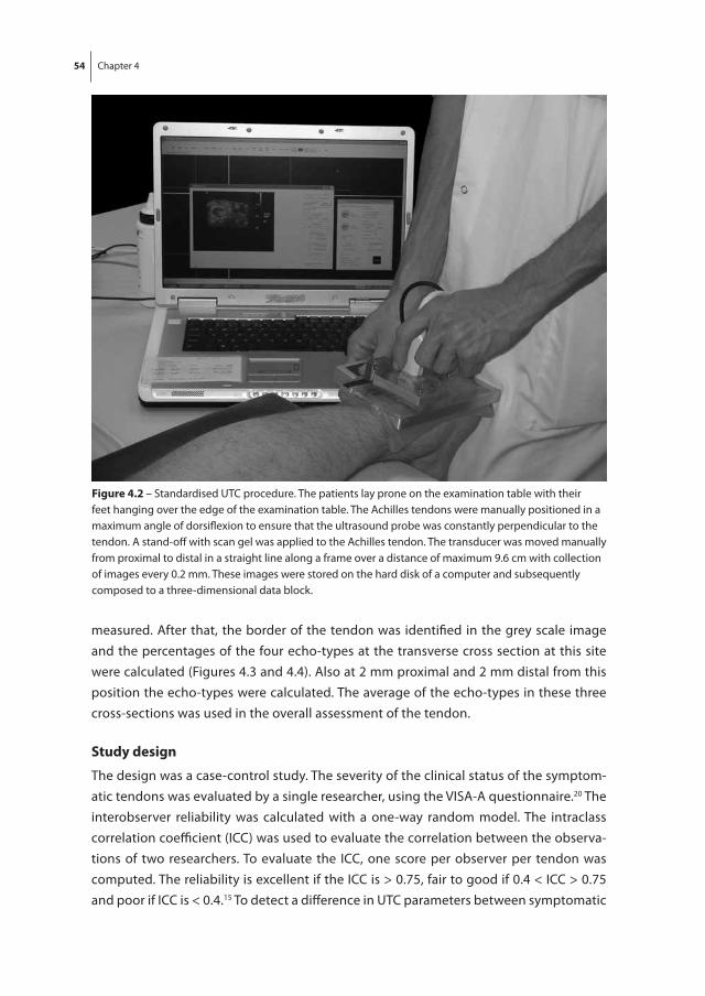
54 Chapter 4
measured. After that, the border of the tendon was identified in the grey scale image and the percentages of the four echo-types at the transverse cross section at this site were calculated (Figures 4.3 and 4.4). Also at 2 mm proximal and 2 mm distal from this position the echo-types were calculated. The average of the echo-types in these three cross-sections was used in the overall assessment of the tendon.
study design
The design was a case-control study. The severity of the clinical status of the symptom-atic tendons was evaluated by a single researcher, using the VISA-A questionnaire.20 The interobserver reliability was calculated with a one-way random model. The intraclass correlation coefficient (ICC) was used to evaluate the correlation between the observa-tions of two researchers. To evaluate the ICC, one score per observer per tendon was computed. The reliability is excellent if the ICC is > 0.75, fair to good if 0.4 < ICC > 0.75 and poor if ICC is < 0.4.15 To detect a difference in UTC parameters between symptomatic
figure 4.2 – Standardised UTC procedure. The patients lay prone on the examination table with their feet hanging over the edge of the examination table. The Achilles tendons were manually positioned in a maximum angle of dorsiflexion to ensure that the ultrasound probe was constantly perpendicular to the tendon. A stand-off with scan gel was applied to the Achilles tendon. The transducer was moved manually from proximal to distal in a straight line along a frame over a distance of maximum 9.6 cm with collection of images every 0.2 mm. These images were stored on the hard disk of a computer and subsequently composed to a three-dimensional data block.

Ultrasonographic Tissue Characterisation of human Achilles tendons 55
figure 4.3 – Tomographic visualisation of a right Achilles tendon in three planes of view, in raw greyscale.
A – Transversal viewB – Sagittal viewC – Coronal viewD – Transversal view with the maximum anterior-posterior (AP) thickness measured (white arrow), being the largest diameter perpendicular to the lateromedial width of the tendon. At this position the outline of the
Achilles tendon was drawn in the transverse image. This cross-section was used for calculation of the percentages of echo-types within the tendon.S = SkinP = Peritendinous spaceAT = Achilles tendonCA = Calcaneal bone
figure 4.4 – Difference between an UTC-processed image of an asymptomatic tendon (A) and a symptomatic tendon (B) in transversal view. The border of the Achilles tendon, which was defined on the grey-scale image, is drawn with the white line. Green pixels represent echo-types I, blue pixels echo-types II, red pixels echo-types III and black pixels echo-types IV. Note the frequently observed increased echo-types III and IV on the posteromedial side within the asymptomatic tendon and the diffuse distribution within the symptomatic tendon.
56 Chapter 4
and asymptomatic tendons, a student t-test was used. Statistics were performed using SPSS version 15.0.0 and significance was assumed for P values less than 0.05. The study protocol was approved by the Medical Ethics Committee of the hospital. Informed con-sent was obtained from all patients.
ResulTs
Patients
In both the symptomatic and asymptomatic group 26 Achilles tendons were included. In the symptomatic group there were 12 male and 14 female tendons. In seven patients symptoms were located in the left Achilles tendon, three patients in the right tendon and eight patients had bilateral complaints. The mean age was 44.9 years (SD 6.2), mean duration of symptoms was 193.9 weeks (SD 277.5) and the mean body mass index (BMI) was 24.8 kg/m² (SD 2.2). The mean AAS was 4.8 (SD 2.0) and the mean VISA-A score was 67.3 (SD 18.2). In the asymptomatic group there were 16 male and 10 female tendons. The mean age was 43.6 years (SD 12.6) and the mean BMI was 26.1 kg/m² (SD 4.3). The mean AAS was 5.5 (SD 1.6). There was no significant difference between the symptom-atic group and asymptomatic group in age (p = 0.63), gender (p = 0.17), BMI (p = 0.20) and AAS (p = 0.15).
uTC measurements
UTC analyses showed a distinct difference between the distributions of the four echo-types in the symptomatic group versus the asymptomatic group. Figure 4.4 represents a typical echo-type distribution of both groups in transversal view. Quantitatively the symptomatic group was significantly different from the asymptomatic group for all echo-types (Table 4.1). Although there was some overlap between the groups, symp-tomatic and asymptomatic tendons could be clearly discriminated, e.g. based on the percentage of pixels in echo-types I+II (51.5% vs. 76.6% in symptomatic vs. asymptom-atic, p < 0.001) (Table 4.1, Figure 4.5). A difference in mean tendon thickness between symptomatic and asymptomatic tendons could also be observed (9.2 mm vs. 6.8 mm in symptomatic vs. asymptomatic, p < 0.001) (Table 4.1). When a threshold of 75% in echo-types I+II is chosen (see dotted line in Figure 4.5), three symptomatic tendons are above this threshold and six asymptomatic tendons are under this threshold (Table 4.2). Thus, nine of the totally 52 tendons were categorised in another group than expected, based on the UTC-findings. With 43 tendons that were categorised as expected, an accuracy of 83% was found. The ICC values for the UTC parameters are presented in Table 4.1.

Ultrasonographic Tissue Characterisation of human Achilles tendons 57
dIsCussIon
This is the first study that quantifies tendon structure in patients with Achilles tendi-nopathy and controls with use of Ultrasonographic Tissue Characterisation (UTC). Symptomatic tendons showed significantly less stability of transverse echo-patterns (less echo-types I+II) than asymptomatic tendons (p < 0.001). The mean AP tendon thickness was 9.2 mm in the symptomatic group and 6.8 mm in the asymptomatic group (p < 0.001). The interobserver reliability of determining the UTC parameters was excel-lent (ICC 0.92-0.95) for quantification of the ultrasonographic tendon structure. This indicates that different observers examining the same tendon have excellent agreement between their findings; a prerequisite for implementation of UTC in the clinical setting. For the evaluation of the tendon AP thickness an ICC of 0.84 was found.
Table 4.1 – Mean percentages of the separate four echo-types and tendon thickness are denoted for the symptomatic and asymptomatic tendons. Echo-types I+II (structure-related) and III+IV (non structure-related) are grouped. The P values for the difference between symptomatic and asymptomatic tendons and ICC values for the two observers are also presented for the echo-types and mean tendon thickness. The difference in all measured UTC parameters between symptomatic and asymptomatic tendons was statistically significant.
echo-types symptomatic tendons (n=26)
Asymptomatic tendons (n=26)
P-value ICC
Mean sd Mean sd
% I 33.9 12.7 48.4 12.3 < 0.001 0.92
% II 17.6 4.9 28.3 7.4 < 0.001 0.92
% III 20.9 7.5 12.5 7.9 < 0.001 0.95
% IV 27.5 11.1 10.8 7.6 < 0.001 0.95
% I + % II 51.5 16.4 76.6 15.1 < 0.001 0.95
% III + % IV 48.4 16.4 23.4 15.1 < 0.001 0.95
Tendon AP diameter (mm)
9.2 1.7 6.8 1.2 < 0.001 0.84
AP – Anterior to PosteriorICC – Intraclass Correlation Coefficient
Table 4.2 – The number of tendons that were symptomatic and asymptomatic are given. With use of the threshold of 75% echo-types I+II, these tendons were divided in groups with normal (> 75% echo-types I+II) or abnormal (< 75% echo-types I+II) tendon structure. Three tendons in the symptomatic group had a normal tendon structure and six tendons in the asymptomatic group had an abnormal tendon structure. The other 43 of totally 52 tendons were expected to be below the threshold (symptomatic tendons) or above the threshold (asymptomatic tendons). Therefore, UTC has an accuracy of 83% ((43/52)x100%).
normal tendon structure
Abnormal tendon structure
Asymptomatic tendon 20 6 26
symptomatic tendon 3 23 26
23 39 52

58 Chapter 4
Recently, other researchers focussed on quantifying tendon integrity.13,14 For instance, Bashford et al.13 attempted to quantify structural changes in tendon tissue by analysing the anisotropy of the “speckle pattern” of the ultrasonographic image. Anisotropy of tendons is based on their unique architecture with tendon bundles organised in lon-gitudinal alignment. As such, this approach is fairly similar to UTC since both methods aim to isolate exclusively structure-related information from a “speckle pattern”. As a consequence of anisotropy, ultrasonography of tendons is sensitive for the tilt of the transducer, possibly leading to a hypoechoic representation of normal tendons.22,23 In case of UTC this artefact is prevented by the tracking device with the transducer clamped in standardised position, in alignment and perpendicular to the tendons long-axis and with fixed transducer-tilt in the transverse plane. The foot was supported with the ankle joint in a maximum dorsiflexion which prevented a sag of the Achilles tendon as much as possible and thus resulted in an optimal angle of insonation. With this technique, only small tilting (lower than three degrees) may have occurred. From previous equine studies, it is known that transducer tilting within three degrees can result in small varia-tions in grey-level.24 However, with use of UTC the outcome in echo-types is determined with the relative grey-value of the contiguous pixels and not the absolute grey-value. Therefore, small tilting would have no effects on the echo-types measured with UTC.
In the study of Bashford et al.13 tendinopathic and healthy tendons could be discrimi-nated with an accuracy of 80%. With optimal settings, an accuracy of 83% was found with use of UTC in this study, which is comparable with the study of Bashford et al.13 (Figure 4.5 and Table 4.2). The advantage of UTC is that a connection with underlying tissue histology was made with this approach in equine tendons.
The normal Achilles tendon has a more or less flattened ellipsoid shape with the axis through maximum AP-thickness frequently somewhat oblique, from anterolateral to-wards posteromedial. Therefore we decided not to measure the maximum AP thickness in the sagittal plane but rather in the transverse image, thus independent of angle of insonation. Maximum AP thickness was defined as the maximum diameter perpendicu-lar to the maximal lateromedial width (Figure 4.3D). In the great majority of diseased tendons at least some increase of cross-sectional area can be observed. Therefore it is relevant to measure the anteroposterior thickness. Aström et al.25 found that a thickness over 10 mm may be related to the presence of a partial rupture. However, it should be noted that these dimensions do not provide information about the tendon structure. With use of UTC, the dimension was measured manually and maybe therefore the in-terobserver reliability was not as excellent as measured by Syha et al.26, who used an automatic identification procedure. With their approach, an inter-reader variability of 3.9% was found.
UTC has clear advantages over conventional ultrasonography, as UTC directly visu-alises and quantifies integrity. Moreover, the stability of the transverse echo-pattern

Ultrasonographic Tissue Characterisation of human Achilles tendons 59
in the tendon long axis is not an absolute value but rather a relative one, and thus independent of ultrasonographic gain settings. This stability refers to stability of the echo pattern in consecutive transverse sections and is thereby a true three-dimensional measure. Furthermore, the tracking-device prevented the transducer from tilting and thus the angle of insonation is standardised and stable. The use of the tracking and data collection system did not only prevent a tilting artefact 22,23 but also facilitated the stor-age and subsequent compounding of transverse images.
There are some limitations in this study. For instance, the group of symptomatic tendons had already been unsuccessfully treated with a heavy load eccentric exercise program. Öhberg and Alfredson 27 reported in an observational follow-up study that all 41 tendons of 30 patients had structural ultrasonographic changes (hypoechoic area or irregular fibre structure). After follow-up (mean 28 months) with a 12-week eccentric exercise program, 37 of 41 tendons had a normalised tendon structure. It is possible that this treatment may alter the tendon structure and therefore result in an improved echopattern in some symptomatic tendons. Similarly we cannot exclude that a certain
figure 4.5 – The distribution of the percentages of echo-types I+II in symptomatic and asymptomatic tendons. A clear discrimination can be made between both groups. With a threshold of 75% echo-types I+II (dotted line), three tendons in the symptomatic group were above this threshold and six tendons in the asymptomatic group were below this threshold (dark bars). The other 43 tendons (grey bars) had a tendon structure that could be expected (abnormal structure in symptomatic tendons and normal structure in asymptomatic tendons).

60 Chapter 4
degree of degeneration could have been present in the asymptomatic group. Subjects were included simply by asking if the subjects never had complaints in the Achilles tendon region, while stricter criteria could have been used. From postmortem studies it is known that asymptomatic tendon degeneration is present in approximately 30%.28 This may implicate that a certain degree of degeneration could have been present in the asymptomatic group.
Based on the anatomical similarities between animal and human tendons 29, the UTC echo-types in human tendons can be expected to be similar to horse tendons. Further analyses that relate UTC outcomes with histology from human Achilles tendon mate-rial (e.g. from patients undergoing surgery) might corroborate tissue characterisation by UTC. Reference sets for different age, gender and activity level might be needed to precisely determine the clinical relevance. The UTC procedure could be even more standardised with standard dorsiflexion of the ankle joint and improved tracking and data collection device. As such UTC might be a very useful quantitative method for the monitoring and evaluation of existing and new treatment protocols for tendinopathy. In addition, it would be very valuable to determine its prognostic value.
ConClusIon
Ultrasonographic Tissue Characterisation (UTC) was transferred from equine validated tests to human Achilles tendons. The method showed excellent interobserver reliability and a significant difference in quantified echo-types between symptomatic and asymp-tomatic tendons, indicating its value for the evaluation of treatment in tendinopathy.
Ultrasonographic Tissue Characterisation of human Achilles tendons 61
RefeRenCes
1. O’Brien M. The anatomy of the Achilles tendon. Foot Ankle Clin 2005; 10(2): 225-38. 2. Maffulli N, Wong J, Almekinders LC. Types and epidemiology of tendinopathy. Clin Sports Med
2003; 22: 675-92. 3. Alfredson H. Chronic midportion Achilles tendinopathy: an update on research and treatment.
Clin Sports Med 2003; 22: 727-41. 4. de Mos M, van El B, de Groot J, Jahr H, van Schie HT, van Arkel ER, Tol H, Heijboer R, van Osch GJ,
Verhaar JA. Achilles tendinosis: changes in biochemical composition and collagen turnover rate. Am J Sports Med 2007; 35: 1549-56.
5. Astrom M, Rausing A. Chronic Achilles tendinopathy. A survey of surgical and histopathologic findings. Clin Orthop 1995; 316: 151-64.
6. Martinoli CM, Derchi LE, Pastorino C, Bertolotto M, Silvestri E. Analysis of echotexture of tendons with US. Radiology 1993; 186: 839-43.
7. van Schie HT, Bakker EM. Structure-related echoes in ultrasonographic images of equine superfi-cial digital flexor tendons Am J Vet Res 2000; 61: 202-9.
8. Bleakney RR, White LM. Imaging of the Achilles tendon. Foot Ankle Clin 2005; 10: 239-54. 9. Reiter M, Ulreich N, Dirisamer A, Tscholakoff D, Bucek RA. Colour and power Doppler sonography
in symptomatic Achilles tendon disease. Int J Sports Med 2004; 25(4): 301-5. 10. Leung JL, Griffith JF. Sonography of chronic Achilles tendinopathy: a case-control study. J Clin
Ultrasound 2008; 36(1): 27-32. 11. Archambault JM, Wiley JP, Bray RC, Verhoef M, Wiseman DA, Elliott PD. Can sonography predict
the outcome in patients with achillodynia? J Clin Ultrasound 1998; 26(7): 335-9. 12. Khan KM, Forster BB, Robinson J, Cheong Y, Louis L, Maclean L, Taunton JE. Are ultrasound and
magnetic resonance imaging of value in assessment of Achilles tendon disorders? A two year prospective study. Br J Sports Med 2003; 37(2): 149-53.
13. Bashford GR, Tomsen N, Arya S, Burnfield JM, Kulig K. Tendinopathy discrimination by use of spatial frequency parameters in ultrasound B-mode images. IEEE Trans Med Imaging 2008; 27(5): 608-15.
14. Meghoufel A, Cloutier G, Crevier-Denoix N, DeGuise JA. A thinning algorithm for equine tendon structure identification from 2 D ultrasound images. IEEE ISBI 2008: 1565-68.
15. van Schie HT, Bakker EM, Jonker AM, van Weeren PR. Computerised ultrasonographic tissue characterisation of equine superficial digital flexor tendons by means of stability quantification of echo patterns in contiguous transverse ultrasonographic images. Am J Vet Res 2003; 64(3): 366-75.
16. van Schie HT, Bakker EM, Jonker AM, van Weeren PR. Efficacy of computerised discrimination between structure-related and non-structure-related echoes in ultrasonographic images for the quantitative evaluation of the structural integrity of superficial digital flexor tendons in horses. Am J Vet Res 2001; 62: 1159-66.
17. van Schie HT, Bakker EM, Cherdchutham W, van Weeren PR. Monitoring of the repair process of surgically created partial lesions in equine superficial digital flexor tendons by means of comput-erised ultrasonography. Am J Vet Res 2009; 70(1): 37-48.
18. de Vos RJ, Weir A, Visser RJ, de Winter TC, Tol JL. The additional value of a night splint to eccentric exercises in chronic midportion Achilles tendinopathy: a randomised controlled trial. Br J Sports Med 2007; 41: e5.

62 Chapter 4
19. de Jonge S, de Vos RJ, Van Schie HT, Verhaar JA, Weir A, Tol JL. One-year follow-up of a randomised controlled trial on added splinting to eccentric exercises in chronic midportion Achilles tendi-nopathy. Br J Sports Med 2008. Epub ahead of print, PMID:18838406.
20. Robinson JM, Cook JL, Purdam C, et al. Victorian Institute Of Sport Tendon Study Group. The VISA-A questionnaire: a valid and reliable index of the clinical severity of Achilles tendinopathy. Br J Sports Med 2001; 35(5): 335-41.
21. Halasi T, Kynsburg A, Tállay A, Berkes I. Development of a new activity score for the evaluation of ankle instability. Am J Sports Med 2004; 32(4): 899-908.
22. Fornage BD. Achilles tendon: US examination. Radiology 1986; 159: 759-64. 23. Fornage BD. The hypoechoic normal tendon: a pitfall. J Ultrasound Med 1987; 6: 19-22. 24. van Schie HT, Bakker, EM, van Weeren PR. Ultrasonographic evaluation of equine tendons: a
quantitative in vitro study of the effects of amplifier gain level, transducer-tilt, and transducer-displacement. Vet. Radiol. Ultrasound. 1999, 40, 151-60.
25. Aström M, Gentz CF, Nilsson P, Rausing A, Sjöberg S, Westlin N. Imaging in chronic Achilles tendi-nopathy: a comparison of ultrasonography, magnetic resonance imaging and surgical findings in 27 histologically verified cases. Skeletal Radiol 1996; 25(7): 615-20.
26. Syha R, Peters M, Birnesser H, Niess A, Hirschmueller A, Dickhuth HH, Sandrock M. Computer-based quantification of the mean Achilles tendon thickness in ultrasound images: effect of tendinosis. Br J Sports Med 2007; 41(12): 897-902.
27. Öhberg L, Lorentzon R, Alfredson H. Eccentric training in patients with chronic Achilles tendinosis: normalised tendon structure and decreased thickness at follow up. Br J Sports Med 2004; 38(1): 8-11.
28. Kannus P, Józsa L. Histopathological changes preceding spontaneous rupture of a tendon. A controlled study of 891 patients. J Bone Joint Surg Am 1991; 73(10): 1507-25.
29. Wang JH. Mechanobiology of tendon. J Biomech 2006; 39: 1563-82. 30. Harris RA, Follett DH, Halliwell M, Wells PN. Ultimate limits in ultrasonic imaging resolution.
Ultrasound Med Biol 1991; 17: 547-58. 31. van Schie HT, Bakker EM, Jonker AM, van Weeren PR. Critical evaluation of ultrasonographic tissue
characterisation of equine superficial digital flexor tendons by means of gray level statistics. Am J Vet Res 2000; 61: 210-19.

Chapter 5Tendon structure is not related to clinical outcome following eccentric exercises in
chronic midportion Achilles tendinopathy
R.J. de VosM.P. Heijboer
H. WeinansJ.A.N. Verhaar
H.T.M. van Schie
Department of Orthopaedics
Erasmus University Medical Centre
Rotterdam, the Netherlands
Manuscript submitted to Eur Radiol (2010)

64 Chapter 5
AbsTRACT
Objective - Quantifying structural tendon changes by means of Ultrasonographic Tissue Characterisation (UTC) and assessing clinical improvement during an eccentric exercise program in patients with chronic midportion Achilles tendinopathy.
Design - Observational study
Setting - Orthopaedic department of the Erasmus University Medical Centre
Patients - Twenty-five patients with clinically established chronic midportion Achilles tendinopathy were included.
Interventions - The patients performed a 16-week eccentric exercise program. One experienced researcher performed the ultrasonographic data collection with the UTC procedure. These data were assessed by a blinded observer.
Main outcome measurements - UTC can quantify tendon structure through reliably measuring the proportion of four different echo-types. The determined echo-types I+II represent organised tendon bundles, while echo-types III+IV represent disorganised tendon structure. The severity of symptoms was established with the validated Victorian Institute of Sports Assessment-Achilles (VISA-A) questionnaire. Follow-up was at 2, 8, 16 and 24 weeks.
Results - The mean percentage of echo-types I+II decreased non-significantly with 0.3% after 24 weeks (p = 0.92, 95% CI -5.8 to 5.3). The mean VISA-A score increased slightly but significantly with 11.3 points after 24 weeks (p = 0.01, 95% CI 2.6 to 20.0). An increase in VISA-A score was not correlated with an increased percentage of echo-types I+II (p = 0.94, r = -0.02) and the baseline percentage of echo-types I+II did not correlate with an increase in VISA-A score (p = 0.74, r = 0.07).
Conclusion - With use of the standardised Ultrasonographic Tissue Characterisation, there was no short-term increase in organised tendon structure following eccentric exercises. Tendon structure organisation was not related to the severity of symptoms and could not be used as predictor for the clinical outcome.
Tendon structure is not related to clinical outcome 65
InTRoduCTIon
Chronic midportion Achilles tendinopathy is a frequent and hard to treat disorder.1,2 The diagnosis is based on the clinical triad of pain, swelling and impaired performance and on histology this condition can be characterised by degenerative changes of the tendon matrix.1 The role of conventional ultrasonography in tendon imaging is questionable, since it is influenced by machine settings, transducer handling of the observer and structural changes can only be assessed qualitatively.3 Therefore, a novel method was recently introduced for the evaluation of human Achilles tendons. Ultrasonographic Tissue Characterisation (UTC) was found to be a reliable method, which was able to distinguish symptomatic from asymptomatic Achilles tendons using quantification of the three-dimensional stability of the echopattern.4 UTC is an established method in veterinary medicine, in which various stages of ultrastructural organisation were related to the histomorphology of equine tendons as reference test.5
Nowadays, a heavy-load eccentric calf-muscle exercise program is prescribed as usual care for patients with chronic midportion Achilles tendinopathy.2 Initially, several stud-ies reported superior results with these exercises 6-8, but subsequent studies failed to achieve a high patient satisfaction.9-11 Eccentric exercises are thought to enhance tendon collagen synthesis 12, but conventional ultrasound might not reliably quantify the struc-tural changes following these exercises.3,4 Therefore, the effects of eccentric exercises on tendon structure are difficult to determine. Furthermore, there is moderate correlation between clinical assessment and appearance on conventional ultrasonography.3
The first aim of this study is to evaluate whether an improvement in tendon structure could be observed after an eccentric exercise program. The second aim is to evaluate the relationship between the change in severity of symptoms and tendon structure as quantified by means of UTC.
MATeRIAls And MeThods
study design
The study design was an observational prospective clinical trial, performed at an or-thopaedic outpatient department in an university medical centre (Erasmus University Medical Centre, Rotterdam, the Netherlands). The study protocol was announced for general practitioners, sport physical therapists and medical specialists in the region.
The study protocol was approved by the regional Medical Ethics Committee of the hospital (Rotterdam, the Netherlands). All patients provided written informed consent.

66 Chapter 5
Patients
Patients could contact the researcher by phone or e-mail when they were interested in participation in this study. The researcher informed the patients about the study if they seemed to be suitable for inclusion. Subsequently an appointment was made at the orthopaedic outpatient department.
Suitability for inclusion was evaluated by one experienced orthopaedic surgeon. Inclusion criteria were an age above 18 years and presence of symptoms for more than three months. The diagnosis was made clinically, based on history and physical examination. All patients had a painful thickened tendon with diminished activities. The tendon thickening was located approximately 2-7 cm proximal from the calcaneal insertion. Exclusion criteria were the preceding participation in an intensive program of heavy-load eccentric calf-muscle exercises and the inability to perform heavy loaded exercises. Patients with insertional disorders, an abnormal Thompson test and systemic illness were also excluded. None of the patients received injection therapy or had under-gone surgical treatment before.
Interventions
After inclusion, all patients were instructed about how to perform the eccentric exercises. The exercises were performed with straight knees and with bent knees as described by Alfredson et al.6 The researcher instructed the exercise program and patients were asked to make a series of supervised eccentric drops to be certain that the technique was cor-rect. The first four weeks, the patients were advised to gradually increase the frequency of eccentric drops with the aim to reach a maximum compliance during the program. After four weeks the original 12-week eccentric exercise program started, so in total the eccentric exercises were performed 16 weeks. The patients were instructed to perform 180 repetitions a day. Patients were informed that these exercises commonly cause pain and that they should accept this if it was not disabling. Increasing load was advised when the exercises could be carried out without any discomfort. All patients were instructed to avoid weight bearing sporting activities for the first eight weeks. After eight weeks, gradually return to sports activities was encouraged if the pain allowed.
outcome measurements
All patients completed a questionnaire consisting of clinical outcome measures and subsequently UTC measurements were performed at baseline and after 2, 8, 16 and 24 weeks.
The severity of symptoms was evaluated by a researcher using the Victorian Institute of Sports Assessment-Achilles (VISA-A) questionnaire.13 This validated and disease-specific questionnaire, which quantifies the pain and activity level, was completed with

Tendon structure is not related to clinical outcome 67
minimum assistance of the researcher. To evaluate the long term results (mean follow-up 2.2 years), this questionnaire was completed once more by e-mail.
To determine the level of sports activity in relation to the load capacity of the Achilles tendon, the Ankle Activity Score (AAS) was obtained. The AAS objectively quantifies the ankle related activity.14
The UTC measurements were done using a standardised protocol described in a previ-ous study.4 The patients lay prone on the examination table with their feet hanging over the edge of an examination table. With a holding device the ankle was positioned in a maximum angle of dorsiflexion, thereby stretching the Achilles tendon. A stand-off with scan gel was applied to the limb under investigation and subsequently a high-resolution 10 MHz linear-array transducer (Smartprobe 10L5, Terason 2000, Teratech, USA) was moved manually along and perpendicular to the Achilles tendon’s long axis over a distance of 9.6 cm.
At every step of 0.2 mm, transverse images were captured with constant settings (fo-cussing, brightness, contrast and gain). The digital scan data were stored on the hard disk of a computer and compounded to a three-dimensional data block which provided the tomographical visualisation of the Achilles tendon in three planes of view: transverse, sagittal and coronal. Furthermore, the processing of contiguous transverse images by means of custom-designed algorithms facilitated the discrimination of four echo-types. Echo-types I and II are generated from a single ultrasound reflection that belongs to typically one interface structure and therefore represent more or less organised tendon bundles. Echo-types III and IV are generated by multiple reflections that interfere as a consequence of multiple interfaces that represent smaller, disorganised and more amorphous or fibrillar structures that have typically been described in tendinotic tissue. The method has been extensively described and validated in equine tendons 5 and to a lesser extent tested in tendinopathy patients versus a control group.4
The thickest part of the tendon in the anterior-posterior (AP) direction was determined and subsequently, the border of the Achilles tendon was discriminated in the grey scale image. At this site, the proportions of the four echo-types at the transverse cross-section were defined and the echo-types were also calculated at 2 mm proximal and distal from this position. An average of the echo-types in these three cross-sections was used in the overall assessment of the tendon.
To determine the location of an area with decreased tendon structure, the tendons on UTC imaging at baseline were also divided into five regions (posteromedial, antero-medial, anterolateral, posterolateral and central). This procedure was executed with an automatic custom-build program. A line was drawn through the largest width of the tendon and perpendicular to this line, a second border was defined being the largest distance in anterior to posterior direction. The intersection of these two lines was used as centre of an oval area in the middle of the tendon. With a cut-off of 99% echo-types

68 Chapter 5
I+II, it was determined whether a location was defined as area with decreased tendon structure organisation. This was determined with use of segmentation, which was based on the persistence of features characteristic for echo-type III or IV over at least 25 im-ages, thus over a distance of 4.8 mm.
statistical evaluation
It was estimated that an increase in the VISA-A score of 12 points would be clinically rel-evant, with a standard deviation of the VISA-A score of 15 points.10,13 We calculated that a sample of 25 was required to detect this increase with a power of 80% with two-sided testing at a significance level of 5%. The Kolmogorov-Smirnov test was used to evaluate normality of distribution of the data. Parametric tests were used in case of normality and non-parametric tests in case of skewed distribution of the data.
The researcher who performed the analyses was blinded to the clinical status of the patients. The analyses were performed with the use of SPSS software (version 16.0.1; SPSS Inc., Chicago, Illinois, USA).
ResulTs
Patients
Twenty-six patients visited the Erasmus University Medical Centre for participation in the study between April 2007 and January 2008. One patient was excluded because of
Table 5.1 – Patient characteristics and outcome variables at baseline.
Patients (n=25)
Mean (sd) age (years) 46 (9.5)
gender
- Male (%) 10 (40)
- Female (%) 15 (60)
Mean (sd) body Mass Index (kg/m2) 25.9 (4.7)
Median (IQR) duration of symptoms (weeks) 36 (20-84)
Active in sports (%) 18 (72)
Mean (sd) Ankle Activity score 4.8 (1.5)
Mean (sd) VIsA-A score 47.3 (16.4)
Mean (sd) echo-type I 46.8 (12.9)
Mean (sd) echo-type II 21.5 (6.2)
Mean (sd) echo-type III 14.2 (7.5)
Mean (sd) echo-type IV 17.5 (9.9)
Mean (sd) echo-type I+II 68.3 (16.8)
Mean (sd) maximum tendon thickness (mm) 9.9 (2.0)

Tendon structure is not related to clinical outcome 69
an insertional disorder and 25 patients were included. The patient characteristics are presented in Table 5.1.
During the study, there were two patients lost to follow-up. These two patients experienced too much pain during the eccentric exercises and did not wish further participation. Three UTC-scans could not be analysed in this study. One patient could not attend at the 16-week follow-up time point and two UTC-scans could not be used for final analysis because of poor scanning technique.
After a mean of 2.2 years follow-up, 21 of these 23 patients completed the VISA-A questionnaire by e-mail. Fifteen patients did not receive other treatment options, one patient received a platelet-rich plasma injection and five patients underwent a surgical procedure (debridement of degenerative tissue) for their Achilles tendon injury within a median (IQR) of 5 months (2-11) after the 24-week follow-up time point.
single point in time
Totally, 115 UTC scans were analysed at all time points. The proportions of echo-types I+II did not correlate with the corresponding VISA-A scores (p = 0.32, r = -0.09).
At baseline, non-structure related echo-types (III+IV) were found on the posterome-dial (n = 23), posterolateral (n = 20), anterolateral (n = 14), anteromedial (n = 21) and central (n = 15) site.
figure 5.1 – There was a slight but significant increase in VISA-A score after 24 weeks but there was no significant change in echo-types I+II within this time period. Error bars denote standard deviations.

70 Chapter 5
Changes over time
The mean (SD) percentage of echo-types I+II decreased non-significantly with 0.3% (12.8) after 24 weeks (p = 0.92, 95% CI -5.8 to 5.3). The mean changes of the VISA-A score and the percentage of echo-types I+II are displayed in Figure 5.1 and the mean change in separate echo-types is shown in Figure 5.2.
The mean (SD) VISA-A score increased significantly with 11.3 (20.1) points after 24 weeks (p = 0.01, 95% CI 2.6 to 20.0). After a mean follow-up of 2.2 years, the mean (SD) VISA-A score was significantly increased with 14.8 (21.3) points in the non-surgically treated patients (p = 0.014, 95% CI 4.1 to 27.9) and non-significantly with 13.2 (20.1) points in the surgically treated patients (p = 0.22, 95% CI -11.8 to 38.2), but there was no significant difference between these two groups (p = 0.88, 95% CI -24.2. to 21.0).
An increased percentage of structure-related echo-types I+II after 24 weeks was not correlated with an increase in VISA-A score after 24 weeks (p = 0.94, r = -0.02) or after a mean of 2.2 years (p = 0.69, r = 0.09) (Figure 5.3A and 5.3B). The baseline percent-age of echo-types I+II did not correlate with an increase in VISA-A score after 24 weeks (p = 0.74, r = 0.07) or after a mean of 2.2 years (p = 0.76, r = 0.07).
There was a correlation between the VISA-A score at baseline and the VISA-A score at 24 weeks (p = 0.002, r = 0.61) and at 2.2 years follow-up (p < 0.001, r = 0.71) (Figure 5.4A and 5.4B).
figure 5.2 – Progression of echo-types I-IV. There were no significant changes in any echo-type from baseline to 24 weeks. Error bars denote standard deviations.

Tendon structure is not related to clinical outcome 71
figure 5.3A – Relationship between change in symptoms at short-term and change in tendon structure. An increased percentage of structure-related echo-types I+II after 24 weeks was not correlated with an increase in VISA-A score after 24 weeks (p = 0.94, r = -0.02).
figure 5.3b – Relationship between change in symptoms at long-term and change in tendon structure. An enhanced percentage of structure-related echo-types I+II after 24 weeks was not correlated with an increase in VISA-A score after a mean of 2.2 years (p = 0.69, r = 0.09).

72 Chapter 5
figure 5.4A – Relationship between severity of symptoms at baseline and at short-term follow-up. There was a correlation between the VISA-A score at baseline and the VISA-A score at 24 weeks follow-up (p = 0.002, r = 0.61).
figure 5.4b – Relationship between severity of symptoms at baseline and at long-term follow-up. There was a correlation between the VISA-A score at baseline and the VISA-A score at a mean of 2.2 years follow-up (p < 0.001, r = 0.71).

Tendon structure is not related to clinical outcome 73
dIsCussIon
In this first prospective observational study on the structural changes measured with Ul-trasonographic Tissue Characterisation during an eccentric exercise program, we could not demonstrate an improvement in tendon structure organisation after 24 weeks. The mean VISA-A score increased slightly but significantly. The severity of symptoms was not related to the tendon structure at a single point in time or in change over time. The degree of tendon structure disorganisation at baseline was not a predictive factor for the change in severity of symptoms after 24 weeks, but the VISA-A score at baseline was a predictor for the VISA-A score at short and long term follow-up.
These findings are important as many researchers have been reporting on the re-lationship between structural changes of the tendon and symptoms.4,15-21 It is known that normal tendons are characterised by a regular echopattern, whereas hypoechoic lesions as a result of collagen disorganisation are frequently described in cases of tendi-nosis.3 In two studies, ultrasonography was performed prior to the start of conservative treatment and the authors found that there was no statistical significant relationship between overall clinical outcome and the degree of ultrasonographic abnormality.15,22 These findings are in line with cohort studies that reported that symptoms do not mirror ultrasonographic appearances 16-18, although in asymptomatic individuals they may be of predictive value in the development of Achilles tendon pain.23
The results of our study, in which a quantified and standardised ultrasonographic method was used, confirm the previous (qualitative) findings. UTC has been used previously as follow-up instrument in equine studies. Several studies in horses with standardised surgical lesions induced in normal flexor tendons, thus without underlying degenerative changes, revealed that healing processes can be monitored quantitatively by means of UTC. The quality of repair could be quantified in vivo by means of UTC and these features corresponded significantly with both histomorphology and tensile prop-erties (post-mortem) at 24 weeks post-surgery.24,25
Previous prospective cohort studies reported that all patients had structural ultra-sonographic changes (hypoechoic area or irregular fibre structure) at baseline, but at long term follow-up (means of 28 and 46 months follow-up) after a 12-week eccentric exercise program, many tendons showed a normalised tendon structure on qualitative assessment with concurrently high patient satisfaction.19,26 In our study, the tendon structure did not change within 24 weeks after eccentric exercise treatment. There may be a few reasons for this discrepancy. The first hypothesis is that structural changes of the tendon may not occur yet within this short period. With use of a microdialysis technique it was found that a 12-week eccentric exercise program for the treatment of Achilles tendinopathy resulted in an increased collagen synthesis (peritendinous type I collagen) without an increased degradation.27 These findings suggest a short term regenerative

74 Chapter 5
effect of eccentric exercise therapy on molecular or fibrillar scale, but it is unknown whether this collagen will be organised into hierarchically arranged tendon bundles.28 This process may be time consuming and future studies should be carried out to evalu-ate whether the UTC appearance will ever become normalised at all, when tendons have been symptomatic before. The second hypothesis is that the study population is differ-ent compared with the previous studies. In our study, 72% of the patients were involved in some degree of sports activity. It is known that sedentary individuals benefit less from eccentric exercise therapy than active people.11 The increase in VISA-A score in our study may therefore be lower than in previous studies that used this outcome measure in an active population.10,29 This finding might be explained by the fact that the VISA-A score also quantifies the activity level, but there may be other reasons why sedentary people do not benefit. It is suggested that the compliance to the eccentric exercises may play a role in the outcome.10 However in our study the compliance was not recorded. A third reason may be that surrounding structures are more important than intratendinous col-lagen as a source of pain. Alfredson stated that surrounding tissues, like the nerves and the blood vessels may be changed after eccentric exercise therapy and therefore result in decreased pain perception.2 Also, in cases of peritendinous pathology only moderate intratendinous changes may be observed but clinically this may result in more pain and reduced function.4
The location of the tendon disorders may be another subject of interest. Gibbon et al.30 reported that most ultrasonographic midportion disorders (91%) were found in the medial segment. They concluded that these disorders are a result of repetitive hyper-pronation of the foot. We found that lesions were present on the posteromedial side in all tendons. The biomechanical cause of these findings may be related to abnormal alignment, but Soleus muscle traction can be another cause since this deep calf muscle attaches to the Achilles tendon in this region, probably leading to torsion of structures during repetitive loading.31 The study design makes it impossible to confirm these hy-potheses.
There are a few limitations in this study. We did not use a control group, so the obser-vations in this study may not merely be accounted to the effects of eccentric exercise therapy. Furthermore, we did not record the compliance to the eccentric exercises. It is known that in an active population more than a quarter of the patients reported per-forming the exercises at less than half of the prescribed intensity.10 It would have been interesting to relate the reported compliance to the change in tendon structure.
We found previously that there is a higher chance of having symptoms in the pres-ence of more echo-types III+IV on UTC, representing loss of normal tendon structure.4 However, based on the findings in the current study, the quantity of baseline tendon structure disorganisation is not related to the clinical outcome at short and long-term follow-up. A hypothesis is that pain is not necessarily related to the restoration of bundle

Tendon structure is not related to clinical outcome 75
organisation, at least not at 24 weeks from baseline. Probably, repair of tendon structure in degenerative tendon tissue takes much more time and is preceded by molecular processes that result in improvement of symptoms.20
We found that there was a relationship between the baseline VISA-A score and the VISA-A score at final follow-up. Therefore, clinical examination remains the cornerstone of evaluating the progress of symptoms in tendinopathy and ultrasonographic imaging may not be useful for predicting symptoms. These findings are relevant for daily clinical practice, but they also lead to additional questions which could be a focus in future studies. In tendinopathy research, accurate information about the long-term progress in large cohort studies is lacking. It is unknown whether patients have a constant clinical progression or a more fluctuating course of symptoms over time. It could be hypoth-esised that a normalised tendon structure does not result in recurrence of symptoms. Another problem is the limited information about the relationship between the sever-ity of symptoms and the previous activity levels. It might be that a slight decrease in activity level results in prolonged period without symptoms.21 Furthermore, we are not informed about the tendon structure, measured with a standardised method, at long term follow-up.
ConClusIon
Ultrasonographic Tissue Characterisation (UTC) was previously transferred from equine validated tests to human Achilles tendons. With this prospective cohort study we found that tendon structure does not improve at short-term following eccentric exercises in Achilles tendinopathy. Furthermore, tendon structure is not related to the clinical sever-ity of symptoms at a single point in time or in change over time.
ACknowledgeMenTs
We would like to thank B.E. Impelmans and I.B. de Groot (Department of Orthopaedics, Erasmus Medical Centre) for help with the study design and collection of data.

76 Chapter 5
RefeRenCes
1. Maffulli N, Wong J, Almekinders LC. Types and epidemiology of tendinopathy. Clin Sports Med 2003; 22(4): 675-92.
2. Alfredson H. Chronic midportion Achilles tendinopathy: an update on research and treatment. Clin Sports Med 2003; 22(4): 727-41.
3. Bleakney RR, White LM. Imaging of the Achilles tendon. Foot Ankle Clin 2005; 10(2): 239-54. 4. van Schie HT, de Vos RJ, de Jonge S, et al. Ultrasonographic tissue characterisation of human
Achilles tendons: quantification of tendon structure through a novel non-invasive approach. Br J Sports Med 2009; PMID: 19666626.
5. van Schie HT, Bakker EM, Jonker AM, et al. Computerised ultrasonographic tissue characterisation of equine superficial digital flexor tendons by means of stability quantification of echo patterns in contiguous transverse ultrasonographic images. Am J Vet Res 2003; 64(3): 366-75.
6. Alfredson H, Pietila T, Jonsson P, et al. Heavy-load eccentric calf muscle training for the treatment of chronic Achilles tendinosis. Am J Sports Med 1998; 26(3): 360-6.
7. Mafi N, Lorentzon R, Alfredson H. Superior short-term results with eccentric calf muscle training compared to concentric training in a randomised prospective multicentre study on patients with chronic Achilles tendinosis. Knee Surg Sports Traumatol Arthrosc 2001; 9(1): 42-7.
8. Fahlstrom M, Jonsson P, Lorentzon R, et al. Chronic Achilles tendon pain treated with eccentric calf-muscle training. Knee Surg Sports Traumatol Arthrosc 2003; 11(5): 327-33.
9. Roos EM, Engstrom M, Lagerquist A, et al. Clinical improvement after 6 weeks of eccentric exercise in patients with mid-portion Achilles tendinopathy–a randomised trial with 1-year follow-up. Scand J Med Sci Sports 2004; 14(5): 286-95.
10. de Vos RJ, Weir A, Visser RJ, et al. The additional value of a night splint to eccentric exercises in chronic midportion Achilles tendinopathy: a randomised controlled trial. Br J Sports Med 2007; 41(7): e5.
11. Sayana MK, Maffulli N. Eccentric calf muscle training in non-athletic patients with Achilles tendi-nopathy. J Sci Med Sport 2007; 10(1): 52-8.
12. Langberg H, Rosendal L, Kjaer M. Training-induced changes in peritendinous type I collagen turnover determined by microdialysis in humans. J Physiol 2001; 534(Pt 1): 297-302.
13. Robinson JM, Cook JL, Purdam C, et al. Victorian Institute Of Sport Tendon Study Group. The VISA-A questionnaire: a valid and reliable index of the clinical severity of Achilles tendinopathy. Br J Sports Med 2001; 35(5): 335-41.
14. Halasi T, Kynsburg A, Tállay A, et al. Development of a new activity score for the evaluation of ankle instability. Am J Sports Med 2004; 32(4): 899-908.
15. Khan KM, Forster BB, Robinson J, et al. Are ultrasound and magnetic resonance imaging of value in assessment of Achilles tendon disorders? A two year prospective study. Br J Sports Med 2003; 37(2): 149-53.
16. Khan KM, Cook JL, Kiss ZS, et al. Patellar tendon ultrasonography and jumper’s knee in elite female basketball players: a longitudinal study. Clin J Sport Med 1997; 7(3): 199-206.
17. Cook JL, Khan KM, Kiss ZS, et al. Prospective imaging study of asymptomatic patellar tendinopa-thy in elite junior basketball players. J Ultrasound Med 2000; 19(7): 473-9.
18. Cook JL, Khan KM, Kiss ZS, et al. Asymptomatic hypoechoic regions on patellar tendon ultra-sound: a 4-year clinical and ultrasound follow-up of 46 tendons. Scand J Med Sci Sports 2001; 11(6): 321-7.

Tendon structure is not related to clinical outcome 77
19. Öhberg L, Lorentzon R, Alfredson H. Eccentric training in patients with chronic Achilles tendinosis: normalised tendon structure and decreased thickness at follow up. Br J Sports Med 2004; 38(1): 8-11.
20. Khan KM, Cook JL, Maffulli N, et al. Where is the pain coming from in tendinopathy? It may be biochemical, not only structural, in origin. Br J Sports Med 2000; 34(2): 81-3.
21. Fredberg U, Stengaard-Pedersen K. Chronic tendinopathy tissue pathology, pain mechanisms, and aetiology with a special focus on inflammation. Scand J Med Sci Sports 2008; 18(1): 3-15.
22. Archambault JM, Wiley JP, Bray RC, et al. Can sonography predict the outcome in patients with achillodynia? J Clin Ultrasound 1998; 26(7): 335-9.
23. Fredberg U, Bolvig L. Significance of ultrasonographically detected asymptomatic tendinosis in the patellar and achilles tendons of elite soccer players: a longitudinal study. Am J Sports Med 2002; 30(4): 488-91.
24. van Schie HT, Bakker EM, Cherdchutham W, et al. Monitoring of the repair process of surgically created partial lesions in equine superficial digital flexor tendons by means of computerised ultrasonography. Am J Vet Res 2009; 70(1): 37-48.
25. van Schie HT. Ultrasonographic tissue characterisation of equine superficial digital flexor ten-dons; Development and applications of computer-aided image analysis. Thesis, 2004.
26. Öhberg L, Alfredson H. Effects on neovascularisation behind the good results with eccentric train-ing in chronic mid-portion Achilles tendinosis? Knee Surg Sports Traumatol Arthrosc 2004; 12(5): 465-70.
27. Langberg H, Ellingsgaard H, Madsen T, et al. Eccentric rehabilitation exercise increases peritendi-nous type I collagen synthesis in humans with Achilles tendinosis. Scand J Med Sci Sports 2007; 17(1): 61-6.
28. de Jonge S, de Vos RJ, van Schie HT, et al. One-year follow-up of a randomised controlled trial on added splinting to eccentric exercises in chronic midportion Achilles tendinopathy. Br J Sports Med. 2008; PMID: 18838406.
29. Brown R, Orchard J, Kinchington M, et al. Aprotinin in the management of Achilles tendinopathy: a randomised controlled trial. Br J Sports Med 2006; 40: 275-79.
30. Gibbon WW, Cooper JR, Radcliffe GS. Distribution of sonographically detected tendon abnor-malities in patients with a clinical diagnosis of chronic Achilles tendinosis. J Clin Ultrasound 2000; 28(2): 61-6.
31. O’Brien M. The anatomy of the Achilles tendon. Foot Ankle Clin 2005; 10(2): 225-38.


Chapter 6Effects of platelet-rich plasma on
ultrasonographic findings in chronic midportion Achilles tendinopathy
R.J. de VosA. WeirJ.L. Tol
J.A.N. VerhaarH. Weinans
H.T.M. van Schie
Department of Orthopaedics
Erasmus University Medical Centre
Rotterdam, the Netherlands
Manuscript submitted to Eur Radiol (2010)

80 Chapter 6
AbsTRACT
Background - Chronic tendinopathy is frequently characterised by a reduced tissue repair and degeneration. Regenerative effects may be achieved with the administration of platelet-rich plasma (PRP), which can release large amounts of growth factors. Labora-tory studies show that PRP can lead to tendon healing by collagen regeneration and stimulation of angiogenesis. To date, the effects of PRP on ultrasonographic findings in tendinopathy have not been established.
Hypothesis - A PRP injection in addition to an eccentric exercise program leads to an enhanced tendon structure and neovascularisation, measured with ultrasonographical techniques, in chronic midportion Achilles tendinopathy.
Study design - Double-blind, randomised, placebo-controlled clinical trial.
Methods - Patients with chronic midportion Achilles tendinopathy were included and randomised to eccentric exercise therapy with either a PRP injection (PRP group) or a saline injection (placebo group). Tendon structure was evaluated by Ultrasonographic Tissue Characterisation (UTC), a novel technique which quantifies the tendon structure into four different echo-types: echo-types I+II represent organised tendon bundles, whereas echo-types III+IV represent a disorganised tendon structure. Colour Doppler ultrasonography (CDU) was used to measure and score the degree of neovascularisa-tion. Follow-up was at 6, 12 and 24 weeks. The study is registered with ClinicalTrials.gov, number NCT00761423.
Results - A significant improvement in echo-types I+II was found after 24 weeks within both the PRP group (n = 27) and the placebo group (n = 27), but there was no significant between-group difference (95% CI -1.6 to 7.8, p = 0.17). After six weeks, the neovascu-larisation score increased within the PRP group (p = 0.001) and the placebo group (p = 0.002), but there was no significant between-group difference in change of neovascu-larisation score at any point in time.
Conclusions - Injecting PRP for the treatment of chronic midportion Achilles tendi-nopathy does not contribute to an increased tendon structure or alter the degree of neovascularisation.
Funding - Biomet Biologics LLC (Warsaw, Indiana, USA).

Effects of platelet-rich plasma on ultrasonographic findings 81
InTRoduCTIon
Chronic midportion Achilles tendinopathy is a degenerative disorder that commonly affects middle-aged individuals.1 The diagnosis is established clinically when there is presence of a painful swollen Achilles tendon in the midportion with a reduced load bearing capacity.15
Conventional ultrasonography is frequently used as an imaging technique for the evaluation of tendon disorders.15 Ultrasonography is an operator-dependent technique and transducer handling and machine settings can influence the image gained. Conse-quently, the interobserver reliability of ultrasonographic assessment of tendon structure is not known and changes in time may be difficult to compare.25 In veterinary medicine a method for computerised Ultrasonographic Tissue Characterisation (UTC) has been de-veloped, in which ultrastructural organisation of tendon tissue could be matched with histomorphology of equine tendons as a reference test.26 Recently this novel technique was found to be a reliable method, which was able to distinguish symptomatic from asymptomatic human Achilles tendons using quantification of the three-dimensional stability of the echopattern over contiguous images.25
Colour Doppler ultrasonography (CDU) is an established method to measure the vas-cularity within and around tendons. Hypervascularity is commonly seen in symptomatic Achilles tendons and is also called neovascularisation.17 The degree of neovascularisa-tion can be measured with an excellent interobserver reliability using a five-point scale (modified Öhberg-score).24 In previous studies it was reported that neovascularisation was present in all symptomatic tendons, which decreased concurrently with a reduc-tion in symptoms after conservative therapy.18,19 An increase in neovascularisation was observed within the first three weeks after applying sclerosing injections, which were successful when the neovascularisation later disappeared after this initial period.2 These findings suggest a beneficial effect of increased neovascularisation within the very first period of treatment and an opposing effect on the longer term.
Injections with platelet-rich plasma (PRP), defined as an increased concentration of platelets with the ability to release several growth factors after degranulation, may improve tissue repair processes and therefore enhance tendon structure.11 A recent laboratory study indicated that PRP application may lead to accelerated remodelling of the damaged tendon collagen tissue with an increased vascularisation.5 These effects have never been measured in clinical studies on patients with tendinopathy.
The first aim of this randomised placebo-controlled clinical trial is to evaluate whether a PRP injection in addition to an eccentric exercise program leads to an improved ten-don structure on UTC. The second aim is to evaluate whether a PRP injection can result in an increased degree of neovascularisation on CDU.

82 Chapter 6
MATeRIAls And MeThods
Patients
This study was part of a randomised, double-blind, placebo-controlled trial which was performed at the sports medical outpatient department of a large district general hospital (the Hague Medical Centre, Leidschendam, the Netherlands).10 The trial was publicised for health professionals and the general public in the Netherlands through advertisements and different media (websites on the internet, folders and regional radio).
The eligibility criteria are listed in Table 6.1. The clinical diagnosis was made when there was presence of a painful and thickened tendon in relation to activity, located at 2-7 cm proximal to the insertion on the calcaneal bone with a duration of at least two months.
Table 6.1 – Inclusion and exclusion criteria.
Inclusion criteria exclusion criteria
Clinical diagnosis “chronic midportion Achilles tendinopathy”
Clinical suspicion of insertional disorders (pain at the site of the insertion of the Achilles tendon on the calcaneal bone)
Age 18-70 years Clinical suspicion of an Achilles tendon rupture (Thompson test abnormal and a palpable “gap”)
Clinical suspicion of plantar flexor tenosynovitis (posteromedial pain when the toes are plantar flexed against resistance)
Clinical suspicion of sural nerve pathology (sensitive disorder in the area of the sural nerve)
Clinical suspicion of peroneal subluxation (visible luxation of the peroneal muscles in combination with localised pain)
Known presence of internal disorders: spondylarthropathy, gout, hyperlipidemia, Rheumatoid Arthritis and sarcoidosis
Condition that prevented the patients from executing an active exercise program
Patient already performed heavy load eccentric calf muscle exercises, according to the schedule of Alfredson et al.1
Patient already received an injection of PRP in this tendon
Patient did not wish, for whatever reason, to undergo one of the two treatments
Known presence of a pregnancy
Condition of the Achilles tendon caused by medications (arising in relation to moment of intake), such as quinolones and statins

Effects of platelet-rich plasma on ultrasonographic findings 83
Procedures
Patients could contact the researcher (RJV), who provided a detailed information folder when patients seemed to be eligible, based on their history findings. When patients decided to participate in the study, an appointment was made at the outpatient sports medicine department.
One experienced sports medicine physician (JLT) examined the eligibility of the pa-tients. When patients met the inclusion criteria, the researcher informed the patients orally about the aims and possible risks of the trial before informed consent was ob-tained.
For every included patient, one PRP injection and one saline injection was prepared. The PRP injection was prepared using the “Recover Platelet Separation Kit” (type “Gravi-tational Platelet Separation” III), in accordance with the system instructions.11 Fifty-four ml of venous blood was collected from the cubital vein of the patient, which was mixed with 6 mL of citrate to prevent early clotting. The mixture was then centrifuged for 15 minutes, giving approximately 6 mL of PRP. To increase the pH, a 0.3 mL of 8.4% sodium bicarbonate buffer was added. A total amount of 4 mL PRP was collected for infiltration. A 4 mL saline injection with an identical syringe and needle was prepared.
All the patients received instructions for the rehabilitation program. During the first 48 hours after the injection the patient could fully weight bear and were advised to walk only short distances indoors. After one week, the exercise program was started. The patients were instructed to start with stretching exercises for one week and after two weeks, a 12-week heavy load eccentric calf muscle exercise program started as described by Alfredson et al.1. The patients were instructed to perform 180 repetitions daily, despite the presence of pain. All patients were instructed to avoid weight bearing sporting activities for the first four weeks of the study. After four weeks, gradual return to sports activities was encouraged.6
Stratification was performed, based on activity-level of the patients, and block-randomisation was done to equalise the number of patients in both groups over time.10 The Ankle Activity Score (AAS) was used to objectively quantify the ankle related ac-tivity level.12 Randomisation was performed using envelopes that were equivalent in appearance and weight. Patients were randomised to the PRP group (PRP injection with eccentric exercises) or the placebo group (saline injection with eccentric exercises) by picking a closed envelop.
An unblinded sports medicine physician (AW) selected the correct injection and blinded the injection with the use of a covering sheath surrounding the syringe. The data of allocation were stored in a secret location. The content of the injection was blinded for the treating sports medicine physician (JLT), the researcher (RJV), the patients and the researcher who did the analysis (RJV).

84 Chapter 6
The injection was given by a blinded sports medicine physician. A local anaesthetic was injected (2 ml of 0.5% marcain) in the skin and subcutaneous tissue. Hypoechoic areas within the thickened part of the tendon were identified in transverse and longi-tudinal plane with use of an ultrasonographic machine (MyLab30; Esaote Piemedical, Maastricht, the Netherlands). The blinded fluid was injected using a 22G needle under ultrasonographical guidance via three puncture locations. Through each puncture location, five small depots were left at several sites into the degenerative areas of the tendon. The first injection was placed at the site of most structural disorganisation and the other two injections were positioned proximal and distal from the first injection.
outcome measurements
Ultrasonographic Tissue Characterisation (UTC)The outcome measurements were determined with the ultrasonographic techniques and were evaluated by a single researcher (RJV) at baseline and after 6, 12 and 24 weeks post-injection. The detailed working mechanisms of Ultrasonographic Tissue Characterisation (UTC) have been previously described in the literature.25,26 The patients were asked to lay prone on the examination table with the affected leg positioned in a holding device, which ensured a standardised dorsiflexion (15 degrees) of the ankle. The UTC procedure and measurements were performed with a high-resolution 10 MHz linear-array transducer (Smartprobe 10L5, Terason 2000, Teratech, USA). The transducer was secured in a frame which was clamped in the holding device to prevent motion of the transducer. This transducer was moved automatically with a constant speed, with use of a driving mechanism, along and perpendicular to the Achilles tendon’s long axis over a distance of 9.6 cm. This custom-designed tracking and data collection device assisted the collection of digital transverse images at regular distances of 0.2 mm. These images were stored on a computer and subsequently used to reconstruct a three-dimensional data block. The processing of contiguous transverse images by means of custom-designed algorithms facilitated the discrimination of four echo-types: echo-types I and II are generated from a single ultrasound reflection that belong to typically one interface structure and therefore represent more or less organised tendon bundles. Echo-types III and IV are generated by multiple reflections that interfere as a consequence of multiple interfaces that represent smaller, disorganised and more amor-phous or fibrillar structures that have been described in tendinotic tissue. The method has been extensively described and validated in equine tendons 26 and to a lesser extent it has also been tested in tendinopathy patients versus a control group.25 Furthermore, this information can be visualised tomographically in three planes of view: transverse, sagittal and coronal.

Effects of platelet-rich plasma on ultrasonographic findings 85
The thickest part of the tendon in the anterior-posterior (AP) direction was identified and subsequently the border of the tendon was identified in the grey-scale image at the thickest part. This procedure was also executed twice at 1.5 cm proximal and distal from the thickest segment, resulting in five bordered slices within 3 cm. Contours were interpolated and subsequently the volume within a 3 cm length of the tendon could be analysed. The proportions of the four echo-types within this volume were calculated. The distance from the thickest part to the calcaneal bone was calculated and used with the follow-up examination to ensure that the same volume of the tendon was evaluated each time.
One trained researcher (RJV) performed all UTC scans and could establish the out-comes with an intraclass correlation coefficient for intra-observer reliability of 0.88 with a mean difference of 0.80% and SD of 3.7 (unpublished data). A second blinded and experienced researcher (JVS) determined whether the UTC scans could be included into the final analysis.
Colour Doppler ultrasonography (CDU)CDU examination was performed with a linear high-frequency 12-15 MHz transducer (MyLab30; Esaote Piemedical, Maastricht, the Netherlands). The Doppler gain was set at 79%, just below random noise. For CDU, the Doppler frequency was set at 6.6 MHz. Patients were examined by one trained researcher (RJV), who participated in previous studies on determination of the neovascularisation score.7,24 Patients lay prone on the examination table with the ankle placed in neutral position during these measurements. To avoid obliteration of the vessels, the pressure of the probe was kept to a minimum. The tendons were examined in longitudinal and transverse planes as described previ-ously.24 The blood vessels observed with CDU were scored using the five-grade modified Öhberg score.7,24 This score was determined as 0 (no vessels visible), 1+ (one vessel, mostly anterior to the tendon), 2+ (one or two vessels throughout the tendon), 3+ (three vessels throughout the tendon), or 4+ (more than three vessels throughout the tendon).
statistical analysis
The power-analysis was based on the primary outcome measure of the trial.10 A total of 27 patients in each arm was required (80% power with two-sided testing at a 5% level of significance).
Analysis was done on the basis of intention-to-treat. The data remained blinded until the last patient was seen for follow-up and after the analysis, enabling a blinded statistical analysis. After that the blinding code was broken. The Kolmogorov-Smirnov test was used to evaluate normality of distribution of the data. Parametric tests were used in case of normality and non-parametric tests in case of skewed distribution of the

86 Chapter 6
data. These tests were performed for within-group and between-group differences in ultrasonographical scores.
Statistics were performed using SPSS version 16.0.1 (SPSS Science, Chicago, Illinois, USA) and significance was assumed when P values were less than 0.05.
The study protocol was approved by the regional Medical Ethics Committee and registered with ClinicalTrials.gov, number NCT00761423.
Role of the funding source
The sponsor of the study (Biomet Biologics LLC, Warsaw, Indiana, USA) had no role in study design, data collection, analysis, interpretation or writing of the article. All authors had full access to the data and made the final decision for publication.
ResulTs
Patients
Patients were included from August 2008 to January 2009. In July 2009, the last patient had an appointment for follow-up. The flow diagram (Figure 6.1) shows the progress of the study participants. During the study, there were no patients lost to follow-up. Eight
Table 6.2 – Patient characteristics and outcome variables at baseline in PRP group and placebo group. There were no significant differences at baseline between both treatment groups.
PRP-group(n=27)
placebo-group(n=27)
Mean (sd) age (years) 49 (8.1) 50 (9.4)
gender
- Male 13 (48%) 13 (48%)
- Female 14 (52%) 14 (52%)
Mean (sd) body Mass Index (kg/m2) 26.8 (3.9) 26.2 (3.5)
Median (IQR) duration of symptoms (weeks) 36 (24-78) 26 (16-104)
Active in sports 22 (81%) 24 (89%)
Mean (sd) Ankle Activity score 5.7 (1.8) 5.5 (1.6)
Mean (sd) echo-type I 50.5 (8.5) 46.5 (7.1)
Mean (sd) echo-type II 26.4 (4.6) 25.6 (5.7)
Mean (sd) echo-type III 13.0 (5.7) 15.7 (5.2)
Mean (sd) echo-type IV 10.1 (5.4) 12.1 (5.6)
Mean (sd) echo-type I+II 76.9 (10.8) 72.1 (10.5)
Mean (sd) maximum tendon thickness (mm) 9.83 (1.78) 9.84 (3.05)
Mean (sd) neovascularisation score 2.26 (1.16) 2.15 (1.35)

Effects of platelet-rich plasma on ultrasonographic findings 87
UTC scans could not be used for final analysis and five CDU examinations could not be performed. The reasons for these missing data are mentioned in Figure 6.1.
At baseline, the patient characteristics and outcome variables did not differ signifi-cantly between the two treatment groups (Table 6.2).
Assessed for eligibility (n= 99) Excluded (n=38)
Reasons: not mentioned (n=10) choose another therapy (n=8) choose a waiting policy (n=8) fear for the injection (n=3) anamnestic insertional tendon
disorder (n=2) tendinopathy for less than two
months (n=2) performed a total eccentric exercise
program (n=2) anamnestic total rupture (n=2) no time (n=1)
UTC-data missing: n=0 CDU-data missing: n=0
Allocated to PRP injection (n=27) UTC-data missing: n=0 CDU-data missing: n=0
Baseline
Analysis 24 weeks
6 weeks
Enrollment
Randomisation
Allocated to saline injection (n=27) UTC-data missing: n=3 Bad scanning technique (n=2) Skin problem (n=1) CDU-data missing: n=0
UTC-data missing: n=1 Skin problem (n=1) CDU-data missing: n=0
Excluded (n= 7) Reasons: insertional tendinopathy (n=4) musculo-tendinous disorder
(n=1) abnormal Thompson test (n=1) PRP treatment elsewhere (n=1)
12 weeks
UTC-data missing: n=1 Lost with data-transmission
(n=1) CDU-data missing: n=3 Repair machine (n=3)
UTC-data missing: n=1 Skin problem (n=1) CDU-data missing: n=2 Repair machine (n=2)
UTC-data missing: n=0 CDU-data missing: n=0
UTC-data missing: n=2 Skin problem (n=1) Reverberation artifact (n=1) CDU-data missing: n=0
figure 6.1 – Progress of patients through the study. Eight UTC scans could not be used for final analysis and five CDU examinations could not be performed.

88 Chapter 6
Mean proportions of uTC echo-types
Within both treatment groups, a significant increase in echo-types I+II could be ob-served after 24 weeks. However, there were no significant between-group differences in change of echo-types I+II. Concurrently there was a significant decrease in echo-types III+IV within both groups. Figure 6.2 illustrates the mean differences in echo-types I+II during follow-up in the PRP group and in the placebo group.
figure 6.2 – Difference in echo-types I+II between the PRP group and the placebo group. Error bars denote standard deviations. Echo-types I+II increased significantly from baseline to 24 weeks within the PRP group (95% CI 6.2 to 12.7, p < 0.001) and the placebo group (95% CI 8.9 to 16.1, p < 0.001). Concurrently, the echo-types III+IV decreased significantly within the PRP group (95% CI -6.2 to -12.8, p < 0.001) and the placebo group (95% CI -8.9 to -16.1, p < 0.001). There was no significant difference in change of echo-types I+II between both treatment groups from baseline to six (95% CI -4.3 to 5.6, p = 0.68), 12 (95% CI -2.1 to 10.6, p = 0.23) and 24 weeks (95% CI -1.6 to 7.8, p = 0.17).
There were no significant differences in the change of any echo-type at any point in time between both treatment groups. The changes in the proportion of echo-types I, II, III and IV over time in the PRP group and placebo group are shown in Figure 6.3A and 6.3B.
Mean neovascularisation score
There was an increase in neovascularisation within six weeks in both groups (PRP group: p = 0.001. placebo group: p = 0.002), but these scores returned to pre-treatment levels after 24 weeks (PRP group: p = 0.89, placebo group: p = 1.00). There were no significant differences in change of neovascularisation score from baseline to six (p = 0.37), 12 (p = 0.44) and 24 weeks (p = 0.75) between both treatment groups. The mean degree of neovascularisation in both treatment groups is shown in Figure 6.4.
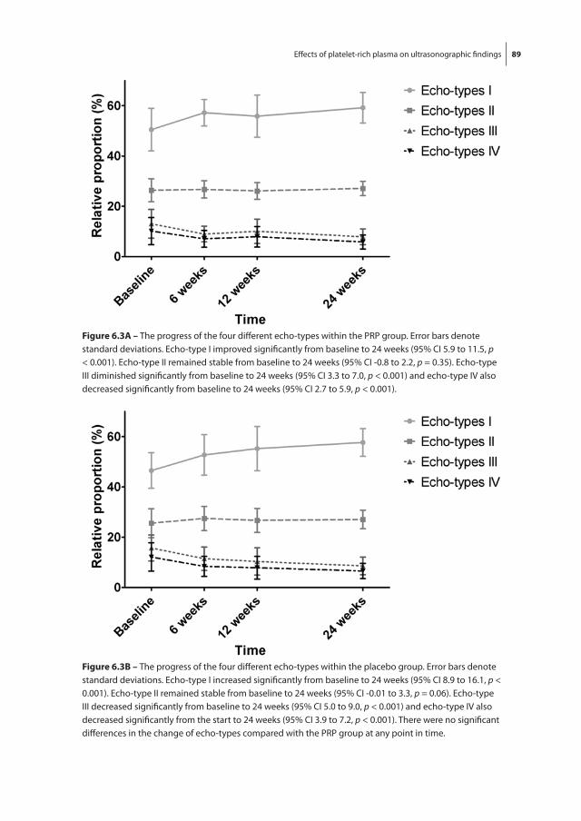
Effects of platelet-rich plasma on ultrasonographic findings 89
figure 6.3A – The progress of the four different echo-types within the PRP group. Error bars denote standard deviations. Echo-type I improved significantly from baseline to 24 weeks (95% CI 5.9 to 11.5, p < 0.001). Echo-type II remained stable from baseline to 24 weeks (95% CI -0.8 to 2.2, p = 0.35). Echo-type III diminished significantly from baseline to 24 weeks (95% CI 3.3 to 7.0, p < 0.001) and echo-type IV also decreased significantly from baseline to 24 weeks (95% CI 2.7 to 5.9, p < 0.001).
figure 6.3b – The progress of the four different echo-types within the placebo group. Error bars denote standard deviations. Echo-type I increased significantly from baseline to 24 weeks (95% CI 8.9 to 16.1, p < 0.001). Echo-type II remained stable from baseline to 24 weeks (95% CI -0.01 to 3.3, p = 0.06). Echo-type III decreased significantly from baseline to 24 weeks (95% CI 5.0 to 9.0, p < 0.001) and echo-type IV also decreased significantly from the start to 24 weeks (95% CI 3.9 to 7.2, p < 0.001). There were no significant differences in the change of echo-types compared with the PRP group at any point in time.

90 Chapter 6
dIsCussIon
This study shows that there is no difference in change of tendon structure within 24 weeks after injecting PRP in addition to eccentric exercises in the treatment of chronic midportion Achilles tendinopathy. In the patients treated with PRP and placebo, there was an initial increase in the degree of neovascularisation, but the change in neovascu-larisation score was not significantly different between both treatment groups.
These findings are important and clinically relevant as PRP is growing in popularity. Recent reviews supported its use for chronic tendon disorders,16,22 but good clinical evidence is lacking.9
Recent studies reported that PRP led to an enhanced tendon structure and increased neovascularisation in standardised surgically-induced tendon lesions in horses.3,4 In the present study, there was an increase in organised tendon structure and initial increase in neovascularisation, but no differences were found between the groups treated with PRP and saline. The main reason may be that a traumatic model was used in the equine study and the effects of PRP might be different in degenerative tendon tissue. It can also be possible that PRP was localised at the site of the lesion for a shorter time in our study because no compartment could be formed within the degenerative areas. It is unknown how long the platelets remain locally at the site of the degenerative lesion. In our study the platelets were not activated prior to injection, based on the assumption that plate-lets can be slowly activated as a consequence of the exposure to tendon collagen.16
figure 6.4 – Change in neovascularisation score in both treatment groups. Error bars denote standard deviations. During the first six weeks, the degree of neovascularisation increased within the PRP group (p = 0.001) and the placebo group (p = 0.002). The change in neovascularisation score from baseline did also not differ significantly at any point in time between the PRP group and the placebo group.

Effects of platelet-rich plasma on ultrasonographic findings 91
The addition of an eccentric exercise program may have resulted in dominating posi-tive effects on tendon structure, thereby overshadowing an effect of PRP. Öhberg et al.20 previously reported that eccentric training improves conventional ultrasonographic findings, using a qualitative evaluation, at long-term follow-up in Achilles tendinosis. It may also be possible that eccentric training adversely affects any regenerative proper-ties of PRP. The study design makes it impossible to rule out this possibility, although an experimental study on Achilles tendons of rats reported that mechanical load may be a prerequisite for the therapeutic effect of platelets.27
In a recent prospective observational study on the effects of eccentric exercises on tendon structure, there was no improvement in tendon structure after 24 weeks.8 This is in contrast with the findings of Öhberg et al.20, and also in the current study we found that tendon structure improved within 24 weeks of follow-up. There are a few possible explanations for this discrepancy. It could be possible that introducing the needle within the tendon improved the tendon structure, since some studies showed positive clinical effects of needling.9 However other mechanisms may be responsible. For instance, the populations between the studies may differ. In the present study there were a high number of patients involved in sports participation with a similar high ankle loading activity, quantified with the Ankle Activity Score. It is known that athletes benefit more from eccentric exercise therapy than sedentary individuals.23 The reason for this finding is unknown, but it has been suggested that the compliance to the eccentric exercises may be of relevance in treatment outcome.6 The reported patient compliance to the eccentric exercises was found to be high in the present study.10
A limitation of the current study may be that the neovascularisation score is not validated.14 In a previous study, we found that this scoring system has an excellent interobserver reliability 24 but the intra-observer reliability is not known and therefore we cannot confirm whether this score could be used in a follow-up study. An in vivo study on equine tendons reported that the number of properly aligned blood vessels, determined in harvested tissue at post mortem, was correlated to the neovascularisation score with the preceding in vivo CDU examination.4 In the present study, we used a com-parable machine and similar settings. Moreover, the measurements in the current study were performed under standardised conditions. The degree of ankle plantar flexion was standardised during the examination and minimal probe pressure should be used to avoid obliteration of vessels.7 Another limitation could be that CDU is less sensitive than Power Doppler ultrasonography for determining the degree of neovascularisation.21 The available machine in our clinic contained merely a Colour Doppler function, but previ-ous studies did also show increased amounts of neovascularisation using CDU.2,4,17-19
Future in vitro studies may be focused on the effects of PRP in the currently developing degenerative models.13,28 The influence of variables that may be of relevance could also be determined, such as the role of platelet activation prior to injection, the duration of

92 Chapter 6
collagen exposition to PRP, the concentrations of platelets and the concentrations of specific growth factors in the releasate.
Based on the current study results, there is no improvement in tendon structure and no effect on neovascularisation with the addition of PRP to eccentric exercises in chronic midportion Achilles tendinopathy, compared with a placebo injection.
ACknowledgeMenTs
All authors declare that we have no conflict of interest. The trial was funded by Biomet Biologics LLC (Warsaw, Indiana) and we thank this company for their support.
We also thank the patients for their participation and the health professionals for referring. We would also like to thank some co-workers of this study, who were not com-pensated for their efforts. We are grateful for the logistic help of Cora van der Heijden, Ank de Vries and Pauline Verschoor (Department of Sports Medicine, the Hague Medical Centre, the Netherlands).
Effects of platelet-rich plasma on ultrasonographic findings 93
RefeRenCes
1. Alfredson H. Chronic midportion Achilles tendinopathy: an update on research and treatment. Clin Sports Med. 2003; 22(4): 727-41.
2. Alfredson H, Öhberg L. Increased intratendinous vascularity in the early period after sclerosing in-jection treatment in Achilles tendinosis: a healing response? Knee Surg Sport Traumatol Arthrosc. 2006; 14(4): 399-401.
3. Bosch G, van Schie HT, de Groot M, Cadby J, Barneveld A, van de Lest CH, van Weeren PR. Effects of Platelet Rich Plasma on the quality of repair of mechanically induced core lesions in equine Superficial Digital Flexor Tendons: a placebo-controlled experimental study. J Orthop Res. 2009; PMID: 19714688.
4. Bosch G, Moleman M, Barneveld A, van Weeren PR, van Schie HT. The effect of Platelet Rich Plasma on the neovascularisation of surgically created equine superficial digital flexor tendon lesions. Accepted for publication in the Scan J Med Sci Sports.
5. de Mos M, van der Windt AE, Jahr H, van Schie HT, Weinans H, Verhaar JA, van Osch GJ. Can Platelet-Rich Plasma Enhance Tendon Repair? A Cell Culture Study. Am J Sports Med. 2008; 36(6): 1171-8.
6. de Vos RJ, Weir A, Visser RJ, Winter TC, Tol JL. The additional value of a night splints to eccentric exercises in midportion chronic Achilles tendinopathy: a randomised controlled trial. Br J Sports Med. 2007; 41(7): e5.
7. de Vos RJ, Weir A, Cobben LP, Tol JL. The value of power Doppler ultrasonography in Achilles tendinopathy: a prospective study. Am J Sports Med. 2007; 35(10): 1696-701.
8. de Vos RJ, Heijboer MP, Weinans H, Verhaar JA, Van Schie HT. Tendon structure is not related to clinical outcome following eccentric exercises in chronic midportion Achilles tendinopathy. Submitted to Eur Radiol.
9. de Vos RJ, van Veldhoven PL, Moen MH, Weir A, Tol JL, Maffulli N. Autologous growth factor injec-tions in chronic tendinopathies: a systematic review. Br Med Bull. 2010; Epub ahead of print.
10. de Vos RJ, Weir A, Van Schie HT, Bierma-Zeinstra SM, Verhaar JA, Weinans H, Tol JL. Platelet-rich plasma injection for chronic Achilles tendinopathy – a randomized controlled trial. JAMA 2010; 303(2): 144-9.
11. Eppley BL, Woodell JE, Higgins J. Platelet quantification and growth factor analysis from platelet-rich plasma: implications for wound healing. Plast Reconstr Surg. 2004; 114(6): 1502-8.
12. Halasi T, Kynsburg A, Tállay A, Berkes I. Development of a new activity score for the evaluation of ankle instability. Am J Sports Med. 2004; 32(4): 899-908.
13. Fu SC, Chan KM, Chan LS, Fong DT, Lui PY. The use of motion analysis to measure pain-related behaviour in a rat model of degenerative tendon injuries. J Neurosci Methods. 2009; 179(2): 309-18.
14. Knobloch K, de Vos RJ, Weir A, Tol JL. The use of a neovascularisation score to predict clinical severity in Achilles tendinopathy. Am J Sports Med. 2008; 36(2): 395-7.
15. Maffulli N, Sharma P, Luscombe KL. Achilles tendinopathy: aetiology and management. J R Soc Med. 2004; 97(10): 472-6.
16. Mishra A, Woodall J Jr, Vieira A. Treatment of tendon and muscle using platelet-rich plasma. Clin Sports Med. 2009; 28(1): 113-25.
17. Öhberg L, Lorentzon R, Alfredson H. Neovascularisation in Achilles tendons with painful tendi-nosis but not in normal tendons: an ultrasonographic investigation. Knee Surg Sports Traumatol Arthrosc. 2001; 9(4): 233-8.

94 Chapter 6
18. Öhberg L, Alfredson H. Effects on neovascularisation behind the good results with eccentric train-ing in chronic mid-portion Achilles tendinosis? Knee Surg Sports Traumatol Arthrosc. 2004; 12(5): 465-70.
19. Öhberg L, Alfredson H. Ultrasound guided sclerosis of neovessels in painful chronic Achilles tendinosis: pilot study of a new treatment. Br J Sports Med. 2002; 36: 173-7.
20. Öhberg L, Lorentzon R, Alfredson H. Eccentric training in patients with chronic Achilles tendinosis: normalised tendon structure and decreased thickness at follow up. Br J Sports Med. 2004; 38(1): 8-11; discussion 11.
21. Richards PJ, Win T, Jones PW. The distribution of microvascular response in Achilles tendonopathy assessed by colour and power Doppler. Skeletal Radiol. 2005; 34(6): 336-42.
22. Sánchez M, Anitua E, Orive G, Mujika I, Andia I. Platelet-rich therapies in the treatment of ortho-paedic sport injuries. Sports Med. 2009; 39(5): 345-54.
23. Sayana MK, Maffulli N. Eccentric calf muscle training in non-athletic patients with Achilles tendi-nopathy. J Sci Med Sport. 2007; 10(1): 52-8.
24. Sengkerij PM, de Vos RJ, Weir A, van Weelde BJ, Tol H. Interobserver Reliability of Neovascularisa-tion Score Using Power Doppler Ultrasonography in Midportion Achilles Tendinopathy. Am J Sports Med. 2009; 37(8): 1627-31.
25. van Schie HT, de Vos RJ, de Jonge S, Bakker EM, Heijboer MP, Verhaar JA, Tol JL, Weinans H. Ultraso-nographic tissue characterisation of human Achilles tendons: quantification of tendon structure through a novel non-invasive approach. Br J Sports Med. 2009; PMID: 19666626.
26. van Schie HT, Bakker EM, Jonker AM, van Weeren PR. Computerised ultrasonographic tissue characterisation of equine superficial digital flexor tendons by means of stability quantification of echo patterns in contiguous transverse ultrasonographic images. Am J Vet Res. 2003; 64(3): 366-75.
27. Virchenko O, Aspenberg P. How can one platelet injection after tendon injury lead to a stronger tendon after 4 weeks? Interplay between early regeneration and mechanical stimulation. Acta Orthop. 2006; 77(5): 806-12.
28. Warden SJ. Development and use of animal models to advance tendinopathy research. Front Biosci. 2009; 14: 4588-97.

Chapter 7The additional value of a night splint to eccentric
exercises in chronic midportion Achilles tendinopathy – a randomised controlled trial
R.J. de VosA. Weir
R.J.A. VisserT.C. de Winter
J.L. Tol
Department of Sports Medicine
The Hague Medical Centre Antoniushove
Leidschendam, the Netherlands
Br J Sports Med 2007; 41(7): e5

96 Chapter 7
AbsTRACT
Aim - To assess whether the use of a night splint is of added benefit on functional out-come in treating chronic midportion Achilles tendinopathy.
Methods - This was a single-blind, prospective, single centre, randomised controlled trial set in the sports medical department, the Hague Medical Centre, the Netherlands. Inclusion criteria were: age 18-70 years, active participation in sports, and tendon pain localised at 2-7 cm from distal insertion. Exclusion criteria were: insertional disorders, partial or complete ruptures, or systemic illness. Seventy tendons were included and randomised into one of two treatment groups: eccentric exercises with a night splint (night splint group, n = 36) or eccentric exercises only (eccentric group, n = 34).
Interventions - Both groups completed a 12-week heavy-load eccentric training pro-gram. One group received a night splint in addition to eccentric exercises. At baseline and follow-up at 12 weeks, patient satisfaction, Victorian Institute of Sports Assessment-Achilles questionnaire (VISA-A) score and reported compliance were recorded by a trained researcher who was blinded to the treatment.
Results - After 12 weeks, patient satisfaction in the eccentric group was 63% compared with 48% in the night splint group. The VISA-A score significantly improved in both groups; in the eccentric group from 50.1 to 68.8 (p = 0.001) and in the night splint group from 49.4 to 67.0 (p < 0.001). There was no significant difference between the two groups in VISA-A score (p = 0.82) and patient satisfaction (p = 0.26).
Conclusion - A night splint is not beneficial in addition to eccentric exercises in the treat-ment of chronic midportion Achilles tendinopathy.

The additional value of a night splint to eccentric exercises 97
InTRoduCTIon
Overuse injury of the Achilles tendon is a common entity in athletes and older athletes are at an increased risk. When the exact origin of tendon pain is unclear, the term tendi-nopathy is preferred.1,2
Scientific evidence for many proposed aetiological factors (biomechanical abnormali-ties, alteration of vascularity, certain systemic diseases, use of certain medications, ge-netic predisposition and excessive load in sports) is scarce.3,4 Consequently, there is no consensus as to which treatment should be used. Many differing treatments have been proposed, but none of these treatments have shown reproducible promising results in randomised trials.5-7
Alfredson et al.8 described a heavy-load eccentric exercise program for 15 athletes with chronic Achilles tendinosis. It consisted of 180 repetitions daily, which were allowed to be painful. The results were excellent in all athletes. In subsequent studies, comparable results were reported.9-12
A recent systematic review on the effect of eccentric overload training, in which nine studies satisfied the methodological criteria, showed that the effects on pain are promis-ing.13 They stated, however, that the weakness of all these publications is that only pain is used when scoring the outcomes. Besides pain, the functional status and (sports) activ-ity should be scored to determine the magnitude of the effect, and they suggest using the validated Victorian Institute of Sports Assessment-Achilles (VISA-A) questionnaire.
The basic mechanisms underlying the effectiveness of eccentric exercises are unknown to date. Alfredson14 suggests a few possible theories. The first theory is that an alteration of pain perception, due to the painful exercises, reduces the symptoms.14 The second theory is based on the ingrowth of neovessels with accompanying nerves around the tendon, which are supposed to be the source of pain. Eccentric exercises should destroy these neovessels.5,14 The third theory is that loading improves the tensile strength of the tendons. It may also induce lengthening of the musculotendinous junction and, therefore, put less strain on the Achilles tendon during movement.14
The use of a night splint has been shown to be effective in cases of chronic plantar fasciitis.15-17 There are several hypotheses supporting the use of a night splint, which provides passive dorsiflexion, in cases of Achilles tendinopathy. One hypothesis is that a night splint would work for Achilles tendinopathy owing to lengthening of muscle and tendon, and the concomitant reduction in strain on the tendon.11 It has been stated that passive dorsiflexion may be helpful in chronic Achilles tendon injury.2
The effect of splinting has been studied twice with differing results. Dijkstra and van Enst18 found clinical benefit using splinting alone. Roos et al.11 reported no difference between the three groups: eccentric exercises, use of a night splint and combination of splinting and eccentric exercises.

98 Chapter 7
The aim of this single-blinded prospective study is to compare the short-term effects on pain and functional outcome of a heavy-load eccentric exercise program, with a heavy-load eccentric program in combination with the use of a night splint.
MATeRIAls And MeThods
Patients
The location for the study was an outpatient sports medicine department in a large dis-trict general hospital (the Hague Medical Centre, the Netherlands). We included patients who were referred by a general practitioner, physical therapist or a medical specialist. Most of the patients had already received another conservative treatment, but none had undergone surgical treatment, a heavy-load eccentric training program or used a night splint before.
Inclusion criteria were patients aged between 18 and 70 years, presence of symptoms for more than two months, active participation in sporting activities before the onset of symptoms and the patients’ wish to return to their original level of sports. All patients had a tendon that was tender on palpation and painful during or after sport. The ten-don thickening was located approximately 2-7 cm proximal to the distal insertion. The diagnosis was made based on clinical examination. Exclusion criteria were the previous performance of an intensive program of heavy-load eccentric exercises and inability to perform heavy-load exercises. Insertional disorders, tendon ruptures and patients with systemic illness were also excluded.
study design
The study design was a single-blinded randomised controlled trial. Patients were ran-domised after initial standardised assessment to one of two treatment groups: eccentric exercises or eccentric exercises in combination with a night splint. The researcher was blinded to the treatment groups. An appointment was made after four weeks to evalu-ate possible problems. Patients were followed-up after 12 weeks.
Patient evaluation
After the patients were informed about the aims and possible risks of the study, they signed the informed consent form. The study protocol was approved by the Medical Ethics Committee, the Hague Medical Centre, the Netherlands.
All patients were examined by the same single researcher using a standard protocol consisting of standardised outcome measures: the VISA-A 19 was completed with mini-mum assistance from the researcher, and patient satisfaction was rated subjectively as poor, fair, good or excellent. A good or excellent result was considered as successful.
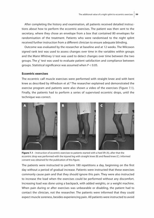
The additional value of a night splint to eccentric exercises 99
After completing the history and examination, all patients received detailed instruc-tions about how to perform the eccentric exercises. The patient was then sent to the secretary, where they chose an envelope from a box that contained 80 envelopes for randomisation of the treatment. Patients who were randomised to the night splint received further instruction from a different clinician to ensure adequate blinding.
Outcome was evaluated by the researcher at baseline and at 12 weeks. The Wilcoxon signed rank test was used to assess changes over time in the variables within groups and the Mann Whitney U test was used to detect changes over time between the two groups. The χ2 test was used to evaluate patient satisfaction and compliance between groups. Statistical significance was assumed when P < 0.05.
eccentric exercises
The eccentric calf muscle exercises were performed with straight knee and with bent knee as described by Alfredson et al.8 The researcher explained and demonstrated the exercise program and patients were also shown a video of the exercises (Figure 7.1). Finally, the patients had to perform a series of supervised eccentric drops, until the technique was correct.
figure 7.1 – Instruction of eccentric exercises to patients started with a heel lift (A), after that the eccentric drop was performed with the injured leg with straight knee (B) and flexed knee (C). Informed consent was obtained for the publication of this figure.
The patients were instructed to perform 180 repetitions a day, beginning on the first day without a period of gradual increase. Patients were instructed that these exercises commonly cause pain and that they should ignore this pain. They were also instructed to increase the load when the exercises could be performed without any discomfort. Increasing load was done using a backpack, with added weights, or a weight machine. When pain during or after exercises was unbearable or disabling, the patient had to contact the clinician, not the researcher. The patients were informed that they could expect muscle soreness, besides experiencing pain. All patients were instructed to avoid

100 Chapter 7
weight-bearing sporting activities for the first four weeks. After four weeks, gradual return to sports activities was encouraged if the pain allowed.
night splint (additional to eccentric exercises)
Patients had a 50% chance of being randomised to receive a night splint (UFO night splint, Somas Industries, Sint Anthonis, the Netherlands) besides the eccentric exercises. In this case, the patient was sent to a different clinician for consultation to ensure blind-ing. The clinician decided which size was suitable for the patient. After that, the ankle positions of 0° and 5° dorsiflexion were marked on the night splint. The patient could easily adjust the splint between these two positions. For the first four weeks, patients had to maintain the neutral position of 0°, and after this period, the position of at least 5° dorsiflexion was used. Patients were told that the night splint should be positioned beyond these 5° dorsiflexion if pain could be tolerated (Figure 7.2). Pain and the extent of sleep disturbances were the main guidelines when positioning the night splint.
figure 7.2 – Night splint fitted in a patient. In the first four weeks, a neutral position of 0° dorsiflexion was maintained (A). After four weeks of treatment, a position of 5° dorsiflexion was advised. Dorsiflexion of more degrees was recommended, when tolerated (B). Informed consent was obtained for the publication of this figure.
Compliance
Patient compliance was measured by daily registration in the form of a diary. The pa-tients had to note their number of repetitive exercises performed. Items on the night splint diary were number of hours the splint was worn and degrees in which the splint was positioned. Compliance was divided into four categories. When less than 25% of the exercises were performed, patient compliance was scored as poor. Between 25% and 50% it was scored as moderate, between 50% and 75% as good, and more than 75% was scored as an excellent compliance.

The additional value of a night splint to eccentric exercises 101
ResulTs
Patients
Between March 2005 and December 2005, 67 patients (79 tendons) visited the Hague Medical Centre for the study. Nine tendons were excluded. Four patients had an inser-tional disorder, two patients already performed a heavy-load eccentric exercise program, one patient had a partial rupture, one patient had a total rupture and one patient was unable to carry out the eccentric exercises.
In all, 58 patients (70 tendons, 46 unilateral and 12 bilateral) met the inclusion criteria. The mean (range) age was 44.6 (26-59) years, mean (range) duration of symptoms 30.7 (2-221) months and the mean (range) body mass index was 25.1 (20.2-34.5) kg/m2. All patients participated actively and most of them had to stop their sporting activities (mean (range) 13 (0-104) weeks).
A total of 70 tendons were randomised into two groups: 34 in the eccentric exercises only (eccentric group) and 36 in the eccentric exercises with the night splint (night splint group). At baseline, there were no statistical differences in patient characteristics between the groups (Table 7.1).
Table 7.1 – Patient characteristics at baseline. No significant differences in patient characteristics were found between the eccentric group and night splint group (night splint in combination with eccentric exercises).
eccentric group night splint group
Variable Mean (sd) Mean (sd)
Patient’s age (years) 44.1 (7.0) 45.1 (8.9)
Male/female 20/12 17/14
Body mass index 25.4 (3.1) 24.9 (3.2)
Duration of symptoms (months) 33.7 (55.6) 27.7 (45.9)
Duration of sports neglect (weeks) 12.6 (24.4) 13.4 (25.1)
There was a significant higher patient satisfaction in the group of patients (n = 9, patient satisfaction 89%) with a duration of symptoms of < 5.5 months when compared to the group of patients (n = 54, patient satisfaction 50%) with a duration of symptoms of > 5.5 months (p = 0.03). There was a significant higher VISA-A score after 12 weeks in the group of patients (n = 9, VISA-A score 87) with a duration of symptoms of < 5.5 months when compared to the group of patients (n = 54, VISA-A score 65) with a duration of symptoms of > 5.5 months (p = 0.02). The VISA-A score at baseline did not differ signifi-cantly between these groups (p = 0.23).
In the night splint group, there were four patients (five tendons) who could not be included in the analysis. One patient (two tendons) was poorly motivated and failed to

102 Chapter 7
attend the follow-up appointments, one patient (one tendon) left to live overseas and two patients (two tendons) stopped using the night splint because of sleep disturbance.
Results within groups
Patient satisfaction in the eccentric group after 12 weeks was excellent or good in 20 (63%) tendons and moderate or poor in 12 (37%) tendons. Patient satisfaction in the night splint group after 12 weeks was excellent or good in 15 (48%) tendons and moder-ate or poor in 16 (52%) tendons.
The VISA-A score in the eccentric group increased significantly from 50.1 at baseline to 68.8 after 12 weeks (p = 0.001). In the night splint group, the VISA-A score also increased significantly from 49.4 at baseline to 67 after 12 weeks (p < 0.001). Figure 7.3 shows the change in VISA-A score within each treatment group.
Compliance in the eccentric groupCompliance reported after 12 weeks in the eccentric group wasexcellent or good in 23 of 32 (72%) patients; patient satisfactionin this group was excellent or good in 16 (70%) patients andmoderate or poor in 7 (30%) patients (table 2).Compliance was moderate or poor in 9 (28%) patients;
patient satisfaction in this group was excellent or good in 4(44%) patients and moderate or poor in 5 (56%) patients. Therewere no statistically significant differences between thesegroups (p=0.194).
Compliance in the night splint groupEccentric exercisesIn the night splint group, reported compliance of the eccentricexercises after 12 weeks was good or excellent in 23 of 31 (74%)patients; patient satisfaction in this group was excellent or goodin 13 (57%) patients and moderate or poor in 10 (43%) patients.Compliance was moderate or poor in 8 (26%) patients;
patient satisfaction in this group was excellent or good in 2(25%) patients and moderate or poor in 6 (75%) patients. Therewere no statistically significant differences between thesegroups (p=0.131).
Night splintCompliance of the night splint after 12 weeks was excellent orgood in 25 of 31 (81%) patients; patient satisfaction in thisgroup was excellent or good in 11 (44%) patients and moderateor poor in 14 (56%) patients.Compliance was moderate or poor in 6 (19%) patients;
patient satisfaction in this group was excellent or good in 4(67%) patients and moderate or poor in 2 (33%) patients. Therewere no statistically significant differences between thesegroups (p=0.931).
Eccentric exercises and night splintIn the night splint group, reported compliance of both theeccentric exercises and the night splint after 12 weeks was goodor excellent in 18 of 31 (58%) patients; patient satisfaction inthis group was excellent or good in 9 (50%) patients andmoderate or poor in 9 (50%) patients. Compliance wasmoderate or poor for both the eccentric exercises and the nightsplint in 1 of 31 patients; patient satisfaction was rated as poor(table 2).
Complications and dropoutsFigure 4 shows the progress of patients through the study. Twopatients (two tendons) did not complete the treatment in the
eccentric group. One patient experienced too much pain andone patient developed a subluxation of the peroneal tendonduring the study, which prevented him from performing theexercises.In a few cases, the night splint caused minor symptoms,
which did not prevent further treatment. Two patientsexperienced painful pressure areas and one patient could notincrease dorsiflexion of the night splint .0˚ because ofparaesthesia of the foot.
DISCUSSIONThis prospective study shows that splinting in addition toeccentric exercises did not lead to a significantly better outcomefor patient satisfaction and VISA-A score.The effect size of eccentric training is still uncertain and few
studies have a sound method.13 The early studies showedpromising results of eccentric training on pain and/or patientsatisfaction,9–12 but functional outcome scores are required.13
The VISA-A questionnaire is a validated score for Achillestendinopathy, which measures pain, and also function in dailyliving and sporting activity. In a recent study in non-athletes,Sayana and Maffulli20 have shown VISA-A scores of approxi-mately 50 after a 12-week heavy-load eccentric exerciseprogramme. In their population, improvement was scored in19 of 34 (56%) patients, which is comparable with our results.Brown et al21 found that only 13% of the patients treated withheavy-load eccentric training and placebo injections hadreturned to sports after 3 months.The patient satisfaction after a heavy-load eccentric exercise
programme (63%) is lower than previously reportedScandinavian studies; overall satisfaction in these trials isaround 80–90%.8–12 An explanation for this result could be thatour population had a longer duration of symptoms. The meanduration of symptoms in previous studies ranged from 5.5 to21 months. Mean duration of symptoms in our population was31.6 months. A longer duration of symptoms is generallythought to be associated with a worse prognosis and we foundthat patients who had symptoms for ,5.5 months had a betteroutcome. Another explanation could be the shorter follow-uptime used in our study. The follow-up time ranged from12 weeks to 3.8 years in previously published Scandinavianstudies. In this study, a follow-up time of 12 weeks was used.This was found to be the case in the study of Brown et al,21 whoshowed that although only 13% had returned to sport after3 months, 85% had returned at 1 year.Dijkstra and Van Enst18 retrospectively studied nine patients
with Achilles tendinopathy and concluded that a night splintreduced pain after a mean duration of 9.4 months. The meanvisual analogue score decreased from 7.3 to 2.7. In a prospective
Figure 3 VISA-A score in the two treatment groups. In both treatmentgroups, VISA-A score improved significantly. There was no significantdifference at baseline and at 12 weeks follow-up between these two groupsafter 12 weeks.
Table 2 Good/excellent patient satisfaction in the twotreatment groups after 12 weeks
Patient satisfaction
Eccentric group(n = 32)
Night splint group(n = 31)
Overall 20 (63) 15 (48)Good/excellentcompliance
16 (70) 9 (50)
Moderate/poorcompliance
4 (44) 1 (0)
Values are n (%). Besides the overall results (2nd row), good/excellentpatient satisfaction was calculated for good/excellent compliance (3rd row)and moderate/poor compliance (4th row). Better patient compliance in theeccentric group tends to lead to a higher patient satisfaction than patientswith a low reported compliance for the eccentric exercises in the eccentricgroup. However, the differences were not statistically significant.
4 of 6 de Vos, Weir, Visser, et al
www.bjsportmed.com
on 19 November 2007 bjsm.bmj.comDownloaded from
figure 7.3 – VISA-A score in the two treatment groups. In both treatment groups, VISA-A score improved significantly. There was no significant difference at baseline and at 12 weeks follow-up between these two groups after 12 weeks.
In the eccentric group, there was an increase in VISA-A score in 78% of the tendons after 12 weeks, compared with baseline. In the night splint group, 71% of the tendons had a higher VISA-A score after 12 weeks.
Results between groups
There was no statistically significant difference in patient satisfaction between the two treatment groups at 12 weeks follow-up (p = 0.26). No significant differences were found in VISA-A score between the treatment groups after 12 weeks treatment (p = 0.82). T

The additional value of a night splint to eccentric exercises 103
Compliance
Table 7.2 shows reported patient compliance after 12 weeks therapy in the two treat-ment groups.
In all, 17 of 63 (27%) subjects reported a moderate or poor compliance after a 12-week program of eccentric exercises (in eccentric and night splint group).
Table 7.2 – Good/excellent patient satisfaction in the two treatment groups after 12 weeks related to the compliance to eccentric exercises. Values are n (%).Besides the overall results (2nd row), good/excellent patient satisfaction was calculated for good/excellent compliance (3rd row) and moderate/poor compliance (4th row). Better patient compliance in the eccentric group tends to lead to a higher patient satisfaction than patients with a low reported compliance for the eccentric exercises in the eccentric group. However, the differences were not statistically significant.
Patient satisfaction
eccentric group (n=32) night splint group (n=31)
overall 20 (63) 15 (48)
good/excellent compliance 16 (70) 9 (50)
Moderate/ Poor compliance 4 (44) 1 (0)
Compliance in the eccentric groupCompliance reported after 12 weeks in the eccentric group was excellent or good in 23 of 32 (72%) patients; patient satisfaction in this group was excellent or good in 16 (70%) patients and moderate or poor in seven (30%) patients (Table 7.2). Compliance was moderate or poor in nine (28%) patients; patient satisfaction in this group was excellent or good in four (44%) patients and moderate or poor in five (56%) patients. There were no statistically significant differences between these groups (p = 0.19).
Compliance in the night splint group
Eccentric exercisesIn the night splint group, reported compliance of the eccentric exercises after 12 weeks was good or excellent in 23 of 31 (74%) patients; patient satisfaction in this group was excellent or good in 13 (57%) patients and moderate or poor in 10 (43%) patients. Com-pliance was moderate or poor in eight (26%) patients; patient satisfaction in this group was excellent or good in two (25%) patients and moderate or poor in six (75%) patients. There were no statistically significant differences between these groups (p = 0.13).
Night splintCompliance of the night splint after 12 weeks was excellent or good in 25 of 31 (81%) patients; patient satisfaction in this group was excellent or good in 11 (44%) patients and moderate or poor in 14 (56%) patients. Compliance was moderate or poor in six (19%) patients; patient satisfaction in this group was excellent or good in four (67%) pa-

104 Chapter 7
tients and moderate or poor in two (33%) patients. There were no statistically significant differences between these groups (p = 0.93).
Eccentric exercises and night splintIn the night splint group, reported compliance of both the eccentric exercises and the night splint after 12 weeks was good or excellent in 18 of 31 (58%) patients; patient satis-faction in this group was excellent or good in nine (50%) patients and moderate or poor in nine (50%) patients. Compliance was moderate or poor for both the eccentric exercises and the night splint in one of 31 patients; patient satisfaction was rated as poor (Table 7.2).
Complications and dropouts
Figure 7.4 shows the progress of patients through the study. Two patients (two tendons) did not complete the treatment in the eccentric group. One patient experienced too much pain and one patient developed a subluxation of the peroneal tendon during the study, which prevented him from performing the exercises.
In a few cases, the night splint caused minor symptoms, which did not prevent further treatment. Two patients experienced painful pressure areas and one patient could not increase dorsiflexion of the night splint > 0° because of paraesthesia of the foot.
dIsCussIon
This prospective study shows that splinting in addition to eccentric exercises did not lead to a significantly better outcome in patient satisfaction and VISA-A score.
The effect size of eccentric training is still uncertain and few studies have a sound method.13 The early studies showed promising results of eccentric training on pain and/or patient satisfaction,9-12 but functional outcome scores are required.13 The VISA-A ques-tionnaire is a validated score for Achilles tendinopathy, which measures pain, and also function in daily living and sporting activity. In a recent study in non-athletes, Sayana and Maffulli 20 have shown VISA-A scores of approximately 50 after a 12-week heavy-load eccentric exercise program. In their population, improvement was scored in 19 of 34 (56%) patients, which is comparable with our results. Brown et al.21 found that only 13% of the patients treated with heavy-load eccentric training and placebo injections had returned to sports after three months.
The patient satisfaction after a heavy-load eccentric exercise program (63%) is lower than previously reported Scandinavian studies; overall satisfaction in these trials is around 80-90%.8-12 An explanation for this result could be that our population had a longer duration of symptoms. The mean duration of symptoms in previous studies ranged from 5.5 to 21 months. Mean duration of symptoms in our population was 31.6

The additional value of a night splint to eccentric exercises 105
months. A longer duration of symptoms is generally thought to be associated with a worse prognosis and we found that patients who had symptoms for < 5.5 months had a better outcome. Another explanation could be the shorter follow-up time used in our study. The follow-up time ranged from 12 weeks to 3.8 years in previously published Scandinavian studies. In this study, a follow-up time of 12 weeks was used. This was found to be the case in the study of Brown et al.21, who showed that although only 13% had returned to sport after three months, 85% had returned at one year.
Dijkstra and van Enst 18 retrospectively studied nine patients with Achilles tendi-nopathy and concluded that a night splint reduced pain after a mean duration of 9.4
randomised controlled trial, Roos et al11 reported that effects ofeccentric exercises alone were comparable to a night splint or acombination of eccentric exercises with a night splint. Agradual onset of the exercises was used during the first weekand when the patient could perform the exercises withoutdiscomfort, an increased load was prescribed. In all, 44 patientswere included in this study. After 6 weeks, pain reduction inthe eccentric group was 27%, in the night splint-only group 19%and in the night splint combined with eccentric group 18%.These differences were not statistically significant between thegroups. After 1 year, there were no differences between groupsand there was significant improvement in pain reduction (35–42%) in all groups compared to baseline. Roos et al11 proposedthat a night splint may negatively influence the positive effectof eccentric exercises to the muscle tendon unit. To ourknowledge, Roos et al11 are the only other researchers to havecompared the eccentric exercise model with the addition of anight splint; a night splint with 0˚dorsiflexion was used. In thisstudy, the patients were instructed to increase dorsiflexion asmuch as they could tolerate. This was done to maximise thehypothesised beneficial effects of splinting. Another differencewas the instruction of the eccentric exercises. Roos et al11 used agradual onset during the first week. In this study, the abruptloading as described by Alfredson et al8 was used.Reported patient compliance for the eccentric exercises was
excellent or good in the majority of the patients in bothtreatment groups, although it should be mentioned that still
more than a quarter of the patients in both groups reportedperforming the exercises at,50% of the prescribed intensity. Ofthe nine studies in the recent systematic review of Kingmaet al,13 only Roos et al11 report the compliance rate in theirfindings. In their study, a good compliance (75% of therecommended exercises) for eccentric exercises in the firstweeks was reported. At 12 weeks, however, only half stillreported a good compliance. Compliance for the night splintwas good for most of the study period.Roos et al11 did not calculate the association between outcome
and compliance. To our knowledge, there are no other studiesin which this relationship is described. In both treatmentgroups, there was a tendency that a better patient compliancefor eccentric exercises lead to better results (table 2). However,the differences were not statistically significant. A possible wayto increase patient compliance in future studies can probably bethrough more frequent patient contact by telephone or email.Although we strictly adhered to our inclusion criteria, we
cannot exclude the occurrence of selection bias. We used achecklist for objective inclusion and exclusion of patients, andwe always discussed with several clinicians when there wasdoubt whether a patient could be included.The researcher gave minimal assistance to patients on the
first occasion they completed the VISA-A questionnaire.Robinson et al19 also always administered the questionnaireonce with an investigator and stated that this would avoidsome but not all potential for observer bias.
Figure 4 Progress of patients through thestudy.
Chronic midportion Achilles tendinopathy 5 of 6
www.bjsportmed.com
on 19 November 2007 bjsm.bmj.comDownloaded from
figure 7.4 – Progress of patients through the study.

106 Chapter 7
months. The mean Visual Analogue Scale score decreased from 7.3 to 2.7. In a prospec-tive randomised controlled trial, Roos et al.11 reported that effects of eccentric exercises alone were comparable to a night splint or a combination of eccentric exercises with a night splint. A gradual onset of the exercises was used during the first week and when the patient could perform the exercises without discomfort, an increased load was pre-scribed. In all, 44 patients were included in this study. After six weeks, pain reduction in the eccentric group was 27%, in the night splint-only group 19% and in the night splint combined with eccentric group 18%. These differences were not statistically significant between the groups. After one year, there were no differences between groups and there was significant improvement in pain reduction (35- 42%) in all groups compared to baseline. Roos et al.11 proposed that a night splint may negatively influence the posi-tive effect of eccentric exercises to the muscle tendon unit. To our knowledge, Roos et al.11 are the only other researchers to have compared the eccentric exercise model with the addition of a night splint; a night splint with 0° dorsiflexion was used. In this study, the patients were instructed to increase dorsiflexion as much as they could tolerate. This was done to maximise the hypothesised beneficial effects of splinting. Another differ-ence was the instruction of the eccentric exercises. Roos et al.11 used a gradual onset during the first week. In this study, the abrupt loading as described by Alfredson et al.8 was used.
Reported patient compliance for the eccentric exercises was excellent or good in the majority of the patients in both treatment groups, although it should be mentioned that still more than a quarter of the patients in both groups reported performing the exercises at < 50% of the prescribed intensity. Of the nine studies in the recent systematic review of Kingma et al.13, only Roos et al.11 report the compliance rate in their findings. In their study, a good compliance (75% of the recommended exercises) for eccentric exercises in the first weeks was reported. At 12 weeks, however, only half still reported a good compliance. Compliance for the night splint was good for most of the study period.
Roos et al.11 did not calculate the association between outcome and compliance. To our knowledge, there are no other studies in which this relationship is described. In both treatment groups, there was a tendency that a better patient compliance for eccentric exercises leads to better results (Table 7.2). However, the differences were not statisti-cally significant. A possible way to increase patient compliance in future studies can probably be through more frequent patient contact by telephone or email.
Although we strictly adhered to our inclusion criteria, we cannot exclude the occur-rence of selection bias. We used a checklist for objective inclusion and exclusion of patients, and we always discussed with several clinicians when there was doubt whether a patient could be included.
The researcher gave minimal assistance to patients on the first occasion they com-pleted the VISA-A questionnaire. Robinson et al.19 also always administered the ques-

The additional value of a night splint to eccentric exercises 107
tionnaire once with an investigator and stated that this would avoid some but not all potential for observer bias.
ConClusIon
In this study, eccentric exercises and eccentric exercises in combination with a night splint reduced pain and improved functional outcome in patients with Achilles tendi-nopathy. The results on pain reduction are less promising than previously reported. Fur-ther trials including functional outcome measures are necessary to evaluate the effect of eccentric exercises. It can be stated that a night splint in addition to eccentric exercises is not beneficial at short-term follow-up. There was a tendency that higher reported patient compliance for eccentric exercises was correlated with a higher patient satisfac-tion. Outcome in patients with a shorter duration of the symptoms (< 5.5 months) was significantly better than in patients with longer duration of symptoms.
ACknowledgeMenT
We are grateful for the help and advice of S. Willemsen (Dept. of Biostatistics, Erasmus Medical Centre, Rotterdam, the Netherlands) in the statistical evaluation of the data.
108 Chapter 7
RefeRenCes
1. Maffulli N, Wong J, Almekinders LC. Types and epidemiology of tendinopathy. Clin Sports Med 2003; 22: 675-92.
2. Schepsis AA, Jones H, Haas AL. Achilles tendon disorders in athletes. Am J Sports Med 2002; 30: 287-305.
3. Maffulli N, Khan KM, Puddu G. Overuse tendon conditions: time to change a confusing terminol-ogy. Arthroscopy 1998; 14: 840-3.
4. Vora AM, Myerson MS, Oliva F, et al. Tendinopathy of the main body of the Achilles tendon. Foot Ankle Clin 2005; 10: 293-308.
5. Alfredson H. Conservative management of achilles tendinopathy: new ideas. Foot Ankle Clin 2005; 10: 321-9.
6. Kader D, Saxena A, Movin T, et al. Achilles tendinopathy: some aspects of basic science and clini-cal management. Br J Sports Med 2002; 36: 239-49.
7. Maffulli N, Sharma P, Luscombe KL. Achilles tendinopathy: aetiology and management. J R Soc Med 2004; 97: 472-6.
8. Alfredson H, Pietila T, Jonsson P, et al. Heavy-load eccentric calf muscle training for the treatment of chronic Achilles tendinosis. Am J Sports Med 1998; 26: 360-6.
9. Mafi N, Lorentzon R, Alfredson H. Superior short-term results with eccentric calf muscle training compared to concentric training in a randomised prospective multicentre study on patients with chronic Achilles tendinosis. Knee Surg Sports Traumatol Arthrosc 2001; 9: 42-7.
10. Fahlstrom M, Jonsson P, Lorentzon R, et al. Chronic Achilles tendon pain treated with eccentric calf-muscle training. Knee Surg Sports Traumatol Arthrosc 2003; 11: 327-33.
11. Roos EM, Engstrom M, Lagerquist A, et al. Clinical improvement after 6 weeks of eccentric exercise in patients with midportion Achilles tendinopathy – a randomised trial with 1-year follow-up. Scand J Med Sci Sports 2004; 14: 286-95.
12. Öhberg L, Lorentzon R, Alfredson H. Eccentric training in patients with chronic Achilles tendino-sis: normalised tendon structure and decreased thickness at follow up [Discussion 11]. Br J Sports Med 2004; 38: 8-11.
13. Kingma JJ, de Knikker R, Wittink HW, et al. Eccentric overload training in patients with a chronic Achilles tendinopathy: a systematic review. Br J Sports Med 2007; 41: e3.
14. Alfredson H. Chronic midportion Achilles tendinopathy: an update on research and treatment. Clin Sports Med 2003; 22: 727-41.
15. Wapner KL, Sharkey PF. The use of night splints for treatment of recalcitrant plantar fasciitis. Foot Ankle 1991; 12: 135-7.
16. Batt ME, Tanji JL, Skattum N. Plantar fasciitis: a prospective randomised clinical trial of the tension night splint. Clin J Sport Med 1996; 6: 158-62.
17. Powell M, Post WR, Keener J, et al. Effective treatment of chronic plantar fasciitis with dorsiflexion night splints: a crossover prospective randomised outcome study. Foot Ankle Int 1998; 19: 10-18.
18. Dijkstra HJ, van Enst GC. The therapeutic value of a G-brace in the management of chronic achil-les tendinosis: a pilot study. Geneeskunde en Sport 2003; 36: 137-40.
19. Robinson JM, Cook JL, Purdam C, et al. The VISA-A questionnaire: a valid and reliable index of the clinical severity of Achilles tendinopathy. Br J Sports Med 2001; 35: 335-41.
20. Sayana MK, Maffulli N. Eccentric calf muscle training in non-athletic patients with Achilles tendi-nopathy. J Sci Med Sport 2007; 10: 52-8.

The additional value of a night splint to eccentric exercises 109
21. Brown R, Orchard J, Kinchington M, et al. Aprotinin in the management of Achilles tendinopathy: a randomised controlled trial. Br J Sports Med 2006; 40: 275-79.


Chapter 8Autologous growth factor injections in
chronic tendinopathy: a systematic review
R.J. de VosP.L.J. van Veldhoven
M.H. MoenA. WeirJ.L. Tol
N. Maffulli
Department of Sports Medicine
The Hague Medical Centre Antoniushove
Leidschendam, the Netherlands
Electronic publication, ahead of print. Br Med Bull 2010

112 Chapter 8
AbsTRACT
Background - Chronic degenerative tendinopathies are frequent and difficult to treat. Tendon healing and regeneration may be improved by injecting autologous growth factors obtained from the patient’s blood. Autologous growth factors can be injected with autologous whole blood or platelet-rich plasma (PRP). There are many proposed treatment options for chronic tendinopathy. Treatments in the form of injections with autologous whole blood or PRP are increasingly used in clinical practice. There are high expectations of these regenerative injections, and there is a clear need for effective conservative therapies.
Methods - Electronic databases were searched for prospective clinical trials on treat-ment with autologous growth factors of patients with chronic tendinopathy. Chronic tendinopathy in this study included wrist extensors, flexors, plantar fasciopathy and patellar tendinopathy. Studies examining the treatment of other tendinopathies were not identified. The Physiotherapy Evidence Database score was used to examine the methodological quality of the assessment, and a qualitative analysis was performed with the levels of evidence.
Results - Eleven studies were suitable for quality assessment. All studies showed that injections of autologous growth factors (whole blood and PRP) in patients with chronic tendinopathy had a significant impact on improving pain and/or function over time. However, only three studies using autologous whole blood had a high methodological quality assessment, and none of them showed any benefit of an autologous growth factor injection when compared with a control group.
Conclusion - At present, there is strong evidence that the use of injections with au-tologous whole blood should not be recommended. There were no high-quality studies found on PRP treatment. There is limited evidence to support the use of injections with PRP in the management of chronic tendinopathy. There is growing interest in the working mechanisms of autologous growth factors. The amount and mixture of growth factors produced using different cell separating systems are largely unknown and it is also uncertain whether platelet activation prior to injection is necessary. These variables should be taken into account when starting clinical studies. A good experimental model for studying tendinopathy would be helpful for basic research. Future clinical studies using a proper control group, randomisation, blinding and validated disease-specific outcome measures for pain and function are needed.

A systematic review on autologous growth factor injections 113
InTRoduCTIon
Chronic painful tendon disorders are common in athletic and sedentary individuals.1-4 They are more common in middle age, and with increasing in sports participation at in-creasing ages, they are becoming more frequent.1,2 The Achilles tendon, patellar tendon, wrist extensors, plantar fascia and supraspinatus tendon are commonly affected larger tendons.5 Multiple aetiological factors probably play a role in the pathogenesis of these conditions.1-5
If the triad of pain, swelling and a reduced load bearing capacity are present, then the correct term for the diagnosis is tendinopathy.1 This is a clinical and not a histopatho-logical diagnosis.1,3 A failed healing repair process at tissue level results in a variety of histopathological changes, including degeneration, of the tendon tissue.3 Tendinopathy leads to a reduction in activity levels and sometimes to cessation of all sporting activi-ties.4
Increasing knowledge of the pathology and pathogenesis of tendinopathy has lead to the introduction of a large number of conservative treatments. At present, the best available evidence points towards the use of heavy load eccentric training programmes.6 Conventional conservative therapy is ineffective in around 25% of patients with Achilles tendinopathy.7 In these patients, surgery can be performed, but this is not always suc-cessful, and the post-operative rehabilitation is slow and time consuming.3,4,6-8 To reduce the need for surgery, more effective conservative therapies are needed.
Recently, research has focused on regenerative therapies with high expectations of success.9,10 The use of autologous growth factors is thought to lead to tendon healing through collagen regeneration and the stimulation of a well-ordered angiogenesis.9,10 These growth factors are administered in the form of autologous whole blood or platelet-rich plasma (PRP).9 Platelets can be isolated using simple cellseparating systems.9,11 The degranulation of the α-granules in the platelets releases many different growth factors that play a role in tissue regeneration processes. Platelet-derived growth factor, trans-forming growth factor-β, vascular-derived endothelial growth factor, epithelial growth factor, hepatocyte growth factor and insulin-like growth factor are examples of such growth factors.9-12 Injections with autologous growth factors are becoming common in clinical practice.10,11
This systematic review examines the literature on the effects of autologous blood and PRP injections in the management of tendinopathies.

114 Chapter 8
MeThods
literature search
A comprehensive, systematic literature search was performed in October 2009. The da-tabases of PubMed, MEDLINE, EMBASE, CINAHL and the Cochrane library were searched without time limits. The following key words were used in differing combinations: “tendinopathy”, “tendinosis”, “tendonitis”, “tendons”, “tennis elbow”, “plantar fasciitis”, “platelet-rich plasma”, “platelet transfusion”, “autologous blood” or “injection”. The search was limited to articles in English, and only human studies were included. All titles and abstracts were assessed by two researchers, and all relevant articles were obtained. All bibliographies were also hand searched to identify further relevant literature.
All relevant articles were read independently in full text by two researchers to assess whether they met the inclusion criteria. If there was a difference in opinion on suitability, a consensus was reached by consulting a third reviewer.
study selection
Articles were suitable (inclusion criteria) if the subjects had been clinically diagnosed as having chronic tendinopathy. The design had to be a prospective clinical study; ran-domised controlled trial (RCT), nonrandomised clinical trial (CCT) or prospective case series. There had to be a well-described intervention in the form of an injection with either PRP or autologous blood. The outcome had to be reported in terms of pain and/or function.
data extraction
Two researchers independently recorded the study design, population, intervention, outcome measure and outcome using standardised data extraction forms.13 To assess the efficacy of the interventions, mean values of the continuous outcomes were ex-tracted from the published articles.
Quality assessment
The studies included were scored using the PEDro (Physiotherapy Evidence Database) score.14 The PEDro score is an 11-point list using yes and no answers. The first statement pertains to the external validity of the study and is not used to compute the final quality score. The score (0-10) is the number of positive answers on questions 2-11. The PEDro items are shown in Table 8.1.
To assess the reliability of consensus ratings using the PEDro scale, a study was con-ducted by Maher et al.14 A random selection of 120 RCTs was assessed four times by four different raters. Intraclass correlation coefficient for consensus ratings using the PEDro scale showed to be 0.68, which compares to a “fair” to “good” reliability.15 It was

A systematic review on autologous growth factor injections 115
suggested that the PEDro scale has sufficient reliability for its use in systematic reviews of physiotherapy trials 14, and recently it has been used in a systematic review on the effects of exercise treatment in tendinopathy.16
A PEDro score of 6 or higher is considered to represent a high-quality study.16 The results of the quality assessments of the individual trials were used to classify the level of evidence.17 This qualitative analysis was performed with five levels of evidence based upon the quality and results of clinical studies:
1. Strong evidence: provided by generally consistent findings in multiple high quality RCTs
2. Moderate evidence: provided by generally consistent findings in one high quality RCTs and one or more lower quality RCTs, or by generally consistent findings in mul-tiple low quality RCTs
3. Limited evidence: provided by only one RCT (either high or low quality) or generally consistent findings in CCTs
4. Conflicting evidence: inconsistent findings in multiple RCTs or CCTs5. No evidence: no RCTs or CCTs
Studies with a high methodological score using the PEDro scale were considered as high-quality studies and those with a low PEDro score were considered low-quality studies.
Table 8.1 – PEDro scale. The score is the number of positive answers on questions 2-11 (0-10).
Items
1. Eligibility criteria were specified
2. Subjects were randomly allocated to groups
3. Allocation was concealed
4. The groups were similar at baseline regarding the most important prognostic indicators
5. There was blinding of all subjects
6. There was blinding of all therapists who administered the therapy
7. There was blinding of all assessors who measured at least one key outcome
8. Measures of at least one key outcome were obtained from more than 85% of the subjects initially allocated to groups
9. All subjects for whom outcome measures were available received the treatment or control condition as allocated or, where this was not the case, data for at least one key outcome were analysed by “intention to treat”
10. The results of between-group statistical comparisons are reported for at least one key outcome
11. The study provides both point measures and measures of variability for at least one key outcome

116 Chapter 8
ResulTs
literature search
Thirteen studies were included after screening. Two studies were excluded (Figure 8.1).43,44 Eleven studies were suitable for quality assessment and were assessed using the PEDro score.
study design
There were six observational non-controlled studies 18-23 and five controlled clinical trials 24-28 of which two were evaluated as having appropriate randomisation.24,28
Participants
The mean number of subjects was 40.5 (SD 24.6) with a range 20-100. Four studies were on patients with chronic tendinopathy of the wrist extensors (tennis elbow) 18,19,23,25 of which one study on both wrist extensor and flexor tendinopathy (golfer’s elbow).25 One study evaluated the treatment effect on tendinopathy of wrist flexors.20 Patients with chronic plantar fasciopathy were treated in three studies 24,27,28 and three studies had examined patients with chronic patellar tendinopathy.21,22,26
Interventions
There were eight studies on the effects of autologous blood injections 18-21,23,24,27,28, of which five studies used this in combination with a local anaesthetic 18-21,24 and the other three studies applied only autologous blood.23,27,28
There were three studies on PRP injections 22,25,26, of which one used an additional local anaesthetic 25 and two did not report whether local anaesthesia were used.22,26 In three studies a single injection 24,25,27, in one study two injections 21 and in two studies three injections 22,26 were used. In the other five studies, a varying number of injections (1-3) were given.18-20,23,28 The PRP was prepared using a single 25 or double 22,26 centrifuging process. In two studies, calcium was added to the PRP for activation of the platelets.22,26
outcome measures
Ten of the 11 studies used a visual analogue scale (VAS) or ordinal scale to measure pain.18-20,22-28 In four studies, the elbow function was quantified using the Nirschl score.18-20,23 The Nirschl score runs from 1; mild pain during activity to 7; constant pain at rest. To our knowledge, there are no data available on the validity of the Nirschl score. One study 21 used the Victorian Institute of Sports Assessment-Patella (VISA-P) score which is a validated outcome measure for patellar tendinopathy that assesses pain and function. It runs from 0 representing maximal pain and minimal function to 100 which represents no pain and maximal functioning. The two other studies on patellar tendinopathy used

A systematic review on autologous growth factor injections 117
the Tegner score to quantify activity level.22,26 The Tegner score runs from 0 to 10 with 0 being invalidated and 10 representing specific professional sports activities. The Tegner score previously showed an acceptable validity in the evaluation of meniscal injuries.29
figure 8.1 – QUOROM statement flow diagram.

118 Chapter 8
One study on plantar fasciopathy used the rearfoot score of the American Orthopaedics Foot and Ankle (AOFAS) to assess function.28 There are no data available on the validity of the AOFAS score for the evaluation of plantar fasciopathy. This score runs from 0 to 100; a score of 100 represents no pain and optimal functioning. One study used the modified Mayo elbow score, which was not recorded at final follow-up.25
outcomes
All the intervention groups reported a significant improvement in the pain and/or func-tion scores with the mean improvement being 66% (SD 19, range 33-100). The outcomes in the control groups also improved significantly in all the studies with a mean improve-ment of 57% (SD 18, range 32-87). These improvements were reported after a mean follow-up of 9.4 months (SD 6.0). There was in none of the included studies a beneficial effect on pain score at final follow-up after autologous growth factor injections when compared with a control group.24,26-28 One study reported a significant improvement on the functional Tegner score when compared with the control group, but the statistical baseline difference in Tegner score between these groups was not reported.26 In four other control groups, there were similar results on pain and/or function when compared with autologous growth factor injections.24,27,28 In two control groups, there was a signifi-cant improvement on pain in favour of the control group.27 Table 8.2 gives an overview of these differences.
sample size calculation
Only one trial reported a sample size calculation.22 Kon et al. reported that 20 cases were needed to detect a clinically important increase of 15 points on the VAS score. All other included studies did not report using a sample size calculation.
Methodological quality
The PEDro scores for the 11 studies are shown in Table 8.3. The scores ranged from 1 to 7 with an average of 3.4 (SD 2.3). Three studies were considered as being high qual-ity (PEDro score ≥ 6) and the other eight studies were of low quality (PEDro score < 6). All the studies reported the inclusion criteria. A comparison to another treatment was performed in five studies, and randomisation was used in two studies. Blinding of the treatment was undertaken for patients in one study 27, for the treating physician in none of the studies and for the outcome assessor in three studies.24,27,28 In three studies, more than 15% of the patients were lost to follow-up 19,20,25, and in four studies, the data analysis was not performed on an “intention to treat” basis.19-21,25 Two studies had poor reporting of the statistical analysis.18,23

A systematic review on autologous growth factor injections 119
Tabl
e 8.
2 –
Incl
uded
stu
dies
. Im
prov
emen
ts w
ere
calc
ulat
ed a
fter
cor
rect
ing
for s
cale
and
bas
elin
e sc
ore.
Refe
renc
en
umbe
r of
par
tici
-pa
nts
stud
y de
sign
Incl
usio
n cr
iter
iaIn
terv
enti
onCo
ntro
l gr
oup(
s)Pr
imar
y ou
tcom
e m
easu
res
follo
w-
up
(mo)
out
com
e in
inte
rven
tion
gr
oup
(% im
prov
emen
t)o
utco
me
in c
ontr
ol
grou
p(s)
(%
impr
ovem
ent)
Edw
ards
and
Ca
land
rucc
io.
2003
18
28Ca
se
serie
sw
rist e
xten
sor
tend
inop
athy
1-3
auto
logo
us
bloo
d in
ject
ion(
s)-
Pain
sca
le (0
-10)
9.5
Mea
n pa
in s
core
: 7.8
to
2.3
(71%
)-
Nirs
chl s
core
(0-7
)M
ean
Nirs
chl s
core
: 6.5
to
2.0
(69%
)
Mis
hra
and
Pave
lko.
200
6 25
20CC
Tw
rist e
xten
sor
and
flexo
r te
ndin
opat
hy
1 PR
P in
ject
ion
C: 1
ana
esth
etic
in
ject
ion
VAS
scor
e (0
-100
)25
.6M
ean
VAS
scor
e: 8
0.3
to
5.7
(93%
)C:
Mea
n VA
S sc
ore:
N
A
Mod
ified
May
o el
bow
sco
re (0
-100
)M
ean
Mod
ified
May
o el
bow
sco
re: N
AM
ean
Mod
ified
May
o el
bow
sco
re: N
A
Sure
sh e
t al.
2006
20
20Ca
se
serie
sw
rist fl
exor
te
ndin
opat
hy2-
3 au
tolo
gous
bl
ood
inje
ctio
ns-
VAS
scor
e (0
-10)
10M
ean
VAS
scor
e: 8
.0 to
2.
2 (7
3%)
-
Nirs
chl s
core
(0-7
)M
edia
n N
irsch
l sco
re: 6
.0
to 1
.0 (8
3%)
Conn
ell e
t al.
2006
19
35Ca
se
serie
sw
rist e
xten
sor
tend
inop
athy
2-3
auto
logo
us
bloo
d in
ject
ions
-VA
S sc
ore
(0-1
0)6
Med
ian
VAS
scor
e: 9
.0 to
0.
0 (1
00%
)-
Nirs
chl s
core
(0-7
)M
edia
n N
irsch
l sco
re: 6
.0
to 0
.0 (1
00%
)

120 Chapter 8
Tabl
e 8.
2 (c
ontin
ued)
Refe
renc
en
umbe
r of
par
tici
-pa
nts
stud
y de
sign
Incl
usio
n cr
iter
iaIn
terv
enti
onCo
ntro
l gr
oup(
s)Pr
imar
y ou
tcom
e m
easu
res
follo
w-
up
(mo)
out
com
e in
inte
rven
tion
gr
oup
(% im
prov
emen
t)o
utco
me
in c
ontr
ol
grou
p(s)
(%
impr
ovem
ent)
Kite
r et a
l. 20
06 28
54RC
Tpl
anta
r fa
scio
path
y1-
3 au
tolo
gous
bl
ood
inje
ctio
n(s)
C1: 1
-2
cort
icos
tero
id
inje
ctio
n(s)
VAS
scor
e (0
-10)
6M
ean
VAS
scor
e: 7
.6 to
2.
4 (6
8%)
C1: M
ean
VAS
scor
e:
7.3
to 2
.6 (6
5%) †
C2: d
ry
need
ling
AOFA
S sc
ore
(0-
100)
Mea
n AO
FAS
scor
e: 7
1.6
to 8
0.9
(33%
)M
ean
AOFA
S sc
ore:
65
.7 to
80.
1 (4
2%) †
C2: M
ean
VAS
scor
e:
6.4
to 2
.0 (6
9%) †
Mea
n AO
FAS
scor
e:
64.1
to 7
8.2
(39%
) †
Ul G
ani e
t al.
2007
23
26Ca
se
serie
sw
rist e
xten
sor
tend
inop
athy
1-2
auto
logo
us
bloo
d in
ject
ion(
s)-
Pain
sca
le (1
-4)
8M
ean
pain
sco
re: 3
.3 to
1.
2 (6
4%)
-
Nirs
chl s
core
(0-7
)M
ean
Nirs
chl s
core
: 5.5
to
2.1
(62%
)
Jam
es e
t al.
2007
21
47Ca
se
serie
spa
tella
r te
ndin
opat
hy2
auto
logo
us
bloo
d in
ject
ions
co
mbi
ned
with
dr
y ne
edlin
g
-VI
SA-P
sco
re (0
-100
)14
.8M
ean
VISA
-P s
core
: 39.
8 to
74.
3 (5
7%)
-
Lee
and
Ahm
ad.
2007
24
64RC
Tpl
anta
r fa
scio
path
y1
auto
logo
us
bloo
d in
ject
ion
C: 1
cor
ticos
te-
roid
inje
ctio
nVA
S sc
ore
(0-1
0)6
Mea
n VA
S sc
ore:
7.3
to
3.6
(51%
)C:
Mea
n VA
S sc
ore:
6.
9 to
2.4
(65%
) †
Kon
et a
l. 20
09 22
20Ca
se
serie
spa
tella
r te
ndin
opat
hy3
PRP
inje
ctio
ns-
EQ-V
AS
scor
e (0
-100
)6
Mea
n EQ
-VA
S sc
ore:
57
to
82 (5
8%)
-
Tegn
er s
core
(0-1
0)M
ean
Tegn
er s
core
: 4 to
7
(50%
)

A systematic review on autologous growth factor injections 121
Tabl
e 8.
2 (c
ontin
ued)
Refe
renc
en
umbe
r of
par
tici
-pa
nts
stud
y de
sign
Incl
usio
n cr
iter
iaIn
terv
enti
onCo
ntro
l gr
oup(
s)Pr
imar
y ou
tcom
e m
easu
res
follo
w-
up
(mo)
out
com
e in
inte
rven
tion
gr
oup
(% im
prov
emen
t)o
utco
me
in c
ontr
ol
grou
p(s)
(%
impr
ovem
ent)
Fila
rdo
et a
l. 20
09 26
31CC
Tpa
tella
r te
ndin
opat
hy3
PRP
inje
ctio
nsC:
exe
rcis
e th
erap
yEQ
-VA
S sc
ore
(0-1
00)
6M
ean
EQ-V
AS
scor
e: 5
2.7
to 7
8.3
(54%
)C:
Mea
n EQ
-VA
S sc
ore:
50.
6 to
73.
5 (4
6%) †
Tegn
er s
core
(0-1
0)M
ean
Tegn
er s
core
: 3.7
to
6.6
(46%
)M
ean
Tegn
er s
core
: 5.
3 to
6.8
(32%
) *
Kala
ci e
t al.
2009
27
100
CCT
plan
tar
fasc
iopa
thy
1 au
tolo
gous
bl
ood
inje
ctio
nC1
: 1 c
ortic
oste
-ro
id in
ject
ion
VAS
scor
e (0
-10)
6M
ean
VAS
scor
e: 6
.8 to
3.
5 (4
8%)
C1: M
ean
VAS
scor
e:
7.0
to 1
.5 (7
8%) ‡
C2: 1
cor
ticos
te-
roid
inje
ctio
n co
mbi
ned
with
dr
y ne
edlin
g
C2: M
ean
VAS
scor
e:
7.2
to 1
.0 (8
7%) ‡
C3: 1
an
aest
hetic
in
ject
ion
com
bine
d w
ith
dry
need
ling
C3: M
ean
VAS
scor
e:
6.7
to 3
.4 (4
8%) †
CCT
– no
nran
dom
ised
clin
ical
tria
lRC
T –
rand
omis
ed c
ontr
olle
d tr
ial
VAS
– Vi
sual
Ana
logu
e Sc
ale
VISA
-P –
Vic
toria
n In
stitu
te o
f Spo
rts
Ass
essm
ent-
Pate
llaAO
FAS
– A
mer
ican
Ort
hopa
edic
s Fo
ot a
nd A
nkle
Sco
re (r
earf
oot s
core
)N
A –
Not
Ava
ilabl
eC
– Co
ntro
l gro
up*
– Si
gnifi
cant
impr
ovem
ent i
n fa
vour
of a
utol
ogou
s gr
owth
fact
or in
ject
ion
† –
No
sign
ifica
nt d
iffer
ence
with
con
trol
gro
up‡
– Si
gnifi
cant
impr
ovem
ent i
n fa
vour
of c
ontr
ol g
roup

122 Chapter 8
level of evidence
Until now, three high-quality studies 24,27,28 on the use of autologous growth factor injections (all used autologous blood injections) for the management of chronic ten-dinopathy showed no significant improvement when compared with a control group. One study showed a significant superior improvement after a corticosteroid injection in comparison with one single autologous blood injection.27 Two of these high-quality studies were RCTs. As such, there is level 1 (strong) evidence of no improvement in pain and/or function in chronic tendinopathy after injecting autologous blood when com-pared with other treatment options. If PRP injections were to be considered separately, three low-quality studies were included, and so there is level 3 (limited) evidence that these injections improve pain and/or function in chronic tendinopathy.
dIsCussIon
A total of 11 articles were suitable for inclusion in this systematic review on the use of autologous growth factors in the treatment of chronic tendinopathy. Three studies, of which two were RCTs, were of high quality. All studies showed an improvement in pain and function scores, but there was no difference when compared with the improvement in pain scores in the control groups. After a qualitative analysis, there was level 1 (strong) evidence that injections with autologous blood were not of benefit. Currently, there is level 3 (limited) evidence that PRP injections improve pain and/or function in chronic tendinopathy.
Table 8.3 – Particular scoring of the included studies for methodological quality according to the PEDro score. The total score was defined by the number of positive answers on questions 2-11 (0-10).
Item Pedro score Total score
Reference 1 2 3 4 5 6 7 8 9 10 11
Edwards en Calandruccio. 2003 18 + − − − − − − + + − − 2/10
Mishra en Pavelko. 2006 25 + − − − − − − − − + + 2/10
Suresh et al. 2006 20 + − − − − − − − − − + 1/10
Connell et al. 2006 19 + − − − − − − − − − + 1/10
Kiter et al. 2006 28 + + − + − − + + + + + 7/10
Ul Gani et al. 2007 23 + − − − − − − + + − − 2/10
James et al. 2007 21 + − − − − − − + − − + 2/10
Lee and Ahmad. 2007 24 + + − + − − + + + + + 7/10
Kon et al. 2009 22 + − − − − − − + + − + 3/10
Filardo et al. 2009 26 + − − − − − − + + + + 4/10
Kalaci et al. 2009 27 + − − − + − + + + + + 6/10

A systematic review on autologous growth factor injections 123
These findings are clinically relevant, as the use of autologous growth factors is gain-ing popularity.9-11 This results in part from laboratory and animal studies showing posi-tive and promising results.30-32 Autologous growth factors have the potential to change collagen production and degradation by influencing matrix regulating enzymes.9,10,33
Laboratory studies showed that the addition of PRP to human tenocytes resulted in cell proliferation, collagen deposition and improved gene expression for matrix degrad-ing enzymes and endogenous growth factors.30 A recent animal study found similar results 31, and the in vivo application of PRP suggested an accelerated remodelling and angiogenic process. Bosch et al.32 performed a placebo-controlled ultrasound study on the recovery of horse tendons using PRP which showed an increase in anti-inflammatory response and fibrillogenesis in the short term. At longer-term follow-up, an increased collagen matrix integrity was found in the PRP treated tendons.
Although the results of laboratory studies are encouraging, they always use healthy tendons or surgically induced lesions given the lack of a good experimental model for tendinopathy. At present, it is unclear whether these results can be extrapolated to ten-dinopathic tendons, and future research in the field of basic science should study this.
This systematic review makes it clear that there is a lack of good quality studies in this field, especially regarding treatment with PRP. The commonest methodological flaws are the lack of a suitable control group, randomisation and blinding of subjects and those involved in the treatment. One research group reported that the study design was a RCT 27, but after critically reading the full-text, it became apparent that this study was a CCT. Another research group selected a very small control group of five patients and reported a significant improvement in pain and function scores in the PRP group compared with this small control group after eight weeks.25 However, the patients in the control group were lost to follow-up already after eight weeks and could not be included in the final analysis. Although there was a consensus that this was a CCT, the authors agreed that the control group was not appropriate. Although these method-ological processes are relatively simple to implement, it does make the research process more intensive and less attractive for potential subjects. It is not uncommon for pilot studies to be performed to assess the effect size of new treatments before progressing to evaluate their use in randomised controlled clinical trials. Lower-quality studies on the management of tendinopathy evidence better results than good-quality studies.34 Future studies should therefore use appropriate randomisation, and all those involved should be blinded to the treatment given.
A few other suggestions on future research can also be made. There may be differ-ences in natural healing response between load-bearing tendons, such as the patellar and Achilles tendon, and non-load-bearing tendons, such as the wrist extensors and flexors. Wrist extensor tendinopathy is a self-limiting disease with 80-90% recovery within one year 35, whereas patients with tendinopathy of the main body of the Achilles

124 Chapter 8
tendon did not improve in a trial with a four month wait and see arm.36 In some studies, the subjects included had a variety of midportion and insertional tendinopathies, and it is unclear whether these can be compared, as these portions of the tendon have dif-fering biomechanical and metabolic properties and responses to treatment.5 This makes comparing the results of studies on differing locations of tendinopathy difficult and emphasises the need of suitable control groups.
Many of the studies on the effect of injections with autologous growth factors used a mixture with local anaesthetic which could lead to bias, as an injection with local anaes-thetic alone led to improvement in a previous trial on elbow tendinopathy.35
Most of the studies included in this review used pain as the primary outcome to assess treatment effect. Only one study had used the VISA-P score, a validated outcome ques-tionnaire for patellar tendinopathy. Four studies used the Nirschl score, which does give a global impression of pain in combination with activity. Outcome assessment should focus on activity as well as pain when studying tendinopathy and where possible use disease-specific validated measures.37 Another important feature of outcome assess-ment is the prior establishment of the clinically important difference. Only one study of those included reported a sample size calculation with the use of a clinically relevant dif-ference.22 In osteoarthritis research, minimally important clinical differences are defined for different outcome measures 38, but these values are lacking in tendinopathy research.
No studies to date have compared an injection with autologous growth factors to a placebo injection. The effects of placebo treatments are greater the more invasive they are 39, and a recent tendinopathy study showed a large effect after a placebo injection was performed.40
Currently, there have also been no studies that have compared autologous blood to PRP. This would be interesting given the larger costs and practical difficulties associated with preparing PRP.
Along with treatment effects it is also necessary to report and monitor for complica-tions.33 In the studies included here, no complications were reported but it is important to monitor for infections, ruptures and possible systemic effects when using autologous growth factors.
There are still many unanswered questions in this field. There has been little research performed on the amount of growth factors produced using different cell separating systems, and what the optimal mixture would be.10,11 It is unclear what the best volume and frequency of the injections is. Moreover, when multiple injections are considered, the ideal period between multiple injections is unknown. It is also uncertain whether platelet activation prior to injection is necessary, as contact with collagen would also lead to platelet degranulation.41,42

A systematic review on autologous growth factor injections 125
ConClusIon
There is strong evidence that autologous blood injections do not improve pain and/or function compared with other treatment options. There is only limited evidence that PRP injections are beneficial. All three high-quality studies on the use of autologous growth factor injections in the management of chronic tendinopathy showed no ben-efit. All studies did show an effect on pain and function in time, but many are seriously methodologically flawed. To date, there is strong evidence that the use of injections with autologous blood should not be recommended, and there is limited evidence to support the use of injections with PRP in the treatment of chronic tendinopathy. Further studies using a proper control group, randomisation, blinding and validated disease-specific outcome measures for pain and function are needed.

126 Chapter 8
RefeRenCes
1. Maffulli N, Wong J, Almekinders LC (2003) Types and epidemiology of tendinopathy. Clin Sports Med, 22, 675-92.
2. Schepsis AA, Jones H, Haas AL (2002) Achilles tendon disorders in athletes. Am J Sports Med, 30, 287-305.
3. Alfredson H (2003) Chronic midportion Achilles tendinopathy: an update on research and treat-ment. Clin Sports Med, 22, 727-41.
4. van Linschoten R, den Hoed PT, de Jongh AC (2007) Guideline ‘Chronic Achilles tendinopathy, in particular tendinosis, in sportsmen/sportswomen’. Ned Tijdschr Geneeskd, 20, 2319-24.
5. Woo SLY, Renström PA (2007) Tendinopathy in athletes, the encyclopaedia of sports medicine, volume XII. Hong Kong: Blackwell Publishing.
6. Andres BM, Murrell GA (2008) Treatment of tendinopathy: what works, what does not, and what is on the horizon. Clin Orthop Relat Res, 466, 1539-54.
7. Kvist M (1994) Achilles tendon injuries in athletes. Sports Med, 18, 173-201. 8. Zwerver J (2008) Patellar tendinopathy (‘jumper’s knee’); a common and difficult-to-treat sports
injury. Ned Tijdschr Geneeskd, 16, 1831-7. 9. Sampson S, Gerhardt M, Mandelbaum B (2008) Platelet rich plasma injection grafts for musculo-
skeletal injuries: a review. Curr Rev Musculoskelet Med, 1, 165-74. 10. Mishra A, Woodall J Jr, Vieira A (2009) Treatment of tendon and muscle using platelet-rich plasma.
Clin Sports Med, 28, 113-25. 11. Dohan Ehrenfest DM, Rasmusson L, Albrektsson T (2009) Classification of platelet concentrates:
from pure platelet-rich plasma (P-PRP) to leucocyte- and platelet-rich fibrin (L-PRF). Trends Bio-technol, 27, 158-67.
12. Eppley BL, Woodell JE, Higgins J (2004) Platelet quantification and growth factor analysis from platelet-rich plasma: implications for wound healing. Plast Reconstr Surg, 114, 1502-8.
13. Moher D, Cook DJ, Eastwood S et al. (1999) Improving the quality of reports of meta-analyses of randomised controlled trials: the QUOROM statement. Quality of Reporting of Meta-analyses. Lancet, 354, 1896-1900.
14. Maher CG, Sherrington C, Herbert RD et al. (2003) Reliability of the PEDro scale for rating quality of randomised controlled trials. Phys Ther, 83, 713-21.
15. Fleiss JL (1986) The Design and Analysis of Clinical Experiments. New York, NY: John Wiley & Sons Inc.
16. Woodley BL, Newsham-West RJ, Baxter GD (2007) Chronic tendinopathy: effectiveness of eccen-tric exercise. Br J Sports Med, 41, 188-98.
17. van Tulder M, Furlan A, Bombardier C, Bouter L (2003) Editorial Board of the Cochrane Collabo-ration Back Review Group. Updated method guidelines for systematic reviews in the cochrane collaboration back review group. Spine (Phila Pa 1976), 28, 1290-9.
18. Edwards SG, Calandruccio JH (2003) Autologous blood injections for refractory lateral epicondy-litis. J Hand Surg [Am], 28, 272-8.
19. Connell DA, Ali KE, Ahmad M et al. (2006) Ultrasound-guided autologous blood injection for ten-nis elbow. Skeletal Radiol, 35, 371-7.
20. Suresh SP, Ali KE, Jones H, Connell DA (2006) Medial epicondylitis: is ultrasound guided autolo-gous blood injection an effective treatment? Br J Sports Med, 40, 935-9.
21. James SL, Ali K, Pocock C et al. (2007) Ultrasound guided dry needling and autologous blood injection for patellar tendinosis. Br J Sports Med, 41, 518-21.

A systematic review on autologous growth factor injections 127
22. Kon E, Filardo G, Delcogliano M et al. (2009) Platelet-rich plasma: new clinical application: a pilot study for treatment of jumper’s knee. Injury, 40, 598-603.
23. Ul Gani N, Butt M, Dhar SA et al. (2007) Autologous Blood Injection in the Treatment of Refractory Tennis Elbow. The Internet Journal of Orthopaedic Surgery, volume 5.
24. Lee TG, Ahmad TS (2007) Intralesional autologous blood injection compared to corticosteroid injection for treatment of chronic plantar fasciitis. A prospective, randomised, controlled trial. Foot Ankle Int, 28, 984-90.
25. Mishra A, Pavelko T (2006) Treatment of chronic elbow tendinosis with buffered platelet rich plasma. Am J Sports Med, 34, 1774-8.
26. Filardo G, Kon E, Della Villa S et al. (2009) Use of platelet-rich plasma for the treatment of refrac-tory jumper’s knee. Int Orthop, PMID: 19641918.
27. Kalaci A, Cakici H, Hapa O et al. (2009) Treatment of plantar fasciitis using four different local injection modalities: a randomised prospective clinical trial. J Am Podiatr Med Assoc, 99, 108-13.
28. Kiter E, Celikbas E, Akkaya S et al. (2006) Comparison of injection modalities in the treatment of plantar heel pain: a randomised controlled trial. J Am Podiatr Med Assoc, 96, 293-6.
29. Briggs KK, Kocher MS, Rodkey WG et al. (2006) Reliability, validity, and responsiveness of the Lysholm knee score and Tegner activity scale for patients with meniscal injury of the knee. J Bone Joint Surg Am, 88, 698-705.
30. De Mos M, van der Windt AE, Jahr H et al. (2008) Can Platelet-Rich Plasma Enhance Tendon Repair? A Cell Culture Study. Am J Sports Med, 36, 1171-8.
31. McCarrel T, Fortier L (2009) Temporal growth factor release from platelet-rich plasma, trehalose lyophilized platelets, and bone marrow aspirate and their effect on tendon and ligament gene expression. J Orthop Res, 27, 1033-42.
32. Bosch G, van Schie HT, de Groot M et al. (2009) Effects of Platelet Rich Plasma on the quality of repair of mechanically induced core lesions in equine Superficial Digital Flexor Tendons: a placebo-controlled experimental study. J Orthop Res, PMID: 19714688.
33. Creaney L, Hamilton B (2008) Growth Factor Delivery Methods in the Management of Sports Injuries: The State of Play. Br J Sports Med, 42, 314-20.
34. Tallon C, Coleman BD, Khan KM, Maffulli N (2001) Outcome of surgery for chronic Achilles tendi-nopathy. A critical review. Am J Sports Med, 29, 315-20.
35. Rineer CA, Ruch DS (2009) Elbow tendinopathy and tendon ruptures: epicondylitis, biceps and triceps ruptures. J Hand Surg [Am], 34, 566-76.
36. Rompe JD, Nafe B, Furia JP, Maffulli N (2007) Eccentric loading, shock-wave treatment, or a wait-and-see policy for tendinopathy of the main body of tendo Achillis: a randomised controlled trial. Am J Sports Med, 35, 374-83.
37. Kingma JJ, de Knikker R, Wittink HM, Takken T (2007) Eccentric overload training in patients with a chronic Achilles tendinopathy: a systematic review. Br J Sports Med, 41, e3.
38. Pham T, van der Heijde D, Altman RD et al. (2004) OMERACT-OARSI initiative: Osteoarthritis Research Society International set of responder criteria for osteoarthritis clinical trials revisited. Osteoarthritis Cartilage, 12, 389-99.
39. Zhang W, Robertson J, Jones AC et al. (2008) The placebo effect and its determinants in osteoar-thritis: meta-analysis of randomised controlled trials. Ann Rheum Dis, 67, 1716-23.
40. Brown R, Orchard J, Kinchington M et al. (2006) Aprotinin in the management of Achilles tendi-nopathy: a randomised controlled trial. Br J Sports Med, 40, 275-9.
41. Anitua E, Sánchez M, Nurden AT et al. (2006) New insights into and novel applications for platelet-rich fibrin therapies. Trends Biotechnol, 24, 227-34.

128 Chapter 8
42. Fufa D, Shealy B, Jacobson M et al. (2008) Activation of platelet-rich plasma using soluble Type I collagen. J Oral Maxillofac Surg, 66, 684-90.
43. Mishra A, Collado H, Fredericson M (2009) Platelet-rich plasma compared with corticosteroid injection for chronic lateral elbow tendinosis. PM R, 1, 366-70.
44. Barrett SL, Erredge SE (2004) Growth factors for chronic plantar fasciitis? Podiatry Today, 17, 36-42.

Chapter 9Platelet-rich plasma injection for chronic Achilles
tendinopathy – a randomised controlled trial
R.J. de VosA. Weir
H.T.M. van SchieS.M.A. Bierma-Zeinstra
J.A.N. VerhaarH. Weinans
J.L. Tol
Department of Sports Medicine
The Hague Medical Centre Antoniushove
Leidschendam, the Netherlands
JAMA 2010; 303(2): 144-9
130 Chapter 9
AbsTRACT
Context - Tendon disorders comprise 30% to 50% of all activity-related injuries; chronic degenerative tendon disorders (tendinopathy) occur frequently and are difficult to treat. Tendon regeneration might be improved by injecting platelet-rich plasma (PRP), an increasingly used treatment for releasing growth factors into the degenerative tendon.
Objective - To examine whether a PRP injection would improve outcome in chronic midportion Achilles tendinopathy.
Design, setting, and patients - A stratified, block-randomised, double-blind, placebo-controlled trial at a single centre (the Hague Medical Centre, Leidschendam, the Neth-erlands) of 54 randomised patients aged 18 to 70 years with chronic tendinopathy 2 to 7 cm above the Achilles tendon insertion. The trial was conducted between August 28, 2008, and January 29, 2009, with follow-up until July 15, 2009.
Intervention - Eccentric exercises (usual care) with either a PRP injection (PRP group) or saline injection (placebo group). Randomisation was stratified by activity level.
Main outcome measures - The validated Victorian Institute of Sports Assessment-Achilles (VISA-A) questionnaire, which evaluated pain score and activity level, was completed at baseline and 6, 12, and 24 weeks. The VISA-A score ranged from 0 to 100, with higher scores corresponding with less pain and increased activity. Treatment group effects were evaluated using general linear models on the basis of “intention-to-treat”.
Results - After randomisation into the PRP group (n = 27) or placebo group (n = 27), there was complete follow-up of all patients. The mean VISA-A score improved signifi-cantly after 24 weeks in the PRP group by 21.7 points (95% confidence interval [CI], 13.0-30.5) and in the placebo group by 20.5 points (95% CI, 11.6-29.4). The increase was not significantly different between both groups (adjusted between-group difference from baseline to 24 weeks, -0.9; 95% CI, -12.4 to 10.6). This CI did not include the predefined relevant difference of 12 points in favour of PRP treatment.
Conclusions - Among patients with chronic Achilles tendinopathy who were treated with eccentric exercises, a PRP injection compared with a saline injection did not result in greater improvement in pain and activity.
Trial registration - Clinicaltrials.gov Identifier: NCT00761423.

Platelet-rich plasma injection for chronic Achilles tendinopathy 131
InTRoduCTIon
Overuse injury of the Achilles tendon is a frequent problem that often affects sport participants but also inactive middle-aged individuals.1,2 An estimated 30% to 50% of all sports-related injuries are tendon disorders.3 Former distance runners have a lifetime risk of 52% for Achilles tendon injury.4 Achilles tendon injuries frequently lead to sport cessation for long periods and may interfere with activities of daily living.5 Conservative treatment is disappointing and 25% to 45% of patients eventually require surgery.1,5 There is a clear need for improved conservative therapy.
Many factors in the aetiology and pathogenesis have been reported, but no study has identified a direct cause-effect relationship.1,2 Previously, the nomenclature “tendinitis” was generally used for chronic tendon disorders, suggesting the presence of inflamma-tion.1,2 Histological studies, however, proved abnormal tissue repair and degeneration, which favoured the term tendinopathy for the clinical triad of pain, swelling, and de-creased activity.1,2 Anti-inflammatory agents, previously used for chronic tendinopathies without appropriate efficacy 1,6, have now been replaced by eccentric exercises as usual care 6 that provide some positive effects on tendon collagen synthesis and may result in a decrease of pain.1,7
The recent introduction of platelet-rich plasma (PRP) injections in tendinopathy raised high expectations.8-11 Platelets derived from whole blood using simple cell-separating systems provide a release of various growth factors that participate in tissue repair processes.9,12,13 Three recent reviews reported promising results of the use of PRP in tendinopathy, although these conclusions were based on laboratory studies and on clinical studies with important limitations.9-11 Although we are aware of no published data on the prevalence of use of this therapy, two recent reviews have suggested that PRP injections for tendinopathy are increasingly used in the clinical setting.13,14
The goal of our double-blind, block-randomised, placebo-controlled trial, the first to our knowledge in this field, was to compare the effects on pain and functional outcome of a PRP injection with a placebo injection, both combined with an eccentric exercise program in patients with chronic midportion Achilles tendinopathy.
MeThods
study design
The stratified, block-randomised, double-blind, placebo-controlled trial was performed at the sports medicine outpatient department in a large district general hospital (The Hague Medical Centre Antoniushove, Leidschendam, the Netherlands). The single centre study was announced to general practitioners, sports medicine physicians, orthopaedic surgeons, physiotherapists, and the general public with advertisements on several web

132 Chapter 9
sites, folders, and regional radio. The PRP treatment was disseminated as a potentially successful treatment for tendinopathies. According to the study protocol, the primary analysis was performed after 24 weeks follow-up. After 24 weeks, blinding was disclosed for the primary researcher. Results at 52 weeks will be used as a secondary outcome to describe the long-term results in a future analysis.
The study protocol was approved by the regional Medical Ethics Committee Zuidwest Holland, Voorburg, the Netherlands. All patients provided written informed consent.
Patients
When patients contacted the researcher by telephone or e-mail, detailed information about the study was given and eligibility was evaluated. If patients seemed to be suit-able for inclusion in the study after this screening, an appointment was made at the sports medicine outpatient department. One experienced sports medicine physician (JLT) evaluated suitability for inclusion.
Inclusion criteria were the presence of chronic midportion Achilles tendinopathy at the age of 18 to 70 years. The diagnosis was made based on clinical findings: all patients had a painful and thickened tendon in relation to activity and on palpation. The tendon pain was located approximately 2 to 7 cm proximal to the insertion on the calcaneus. Symptoms had to have been present for at least two months.
Exclusion criteria were (1) clinical suspicion of other musculoskeletal (insertional dis-orders and tendon rupture) injuries, inflammatory internal disorders, or use of specific medications that can cause tendinopathy (fluoroquinolones); (2) previous performance of a complete heavy load eccentric exercise program 1 or inability to perform it; or (3) a previous injection with PRP. Detailed information regarding the inclusion and exclusion criteria is described at clinicaltrials.gov (Identifier: NCT00761423) or can be obtained from the corresponding author (RJV).
Procedures
A researcher (RJV) prepared a PRP injection and a saline injection for every patient. The PRP injection was prepared using the “recover platelet separation kit”, in accordance with the system instructions.12 Fifty-four millilitres of venous blood was collected from the cubital vein. The whole blood was mixed with 6 mL of citrate to prevent early clotting. After blood collection and 15 minutes of centrifugation, PRP was obtained. To match the pH of PRP with the pH of the tendon tissue, 0.3 mL of 8.4% sodium bicarbonate buffer was added. One millilitre of the PRP was collected for evaluation of possible contamina-tion of PRP after the preparation, which was cultured and analysed on microbial growth by the department of medical microbiology, the Hague Medical Centre, Leidschendam, the Netherlands. Four millilitres of PRP was collected for infiltration and 4 mL of isotonic saline was also prepared in an identical syringe.

Platelet-rich plasma injection for chronic Achilles tendinopathy 133
Randomisation
The pre-injury activity level may be a confounder for the primary outcome in this study, because it evaluates pain and activity.15 Stratification was used to divide the number of active patients between the two treatment groups. This stratification was based on the Ankle Activity Score, which objectively quantifies the ankle-related activity.16 An Ankle Activity Score of 4 or more points indicated a high activity level and a score of less than 4 points indicated a low activity level. Randomisation was performed using sealed opaque, identical envelopes. The envelopes were evenly distributed in the high- and low activity box. The patient was then randomised into one of the two treatment groups by choosing a closed envelope. To ensure balance in the number of patients between the groups, a block-randomisation was performed (block size of 12 participants).
One unblinded sports medicine physician (AW) selected the correct injection and blinded the injection with the use of a covering sheath surrounding the syringe and hub of the needle. To ensure concealment of allocation, data on allocation were stored in a secret location. The content of the injection was blinded for the treating sports medicine physician, researcher, and patients.
Intervention
A blinded sports medicine physician (JLT) performed the injection. First, a local anaes-thetic was injected (2 mL of 0.5% marcain) in the skin and subcutaneous tissue. Using an ultrasonographic machine (MyLab30; Esaote Piemedical, Maastricht, the Netherlands), the tendon structure was imaged and the blinded fluid was injected using a 22-gauge needle through three different puncture locations. Through each puncture location, five small depots were left at several sites in the degenerative area of the main body of the tendon with Colour Doppler guidance. Immediately after the injection, the patients lay prone on the examination table for 10 minutes.
All patients received detailed instructions on the standardised rehabilitation program.1 During the first 48 hours after the injection, patients were only allowed to walk short dis-tances indoors. During days three to seven postinjection, walks up to 30 minutes were allowed. After the first week, the exercise program was started and consisted of one week of stretching exercises and then a 12-week daily eccentric exercise program (180 repetitions).1 Eccentric exercises were done by performing “heel drops” on a step. The specific action of this eccentric exercise movement is the stretch of the Achilles tendon with concurrently contraction of the calf muscle.
All patients were instructed to avoid weight-bearing sporting activities for the first four weeks. After four weeks, a gradual return to sports activities was encouraged. The intensity of sports activities could be increased when there was only mild pain (maxi-mum score of 3 on a scale from 0-10, with 0 representing no pain and 10 representing maximum pain) and no increase in morning stiffness.

134 Chapter 9
Patients were instructed to avoid the use of co-interventions within the follow-up period. Acetaminophen (500 mg) could be used as rescue medication.
outcome measures
All patients completed a questionnaire consisting of standardised outcome measures at baseline and after 6, 12, and 24 weeks. The primary outcome measure was the Victorian Institute of Sports Assessment-Achilles (VISA-A) questionnaire, which quantifies the pain and activity level.17 The VISA-A score ranged from 0 to 100, where 0 denotes no activity and maximum pain and 100 denotes maximum activity and no pain. This is a validated questionnaire, specifically designed for evaluating outcome in Achilles tendinopathy. Secondary outcome measures were subjective patient satisfaction, return to sports, and adherence of the eccentric exercises. Patient satisfaction was subjectively rated as poor, fair, good, or excellent. A good or excellent result was determined as successful. The re-turn to sports level was divided into five groups (not active in sports, no return to sports, returning to sport but not in desired sport, returning to desired sport but not at the pre-injury level, and returning to pre-injury level in the desired sport). We determined the patient’s return to desired sport, regardless of the level. The patients received forms to keep daily logs for the eccentric exercises. At follow-up, the subjective adherence of the patients was determined by asking which percentage of the prescribed repetitions the patients had accomplished.
statistical analysis
Based on previous studies, our alternative hypothesis was that in the group treated with a PRP injection (PRP group), the VISA-A score would be 12 points higher in comparison with the group treated with a saline injection (placebo group).15,18,19 The SD of the VISA-A score was estimated at 15 points.17-19 We calculated that a sample of 27 in each group was required for the study to detect this difference, with a power of 80% with two-sided testing at a significance level of 0.05 and assuming 10% loss to follow-up. The patients were analysed by “intention-to-treat”. To test for the effect of treatment on the between-group difference in primary outcome, we used the repeated measurement general linear model. Changes from baseline to all follow-up time points were included in the model. Adjustments were made for those variables that influenced the primary outcome with P < 0.10. We evaluated secondary outcomes with use of a generalised estimating equations model.
The researcher (RJV) who performed the analyses was blinded to the allocated treat-ment. The analyses were performed by using SPSS version 16.0.1 (SPSS Inc, Chicago, Illinois, USA).

Platelet-rich plasma injection for chronic Achilles tendinopathy 135
ResulTs
Between August 28, 2008, and January 29, 2009, 99 patients contacted the researcher (RJV) for information on the study. The flow of patients through the trial is shown in Figure 9.1, and the baseline characteristics of the patients in both treatment groups are shown in Table 9.1. During the study, there were no patients lost to follow-up and there were no missing data. The end of the follow-up period was on July 16, 2009.
The mean VISA-A score improved significantly after 24 weeks within the PRP group by 21.7 points (95% confidence interval [CI], 13.0-30.5) and within the placebo group by 20.5 points (95% CI, 11.6-29.4) (Figure 9.2).
Screened for eligibility (n= 99) Excluded after screening (n=38)
Reasons: refused to participate (n=10) choose another therapy (n=8) choose a waiting policy (n=8) fear of the injection (n=3) insertional tendon disorder (n=2) complaints for less than two
months (n=2) performed a total eccentric exercise
program (n=2) total rupture (n=2) no time (n=1)
Analysed (n=27)
Lost to follow-up (n=0)
Allocated to PRP-injection and EET (n=27)
Analysed (n=27)
Allocation
Analysis
Follow-Up
Clinical assessment at the research location
Randomisation
Allocated to saline-injection and EET (n=27)
Lost to follow-up (n=0)
Excluded after examination (n= 7) Reasons: insertional tendinopathy (n=4) musculo-tendinous disorder (n=1) abnormal Thompson test (n=1) PRP treatment elsewhere (n=1)
figure 9.1 – Progress of patients through the study.PRP indicates platelet-rich plasma; EET, eccentric exercise therapy. A Thompson test suggests the presence of a tendon rupture.

136 Chapter 9
Variables that were considered as important predictors of the primary outcome (VISA-A score) were the baseline VISA-A score (p = 0.03) and duration of symptoms (p = 0.06). After adjustment for these variables, there was no significant difference in improvement on the VISA-A score at 6, 12, and 24 weeks follow-up between these two treatment groups; between-group differences were 2.5 (95% CI, -6.9 to 11.9), -1.6 (95% CI, -11.9 to 8.7), and -0.9 (95% CI, -12.4 to 10.6), respectively (positive values favour the PRP group) (Table 9.2).
There was no significant difference in secondary outcome measures. Subjective patient satisfaction (after 24 weeks: -4.1%; 95% CI, -25.8% to 17.7%) and number of patients re-turning to their desired sport (after 24 weeks: 1.4%; 95% CI, -17.0% to 19.8%) are shown in Table 9.2 (positive values favour the PRP group).
The mean (SD) percentage of reported adherence for the eccentric exercises in the PRP group was 70.9% (27.0) and in the placebo group was 74.6% (17.3). There was no significant difference between both groups (95% CI, -16.1 to 8.7).
Table 9.1 – Baseline characteristics in the PRP group and placebo group.a
Abbreviations: BMI, body mass index, calculated as weight in kilograms divided by height in meters squared; IQR, interquartile range; PRP, platelet-rich plasma; VISA-A, Victorian Institute of Sports Assessment-Achilles.
PRP-group (n=27) placebo-group (n=27)
Age (years), mean (sd) 49 (8.1) 50 (9.4)
gender, n (%)
- Male 13 (48) 13 (48)
duration of symptoms (weeks), median (IQR)
36 (24-78) 26 (16-104)
Activity, n (%)
- Active in sports 22 (81) 24 (89)
- Sedentary 5 (19) 3 (11)
level of sports, n (%)
- Competitive 6 (27) 3 (13)
- Recreational 16 (73) 21 (87)
sports activity at baseline, n (%)
- Unchanged 2 (9) 9 (37)
- Reduced 8 (36) 5 (21)
- Ceased 12 (55) 10 (42)
duration of sports cessation (weeks), mean (sd)
11 (16) 12 (23)
body Mass Index (kg/m2), mean (sd)
26.8 (3.9) 26.2 (3.5)
VIsA-A score, mean (sd) 46.7 (16.2) 52.6 (19.0)
a Data are presented as No. (%) unless otherwise specified.

Platelet-rich plasma injection for chronic Achilles tendinopathy 137
One patient in the PRP group used a tendon binding band during the follow-up pe-riod and one patient in the placebo group applied foot orthotics. The eccentric exercises were continued with a lower frequency after the 12-week program in the PRP group by 15 patients and in the placebo group by 17 patients (p = 0.58).
There was no microbial growth found in the collected PRP samples, and no complica-tions (infections, hematomas, or ruptures) were reported after the treatments.
CoMMenT
In the first, to our knowledge, double-blind, block-randomised, placebo-controlled trial on the clinical use of a PRP injection, there was no benefit on pain and function. There were also no significant differences observed in the secondary outcome measures (subjective patient satisfaction and return to sports activity).
These findings are important and clinically relevant as PRP is thought to be growing in popularity and recent reviews supported its use for chronic tendon disorders.9-11 These conclusions were drawn based on laboratory studies and small clinical studies. Some of the released growth factors, such as vascular endothelial growth factor, platelet-derived growth factor, and transforming growth factor β, have the potential to play a role in regen-eration of tendon tissue through increased tendon cell proliferation, collagen synthesis,
figure 9.2 – Individual changes in the VISA-A Score from baseline in patients treated with PRP and placebo. Adjusted between group mean differences from baseline to 6, 12, and 24 weeks are shown with 95% confidence intervals.VISA-A indicates Victorian Institute of Sports Assessment-Achilles; PRP, platelet-rich plasma.
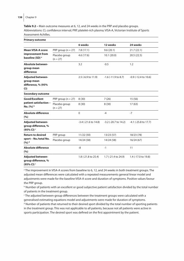
138 Chapter 9
Table 9.2 – Main outcome measures at 6, 12, and 24 weeks in the PRP and placebo groups.Abbreviations: CI, confidence interval; PRP, platelet-rich plasma; VISA-A, Victorian Institute of Sports Assessment-Achilles.
Primary outcome
6 weeks 12 weeks 24 weeks
Mean VIsA-A score improvement from baseline (sd) a
PRP group (n = 27) 7.8 (17.1) 9.6 (20.1) 21.7 (22.1)
Placebo group (n = 27)
4.6 (17.6) 10.1 (20.0) 20.5 (22.5)
Absolute between-group mean difference
3.2 -0.5 1.2
Adjusted between-group mean difference, % (95% CI)
2.5 (-6.9 to 11.9) -1.6 (-11.9 to 8.7) -0.9 (-12.4 to 10.6)
secondary outcome
good/excellent patient satisfaction – no. (%) b
PRP group (n = 27) 8 (30) 7 (26) 15 (56)
Placebo group (n = 27)
8 (30) 8 (30) 17 (63)
Absolute difference (%)
0 -4 -7
Adjusted between-group difference, % (95% CI) c
-3.4 (-21.6 to 14.8) -3.2 (-20.7 to 14.2) -4.1 (-25.8 to 17.7)
Return to desired sport – no./total no. (%) d
PRP group 11/22 (50) 13/23 (57) 18/23 (78)
Placebo group 14/24 (58) 14/24 (58) 16/24 (67)
Absolute difference (%)
-8 -1 11
Adjusted between-group difference, % (95% CI) c
1.8 (-21.8 to 25.4) 1.7 (-21.4 to 24.9) 1.4 (-17.0 to 19.8)
a The improvement in VISA-A scores from baseline to 6, 12, and 24 weeks in both treatment groups. The adjusted mean differences were calculated with a repeated measurements general linear model and adjustments were made for the baseline VISA-A score and duration of symptoms. Positive values favour the PRP group.b Number of patients with an excellent or good subjective patient satisfaction divided by the total number of patients in the treatment group.c The adjusted between-group differences between the treatment groups were calculated with a generalised estimating equations model and adjustments were made for duration of symptoms.d Number of patients that returned to their desired sport divided by the total number of sporting patients in the treatment group. This was not applicable to all patients, because not all patients were active in sports participation. The desired sport was defined on the first appointment by the patient.

Platelet-rich plasma injection for chronic Achilles tendinopathy 139
and vascularisation. In in vitro and animal studies, positive effects on tendon collagen tissue and vascularisation were reported.20-23 However, these studies using healthy ten-dons or traumatically induced lesions as a good experimental model for tendinopathy is lacking, and it is unknown whether the results of these studies also apply to degenerative tendon disorders. The two small clinical studies on PRP in tendinopathy showed a good ef-fect on pain scores and patient satisfaction but had important limitations, such as the lack of a proper control group, disease-specific and validated outcome measurements, and blinding procedure.24,25 Treatments in tendinopathy that seem to be effective in poor qual-ity studies frequently fail to show clinical benefit when assessed in good clinical studies.26
Our study showed no statistically significant difference in outcome between the two groups and the CIs did not include the predefined difference used in the power cal-culation (12 points on the VISA-A score). We defined this clinically relevant difference as 12 points, based on previous studies.15,18,19 There is no official agreement on the minimal clinical important difference for the VISA-A score, but on other comparable studies in mus-culoskeletal medicine, this is reported to be 10% to 15% of the scale.27-29 Our predefined clinically relevant difference of 12 points is reasonably located between this accepted minimal clinical important difference of 10% to 15% of the scale. The estimated difference was determined for 24 weeks, but due to the demands of active patients and the claimed fast recovery after PRP administration, we were also interested in the 6- and 12-week results. The reason why both treatment groups show clinical progression in our study, but also in other studies on PRP, is likely due to the fact that exercises were performed. Eccen-tric exercises have been shown to be effective in previous randomised trials.18,30 After four months, eccentric exercises proved to improve pain and function in contrast with a wait-and-see policy 30, although there is no convincing evidence that eccentric exercise therapy is more effective than other forms of exercise.31,32 Another possible explanation for the improvement in the placebo group might be that the placebo response is amplified when a treatment is invasive and raises high expectations.33 This has also been demonstrated previously in a study on the value of injection therapy for the treatment of tendinopathy.34
A limitation of our study is that the amount of platelets and the quantity of activated growth factors that were present in the PRP injections was unknown. Nonetheless, good platelet collection efficiency was reported with the use of the platelet separation system used in our study, and a positive correlation between the number of platelets and the harvest of growth factors has been shown.10,12 Another variable that may be of interest is the length of time that the platelets remain at the site after injection into the degenerative area. Platelets are slowly activated by exposure to tendon collagen 10, but it might be that due to the pressure within the tendon a large amount of PRP diffused rapidly out of the tendon, thereby reducing its effect. However, in a laboratory study, an increase in tendon collagen synthesis was also found even with the use of a lower PRP

140 Chapter 9
concentration (20%) in healthy human tendon cells, which may be more comparable with the concentration reached during in vivo administration.20
Moreover, the PRP preparation and injection was performed as the usual generally accepted procedure in daily clinical practice.12 The lack of a group that received only a PRP injection without eccentric exercises may be regarded as another limitation of the study. Until now, all studies reporting clinical effects in tendinopathy have used it in combination with exercises. Although it is unlikely that eccentric exercises have had a negative effect on the PRP treatment, as shown by an animal model in which tendon mechanical properties were improved when PRP was combined with mechanical stress, the study design makes it impossible to rule this out.22
In the future, laboratory studies could examine which fraction of an injected substance remains within the degenerative tendon. This information may be useful for an accurate design of laboratory studies and implementation in clinical research. The Achilles ten-don midportion is an ideal location for further clinical research in tendinopathy, because it is not affected by accompanying pathology 8, not self limiting at midterm 30, and there is a disease-specific validated outcome measure.17
Among patients with chronic midportion Achilles tendinopathy treated with an eccentric exercise program, a PRP injection compared with a saline injection did not result in greater improvement in pain and activity. Therefore, we do not recommend this treatment for chronic midportion Achilles tendinopathy.
fundIng/suPPoRT
This work was funded by Biomet Biologics LLC, Warsaw, Indiana, USA.
Role of The sPonsoR
Biomet Biologics LLC had no role in the design and conduct of the study; in the collec-tion, management, analysis, and interpretation of the data; or in the preparation, review, or approval of the manuscript.
AddITIonAl ConTRIbuTIons
We thank Max Reijman, PhD, and Jan H. Waarsing, PhD (Department of Orthopaedics, Erasmus University Medical Centre, Rotterdam, the Netherlands), for their help with the study design, statistical evaluation, and interpretation of the data; Sten P. Willemsen

Platelet-rich plasma injection for chronic Achilles tendinopathy 141
(Department of Biostatistics, Erasmus University Medical Centre), for his independent statistical analysis; and Cora van der Heijden, Ank de Vries, and Pauline Verschoor (De-partment of Sports Medicine, the Hague Medical Centre Antoniushove, Leidschendam, the Netherlands), for their logistic help. These individuals were not compensated for their efforts. We thank Biomet Biologics LLC for their support. We also thank the patients for their participation and the health professionals for referring them to the study.
142 Chapter 9
RefeRenCes
1. Alfredson H. Chronic midportion Achilles tendinopathy: an update on research and treatment. Clin Sports Med 2003; 22: 727-41.
2. Maffulli N, Wong J, Almekinders LC. Types and epidemiology of tendinopathy. Clin Sports Med 2003; 22: 675-92.
3. Järvinen TA, Kannus P, Maffulli N, Khan KM. Achilles tendon disorders: aetiology and epidemiol-ogy. Foot Ankle Clin 2005; 10: 255-66.
4. Zafar MS, Mahmood A, Maffulli N. Basic science and clinical aspects of achilles tendinopathy. Sports Med Arthrosc. 2009; 17: 190-7
5. Maffulli N, Sharma P, Luscombe KL. Achilles tendinopathy: aetiology and management. J R Soc Med 2004; 97: 472-76.
6. Rees JD, Maffulli N, Cook J. Management of tendinopathy. Am J Sports Med. 2009; 37: 1855-67. 7. Langberg H, Ellingsgaard H, Madsen T, et al. Eccentric rehabilitation exercise increases peritendi-
nous type I collagen synthesis in humans with Achilles tendinosis. Scand J Med Sci Sports 2007; 17: 61-6.
8. Woo SL, Renstrőm PA, Arnoczky SP. Tendinopathy in athletes, the encyclopaedia of sports medi-cine, volume XII. Blackwell Publishing; Hong Kong, China: 2007.
9. Sampson S, Gerhardt M, Mandelbaum B. Platelet rich plasma injection grafts for musculoskeletal injuries: a review. Curr Rev Musculoskelet Med 2008; 1: 165-74.
10. Mishra A, Woodall J Jr, Vieira A. Treatment of tendon and muscle using platelet-rich plasma. Clin Sports Med 2009; 28: 113-25.
11. Sánchez M, Anitua E, Orive G, et al. Platelet-rich therapies in the treatment of orthopaedic sport injuries. Sports Med 2009; 39: 345-54.
12. Eppley BL, Woodell JE, Higgins J. Platelet quantification and growth factor analysis from platelet-rich plasma: implications for wound healing. Plast Reconstr Surg 2004; 114: 1502-8.
13. Dohan Ehrenfest DM, Rasmusson L, Albrektsson T. Classification of platelet concentrates: from pure platelet-rich plasma (P-PRP) to leucocyte- and platelet-rich fibrin (L-PRF). Trends Biotechnol 2009; 27: 158-67.
14. Alsousou J, Thompson M, Hulley P, et al. The biology of platelet-rich plasma and its application in trauma and orthopaedic surgery: a review of the literature. J Bone Joint Surg Br 2009; 91: 987-96.
15. Sayana MK, Maffulli N. Eccentric calf muscle training in non-athletic patients with Achilles tendi-nopathy. J Sci Med Sport 2007; 10: 52-58.
16. Halasi T, Kynsburg A, Tállay A, Berkes I. Development of a new activity score for the evaluation of ankle instability. Am J Sports Med 2004; 32: 899-908.
17. Robinson JM, Cook JL, Purdam C, et al. The VISA-A questionnaire: a valid and reliable index of the clinical severity of Achilles tendinopathy. Br J Sports Med 2001; 35: 335-41.
18. de Vos RJ, Weir A, Visser RJ, de Winter TC, Tol JL. The additional value of a night splint to eccentric exercises in chronic midportion Achilles tendinopathy: a randomised controlled trial. Br J Sports Med 2007; 41: e5.
19. James SL, Ali K, Pocock C, et al. Ultrasound guided dry needling and autologous blood injection for patellar tendinosis. Br J Sports Med 2007; 41: 518-21.
20. De Mos M, van der Windt AE, Jahr H, et al. Can Platelet-Rich Plasma Enhance Tendon Repair? A Cell Culture Study. Am J Sports Med 2008; 36: 1171-8.

Platelet-rich plasma injection for chronic Achilles tendinopathy 143
21. Bosch G, van Schie HT, de Groot M, et al. Effects of Platelet Rich Plasma on the quality of repair of mechanically induced core lesions in equine Superficial Digital Flexor Tendons: a placebo-controlled experimental study. J Orthop Res 2009; PMID: 19714688.
22. Virchenko O, Aspenberg P. How can one platelet injection after tendon injury lead to a stronger tendon after 4 weeks? Interplay between early regeneration and mechanical stimulation. Acta Orthop 2006; 77: 806-12.
23. McCarrel T, Fortier L. Temporal growth factor release from platelet-rich plasma, trehalose ly-ophilized platelets, and bone marrow aspirate and their effect on tendon and ligament gene expression. J Orthop Res 2009; 27: 1033-42.
24. Mishra A, Pavelko T. Treatment of chronic elbow tendinosis with buffered platelet-rich plasma. Am J Sports Med 2006; 34: 1774-8.
25. Kon E, Filardo G, Delcogliano M, et al. Platelet-rich plasma: new clinical application: a pilot study for treatment of jumper’s knee. Injury 2009; 40: 598-603.
26. Tallon C, Coleman BD, Khan KM, Maffulli N. Outcome of surgery for chronic Achilles tendinopathy. A critical review. Am J Sports Med 2001; 29: 315-20.
27. Pham T, van der Heijde D, Altman RD, et al. OMERACT-OARSI initiative: Osteoarthritis Research So-ciety International set of responder criteria for osteoarthritis clinical trials revisited. Osteoarthritis Cartilage. 2004; 12: 389-99.
28. Rozendaal RM, Koes BW, van Osch GJ, et al. Effect of glucosamine sulfate on hip osteoarthritis: a randomised trial. Ann Intern Med. 2008; 148: 268-77.
29. Ostelo RW, Deyo RA, Stratford P, et al. Interpreting change scores for pain and functional status in low back pain: towards international consensus regarding minimal important change. Spine (Phila Pa 1976). 2008; 33: 90-4.
30. Rompe JD, Nafe B, Furia JP, Maffulli N. Eccentric loading, shock-wave treatment, or a wait-and-see policy for tendinopathy of the main body of tendo Achillis: a randomised controlled trial. Am J Sports Med 2007; 35: 374-83.
31. Kingma JJ, de Knikker R, Wittink HM, Takken T. Eccentric overload training in patients with chronic Achilles tendinopathy: a systematic review. Br J Sports Med 2007; 41: e3.
32. Wasielewski NJ, Kotsko KM. Does eccentric exercise reduce pain and improve strength in physi-cally active adults with symptomatic lower extremity tendinosis? A systematic review. J Athl Train 2007; 42: 409-21.
33. Zhang W, Robertson J, Jones AC, et al. The placebo effect and its determinants in osteoarthritis: meta-analysis of randomised controlled trials. Ann Rheum Dis. 2008; 67: 1716-23.
34. Brown R, Orchard J, Kinchington M, et al. Aprotinin in the management of Achilles tendinopathy: a randomised controlled trial. Br J Sports Med 2006; 40: 275-9.


Chapter 10General Discussion


General Discussion 147
dIsCussIon
Chronic midportion Achilles tendinopathy is a frequently occurring disorder which is difficult to treat. It is generally accepted to diagnose this condition based upon clini-cal findings in presence of pain and swelling of the Achilles tendon in the midportion combined with impaired function.1-3 When this clinical diagnosis is made, there is a high probability that histology will show degenerative changes.4 An abundance of options are currently available for the treatment of tendinopathy, but scientific evidence for their effectiveness is scarce.5-7 The working mechanisms of various treatment options are poorly understood and there are few predictive factors for treatment outcome, which makes it difficult to provide an accurate prognosis for patients. Tendinopathy frequently leads to frustration for patients and treating physicians and it is one of the most chal-lenging areas in sports medicine and orthopaedics.
Increasing interest in tendinopathy research has led to rapid development of knowl-edge in this field. Especially in the fields of imaging and treatment there have been many publications in the last decade. There are, however, still many unanswered questions in these areas. The work in this thesis attempts to improve the knowledge on imaging modalities and the effects of several treatment options. We hoped to find predictive fac-tors for clinical outcome and we followed ultrasonographical changes after treatment. The clinical effect of eccentric exercise therapy, splinting and an injection of platelet-rich plasma were studied. With these objectives we hoped to contribute to an increased un-derstanding of the working mechanisms of treatment and aid the clinician in improving patient outcome in tendinopathy.
ulTRAsonogRAPhy
neovascularisation
Healthy tendons are relatively hypovascular. In a case-control study it was shown that blood flow could not be measured using Colour Doppler ultrasonography (CDU) in asymptomatic tendons, but in symptomatic tendons there was an increased blood flow.8 This increased blood flow has been referred to as “neovascularisation” in the literature, but to date it is unknown whether this phenomenon is the result of newly formed blood vessels. We showed that neovascularisation can also be detected in asymptomatic tendons.9 Whether this is due to underlying pathology of the tendon tissue or a normal increased tendon Doppler flow needs to be determined. We can state that Doppler flow is less frequent in healthy tendons than in tendinopathic tendons, which may be caused by the physiologically low blood supply of healthy tendons. Several studies have re-ported that in cases of tendinopathy an increased blood flow is frequently observed on

148 Chapter 10
Doppler ultrasonography.8,10-12 This is thought to be part of the repair process secondary to degeneration.13,14 The reported prevalence of this “neovascularisation” in chronic ten-dinopathy ranges from 47% to 100%.8,10-12,15-18 At present it is unknown how these large differences can be explained. A problem when comparing these results may be the lack of a standardised approach in assessing the degree of neovascularisation. Cook et al.19 reported that the measurement of the length of the neovascularisation within patellar tendons using CDU could be performed with a high reproducibility. We confirmed these findings in Achilles tendons with an easy to use five-grade scale on Power Doppler ultra-sonography (PDU) with an excellent interobserver reliability.9 Another relevant finding is that prior physical activity can influence the amount of neovascularisation, even in asymptomatic individuals.20,21 This emphasises the need for a standardised examination, as proposed in our studies.9,22
Research in the evaluation of equine tendon injuries used comparable standardised settings and examined the neovascularisation with CDU.23 These authors found that the number of regularly arranged blood vessels within the endotenon, determined on harvested tissue after immunohistochemical staining, was closely related to the degree of neovascularisation obtained with CDU prior to euthanasia of the horses. This result supports the hypothesis that standardised ultrasonographic Doppler measurement of neovascularisation can accurately determine the number of blood vessels within tendons. That the location of the neovascularisation in human Achilles tendinopathy may differ from that in equine tendinopathy, is a possible weakness of this hypothesis. In human tendinopathy, the neovascularisation is frequently located at the anterior side of the tendon and a diffuse distribution throughout the tendon can also be observed. This may cause a different representation of neovascularisation using ultrasonographic Doppler measurement.
The clinical implications of increased neovascularisation, measured with Doppler ultrasonography, remain unknown. Several studies have examined the relationship between the amount of neovascularisation and clinical severity of symptoms with conflicting results.8,10-12 Our findings supported a previous report 12 that presence of neovascularisation before the start of conservative treatment had no role in prediction of the clinical outcome, measured with a functional score, at short-term follow-up. In our study we found that there was no difference in change of the Victorian Institute of Sports Assessment-Achilles (VISA-A) score at short-term follow-up after conservative treatment when tendons without neovascularisation were compared with tendons with neovascularisation at baseline.22 However the neovascularisation can be used as target of treatment, since several studies found beneficial effects after obliterating neovascu-larisation with sclerosing agents.15-17,24,25 A decrease in neovascularisation after injecting sclerosing agents was found to result in higher patient satisfaction, although an increase in neovascularisation has been observed shortly after the injections.24
General Discussion 149
Recently, interesting hypotheses have been postulated about the role of neovasculari-sation in the treatment response in tendinopathy. It is thought that an increase in neo-vascularisation in the early phase of treatment would be beneficial. Consequently, it ap-peared that the local administration of platelet-rich plasma (PRP) would be a reasonable manner to achieve this. Platelet degranulation leads to a significant increase in several growth factors.26 One of these; vascular endothelial growth factor (VEGF), is known to be a potent stimulator of angiogenesis.23,27 A laboratory study showed that PRP induced endogenous VEGF production in tendon cells.28 A study on surgically created lesions in equine tendons reported that the neovascularisation, measured with CDU, increased sig-nificantly in tendons treated with PRP compared to controls.23 We described the effects of PRP in combination with eccentric exercise therapy on the neovascularisation score in patients with chronic tendinopathy. There was a short-term increase and subsequent decrease of neovascularisation, but this pathway was not significantly different from the placebo group.29 It is not clear whether this initial increase is a result of tendon needling or a normal response over time after the start of eccentric exercises. In our study, the effects on neovascularisation may have been overshadowed by the eccentric exercise therapy. Another hypothesis is that in degenerative tendinopathy the in vivo response to growth factor delivery is smaller than expected from laboratory and equine studies.23,27,30
A novel theory is that not the neovessels, but the accompanying nerves are the source of pain in chronic tendinopathy.3 There is a lack of innervation in healthy tendon tissue, but the amount of nerves may be increased in the presence of pathological neovascularisation.31 This theory was supported after the detection of several sensory neurotransmitters, such as glutamate, using a microdialysis technique 32 and staining of pathologic tissue specimens.31 Recently, however, researchers studied the ventral part of the paratenon after biopsy, where the neovessels are frequently observed.31 Within this area there were more sympathetic nerves than sensory nerves with a pronounced occurrence of α-adrenoreceptors, which are known to mediate constriction of blood vessels. These authors hypothesised that the “neovascularisation”, observed with Dop-pler ultrasonography, is the measurement of an increased pathological blood vessel constriction with concomitant increased blood flow. This flow may be increased, due to an equal blood supply through the narrower constricted vessels. A previous report 11 already stated that there may be a slower flow within the physiologically present blood vessels, and this can, despite the presence of vessels, not be detectable with ultrasono-graphic Doppler techniques.10 Research in this area is ongoing and can help us in the search on the aetiology of tendon pain.
Future studies may address the role of neovascularisation in relationship to the clinical severity. It would be interesting to examine the test-retest characteristics and validity of determining the degree of neovascularisation. More recently, researchers have been focussing on the in vivo microcirculatory appearance of tendons using laser Doppler

150 Chapter 10
flowmetry.33 Tendon capillary blood flow could be determined quantitatively and more accurately with this novel technique. A case-control study reported an increased micro-circulatory blood flow and a recent cohort study showed a decreased capillary blood flow in the Achilles tendon insertion and midportion after eccentric exercise therapy.34 This standardised technique makes it attractive to follow patients during conservative therapy and relate the microcirculatory changes to the clinical outcome in larger studies.
Tendon structure
Normal tendons are characterised by a hierarchically arranged structure 35 and on ultrasonography (US), the echogenicity of tendons is based on the density and arrange-ment of this collagenous matrix. Disorganisation of tendon structure leads to a loss in echogenicity and consequently, structural abnormalities and hypoechoic lesions are frequently observed in cases of tendinopathy.36 Several studies reported these observa-tions in case-control studies 11,18, using conventional US, which may be limited by its qualitative assessment, susceptibility to transducer handling and influence of machine settings. A novel ultrasonographic method, called Ultrasonographic Tissue Characterisa-tion (UTC), was previously introduced in veterinary medicine and developed for the im-aging of equine tendons. We studied UTC with the aim of overcoming the limitations of conventional US. The method had a high interobserver reliability in quantifying tendon structure organisation and we found a significant difference in tendon structure between symptomatic and asymptomatic tendons.37 UTC is a standardised procedure that is now performed using an automated moving transducer and a holding device which ensures a standardised dorsiflexion of the ankle. With these settings we confirmed that the whole procedure, including image analysis, can be performed with a very good intra-observer reliability.29 Although it could be assumed that measurements from equine tendons do not differ considerably from human tendons, analyses that relate UTC outcomes with histology from human Achilles tendon material (e.g. from patients undergoing surgery or from post-mortem material) should confirm tissue characterisation by UTC. Problems with such experiments would encompass ethical considerations and it would be difficult to match the excised tissue with the corresponding location on the UTC images.
Despite the fact that patients with tendinopathy have tendons with more disorgan-ised structure, we did not find a relationship between the degree of tendon structure disorganisation and clinical outcome at a single point in time or in changes over time.38 These findings are in keeping with previous conventional ultrasonographic studies, and it could be speculated that there is a missing link between the amount of pain and the degree of tendon structural disorganisation on ultrasound. Studying the role of several biochemical agents that could contribute to the pain experienced by irritating nociceptors in close proximity to the tendon has been advocated.39 Many biochemical markers have been proposed to be responsible 40, but to date we can only speculate as
General Discussion 151
to whether they are causative or a result of tendon degeneration, since their relationship to the severity of symptoms is not known.
From a long-term conventional ultrasonographic follow-up study, it is known that tendon structure disorganisation can improve after an eccentric exercise program.41 In an observational study, using the standardised UTC method, we did not find that tendon structure disorganisation improved after 24 weeks following eccentric exercises.38 On the contrary, in a subsequent comparable study we reported that tendon structure improved after eccentric exercises, although in this study these effects might have been induced by the effect of intratendinous needling.29 Other possible reasons for this discrepancy are the differing frequency of the sports participation and the intensity of the activity level of the patients between the studies. It is known that sedentary patients profit less from eccentric exercise therapy than athletic patients.42 Long-term follow-up with UTC data of treated tendons is not yet available.
There are data available of the value of tendon structure disorganisation in asymp-tomatic individuals, measured with qualitative US. Ultrasonographic studies on asymp-tomatic athletes showed that spindle-shaped thickening or hypoechoic areas may be present without the presence of symptoms.39,43 Almost half of elite soccer players with ultrasonographic abnormalities developed symptoms within one year.43 Still, more than a half of these individuals did not develop symptoms and some of them had a normal ul-trasonographic appearance at one year follow-up. The lack of correlation between tissue damage and pain is common in other areas of sports medicine and orthopaedics. It is unknown why some structural abnormalities are relevant in the development of symp-toms, while others are not. Degeneration of tendon tissue is found in many previously healthy individuals on post-mortem study 44, which indicates that a certain degree of degenerative change may be more frequent with increasing age. One could hypothesise that in vivo measurements of tendon structure organisation using UTC could determine the normal degree of tendon structure organisation related to age and activity levels of people.
To date it is uncertain why in one study ultrasonographic imaging could be used as a predictive factor for the development of symptoms in asymptomatic individuals 43, while this is not possible during a course of therapy in patients suffering from tendinop-athy as found in several studies.38,39 Recently, some researchers proposed that chronic degenerative tendinopathy can be compared with an iceberg, with pain being the tip of it. Moderate intratendinous degenerative changes may therefore be asymptomatic for long periods and a further increase in tendon structure disorganisation can lead to reaching the pain threshold.40 This may also explain why most Achilles tendons are not painful prior to rupturing.44 The “iceberg theory” can explain the relapse of symptoms af-ter starting sports activities too quickly in the rehabilitation period, but in our studies on treatment effects we have also noticed tendons with an obvious increase in organised

152 Chapter 10
tendon structure but a persistence of symptoms. These findings further emphasise the need for more research to the source of pain.
TReATMenT ModAlITIes
The effects of eccentric exercises with or without a night splint were evaluated in a randomised controlled clinical trial with short term follow-up. At three months, and also one year follow-up, there was no enhanced improvement of the VISA-A score with the addition of a night splint.45,46 This study was adequately powered, but a weakness of this study was that tendons were randomised instead of patients, which could have caused a bias. Healing processes and pain perception are very dependent on intrinsic patient characteristics and thus influence clinical outcome measured with a questionnaire. The effect of eccentric exercises on patient satisfaction was apparently lower in our study than previously reported.47-49 This could have been caused by a longer mean symptom duration, and the relatively short follow-up time could also be a reason. Recent system-atic reviews stated that it is uncertain whether eccentric exercises are more effective than other forms of exercise for tendinopathy.5,7,50 Only one clinical trial has compared an eccentric exercise program to concentric exercises, but these programs were very dissimilar.48 Furthermore, it is unknown whether an intensive program, with 180 repeti-tions daily, is necessary.51 Based on common physiological principles, it could be argued to rest between two days of heavy load training.
A highly significant correlation between the reported compliance to the exercises and the clinical outcome could not be found in our clinical studies. Another factor that is likely to be related to the clinical outcome is the performance of a supervised exercise program, rather than home-based exercises.52 These additional data are very relevant for the improvement of outcome after exercise protocols. While clinicians may choose to continue prescribing an eccentric exercise program for chronic midportion Achilles tendinopathy, they should be aware of the lack of evidence of a superior effectiveness compared with other management options. When eccentric exercises are prescribed as usual care, a substantial number of patients will have a positive response. There is currently no evidence to guide further treatment options in non-responders. Further studies are warranted in this area.
There have been many hypotheses about the working mechanisms of eccentric exercise therapy. These include an alteration of pain perception, an obliteration of the neovessels and their accompanying nerves, improvement of the tensile tendon strength and lengthening of the muscle tendon unit.3,45 Others described later on that eccentric exercise therapy can lead to an increased collagen synthesis 53, and recently researchers found that during an eccentric exercise movement there was a sinusoidal pattern of the

General Discussion 153
tendon load, which was not observed during concentric exercises.54 This might be an important stimulus for the remodelling of tendon tissue. However, these findings do not explain why patients improve clinically in the short-term while tendon structure, measured with UTC, does not.
In recent years, scientific research and technology in the field of regenerative medi-cine has provided a new perspective on management of chronic tendon injuries by delivering growth factors in an attempt to initiate tissue healing.23,55-57 One approach to achieve this is the use of platelet-rich plasma (PRP). Degranulation of platelets leads to a release of various growth factors, of which platelet derived growth factor (PDGF), transforming growth factor-β (TGF-β), vascular endothelial growth factor (VEGF) and insulin-like growth factor (IGF-I) are frequently reported in sports medical literature.58 Recent laboratory and equine tendon studies suggested that the in vivo application of PRP can increase tendon collagen synthesis and vascularity and that it may be a good treatment option for tendinopathy.23,30 These previous studies used healthy tendons or traumatically induced lesions due to a lack of a suitable experimental model for tendi-nopathy. It is unknown whether the results of these studies also apply to tendons of patients with clinically established tendinopathy. Since there is an increase of catabolic enzymes in degenerative tendinopathy 59, it could be hypothesised that the metabolic environment in degenerative tendons is not ideal for growth factors. Therefore it would be interesting to assess whether PRP has also positive effects in degenerative tendon tissue. New animal models are developing, but due to the unknown pathogenesis it is only possible to reproduce similar histopathological features of tendinopathy at its end stage.60,61 As a result, there may be good reasons to use tendon tissue explants from humans undergoing surgery for their tendinopathy. Although, such experiments would also have limitations.
There are many questions remaining regarding PRP treatment.62 Study results vary in the platelet concentration and quantity of different growth factors present in the PRP substance. Some advocate the presence of white blood cells in the concentrate, while others propose that this might have a negative influence. Also, there are many prepara-tion protocols, kits, centrifuges and methods to trigger platelet activation before its use. There is little information regarding the optimal timing of injection and the specific volume to use. All these variables account for difficulties with interpretation of study results.
Despite its worldwide clinical use, our systematic review showed that there was lim-ited evidence for recommending injections with PRP as a treatment for tendinopathy.63 The clinical studies that assessed the effects of PRP did not use an adequate control group, and even if a control group was present, the methodological quality was poor. To assess the effect of a PRP injection in chronic midportion Achilles tendinopathy, we performed a double-blind randomised placebo-controlled clinical trial.64 Improvement

154 Chapter 10
in functional outcome scores were equal within both treatment groups and clinically rel-evant differences were not found within the 95% confidence interval and are therefore unlikely. This is in contrast to the positive findings in previous laboratory and low-quality clinical studies. There may be a few explanations for these discrepancies. First of all, we described that the previous clinical studies had serious methodological limitations. It has been previously found that the best results in the management of tendinopathy were found in the weakest studies.65 Secondly; it is unknown whether the PRP remained at the site of injection. Further studies could assess where the injected fluid is located and for how long the substance remains within the tendon by performing cadaver stud-ies or, more specifically, using an in vivo labelled tracer. If the fluid remains present for a certain time, then further laboratory tests could determine the effect of PRP within this time frame. Thirdly, little information is present on the effects of pre-activation of platelets, and the ideal dose and frequency of injecting PRP.
A criticism on our double-blind randomised controlled trial has been that PRP treat-ment should be reserved for patients with severe tendinopathy and not for patients who had not already received usual care.66 The suggestion that PRP treatment can play a role for patients with resistant tendinopathy that fails to respond to eccentric loading cannot be supported with evidence from the literature. However, whether this specific patient group may benefit from PRP treatment warrants further investigation. Currently, there are no definitions for the resistance of tendinopathy. Our study population had on average complaints for 16 months (median duration of symptoms 8 months) and persistent restrictions during activities of daily living and sports.64 Another limitation may be that the eccentric exercises “overshadowed” the possible beneficial effect of PRP. If a tendon is already being stimulated to heal as fast as possible, there wouldn’t be much additional benefit to demonstrate by adding a second treatment with these small groups. However, this is the crux of the matter. The larger the numbers of patients needed to achieve significance, the smaller the clinically relevant effect. Prior to the start of our randomised trial, there were indications that PRP would provide large benefits, also in combination with standardised stretching and strengthening programs. A power analysis was performed to make sure the study would be able to detect a clinically relevant effect. We do not recommend using PRP for Achilles tendinopathy, since this painful and expensive treatment does not result in a clinically relevant effect in addition to an exercise program. Furthermore, we evaluated the additive value of a PRP injection to these exercises, given that the exercises have been shown to be effective and should be included in usual care. In addition, to inject PRP in isolation is not recommended because in an animal study the improvement in tendon mechanical properties occurred when PRP was combined with mechanical stress.67 Moreover, all clinical PRP studies reporting effects in tendinopathy have used it in combination with strengthening programs.63 Therefore, which of the two treatments, PRP or exercise, is more effective

General Discussion 155
can not be evaluated in our trial. Such a trial would be of interest if there were reasons to believe that PRP injection alone is better than exercise treatment alone, or to assess the cost-effectiveness. We considered adding a third treatment arm with eccentric exercises alone to analyse the effects of needling, but opted against this as it would then be impossible to perform blinding, adversely affecting the methodological quality. Future well-designed studies should be conducted to investigate whether these results are reproducible and whether this could be extrapolated to other tendon locations and other tissues.
A very recent high-quality study on the treatment of patients with chronic tendinopa-thy of the wrist extensors described that PRP significantly reduced pain and increased function, when compared to a corticosteroid injection.68 This result is in contrast with our findings, which could be explained by the difference in location of tendinopathy. Another difference is the treatment in the control group. Although, we should be cau-tious with comparison between different studies, a review on the treatment effects in tendinopathy of the wrist extensors found that corticosteroids are less favourable than a wait-and-see policy after six months follow-up.69 This makes it unclear as to whether the difference observed is due to the beneficial effect of PRP or the detrimental effect of corticosteroids. This stresses the need for placebo-controlled studies. Although basic research on the effects of PRP would provide interesting results, it should be kept in mind that after these high-quality studies, there is currently conflicting evidence to sup-port using PRP injections in tendinopathy. Moreover, improving collagen synthesis and increasing vascularisation might not be related to reducing symptoms. Further research on the value of improved tendon structure at long-term in relation to the recurrence of symptoms is needed.
Clinical researchers in the field of tendinopathy should address some essential items when starting a clinical trial; the VISA-A score should be used since this is a validated and disease-specific questionnaire.70 One should bear in mind that minimum assistance is required since some questions (regarding the duration of morning stiffness and activity level) are frequently misunderstood, which may result in an altered score. Robinson et al. also mentioned that explanation is needed for some questions. 70 Furthermore, the outcome is influenced by prior activity of the patients and therefore researchers should only include active patients or use stratification to equalise the activity level in both groups. When patients with bilateral symptoms are included, another problem may occur using this outcome measure because the outcome of the treated tendon might be influenced by the contralateral tendon. The VISA-A scale assesses sporting activities which can be restricted by pain in a symptomatic contralateral tendon. This is hard to prevent because symptoms are commonly bilateral and even if strictly patients with unilateral symptoms are included, there is a certain possibility that symptoms will arise on the contralateral side within the follow-up period. Another recommendation would

156 Chapter 10
be that randomisation should apply to patients and not tendons, as stated before in this chapter. Finally, it should be emphasised that researchers base their sample size calcula-tions on clinically relevant between-group differences, rather than attempt to achieving statistically significant differences.
fuTuRe PRosPeCTs
We studied the role of imaging and treatment modalities in chronic midportion Achilles tendinopathy. We started with a prospective randomised controlled clinical trial. From this study, a number of hypotheses and questions arose.
Ultrasonographic and therapeutic studies were subsequently performed to examine the reliability of the measurements and the ultrasonographical and clinical effects of several treatment options. Again, new hypotheses have appeared and we feel that future research should focus on the following topics:
1. What is the normal distribution of tendon structure, measured with UTC, in a group of asymptomatic individuals with a special focus on the age and activity level of these individuals?
2. Does tendon structure ever become normal in patients treated for there tendinopa-thy and is there a relationship between lack of improvement in tendon structure and recurrence of symptoms at long-term follow-up?
3. What is the clinical effect of the current usual care, concerning the dose of the ex-ercises performed and the effects of the eccentric drop rather than the concentric phase of the exercises?
4. What is the best treatment option for patients with Achilles tendinopathy who do not respond to an eccentric exercise program?

General Discussion 157
RefeRenCes
1. Maffulli N, Wong J and Almekinders LC. Types and epidemiology of tendinopathy. Clin Sports Med, 2003; 22(4): 675-92.
2. Schepsis AA, Jones H and Haas AL. Achilles tendon disorders in athletes. Am J Sports Med, 2002; 30(2): 287-305.
3. Alfredson, H. Chronic midportion Achilles tendinopathy: an update on research and treatment. Clin Sports Med, 2003; 22(4): 727-41.
4. Maffulli N, Kenward MG, Testa V, Capasso G, Regine R, King JB. Clinical diagnosis of Achilles tendi-nopathy with tendinosis. Clin J Sport Med, 2003; 13(1): 11-5.
5. Kingma JJ, de Knikker R, Wittink HW, Takken T. Eccentric overload training in patients with a chronic Achilles tendinopathy: a systematic review. Br J Sports Med, 2006; 41(6): e3
6. Rees JD, Maffulli N, Cook J. Management of tendinopathy. Am J Sports Med, 2009; 37(9): 1855-67. 7. Wasielewski NJ, Kotsko KM. Does eccentric exercise reduce pain and improve strength in physi-
cally active adults with symptomatic lower extremity tendinosis? A systematic review. J Athl Train, 2007; 42: 409-21.
8. Öhberg L, Lorentzon R, Alfredson H. Neovascularisation in Achilles tendons with painful tendi-nosis but not in normal tendons: an ultrasonographic investigation. Knee Surg Sports Traumatol Arthrosc, 2001; 9(4): 233-8.
9. Sengkerij PM, de Vos RJ, Weir A, van Weelde BJ, Tol JL. Interobserver reliability of neovascular-ization score using power Doppler ultrasonography in midportion achilles tendinopathy. Am J Sports Med, 2009; 37(8): 1627-31.
10. Peers KH, Brys PP, Lysens RJ. Correlation between power Doppler ultrasonography and clinical severity in Achilles tendinopathy. Int Orthop, 2003; 27(3): 180-3.
11. Reiter M, Ulreich N, Dirisamer A, Tscholakoff D, Bucek RA. Colour and power Doppler sonography in symptomatic Achilles tendon disease. Int J Sports Med, 2004; 25(4): 301-5.
12. Zanetti M, Metzdorf A, Kundert HP, Zollinger H, Vienne P, Seifert B, Hodler J. Achilles tendons: clini-cal relevance of neovascularization diagnosed with power Doppler US. Radiology, 2003; 227(2): 556-60.
13. Aström M, Westlin N. Blood flow in chronic Achilles tendinopathy. Clin Orthop Relat Res, 1994; (308): 166-72.
14. Movin T, Gad A, Reinholt FP, Rolf C. Tendon pathology in long-standing achillodynia. Biopsy find-ings in 40 patients. Acta Orthop Scand, 1997; 68(2): 170-5.
15. Öhberg L, Alfredson H. Ultrasound guided sclerosis of neovessels in painful chronic Achilles tendinosis: pilot study of a new treatment. Br J Sports Med, 2002; 36: 173-7.
16. Alfredson H, Öhberg L. Sclerosing injections to areas of neo-vascularisation reduce pain in chronic Achilles tendinopathy: a double-blind randomised controlled trial. Knee Surg Sports Traumatol Arthrosc, 2005; 13: 338-44.
17. Lind B, Öhberg L, Alfredson H. Sclerosing polidocanol injections in mid-portion Achilles tendi-nosis: remaining good clinical results and decreased tendon thickness at 2-year follow-up. Knee Surg Sports Traumatol Arthrosc, 2006; 14: 1327-32.
18. Leung JL, Griffith JF. Sonography of chronic Achilles tendinopathy: a case-control study. J Clin Ultrasound, 2008; 36: 27-32.
19. Cook JL, Ptazsnik R, Kiss ZS, Malliaras P, Morris ME, De Luca J. High reproducibility of patellar tendon vascularity assessed by colour Doppler ultrasonography: a reliable measurement tool for quantifying tendon pathology. Br J Sports Med, 2005; 39: 700-3.

158 Chapter 10
20. Boesen MI, Boesen A, Koenig MJ, Bliddal H, Torp-Pedersen S. Ultrasonographic investigation of the Achilles tendon in elite badminton players using color Doppler. Am J Sports Med, 2006; 34: 2013-21.
21. Boesen MI, Koenig MJ, Torp-Pedersen S, Bliddal H, Langberg H. Tendinopathy and Doppler activ-ity: the vascular response of the Achilles tendon to exercise. Scand J Med Sci Sports, 2006; 16: 463-69.
22. de Vos RJ, Weir A, Cobben LP, Tol JL. The value of power Doppler ultrasonography in Achilles tendinopathy: a prospective study. Am J Sports Med, 2007; 35(10): 1696-701.
23. Bosch G, van Schie HT, de Groot M, Cadby J, Barneveld A, van de Lest CH, van Weeren PR. Effects of Platelet Rich Plasma on the quality of repair of mechanically induced core lesions in equine Superficial Digital Flexor Tendons: a placebo-controlled experimental study. J Orthop Res, 2010; 28(2): 211-17.
24. Alfredson H, Öhberg L. Increased intratendinous vascularity in the early period after sclerosing injection treatment in Achilles tendinosis: a healing response? Knee Surg Sports Traumatol Arthrosc, 2006; 14: 399-401.
25. Alfredson H, Lorentzon R. Sclerosing polidocanol injections of small vessels to treat the chronic painful tendon. Cardiovasc Hematol Agents Med Chem, 2007; 5(2): 97-100.
26. Eppley BL, Woodell JE, Higgins J. Platelet quantification and growth factor analysis from platelet-rich plasma: implications for wound healing. Plast Reconstr Surg, 2004; 114(6): 1502-8.
27. McCarrel T, Fortier L. Temporal growth factor release from platelet-rich plasma, trehalose ly-ophilized platelets, and bone marrow aspirate and their effect on tendon and ligament gene expression. J Orthop Res, 2009; 27(8): 1033-42.
28. Anitua E, Andía I, Sanchez M, Azofra J, del Mar Zalduendo M, de la Fuente M, Nurden P, Nurden AT. Autologous preparations rich in growth factors promote proliferation and induce VEGF and HGF production by human tendon cells in culture. J Orthop Res, 2005; 23(2): 281-6.
29. de Vos RJ, Weir A, Van Schie HT, Verhaar JA, Weinans H, Tol JL. Effects of platelet-rich plasma on Ultrasonographic findings in chronic midportion Achilles tendinopathy. Submitted to Eur Radiol.
30. de Mos M, van der Windt AE, Jahr H, van Schie HT, Weinans H, Verhaar JA, van Osch GJ. Can Platelet-Rich Plasma Enhance Tendon Repair? A Cell Culture Study. Am J Sports Med, 2008; 36(6): 1171-8.
31. Andersson G, Danielson P, Alfredson H, Forsgren S. Nerve-related characteristics of ventral paratendinous tissue in chronic Achilles tendinosis. Knee Surg Sports Traumatol Arthrosc, 2007; 15(10): 1272-9.
32. Alfredson H, Thorsen K, Lorentzon R. In situ microdialysis in tendon tissue: high levels of glu-tamate, but not prostaglandin E2 in chronic Achilles tendon pain. Knee Surg Sports Traumatol Arthrosc, 1999; 7(6): 378-81.
33. Knobloch K. The role of tendon microcirculation in Achilles and patellar tendinopathy. J Orthop Surg Res, 2008; 30; 3:18.
34. Knobloch K. Eccentric training in Achilles tendinopathy: is it harmful to tendon microcirculation? Br J Sports Med, 2007; 41(6): e2; discussion e2.
35. O’Brien M. The anatomy of the Achilles tendon. Foot Ankle Clin, 2005; 10(2): 225-38. 36. Bleakney RR, White LM. Imaging of the Achilles tendon. Foot Ankle Clin, 2005; 10: 239-54. 37. van Schie HT, de Vos RJ, de Jonge S, Bakker EM, Heijboer MP, Verhaar JA, Tol JL, Weinans H.
Ultrasonographic tissue characterisation of human Achilles tendons: quantification of tendon structure through a novel non-invasive approach. Br J Sports Med. 2009. Epub ahead of print, PMID: 19666626.

General Discussion 159
38. de Vos RJ, Heijboer MP, Weinans H, Verhaar JA, Van Schie HT. Tendon structure is not related to clinical outcome following eccentric exercises in chronic midportion Achilles tendinopathy. Submitted to Eur Radiol.
39. Khan KM, Cook JL, Maffulli N, Kannus P. Where is the pain coming from in tendinopathy? It may be biochemical, not only structural, in origin. Br J Sports Med, 2000; 34(2): 81-3.
40. Fredberg U, Stengaard-Pedersen K. Chronic tendinopathy tissue pathology, pain mechanisms, and etiology with a special focus on inflammation. Scand J Med Sci Sports, 2008; 18(1): 3-15.
41. Öhberg L, Lorentzon R, Alfredson H. Eccentric training in patients with chronic Achilles tendino-sis: normalised tendon structure and decreased thickness at follow up. Br J Sports Med, 2004; 38: 8-11.
42. Sayana MK, Maffulli N. Eccentric calf muscle training in non-athletic patients with Achilles tendi-nopathy. J Sci Med Sport, 2007; 10: 52-8.
43. Fredberg U, Bolvig L. Significance of ultrasonographically detected asymptomatic tendinosis in the patellar and achilles tendons of elite soccer players: a longitudinal study. Am J Sports Med, 2002; 30(4): 488-91.
44. Kannus P, Józsa L. Histopathological changes preceding spontaneous rupture of a tendon. A controlled study of 891 patients. J Bone Joint Surg Am, 1991; 73(10): 1507-25.
45. de Vos RJ, Weir A, Visser RJ, de Winter TC, Tol JL. The additional value of a night splint to eccentric exercises in chronic midportion Achilles tendinopathy: a randomised controlled trial. Br J Sports Med, 2007; 41(7): e5.
46. de Jonge S, de Vos RJ, van Schie HT, Verhaar JA, Weir A, Tol JL. One-year follow-up of a randomised controlled trial on added splinting to eccentric exercises in chronic midportion Achilles tendi-nopathy. Br J Sports Med, 2008. Epub ahead of print, PMID: 18838406.
47. Alfredson H, Pietilä T, Jonsson P, Lorentzon R. Heavy-load eccentric calf muscle training for the treatment of chronic Achilles tendinosis. Am J Sports Med, 1998; 26(3): 360-6.
48. Mafi N, Lorentzon R and Alfredson H. Superior short-term results with eccentric calf muscle train-ing compared to concentric training in a randomized prospective multicenter study on patients with chronic Achilles tendinosis. Knee Surg Sports Traumatol Arthrosc, 2001; 9(1): 42-7.
49. Fahlstrom M, Jonsson P, Lorentzon R, Alfredson H. Chronic Achilles tendon pain treated with ec-centric calf-muscle training. Knee Surg Sports Traumatol Arthrosc, 2003; 11(5): 327-33.
50. Woodley BL, Newsham-West RJ, Baxter GD. Chronic tendinopathy: effectiveness of eccentric exercise. Br J Sports Med, 2007; 41(4): 188-98.
51. Meyer A, Tumilty S, Baxter GD. Eccentric exercise protocols for chronic non-insertional Achilles tendinopathy: how much is enough? Scand J Med Sci Sports, 2009; 19(5): 609-15.
52. Stasinopoulos D, Stasinopoulou K, Stasinopoulos I, Manias P. Comparison of effects of a home exercise programme and a supervised exercise programme for the management of lateral elbow tendinopathy. Br J Sports Med, 2009. Epub ahead of print, PMID: 19887440.
53. Langberg H, Ellingsgaard H, Madsen T, Jansson J, Magnusson SP, Aagaard P, Kjaer M. Eccentric rehabilitation exercise increases peritendinous type I collagen synthesis in humans with Achilles tendinosis. Scand J Med Sci Sports, 2007; 17(1): 61-6.
54. Rees JD, Wolman RL, Wilson A. Eccentric exercises; why do they work, what are the problems and how can we improve them? Br J Sports Med, 2009; 43(4): 242-6.
55. Mishra A, Woodall J Jr, Vieira A. Treatment of tendon and muscle using platelet-rich plasma. Clin Sports Med, 2009; 28(1): 113-25.
56. Sánchez M, Anitua E, Orive G, Mujika I, Andia I. Platelet-rich therapies in the treatment of ortho-paedic sport injuries. Sports Med, 2009; 39(5): 345-54.

160 Chapter 10
57. Sampson S, Gerhardt M, Mandelbaum B. Platelet rich plasma injection grafts for musculoskeletal injuries: a review. Curr Rev Musculoskelet Med, 2008; 1(3-4): 165-74.
58. Creaney L, Hamilton B. Growth factor delivery methods in the management of sports injuries: the state of play. Br J Sports Med, 2008; 42(5): 314-20.
59. de Mos M, van El B, DeGroot J, Jahr H, van Schie HT, van Arkel ER, Tol H, Heijboer R, van Osch GJ, Verhaar JA. Achilles tendinosis: changes in biochemical composition and collagen turnover rate. Am J Sports Med, 2007; 35(9): 1549-56.
60. Fu SC, Chan KM, Chan LS, Fong DT, Lui PY. The use of motion analysis to measure pain-related behaviour in a rat model of degenerative tendon injuries. J Neurosci Methods, 2009; 179(2): 309-18.
61. Warden SJ. Development and use of animal models to advance tendinopathy research. Front Biosci, 2009; 14: 4588-97.
62. M-Dan O, Mann G, Maffulli N. Platelet-rich plasma: any substance into it? Br J Sports Med, 2010. Epub ahead of print, PMID: 20133324.
63. de Vos RJ, van Veldhoven PL, Moen MH, Weir A, Tol JL, Maffulli N. Autologous growth factor injec-tions in chronic tendinopathy: a systematic review. Br Med Bull, 2010. Epub ahead of print, PMID: 20197290.
64. de Vos RJ, Weir A, Van Schie HT, Bierma-Zeinstra SM, Verhaar JAN, Weinans H, Tol JL. Platelet-rich plasma injection for chronic Achilles tendinopathy – a randomized controlled trial. JAMA, 2010; 303(2): 144-9.
65. Tallon C, Coleman BD, Khan KM, Maffulli N. Outcome of surgery for chronic Achilles tendinopathy. A critical review. Am J Sports Med, 2001; 29: 315-20.
66. de Vos RJ, Tol JL, Verhaar JA. Platelet-rich plasma for treatment of Achilles tendinopathy – Author reply. JAMA, 2010; 303(17): 1697-8.
67. Virchenko O, Aspenberg P. How can one platelet injection after tendon injury lead to a stronger tendon after 4 weeks? interplay between early regeneration and mechanical stimulation. Acta Orthop, 2006; 77(5): 806-12.
68. Peerbooms JC, Sluimer J, Bruijn DJ, Gosens T. Positive effect of an autologous platelet concentrate in lateral epicondylitis in a double-blind randomized controlled trial: platelet-rich plasma versus corticosteroid injection with a 1-year follow-up. Am J Sports Med, 2010; 38: 255-62.
69. Smidt N, van der Windt DA. Tennis elbow in primary care. BMJ, 2006; 333 (7575): 927-8. 70. Robinson JM, Cook JL, Purdam C, Visentini PJ, Ross J, Maffulli N, Taunton JE, Khan KM. The VISA-A
questionnaire: a valid and reliable index of the clinical severity of Achilles tendinopathy. Br J Sports Med, 2001; 35(5): 335-41.

Chapter 11Summary


Summary 163
In Chapter 1 an overview is given from the literature of the anatomy, histopathology, clinical features, additional diagnostics and treatment options of chronic midportion Achilles tendinopathy.
The aim of this thesis was to evaluate the role of Doppler ultrasound imaging in the clinical setting, to test a recently developed ultrasonographic method that quantifies tendon structure and determine its relation to clinical severity of symptoms, to review the clinical effects of autologous growth factors and determine the treatment effects of eccentric exercises, splinting and an injection with platelet-rich plasma in prospective randomised studies.
Tendons have a low physiological blood supply, whereas in patients with tendinopathy an increase in blood flow is frequently observed when Doppler ultrasound is used. This neovascularisation can be regarded as a pathological vascular ingrowth into the tendon and is interpreted as a possible failed healing response. In Chapter 2 the reliability is described of determining ultrasonographic neovascularisation using Power Doppler ultrasonography (PDU). Many studies have been using a degree of neovascularisation on Doppler ultrasonography, but the reliability of grading neovascularisation has not been examined previously.
The major aim of this study was to evaluate the interobserver reliability of the ultra-sonographic degree of neovascularisation using the modified Öhberg score, which is an easy to use five-point grade scale. Patients with midportion Achilles tendinopathy underwent a PDU examination of both Achilles tendons. In total eight radiologists participated in the study and each tendon was scored twice by different radiologists. To record the severity of symptoms, the patients completed the Victorian Institute of Sports Assessment-Achilles (VISA-A) questionnaire.
Thirty-three symptomatic and 17 asymptomatic Achilles tendons from 25 consecutive patients were included. The intraclass correlation coefficient was 0.85, which represents an excellent interobserver reliability for determining the degree of neovascularisation using PDU. Neovascularisation was present in 70% of the symptomatic tendons and in 29% of the asymptomatic tendons. There was no significant correlation between the degree of neovascularisation and the severity of symptoms.
It was concluded that different radiologists have excellent agreement between their findings when examining the same tendon at a single point in time. We expressed that future studies may examine test-retest characteristics and validity of the assessment to provide more accurate insight into the course of neovascularisation in follow-up studies.
In Chapter 3 the value of the neovascularisation score was determined in a cohort study. This study was part of a randomised controlled clinical trial. The PDU neovascularisation

164 Chapter 11
score was correlated with the severity of symptoms at a single point in time and its value in predicting clinical outcome was established.
Patients with the clinical diagnosis of chronic midportion Achilles tendinopathy were included. All patients were prescribed a 12-week heavy-load eccentric training program. The VISA-A score and the degree of neovascularisation (five-grade scale) were obtained at baseline and after 12 weeks.
Seventy tendons from 58 patients with chronic midportion Achilles tendinopathy were included, and 63 symptomatic tendons were included in the final analysis. Baseline neovascularisation scores were 23 grade 0 (37% no neovessels), 18 grade 1, 8 grade 2, 8 grade 3, and 6 grade 4 (63% neovascularisation grades 1-4). At baseline, neovascularisa-tion was not significantly correlated with the VISA-A score, but at 12-week follow-up, the neovascularisation score was significantly correlated with the VISA-A score. No significant differences were found in improvement of the VISA-A score after treatment between patients with neovessels (grades 1-4) or without neovessels (grade 0) at base-line.
We concluded that there was no relationship between severity of symptoms and degree of neovascularisation before treatment, yet after treatment, a relationship was found. Furthermore, we found that performing PDU at baseline had no prognostic value on clinical outcome, which is a relevant finding for the daily clinical practice.
Normal tendons consist of a hierarchically arranged structure of tendon tissue. Ultra-sonography is a well-suited method to depict this ordered structure. In patients with tendinopathy a disorganisation of tendon structure can be observed. Unfortunately, conventional ultrasonography has the disadvantages that can be markedly influenced by several factors and, furthermore, a quantitative analysis is not possible. In Chapter 4 we introduced a novel ultrasonographic technique, which was previously developed in equine tendons with histomorphology as reference test, in human tendons. We as-sessed if three-dimensional imaging of the Achilles tendon by this Ultrasonographic Tissue Characterisation (UTC) was able to differentiate between symptomatic and asymptomatic tendons.
Twenty-six tendons from patients with chronic midportion Achilles tendinopathy and 26 “healthy” control tendons were included. All tendons were scanned using the UTC procedure, which was executed by one single researcher. The blinded data were randomised and outcome measures were determined by two independent observers. The raw ultrasonographic images were analysed by quantifying the three-dimensional stability of grey-scale echopatterns over contiguous transverse images. This three-dimensional stability was related to tendon structure disorganisation in previous stud-ies. UTC categorises four different echo-types that represent: I) highly stable; II) medium stable; III) highly variable and IV) constantly low intensity and variable distribution. The
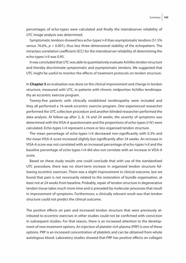
Summary 165
percentages of echo-types were calculated and finally the interobserver reliability of UTC image analysis was determined.
Symptomatic tendons showed less echo-types I+II than asymptomatic tendons (51.5% versus 76.6%, p < 0.001), thus less three-dimensional stability of the echopattern. The intraclass correlation coefficient (ICC) for the interobserver reliability of determining the echo-types I+II was 0.95.
It was concluded that UTC was able to quantitatively evaluate Achilles tendon structure and thereby discriminate symptomatic and asymptomatic tendons. We suggested that UTC might be useful to monitor the effects of treatment protocols on tendon structure.
In Chapter 5 an evaluation was done on the clinical improvement and change in tendon structure, measured with UTC, in patients with chronic midportion Achilles tendinopa-thy an eccentric exercise program.
Twenty-five patients with clinically established tendinopathy were included and they all performed a 16-week eccentric exercise program. One experienced researcher performed the UTC collection procedure and another blinded researcher performed the data analysis. At follow-up after 2, 8, 16 and 24 weeks, the severity of symptoms was determined with the VISA-A questionnaire and the proportions of echo-types (I-IV) were calculated. Echo-types I+II represent a more or less organised tendon structure.
The mean percentage of echo-types I+II decreased non-significantly with 0.3% and the mean VISA-A score increased slightly but significantly after 24 weeks. An increase in VISA-A score was not correlated with an increased percentage of echo-types I+II and the baseline percentage of echo-types I+II did also not correlate with an increase in VISA-A score.
Based on these study results one could conclude that with use of the standardised UTC procedure, there was no short-term increase in organised tendon structure fol-lowing eccentric exercises. There was a slight improvement in clinical outcome, but we found that pain is not necessarily related to the restoration of bundle organisation, at least not at 24 weeks from baseline. Probably, repair of tendon structure in degenerative tendon tissue takes much more time and is preceded by molecular processes that result in improvement of symptoms. Furthermore, a clinically relevant result was that tendon structure could not predict the clinical outcome.
The positive effects on pain and increased tendon structure that were previously at-tributed to eccentric exercises in other studies could not be confirmed with conviction in subsequent studies. For that reason, there is an increased attention to the develop-ment of new treatment options. An injection of platelet-rich plasma (PRP) is one of these options. PRP is an increased concentration of platelets and can be obtained from whole autologous blood. Laboratory studies showed that PRP has positive effects on collagen

166 Chapter 11
synthesis and angiogenesis. In Chapter 6 it was described whether a PRP injection in ad-dition to an eccentric exercise program could lead to an improved tendon structure and increased neovascularisation, measured with ultrasonographical techniques, in patients with chronic midportion Achilles tendinopathy.
Patients were included based on the clinical diagnosis of tendinopathy and this study was part of a double-blind randomised placebo-controlled clinical trial. After inclusion, 54 patients were randomised to eccentric exercise therapy with either a PRP injection (PRP group) or a saline injection (placebo group). Tendon structure organisation was evaluated by means of UTC, which could be performed with a very high intra-observer reliability. The determined echo-types I+II represented a more or less organised tendon structure. Colour Doppler ultrasonography (CDU) was used to score the degree of neo-vascularisation. Follow-up was at 6, 12 and 24 weeks.
Although echo-types I+II increased significantly from baseline within both treatment groups, there were no significant between-group differences. After the first six weeks, the neovascularisation score increased within the PRP group and the placebo group, but there was no significant difference in change of the neovascularisation score between both groups at any point in time.
Although positive effects of PRP administration on collagen synthesis and angiogen-esis were described in previous laboratory studies, this study described that in vivo PRP injections do not contribute to an increase in organised tendon structure and there is no significant effect on neovascularisation, compared with a placebo injection.
Overuse injuries of the plantar fascia are frequently treated with a splint, and therefore it was hypothesised that this treatment method has also beneficial effects in patients with tendinopathy of the Achilles tendon. In Chapter 7 the results of a single-blind randomised clinical trial on eccentric exercise therapy and additional splinting were presented.
Patients with chronic midportion Achilles tendinopathy were included. All patients were prescribed a 12-week eccentric exercise program and half of the group received an additional night splint. Evaluation was done recording the subjective patient satis-faction, the VISA-A score and the reported compliance. The researcher who evaluated the outcome measures was blinded to the allocated therapy. Patients were followed-up after 12 weeks.
Seventy tendons were included and randomised into one of the two treatment groups: eccentric exercises with a night splint (night splint group, n=36) or eccentric exercises only (eccentric group, n=34). After 12 weeks, the patient satisfaction did not differ between the eccentric group (63% excellent or good) and the night splint group (48% excellent or good). The VISA-A score improved significantly within both groups, but there was also no significant difference between the two groups in improvement

Summary 167
of the VISA-A score. Seventeen of 63 (27%) subjects reported a compliance of less than 50% on eccentric exercise therapy. In both treatment groups, there was a tendency that a better patient compliance for eccentric exercises leads to better results, but the differ-ences were not statistically significant.
These findings imply that a night splint is not beneficial in addition to eccentric exercises on short-term follow-up in the treatment of chronic midportion Achilles tendi-nopathy. This study also revealed that the clinical improvement after eccentric exercise therapy is less promising than previously reported. We recommend that further trials including functional outcome measures are necessary to evaluate the effect of eccentric exercises.
Endogenous growth factors may be obtained from autologous whole blood or PRP. The promising effects of autologous growth factors have received increased attention in the treatment of tendon injuries. In Chapter 8 we systematically reviewed the clinical effects of injections of autologous growth factors as a treatment for tendinopathy.
Several electronic databases were explored for prospective clinical trials on treatment with autologous growth factors (autologous blood or PRP) for patients with chronic tendinopathy. Another inclusion criterion was that pain and/or function was quantified as outcome measure. The Physiotherapy Evidence Database (PEDro) score was used to examine the methodological quality of the assessment, and a qualitative analysis was performed to establish the levels of evidence.
Eleven clinical trials were included; eight studies described the effects of autologous blood injections and three were performed on the effects of PRP injections. All stud-ies showed that injections of autologous growth factors had a significant impact on improving pain and/or function over time. However, only three included studies were determined as having a high methodological quality, and none of these showed any benefit of an autologous growth factor injection when compared to a control group.
We concluded that, based on these results, there is strong evidence that the use of injections with autologous blood is not beneficial for the improvement in symptoms. There is only limited evidence to support the use of injections with PRP in the manage-ment of chronic tendinopathy. Since these therapies are increasingly applied in the daily clinical practice, these conclusions are most relevant. Injections of autologous growth factors should not be recommended until further clinical research would support its use.
In Chapter 9 a stratified, block-randomised, double-blind, placebo-controlled clinical trial was described to examine whether a PRP injection would improve outcome in chronic midportion Achilles tendinopathy.
Patients with chronic tendinopathy 2-7 cm above the Achilles tendon insertion were included. A 12-week eccentric exercise program was performed with either a PRP in-

168 Chapter 11
jection (PRP group) or saline injection (placebo group). Randomisation was stratified by activity level. The VISA-A score, subjective patient satisfaction and return to desired sports were obtained at follow-up after 6, 12 and 24 weeks. Treatment group effects were evaluated using general linear models on the basis of intention-to-treat and adjustments were made for affecting variables. A sample size calculation revealed that 54 patients were required to detect a clinically relevant difference of 12 points on the VISA-A score.
Fifty-four patients were included and after randomisation into the PRP group (n=27) or placebo group (n=27) there were no patients lost to follow-up. The mean VISA-A score improved significantly after 24 weeks in the PRP group and in the placebo group, but the increase was not significantly different between both groups. The 95% confidence inter-val of the adjusted between-group difference did not include the predefined relevant difference of 12 points in favour of PRP treatment. There was no significant difference in patient satisfaction after 24 weeks between the PRP group (56% excellent or good) and the placebo group (63% excellent or good). The percentage of patients that returned to the desired sport did also not differ significantly (PRP group: 78%, placebo group: 67%).
This study demonstrated that there was no additional clinical benefit of a PRP injec-tion to the usual care (eccentric exercise therapy). A PRP injection, as performed in daily clinical practice, should not be recommended for the treatment of chronic midportion Achilles tendinopathy. It could be hypothesised whether these results can be extrapo-lated to other tendinopathies; further studies need to assess this.
Finally the main results of this thesis are described in Chapter 10 in relation to each other. These findings are integrated into the current scientific knowledge. Recommendations are made for future research, but also the gaps and obstacles will be demonstrated in the clinical studies on tendinopathy. New studies could potentially be targeted at (1) studying the normal distribution of the tendon structure, measured with UTC, in a group of asymptomatic individuals with a special focus on the age and activity level; (2) the assessment of tendon structure and its relationship with the symptoms at long-term; (3) studying the clinical effects of eccentric exercise therapy, with respect to the performed dose of the exercises, and (4) determining which treatment will be most effective for non-responders to eccentric exercise therapy. This knowledge will increase understanding of the exact value of tendon structure for different patient groups, provide knowledge of the response to therapy of tendinotic tissue and it will eventually lead to more effective conservative treatment options.

AppendicesNederlandse samenvatting
Dankwoord
Curriculum Vitae
PhD Portfolio Summary
List of publications
VISA-A questionnaire


Nederlandse samenvatting 171
Nederlandse samenvatting
Surmenageletsel van pezen is een frequent voorkomend probleem dat voornamelijk sporters, maar ook inactieve personen treft. De aandoening treedt vaker op bij mensen van middelbare leeftijd. Met de toename van sportactiviteit in de bevolking stijgt ook het aantal overbelastingsblessures van pezen. Vooralsnog werd aangenomen dat een verminderd herstelmechanisme op weefselniveau met uiteindelijk degeneratie van het peesweefsel de oorzaak is van het persisteren van de klachten. Bij de trias van pijn, zwelling en verminderde belastbaarheid van de pees wordt de term tendinopathie geprefereerd. Tendinopathie van de Achillespees wordt vaak in het middendeel van de pees ofwel midportion, net boven de aanhechting op het hielbeen, gediagnosticeerd. Door de oppervlakkige positie leent dit deel van de pees zich uitstekend voor echografi-sche beoordeling. Tendinopathie wordt met echografische technieken gekarakteriseerd door een verminderde structuur van het peesweefsel en met toevoeging van Doppler kan een verhoogde bloedstroom van de pees worden waargenomen, in de literatuur aangeduid met de term “neovascularisatie”.
Chronische Achilles tendinopathie leidt veelvuldig tot een vermindering van sportactiviteiten en kan zelfs carrièrebedreigend zijn voor een sporter. Conventionele conservatieve therapie is niet zelden onvoldoende effectief en in dat geval kan bij deze aandoeningen een chirurgische behandeling ingezet worden. Hoewel een chirurgische behandeling in veel gevallen uiteindelijk leidt tot een klachtenvermindering, blijkt ook deze behandeling niet altijd succesvol en is de postoperatieve revalidatie tijdsintensief. Om een operatie te voorkomen is het noodzakelijk om de effectiviteit van conservatieve therapie te verhogen.
Met dit proefschrift willen we een bijdrage leveren aan de toenemende kennis van de beeldvorming en behandeling van chronische Achilles tendinopathie. Het uiteindelijke doel is dat deze kennis leidt tot een verbeterde handelswijze in de klinische praktijk. Daarbij ligt de focus op de betrouwbaarheid en voorspellende waarde van afbeeldende echografische technieken en op de effectiviteit van enkele nieuwe methoden voor de conservatieve behandeling.
In hoofdstuk 1 werd een overzicht van de literatuur gegeven van de normale anatomie van peesweefsel, alsmede van de histologische veranderingen die karakteristiek zijn voor tendinopathie. Daarnaast werden de klinische kenmerken van tendinopathie uiteengezet en bovendien werd beschreven welke frequent aanbevolen aanvullende afbeeldende technieken de behandelaar tot zijn beschikking heeft in de beoordeling

172 Appendices
van tendinopathie. Er werd tevens een overzicht gegeven van enkele veelbelovende behandelingsmogelijkheden van chronische midportion Achilles tendinopathie.
Het hoofdstuk sluit af met de specifieke doelstellingen van dit proefschrift en een beschrijving van de inhoud. Specifieke doelen van dit proefschrift waren het testen van de betrouwbaarheid en de klinische waarde van echografische bepaling van de neovascularisatie en structuur van het peesweefsel voor de behandeling en in klinische gerandomiseerde studies te onderzoeken wat het effect is van enkele veelbelovende therapieën op korte- en lange termijn, zoals excentrische oefentherapie, de toevoeging van een nachtspalk en een toegevoegde injectie met plaatjes-rijk plasma.
De bloedvoorziening in pezen is in fysiologische omstandigheden relatief laag. Bij patiënten met tendinopathie wordt juist frequent een toename gezien van de bloed-doorstroming, gemeten met Doppler echografie. Deze zogenaamde neovascularisatie is een pathologische ingroei van bloedvaten in de pees en wordt gezien als een mogelijk falend herstelproces. In hoofdstuk 2 werd de betrouwbaarheid van de bepaling van echografische neovascularisatie met behulp van Power Doppler ultrasonography (PDU) beschreven. Veel studies beschrijven een gradatie van neovascularisatie bij patiënten met tendinopathie, maar de betrouwbaarheid van de indeling van neovascularisatie was niet eerder onderzocht.
Het belangrijkste doel van deze studie was om de inter-observer betrouwbaarheid van de graad van neovascularisatie te bepalen met behulp van de gemodificeerde Öhberg score. Dit is een praktisch bruikbare gradatie van 0-4. Patiënten met Achilles tendinopathie ondergingen een PDU onderzoek van beide Achillespezen. In totaal namen acht radiologen deel aan de studie en elke pees werd beoordeeld door twee verschillende radiologen. Voor het bepalen van de ernst van de symptomen voltooiden de patiënten de Victorian Institute of Sports Assessment-Achilles (VISA-A) vragenlijst.
Drieëndertig symptomatische en 17 asymptomatische Achillespezen van 25 opeen-volgende patiënten werden geïncludeerd. De intra-class correlatiecoëfficiënt was 0.85 en dit kan worden beschouwd als een uitstekende betrouwbaarheid voor de bepaling van de graad van neovascularisatie met PDU tussen verschillende waarnemers. Neo-vascularisatie was aanwezig in 70% van de symptomatische pezen en in 29% van de asymptomatische pezen. Er was geen significante correlatie tussen de mate van neovas-cularisatie en de ernst van de symptomen.
Er werd geconcludeerd dat de beoordeling van dezelfde Achillespees op één moment in de tijd door verschillende radiologen kan worden uitgevoerd met een uitstekende overeenkomst. Toekomstige studies zouden zich kunnen richten op de test-retest reproduceerbaarheid en de validiteit van de gradatie om een beter inzicht te kunnen verschaffen in het beloop van neovascularisatie bij patiënten.

Nederlandse samenvatting 173
In het onderzoek dat beschreven is in hoofdstuk 3 werd de waarde van de neovascula-risatie score bepaald in een cohort studie. De neovascularisatie score werd gecorreleerd met de ernst van de symptomen op één moment in de tijd en onderzocht werd wat de betekenis was van de gevonden score voor het voorspellen van de klinische uitkomst.
Patiënten met de klinische diagnose van chronische midportion Achilles tendinopa-thie werden geïncludeerd. Aan alle patiënten werd 12 weken excentrische oefentherapie voorgeschreven. De VISA-A score en de neovascularisatie score werden bepaald voor de start van het onderzoek en na 12 weken.
Zeventig pezen van 58 patiënten met chronische midportion Achilles tendinopathie werden geïncludeerd, en 63 symptomatische pezen werden uiteindelijke geanalyseerd. Vóór de start van de behandeling waren de neovascularisatie scores als volgt verdeeld: 23 graad 0 (37% geen neovascularisatie), 18 graad 1, 8 graad 2, 8 graad 3 en 6 graad 4 (63% neovascularisatie score 1-4). Op baseline was de neovascularisatie score niet significant gecorreleerd met de VISA-A score, maar na 12 weken was een hogere neo-vascularisatie score wel gecorreleerd met een slechtere VISA-A score. Er werden geen significante verschillen gevonden in de verbetering van de VISA-A score na behandeling van patiënten met neovascularisatie (graad 1-4) of zonder neovascularisatie (graad 0) op baseline.
We concludeerden dat er geen relatie is tussen de ernst van de symptomen en de mate van neovascularisatie vóór de behandeling, maar na de behandeling bleek een lagere graad van neovascularisatie gunstiger te zijn in relatie tot de ernst van de symp-tomen op dat moment. Bovendien vonden we dat het uitvoeren van PDU voor de start van conservatieve behandeling geen voorspellende waarde heeft voor de klinische uitkomst, wat een relevante bevinding is voor de dagelijkse klinische praktijk.
Normale pezen hebben een hiërarchisch gerangschikte structuur van het peesweefsel. Deze goed geordende structuur kan uitstekend worden afgebeeld met echografie. Bij patiënten met tendinopathie kan een desorganisatie van peesstructuur worden waar-genomen. Helaas heeft conventionele echografie het nadeel dat deze in hoge mate kan worden beïnvloed door verscheidene factoren en bovendien is een kwantitatieve analy-se niet mogelijk. In hoofdstuk 4 introduceerden we een nieuwe echografische techniek in humane pezen, die eerder werd ontwikkeld voor paardenpezen met histologische beelden als referentie-test. De hypothese was dat met behulp van driedimensionale beeldvorming van de Achillespees door deze Ultrasonographic Tissue Characterisation (UTC) onderscheid kon worden gemaakt tussen symptomatische en asymptomatische pezen.
Zesentwintig pezen van patiënten met chronische midportion Achilles tendinopathie en 26 “gezonde” controle pezen werden geïncludeerd. Alle pezen werden in beeld gebracht met behulp van de UTC-procedure die werd uitgevoerd door één ervaren
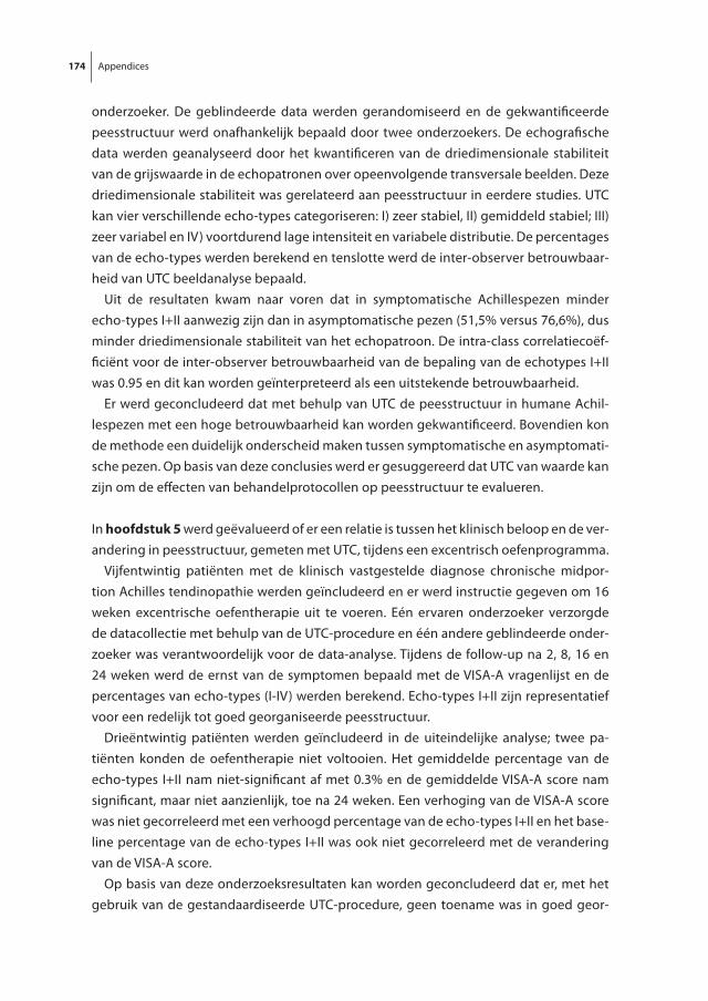
174 Appendices
onderzoeker. De geblindeerde data werden gerandomiseerd en de gekwantificeerde peesstructuur werd onafhankelijk bepaald door twee onderzoekers. De echografische data werden geanalyseerd door het kwantificeren van de driedimensionale stabiliteit van de grijswaarde in de echopatronen over opeenvolgende transversale beelden. Deze driedimensionale stabiliteit was gerelateerd aan peesstructuur in eerdere studies. UTC kan vier verschillende echo-types categoriseren: I) zeer stabiel, II) gemiddeld stabiel; III) zeer variabel en IV) voortdurend lage intensiteit en variabele distributie. De percentages van de echo-types werden berekend en tenslotte werd de inter-observer betrouwbaar-heid van UTC beeldanalyse bepaald.
Uit de resultaten kwam naar voren dat in symptomatische Achillespezen minder echo-types I+II aanwezig zijn dan in asymptomatische pezen (51,5% versus 76,6%), dus minder driedimensionale stabiliteit van het echopatroon. De intra-class correlatiecoëf-ficiënt voor de inter-observer betrouwbaarheid van de bepaling van de echotypes I+II was 0.95 en dit kan worden geïnterpreteerd als een uitstekende betrouwbaarheid.
Er werd geconcludeerd dat met behulp van UTC de peesstructuur in humane Achil-lespezen met een hoge betrouwbaarheid kan worden gekwantificeerd. Bovendien kon de methode een duidelijk onderscheid maken tussen symptomatische en asymptomati-sche pezen. Op basis van deze conclusies werd er gesuggereerd dat UTC van waarde kan zijn om de effecten van behandelprotocollen op peesstructuur te evalueren.
In hoofdstuk 5 werd geëvalueerd of er een relatie is tussen het klinisch beloop en de ver-andering in peesstructuur, gemeten met UTC, tijdens een excentrisch oefenprogramma.
Vijfentwintig patiënten met de klinisch vastgestelde diagnose chronische midpor-tion Achilles tendinopathie werden geïncludeerd en er werd instructie gegeven om 16 weken excentrische oefentherapie uit te voeren. Eén ervaren onderzoeker verzorgde de datacollectie met behulp van de UTC-procedure en één andere geblindeerde onder-zoeker was verantwoordelijk voor de data-analyse. Tijdens de follow-up na 2, 8, 16 en 24 weken werd de ernst van de symptomen bepaald met de VISA-A vragenlijst en de percentages van echo-types (I-IV) werden berekend. Echo-types I+II zijn representatief voor een redelijk tot goed georganiseerde peesstructuur.
Drieëntwintig patiënten werden geïncludeerd in de uiteindelijke analyse; twee pa-tiënten konden de oefentherapie niet voltooien. Het gemiddelde percentage van de echo-types I+II nam niet-significant af met 0.3% en de gemiddelde VISA-A score nam significant, maar niet aanzienlijk, toe na 24 weken. Een verhoging van de VISA-A score was niet gecorreleerd met een verhoogd percentage van de echo-types I+II en het base-line percentage van de echo-types I+II was ook niet gecorreleerd met de verandering van de VISA-A score.
Op basis van deze onderzoeksresultaten kan worden geconcludeerd dat er, met het gebruik van de gestandaardiseerde UTC-procedure, geen toename was in goed geor-

Nederlandse samenvatting 175
ganiseerde peesstructuur op korte termijn na de excentrische oefentherapie. Er was een lichte verbetering van de klinische uitkomst, maar het bleek dat de ernst van de symptomen niet noodzakelijkerwijs verband houdt met het herstel van de organisatie in peesbundels, althans niet na 24 weken. Waarschijnlijk neemt het herstel van de pees-structuur in degeneratief peesweefsel veel meer tijd in beslag en mogelijk wordt dit voorafgegaan door moleculaire processen die leiden tot verbetering van de symptomen. Een klinisch relevant resultaat was dat de mate van desorganisatie in peesstructuur het klinisch beloop niet kan voorspellen.
De positieve effecten op pijn en toename van georganiseerde peesstructuur die voorheen werden toegeschreven aan excentrische oefentherapie kunnen in vervolg-onderzoeken niet overtuigend worden bevestigd. Daarom is er veel aandacht voor de ontwikkeling van nieuwe behandelopties. Een injectie met plaatjes-rijk plasma (PRP) is één van deze opties. PRP bevat een verhoogde concentratie aan bloedplaatjes en kan uit lichaamseigen bloed worden verkregen. Uit laboratoriumonderzoeken komt naar voren dat PRP positieve effecten heeft op collageen synthese en vaatnieuwvorming. In hoofdstuk 6 werd beschreven of een PRP injectie in aanvulling op een excentrisch oefenprogramma zou kunnen leiden tot een verbeterde peesstructuur en een toename in neovascularisatie, gemeten met de echografische technieken.
Patiënten werden toegelaten in de studie op basis van de klinische diagnose chro-nische midportion Achilles tendinopathie en deze studie was onderdeel van een dub-belblind, gerandomiseerd, placebo-gecontroleerd onderzoek. Na inclusie werd aan 54 patiënten de excentrische oefentherapie voorgeschreven met ofwel een PRP injectie (PRP-groep) of een injectie met zoutoplossing (placebo-groep). Peesstructuur werd geëvalueerd door middel van de UTC-procedure en Colour Doppler ultrasonography (CDU) werd gebruikt voor bepaling van de neovascularisatie score. Follow-up metingen werden verricht na 6, 12 en 24 weken.
Hoewel de echo-types I+II aanzienlijk toenamen ten opzichte van vóór de behande-ling in beide behandelgroepen, waren er geen significante verschillen tussen de groe-pen. Na de eerste zes weken was de neovascularisatie score toegenomen in zowel de PRP-groep als de placebo-groep, maar er was geen significant verschil in verandering van de neovascularisatie score tussen beide groepen in de loop van de tijd.
Hoewel er positieve effecten van PRP toediening op de collageen synthese en vaatnieuwvorming werden beschreven in eerdere studies, toont dit onderzoek aan dat in vivo PRP toediening niet bijdraagt aan een toename van de verbetering van de peesstructuur en is er geen significant effect op neovascularisatie, vergeleken met een placebo injectie.

176 Appendices
Omdat overbelastingsblessures van de fascia plantaris frequent worden behandeld met een spalk, werd de hypothese geformuleerd dat deze behandelmethode ook gunstige effecten zou kunnen hebben bij patiënten met tendinopathie van de Achillespees. In hoofdstuk 7 werden de klinische resultaten van een gerandomiseerd onderzoek beschreven, waarbij de waarde van een nachtspalk in aanvulling op excentrische oefen-therapie werd onderzocht.
Eén van de inclusiecriteria was de diagnose chronische midportion Achilles tendino-pathie. Aan iedere patiënt werd 12 weken excentrische oefentherapie voorgeschreven en de helft van de groep kreeg tevens een nachtspalk. Uitkomstmaten waren de sub-jectieve patiënt tevredenheid, de VISA-A score en de gerapporteerde therapietrouw. De onderzoeker, die de resultaten evalueerde, was geblindeerd. De patiënten werden op baseline en na 12 weken beoordeeld.
Zeventig pezen werden geïncludeerd en gerandomiseerd in één van de twee behan-delgroepen: excentrische oefeningen met een nachtspalk (nachtspalk-groep, n = 36) of excentrische oefeningen alleen (excentrische groep, n = 34). Na 12 weken was de subjectieve patiënt tevredenheid niet significant verschillend tussen de excentrische groep (63% uitstekend of goed) en de nachtspalk-groep (48% uitstekend of goed). De VISA-A score verbeterde significant in beide groepen, maar er was ook geen significant verschil tussen de twee groepen in de verbetering van de VISA-A score. Zeventien van de 63 (27%) patiënten rapporteerden dat ze minder dan 50% van de voorgeschreven excentrische oefeningen hadden uitgevoerd. In beide behandelgroepen was er een tendens dat een betere therapietrouw voor excentrische oefeningen leidt tot betere resultaten, maar de verschillen waren niet statistisch significant.
Deze bevindingen gaven aan dat een nachtspalk niet leidt tot een verbetering in symptomen in aanvulling op excentrische oefeningen op korte termijn bij de behande-ling van chronische midportion Achilles tendinopathie. In deze studie bleek ook dat de klinische verbetering na excentrische oefentherapie minder effectief is dan werd gevonden in eerdere studies. Onze aanbevelingen waren dat toekomstige studies naar het effect van therapie in tendinopathie niet alleen pijn en patiënt tevredenheid zouden moeten evalueren, maar ook de activiteit en functie. De VISA-A vragenlijst is hiervoor een zeer geschikt instrument.
Lichaamseigen groeifactoren kunnen worden verkregen uit autoloog onbewerkt bloed of uit PRP. Door de veelbelovende effecten van autologe groeifactoren is er veel aandacht voor ontstaan bij de behandeling van peesblessures. In hoofdstuk 8 werd een systematisch onderzoek beschreven naar de klinische effecten van injecties van autologe groeifactoren als behandeling voor tendinopathie.
Verschillende elektronische databases werden doorzocht naar klinische studies die effecten van de behandeling met autologe groeifactoren (autoloog bloed of PRP) on-
Nederlandse samenvatting 177
derzochten bij patiënten met chronische tendinopathie. Een ander criterium was dat als uitkomstmaat pijn en/of functie werd gebruikt. De Physiotherapy Evidence Database (PEDro) score werd gebruikt om de methodologische kwaliteit van de artikelen te beoor-delen en een kwalitatieve analyse werd uitgevoerd om de bewijskracht vast te stellen.
Elf klinische onderzoeken werden geïncludeerd; acht studies beschreven de effecten van autoloog bloed injecties en drie werden uitgevoerd naar de effecten van PRP injec-ties. Alle studies toonden aan dat injecties met autologe groeifactoren een significante invloed hadden op de verbetering van pijn en/of functie in de loop van de tijd. Echter, slechts drie geïncludeerde studies werden beoordeeld als onderzoek met methodolo-gisch hoge kwaliteit en geen van deze studies rapporteerde een toegenomen verbete-ring in pijnscore na autologe groeifactor injecties in vergelijking met een controlegroep.
Daarom hebben we geconcludeerd dat er op basis van deze resultaten sterke aanwij-zingen zijn dat het gebruik van injecties met autoloog bloed niet leidt tot toegenomen verbetering van de symptomen. Er is slechts beperkt bewijs voor het effect van injecties met PRP in de behandeling van chronische tendinopathie. Omdat deze therapieën steeds meer worden toegepast in de dagelijkse klinische praktijk, zijn deze conclusies zeer relevant.
hoofdstuk 9 beschreef een gestratificeerd, gerandomiseerd, dubbelblind, placebo-gecontroleerd klinisch onderzoek naar het effect van een PRP injectie op de ernst van de symptomen bij patiënten met chronische midportion Achilles tendinopathie.
Patiënten met chronische tendinopathie 2-7 cm boven de aanhechting van de Achil-lespees op het hielbeen werden geïncludeerd. Twaalf weken excentrische oefentherapie werd uitgevoerd met ofwel een PRP injectie (PRP-groep) of een injectie met zoutoplos-sing (placebo-groep). Randomisatie was gestratificeerd voor activiteitenniveau. De VISA-A score, subjectieve patiënt tevredenheid en terugkeer in de gewenste sport werden gemeten bij de follow-up na 6, 12 en 24 weken. Met behulp van een power-analyse werd berekend dat 54 patiënten waren benodigd om een klinisch relevant verschil van 12 punten op de VISA-A score aan te kunnen tonen. Analyses werden uitgevoerd met een general linear model, waarin kon worden geadjusteerd voor beïnvloedende variabelen.
Vierenvijftig patiënten werden geïncludeerd en na randomisatie in de PRP groep (n = 27) of de placebo groep (n = 27) konden alle patiënten de studie voltooien. De gemid-delde VISA-A score verbeterde significant na 24 weken in zowel de PRP groep als in de placebo groep, maar de stijging was niet significant verschillend tussen beide groepen. Het 95% betrouwbaarheidsinterval van het geadjusteerde verschil tussen de groepen bevatte niet het vooraf gedefinieerde klinisch relevante verschil van 12 punten toename na de PRP behandeling. Er was geen significant verschil in patiënt tevredenheid na 24 weken tussen de PRP-groep (56% uitstekend of goed) en de placebo-groep (63% uitstekend of goed). Het percentage patiënten dat terugkeerde in de gewenste sport

178 Appendices
verschilde ook niet significant tussen beide groepen (PRP-groep: 78%, placebo-groep: 67%).
Dit onderzoek heeft aangetoond dat er geen toegevoegde waarde is van een PRP injectie. Een PRP injectie, zoals uitgevoerd in de dagelijkse klinische praktijk, zou niet moeten worden aanbevolen voor de behandeling van chronische midportion Achil-les tendinopathie. Er zou kunnen worden verondersteld dat deze resultaten kunnen worden geëxtrapoleerd naar andere tendinopathieën; echter, toekomstige studies zijn benodigd om hierover een definitieve uitspraak te kunnen doen.
In hoofdstuk 10 werden tot slot de belangrijkste resultaten van dit proefschrift in relatie tot elkaar besproken. Deze bevindingen werden afgespiegeld tegen de actuele wetenschappelijke kennis. Er werden aanbevelingen gedaan voor toekomstig onder-zoek, maar ook werd er aandacht geschonken aan de hiaten en obstakels in de klinische onderzoeken naar tendinopathie. Nieuwe onderzoeken zouden mogelijk kunnen worden gericht op (1) het bestuderen van de normale verdeling van de peesstructuur, gemeten met UTC, in een groep van asymptomatische personen met een speciale focus op de leeftijd en het activiteitsniveau; (2) de beoordeling van peesstructuur en de relatie tot de symptomen op lange termijn; (3) het bestuderen van het klinische effect van de excentrische oefentherapie, met betrekking tot de voorgeschreven dosering van de oefeningen; en (4) het bepalen van de meest effectieve therapie bij patiënten die niet reageren op excentrische oefentherapie. Deze kennis zal meer inzicht geven in de exacte waarde van peesstructuur voor verschillende patiëntengroepen, meer kennis verschaf-fen in de respons van degeneratief peesweefsel op therapie en het zal uiteindelijk leiden tot effectievere conservatieve behandelmethoden.

Dankwoord 179
Dankwoord
Hooggeleerde heer, beste Prof.dr. J.A.N. Verhaar, dank dat u mij de mogelijkheid heeft geboden om mijn wetenschappelijke ervaringen te continueren in een promotietraject. Ik heb altijd kunnen rekenen op uw steun en adviezen. In de periode na het verschijnen van uw proefschrift is het aantal publicaties met wetenschappelijk onderzoek naar tendinopathieën exponentieel toegenomen. In dit proefschrift zijn de nieuwe inzichten beschreven en ik hoop van harte dat het peesonderzoek op zowel klinisch- als basaal wetenschappelijk niveau kan worden voortgezet op de afdeling Orthopaedie van het Erasmus MC en dat de vruchtbare samenwerking met de Sportgeneeskunde kan wor-den voortgezet.
Hooggeleerde Prof.dr.ir. H. Weinans, beste Harrie, aan het einde van mijn co-schappen mocht ik bij je op gesprek komen voor de bespreking van een promotietraject. We waren er snel uit en ik kon aan de slag als promovendus. Ik merkte al binnen korte tijd dat je ondanks de vurige en doortastende discussies heel flexibel kon zijn. Je wist op moeilijke momenten alle wetenschappelijke neuzen dezelfde kant op te laten wijzen. Ik heb de lage drempel om problemen te bespreken zeer gewaardeerd en wil je bedanken voor de begeleiding in de ‘wetenschappelijke jungle’.
Dr. J.L. Tol, beste Hans, ons eerste contact dateert al weer van 2004. Na een schriftelijk verzoek tot het verrichten van wetenschappelijk onderzoek op een Sportgeneeskundige afdeling werd er enthousiast gereageerd vanuit Leidschendam. Na een kort overleg besloten we ons te richten op de diagnostiek en behandeling van Achillespeesblessures. In de opzet van een gerandomiseerd klinisch onderzoek werd ik volkomen vrijgelaten. In onze besprekingen was het steevaste antwoord: “Het is jouw onderzoek, dus jij bent de baas”. Maar beschikbaar was je altijd. Mijn ingeleverde manuscripten werden tussen je poli afspraken door met een rode pen beklad en binnen enkele uren geretourneerd. In je commentaar was je altijd kritisch en rechtlijnig, maar de keuze of hiermee wat werd gedaan lag altijd bij de onderzoeker. Het wierp zijn vruchten af, want na ruim een jaar waren enkele publicaties in vooraanstaande (sport)medische tijdschriften een feit. Daar-naast heb ik je van dichtbij meegemaakt tijdens het congres in Cyprus, waar ik samen met Evelien nog als oppas voor ‘de kleine aap’ heb gefungeerd. Voor een training van het Medical Dutch Soccer team wist de toenmalige student je hele koelkast leeg te eten en met hetzelfde team hebben we in Spanje en Italië furore gemaakt door tweemaal de gouden medaille op te halen. Het bleef niet bij dit alles, want aan het einde van mijn wetenschappelijke stage werd door jou de volgende missie geïnitieerd. De Sport-

180 Appendices
geneeskunde zou gebaat zijn bij een AGIKO constructie, waarbij een opleiding wordt gecombineerd met een promotieonderzoek. Daarin zag je mij als ideale kandidaat en ondanks je drukke werkzaamheden op alle vlakken wist je ook dit, na alle bureaucra-tische hindernissen, te bewerkstelligen. Het mag dan ook geen verrassing zijn dat ik je ongelooflijk dankbaar ben voor de begeleiding in deze lange periode en ik hoop in de toekomst de samenwerking te kunnen continueren.
Dr. H.T.M. van Schie, beste Hans, je startte in Leidschendam op de poli Sportgeneeskunde als ‘vreemde eend in de bijt’ met humaan onderzoek naar Achillespeesklachten. Met een nieuw echo-apparaat wist je zowel de patiënten als de artsen enorm te interesseren. Ook ik viel voor een prominente rol van deze UTC in mijn promotieonderzoek. Altijd was je bezig met het optimaliseren van de apparatuur. Gedurende deze periode botsten we wel eens door onze meningsverschillen. Toch heb ik heel veel geleerd van jouw enorme kennis van echografie en basaal peesonderzoek, waarvoor ik je dankbaar ben. En, niet onbelangrijk, tijdens de verrichte klinische onderzoeken met de UTC bemerkte ik steeds weer het enthousiasme van de betrokken artsen en patiënten. Ik hoop dat de ontwikke-ling van de UTC kan worden voortgezet.
Beste Don de Winter, ik heb slechts een deel van de enorme ontwikkeling meegemaakt op de polikliniek Sportgeneeskunde in Leidschendam. Ik heb in deze periode bewonde-ring gehad voor jouw rol hierin. Het is voor een promovendus een stimulans om te zien hoe trots je bent op het feit dat er veel wetenschap wordt bedreven op deze afdeling. Ik ben er ook trots op om deel uit te maken van deze gezellige en professionele groep op de afdeling, bedankt daarvoor.
Beste Adam Weir, je was als arts-assistent al de ‘geek’ op wetenschappelijk sportmedisch gebied. Het bijzondere is dat je deze kennis ook durft toe te passen in de dagelijkse praktijk met daarnaast een sterke empathie voor de patiënt die tegenover je zit. Als co-assistent heb ik daarom heel veel geleerd van je werkwijze en ik hoop later ook op een dergelijke manier mijn professie te kunnen uitoefenen. Tevens zou ik in de toekomst nog graag vaker met je samenwerken.
Beste drs. M.P. Heijboer, beste Rien, bij ieder contact dat we hadden werd eerst het voorbijgaande voetbalweekend geëvalueerd. U was altijd op de hoogte van het voet-balnieuws in de regio. Ik heb heel veel geleerd als vrijwilliger op uw beroemde sport-poli’s en was blij dat ik af en toe een teamgenoot tussen de linies van het spreekuur mocht doorschuiven. Als onderzoeker wil ik u ook bedanken voor uw rol in het klinisch onderzoek en het aanleveren van peesweefsel vanaf de operatiekamer.

Dankwoord 181
Beste dr. S.M.A. Bierma-Zeinstra, beste Sita, u werd pas in een later stadium geconsul-teerd voor de interpretatie van onze PRICT studie. Daarbij is er een nieuwe wereld voor me opengegaan in het klinische onderzoek. Ik zou in de toekomst graag meer van u leren om zo het klinisch onderzoek in de Sportgeneeskunde op een hoger niveau te brengen.
Dear Prof.dr. N. Maffulli, dear Nicola, as a starting medical student I already read many of your published articles in the field of tendinopathy. To my opinion, it was a big op-portunity to write an article with one of the leading tendon researchers after we met in Edinburgh last year. I hope this will lead to an interesting cooperation, up to many more together!
Beste Cora, Ank en Paulien. De vele klinische onderzoeken in het Antoniushove zieken-huis hebben jullie bergen extra werk opgeleverd. De start van ieder nieuw onderzoek zorgde weer voor een golf van vragen. Toch waren jullie altijd positief gestemd en wilden jullie graag meedenken. Ik wil jullie heel graag bedanken voor deze hulp en de prettige samenwerking.
De sportartsen (in opleiding) die ik in mijn onderzoeksperiode heb meegemaakt wil ik graag bedanken voor de medewerking en voor het verwijzen van patiënten. Met som-migen heb ik ook samen geschreven en gepubliceerd. Peter, Maarten, Pieter en Rhijn, bedankt voor jullie inspanningen en zodoende de bijdrage aan dit proefschrift.
De radiologen en orthopeden van het Antoniushove ziekenhuis waren van belang bij respectievelijk de consulten en verwijzingen van de patiënten. Graag wil ik iedereen die een actieve bijdrage heeft geleverd binnen deze vakgroepen bedanken voor deze belangrijke inzet.
In 2008 werd de ‘Dutch TENDON group’ in het leven geroepen. Uit het hele land komen halfjaarlijks vele peesonderzoekers bijeen. Ik wil de oprichters en leden bedanken voor de inzet die is geleverd tijdens deze bijeenkomsten en ik hoop dat we hieraan een goed vervolg kunnen geven.
Met veel plezier heb ik 2 jaar lang in het orthopaedisch lab gewerkt in Rotterdam. De peesonderzoekers, Marieke, Jennifer en Suzan, vormden een exponentieel groeiende populatie binnen het lab. Marieke, jij hebt het belangrijke voorwerk gedaan als eerste peesonderzoeker in het orthopaedisch lab. Ik heb de geduldige uitleg en prettige overdracht van de lopende onderzoeken zeer gewaardeerd. Jennifer, helaas heb je het basale peesonderzoek niet kunnen continueren. Desalniettemin wens ik je veel succes in je verdere carrière. Suzan, wat goed dat je de nieuwe ontwikkelingen in het

182 Appendices
peesonderzoek een vervolg geeft. Hopelijk kun je de mogelijkheden in het lab optimaal benutten en ik blijf daarbij graag aan de zijlijn betrokken. Tom en Olav, ik heb genoten van de eindeloze serieuze en onzinnige (wetenschappelijke) discussies en natuurlijk van de ‘casus del dia’. Yvonne B, ik had wat moeite met de transformatie naar een roze werkkamer maar uiteindelijk hebben we een goede en gezellige werkplek gecreëerd. Mede-wetenschappers Gerjo, Ruud, Eric, Nicole, Erwin, Holger, Anna, Katja, Carola, Mar-tine, Yvonne S, Rianne, Gerben, Michiel, Stefan, Esther, Sandra en Wendy, bedankt voor jullie feedback en het was prettig dat jullie mijn collega’s waren in dit promotietraject. Ook de wetenschappers en medewerkers van de kliniek orthopaedie in het Erasmus MC wil ik bedanken voor hun bijdrage.
Beste paranimfen, Stian en Martijn, vrienden voor het leven. In de ‘jungle’ van de voetbal-wereld hebben we alles meegemaakt, van het spelen in de kelder van het amateurvoet-bal tot wedstrijden tegen gerenommeerde clubs in het betaalde voetbal. Daarbuiten was er altijd wel tijd voor een hapje of een drankje en dan het liefst ongepland. Ik hoop dat we hiervoor in de toekomst tijd voor kunnen blijven vrijmaken. Ik twijfel er niet aan dat ik door jullie inzet ook op dit toneel kan pieken op het moment dat het gevraagd wordt.
En dan mijn familie, in het bijzonder Henk, Anneke, Marieke en Jasper, ik heb altijd veel liefde en steun gehad gedurende mijn opleidingen. Jullie weten dat ik er niet de persoon naar ben om dit met veel woorden uit te spreken, maar ik wil jullie bij deze gelegenheid heel graag bedanken voor alles dat jullie me hebben gegeven.
Lieve, lieve Evelien, we vonden elkaar in de saaie collegebanken maar tot op de dag van vandaag blijkt hieruit een levendige relatie te zijn ontstaan. Ik heb ontzettend veel bewondering voor je enthousiasme, doorzettingsvermogen en liefdevolle karakter. Daarboven vind ik je een fantastische vrouw! Ik ben ervan overtuigd dat we een prach-tige toekomst tegemoet gaan in ons nieuwe paleis.

Curriculum vitae 183
Curriculum vitae
Robert Johannes de Vos werd geboren op 27 februari 1982 te Gouda. Van 1994 tot en met 2000 doorliep hij het atheneum op de Scholengemeenschap Willem van Oranje te Oud-Beijerland. Na het behalen van zijn VWO diploma startte hij aansluitend met de studie Geneeskunde aan de Erasmus Universiteit te Rotterdam, waarvoor hij in 2002 zijn propedeuse behaalde. In het kader van zijn afstudeeronderzoek heeft hij 12 maanden stage gelopen op de afdeling Sportgeneeskunde in het Medisch Centrum Haaglanden te Leidschendam onder begeleiding van dr. Hans Tol, waarna het doctoraal werd verkre-gen. Het artsexamen legde hij in januari 2008 cum laude af.
Vanaf januari 2008 werd hij door Prof.dr. Jan Verhaar en Prof.dr.ir. Harrie Weinans aangesteld als promovendus op reeds bestaande en nieuwe peesprojecten in het orthopaedisch laboratorium van het Erasmus Medisch Centrum te Rotterdam. Dit promotietraject werd gecombineerd met de opleiding Sportgeneeskunde, waardoor hij als eerste Assistent Geneeskundige In opleiding tot Klinisch Onderzoeker (AGIKO) Sportgeneeskunde in Nederland was aangesteld. Dit heeft, naast een gegarandeerde opleidingsplaats, geleid tot een intensievere samenwerking op wetenschappelijk ge-bied tussen het Erasmus Medisch Centrum en Medisch Centrum Haaglanden. Tevens heeft dit geleid tot de totstandkoming van dit proefschrift.
Robert-Jan de Vos is sinds november 2009 samenwonend met Evelien Kerkhof. Sinds 3 jaar voetbalt hij wekelijks op het hoogste nationale amateurniveau. Momenteel is hij werkzaam als arts-assistent Sportgeneeskunde voor de deelstage Orthopaedie in het Medisch Centrum Haaglanden te Leidschendam onder hoofdopleider drs. Don de Winter.


PhD Portfolio Summary 185
PhD Portfolio Summary
summary of Phd training and teaching activities
Name PhD student: R.J. de VosErasmus MC Department: Orthopaedic Research LaboratoryPhD period: 2008-2009Promotors: Prof.dr.ir. H. Weinans, Prof.dr. J.A.N. VerhaarSupervisors: dr. J.L. Tol, dr. H.T.M. van Schie
1. Phd trainingyear
In-depth courses
Introduction to data-analysis (NIHES) 2009
Cohort studies (NIHES) 2009
Biomedical English Writing and Communication (EUR) 2009
Klinische epidemiologie (NIOS) 2009
Injectietechnieken bewegingsapparaat (NIOS) 2009
Presenteren (Postgrade) 2009
Basiscursus Sportgeneeskunde (NIOS) 2009
(Inter)national Conferences and podium presentations
Chronic midportion Achilles tendinopathySymposium medisch centrum voor bewegingDen Haag, the Netherlands
2005
The additional value of a night splint to eccentric exercises in chronic midportion Achilles Tendinopathy: a randomised controlled trialEuropean Sports Medicine CongressLimassol, Cyprus
2005
Gebruik van Power Doppler echografie in chronische midportion Achilles tendinopathie: een prospectieve klinische trialVSG congres Sport, Bewegen & GezondheidNoordwijkerhout, the NetherlandsAwarded 3rd best abstract price Stichting Sport & Orthopaedie
2006
The value of Ultrasound in assessment of Chronic midportion Achilles tendinopathy: a prospective clinical studyWorld Congress 2007; Society for Tennis Medicine and ScienceAntwerpen, Belgium
2007
Invloed van patiëntfactoren op uitkomst na excentrische oefentherapie in chronische midportion Achilles tendinopathieVSG congres “Sport, Bewegen & Gezondheid”Noordwijkerhout, the Netherlands
2007
186 Appendices
Interobserver betrouwbaarheid van Power Doppler echografie in midportion Achilles tendinopathieVSG congres “Scope on sports”Ermelo, the Netherlands
2008
Prevalentie van Femoro Acetabular Impingement bij patiënten met chronische adductor gerelateerde pijnVSG congres “Scope on sports”Ermelo, the Netherlands
2008
The additional value of splinting to eccentric exercises in chronic midportion Achilles Tendinopathy: a randomised controlled trial with one-year follow-up 9th IFOMT CongressRotterdam, the Netherlands
2008
Colour Doppler echografie tijdens follow-up van patiënten met chronische midportion Achilles tendinopathie; een prospectief klinisch onderzoekVSG congres “Sport, Bewegen & Gezondheid”Noordwijkerhout, the Netherlands
2008
Ultrasonographic Tissue Characterisation of Human Achilles TendonsBASEM Annual CongressEdinburgh, Scotland
2009
Een dubbelblind gerandomiseerd placebo-gecontroleerd klinisch onderzoek naar het effect van een plaatjes-rijk plasma injectie in chronische midportion Achilles tendinopathieWetenschapsdag Orthopaedie Erasmus MCRotterdam, the Netherlands
2009
Ultrasonographic Tissue Characterisation in humane Achillespezen: kwantificatie van peesstructuur met een nieuwe non-invasieve methodeVSG congres “Sport, Bewegen & Gezondheid”Noordwijkerhout, the Netherlands
2009
Een dubbelblind gerandomiseerd placebo-gecontroleerd klinisch onderzoek naar het effect van een plaatjes-rijk plasma injectie in chronische midportion Achilles tendinopathieVSG congres “Sport, Bewegen & Gezondheid”Noordwijkerhout, the NetherlandsAwarded best abstract price Stichting Sport & Orthopaedie
2009
Should we apply platelet-rich plasma to elite soccer players?“FA medical society meeting” – Invited lectureLondon, United Kingdom
2010
Effects of platelet-rich plasma on Ultrasonographic findings in chronic midportion Achilles tendinopathyNVA-VSG congress ‘New Horizons in Arthroscopy’Noordwijk aan Zee, the Netherlands
2010
2. Teatching activitiesyear
lecturing
Chronic midportion Achilles tendinopathyMFVR symposium “Beweeg, maar sport met mate”Erasmus MC, Rotterdam, the Netherlands
2006
Beeldvorming bij Femoro Acetabulair ImpingementSportrefereeravond OrthopaedieErasmus MC, Rotterdam, the Netherlands
2007

PhD Portfolio Summary 187
Achilles tendinopathiecollege tweedejaars geneeskunde studentenErasmus MC, Rotterdam, the Netherlands
2008, 2009
Achilles tendinopathie – diagnostiek en behandelingonderwijs arts-assistenten orthopaedieErasmus MC, Rotterdam, the Netherlands
2009
Een dubbelblind gerandomiseerd placebo-gecontroleerd klinisch onderzoek naar het effect van een plaatjes-rijk plasma injectie in chronische midportion Achilles tendinopathieSportrefereeravond OrthopaedieErasmus MC, Rotterdam, the Netherlands
2009
Achilles tendinopathie – diagnostiek en behandelingonderwijs arts-assistenten orthopaedieMedisch Centrum Haaglanden, Den Haag, the Netherlands
2009
supervising practicals
“Conservatieve behandeling van peesaandoeningen: excentrische oefentherapie of shockwave?”Begeleiding review tweedejaars geneeskunde studenten EMC
2008
“Conservatieve behandeling van peesaandoeningen: effecten van autologe groeifactoren in tendinopathie?”Begeleiding review tweedejaars geneeskunde studenten EMC
2009
year
other
Bestuurslid Wetenschapscommissie, Juniorkamer Vereniging voor Sportgeneeskunde 2009
Reviewer sportmedisch tijdschrift Sport & Geneeskunde 2008
Reviewer tijdschrift internationale medische tijdschriften (o.a. American Journal of Sports Medicine, Journal of American Medical Association), onder supervisie van dr. J.L. Tol
2009


List of Publications 189
List of Publications
Weir A, de Vos RJ, de Winter TC.Iliotibiale band frictiesyndroom.Geneeskunde & Sport, jaargang 38, nummer 6, december 2005.
de Vos RJ, Weir A, Tol, JL.Ontwikkelingen in de behandeling van chronische tendinopathieën.Geneeskunde & Sport. Jaargang 39, nummer 3, juni 2006.
de Vos RJ, Weir A, Visser RJ, de Winter TC, Tol JL.The additional value of a night splint to eccentric exercises in chronic midportion Achil-les tendinopathy: a randomised controlled trial.Br J Sports Med. 2007; 41(7): e5.
de Vos RJ, Weir A, Cobben LP, Tol JL.The Value of Power Doppler ultrasonography in Achilles Tendinopathy: A Prospective Study.Am J Sports Med. 2007; 35(10): 1696-701.
de Vos RJ, Dijksterhuis MG.Sport tijdens de zwangerschap, een case report en literatuuroverzicht.Sport & Geneeskunde. Jaargang 40, nummer 4, oktober 2007.
de Vos RJ, Weir A, Tol, JL.Reply letter to the editor: The use of a neovascularisation score to predict clinical sever-ity in Achilles tendinopathy.Am J Sports Med. 2008; 36(2): 395-7.
de Jonge S, de Vos RJ, van Schie HT, Verhaar JA, Weir A, Tol JL.One-year follow-up of a randomised controlled trial on added splinting to eccentric exercises in chronic midportion Achilles tendinopathy.Br J Sports Med. 2008; Epub ahead of print.
Moen MH, de Vos RJ, van Arkel ER, Weir A, Moussavi J, Kraan T, de Winter TC.De meest waardevolle klinische schoudertesten. Een literatuuronderzoek.Sport & Geneeskunde. Jaargang 41, nummer 4, oktober 2008. Aanmoedigingsprijs Sport & Geneeskunde 2008

190 Appendices
Moen MH, de Vos RJ, van Arkel ER, Weir A, Moussavi J, Kraan T, de Winter TC.Reply letter to the editor: De meest waardevolle klinische schoudertesten.Sport & Geneeskunde. Jaargang 42, nummer 1, februari 2009.
Sengkerij PM, de Vos RJ, Weir A, van Weelde BJ, Tol JL.Interobserver reliability of neovascularisation score using power Doppler ultrasonogra-phy in midportion Achilles tendinopathy.Am J Sports Med. 2009; 37(8): 1627-31.
van Schie HT, de Vos RJ, de Jonge S, Bakker EM, Heijboer MP, Verhaar JA, Tol JL, Weinans H.Ultrasonographic Tissue Characterisation of human Achilles tendons: quantification of tendon structure through a novel non-invasive approach.Br J Sports Med. 2009; Epub ahead of print.
Weir A, de Vos RJ, Moen M, Hölmich P, Tol JL.Prevalence of radiological signs of femoroacetabular impingement in patients present-ing with long standing adductor related groin pain.Br J Sports Med. 2009; Epub ahead of print.
de Vos RJ.Boekbespreking proefschrift M. de Mos: Tendon Cell Behavior and Matrix Remodeling in Degenerative Tendinopathy.Sport & Geneeskunde. Jaargang 42, nummer 2, mei 2009.
de Vos RJ, van Veldhoven PL.Behandeling van chronische tendinopathieën met autologe groeifactoren. Een over-zicht van de literatuur.Sport & Geneeskunde. Jaargang 42, nummer 3, augustus 2009.
de Vos RJ.Boekbespreking leerboek orthopaedie: Musculoskeletale Aandoeningen.Sport & Geneeskunde. Jaargang 43, nummer 1, februari 2010.
de Vos RJ, Weir A, Van Schie HT, Bierma-Zeinstra SM, Verhaar JA, Weinans H, Tol JL.Platelet-rich plasma injection for chronic Achilles tendinopathy – a randomised con-trolled trial.JAMA. 2010; 303(2): 144-9.

List of Publications 191
de Vos RJ, van Veldhoven PL, Moen MH, Weir A, Tol JL, Maffulli N.Autologous growth factor injections in chronic tendinopathy: a systematic review.Br Med Bull. 2010; Epub ahead of print.
Moen MH, de Vos RJ, Ellenbecker TS, Weir A.Clinical tests in shoulder examination: how to perform them.Br J Sports Med. 2010; 44(5): 370-5.
de Vos RJ, Tol JL, Verhaar JA.Platelet-rich plasma for treatment of Achilles tendinopathy – Author replyJAMA. 2010; 303(17): 1697-8.
de Vos RJ.Editorial: Richtlijnontwikkeling in de SportgeneeskundeSport & Geneeskunde. Jaargang 43, nummer 2, mei 2010.
Paoloni JA, de Vos RJ, Hamilton B, Murrell GA, Orchard JPlatelet-rich plasma treatment for ligament and tendon injuries.Clin J Sports Med. 2010; Epub ahead of print.


193VISA-A questionnaire
VISA-A questionnaire
ITEM REDUCTION
A focus group consisting of the principal ques-tionnaire developer, a primary care sportsmedicine doctor, and two physiotherapistsreviewed the items generated. They decidedthat three domains—pain, functional status,and activity—were necessary, and they allo-cated three questions to each domain (fig1).24–27
ITEM SCALING
A visual analogue scale (VAS) is more accurateand sensitive than categorical verbal scales.25 28–
32 The first six questions use a VAS so that thepatient may report magnitude of a continuumof subjective symptoms. Activity is bestmeasured using a categorical rating systembased on an incremental range of values.33
Thus the final two questions used a categoricalrating scale.
PRETESTING
Before being shown the working version of thequestionnaire, a group of 15 clinicians expert inthe field of tendon injuries were asked to identify
questions they felt were important in assessingthe severity of Achilles tendon disorders. Thegroup comprised eight physiotherapists, fourprimary care doctors, one orthopaedic surgeon,and one rehabilitation specialist.
The same 15 participants were then shownthe VISA-A score and asked to evaluate thequestionnaire. They were specifically asked ifthere were any questions they would add,delete, or modify. Fourteen of the participantshad no questions to add, and none wanted anydeleted or modified.
WEIGHTING
The questionnaire tests the three significantdomains of dysfunction with three questionsfor each (question 8 is eVectively two ques-tions, one relating to pain with activity and theother to duration of activity). By removingredundancies and eliminating items of lessimportance, weighting of the remaining itemsmay be the same (each question is scored out of10) without aVecting the value of the question-naire.20 The final version of the questionnaire
Figure 1 The VISA-A questionnaire. Eight questions are used to determine the VISA-A score (continued). The VISA-Aquestionnaire can be dowloaded in full at http://bjsm.bmjjournals/cgi/content/full/35/5/336/DC1.
IN THIS QUESTIONNAIRE, THE TERM PAIN REFERS SPECIFICALLY TO PAIN IN THE ACHILLES
TENDON REGION
1. For how many minutes do you have stiffness in the Achilles region on first getting up?
2. Once you are warmed up for the day, do you have pain when stretching the Achilles
tendon fully over the edge of a step? (keeping knee straight)
3. After walking on flat ground for 30 minutes, do you have pain within the next 2 hours?
(If unable to walk on flat ground for 30 minutes because of pain, score 0 for this
question).
strongseverepain
100 min
no pain
0 min
POINTS
POINTS
POINTS
0 1 2 3 4 5 6 7 98 10
0 1 2 3 4 5 6 7 98 10
strongseverepain
no pain
0 1 2 3 4 5 6 7 98 10
336 Robinson, Cook, Purdam, et al
www.bjsportmed.com
was called the Victorian Institute of SportAssessment-Achilles questionnaire (VISA-A)(fig 1).
RELIABILITY AND VALIDITY TESTING
We administered the VISA-A questionnaire tofour populations: group 1 (non-surgical pa-tients; n = 45) attended a primary care sportsmedicine clinic; group 2 (presurgical patients;n = 14) had been referred to a sportsorthopaedist for tendon surgery; group 3 (uni-versity students; n = 63) represented a conven-ience sample of young normally active peopleto serve as a control group; group 4 (membersof a running club; n = 24) represented active,but non-injured people whose age matched thepatient groups. As imaging does not provide ayardstick for tendon disorders, diagnosis wasby assessment of two expert clinicians, as hasbeen justified in other tendon studies.34–36 For
inclusion in the study, subjects in all groupshad to be older than 18 who were able to givewritten informed consent. For groups 1 and 2,subjects had to have a diagnosis of Achillestendinosis, paratendinitis, or partial rupturewith or without a retrocalcaneal or Achillesbursitis. For groups 3 and 4, subjects had tobelong to the two groups as defined. Womenwho were pregnant or nursing were excluded,as were patients with a total rupture of theAchilles tendon. Subjects with previous or cur-rent Achilles tendon symptoms, but who werenot currently undergoing treatment for thecondition, were not excluded from the controlgroups as these groups were designed to reflectthe populations.
We tested construct validity in two ways.Firstly, the 45 non-surgical patients in group 1completed the VISA-A test and two other
4. Do you have pain walking downstairs with normal gait cycle?
5. Do you have pain during or immediately after doing 10 (single leg) heel raises from a
flat surface?
6. How many single leg hops can you do without pain?
7. Are you currently undertaking sport or other physical activity?
0 Not at all
4 Modified training ± modified competition
7 Full training ± competition but not at same level as when symptoms began
10 Competing at the same or higher level as when symptoms began
strongseverepain
no pain
POINTS
0 1 2 3 4 5 6 7 98 10
strongseverepain
no pain
POINTS
0 1 2 3 4 5 6 7 98 10
0 10
POINTS
POINTS
0 1 2 3 4 5 6 7 98 10
Figure 1 continued
Clinical measure of Achilles tendinopathy 337
www.bjsportmed.com

194 Appendices
was called the Victorian Institute of SportAssessment-Achilles questionnaire (VISA-A)(fig 1).
RELIABILITY AND VALIDITY TESTING
We administered the VISA-A questionnaire tofour populations: group 1 (non-surgical pa-tients; n = 45) attended a primary care sportsmedicine clinic; group 2 (presurgical patients;n = 14) had been referred to a sportsorthopaedist for tendon surgery; group 3 (uni-versity students; n = 63) represented a conven-ience sample of young normally active peopleto serve as a control group; group 4 (membersof a running club; n = 24) represented active,but non-injured people whose age matched thepatient groups. As imaging does not provide ayardstick for tendon disorders, diagnosis wasby assessment of two expert clinicians, as hasbeen justified in other tendon studies.34–36 For
inclusion in the study, subjects in all groupshad to be older than 18 who were able to givewritten informed consent. For groups 1 and 2,subjects had to have a diagnosis of Achillestendinosis, paratendinitis, or partial rupturewith or without a retrocalcaneal or Achillesbursitis. For groups 3 and 4, subjects had tobelong to the two groups as defined. Womenwho were pregnant or nursing were excluded,as were patients with a total rupture of theAchilles tendon. Subjects with previous or cur-rent Achilles tendon symptoms, but who werenot currently undergoing treatment for thecondition, were not excluded from the controlgroups as these groups were designed to reflectthe populations.
We tested construct validity in two ways.Firstly, the 45 non-surgical patients in group 1completed the VISA-A test and two other
4. Do you have pain walking downstairs with normal gait cycle?
5. Do you have pain during or immediately after doing 10 (single leg) heel raises from a
flat surface?
6. How many single leg hops can you do without pain?
7. Are you currently undertaking sport or other physical activity?
0 Not at all
4 Modified training ± modified competition
7 Full training ± competition but not at same level as when symptoms began
10 Competing at the same or higher level as when symptoms began
strongseverepain
no pain
POINTS
0 1 2 3 4 5 6 7 98 10
strongseverepain
no pain
POINTS
0 1 2 3 4 5 6 7 98 10
0 10
POINTS
POINTS
0 1 2 3 4 5 6 7 98 10
Figure 1 continued
Clinical measure of Achilles tendinopathy 337
www.bjsportmed.com

195VISA-A questionnaire
generic tendon grading systems37 38 at one visit.Secondly, we tested the VISA-A scale in bothsurgical patients (group 2) who are generallyconsidered to have the most significant degreeof disease, and two control populations (groups3 and 4).
All patients in group 1 attended our researchcentre on two occasions one week apart andcompleted the VISA-A questionnaire threetimes to test its reliability. At either the first orsecond visit, patients completed the question-naire twice with a 60 minute interval to meas-ure test-retest reliability. Short term reliabilitywas measured by comparing VISA-A scores atone week follow up with the baseline measure.To test intertester reliability, the chief investiga-tor (JR) and one of two other trained research-ers administered the VISA-A questionnaire in asubset of 16 subjects. We also tested intertesterand test-retest reliability studies in a controlgroup (group 4). We did not test reliability in
the surgical patients as they travelled to a terti-ary referral centre and were not available forthe one week follow up (short term stability)measure.
STATISTICAL METHODS
All data were entered on a personal computerand results analysed using SPSS Version 7.0.Descriptive data are reported as mean, stand-ard deviation, and 95% confidence interval.Correlation of VISA-A scores in group 1patients with the other scoring scales was bySpearman’s rank correlation coeYcient fornon-parametric data, as the data were not nor-mally distributed. Reliability data were ana-lysed by Pearson’s r, as these data werenormally distributed. VISA-A scores in thevarious study groups were compared using sin-gle factor analysis of variance with Tukey’s posthoc test.
NIL
0
>30 mins
10
8. Please complete EITHER A, B or C in this question.
A. If you have no pain while undertaking Achilles tendon loading sports, for how
long can you train/practise?
NIL
0
1–10 mins
7
11–20 mins
14
21–30 mins
21
>30 mins
30
C. If you have pain that stops you from completing your training/practice in Achilles tendon
loading sports, for how long can you train/practise?
21–30 mins
7
B.
OR
OR
TOTAL SCORE ( /100) %
If you have some pain while undertaking Achilles tendon loading sports, but it
does not stop you from completing your training/practice, for how long can you
train/practise?
If you have no pain while undertaking Achilles tendon loading sports please
complete Q8A only.
If you have pain while undertaking Achilles tendon loading sports but it does
not stop you from completing the activity, please complete Q8B only.
If you have pain that stops you from completing Achilles tendon loading
sports, please complete Q8C only.
•
•
•
POINTS
POINTS
NIL
0
1–10 mins
4
11–20 mins
10
21–30 mins
14
>30 mins
20
1–10 mins
2
11–20 mins
5
POINTS
Figure 1 continued
338 Robinson, Cook, Purdam, et al
www.bjsportmed.com
Robinson JM et al. Br J Sports Med 2001; 35: 355-341

Printing of this thesis was financially supported by:
Vereniging voor SportgeneeskundeNederlandse Orthopaedische VerenigingErasmus Universiteit RotterdamMedisch Centrum HaaglandenStichting Anna FondsStichting ReumafondsJ.E. Jurriaanse StichtingBiomet Nederland B.V.Smith & Nephew Nederland B.V.Johnson & Johnson Medical B.V.Boehringer Ingelheim B.V.Bauerfeind Benelux B.V.Arthrex Nederland B.V.
Related Documents