1 Imaging and Surgical Options of Cholecystitis, Cholelithiasis, and Functional Gallbladder Disorder: A Case Report Abstract There are three common types of gallbladder disorders, Cholelithiasis, Cholecystitis, and Functional Gallbladder Disorder. Cholelithiasis is a disease characterized by the formation of gallstones and/or the accumulation of biliary sludge in the gallbladder or cystic duct. Cholecystitis is a disease characterized by chronic or acute infection and inflammation of the gallbladder. Functional gallbladder disorder is a disease characterized by an inability of the gallbladder to contract or to release bile. Imaging modalities that are commonly used to evaluate these conditions are, ultrasound, MRI, and nuclear medicine. After the patient has been positively diagnosed there are two surgical options, cholecystectomy and cholecystotomy, although cholecystectomy is preferred. Introduction The gallbladder is an accessory organ of the digestive system it is located under the liver and is attached to the biliary system. It is responsible for the storage and controlled release of bile. When a subject consumes fatty food the gallbladder is stimulated to contract and it releases a proportionate amount of bile into the biliary system, the bile will then travel through the common bile duct into the small intestine. 1 The gallbladder attaches to the biliary system via the cystic duct, which is a short and crooked canal that connects the gallbladder to the common bile duct. This duct descends down from that intersection to its attachment point in the duodenum called the hepatopancreatic ampulla, the sphincter of Oddi, or the ampulla of Vater. This attachment point is a small sphincter of muscle, which is a secondary point of regulation for the introduction of bile into the digestive system (see Figure 1). Once there, bile assists in the digestion of fats and acts as an emulsifying agent allowing them to be more easily broken down into smaller and more manageable molecules. Bile is produced in the liver then is stored and concentrated in the gallbladder and its release is controlled by the body’s production of the chemical cholecystokinin, which is produced in response to the presence of fatty substances in the duodenum. Presence of the hormone cholecystokinin in the patient ’ s blood causes the gallbladder to contract and the sphincter of Oddi to relax. 1 Several methods have been used over the years to image the gallbladder and to investigate possible malformation or dysfunction. An

Welcome message from author
This document is posted to help you gain knowledge. Please leave a comment to let me know what you think about it! Share it to your friends and learn new things together.
Transcript
1
Imaging and Surgical Options of Cholecystitis, Cholelithiasis, and
Functional Gallbladder Disorder: A Case Report
Abstract
There are three common types of gallbladder disorders, Cholelithiasis, Cholecystitis, and
Functional Gallbladder Disorder. Cholelithiasis is a disease characterized by the formation of
gallstones and/or the accumulation of biliary sludge in the gallbladder or cystic duct.
Cholecystitis is a disease characterized by chronic or acute infection and inflammation of the
gallbladder. Functional gallbladder disorder is a disease characterized by an inability of the
gallbladder to contract or to release bile. Imaging modalities that are commonly used to evaluate
these conditions are, ultrasound, MRI, and nuclear medicine. After the patient has been
positively diagnosed there are two surgical options, cholecystectomy and cholecystotomy,
although cholecystectomy is preferred.
Introduction
The gallbladder is an accessory organ of the digestive system it is located under the liver
and is attached to the biliary system. It is responsible for the storage and controlled release of
bile. When a subject consumes fatty food the gallbladder is stimulated to contract and it releases
a proportionate amount of bile into the biliary system, the bile will then travel through the
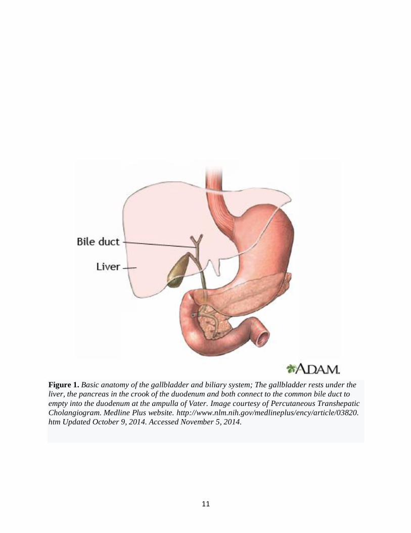
common bile duct into the small intestine.1 The gallbladder attaches to the biliary system via the
cystic duct, which is a short and crooked canal that connects the gallbladder to the common bile
duct. This duct descends down from that intersection to its attachment point in the duodenum
called the hepatopancreatic ampulla, the sphincter of Oddi, or the ampulla of Vater. This
attachment point is a small sphincter of muscle, which is a secondary point of regulation for the
introduction of bile into the digestive system (see Figure 1). Once there, bile assists in the
digestion of fats and acts as an emulsifying agent allowing them to be more easily broken down
into smaller and more manageable molecules. Bile is produced in the liver then is stored and
concentrated in the gallbladder and its release is controlled by the body’s production of the
chemical cholecystokinin, which is produced in response to the presence of fatty substances in
the duodenum. Presence of the hormone cholecystokinin in the patient’s blood causes the
gallbladder to contract and the sphincter of Oddi to relax.1
Several methods have been used over
the years to image the gallbladder and to investigate possible malformation or dysfunction. An

2
obsolete method of imaging called an Oral Cholecystogram was originally used to image the
gallbladder; it consisted of the patient orally ingesting contrast and a technologist obtaining
oblique radiographs of the gallbladder. This method outlined the cystic duct and the, neck, body,
and fundus, of the gallbladder as they were filled with contrast.1
This eventually gave way to the
use of medical sonography to diagnose gallbladder disorders and this method offers several
benefits that a conventional oral cholecystogram does not. Ultrasound imaging of the gallbladder
allows for the detection of small calculi that could not be visualized previously, there is little
patient preparation required as the patient need only fast for eight hours prior to examination, no
contrast is required, and because ultrasound uses sound waves there is no radiation dose to the
patient.1 A second modern method is the nuclear medicine HIDA scan. The HIDA scan measures
the ejection fraction of the gallbladder to determine overall emptying efficiency, and is more of a
functional scan than a structural one.2
A third method of imaging is MRI
cholangiopancreatography.3
Literature Review
There are three distinct pathologies that manifest in the gallbladder, functional
gallbladder disorder, cholecystitis, and cholelithiasis. Functional gallbladder disorder is
essentially a motility disorder and its causes are not well understood. “Presumably, the pain
associated with functional gallbladder disorder may occur due to increased gallbladder pressure
caused by either structural or functional outflow obstruction. Similar to other functional GI
disorders the pathophysiology of functional gallbladder disorder remains poorly understood and
may, in fact, represent a constellation of mechanisms” 4(p.370)
There are wide and varied opinions
on the cause of functional gallbladder disorder, and none has been definitively decided on by the
medical community. “Multiple theories of pathogenesis have been proposed including
cholesterolosis, microlithiasis, biliary sludge, chronic cholecystitis, gallbladder dysmotility,
narrowed cystic duct, cystic duct spasm, sphincter of Oddi dysfunction, and visceral
hypersensitivity.”4(p.370)
Functional gallbladder disorder is define by ROME III criteria as an
ejection fraction under 40% and an absence of pain for one year following cholecystectomy.2
A common method of diagnosis for Functional Gallbladder disorder is a functional
assessment and measure of the gallbladder ejection fraction (GBEF) after gallbladder stimulation
via Cholecystokinin. “Presently, CCK-CS with measurement of the GBEF is the most commonly
used test to aid in the diagnosis of functional gallbladder disorder.”4(p.372)
This nuclear-medicine
3
test, known as a HIDA scan, measures the gallbladder’s ability to empty over a period of 30 -60
minutes and rates the emptying efficiency as a percentage, generally a rating under 35% is
considered abnormal, and can merit cholecystectomy. This test is minimally invasive and only
requires a simple injection of cholecystokinin and radionuclides, then a period of observation
where the radionuclides are collected within the gallbladder and the rate at which the gallbladder
empties itself is measured and evaluated. Because this test requires the stimulation of the
gallbladder it can result in some pain and discomfort for the patient, often initiating the
symptoms the patient sought care for.2
“Acute or chronic cholecystitis is the inflammation of the gallbladder. In acute
cholecystitis, often a blockage of the cystic duct restricts the flow of bile from the gallbladder
into the common bile duct. The blockage is frequently due to a stone lodged in the neck of the
gallbladder. Over time, the bile begins to irritate the inner lining of the gallbladder, and it
becomes inflamed.”1(p.449)
The symptoms of acute cholecystitis can include, but are not limited
to, abdominal pain, right upper quadrant pain, and fever. Acute cholecystitis can also result from
bacterial infection, and gas producing bacteria can cause the development of a gangrenous
gallbladder.1
In contrast, chronic cholecystitis will almost always be associated with the
formation of gallstones, but could also be the result of pancreatitis or carcinoma of the
gallbladder. Its common symptoms are, right upper quadrant pain and nausea or heartburn after
the patient eats. It may also result in the thickening or calcification of the gallbladder wall.
Chronic cholecystitis often manifests in a series of attacks that follow meals and can last for up
to four hours.1
For patients with acute cholecystitis there are two main treatment options;
cholecystotomy and cholecystectomy, and there is a great deal of debate on when one is the best
choice over the other. However a study by Anderson et al as cited by Knab, Boller and
Mahvi5(p.465)
demonstrated that if the patient is a surgical candidate then the treatment option that
leads to the most optimal outcome for the patient is cholecystectomy. This resulted in a shorter
hospital stay, lower overall medical expenses, decreased complication rates, and little to no
increase in mortality. However if the patient was a poor surgical candidate that a cholecystotomy
was a useful treatment option but the patient will almost always have to return to the hospital for
a cholecystectomy at a later date as their symptoms return or worsen. Cholecystotomy being a

4
temporary fix at best and generally only performed in the event that a cholecystectomy would be
too dangerous because of secondary health issues in the patient.
Cholelithiasis is a chronic condition characterized by the presence of abnormal
calcifications or stones within the gallbladder. Increased body levels of bilirubin, cholesterol or
calcium can lead to the formation of gallstones. Those patients at highest risk for the
development of gallstones are women and the obese. Cholelithiasis accounts for nearly 90% of
all gallbladder and biliary duct disorders. The symptoms of cholelithiasis can include but are not
limited to; right upper quadrant pain after a meal, nausea and in some cases vomiting, and in
patients with a complete biliary blockage jaundice can develop.6
However gallstones are not all
the same nor do they result from the same dietary or disease mechanisms. “Gallbladder stones
were classified into eight types and more than ten subtypes according to the systematic
classification. These included cholesterol stones, pigment stones, calcium carbonate stones,
phosphate stones, calcium stearate stones, protein stones, cysteine stones and mixed stones.”6(p.4)
This wide variety of stone types and corresponding formation pathways leads to the high
numbers of patients suffering from gallstone related symptoms and the high number of gallstone
related cholecystectomies performed each year. The majority of gallstones cannot be visualized
radiographically without contrast media, this is a result of their composition. Those that require
contrast media to be visualized are cholesterol gallstones and gallstones comprised of cholesterol
and crystalline salts. Cholesterol gallstones comprise about sixty percent of all gallstones, and
stones made of cholesterol and crystalline salts account for an additional twenty five to thirty
percent of all gallstones. Those stones that can be visualized without contrast media account for
only ten to fifteen percent and are composed of calcium crystalline salts.1
Therefore it is often
necessary the to use contrast media to evaluate for the presence of gallstones or calcifications.
Diagnostic Imaging Procedures
There are three primary imaging procedures used to image the gallbladder to
diagnose acute cholecystitis, cholelithiasis, or functional gallbladder disorder, they are
abdominal ultrasound, nuclear medicine HIDA scan, and magnetic resonance imaging
cholangiopancreatography. Abdominal ultrasound is an outpatient procedure where the
sonographer manually examines the patient’s abdominal organs via an ultrasound transducer (see
Figure 4); it is useful for detecting stones and gallbladder wall thickening. Since ultrasound
relies on sound waves and is non-invasive this is a safe and inexpensive option for patients.7
5
Second is nuclear medicine HIDA scan, “Hepatobiliary Scintigraphy is a radionuclide diagnostic
imaging study (Including planar imaging, SPECT, or hybrid imaging such as SPECT/CT) that
evaluates hepatocellular function and the biliary system by tracing the production and flow of
bile from the formative phase in the liver, and its passage through the biliary system and into the
small intestine”8(p.211)
. This study consists of an initial radionuclide injection followed by a series
of images taken over a one-hour period. During this hour images are captured at one-minute
intervals and the activity within the gallbladder is assessed (see Figure 3). If acute cholecystitis
is suspected then follow up images can be requested at the four hour and twenty-four hour
marks. This test can also evaluate gallbladder contractibility and the gallbladder ejection fraction
and is the main method for diagnosing functional gallbladder disorder. To evaluate gallbladder
motility the nuclear medicine technologist injects sincalide after the initial one-hour imaging
period, then continues capturing images of the gallbladder as it is stimulated to contract. The
degree by which the gallbladder is able to empty after a one-hour period is recorded as a
percentage and labeled the GBEF.9 The third exam for gallbladder imaging is magnetic
resonance cholangiopancreatography, MRCP requires an MRI screening and the intravenous
injection of contrast. It is a fairly routine exam and can be performed as part of a routing
abdominal MRI (see Figure 2). This exam is capable of obtaining high quality diagnostic images
that would otherwise require a more invasive procedure such as ERCP. This exam carries a small
risk of infection and allergic reaction it is an outpatient procedure and should be completed in
less than one hour. MRI provides unparalleled soft tissue detail and can evaluate both structure
and function of the gallbladder. All three are effective and accurate however there is some
dispute as to the best choice between nuclear medicine ultrasound, and MRI for diagnosing acute
cholecystitis specifically. A recent study done by the Surgical department of St Joseph Mercy
Hospital in Ann Arbor Michigan compared the diagnostic effectiveness of ultrasound and HIDA
scan.10
When determining whether abdominal ultrasound has given a positive result for
cholecystitis the radiologist must consider several factors. The patient should display; a
sonographic murphy sign, evidence of a thickening of the gallbladder wall of greater five
millimeters, evidence of pericholecystic fluid, and the presence of hydrops with increased
transverse gallbladder diameter. The radiologist may also consider the presence and location of
biliary stones or sludge.10
In order for a radiologist to determine that a patient has tested positive
for cholecystitis after a HIDA scan the patient must; possess a gallbladder that was not visualized
6
noting persistent blockage of the cystic duct but not partial or complete blockage of common bile
duct.10
It was found that in cases where patients whose ultrasound result was negative but their
HIDA scan result was positive 89.9% were diagnosed intra-operatively with acute cholecystitis.
Of the patients who tested negative on their HIDA scan but positive of their abdominal
ultrasound 55% were diagnosed intra-operatively with acute cholecystitis. Researchers found
that the combined sensitivity of performing both abdominal ultrasound and HIDA scan improved
upon the diagnostic accuracy of performing only abdominal ultrasound by 24.4% and improved
on the diagnostic accuracy of performing only HIDA scan by 6%.10
This means that the greatest
diagnostic accuracy is achieved by performing both abdominal ultrasound and nuclear medicine
HIDA scan, and that the next most accurate option is HIDA scan alone, and the least accurate
diagnostic method for acute cholecystitis is abdominal ultrasound. Another factor to consider is
the cost and safety of the different imaging procedures as it relates to their relative diagnostic
efficiency. A study by Kiewiet et al as cited in Knab, Boller, and Mahvi5(p.459)
demonstrated that
the nuclear medicine study Cholescintigraphy is significantly superior to ultrasound for the
detection of acute cholecystitis, however this comes at the cost of time, affordability, availability
and safety. “If acute cholecystitis is highly suspected, US is likely the ideal choice given its
widespread availability, quick administration time, low cost, and patient safety profile. If the
diagnosis of acute cholecystitis is in question and 1 imaging study was equivocal, HIDA is likely
the better choice given its superior sensitivity compared with both ultrasound and MRI.”5(p.460)
This lends itself to a step-wise process of diagnosis, where the cheaper and easier imaging
options are explored before resorting to more invasive and expensive methods.
Invasive/Intraoperative Imaging Procedures
There are three primary invasive/intraoperative fluoroscopic exams used to image the
gallbladder and biliary system, and to treat complications from cholelithiasis. They are
endoscopic retrograde cholangiopancreatography (ERCP), percutaneous transhepatic
cholangiogram, and intraoperative cholangiogram. ERCP is a procedure where an endoscope is
inserted into the patient’s mouth and travels down the digestive system until it reaches the
duodenum. There the physician will thread a small tube through the papilla and into the common
bile duct. The physician then injects contrast and fluoroscopically evaluates the outlined biliary
anatomy. This contrast will highlight the common bile duct and biliary tree and reveal the
presence of stones or obstructions. Should there be a need to remove any stones or obstructions
7
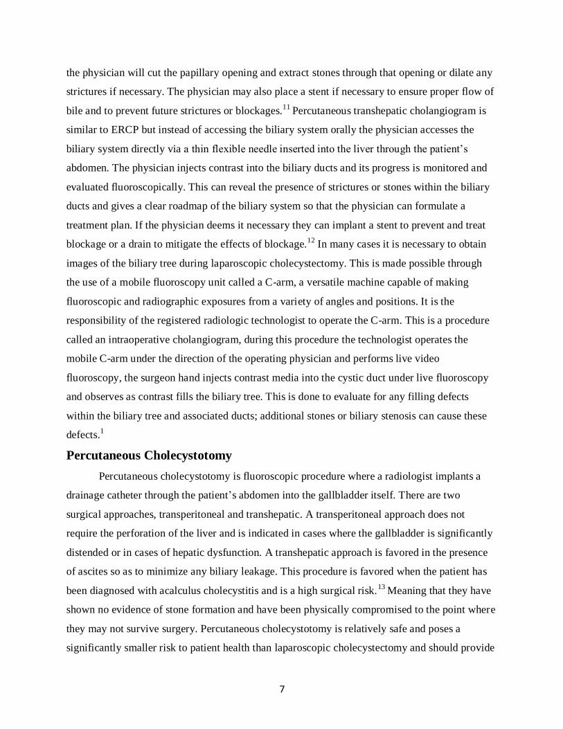
the physician will cut the papillary opening and extract stones through that opening or dilate any
strictures if necessary. The physician may also place a stent if necessary to ensure proper flow of
bile and to prevent future strictures or blockages.11
Percutaneous transhepatic cholangiogram is
similar to ERCP but instead of accessing the biliary system orally the physician accesses the
biliary system directly via a thin flexible needle inserted into the liver through the patient’s
abdomen. The physician injects contrast into the biliary ducts and its progress is monitored and
evaluated fluoroscopically. This can reveal the presence of strictures or stones within the biliary
ducts and gives a clear roadmap of the biliary system so that the physician can formulate a
treatment plan. If the physician deems it necessary they can implant a stent to prevent and treat
blockage or a drain to mitigate the effects of blockage.12
In many cases it is necessary to obtain
images of the biliary tree during laparoscopic cholecystectomy. This is made possible through
the use of a mobile fluoroscopy unit called a C-arm, a versatile machine capable of making
fluoroscopic and radiographic exposures from a variety of angles and positions. It is the
responsibility of the registered radiologic technologist to operate the C-arm. This is a procedure
called an intraoperative cholangiogram, during this procedure the technologist operates the
mobile C-arm under the direction of the operating physician and performs live video
fluoroscopy, the surgeon hand injects contrast media into the cystic duct under live fluoroscopy
and observes as contrast fills the biliary tree. This is done to evaluate for any filling defects
within the biliary tree and associated ducts; additional stones or biliary stenosis can cause these
defects.1
Percutaneous Cholecystotomy
Percutaneous cholecystotomy is fluoroscopic procedure where a radiologist implants a
drainage catheter through the patient’s abdomen into the gallbladder itself. There are two
surgical approaches, transperitoneal and transhepatic. A transperitoneal approach does not
require the perforation of the liver and is indicated in cases where the gallbladder is significantly
distended or in cases of hepatic dysfunction. A transhepatic approach is favored in the presence
of ascites so as to minimize any biliary leakage. This procedure is favored when the patient has
been diagnosed with acalculus cholecystitis and is a high surgical risk.13
Meaning that they have
shown no evidence of stone formation and have been physically compromised to the point where
they may not survive surgery. Percutaneous cholecystotomy is relatively safe and poses a
significantly smaller risk to patient health than laparoscopic cholecystectomy and should provide

8
relief from symptoms within 48 hours. Patients continue to undergo regular imaging of the
biliary system in the days and weeks following the procedure, and when the patient’s gallbladder
and biliary ducts return to a healthy appearance the catheter can be removed.13
The major risks of
the procedure are; tube dislodgement, biliary peritonitis, hemorrhage, and intestinal perforation.
Cholecystotomy is an effective management tool for acalculus cholecystitis but is not in itself a
cure, it can mitigate patient symptoms but it will often be followed by an effective surgical
procedure such as laparoscopic cholecystectomy.
Laparoscopic Cholecystectomy
“Laparoscopic cholecystectomy provides a less invasive approach for the removal of
diseased gallbladders. The surgeon makes a small opening in the umbilicus and passes an
endoscope into the abdominal cavity”1(p.607)
There are many advantages to laparoscopic
cholecystectomy; it can be performed as an outpatient procedure, as it is performed
laparoscopically it is minimally invasive and results in lower risk and faster recovery times, it
results in a shorter hospital stay than open cholecystectomy and many patients are able to return
home the day of their surgery and are able to return to work within two or three days.1
The
majority of patients who undergo laparoscopic cholecystectomy experience marked relief from
their pre-surgical symptoms however some abdominal pain, even after the recovery period, is
common.14
Out of one hundred and twenty six patients surveyed 60.3% reported a complete
absence of pain after an interval of ten years, but 90% report operative success when surveyed
after an interval of five years. There was no significant concentration of results around age,
gender, or ethnic group, meaning that surgical outcomes were good overall.14
This indicates that
a patient’s definition of operative success is not necessarily determined by the absence of pain
but rather the degree by which the patient’s symptoms are improved, and could be influenced
heavily by the patient’s preconceptions and expectations of how completely a cholecystectomy
could rectify their symptoms. There appears to be little variance in quality of surgical outcome
across race, gender, and age barriers among patients surveyed. However there is a real need for
realistic expectation on the part of the patient as it relates to surgical success, “The discrepancy
between outcome measures highlights the need for setting realistic expectations prior to
cholecystectomy”14(p.7)
. There is a very real need to properly educate the patient and follow up
with the patient at set intervals post surgically. The patient must understand that while
cholecystectomy will in all likelihood relieve the majority of their symptoms that there is a

9
possibility of continued discomfort, and that this does not mean that the surgery was ineffective
but only that it was not able to rectify all of the patients pathology.
Conclusion
Gallbladder disorders have become increasingly common in recent years and medical
imaging plays in important role in the diagnosis and treatment of gallbladder disease. There are
many imaging procedures and multiple treatment options and it is up to the physician’s
discretion which procedures suit a patient’s diagnosis best. Regardless of their specialty, imaging
professionals will likely be required to perform gallbladder imaging, no single modality is a best
choice for imaging the gallbladder and each is useful for observing different pathology.
Ultrasound is useful for describing gallbladder wall thickening and the presence of stones with
low cost and a short procedure time. MRI provides unparalleled tissue detail at the expense of
greater cost and increased scan time. Nuclear medicine clearly demonstrates gallbladder function
and emptying efficiency but is also expensive and lengthy, diagnostic radiography can provide
intra-operative imaging of the gallbladder during ERCP, cholecystectomy, cholecystotomy, and
percutaneous cholangiogram.

10
References
1. Kenneth L. Bontrager, John P. Lampignano. Textbook of Radiographic Positioning and
Related Anatomy eighth edition. St. Louis, MO: ELSEVIER; 2014.
2. DuCoin C, Faber R, Iligan M, Ruderman W, Wier D, et al. Normokinetic biliary dyskinesia:
a novel diagnosis. Surg Endosc, 2012; doi 10.1007/s00464-012-2342-0.
3. Magnetic Resonance Cholangiopancreatography. Radiology Info website.
http://www.radiologyinfo.org/en/info.cfm?pg=mrcp Reviewed May 2. 2013. Accessed
November 5, 2014.
4. Hansel Stephanie L. DiBaise John K. . Functional Gallbladder Disorder: Gallbladder
Dyskinesia. Gastroenterol Clin North Am. 2010; 39: 369-379.
5. Knab LM, Boller AM, Mahvi DM, et al. Cholecystitis. Surg Clin North Am. 2014;94(2):455-
470.
6. Tie Quiao, Rui-hong Ma, Xioa-bing Luo, Liu-quing Yang, Zhen-liang Luo, Pei-ming Zheng
et al. The Systematic Classification of Gallbladder Stones, PLoS One, 2013; doi:
10.1371/journal.pone.0074887.
7. Abdominal Ultrasound. Radiology Info website.
http://www.radiologyinfo.org/en/info.cfm?pg=mrcp Reviewed May 2. 2013. Accessed
November 5, 2014.
8. Nuclear Medicine – Hepatobiliary. Radiology Info website.
http://www.radiologyinfo.org/en/info.cfm?pg=hepatobiliary Reviewed February 12, 2014.
Accessed November 7, 2014.
9. Tulchinsky M. Ciak BW, Delbeke D, Hilson A, Holes-Lewis KA, Stabin MG, Ziessman HA
et al. SNM Practice Guideline For Hepatobiliary Scintigraphy. J Nucl Med Technol. 2010;
38: 210-218.
10. Kaoutzaniz C, Davies E, Leichtle S.W, Welch K.B, Winter S, Lapman R, Arneson W, et al.
Abdominal ultrasound versus hepato-imino diacetic acid scan in diagnosing acute
cholecystitis - what is the real benefit? J Surg Res, 2014; 188: 45-52.
11. Pateint Center - ERCP. American Gastroenterological Association website.
http://www.gastro.org/patient-center/procedures/ercp Published June 2013. Accessed
November 5, 2014.
12. Percutaneous Transhepatic Cholangiogram. Medline Plus website.
http://www.nlm.nih.gov/medlineplus/ency/article/003820.htm Updated October 9, 2014.
Accessed November 5, 2014.
13. Ramakrishnan KG, Menon S, Matthew A, Mohan M et al. Percutaneous cholecystotomy – an
alternative to surgery in a high-risk patient with acalculus cholecystitis Indian J Radiol
Imaging. 2003; 13: 307-310.
14. Lamberts MP, Oudsten BD, Keus F, et al. Patient-Reported outcomes of symptomatic
cholelithiasis patients following cholecystectomy after at least 5 years follow-up. Surg
Endosc. 2014; doi 10.1007/s00464-014-3619-2.
11
Figure 1. Basic anatomy of the gallbladder and biliary system; The gallbladder rests under the
liver, the pancreas in the crook of the duodenum and both connect to the common bile duct to
empty into the duodenum at the ampulla of Vater. Image courtesy of Percutaneous Transhepatic
Cholangiogram. Medline Plus website. http://www.nlm.nih.gov/medlineplus/ency/article/03820.
htm Updated October 9, 2014. Accessed November 5, 2014.

12
Figure 2. Magnetic resonance image of the gallbladder and associated anatomy; gallbladder is
highlighted below the biliary tree, come duodenum can be seen and some stomach can be
visualized. Image courtesy of Magnetic Resonance Cholangiopancreatography. Radiology Info
website. http://www.radiologyinfo.org/en/info.cfm?pg=mrcp Reviewed May 2. 2013. Accessed
November 5, 2014.

13
Figure 3. Chronological representation of nuclear medicine HIDA scan demonstrating
gallbladder filling. Image courtesy of Nuclear Medicine – Hepatobiliary. Radiology Info website.
http://www.radiologyinfo.org/en/info.cfm?pg=hepatobiliary Reviewed February 12, 2014.
Accessed November 7, 2014

14
15. Figure 4. Ultrasound of the gallbladder and common bile duct; gallbladder is labeled B and
common bile duct is labeled A. Image courtesy of Abdominal Ultrasound. Radiology Info
website. http://www.radiologyinfo.org/en/info.cfm?pg=mrcp Reviewed May 2. 2013.
Accessed November 5, 2014.
Related Documents