Image-guided femtosecond laser–assisted cataract surgery in Peters anomaly type 2 Joshua H. Hou, MD, Joao Crispim, MD, Maria S. Cortina, MD, Jose de la Cruz, MD We describe a technique for image-guided femtosecond laser–assisted cataract surgery in a case of Peters anomaly type 2. The femtosecond laser technology enabled reliable construction of a complete capsulotomy despite central corneal opacification and a tented anterior capsule. Use of an image-guided femtosecond laser for cataract extraction in Peters anomaly type 2 is a safe method for removing these complex cataracts while preserving capsular and corneal integrity. Financial Disclosure: Dr. De la Cruz is a consultant to Alcon Surgical, Inc. No other author has a financial or proprietary interest in any material or method mentioned. J Cataract Refract Surg 2015; 41:2353–2357 Q 2015 ASCRS and ESCRS Online Video Peters anomaly is a rare congenital ocular disorder characterized by central corneal opacification with absent endothelium, Descemet membrane, and poste- rior stroma. 1,2 This ocular malformation is believed to be due to abnormal migration of neural crest cells to- ward the posterior cornea during embryogenesis. 3 Type 1 Peters anomaly is characterized by corneal leu- koma with iridocorneal adhesions, 4 and type 2 is char- acterized by corneal leukoma with cataract and corneolenticular adhesions. 4 In patients with Peters anomaly type 2, visually sig- nificant cataracts can be present at birth or develop early in life. 5 Poor visualization through the cornea, tenting of the anterior capsule, and traction from cor- neolenticular adhesions on adjacent Descemet mem- brane can make manual capsulorhexis construction and cataract surgery challenging. As a result, cataract extraction without concurrent penetrating kerato- plasty (PKP) has rarely been reported in Peters anom- aly patients. 6 However, the prognosis for PKP in these patients is extremely poor. In a review of the literature by Bhandari et al., 4 PKP failed in 85.8% of reported cases of Peters anomaly type 2. Given the poor outcome, cataract surgery alone is an important option when vision loss is out of proportion to the degree of corneal opacification in Peters anomaly patients. With femtosecond laser technology and intraopera- tive image guidance, options for overcoming major challenges in otherwise difficult cataract cases are now available. 7,8 We present the use of image-guided femtosecond laser technology for performing cataract surgery alone in Peters anomaly type 2. SURGICAL TECHNIQUE Preoperative assessment of pupil dilation (4.0 mm or greater) and presence of iridocorneal or iridolenticu- lar adhesions is important in determining whether a patient with Peters anomaly is a candidate for femto- second laser–assisted cataract extraction. Iridocor- neal or iridolenticular adhesions that encroach on the central 4.0 mm of the visual axis may interrupt the laser capsulotomy and may necessitate decentra- tion of the capsulotomy or exclusion of the patient. Submitted: June 2, 2015. Final revision submitted: June 28, 2015. Accepted: July 1, 2015. From the Department of Ophthalmology and Visual Sciences (Hou, Crispim, Cortina, de la Cruz), University of Illinois Eye and Ear Infirmary, Chicago, Illinois, and the Department of Ophthal- mology and Visual Neurosciences (Hou), University of Minnesota, Minneapolis, Minnesota, USA; the Department of Ophthalmology and Visual Sciences (Crispim), Federal University of Sao Paulo, Sao Paulo, Brazil. Supported by unrestricted institutional grants from Research to Prevent Blindness, New York, New York, USA. Presented at the ASCRS Symposium on Cataract, IOL and Refrac- tive Surgery, San Diego, California, USA, April 2015. Corresponding author: Joshua H. Hou, MD, 420 Delaware Street Southeast, MMC 493, Minneapolis, Minnesota 55455, USA. E-mail: [email protected]. Q 2015 ASCRS and ESCRS Published by Elsevier Inc. http://dx.doi.org/10.1016/j.jcrs.2015.10.045 2353 0886-3350 TECHNIQUE

Welcome message from author
This document is posted to help you gain knowledge. Please leave a comment to let me know what you think about it! Share it to your friends and learn new things together.
Transcript

TECHNIQUE
SubmittedFinal revisAccepted:
From the(Hou, CrisEar Infirmmology anMinneapoand VisuaSao Paulo
SupportedPrevent B
Presentedtive Surge
CorresponSoutheastE-mail: jh
Q 2015 ASC
Published by
Image-guided femtosecond laser–assistedcataract surgery in Peters anomaly type 2
Joshua H. Hou, MD, Joao Crispim, MD, Maria S. Cortina, MD, Jose de la Cruz, MD: Junion sJuly
Deppim,ary,d Vislis, Ml Sci, Bra
bylindn
at thry, S
ding, MMou.jh
RS an
Elsev
We describe a technique for image-guided femtosecond laser–assisted cataract surgery in a caseof Peters anomaly type 2. The femtosecond laser technology enabled reliable construction of acomplete capsulotomy despite central corneal opacification and a tented anterior capsule. Useof an image-guided femtosecond laser for cataract extraction in Peters anomaly type 2 is a safemethod for removing these complex cataracts while preserving capsular and corneal integrity.
Financial Disclosure: Dr. De la Cruz is a consultant to Alcon Surgical, Inc. No other author has afinancial or proprietary interest in any material or method mentioned.
J Cataract Refract Surg 2015; 41:2353–2357 Q 2015 ASCRS and ESCRS
Online Video
Peters anomaly is a rare congenital ocular disordercharacterized by central corneal opacification withabsent endothelium, Descemet membrane, and poste-rior stroma.1,2 This ocular malformation is believed tobe due to abnormal migration of neural crest cells to-ward the posterior cornea during embryogenesis.3
Type 1 Peters anomaly is characterized by corneal leu-komawith iridocorneal adhesions,4 and type 2 is char-acterized by corneal leukoma with cataract andcorneolenticular adhesions.4
In patients with Peters anomaly type 2, visually sig-nificant cataracts can be present at birth or develop
e 2, 2015.ubmitted: June 28, 2015.1, 2015.
artment of Ophthalmology and Visual SciencesCortina, de la Cruz), University of Illinois Eye andChicago, Illinois, and the Department of Ophthal-ual Neurosciences (Hou), University of Minnesota,innesota, USA; the Department of Ophthalmologyences (Crispim), Federal University of Sao Paulo,zil.
unrestricted institutional grants from Research toess, New York, New York, USA.
e ASCRS Symposium on Cataract, IOL and Refrac-an Diego, California, USA, April 2015.
author: Joshua H. Hou, MD, 420 Delaware StreetC 493, Minneapolis, Minnesota 55455, USA.
d ESCRS
ier Inc.
early in life.5 Poor visualization through the cornea,tenting of the anterior capsule, and traction from cor-neolenticular adhesions on adjacent Descemet mem-brane can make manual capsulorhexis constructionand cataract surgery challenging. As a result, cataractextraction without concurrent penetrating kerato-plasty (PKP) has rarely been reported in Peters anom-aly patients.6 However, the prognosis for PKP in thesepatients is extremely poor. In a review of the literatureby Bhandari et al.,4 PKP failed in 85.8% of reportedcases of Peters anomaly type 2. Given the pooroutcome, cataract surgery alone is an important optionwhen vision loss is out of proportion to the degreeof corneal opacification in Peters anomaly patients.
With femtosecond laser technology and intraopera-tive image guidance, options for overcoming majorchallenges in otherwise difficult cataract cases arenow available.7,8 We present the use of image-guidedfemtosecond laser technology for performing cataractsurgery alone in Peters anomaly type 2.
SURGICAL TECHNIQUE
Preoperative assessment of pupil dilation (4.0 mm orgreater) and presence of iridocorneal or iridolenticu-lar adhesions is important in determining whether apatient with Peters anomaly is a candidate for femto-second laser–assisted cataract extraction. Iridocor-neal or iridolenticular adhesions that encroach onthe central 4.0 mm of the visual axis may interruptthe laser capsulotomy and may necessitate decentra-tion of the capsulotomy or exclusion of the patient.
http://dx.doi.org/10.1016/j.jcrs.2015.10.045 23530886-3350
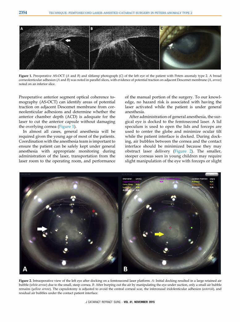
Figure 1. Preoperative AS-OCT (A and B) and slitlamp photograph (C) of the left eye of the patient with Peters anomaly type 2. A broadcorneolenticular adhesion (A and B) was noted in parallel slices, with evidence of potential traction on adjacent Descemet membrane (A, arrow)noted on an inferior slice.
2354 TECHNIQUE: FEMTOSECOND LASER–ASSISTED CATARACT SURGERY IN PETERS ANOMALY TYPE 2
Preoperative anterior segment optical coherence to-mography (AS-OCT) can identify areas of potentialtraction on adjacent Descemet membrane from cor-neolenticular adhesions and determine whether theanterior chamber depth (ACD) is adequate for thelaser to cut the anterior capsule without damagingthe overlying cornea (Figure 1).
In almost all cases, general anesthesia will berequired given the young age of most of the patients.Coordinationwith the anesthesia team is important toensure the patient can be safely kept under generalanesthesia with appropriate monitoring duringadministration of the laser, transportation from thelaser room to the operating room, and performance
Figure 2. Intraoperative view of the left eye after docking on a femtosecobubble (white arrow) due to the small, steep cornea. B: After burping out thremains (yellow arrow). The capsulotomy is adjusted to avoid the centralresidual air bubbles under the contact patient interface.
J CATARACT REFRACT SURG - V
of the manual portion of the surgery. To our knowl-edge, no hazard risk is associated with having thelaser activated while the patient is under generalanesthesia.
After administration of general anesthesia, the sur-gical eye is docked to the femtosecond laser. A lidspeculum is used to open the lids and forceps areused to center the globe and minimize ocular tiltwhile the patient interface is docked. During dock-ing, air bubbles between the cornea and the contactinterface should be minimized because they mayobstruct laser delivery (Figure 2). The smaller,steeper corneas seen in young children may requireslight manipulation of the eye with forceps or slight
nd laser platform. A: Initial docking resulted in a large retained aire air by manipulating the eye under suction, only a small air bubblecorneal scar, the inferonasal iridolenticular adhesion (asterisk), and
OL 41, NOVEMBER 2015

Figure 3. Intraoperative AS-OCT ofthe left eye showing (A) a scrolledcross-sectional display along the in-tended circumference of the capsuloto-my and (B) a single axial cross-sectionalong the axis of greatest lens tilt. A:The height and position of the capsu-lotomy is adjusted to ensure that theanterior (white arrow) and posteriorlimits (gray arrow) of the laser cutcontain the anterior capsule for thefull 360-degree circumference of thecapsulotomy. Due to multiple peaks(yellow arrowheads) and troughs (bluearrowheads) in the distorted anteriorlens capsule, alignment of the anteriorand posterior limits of the laser cut ona single peak or trough can result in anincomplete capsulotomy. B: On axialcross-section, care should be taken toensure the intended path of the laser(demarcated by the purple lines) doesnot hit the cornea. Since the anteriorchamber in Peters anomaly type 2 isdeeper peripherally, smaller capsulot-omies (yellow lines) are more likely tohit the cornea. Air bubbles (asterisk) be-tween the cornea and the patient inter-face can cause shadowing.
2355TECHNIQUE: FEMTOSECOND LASER–ASSISTED CATARACT SURGERY IN PETERS ANOMALY TYPE 2
movement of the head while the eye is docked undersuction to burp out retained bubbles. Shadowing inthe path of the laser on the intraoperative AS-OCToverlay may be due to bubbles and may warrantredocking (Figure 3).
Adjustments should be made to the capsulotomysize and centration based on the surgeon's view ofthe eye provided by the laser platform (Figure 2).Care should be taken to avoid any iridolenticular ad-hesions. The diameter of the capsulotomy should belarge enough to ensure that the central corneal opacitydoes not obscure the path of the laser. Widening thediameter of the capsulotomy or decentering the capsu-lotomy may be necessary to create adequate ACD be-tween the corneal endothelium and the distortedanterior lens capsule (Figure 3).
Using the AS-OCT imaging data provided by thefemtosecond laser platform, the height of the anteriorcapsulotomy should be adjusted to encompass theanterior capsule surface within the anterior and poste-rior limits of the laser cut for the entire 360-degreecircumference of the capsulotomy (Figure 3). Careshould be taken to ensure that the capsulotomy lasercut does not hit the cornea. Since the distorted anteriorcapsulemay havemultiple peaks and troughs along the
J CATARACT REFRACT SURG - V
circumference of the intended capsulotomy, centeringthe anterior and posterior limits of the laser at a singlepeak or trough, respectively, (the method typicallyused to compensate for simple lens tilt) may result inan incomplete capsulotomy. Intraoperative AS-OCTproviding a scrolled scan of the circumference of thecapsulotomy is crucial for visually confirming that therange of the laser cut includes the anterior capsule forthe entire 360-degree circumference of the capsulotomy(Video 1, available at: http://jcrsjournal.org). Aftercapsulotomy adjustments have been made, the lasercan be delivered per routine.
In the operating room, 2 paracentesis incisions aremade to facilitate cataract removal. Entering the ante-rior chamber with the paracentesis blade should bedone carefully since the anterior capsulemay be tentedif a complete laser capsulotomywas not achieved. Try-pan blue followed by injection of an ophthalmic visco-surgical device (OVD) to clear the dye can then be usedto stain the margins of the capsulotomy. A completecapsulotomy should be visible with a notable gap be-tween central anterior capsule tissue that is stilladherent to the cornea and peripheral anterior capsulethat has relaxed and flattened (Figure 4). Gentlemanipulation of the adherent anterior capsule tissue
OL 41, NOVEMBER 2015
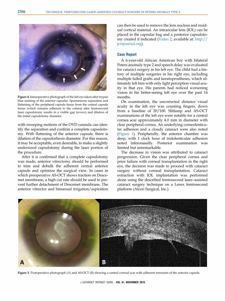
Figure 4. Intraoperative photograph of the left eye taken after trypanblue staining of the anterior capsular. Spontaneous separation andflattening of the peripheral capsule tissue from the central capsuletissue (which remains adherent to the cornea) after femtosecondlaser capsulotomy results in a visible gap (arrows) and dilation ofthe initial capsulotomy diameter.
2356 TECHNIQUE: FEMTOSECOND LASER–ASSISTED CATARACT SURGERY IN PETERS ANOMALY TYPE 2
with sweeping motions of the OVD cannula can iden-tify the separation and confirm a complete capsuloto-my. With flattening of the anterior capsule, there isdilation of the capsulorhexis diameter. For this reason,it may be acceptable, even desirable, to make a slightlyundersized capsulotomy during the laser portion ofthe procedure.
After it is confirmed that a complete capsulotomywas made, anterior vitrectomy should be performedto trim and debulk the adherent central anteriorcapsule and optimize the surgical view. In cases inwhich preoperative AS-OCT shows traction on Desce-met membrane, a high cut rate should be used to pre-vent further detachment of Descemet membrane. Theanterior vitrector and bimanual irrigation/aspiration
Figure 5. Postoperative photograph (A) and AS-OCT (B) showing a centra
J CATARACT REFRACT SURG - V
can then be used to remove the lens nucleus and resid-ual cortical material. An intraocular lens (IOL) can beplaced in the capsular bag and a posterior capsuloto-my created if indicated (Video 2, available at: http://jcrsjournal.org).
Case Report
A 6-year-old African American boy with bilateralPeters anomaly type 2 and speech delaywas evaluatedfor cataract surgery in his left eye. The child had a his-tory of multiple surgeries in his right eye, includingmultiple failed grafts and keratoprostheses, which ul-timately left himwith only light perception visual acu-ity in that eye. His parents had noticed worseningvision in his better-seeing left eye over the past 14months.
On examination, the uncorrected distance visualacuity in the left eye was counting fingers, downfrom a baseline of 20/100. Slitlamp and AS-OCTexaminations of the left eye were notable for a centralcornea scar approximately 4.0 mm in diameter withclear peripheral cornea. An underlying corneolenticu-lar adhesion and a cloudy cataract were also noted(Figure 1). Peripherally, the anterior chamber wasdeep, with 1 clock hour of iridolenticular adhesionnoted inferonasally. Posterior examination waslimited but unremarkable.
The decrease in vision was attributed to cataractprogression. Given the clear peripheral cornea andprior failure with corneal transplantation in the righteye, the decision was made to proceed with cataractsurgery without corneal transplantation. Cataractextraction with IOL implantation was performedalone using the described femtosecond laser–assistedcataract surgery technique on a Lensx femtosecondplatform (Alcon Surgical, Inc.).
l corneal scar with adherent remnants of the anterior capsule.
OL 41, NOVEMBER 2015

2357TECHNIQUE: FEMTOSECOND LASER–ASSISTED CATARACT SURGERY IN PETERS ANOMALY TYPE 2
At the 1-month follow-up, examination under anes-thesia was notable for a central corneal scar with clearperipheral cornea and a well-centered posterior cham-ber IOL (Figure 5, A). At the final examination, 7months after surgery, the corrected visual acuity was20/200 at distance and 20/60 at near (with the childmaneuvering his head and the near card to seearound the residual central corneal opacity at near).Remnants of the adherent anterior capsule were notedon AS-OCT imaging (Figure 5, B).
DISCUSSION
Cataract surgery in Peters anomaly type 2 can be chal-lenging due to the presence of corneal scarring, whichobscures the view, and corneolenticular adhesions,which distort and place tension on the anterior capsule.Manual continuous curvilinear capsulotomy tech-niques that rely on a flat anterior capsule surface canbe difficult to control when the capsule is tented up.A manual can-opener capsulotomy may be an alterna-tive but carries the risk for radial capsule tears duringhydrodissection, hydrodelineation, and lens removal.Manual manipulation of the anterior capsule in thepresence of corneolenticular adhesions may also leadto traction and detachment of adjacent Descemet mem-brane. Because of these challenges, cataract surgery israrely performed without concurrent penetrating kera-toplasty in cases of Peters anomaly type 2.6
Our experience suggests the femtosecond laser hasthe potential to perform a well-centered complete cap-sulotomy in cases of Peters anomaly type 2 in whichthe peripheral cornea remains clear. Care must betaken when adjusting the height and depth of thefemtosecond laser cut to ensure a complete capsuloto-my and avoid damaging the endothelium. Anteriorsegment OCT can accurately determine the appro-priate position and tilt of the anterior capsulotomydespite the distorted anatomy. In cases in which theanterior chamber is shallow, an increase in the diam-eter of the capsulotomy may be necessary to movethe incisions more peripherally, where the chamberis deeper. In cases with significant tenting of the ante-rior capsule, the final diameter of the anterior capsulot-omy is likely to be larger than expected after thecapsulotomy detaches from the cornea and lies flaton the lens surface. With minor intraoperative adjust-ments to the femtosecond laser based on the associatedintraoperative AS-OCT, femtosecond laser–assistedcataract surgery appears to be safe and effective incases of Peters anomaly type 2.
J CATARACT REFRACT SURG - VO
WHAT WAS KNOWN
� Femtosecond laser–assisted cataract surgery is an effec-tive technique for performing routine cataract surgery inpatients with clear corneas.
� Cataract surgery alone is rarely performed in patients withPeters anomaly type 2 despite the fact that graft failurefollowing penetrating keratoplasty surgery with cataractextraction is very high in these patients.
WHAT THIS PAPER ADDS
� Femtosecond laser–assisted cataract surgery is a useful tech-nique for performing laser capsulotomy and cataract removalwithout PKP surgery in patients with Peters anomaly type 2.
� Intraoperative ocular coherence tomography can provideaccurate and reliable guidance for placement of thefemtosecond laser–assisted anterior capsulotomy despitedistorted anatomy in patients with Peters anomaly type 2.
REFERENCES1. Yang LLH, Lambert SR, Drews-Botsch C, Stulting RD. Long-
term visual outcome of penetrating keratoplasty in infants and
children with Peters anomaly. J AAPOS 2009; 13:175–180
2. Yang LLH, Lambert SR. Peters’ anomaly. A synopsis of surgical
management and visual outcome. Ophthalmol Clin North Am
2001; 14(3):467–477
3. Ozeki H, Shirai S, Nozaki M, Sakurai E, Mizuno S, Ashikari M,
Matsunaga N, Ogura Y. Ocular and systemic features of Peters’
anomaly. Graefes Arch Clin Exp Ophthalmol 2000; 238:833–839
4. Bhandari R, Ferri S, Whittaker B, Liu M, Lazzaro DR. Peters
anomaly: review of the literature. Cornea 2001; 30:939–944
5. Withers SJ, Gole GA, Summers KM. Autosomal dominant cata-
racts and Peters anomaly in a largeAustralian family. Clin Genet
1999; 55:240–247
6. Nishide T, Nakanishi M, Hayakawa N, Kimura I, Mizuki N. Cata-
ract surgery for tilted lens in peters’ anomaly type 2. Case Rep
Ophthalmol 2013; 4:134–137
7. Martin AI, Hodge C, LawlessM, Roberts T, Hughes P, Sutton G.
Femtosecond laser cataract surgery: challenging cases. Curr
Opin Ophthalmol 2014; 25:71–80
8. Majander AS, Lindahl PM, Vasara LK, Krootila K. Anterior
segment optical coherence tomography in congenital corneal
opacities. Ophthalmology 2012; 119:2450–2457
L
41, NOVEMBER 2015First author:Joshua H. Hou, MD
Department of Ophthalmology &Visual Sciences, University of IllinoisEye & Ear Infirmary, Chicago,Illinois, USA
Related Documents