RESEARCH ARTICLE Open Access Illumina MiSeq 16S amplicon sequence analysis of bovine respiratory disease associated bacteria in lung and mediastinal lymph node tissue Dayle Johnston 1,2 , Bernadette Earley 1 , Paul Cormican 1 , Gerard Murray 3 , David Anthony Kenny 1 , Sinead Mary Waters 1 , Mark McGee 4 , Alan Kieran Kelly 2 and Matthew Sean McCabe 1* Abstract Background: Bovine respiratory disease (BRD) is caused by growth of single or multiple species of pathogenic bacteria in lung tissue following stress and/or viral infection. Next generation sequencing of 16S ribosomal RNA gene PCR amplicons (NGS 16S amplicon analysis) is a powerful culture-independent open reference method that has recently been used to increase understanding of BRD-associated bacteria in the upper respiratory tract of BRD cattle. However, it has not yet been used to examine the microbiome of the bovine lower respiratory tract. The objective of this study was to use NGS 16S amplicon analysis to identify bacteria in post-mortem lung and lymph node tissue samples harvested from fatal BRD cases and clinically healthy animals. Cranial lobe and corresponding mediastinal lymph node post-mortem tissue samples were collected from calves diagnosed as BRD cases by veterinary laboratory pathologists and from clinically healthy calves. NGS 16S amplicon libraries, targeting the V3-V4 region of the bacterial 16S rRNA gene were prepared and sequenced on an Illumina MiSeq. Quantitative insights into microbial ecology (QIIME) was used to determine operational taxonomic units (OTUs) which corresponded to the 16S rRNA gene sequences. Results: Leptotrichiaceae, Mycoplasma, Pasteurellaceae, and Fusobacterium were the most abundant OTUs identified in the lungs and lymph nodes of the calves which died from BRD. Leptotrichiaceae, Fusobacterium, Mycoplasma, Trueperella and Bacteroides had greater relative abundances in post-mortem lung samples collected from fatal cases of BRD in dairy calves, compared with clinically healthy calves without lung lesions. Leptotrichiaceae, Mycoplasma and Pasteurellaceae showed higher relative abundances in post-mortem lymph node samples collected from fatal cases of BRD in dairy calves, compared with clinically healthy calves without lung lesions. Two Leptotrichiaceae sequence contigs were subsequently assembled from bacterial DNA-enriched shotgun sequences. Conclusions: The microbiomes of the cranial lung lobe and mediastinal lymph node from calves which died from BRD and from clinically healthy H-F calves have been characterised. Contigs corresponding to the abundant Leptotrichiaceae OTU were sequenced and found not to be identical to any known bacterial genus. This suggests that we have identified a novel bacterial species associated with BRD. Keywords: Bovine respiratory disease, lung microbiome, 16S sequencing, diagnostics * Correspondence: [email protected] 1 Animal and Bioscience Research Department, Animal & Grassland Research and Innovation Centre, Teagasc Grange, Dunsany, Co. Meath, Ireland Full list of author information is available at the end of the article © The Author(s). 2017 Open Access This article is distributed under the terms of the Creative Commons Attribution 4.0 International License (http://creativecommons.org/licenses/by/4.0/), which permits unrestricted use, distribution, and reproduction in any medium, provided you give appropriate credit to the original author(s) and the source, provide a link to the Creative Commons license, and indicate if changes were made. The Creative Commons Public Domain Dedication waiver (http://creativecommons.org/publicdomain/zero/1.0/) applies to the data made available in this article, unless otherwise stated. Johnston et al. BMC Veterinary Research (2017) 13:118 DOI 10.1186/s12917-017-1035-2

Welcome message from author
This document is posted to help you gain knowledge. Please leave a comment to let me know what you think about it! Share it to your friends and learn new things together.
Transcript

RESEARCH ARTICLE Open Access
Illumina MiSeq 16S amplicon sequenceanalysis of bovine respiratory diseaseassociated bacteria in lung and mediastinallymph node tissueDayle Johnston1,2, Bernadette Earley1, Paul Cormican1, Gerard Murray3, David Anthony Kenny1,Sinead Mary Waters1, Mark McGee4, Alan Kieran Kelly2 and Matthew Sean McCabe1*
Abstract
Background: Bovine respiratory disease (BRD) is caused by growth of single or multiple species of pathogenic bacteria inlung tissue following stress and/or viral infection. Next generation sequencing of 16S ribosomal RNA gene PCR amplicons(NGS 16S amplicon analysis) is a powerful culture-independent open reference method that has recently been used toincrease understanding of BRD-associated bacteria in the upper respiratory tract of BRD cattle. However, it has not yetbeen used to examine the microbiome of the bovine lower respiratory tract. The objective of this study was to use NGS16S amplicon analysis to identify bacteria in post-mortem lung and lymph node tissue samples harvested from fatal BRDcases and clinically healthy animals. Cranial lobe and corresponding mediastinal lymph node post-mortem tissue sampleswere collected from calves diagnosed as BRD cases by veterinary laboratory pathologists and from clinically healthy calves.NGS 16S amplicon libraries, targeting the V3-V4 region of the bacterial 16S rRNA gene were prepared and sequenced onan Illumina MiSeq. Quantitative insights into microbial ecology (QIIME) was used to determine operational taxonomic units(OTUs) which corresponded to the 16S rRNA gene sequences.
Results: Leptotrichiaceae, Mycoplasma, Pasteurellaceae, and Fusobacterium were the most abundant OTUs identified inthe lungs and lymph nodes of the calves which died from BRD. Leptotrichiaceae, Fusobacterium, Mycoplasma, Trueperellaand Bacteroides had greater relative abundances in post-mortem lung samples collected from fatal cases of BRD in dairycalves, compared with clinically healthy calves without lung lesions. Leptotrichiaceae, Mycoplasma and Pasteurellaceaeshowed higher relative abundances in post-mortem lymph node samples collected from fatal cases of BRD in dairycalves, compared with clinically healthy calves without lung lesions. Two Leptotrichiaceae sequence contigs weresubsequently assembled from bacterial DNA-enriched shotgun sequences.
Conclusions: The microbiomes of the cranial lung lobe and mediastinal lymph node from calves which died from BRDand from clinically healthy H-F calves have been characterised. Contigs corresponding to the abundant LeptotrichiaceaeOTU were sequenced and found not to be identical to any known bacterial genus. This suggests that we have identifieda novel bacterial species associated with BRD.
Keywords: Bovine respiratory disease, lung microbiome, 16S sequencing, diagnostics
* Correspondence: [email protected] and Bioscience Research Department, Animal & Grassland Researchand Innovation Centre, Teagasc Grange, Dunsany, Co. Meath, IrelandFull list of author information is available at the end of the article
© The Author(s). 2017 Open Access This article is distributed under the terms of the Creative Commons Attribution 4.0International License (http://creativecommons.org/licenses/by/4.0/), which permits unrestricted use, distribution, andreproduction in any medium, provided you give appropriate credit to the original author(s) and the source, provide a link tothe Creative Commons license, and indicate if changes were made. The Creative Commons Public Domain Dedication waiver(http://creativecommons.org/publicdomain/zero/1.0/) applies to the data made available in this article, unless otherwise stated.
Johnston et al. BMC Veterinary Research (2017) 13:118 DOI 10.1186/s12917-017-1035-2

BackgroundBovine respiratory disease (BRD) is associated withsignificant morbidity and mortality in cattle worldwide,and contributes to substantial economic losses [1–3].Clinical signs associated with BRD typically includeelevated rectal temperature, increased respiratory rate,nasal and ocular discharges, cough, dyspnea, decreasedappetite and depression [2, 4, 5]. In Ireland, BRD is aleading cause of mortality in calves between one and 5months of age [6].BRD results from a multifactorial aetiology of infectious
agents, host factors, environmental stress factors and theirinteractions [7–9]. Primary pathogens including bovineherpesvirus 1 (Bo-HV1), bovine respiratory syncytial virus(BRSV), bovine parainfluenza 3 virus (BPIV-3), bovineviral diarrhoea virus (BVDV), bovine coronavirus (BCoV)and Mycoplasma bovis, are thought to be generallyresponsible for the onset of BRD [1, 4, 10–12] althoughrecent viral metagenomic NGS studies suggest that thereare many more uncharacterised BRD-associated virusesthan was previously thought [13, 14]. BRD-associated viraland Mycoplasmal pathogens damage the lungs by causingimmunosuppression, ciliary dysfunction and cellularnecrosis [15, 16]. This damage allows proliferation andcolonisation by secondary bacterial pathogens, commonlyincluding Mannheimia haemolytica, Pasteurellamultocida, Histophilus somni, Mycoplasma bovis andTrueperella pyogenes, which are normally commensal inthe nasopharyngeal region of cattle [1, 7, 11, 17–19].These bacteria evade the lung’s remaining immunedefences and their virulence factors cause progression ofthe disease if left untreated [7, 11, 15, 16].Currently available vaccinations and antibiotic treat-
ments are targeted against the known viruses and bacteriaassociated with BRD. Poor efficacy of vaccination andantimicrobial treatments against BRD associated bacteria[20–22] may partially be due to the involvement ofunknown bacterial pathogens in the disease.The methods that are currently used for identifica-
tion of specific bacteria associated with fatal cases ofBRD are culture, immunohistochemistry, in-situ hy-bridisation and multiplex and uniplex polymerasechain reaction (PCR) [23]. However, these techniquesare time consuming and they have several limitations.Although culture is considered the gold standard forpathogen identification [24], the main limitation ofthis technique is that many bacterial species asso-ciated with BRD are difficult to culture [25, 26]. PCRhas been demonstrated to be a more sensitive methodthan culture for identification of bacteria in bovinepneumonic lung tissue [27, 28]. Although real-timeqPCR is commonly used in veterinary diagnosticlaboratories, only known bacteria can be identifiedusing this “closed reference” technique and each
qPCR assay is generally designed to detect one indi-vidual bacterial species.Next generation sequencing of 16S ribosomal RNA gene
PCR amplicons (NGS 16S amplicon analysis) has beenused for identification of bacteria present in diversesample types including clinical isolates [29, 30], the bovinerumen [31], human nasal lavage [32] and nasopharyngealswabs from feedlot cattle [26, 33]. As primers bind toconserved regions of the 16S ribosomal RNA (rRNA) gene[34] and phylogenetically variable regions are amplifiedand subsequently sequenced (typically generating 10,000–100,000 sequences per sample), identification of thebacteria present (both known and unknown) in thesample is possible at unprecedented depth [35].The aim of this study was to use NGS 16S amplicon
analysis to identify unculturable and previouslyunknown bacteria which may play a pathogenic role inBRD. For this we analysed DNA from post-mortem cra-nial lung lobe and mediastinal lymph node tissue frombeef and dairy calves with BRD diagnosed as the causeof death, and from clinically healthy Holstein-Friesian(H-F) calves. We found highly abundant 16S sequencefor which the closest match was Leptotrichiaceae. Thisputative Leptotrichiaceae 16S sequence was present inlung and lymph node tissue from fatal BRD cases and inlung lesions from clinically healthy calves but was absentin lesion-free lungs from clinically healthy calves. To ourknowledge this is the first report of detection of thisputative Leptotrichiaceae species in bovine lung lesionsin cattle with BRD and also the first report of NGS 16Samplicon analysis of lung tissue and lung-associatedlymph nodes in cattle with BRD.
MethodsPost-mortem tissue sample collectionThirty-eight cranial lung lobe tissue samples were col-lected post-mortem from 32 beef calves and 6 dairy calveswhich were submitted from farms in three different re-gions in Ireland. Mediastinal lymph node tissue was alsocollected from 32 of these animals. BRD was diagnosed ongross examination at three regional veterinary laboratories(RVLs) (Athlone, Kilkenny and Sligo) by experiencedpathologists. Bacteriological culture, virological and/orbacteriological PCR and histology for identification ofaetiological agents (Table 1 and Additional file 1) wereperformed at the RVLs. In all cases, lung tissue sampleswere harvested from lesions (lesions were defined asmacroscopic consolidation or abscessation of lung tissue)present on the cranial lobes of the lung. Approximately1 g of each of these tissues was collected from each calf(comprising 13 beef and dairy breeds) (Table 1). Six ofthese post-mortem lung tissue samples were frozen at-80 °C immediately following collection. The remaininglung and lymph node post-mortem tissue samples were
Johnston et al. BMC Veterinary Research (2017) 13:118 Page 2 of 18
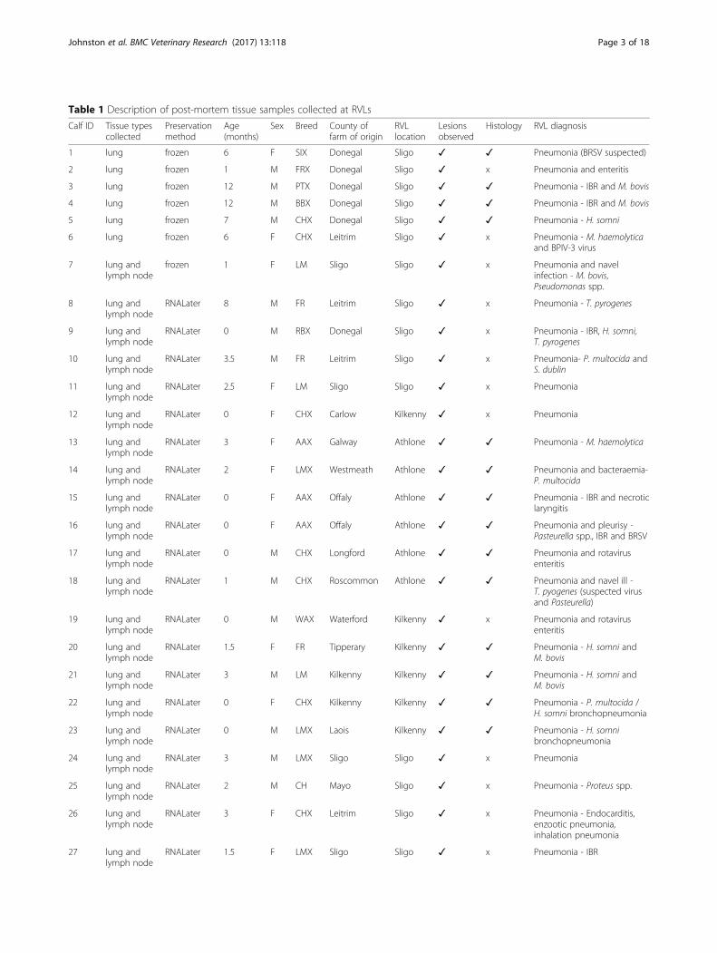
Table 1 Description of post-mortem tissue samples collected at RVLs
Calf ID Tissue typescollected
Preservationmethod
Age(months)
Sex Breed County offarm of origin
RVLlocation
Lesionsobserved
Histology RVL diagnosis
1 lung frozen 6 F SIX Donegal Sligo ✓ ✓ Pneumonia (BRSV suspected)
2 lung frozen 1 M FRX Donegal Sligo ✓ x Pneumonia and enteritis
3 lung frozen 12 M PTX Donegal Sligo ✓ ✓ Pneumonia - IBR and M. bovis
4 lung frozen 12 M BBX Donegal Sligo ✓ ✓ Pneumonia - IBR and M. bovis
5 lung frozen 7 M CHX Donegal Sligo ✓ ✓ Pneumonia - H. somni
6 lung frozen 6 F CHX Leitrim Sligo ✓ x Pneumonia - M. haemolyticaand BPIV-3 virus
7 lung andlymph node
frozen 1 F LM Sligo Sligo ✓ x Pneumonia and navelinfection - M. bovis,Pseudomonas spp.
8 lung andlymph node
RNALater 8 M FR Leitrim Sligo ✓ x Pneumonia - T. pyrogenes
9 lung andlymph node
RNALater 0 M RBX Donegal Sligo ✓ x Pneumonia - IBR, H. somni,T. pyrogenes
10 lung andlymph node
RNALater 3.5 M FR Leitrim Sligo ✓ x Pneumonia- P. multocida andS. dublin
11 lung andlymph node
RNALater 2.5 F LM Sligo Sligo ✓ x Pneumonia
12 lung andlymph node
RNALater 0 F CHX Carlow Kilkenny ✓ x Pneumonia
13 lung andlymph node
RNALater 3 F AAX Galway Athlone ✓ ✓ Pneumonia - M. haemolytica
14 lung andlymph node
RNALater 2 F LMX Westmeath Athlone ✓ ✓ Pneumonia and bacteraemia-P. multocida
15 lung andlymph node
RNALater 0 F AAX Offaly Athlone ✓ ✓ Pneumonia - IBR and necroticlaryngitis
16 lung andlymph node
RNALater 0 F AAX Offaly Athlone ✓ ✓ Pneumonia and pleurisy -Pasteurella spp., IBR and BRSV
17 lung andlymph node
RNALater 0 M CHX Longford Athlone ✓ ✓ Pneumonia and rotavirusenteritis
18 lung andlymph node
RNALater 1 M CHX Roscommon Athlone ✓ ✓ Pneumonia and navel ill -T. pyogenes (suspected virusand Pasteurella)
19 lung andlymph node
RNALater 0 M WAX Waterford Kilkenny ✓ x Pneumonia and rotavirusenteritis
20 lung andlymph node
RNALater 1.5 F FR Tipperary Kilkenny ✓ ✓ Pneumonia - H. somni andM. bovis
21 lung andlymph node
RNALater 3 M LM Kilkenny Kilkenny ✓ ✓ Pneumonia - H. somni andM. bovis
22 lung andlymph node
RNALater 0 F CHX Kilkenny Kilkenny ✓ ✓ Pneumonia - P. multocida /H. somni bronchopneumonia
23 lung andlymph node
RNALater 0 M LMX Laois Kilkenny ✓ ✓ Pneumonia - H. somnibronchopneumonia
24 lung andlymph node
RNALater 3 M LMX Sligo Sligo ✓ x Pneumonia
25 lung andlymph node
RNALater 2 M CH Mayo Sligo ✓ x Pneumonia - Proteus spp.
26 lung andlymph node
RNALater 3 F CHX Leitrim Sligo ✓ x Pneumonia - Endocarditis,enzootic pneumonia,inhalation pneumonia
27 lung andlymph node
RNALater 1.5 F LMX Sligo Sligo ✓ x Pneumonia - IBR
Johnston et al. BMC Veterinary Research (2017) 13:118 Page 3 of 18

cut into slices less than 0.5 cm thick, placed in RNALaterRNA Stabilization Reagent (Qiagen, Manchester, UK),stored at 4 °C overnight, and subsequently stored at -20 °C, according to the manufacturer’s instructions (Table 1).
Bacteriology, virology and histology performed at RVLsAerobic bacteriology of swab samples from pneumoniclungs was performed on blood agar and McConkey agarat 37 °C for 2 days (n = 36) (Additional file 1), as describedby Murray et al. [36]. Culture on chocolate agar under 8%CO2 for 3 days and on xylose lysine desoxycholate agar(following selenite enrichment) for 2 days at 37 °C wasalso conducted for the identification of Histophilus somni(n = 13) and Salmonella species (n = 24), respectively(Additional file 1) [36].Lung and tracheal mucosa were submitted for detection
of Mycoplasma bovis (n = 15), Histophilus somni (n = 11),Mannheimia haemolytica (n = 4) and Pasteurella multo-cida (n = 1), by real-time qPCR analyses (Additional file 1),which were performed as previously described [28, 37–39].Real-time qPCR was also used to identify Bo-HV1 (n = 36)and reverse transcriptase qPCR was used to identify BRSV(n = 38), BPIV-3 (n = 35) and BCoV (n = 34) (Additionalfile 1) as previously described [12]. Positive and negative(no template) controls were included in each qPCR assay.
Sections for histology were taken from the border ofgrossly visible lesions on the cranial lobe (n = 21) (Table 1and Additional file 1). Sections were fixed by placing themin 10% neutral-buffered formalin for 4 days, embeddingthem in paraffin wax, cutting them with a microtome andstaining them with hematoxylin and eosin, as previouslydescribed by Murray et al. [36].
Clinically healthy calves: health assessmentsTo serve as a comparison, 20 lung (cranial lobe) and 20corresponding mediastinal lymph node tissue sampleswere also sourced from clinically healthy H-F calveswhich were slaughtered at approximately 2.5 months ofage. These calves all recorded good performance prior toslaughter and achieved an average daily weight gainwithin a 0.25 standard deviation of their expectedaverage daily gains.Clinical health assessments of these calves were
performed on the morning before slaughter using theWisconsin calf health scoring criteria (https://www.vet-med.wisc.edu/dms/fapm/fapmtools/8calf/calf_respiratory_scoring_chart.pdf) to determine a respiratory score. A calfwas considered to have respiratory disease if it had a re-spiratory score greater than or equal to 5 as it was thenshowing at least two signs of respiratory disease [5]. A full
Table 1 Description of post-mortem tissue samples collected at RVLs (Continued)
28 lung andlymph node
RNALater 1.5 F AAX Sligo Sligo ✓ x Pneumonia - IBR andProteus spp.
29 lung andlymph node
RNALater 12 M CHX Donegal Sligo ✓ ✓ Pneumonia - (possible IBR)and meningitis
30 lung andlymph node
RNALater 1 M AA Sligo Sligo ✓ x Pneumonia - M. bovis
31 lung andlymph node
RNALater 2.5 F LMX Donegal Sligo ✓ x Pneumonia - M. bovis
32 lung andlymph node
RNALater 3 F LMX Kilkenny Kilkenny ✓ ✓ Pneumonia - interstitialpneumonia
33 lung andlymph node
RNALater 2.5 M FR Laois Kilkenny ✓ x Pneumonia - P. multocida
34 lung andlymph node
RNALater 5 M AAX Kilkenny Kilkenny ✓ ✓ Pneumonia - P. multocida,T. pyrogenes chronicbronchopneumonia
35 lung andlymph node
RNALater 4.5 F LMX Sligo Sligo ✓ ✓ Pneumonia - Pasteurellosis
36 lung andlymph node
RNALater 1 M CHX Longford Athlone ✓ ✓ Pneumonia - suppurativebronchial pneumonia.
37 lung andlymph node
RNALater 1.5 M CHX Galway Athlone ✓ ✓ Pneumonia - M. haemolytica
38 lung andlymph node
RNALater 3.5 F AAX Offaly Athlone ✓ ✓ Pneumonia - H. somni,M. bovis, M. haemolytica,P. multocida, T. pyogenes
Lesions were defined as macroscopic consolidation or abscessation of lung tissue.M male, F female, AA Aberdeen Angus, AAX Aberdeen Angus cross, BBX Belgium Blue cross, CH Charolais, CHX Charolais cross, FR Friesian, FRX Friesian cross, LMLimousin, LMX Limousin cross, PTX Parthenaise cross, RBX Rotbunt cross, SIX simmental cross, WAX wagyu cross, ✓ = performed and confirmed bovine respiratorydisease, x = not performed, H. somni = Histophilus somni, M. bovis = Mycoplasma bovis, M. haemolytica = Mannheimia haemolytica, P. multocida = Pasteurellamultocida, S. dublin = Salmonella dublin, T. pyrogenes = Trueperella pyrogenes, BRSV = bovine respiratory syncytial virus, IBR = infectious bovine rhinotracheitis,BPIV-3 = bovine parainfluenza 3 virus.
Johnston et al. BMC Veterinary Research (2017) 13:118 Page 4 of 18

clinical history was available for these calves including arecord of veterinary treatments for BRD.
Clinically healthy control calves: collection of post-mortemtissue samplesThe lungs were harvested from the calves immediatelyfollowing slaughter at Teagasc Ashtown ResearchCentre. All surfaces and forceps were cleaned with 20%Domestos extended germ kill bleach (1% final sodiumhypochlorite solution) (Unilever, Surrey, UK Ltd) Viru-solve® + (Amity International Healthcare; Barnsley, UK),70% ethanol and RNaseZAP™ (Sigma-Aldrich® IrelandLtd., Wicklow, Ireland) initially and between samples.The lungs were visually examined and the presence
and number of lesions was recorded. Tissue samplesfrom the cranial lobe and mediastinal lymph node wereremoved using sterile scalpel blades and forceps. Tissuesamples were cut into slices less than 0.5 cm thick,placed in RNALater RNA Stabilization Reagent, storedat 4 °C overnight, and subsequently stored at -20 °C, ac-cording to the manufacturer’s instructions. When lesionswere present on the cranial lobe, the lesions, rather thanthe lesion-free regions, were sampled.
DNA extractionDNA extraction from post-mortem tissue samples wascarried out in a class II biosafety cabinet. Forceps,homogeniser and all work surfaces were cleaned initiallyand between samples using 20% sodium hypochloritesolution, Virusolve®+, 70% ethanol and RNaseZAP™.Homogeniser blades were sonicated for 10 min at 60 °C inmolecular grade water (Sigma, Ireland), then rinsed byrunning while submerged in 75% ethanol and moleculargrade water. DNA was extracted from post-mortem tissuesamples using the Qiagen QIAamp Cador pathogen minikit (with pre-treatments T2 (enzymatic digestion of tissue)and B1 (for difficult-to-lyse bacteria in pre-treated tissue))(Qiagen, Manchester, UK) according to the manufacturer’sinstructions with some modifications to pre-treatment T2.A small piece of each tissue sample was removed using
sterile scalpel blades and forceps, placed in a sterilePetri-dish, weighed, immediately submerged in bufferATL (Qiagen, Manchester, UK) (360 μl per 50 mg tissue)and homogenised using a hand-held homogeniser (PRO200, Bio-Gen Series; PRO Scientific Inc. Oxford, USA).Following homogenisation, 205 μl homogenised tissuewas transferred to a 1.5 ml microcentrifuge tube (sterile)and 20 μl proteinase K was added. The microcentrifugetube was placed in a shaking incubator overnight(300 rpm, 56 °C).Pre-treatment B1 and the subsequent purification of
pathogen nucleic acids from fluid samples were carriedout according to the manufacturer’s instructions. Follo-wing nucleic acid extraction, RNA was removed using
RNaseA solution, 4 mg/ml (Promega, Southampton,UK). This was achieved by adding 5 μl RNaseA solutionto 100 μl of the sample’s purified nucleic acid eluate andincubating at 37 °C for 20 min. Subsequently, the DNAwas purified using a Zymo genomic DNA clean &concentrator™-10 kit (Zymo Research Corp, Irvine, CA,USA), according to the manufacturer’s instructions. ANanodrop spectrophotometer (NanoDrop Technologies,Wilmington, DE, USA) was used to quantify the DNA.All waste was autoclaved appropriately prior to disposal.
16S amplicon library preparation and sequencingOne hundred and sixteen 16S rRNA gene amplicon libra-ries (including 6 water control libraries) were prepared byPCR amplification of an approximate 467 bp region withinthe hypervariable (V3-V4) region of the 16S rRNA gene inbacteria, from 50 ng of each of the extracted and purifiedDNA from lung and lymph node tissue, and moleculargrade water (non-template control), respectively, accor-ding to the Illumina 16S metagenomic sequencing libraryprotocol, with modifications.PCR was initially performed with broad spectrum 16S
rRNA primers (forward primer: 5′-TCGTCGGCAGCGTCAGATGTGTATAAGAGACAGCCTACGGGNGGCWGCAG-3′, reverse primer: 5′-GTCTCGTGGGCTCGGAGATGTGTATAAGAGACAGGACTACHVGGGTATCTAATCC-3′) [34], using Kapa HiFi HotStart 2× ReadyMixDNA polymerase (Kapa Biosystems Ltd., London, UK).Cycle conditions were 95 °C (3 min), then 35 cycles of95 °C (30 s), 63 °C (30 s), 72 °C (30 s), then a final exten-sion of 72 °C (5 min). Libraries were purified usingAMPure XP beads (LABPLAN; Naas, Ireland) accordingto the Illumina 16S metagenomic sequencing libraryprotocol. Dual indices and Illumina sequencing adaptersfrom the Illumina Nextera XT index kits v2 B and C(Illumina, San Diego, USA) were added to the targetamplicons in a second PCR step using Kapa HotStartHiFi 2× ReadyMix DNA polymerase (Kapa BiosystemsLtd., London, UK). Cycle conditions were 95 °C (3 min),then 9 cycles of 95 °C (30 s), 55 °C (30 s), 72 °C (30 s),then a final extension of 72 °C (5 min). Libraries wereagain purified using AMPure XP beads (LABPLAN;Naas, Ireland) according to the Illumina 16S metage-nomic sequencing library protocol.Libraries were measured for purity and quantity on a
Nanodrop 1000 spectrophotometer. The barcoded ampli-con libraries were combined in equal concentrations intoa single pool according to their Nanodrop quantificationmeasurement. Two μl of each negative control library wasadded to the pool. The library pool was then quantifiedusing the KAPA SYBR FAST Universal qPCR kit with Illu-mina Primer Premix (Kapa Biosystems Ltd., London, UK)and the size was assessed with an Agilent DNA 1000 Kit(Agilent Technologies Ireland Ltd., Dublin, Ireland) on an
Johnston et al. BMC Veterinary Research (2017) 13:118 Page 5 of 18
Agilent 2100 Bioanalyser (Agilent Technologies IrelandLtd., Dublin, Ireland).The library pool was diluted and denatured according
to the Illumina MiSeq library preparation guide. Theamplicon library (8 pM) was spiked with 30% denaturedand diluted PhiX Illumina control library version 3 (12.5pM). The sequencing run was conducted on theIllumina MiSeq using the 500 cycle MiSeq reagent kit(version 2) with paired 250 bp reads. All sequence dataproduced in this study has been deposited to NCBI SRArepository and are available through series accessionnumber SRP080306.
Bioinformatic analysis of amplicon library sequencesRaw sequence reads for all samples in the study werequality controlled using the BBduk (https://sourcefor-ge.net/projects/bbmap/) Java package. This was used totrim low quality bases (<20 Phred score) from the 3′ endof sequence read pairs and identify and remove adaptorsequences. Illumina paired reads with an insert size(length of template molecule) that was shorter than thesum of the lengths of read 1 and read 2 were merged intoa single, longer read. Size selection of 467 bp ±20 bpsequences was performed with an in-house Perl script.The wrapper package Quantitative Insights Into Mi-
crobial Ecology (QIIME) [40] was used to determinethe operational taxonomic units (OTU)s which corre-sponded to the 16S rRNA gene sequences in eachsample. Sequences were clustered into individualOTUs at a default similarity level of 97% using anopen reference picking strategy, and subsequently, asingle representative sequence from each clusteredOTU was used to align to the Greengenes database.The RDP Classifier [41], using a minimum confidencecut off of 0.8, was used to determine the taxonomicclassification for each OTU. Any OTUs with fewerthan 50 sequences across all samples were excludedfrom further analysis. All OTUs with only a singleread count in any sample were removed from theanalysis.
Statistical analysisDifferences between relative abundances and the presenceof bacterial OTUs between post-mortem lung tissuefrozen at -80 °C upon collection and post-mortem lungtissue preserved with RNA-Later, from beef calves whichdied from BRD, were calculated in GraphPad Prism 6(version 6.04) using the Mann-Whitney U test andthe Fisher’s exact test, respectively.The Mann-Whitney U test and the Fisher’s exact test in
GraphPad Prism 6 (version 6.04) were used to determineif differences existed between the relative abundances andthe presence, respectively, of bacterial OTUs in the lungand lymph node tissues between dairy calves which died
from BRD and clinically healthy dairy calves without lunglesions present at slaughter.Stacked bar charts displaying relative abundances of
bacterial OTUs in the specific tissue types were preparedin GraphPad Prism 6 (version 6.04).
Microbial DNA enrichment and metagenomics shotgunsequencingA lung sample from calf number 23 was chosen forshotgun sequencing as it had a high prevalence of agenus of interest (Leptotrichiacea) and did not con-tain many other different bacterial genera. DNA wassuspended in Tris-EDTA buffer and re-quantifiedusing the Nanodrop spectrophotometer. One μg ofthe DNA was enriched for microbial DNA by selec-tive binding and removal of the CpG-methylated hostDNA using the NEBNext® Microbiome DNA Enrich-ment Kit (New England Biolabs (UK) Ltd., Hitchin,Herts, UK). The microbial-enriched DNA sample wasdiluted and quantified with an Agilent high sensitivityDNA kit (Agilent Technologies Ireland Ltd., Dublin,Ireland) on an Agilent 2100 Bioanalyser.
Shotgun library preparation and sequencingOne nanogram of microbial-enriched DNA was used toprepare a library using the Illumina Nextera®XT DNALibrary Preparation Kit, according to the manufacturer’sinstructions. Estimation of library size was performedusing an Agilent high sensitivity DNA kit on an Agilent2100 Bioanalyser and the library was quantified usingthe KAPA SYBR FAST Universal qPCR kit with IlluminaPrimer Premix. The library was diluted and denaturedaccording to the Illumina MiSeq library preparationguide. The 8 pM shotgun library was spiked with 1% de-natured and diluted PhiX Illumina control libraryversion 3 (12.5 pM). A 250 bp paired end sequencingrun was conducted on an Illumina MiSeq using a500 cycle MiSeq reagent kit (version 2).
Bioinformatic analysis of microbial DNA enriched shotgunsequenceThe BBduk (https://sourceforge.net/projects/bbmap/) Javapackage was used to trim low quality bases (<20 Phredscore) from the 3′ end of raw sequence read pairs and toidentify and remove adaptor sequence. All surviving readpairs were mapped to the bovine genome (v.UMD3.1)using bwa [42] to identify host sequences. Approximately100,000 paired-end sequences with no full-lengthalignment to the bovine genome were retained for de novoassembly. Contig assemblies were generated usingOMEGA metagenome assembler (V1.0.2) with defaultsettings. BLAST (BLASTN 2.3.0+) searches against thenon-redundant nucleotide collection database on theNCBI website [43, 44] were conducted to determine
Johnston et al. BMC Veterinary Research (2017) 13:118 Page 6 of 18

known bacterial sequences that were most similar to thecontig assemblies.
ResultsHealth assessments of clinically healthy calvesAll of the clinically healthy H-F calves were assessed asclinically healthy on the day of slaughter and had respira-tory scores based on the Wisconsin calf health scoringcriteria [5] of less than 5 (Additional file 2).
Lung lesions in clinically healthy calvesLesions (macroscopic consolidation or abscessation oflung tissue) were observed on lungs from 12 H-F calvesfrom the ‘clinically healthy’ group immediately post-slaughter (Additional file 3). Seven of these calves hadlesions only on the cranial lobes, two had lesions on themiddle lobe, two had lesions on both cranial and middlelobes and one calf had lesions on both the cranial andcaudal lobes (Additional file 3). Eight calves from the‘clinically healthy’ group had healthy lungs with nolesions present (Additional file 3).In total, 5 out of the 20 H-F calves from the ‘clini-
cally healthy’ group had recorded incidents of BRD(Additional file 3). These calves were all treated withNuflor (Merck Animal Health, New Jersey) andMetacam (Boehringer Ingelheim Vetmedica GmbHm,Germany) and one calf was also treated with Dexametha-sone (Norbrook, UK) and Zuprevo (Merck Animal Health,New Jersey). These antibiotic treatments were received atleast 41 days before slaughter. All calves with previousrecorded incidents of BRD had lung lesions present atslaughter and 7 calves with no recorded incidents ofBRD also had lung lesions observed at slaughter(Additional file 3). Furthermore, one calf with lunglesions received Primidoxine (Norbrook, UK) andASGold (Volac, UK) for treatment of enteritis 70 daysbefore slaughter and one calf with lung lesionspresent received Dexamethasone and Nuflor 10 daysbefore slaughter due to a reaction to vaccination.
Negative control amplicon library PCRTo test for contaminating 16S DNA in the PCR reagentsthat we used for generation of amplicon libraries, weconducted six negative (non-template) control ampliconlibrary reactions by substituting template DNA formolecular grade water. Following PCR amplification andagarose gel electrophoresis, no amplicon bands wereobserved in the gel (Additional file 4 (A)) and no peakswere observed on the electropherogram images obtainedfrom the DNA 1000 chip on an Agilent 2100 Bioanalyser(Additional file 4 (C)). In contrast, bands of the expectedamplicon size (467 bp) were observed on the gel for theselected lung tissue DNA libraries and the pooled lungand lymph node tissue DNA libraries (Additional file 4
(A)). Furthermore, the electropherogram images for theselected lung tissue DNA libraries and the pooled lungand lymph node tissue DNA libraries also confirmed thatthese libraries contained DNA fragments correspondingto the expected amplicon size (Additional file 4 (B)).
Taxonomic classificationOne hundred and fifteen bacterial OTUs were identi-fied overall. Seventy-two were identified to genuslevel. Additionally, 32 OTUs could only be identifiedas far as family level, 7 OTUs could only be identifiedto order, 2 to class level and 2 only to phylum level(Additional files 5 and 6).
Low numbers of classifiable reads in clinically healthyanimalsEven though equimolar amounts of each library wereadded to the pool that was sequenced on the MiSeq, thenumber of reads that were assigned to bacterial OTUsthat could be classified by QIIME were much higher inlung and lymph node tissue from fatal BRD cases thanfrom clinically healthy animals (Fig. 1). Unexpectedly,the PCR amplicon library yields were not different be-tween BRD cases and clinically healthy animals. This in-dicates that in the clinically healthy animals, the 16Sprimers were amplifying a sequence in the lung andlymph node that was not on the 16S Greengenesdatabase.
OTU abundance in lung tissueThe top 5 most abundant OTUs in the post-mortem lungsamples from the beef calves which died from BRD wereclassified by QIIME as Pasteurellaceae ((mean relativeabundance (S.E.M)) 22.4 (5.84)%), Mycoplasma (15.1(5.03)%), Leptotrichiaceae (14.9 (4.40)%), Clostridium (6.4(4.04)%) and Fusobacterium (5.1 (2.14)%). The top 5 mostabundant OTUs in the post-mortem lung tissue samplesfrom dairy calves which died from BRD were classified asLeptotrichiaceae (26.0 (14.0)%), Mycoplasma (22.0(11.00)%), Pasteurellaceae (16.0 (7.40)%), Fusobacterium(15.0 (8.70)%) and Bacteroides (8.1 (6.00)%). The mostabundant OTUs within the lung tissue samples from theclinically healthy H-F calves with lung lesions were classi-fied as Leptotrichiaceae (20.0 (9.90)%), Mycoplasma (15.5(7.42)%), Prevotella (12.3 (4.31)%) Pasteurellaceae (10.2(6.44)%), and Actinobacillus (4.6 (4.59)%). Leptotrichiaceaeand Fusobacterium were undetectable in the lesion-freelung samples from the clinically healthy H-F calves. Themost abundant OTUs in lesion-free samples werePrevotella (30.7 (7.56)%), Bacteroides (7.4 (7.38)%),Pasteurellaceae (6.6 (5.03)%), S24–7 (6.0 (5.24)%), andClostridium (5.5 (3.78)%).
Johnston et al. BMC Veterinary Research (2017) 13:118 Page 7 of 18

OTU abundance in lymph node tissueThe most abundant OTUs in lymph node tissue frombeef calves which died from BRD were classified asPasteurellaceae (18.0 (5.66)%), Clostridium (9.6 (5.30)%),Fusobacterium (7.3 (2.84)%), Leptotrichiaceae (6.8(2.66)%) and Prevotella (6.0 (2.90)%). Clostridium (21.9(19.11)%), Fusobacterium (21.7 (14.81)%), Mycoplasma(21.3 (19.67)%), Leptotrichiaceae (13.7 (10.68)%) andPasteurellaceae (8.1 (5.46)%) were the most abundantOTUs in post-mortem lymph node samples from dairycalves which died from BRD. The most abundant OTUsin post-mortem lymph node samples from the clinicallyhealthy H-F calves with lung lesions were classified asPrevotella (21.9 (10.40)%), Lysinibacillus (16.7 (11.24)%),Cupriavidus (8.4 (8.10)%), Clostridium (7.5 (5.64)%) andLeptotrichiaceae (6.8 (3.79)%). Notably, Leptotrichiaceaeand Fusobacterium OTUs were not detected in the
lymph node samples from the clinically healthy H-Fcalves without lung lesions. Prevotella (42.2 (11.63)%),Enterobacteriaceae (12.5 (12.50)%), Bacteroides (10.0(9.96)%), Succinivibrionaceae (7.9 (4.73)%), and Clostri-diales (3.9 (2.27)%) were the most abundant OTUs thatwere detected in these lymph nodes.
Differences between lung and lymph node microbiomesAlthough OTU relative abundances in lung and lymphnode were similar in some calves, the microbiomes ofthese two tissues were quite different in most of the ani-mals. For example, more than half of the OTUs were iden-tified as Clostridium in the lymph node of calf number 34whereas no Clostridium was detected in the lung of thisanimal. Furthermore, Ureaplasma was the main generadetected in the lung of calf number 36 but this genera wasnot detected in the lymph node of this animal (Fig. 2).
Fig. 1 Number of reads (classified as bacterial) within post-mortem lung and mediastinal lymph node tissues corresponding to each sample type;calves which died from the BRD (n = 38), and healthy Holstein-Friesian calves (with (n = 12) and without (n = 8) lung lesions). Fatal BRD - RVL = samplesfrom calves with the bovine respiratory disease complex collected at regional veterinary laboratories. Healthy – lesions present = samples from healthyHolstein-Friesian calves with lung lesions. Healthy – no lesions = samples from healthy Holstein-Friesian calves without lung lesions. Boxplot = Tukeyboxplot (lowest datum (whisker) is within a 1.5 interquartile range of the lower quartile, highest datum (whisker) is within a 1.5 interquartile range ofthe upper quartile, outliers (individual points) fall above and below these whiskers)
Johnston et al. BMC Veterinary Research (2017) 13:118 Page 8 of 18

Undetected and detected genera in clinically healthylesion-free lungs and lymph nodesOTUs that were classified as Leptotrichiaceae, Fusobac-terium, Pasteurella, Trueperella, Helcococcus, and Urea-plasma were abundant in lung and lymph node tissuesamples from both beef and dairy calves that died fromBRD, but these genera were not detected in either lungor lymph node tissue samples from the subset of clini-cally healthy H-F calves which had no observable lunglesions (n = 8) (Figs. 2 and 3).At least two putative BRD-associated genera were de-
tected in the lesion-free lungs. Mycoplasma was detected inthree lung (6L, 19L and 13L) and one lymph node tissuesample (5LN) (Fig. 3). Although relative abundances ofMycoplasma were reasonably high in 6 L and 19L, the readcounts of this genera were negligable (6L = 308 reads) com-pared to the fatal BRD cases, several of which had readcounts of >10,000 (Additional file 5). Sequences that wereclassified as Pasteurellaceae, a family containing speciesthat are commonly associated with BRD were detected inthree lung tissue and one lymph node tissue sample from
the lesion-free calves (Fig. 3) but, again, compared to thefatal BRD cases these were negligable. The most abundantbacterial genus detected in the lesion-free samples was Pre-votella which is more commonly associated with therumen. However, the average raw read counts for Prevotellawere not significantly different (P > 0.05) between the fatalBRD (average 212, standard deviation 446) cases and thelesion-free animals (average 498, standard deviation 610).
Comparison of OTU abundance between lung samplespreserved with RNALater and frozen lung samplesThere were no differences in the relative abundances orthe presence of bacterial OTUs within post-mortem lungtissue samples which were frozen at -80 °C immediatelyfollowing collection and post-mortem lung tissuesamples preserved with RNALater.
Comparison of OTU abundance between dairy calveswhich died from BRD and clinically healthy calvesBacterial OTUs associated with BRD including Fusobac-terium, Mycoplasma, Trueperella and Bacteroides, and an
Fig. 2 Percentage relative abundance of bacteria in post-mortem lung (L) and corresponding lymph node (LN) samples from calves which died frombovine respiratory disease. Adjacent profiles of L and LN are from the same animal. Athlone = sample collected at Athlone regional veterinarylaboratory (RVL). Kilkenny = sample collected at Kilkenny RVL. Sligo = sample collected at Sligo RVL. Grey bars represent infrequently occurring OTUs(see Additional file 6 for full details of all bacterial OTU classifications)
Johnston et al. BMC Veterinary Research (2017) 13:118 Page 9 of 18

OTU not yet associated with BRD (Leptotrichiaceae), hadgreater relative abundances and were present more often,among the lung tissue samples collected from the dairycalves which died from BRD compared with the lung tis-sue samples collected from the clinically healthy H-Fcalves with no lung lesions (P < 0.05) (Additional file 7).Leptotrichiaceae, Mycoplasma and Pasteurellaceae had
greater relative abundances (P < 0.05) (Additional file 8)and were present more often (P < 0.05) among thelymph node tissue samples collected from the dairycalves which died from BRD compared with the lymphnode tissue samples collected from the clinically healthyH-F calves with no observable lung lesions.
Comparison of 16S rRNA gene amplicon sequencingand RVL qPCRThis 16S rRNA gene amplicon sequencing assay identifiedmany more bacteria present in post-mortem lung andlymph node tissue from calves which died from BRD than
were identified from the same calves at the RVLs usingboth culture and uniplex PCRs on swab samples from thepneumonic lungs (Additional file 9). An OTU represen-ting the species detected by PCR in RVLs was identifiedusing the 16S rRNA gene amplicon sequencing assay inall but one case (calf I.D. = 30) (Additional file 9). How-ever, in seven calves (calf I.D. = 9, 19, 22, 23, 32, 35, 37)the Mycoplasma OTU was identified using the 16S rRNAgene amplicon sequencing assay even though a PCR testat the RVLs had confirmed no Mycoplasma bovis present(Additional file 9).The 16S rRNA gene amplicon sequencing assay identi-
fied bacterial OTUs in all ten calves (calf I.D. = 1, 5, 11,12, 17, 30, 31, 32, 35, 36) which were culture negative atthe RVLs (Additional file 9). Two cases of Escherichiacoli (calf I.D. = 4, 27), one case of Pseudomonas species(calf I.D. = 7), one case of Trueperella pyogenes (calfI.D. = 18) and one case of Salmonella dublin (calfI.D. = 3) were cultured at RVLs but were not
Fig. 3 Percentage relative abundance of bacteria in post-mortem lung (L) and corresponding lymph node (LN) samples from clinically healthyH-F calves with lung lesions (n = 12) and without lung lesions (n = 8). Adjacent profiles of L and LN are from the same animal. The first sevencalves received antibiotic treatment during their lifetimes. Grey bars represent infrequently occurring OTUs (see Additional file 6 for full detailsof all bacterial OTU classifications)
Johnston et al. BMC Veterinary Research (2017) 13:118 Page 10 of 18

identified by the 16S rRNA gene amplicon sequencingassay (Additional file 9).
Metagenomic shotgun sequencing of a microbial DNAenriched post-mortem lung tissue sampleOne of the post-mortem lung DNA samples (from calfnumber 23) was enriched for microbial DNA andcompletely sequenced. However, despite microbial DNAenrichment, 99% of the reads in the run mapped to thebovine genome. Approximately 100,000 sequences didnot map to the bovine genome and were assembled intocontigs. Most of these contigs aligned to the PhiX174bacteriophage genome but three contigs (contig1 = 2647 bp, contig 2 = 1376 bp, contig 3 = 744 bp)aligned to bacterial sequences. PhiX sequence was fromthe pre-prepared non-indexed PhiX V3 library (suppliedby Illumina) which was added, as a quality control, tothe microbial DNA-enriched library prior to sequencingon the MiSeq. Fluorescence from the bases in the non-indexed PhiX clusters bleeds into the fluorescence in theadjacent indexed clusters. Additional file 10 contains thesequences of the contigs which aligned to bacterialsequences. These contigs were blasted against the non-redundant nucleotide collection database on the NCBIwebsite using the BLASTN 2.3.0+ [45, 46] programmein order to determine the most similar known bacterialsequences.The top 10 blast hits for each contig are shown in
Table 2. No sequences on the database were found to be100% identical to any of the complete assembled contigs(Table 2). The top hit for contig number 1 was Sneathiaamnii strain SN35 (GenBank: CP011280.1) (Table 2). Itwas 92% identical to 99% of contig number 1 (Table 2).The top hit for contig number 2 was also Sneathiaamnii strain SN35 (GenBank: CP011280.1) (Table 2). Itwas 93% identical to 99% of contig number 2 (Table 2).Interestingly, contig number 2 was 100% identical to twoidentical partial 16S rRNA gene sequences (GenBank:KR514404.1) obtained from uncultured bacterial clonesin the bovine reproductive tract of farm animals atUniversity College Dublin (Table 2). Contig number 3was 99% identical to 100% of Histophilus somni strain2336 23S ribosomal RNA gene, complete sequence(Table 2).
DiscussionAt the time of writing there was a paucity of reports onhigh throughput 16S amplicon sequencing and metage-nomic shotgun sequencing in lung tissue in cattle andsheep. A detailed recent study was conducted on thelung microbiome in sheep [42] but, to our knowledge,the present report is the first high throughput 16Samplicon sequencing study in lung and correspondingmediastinal lymph nodes in multiple cattle with BRD.
The cranial lung lobe tissue was chosen for investigationas it is the most common site of BRD lesions [43, 44].Mediastinal lymph node tissue was also examined as re-spiratory disease causing viruses and bacteria often dis-seminate to these lymph nodes from the lungs [47–50].To gain an understanding of the bacteria associated withBRD cases in Ireland, we analysed lung tissue from ‘real’BRD fatalities on a number of farms in three differentregions of the country. Obtaining lung and lymph nodetissue from healthy ‘matched’ controls (i.e. same breed,sex, sire, age, farm) was not feasible in this study as itwould have required slaughtering a healthy animal onthe same farm as every BRD case. Instead, we had accessto healthy calves which were being slaughtered as partof another study from which we took lung and lymphnode tissue. In the present study, the composition ofbacteria in the lungs and lymph nodes were verydifferent between animals. In several cases, bacterialgenera were different in lung and mediastinal lymphnode tissues from the same calf.In humans, the lungs of clinically healthy individuals
were originally thought to be sterile [51]. However, recentstudies have demonstrated that small numbers of bacteriainhabit the lungs of clinically healthy humans and sheep[44, 51]. The current study also indicated that there arelow numbers of bacteria in the lungs and additionally inthe lymph nodes of clinically healthy calves. Prevotellawas the most prevalent genus in terms of relativeabundance in the lesion-free lungs but in terms of rawread counts, similar numbers of reads were detected infatal BRD cases. This is a highly abundant anaerobic genusin the rumen so unlikely to actively grow in aerobic lungtissue and probably passes into the upper regions of thelung by inhalation when rumen regurgitation occurs. Apossible explanation for the greater number of bacterialreads found in fatal cases of BRD compared with the clin-ically healthy calves (with and without lung lesions) inboth tissues, is that the disease may cause a proliferationof bacteria that can overcome the natural “equilibrium” inhealthy individuals.Although the 20 H-F calves in the “clinically healthy”
group displayed no clinical signs of disease at slaughterand only 5 of these calves had been previously treated forBRD, lung lesions were observed in 12 of these 20 animals(60%). This is similar to observations by Schneider et al.[52] and Wittum et al. [53], who reported that 61% and68%, of feedlot steers with no recorded history of BRDpresented with lung lesions at slaughter, respectively [53].These authors suggested that many cases of BRD can bemissed or may be sub-clinical [52].Leptotrichiaceae, Fusobacterium, Mycoplasma, True-
perella and Bacteroides were more abundant amongpost-mortem lung tissue samples from dairy calveswhich died from BRD compared to the lesion-free lung
Johnston et al. BMC Veterinary Research (2017) 13:118 Page 11 of 18

Table 2 Assembled bacterial contigs description and top BLAST hits
Contig number Top 10 BLAST hits Query cover Percentage identity
1 (2647 bp) 1.Sneathia amnii strain SN35, complete genome CP011280.1 99% 92%
2. Streptobacillus moniliformis DSM 12112, complete genome CP001779.1 96% 90%
3. Streptobacillus moniliformis strain DSM 12112 23S ribosomal RNA gene,complete sequence NR_076688.1
96% 90%
4. Sebaldella termitidis strain ATCC 33386 23S ribosomal RNA gene,complete sequence NR_076681.1
96% 89%
5. Sebaldella termitidis ATCC 33386, complete genome CP001739.1 96% 89%
6. Leptotrichia buccalis strain C-1013-b 23S ribosomal RNA gene, completesequence NR_076664.1
96% 88%
7. Leptotrichia buccalis DSM 1135, complete genome CP001685.1 96% 88%
8. Leptotrichia sp. oral taxon 212 strain W10393, complete genomeCP012410.1
96% 87%
9. Fusobacterium hwasookii ChDC F206, complete genome CP013336.1 96% 83%
10. Propionigenium maris 23S rRNA gene, strain DSM 9537 T AJ307979.1 93% 83%
2 (1376 bp) 1. Sneathia amnii strain SN35, complete genome CP011280.1 99% 93%
2. Uncultured bacterium clone T21VE9_4 16S ribosomal RNA gene,partial sequence KR514404.1
67% 100%
3. Uncultured bacterium clone T21UE13_2 16S ribosomal RNA gene,partial sequence KR514401.1
67% 100%
4. Uncultured bacterium clone T21VE9_24 16S ribosomal RNA gene,partial sequence KR514494.1
67% 99%
5. Uncultured bacterium clone T21VE9_36 16S ribosomal RNA gene,partial sequence KR514405.1
67% 99%
6. Uncultured bacterium clone T21UE13_23 16S ribosomal RNA gene,partial sequence KR514403.1
67% 99%
7. Uncultured bacterium clone T21UE13_12 16S ribosomal RNA gene,partial sequence KR514402.1
67% 99%
8. Uncultured bacterium clone H94 16S ribosomal RNA gene, partialsequence KC894542.1
69% 99%
9. Uncultured bacterium clone H35 16S ribosomal RNA gene, partialsequence KC894531.1
69% 99%
10. Uncultured bacterium clone T21UE060212_22 16S ribosomal RNAgene, partial sequence KR514400.1
67% 99%
3 (744 bp) 1. Histophilus somni strain 2336 23S ribosomal RNA gene, completesequence NR_103965.1
100% 99%
2. Histophilus somni strain 129P 23S ribosomal RNA gene, completesequence NR_076444.1
100% 99%
3. Haemophilus somnus 2336, complete genome CP000947.1 100% 99%
4. Haemophilus somnus 129PT, complete genome CP000436.1 100% 99%
5. Pasteurella multocida subsp. multocida OH4807, complete genomeCP004391.1
100% 94%
6. Pasteurella multocida subsp. multocida str. HN06, complete genomeCP003313.1
100% 94%
7. Pasteurella multocida subsp. multocida PMTB2.1, complete genomeCP007205.1
100% 94%
8. Pasteurella multocida subsp. multocida strain Pm70 23S ribosomal RNAgene, complete sequence NR_103956.1
100% 94%
9. Pasteurella multocida subsp. multocida str. Pm70, complete genomeAE004439.1
100% 94%
10. Pasteurella multocida strain Pm-3, complete genome CP014618.1 100% 94%
Database = Non redundant (nr) Nucleotide collection (nt).Program = BLASTN 2.3.0+ [45, 46]
Johnston et al. BMC Veterinary Research (2017) 13:118 Page 12 of 18

tissue samples from clinically healthy H-F calves.Additionally, Leptotrichiaceae, Mycoplasma and Pasteur-ellaceae had significantly greater relative abundancesamong post-mortem lymph node tissue samples fromdairy calves which died from BRD relative to the clinicallyhealthy, lung lesion-free H-F calves. It is also possible thatother bacterial OTUs were more abundant and presentmore often within tissue samples from BRD-affectedrelative to clinically healthy dairy calves, however, as thisstudy lacked power due to restricted sample sizes of BRD-affected dairy calves and lung lesion-free, clinically healthydairy calves, these differences were not statisticallydifferent.It is possible that the bacterial OTUs present among
the samples obtained from the RVLs were the result ofcontaminating bacteria which propagated post-mortem.However, this is unlikely as most bacterial OTUs foundamong these samples were also found in the samplesfrom the clinically healthy calves with lung lesions, inwhich the samples were obtained immediately followingslaughter.
LeptotrichiaceaeThe Leptotrichiaceae OTU was more abundant withinpost-mortem lung and lymph node tissue samples fromdairy calves which died from BRD relative to post-mortemlung and lymph node tissue samples from clinicallyhealthy, lung lesion-free, H-F calves. Additionally, it wasone of the most abundant OTUs identified within tissuesamples from beef and dairy calves which died from BRDand was absent in samples from clinically healthy H-Fcalves which had no lung lesions.The prevalence of OTUs that were classified as the
bacterial family Leptotrichiaceae in lung and lymph nodetissue from fatal BRD cases and lung lesions from clini-cally healthy cattle was of particular interest as membersof this family were recently proposed to be associatedwith lung disease in humans. Leptotrichiaceae speciesare not currently associated with BRD and have not beenpreviously described in lungs or mediastinal lymphnodes of cattle. All members of this bacterial family arefacultative to obligate anaerobic Gram-negative rods[54]. They can occur in anoxic environments as well asoral and intestinal environments [54]. Although little isknown about their role in disease, mainly due to the dif-ficulties associated with their isolation and identification,they have been suggested as emerging pathogens [55]. Amember of this family within the genus Leptotrichia wasidentified in bronchoalveolar lavage fluid from an elderlyman with pneumonia and was hypothesized to be respon-sible for the disease [56]. Additionally, a member of thegenus Sneathia was hypothesised to be responsible forlate-onset bronchiolitis obliterans syndrome in a lungtransplant recipient [57]. Furthermore, species within the
Leptotrichia genus have been isolated from blood culturesof patients with lesions in the oral mucosa [55] and fromblood cultures from patients with anaerobic bloodstreaminfection receiving high-dose chemotherapy [58].As the OTU sequence for Leptotrichiaceae found in
the present study was not identical to any knowngenus we wanted to achieve better classification withlonger DNA sequences than was possible with the472 bp V3-V4 16S sequence. To do this we partiallysequenced the genome of this unknown species ofbacteria by metagenomic shotgun sequencing and denovo assembly of a bacterial DNA enriched post-mortem lung tissue sample. From this we obtainedthree bacterial sequence contigs which were 2647 bp,1376 bp and 744 bp long. The 744 bp contig corre-sponded to a known BRD-associated bacterial species,Histophilus somni. A BLAST search against the NCBIcultured bacteria database showed the 2647 bp and1376 bp contigs were most similar (92 and 93%identity respectively) to Sneathia amnii. A BLASTsearch against the nr/nt collection showed that partof one of the Leptotrichiaceae contigs (67%) was100% identical to an uncultured clone. As thisuncultured clone (GenBank: KR514404.1, submittedby Lu, J) was obtained from the bovine reproductivetract of farm animals at University College Dublin, itappears to be a novel species that is present in atleast two microbiomes in cattle. From the presentstudy it is not possible to infer whether this novelLeptotrichiaceae species is pathogenic and causinglung lesions in cattle or wheteher it is merely able togrow opportunistically in the relatively anaerobic lunglesions (some Leptotrichiaceae are facultative anaerobes)which have been caused by known pathogens. In lung tis-sue samples from the calves in this study, the Leptotrichia-ceae OTU always co-occurred with either Pasteurellaceae,Mycoplasma or Fusobacterium OTUs.
PasteurellaceaePasteurellaceae was one of the most abundant OTUsidentified overall and was consistently more abundantin post-mortem lymph node samples from dairycalves which died from BRD compared with thosefrom clinically healthy, lung lesion-free, H-F calves.Many bacterial species implicated in BRD, includingMannheimia haemolytica, Histophilus somni, Biber-steinia trehalosi and Pasteurella multocida, belong tothe Pasteurellaceae family. Indeed, these bacterialspecies possess many virulence factors which enablethem to become pathogenic [17–19, 38, 59, 60].Furthermore they have been commonly isolated fromthe lungs and respiratory tracts of cattle with BRDand healthy cattle [11, 17, 19, 22, 61–64].
Johnston et al. BMC Veterinary Research (2017) 13:118 Page 13 of 18

FusobacteriumFusobacterium was one of the most abundant OTUsidentified. Furthermore, within the post mortem lungtissue samples, it was found to be more abundant amongsamples from dairy calves which died from BRD relativeto the samples from the clinically healthy, lung lesion-free, H-F calves. Additionally, it was not present withinany of the tissue samples from the calves that had nolung lesions. This was not surprising as Fusobacteriumspecies are commonly isolated from chronic, abscessinglung lesions in cattle with BRD [11]. Furthermore, thisgenus contains the anaerobic species Fusobacteriumnecrophorum, which is a significant opportunistic animalpathogen with several virulence factors [65] and it hasbeen previously isolated from ruminant respiratorytracts [63, 66] and pneumonic bovine lungs [67].
MycoplasmaMycoplasma was one of the most abundant OTUspresent in the post-mortem lung and lymph nodesamples. It was found to be more abundant amongthe tissue samples from dairy calves which died fromBRD relative to the tissue samples from the clinicallyhealthy, lung lesion-free, H-F calves. This is consistentwith previous studies which reported Mycoplasma tobe one of the dominant genera in nasopharyngealswab samples from cattle at feedlot [26, 33], despitebeing infrequent in cattle at feedlot entry [26].Additionally, this result is concordant with previousobservations which report that Mycoplasma speciesbovis, dispar and bovirhinis, were identified moreoften in pneumonic lungs and respiratory tracts com-pared with clinically healthy lungs and respiratorytracts [25, 62, 68, 69].Although Mycoplasma bovis is a recognised BRD patho-
gen [70] and is commonly screened for in veterinary diag-nostic laboratories [71, 72], the other major Mycoplasmaspecies are not generally screened. However, they may beresponsible for BRD as Mycoplasma dispar is a recognisedpathogenic Mycoplasma species [10], capable of colonisingthe lower respiratory tract and caused pneumonia wheninoculated into gnotobiotic calves [73]. Moreover, it hasbeen cultured from the respiratory tracts of calvespresenting with BRD [63, 74] and has been isolatedfrom lavage fluids of calves with recurrent respiratorydisease [68]. Furthermore, although Mycoplasmaarginine and Mycoplasma bovirhinis did not causepneumonia following inoculation into gnotobioticcalves, these species have also been isolated from lav-age fluids of calves with recurrent respiratory disease[68] and pneumonic lungs [68, 69] and have beensuggested to act as co-pathogens which may intensifyrespiratory disease symptoms [69, 74].
UreaplasmaUreaplasmas, also belong to the same family as theMycoplasmas, and are pathogenic bacteria which wereinitially associated with urogenital tract infections buthave also been isolated from pneumonic bovine lungs[10, 68, 75]. The species Ureaplasma diversum has beenassociated with clinical respiratory disease [75, 76]. AsUreaplasma was found to be present in pneumonic lungtissue samples from calves which died from BRD, thisgenus may be an important contributor to BRD which isoften overlooked.
BacterioidesBacterioides species are associated with bacterial pneu-monia [1, 18]. They had high relative abundances withinthe post-mortem lung and lymph node tissue samplestaken from beef and dairy calves which died from BRD.Furthermore, they were more abundant among samplesfrom dairy calves which died from BRD compared withclinically healthy lesion-free dairy calves. This result wasexpected as Bacterioides species have been commonlyisolated from chronic, abscessing lung lesions in cattlewith BRD [11] and have previously been cultured frompneumonic bovine lungs [67].
NGS 16S compared to RVL resultsThe bacterial 16S rRNA gene amplicon sequencing assayidentified many more bacteria from calves which diedfrom BRD than the culture and PCR tests carried out atthe RVLs. However, at the RVLs, there were severalcases where bacterial species were identified by culture,and one case where a bacterial species was identified byPCR, which were not identified by the bacterial 16SrRNA gene amplicon sequencing assay. This may bebecause only a small part of the cranial lobe region ofthe lung and the mediastinal lymph node were examinedwith the 16S sequencing assay, whereas culture wasperformed on swab samples covering the whole lungarea at RVLs. In the present study, the 16S rRNA geneamplicon assay showed that many bacteria which arenot currently screened for by PCR or cultured at RVLswere found to be present within the cranial lung lobesand mediastinal lymph nodes from calves which diedfrom BRD, including Leptotrichiaceae, Fusobacterium,Helcococcus, Mycoplasma, Ureaplasma, and Bacteroides.Although Mycoplasma bovis is currently screened for
by PCR in RVLs, the bacterial 16S rRNA gene ampliconsequencing assay found the Mycoplasma OTU to bepresent in seven calves which tested negative forMycoplasma bovis by PCR at RVLs. This is most likelybecause other species belonging to the Mycoplasmagenus, other than Mycoplasma bovis, were presentwithin these samples. Despite Helcococcus species notbeing currently associated with BRD, Helcococcus ovis
Johnston et al. BMC Veterinary Research (2017) 13:118 Page 14 of 18

has been isolated from sheep with respiratory disease [77],a calf with valvular endocarditis [78] and from a horsewith a pulmonary abscess [79]. Therefore, implementationof this 16S rRNA amplicon sequencing assay at veterinarydiagnostic laboratories would enable discovery of more ofthe bacteria responsible for each case of BRD as thepresent study has highlighted that this assay can identifymore known bacteria (both culturable and non-culturable) than culture, and can also identify yet un-known and unculturable bacteria in tissue samples, whichcannot be identified by culture and PCR tests currentlycarried out at veterinary diagnostic laboratories.Next generation sequencing can revolutionise BRD bac-
terial diagnostics. By using a single 16S rRNA amplicon se-quencing assay, the bacteria present in a tissue sample canbe accurately identified. A limitation of the Illumina MiSeqbased bacterial 16S rRNA gene amplicon sequencing assayis that currently, it is unable to identify bacterial OTUs tospecies level due to the fact that the maximum read lengthis <550 base pairs. However, the entire 16S rRNA gene canbe sequenced with Pacific Biosciences’ single molecule,real-time sequencing technology but this is expensive andthe error rate for individual reads are high [80].
ConclusionsIn conclusion, we have shown that a convenient, singleuniversal bacterial 16S rRNA gene amplicon sequencingassay can detect the bacterial OTUs present in a BRD-affected lung or lymph node sample. Using this assay,we have characterised the microbiomes of the craniallung lobe and mediastinal lymph node from calves whichdied from BRD and from clinically healthy H-F calves.We have confirmed that the cranial region of bovinelungs are not sterile environments and that somebacteria associated with BRD can be present within thelung and lymph node tissues of clinically healthy calvesas well as those calves which were suffering from BRD.However, the frequency of the detection of bacteria asso-ciated with BRD is lower in the lungs and lymph nodesof clinically healthy calves compared with calves whichdied from BRD.Furthermore, we have identified bacterial sequences cor-
responding to an unidentified species, which is classifiedas a member of the bacterial family Leptotrichiaceae, thatwas a dominant species in bovine lung lesions in BRDcases and absent in healthy lesion-free lung tissue. There-fore, this 16S rRNA gene amplicon sequencing assay haspotential to expedite BRD diagnosis and identify as of yetunknown bacteria which may be key players in BRDdevelopment and progression. Furthermore, results fromthe 16S rRNA gene amplicon sequencing assay, presentedin this paper, highlight that continually focusing on bac-terial agents that have been commonly associated withBRD could lead to common commensals that may offer a
pathogenic threat being neglected. This could result inincreased morbidity, increased mortality, and decreasedperformance in beef and dairy calves.The present study shows that further NGS study of
large numbers of healthy and BRD lung and lymph nodetissue samples to generate a comprehensive view of themicrobiome of healthy and diseased bovine lungs iswarranted.
Additional files
Additional file 1: Description of post-mortem tissue samples collected,sample preservation methods and the diagnostics tests performed onthese samples at regional veterinary laboratories (RVL)s. (XLSX 18 kb)
Additional file 2: Clinical assessment report from the clinically healthycalves on the morning before slaughter. (DOCX 31 kb)
Additional file 3: Description of the lungs from the clinically healthycalves observed following slaughter. (DOCX 14 kb)
Additional file 4: Agilent 2100 bioanalyzer DNA Electrophoresis assayresults (DOCX 222 kb)
Additional file 5: Operational taxonomic unit absolute read countstable. (XLSX 5305 kb)
Additional file 6: Operational taxonomic unit relative abundance table.(XLSX 78 kb)
Additional file 7: Comparison of OTU abundance between post-mortem lung tissue samples from dairy calves which died from BRD(n = 6) and clinically healthy calves without lung lesions (n = 8). (DOCX35 kb)
Additional file 8: Comparison of OTU abundance between post-mortem lymph node tissue samples from dairy calves which died fromBRD (n = 5) and clinically healthy calves without lung lesions (n = 8).(DOCX 33 kb)
Additional file 9: Description of the results of the PCR and culture testscarried out at the regional veterinary laboratories and the correspondingresults of the OTUs identified by 16S rRNA amplicon sequencing. (DOCX24 kb)
Additional file 10: Assembled bacterial contig sequences. (DOCX 30 kb)
AbbreviationsBCoV: Bovine coronavirus; Bo-HV1: Bovine herpesvirus 1; BPIV-3: Bovineparainfluenza 3 virus; BRD: Bovine respiratory disease; BRSV: Bovine respiratorysyncytial virus; BVDV: Bovine viral diarrhoea virus; H-F: Holstein-Friesian; NGS 16Samplicon analysis: Next generation sequencing of 16S ribosomal RNA gene PCRamplicons; OTU: Operational taxonomic unit; QIIME: Quantitative insights intomicrobial ecology; rRNA: Ribosomal RNA; RVL: Regional veterinary laboratory
AcknowledgementsThe authors would like to acknowledge the veterinary pathlogogists andtechnicians at the regional veterinary laboratories for sample acquisitionand collection.
FundingThis research, including all experimental procedures, was funded by the IrishGovernment under the National Development Plan 2007–2013, Departmentof Agriculture, Food and the Marine (DAFM) Research Stimulus Fund 11/S/131 (B. Earley, Principal Investigator) and EU project: 311,825 (B. Earley,Principal Investigator). The healthy calves were part of a study funded byDAFM Research Stimulus Fund 11/S/322 (D.A. Kenny, Principal Investigator).The funding bodies were not involved in the design of the study andcollection, analysis, and interpretation of data or in writing of the manuscript.
Availability of data and materialsAll sequence data produced in this study has been deposited to NCBI SRArepository and are available through series accession number SRP080306.
Johnston et al. BMC Veterinary Research (2017) 13:118 Page 15 of 18

Authors’ contributionsBE, MSM, DAK, MM, SW, AKK, PC and DJ designed the study. GM obtainedthe samples from fatal BRD cases at the regional veterinary laboratories (RVL)and performed culture and PCR at the RVL. DJ and MSM performed the NGSexperiments. PC performed the bioinformatics analysis. DJ and MSM wrotethe manuscript. All authors contributed to data interpretation and approvedthe final version of the manuscript.
Competing interestsThe authors declare that they have no competing interests.
Consent for publicationNot applicable.
Ethics approval and consent to participateTissue samples from healthy calves were harvested from a cohort ofslaughtered calves in an EU licenced abattoir as part of a separate studyexamining the effect of early life nutrition on adipogenesis in calves. Thislatter study was licenced (project authorisation number: AE19132/P014) bythe Irish Health Products Regulatory Authority in accordance with theEuropean Union (Protection of Animals used for Scientific Purposes)Regulations 2012 (SI 543/2012), the European Union (Protection of Animalsused for Scientific Purposes) (Amendment) Regulations 2013 (SI 434/2013)and the European Union (Protection of Animals used for Scientific Purposes)(Amendment) Regulations 2014 (SI 174/2014). As the tissue samples used inthe current study were obtained post-mortem, this study, therefore, did notrequire a separate licence.Tissue samples from calves which died from BRD were sourced from calveswhich succumbed to disease on-farm and were submitted for post-mortemexamination. No permissions or licences were required to sample these animalsas they were being sampled as part of a diagnostic process by the Departmentof Agriculture to achieve a diagnosis for the herd. None of the sick animalswere euthanised for the purposes of this study.
Publisher’s NoteSpringer Nature remains neutral with regard to jurisdictional claims inpublished maps and institutional affiliations.
Author details1Animal and Bioscience Research Department, Animal & Grassland Researchand Innovation Centre, Teagasc Grange, Dunsany, Co. Meath, Ireland. 2Schoolof Agriculture Food Science and Veterinary Medicine, University CollegeDublin, DublinBelfield, Dublin 4, Ireland. 3Department of Agriculture, Foodand the Marine, Regional Veterinary Laboratory, Sligo, Co. Sligo, Ireland.4Livestock Systems Research Department, Animal & Grassland Research andInnovation Centre, Teagasc Grange, Dunsany, Co. Meath, Ireland.
Received: 26 November 2016 Accepted: 21 April 2017
References1. Griffin D, Chengappa MM, Kuszak J, McVey DS. Bacterial pathogens of
the bovine respiratory disease complex. Vet Clin N Am Food AnimPract. 2010;26:381–94.
2. Taylor JD, Fulton RW, Lehenbauer TW, Step DL, Confer AW. Theepidemiology of bovine respiratory disease: What is the evidence forpreventive measures? Can. Vet. J. 2010;51:1351–9.
3. Brooks KR, Raper KC, Ward CE, Holland BP, Krehbiel CR, Step DL. Economiceffects of bovine respiratory disease on feedlot cattle during backgroundingand finishing phases. Prof. Anim. Sci. 2011;27:195–203.
4. Arcangioli M-A, Duet A, Meyer G, Dernburg A, Bézille P, Poumarat F, LeGrand D. The role of Mycoplasma bovis in bovine respiratory diseaseoutbreaks in veal calf feedlots. Vet J. 2008;177:89–93.
5. McGuirk SM, Peek SF. Timely diagnosis of dairy calf respiratory disease usinga standardized scoring system. Anim Health Res Rev. 2014;15:145–7.
6. DAFM/AFBI. All-Ireland animal disease surveillance report 2014. https://www.agriculture.gov.ie/media/migration/animalhealthwelfare/labservice/rvlreports/2014_All_Island_Disease_Surveillance_Report.pdf.
7. Edwards TA. Control methods for bovine respiratory disease for feedlotcattle. Vet Clin N Am Food Anim Pract. 2010;26:273–84.
8. Hay KE, Barnes TS, Morton JM, Clements ACA, Mahony TJ. Risk factors forbovine respiratory disease in Australian feedlot cattle: Use of a causaldiagram-informed approach to estimate effects of animal mixing andmovements before feedlot entry. Prev. Vet. Med. 2014;117:160–9.
9. Neibergs HL, Seabury CM, Wojtowicz AJ, Wang Z, Scraggs E, Kiser JN,Neupane M, Womack JE, Van Eenennaam A, Hagevoort GR, et al.Susceptibility loci revealed for bovine respiratory disease complex inpre-weaned holstein calves. BMC Genomics. 2014;15:1164.
10. Howard CJ. Mycoplasmas and bovine respiratory disease: studies related topathogenicity and the immune response–a selective review. Yale J. Biol.Med. 1983;56:789–97.
11. Callan R, Garry F. Biosecurity and bovine respiratory disease. VeterinaryClinics: Food Animal Practice. 2002;18:57–77.
12. O'Neill R, Mooney J, Connaghan E, Furphy C, Graham DA. Patterns ofdetection of respiratory viruses in nasal swabs from calves in Ireland: aretrospective study. Vet Rec. 2014;175:351.
13. Ng TFF, Kondov NO, Deng X, Van Eenennaam A, Neibergs HL, Delwart E. Ametagenomics and case-control study to identify viruses associated withbovine respiratory disease. J Virol. 2015;89:5340–9.
14. Mitra N, Cernicchiaro N, Torres S, Li F, Hause BM. Metagenomiccharacterization of the virome associated with bovine respiratory disease infeedlot cattle identified novel viruses and suggests an etiologic role forinfluenza D virus. J Gen Virol. 2016;97:1771–84.
15. Caswell JL. Failure of respiratory defenses in the pathogenesis of bacterialpneumonia of cattle. Vet Pathol. 2014;51:393–409.
16. Gershwin LJ, Van Eenennaam AL, Anderson ML, McEligot HA, Shao MX,Toaff-Rosenstein R, Taylor JF, Neibergs HL, Womack J. Single pathogenchallenge with agents of the bovine respiratory disease complex. PLoS One.2015;10:e0142479.
17. Dabo SM, Taylor JD, Confer AW. Pasteurella multocida and bovinerespiratory disease. Anim Health Res Rev. 2007;8:129–50.
18. Panciera RJ, Confer AW. Pathogenesis and pathology of bovine pneumonia.Vet Clin N Am Food Anim Pract. 2010;26:191–214.
19. Griffin D. Bovine pasteurellosis and other bacterial infections of therespiratory tract. Vet Clin N Am Food Anim Pract. 2010;26:57–71.
20. McVey DS. BRD research needs in the next 10–20 years. Anim Health ResRev. 2009;10:165–7.
21. Larson RL, Step DL. Evidence-based effectiveness of vaccination againstMannheimia haemolytica, Pasteurella multocida, and Histophilus somni infeedlot cattle for mitigating the incidence and effect of bovine respiratorydisease complex. Vet Clin N Am Food Anim Pract. 2012;28:97–106.e107.
22. Gagea MI, Bateman KG, van Dreumel T, McEwen BJ, Carman S, ArchambaultM, Shanahan RA, Caswell JL. Diseases and pathogens associated withmortality in Ontario beef feedlots. J Vet Diagn Investig. 2006;18:18–28.
23. Fulton RW, Confer AW. Laboratory test descriptions for bovine respiratorydisease diagnosis and their strengths and weaknesses: Gold standards fordiagnosis, do they exist? Can. Vet. J. 2012;53:754–61.
24. Padmanabhan R, Mishra AK, Raoult D, Fournier P-E. Genomics andmetagenomics in medical microbiology. J Microbiol Methods. 2013;95:415–24.
25. Marques LM, Buzinhani M, Yamaguti M, Oliveira RC, Ferreira JB, Mettifogo E,Timenetsky J. Use of a polymerase chain reaction for detection of MycoplasmaDispar in the nasal mucus of calves. J Vet Diagn Investig. 2007;19:103–6.
26. Holman DB, Timsit E, Alexander TW. The nasopharyngeal microbiota offeedlot cattle. Sci. Rep. 2015;5:15557.
27. Tegtmeier C, Angen Ø, Ahrens P. Comparison of bacterial cultivation, PCR,in situ hybridization and immunohistochemistry as tools for diagnosis ofHaemophilus somnus pneumonia in cattle. Vet Microbiol. 2000;76:385–94.
28. Bell CJ, Blackburn P, Elliott M, Patterson TIAP, Ellison S, Lahuerta-Marin A,Ball HJ. Investigation of polymerase chain reaction assays to improvedetection of bacterial involvement in bovine respiratory disease. J Vet DiagnInvestig. 2014;26:631–4.
29. Woo PCY, Lau SKP, Teng JLL, Tse H, Yuen KY. Then and now: use of 16S rDNAgene sequencing for bacterial identification and discovery of novel bacteria inclinical microbiology laboratories. Clin Microbiol Infect. 2008;14:908–34.
30. Taylor JD, Doyle DJ, Blackall PJ, Confer AW. Use of REP-PCR and 16S rRNAgene sequencing for comparison of Mannheimia haemolytica isolatesobtained from fatal cases of bovine respiratory disease in the USA andAustralia. Aust Vet J. 2014;92:15–23.
31. McCabe MS, Cormican P, Keogh K, O’Connor A, O’Hara E, Palladino RA,Kenny DA, Waters SM. Illumina miSeq phylogenetic amplicon sequencingshows a large reduction of an uncharacterised Succinivibrionaceae and an
Johnston et al. BMC Veterinary Research (2017) 13:118 Page 16 of 18

increase of the Methanobrevibacter gottschalkii clade in feed restricted cattle.PLoS One. 2015;10:e0133234.
32. Allen EK, Koeppel AF, Hendley JO, Turner SD, Winther B, Sale MM.Characterization of the nasopharyngeal microbiota in health and duringrhinovirus challenge. Microbiome. 2014;2:22.
33. Timsit E, Workentine M, Schryvers AB, Holman DB, van der Meer F, AlexanderTW. Evolution of the nasopharyngeal microbiota of beef cattle from weaningto 40 days after arrival at a feedlot. Vet Microbiol. 2016;187:75–81.
34. Klindworth A, Pruesse E, Schweer T, Peplies J, Quast C, Horn M, Glöckner FO.Evaluation of general 16S ribosomal RNA gene PCR primers for classical andnext-generation sequencing-based diversity studies. Nucleic Acids Res. 2013;41:e1.
35. Sontakke S, Cadenas MB, Maggi RG, Diniz PPVP, Breitschwerdt EB. Use ofbroad range16S rDNA PCR in clinical microbiology. J Microbiol Methods.2009;76:217–25.
36. Murray GM, More SJ, Sammin D, Casey MJ, McElroy MC, O’Neill RG, Byrne WJ,Earley B, Clegg TA, Ball H, et al. Pathogens, patterns of pneumonia, andepidemiologic risk factors associated with respiratory disease in recentlyweaned cattle in Ireland. J Vet Diagn Investig. 2017;29:20–34.
37. Sachse K, Salam HSH, Diller R, Schubert E, Hoffmann B, Hotzel H. Use of anovel real-time PCR technique to monitor and quantitate Mycoplasmabovis infection in cattle herds with mastitis and respiratory disease. Vet J.2010;186:299–303.
38. Klima CL, Alexander TW, Hendrick S, McAllister TA. Characterization ofMannheimia haemolytica isolated from feedlot cattle that were healthy ortreated for bovine respiratory disease. Can J Vet Res. 2014;78:38–45.
39. Liu D, Lawrence ML, Austin FW. Specific PCR identification of Pasteurellamultocida based on putative transcriptional regulator genes. J MicrobiolMethods. 2004;58:263–7.
40. Caporaso JG, Kuczynski J, Stombaugh J, Bittinger K, Bushman FD, CostelloEK, Fierer N, Pena AG, Goodrich JK, Gordon JI, et al. QIIME allows analysis ofhigh-throughput community sequencing data. Nat Methods. 2010;7:335–6.
41. Wang Q, Garrity GM, Tiedje JM, Cole JR. Naïve bayesian classifier for rapidassignment of rRNA sequences into the new bacterial taxonomy. ApplEnviron Microbiol. 2007;73:5261–7.
42. Glendinning L, Wright S, Pollock J, Tennant P, Collie D, McLachlan G. Variabilityof the Sheep Lung Microbiota. Appl Environ Microbiol. 2016;82:3225–38.
43. Epperson WB. A preliminary assessment of lung lesion distribution in fedcattle, South Dakota Beef Report, paper 4. Department of VeterinaryScience, South Dakota State University, Brookings, SD.; 2003. http://openprairie.sdstate.edu/sd_beefreport_2003/4.
44. Prohl A, Ostermann C, Lohr M, Reinhold P. The bovine lung in biomedicalresearch: Visually guided bronchoscopy, intrabronchial inoculation and invivo sampling techniques. J Vis Exp. 2014;89:51557. doi:10.3791/51557.
45. Zhang Z, Schwartz S, Wagner L, Miller W. A greedy algorithm for aligningDNA sequences. J Comput Biol. 2000;7:203–14.
46. Morgulis A, Coulouris G, Raytselis Y, Madden TL, Agarwala R, Schaffer AA.Database indexing for production MegaBLAST searches. Bioinformatics.2008;24:1757–64.
47. Valarcher J-F, Bourhy H, Lavenu A, Bourges-Abella N, Roth M, Andreoletti O,Ave P, Schelcher F. Persistent infection of b lymphocytes by bovinerespiratory syncytial virus. Virology. 2001;291:55–67.
48. Bashiruddin JB, de Santis P, Persson A, Ball H, Regalla J. Detection ofMycoplasma mycoides subspecies mycoides SC in bovine lung and lymphnode tissues by culture, sandwich ELISA and polymerase chain reactionsystems. Res Vet Sci. 2005;78:199–205.
49. Bosio CM, Bielefeldt-Ohmann H, Belisle JT. Active suppression of thepulmonary immune response by Francisella tularensis Schu4. J Immunol.2007;178:4538–457.
50. Rosendahl A, Bergmann S, Hammerschmidt S, Goldmann O, Medina E. Lungdendritic cells facilitate extrapulmonary bacterial dissemination duringpneumococcal pneumonia. Front Cell Infect Microbiol. 2013;3:PMC3689026.
51. Beck JM, Young VB, Huffnagle GB. The microbiome of the lung. Transl Res.2012;160:258–66.
52. Schneider M, Tait R, Busby W, Reecy J. An evaluation of bovine respiratorydisease complex in feedlot cattle: Impact on performance and carcass traitsusing treatment records and lung lesion scores. J Anim Sci. 2009;87:1821–7.
53. Wittum TE, Woollen NE, Perino LJ, Littledike ET. Relationships among treatmentfor respiratory tract disease, pulmonary lesions evident at slaughter, and rate ofweight gain in feedlot cattle. J Am Vet Med Assoc. 1996;209:814–8.
54. Gupta RS, Sethi M. Phylogeny and molecular signatures for the phylumFusobacteria and its distinct subclades. Anaerobe. 2014;28:182–98.
55. Eribe ERK, Olsen I. Leptotrichia species in human infections. Anaerobe.2008;14:131–7.
56. Kawanami T, Fukuda K, Yatera K, Kido T, Yoshii C, Taniguchi H, Kido M.Severe pneumonia with Leptotrichia sp. detected predominantly inbronchoalveolar lavage fluid by use of 16S rRNA gene sequencinganalysis. J Clin Microbiol. 2009;47:496–8.
57. Charlson ES, Diamond JM, Bittinger K, Fitzgerald AS, Yadav A, Haas AR,Bushman FD, Collman RG. Lung-enriched organisms and aberrant bacterialand fungal respiratory microbiota after lung transplant. Am J Respir CritCare Med. 2012;186:536–45.
58. Couturier MR, Slechta ES, Goulston C, Fisher MA, Hanson KE. Leptotrichiabacteremia in patients receiving high-dose chemotherapy. J Clin Microbiol.2012;50:1228–32.
59. Rice JA, Carrasco-Medina L, Hodgins DC, Shewen PE. Mannheimiahaemolytica and bovine respiratory disease. Anim Health Res Rev.2007;8:117–28.
60. Corbeil LB. Histophilus somni host–parasite relationships. Anim Health ResRev. 2007;8:151–60.
61. Welsh RD, Dye LB, Payton ME, Confer AW. Isolation and antimicrobialsusceptibilities of bacterial pathogens from bovine pneumonia: 1994–2002.J Vet Diagn Investig. 2004;16:426–31.
62. Allen J, Viel L, Bateman K, Rosendal S, Shewen P, Physick-Sheard P. Themicrobial flora of the respiratory tract in feedlot calves: associationsbetween nasopharyngeal and bronchoalveolar lavage cultures. Can J VetRes. 1991;55:341–6.
63. Nikunen S, Härtel H, Orro T, Neuvonen E, Tanskanen R, Kivelä SL, Sankari S,Aho P, Pyörälä S, Saloniemi H, Soveri T. Association of bovine respiratorydisease with clinical status and acute phase proteins in calves. CompImmunol Microbiol Infect Dis. 2007;30:143–51.
64. Taylor JD, Holland BP, Step DL, Payton ME, Confer AW. Nasal isolationof Mannheimia haemolytica and Pasteurella multocida as predictors ofrespiratory disease in shipped calves. Res Vet Sci. 2015;99:41–5.
65. Nagaraja TG, Narayanan SK, Stewart GC, Chengappa MM. Fusobacteriumnecrophorum infections in animals: Pathogenesis and pathogenicmechanisms. Anaerobe. 2005;11:239–46.
66. Jang SS, Hirsh DC. Characterization, distribution, and microbiologicalassociations of Fusobacterium spp. in clinical specimens of animal origin.J Clin Microbiol. 1994;32:384–7.
67. Chirino-Trejo JM, Prescott JF. The identification and antimicrobialsusceptibility of anaerobic bacteria from pneumonic cattle lungs. Can. J.Comp. Med. 1983;47:270–5.
68. Thomas A, Dizier I, Trolin A, Mainil J, Linden A, Ball H, Bell C. Isolation ofMycoplasma species from the lower respiratory tract of healthy cattle andcattle with respiratory disease in Belgium. Vet Rec. 2002;151:472–6.
69. Gagea MI, Bateman KG, Shanahan RA, van Dreumel T, McEwen BJ, CarmanS, Archambault M, Caswell JL. Naturally occurring Mycoplasmabovis—associated pneumonia and polyarthritis in feedlot beef calves. J VetDiagn Investig. 2006;18:29–40.
70. Bürki S, Frey J, Pilo P. Virulence, persistence and dissemination ofMycoplasma bovis. Vet Microbiol. 2015;179:15–22.
71. Blackburn P, Brooks C, McConnell W, Ball HJ. Isolation of Mycoplasma bovisfrom cattle in Northern Ireland from 1999 to 2005. Vet Rec. 2007;161:452–3.
72. Byrne WJ, McCormack R, Egan J, Brice N, Ball HJ, Markey B. Isolation ofMycoplasma bovis from bovine clinical samples in the Republic of Ireland.Vet Rec. 2001;148:331–3.
73. Gourlay RN, Howard CJ, Thomas LH, Wyld SG. Pathogenicity of someMycoplasma and Acholeplasma species in the lungs of gnotobiotic calves.Res Vet Sci. 1979;27:233–7.
74. Miles K, McAuliffe L, Ayling RD, Nicholas RAJ. Rapid detection ofMycoplasma dispar and M. bovirhinis using allele specific polymerase chainreaction protocols. FEMS Microbiol Lett. 2004;241:103–7.
75. Larsen LE, Tegtmeier C, Pedersen E. Bovine respiratory syncytial virus(BRSV) pneumonia in beef calf herds despite vaccination. Acta VetScand. 2001;42:113–21.
76. Autio T, Pohjanvirta T, Holopainen R, Rikula U, Pentikäinen J, Huovilainen A,Rusanen H, Soveri T, Sihvonen L, Pelkonen S. Etiology of respiratory diseasein non-vaccinated, non-medicated calves in rearing herds. Vet Microbiol.2007;119:256–65.
77. Zhang Y, Cui J, Parkinson A, Hayes J, Ott K, Byrum B. Isolation ofHelcococcus Ovis from Sheep with Pleuritis and Bronchopneumonia.J Vet Diagn Investig. 2009;21:164–6.
Johnston et al. BMC Veterinary Research (2017) 13:118 Page 17 of 18

78. Post KW, Rushton SD, Billington SJ. Valvular endocarditis associatedwith Helcococcus ovis infection in a bovine. J Vet Diagn Investig.2003;15:473–5.
79. Rothschild CM, Oaks JL, Schaupp JK, Rurangirwa FR, Sellon DC, Hines MT.Helcococcus ovis Isolated from a Pulmonary Abscess in a Horse. J ClinMicrobiol. 2004;42:2224–6.
80. Roberts R, Carneiro M, Schatz M. The advantages of SMRT sequencing.Genome Biol. 2013;14:405.
• We accept pre-submission inquiries
• Our selector tool helps you to find the most relevant journal
• We provide round the clock customer support
• Convenient online submission
• Thorough peer review
• Inclusion in PubMed and all major indexing services
• Maximum visibility for your research
Submit your manuscript atwww.biomedcentral.com/submit
Submit your next manuscript to BioMed Central and we will help you at every step:
Johnston et al. BMC Veterinary Research (2017) 13:118 Page 18 of 18
Related Documents