International Journal of Innovative Research in Advanced Engineering (IJIRAE) ISSN: 2349-2163 Volume 1 Issue 10 (November 2014) www.ijirae.com ______________________________________________________________________________________________________ © 2014, IJIRAE- All Rights Reserved Page -1 Graphene and its Influence in the Improvement of Surface Plasmon Resonance (SPR) Based Sensors: a Review Nasih Hma Salah* , 1 , David Jenkins 1 , Richard Handy 1 1 Faculty of Science and Environment, Plymouth University, Plymouth PL4 8AA, UK Abstract— A biosensor based on the principle of surface Plasmon resonance (SPR) and containing graphene is an efficient technique which allows quantification of biomolecular interactions in real-time. It can be executed in a setting free from label. Kretschmann configuration has turned into the most extensively used technique for excitation of Plasmon. Several researchers studying ligand-analyte interactions have used surfaces functionalized with different biomolecules like peptides, proteins, glycans, ribonucleic acids and deoxyribonucleic acids. It has been reported that the efficiency of an SPR sensor can be improved by post-coating the film with a fine layer of dielectric top. However, this improvement largely depends on thickness and dielectric constant of the upper layer. For the SPR sensors, graphene has proved to be an appropriate dielectric top layer. This paper would show in cases where graphene has been integrated in biosensor chips, sensitivity of the biosensors increases by 100% since the chalcogenide glass offers better detection as compared to the silica glass. Due to unique characters of silver and gold, their optical properties have proved to be tunable within the SPR sensors. This is significant as light matter coupling in absorption is crucial for optical detection. Moreover, in this review it has been theoretically and experimentally demonstrated that absorption potential of graphene is of universal value for visible light. Keywords: graphene, Surface plasmon resonance, biosensor I. INTRODUCTION Plasmonic is the field which has attracted the interest of researchers from different fields i.e. those investigating plasmonic- based lasers, computer processors and better treatments for cancer [1]. This article aims to review scholarly and peer reviewed articles related to surface plasmon resonance (SPR) graphene-based optical bio-sensing applications. Surface Plasmon resonance biosensors have turned into the analytical tools which are widely used [2]. These biosensors refer to optical sensors which make use of surface Plasmon polariton waves for analysing the interaction between sensing surface and biomolecules [1, 2]. A minute alteration in amount of biomolecule causes localized change in refractive index in close vicinity with the metal in the sensing medium. More importantly, change in refractive index (n) causes parallel alteration in the propagation constant of SPP which is usually measurable optically through the attenuated total internal reflection (ATR) technique. A remarkable feature of these biosensors is that they are highly sensitive to small alterations in the refractive index of a medium as shown in the Figure (1) [1-3]. An alteration in the refractive index of the medium being analysed causes particular changes in the surface Plasmon resonance properties such as SPR signal’s full half width, quantity of reflected light and the angle of SPR minimum [4]. Efficiency of an SPR sensor i.e. its specificity and sensitivity is determined by the optical and chemical stability of the sensing and the plasmonic layers. Prism glass of regular and high refractive index coated with silver or gold films of very small thickness are mostly used. Surface plasmon wave propagation at frequencies of visible light is facilitated by these metals at the metal dielectric interface [5]. Fig. (1) Illustrates immunoassay technique of the surface plasmon resonance. Functionalised SPR sensor layer on top of a conventional glass prism/ gold plasmonic system (left). The right side shows the sensogram response changing following binding event [1].

Welcome message from author
This document is posted to help you gain knowledge. Please leave a comment to let me know what you think about it! Share it to your friends and learn new things together.
Transcript
International Journal of Innovative Research in Advanced Engineering (IJIRAE) ISSN: 2349-2163 Volume 1 Issue 10 (November 2014) www.ijirae.com
______________________________________________________________________________________________________ © 2014, IJIRAE- All Rights Reserved Page -1
Graphene and its Influence in the Improvement of Surface Plasmon Resonance (SPR) Based Sensors: a Review
Nasih Hma Salah*, 1
, David Jenkins 1, Richard Handy
1
1 Faculty of Science and Environment, Plymouth University, Plymouth PL4 8AA, UK
Abstract— A biosensor based on the principle of surface Plasmon resonance (SPR) and containing graphene is an efficient technique which allows quantification of biomolecular interactions in real-time. It can be executed in a setting free from label. Kretschmann configuration has turned into the most extensively used technique for excitation of Plasmon. Several researchers studying ligand-analyte interactions have used surfaces functionalized with different biomolecules like peptides, proteins, glycans, ribonucleic acids and deoxyribonucleic acids. It has been reported that the efficiency of an SPR sensor can be improved by post-coating the film with a fine layer of dielectric top. However, this improvement largely depends on thickness and dielectric constant of the upper layer. For the SPR sensors, graphene has proved to be an appropriate dielectric top layer. This paper would show in cases where graphene has been integrated in biosensor chips, sensitivity of the biosensors increases by 100% since the chalcogenide glass offers better detection as compared to the silica glass. Due to unique characters of silver and gold, their optical properties have proved to be tunable within the SPR sensors. This is significant as light matter coupling in absorption is crucial for optical detection. Moreover, in this review it has been theoretically and experimentally demonstrated that absorption potential of graphene is of universal value for visible light. Keywords: graphene, Surface plasmon resonance, biosensor
I. INTRODUCTION
Plasmonic is the field which has attracted the interest of researchers from different fields i.e. those investigating plasmonic-based lasers, computer processors and better treatments for cancer [1]. This article aims to review scholarly and peer reviewed articles related to surface plasmon resonance (SPR) graphene-based optical bio-sensing applications. Surface Plasmon resonance biosensors have turned into the analytical tools which are widely used [2]. These biosensors refer to optical sensors which make use of surface Plasmon polariton waves for analysing the interaction between sensing surface and biomolecules [1, 2]. A minute alteration in amount of biomolecule causes localized change in refractive index in close vicinity with the metal in the sensing medium. More importantly, change in refractive index (n) causes parallel alteration in the propagation constant of SPP which is usually measurable optically through the attenuated total internal reflection (ATR) technique. A remarkable feature of these biosensors is that they are highly sensitive to small alterations in the refractive index of a medium as shown in the Figure (1) [1-3]. An alteration in the refractive index of the medium being analysed causes particular changes in the surface Plasmon resonance properties such as SPR signal’s full half width, quantity of reflected light and the angle of SPR minimum [4]. Efficiency of an SPR sensor i.e. its specificity and sensitivity is determined by the optical and chemical stability of the sensing and the plasmonic layers. Prism glass of regular and high refractive index coated with silver or gold films of very small thickness are mostly used. Surface plasmon wave propagation at frequencies of visible light is facilitated by these metals at the metal dielectric interface [5].
Fig. (1) Illustrates immunoassay technique of the surface plasmon resonance. Functionalised SPR sensor layer on top of a conventional glass prism/ gold
plasmonic system (left). The right side shows the sensogram response changing following binding event [1].
International Journal of Innovative Research in Advanced Engineering (IJIRAE) ISSN: 2349-2163 Volume 1 Issue 10 (November 2014) www.ijirae.com
______________________________________________________________________________________________________ © 2014, IJIRAE- All Rights Reserved Page -2
Mostly gold is preferred owing to its stable chemical and optical characteristics. Still, gold films have not proved to be efficient in developing highly sensitive SPR sensors. Whereas, silver films appear to be the most suitable candidate for visible range since Plasmon coupling demonstrates sharper angular resonance as compared to gold thereby improving the sensitivity of the sensor [6]. But, there are certain limitations which have reduced the utilization of silver for SPR sensing. Most important limitation is that it is not chemically stable. This problem can be avoided by using lamellar structures in biosensors as an alternative. In such an approach, fine films of metal oxides like SnO2 or ITO of 5-10nm thickness are coated on silver film. This results in an improvement in sensitivity [7-10]. Lockett et al. have used a similar strategy by coating silver substrates with a fine layer of amorphous carbon [9]. Furthermore, sputtering few nanometre of gold on top silver layer has been suggested and considered as a protective layer by Nasih et al. [1]. On top of that, researchers have made theoretical propositions that graphene can be used as a substitute for coating sliver [11] and gold [12] as shown in figure 3b. It was found that graphene’s refractive index in range of visible light is n=3+iCλ/3. In this expression, λ refers to wavelength (µm) and C refers to the constant its value is 5.446 µm−1 [13]. More importantly, graphene offers several benefits when employed in sensing devices. Findings of a research conducted by Zhou et al. (2012) indicate that utilization of graphene in the SPR sensing devices results in improvement in subnanometer scale at the single-point defect level [14]. The researchers also determined that utilization of doped monolayer graphene can bring about more improvements the applications currently available for fabrication of quantum plasmonic and atomic-scale nanoplasmonic instruments. Certain unique features of graphene make it a very promising candidate for developing innovative applications related to the SPR imaging [14]. For instance, it has been determined during a research study that in case of graphene-based biosensors, deposition of additional graphene layers over gold film together with utilization of various coupling configurations of laser beam can result in significant improvement in sensitivity of the device. The researchers have proposed that sensitivity of the SPR sensing device increases with the increase in number of graphene layers, to certain numbers, being deposited over the metal film [15]. Significance of using graphene in sensitive imaging applications has also been reported by some other research studies. For example, Choi et al., (2011) carried out a research on graphene-augmented imaging [11]. According to these researchers, taking into consideration the recent advancements made in the deposition of graphene, it can be anticipated that in coming years, sensing devices having silver films coated with single sheets of graphene would be developed. These devises are anticipated to offer several different technological advantages for execution of sensing analysis. Latest graphene-based SPR sensors have been discussed in this review paper. The article begins with elaboration of general over layer with plasmonic substrates. Various approaches for fabricating reproducible and stable graphene-metal interfaces are also discussed. This would be followed by analysis of the significance of graphene layer as a protective and sensing layer on the plasmonic layers such as silver or gold. In addition to these, some application of these interfaces for future mechanisms are outlined to illustrate the direction that might be taken to integrate graphene in sensor devices.
II. OVERLAYERS of GRAPHENE SUBSTRATES INTERFACE
It has been accepted that graphene can be given the name of “miracle substance of 21st century” as it is anticipated that it would continue to positively affect the development of nano and micro-electronic devices as well as the efficiency of photo voltaic cells employed in applications of solar energy [16, 17]. In actual, a number of outstanding characters are demonstrated by graphene. Graphene can take the form of bucky balls of fullerenes. Similarly it can form a carbon nano tube (CNT) if a graphene layer is enfolded. The CNTs are of crucial importance since they can act as a constituent of anti-ageing creams. Also, their devastating effect of main organs of rainbow trout has also been determined [18]. All around the world, number of research studies is being carried out to explore deposition of graphene layer over metal films. The main two reasons behind this are metal films can be employed for development of graphene layers having varying thicknesses and the graphene layer can be transfer onto a polymer or insulating support because of its amazing characteristics [19]. Deposition of graphene on a polymer or insulator support serves to be a beneficial approach which has been employed in developing flexible touch screens. It has been reported by researchers that growing graphene layers on metal films followed by their transference onto a polymer or insulating support is convenient enough to be used in the graphene-based applications.
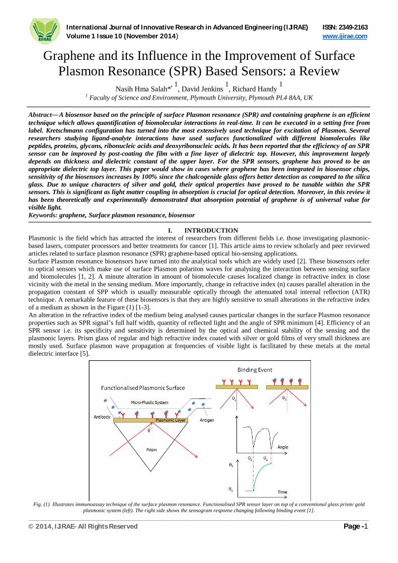
A typical graphene-based interface containing metal comprises of the below mentioned areas and parts as shown in the figure (2). First of all, metal atoms of the upper layer are encircled by carbon atoms in the ATOP (circles) position. They are positioned in the FOC and HCP hollow areas of RH (111) arranged above S-1 and S Rh-layers, respectively (Voloshina and Dedkov, 2012). Another characteristic is the FCC (squares) area where FOC hollow region of the Rh (111) surface is encircled by carbon atoms and are positioned in the uppermost and HCP hollow areas of the Rh (111) arrange above (S) and (S-1) [19]. Furthermore, the HCP triangle area where the hcp hollow region of the RH (111) surface is encircled by carbon atoms and are positioned in the fcc and uppermost hollow regions of RH (111) arrange above (S-2) and (S) RH-layers respectively [19]. One more feature has been reported as the BRIDGE position where Rh atom connects the carbon atoms present in (S) layer.
International Journal of Innovative Research in Advanced Engineering (IJIRAE) ISSN: 2349-2163 Volume 1 Issue 10 (November 2014) www.ijirae.com
______________________________________________________________________________________________________ © 2014, IJIRAE- All Rights Reserved Page -3
Figure (2). Crystallographic structure of graphene deposited on Ir (111). Highly symmetrical regions are marked by stars, down-triangles, rectangle and
circle for BRIDGE, HCP and FCC and ATOP positions [19]. According to researchers, the “density functional theory (DFT)” has been most extensively employed for investigating the graphene-metal systems.
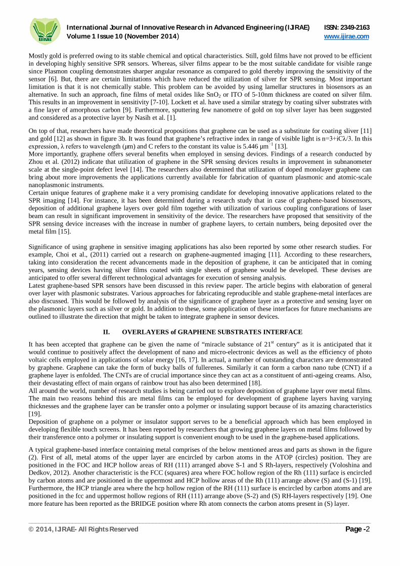
III. TRANSFERRING GRAPHENE ONTO SILVER and GOLD SUBSTRATES Graphene can be deposited or grown on silver or gold interfaces in a number of ways. Chemical vapour deposition (CVD) technique involves exposure of metal surfaces to the gaseous graphene which condenses on the metal. Researchers have demonstrated that this method is a good option to be used since the number of graphene layers deposited on metal surface can be controlled during the course of this technique [13]. In addition, another method is the dry-transfer method which is similar to wire bonding. During this method, top graphene layer is transferred onto thin films of silver of gold. This is also a quick method of graphene deposition on metal films in a reproducible way [13]. This method is based on utilization of commercial thermal release tape. In this method, a graphene sheet of 1mm2 is transferred mechanically on the metal. The graphene sheet is actually grown by the CVD on silicon coated with nickel. Tape is released via annealing the interface for two to three minutes at 120oC. This is followed by rinsing the tape with acetone. Next step is annealing for 360 minutes at a temperature of 500oC resulting in removal of any tape residues [13]. During a CVD process, copper foils were employed to grow graphene layers on the surface of metal, as demonstrated in the figure (3). A quartz chamber blown with argon and hydrogen gases was used to heat copper foils at temperature reaching up to 1000oC. After this, samples were left for annealing at the same temperature for twenty minutes resulting in reduction of oxide layer. The chamber was then blown with methane gas for twenty minutes at a rate of 15 sccm(standard cubic centimetres per minute) while the pressure inside the chamber was maintained at 5torr until the methane blow was stopped. Finally, the temperature of chamber was allowed to reduce to room temperature and copper foils coated with graphene were spin coated with a photoresist. At the completion of process, around 2% absorption was recorded for every layer of graphene [20].
FIG. (3). Demonstration of transferring a single layer of graphene from copper onto gold substrates [20].
International Journal of Innovative Research in Advanced Engineering (IJIRAE) ISSN: 2349-2163 Volume 1 Issue 10 (November 2014) www.ijirae.com
______________________________________________________________________________________________________ © 2014, IJIRAE- All Rights Reserved Page -4
Basically, remarkable advantages offered by graphene-based SPR sensors. One of the important improvement is enhancement in the adsorption potential of organic molecules due to 휋 − 휋 bonding. Another advantages are the protection of surface of the plasmonic layer in aqua environment from oxidization and reduction in adverse effects on characters of plasma [20].
IV. SENSING PROCESS
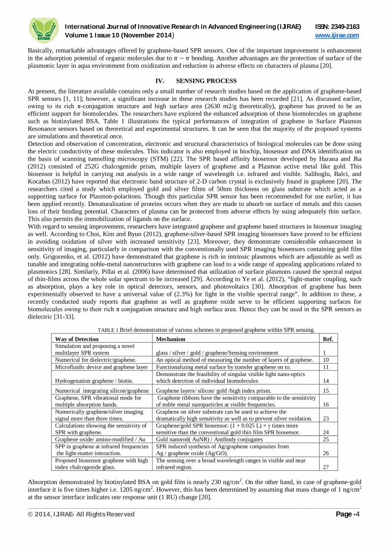
At present, the literature available contains only a small number of research studies based on the application of graphene-based SPR sensors [1, 11]; however, a significant increase in these research studies has been recorded [21]. As discussed earlier, owing to its rich π-conjugation structure and high surface area (2630 m2/g theoretically), graphene has proved to be an efficient support for biomolecules. The researchers have explored the enhanced adsorption of these biomolecules on graphene such as biotinylated BSA. Table 1 illustrations the typical performances of integration of graphene in Surface Plasmon Resonance sensors based on theoretical and experimental structures. It can be seen that the majority of the proposed systems are simulations and theoretical once. Detection and observation of concentration, electronic and structural characteristics of biological molecules can be done using the electric conductivity of these molecules. This indicator is also employed in biochip, biosensor and DNA identification on the basis of scanning tunnelling microscopy (STM) [22]. The SPR based affinity biosensor developed by Harana and Jha (2012) consisted of 252G chalcogenide prism, multiple layers of graphene and a Plasmon active metal like gold. This biosensor is helpful in carrying out analysis in a wide range of wavelength i.e. infrared and visible. Salihoglu, Balci, and Kocabas (2012) have reported that electronic band structure of 2-D carbon crystal is exclusively found in graphene [20]. The researchers cited a study which employed gold and silver films of 50nm thickness on glass substrate which acted as a supporting surface for Plasmon-polaritons. Though this particular SPR sensor has been recommended for use earlier, it has been applied recently. Denaturalization of proteins occurs when they are made to absorb on surface of metals and this causes loss of their binding potential. Characters of plasma can be protected from adverse effects by suing adequately thin surface. This also permits the immobilization of ligands on the surface. With regard to sensing improvement, researchers have integrated graphene and graphene based structures in biosensor imaging as well. According to Choi, Kim and Byun (2012), graphene-silver-based SPR imaging biosensors have proved to be efficient in avoiding oxidation of silver with increased sensitivity [23]. Moreover, they demonstrate considerable enhancement in sensitivity of imaging, particularly in comparison with the conventionally used SPR imaging biosensors containing gold film only. Grigorenko, et al. (2012) have demonstrated that graphene is rich in intrinsic plasmons which are adjustable as well as tunable and integrating noble-metal nanostructures with graphene can lead to a wide range of appealing applications related to plasmonics [28]. Similarly, Pillai et al. (2006) have determined that utilization of surface plasmons caused the spectral output of thin-films across the whole solar spectrum to be increased [29]. According to Ye et al. (2012), “light-matter coupling, such as absorption, plays a key role in optical detectors, sensors, and photovoltaics [30]. Absorption of graphene has been experimentally observed to have a universal value of (2.3%) for light in the visible spectral range”. In addition to these, a recently conducted study reports that graphene as well as graphene oxide serve to be efficient supporting surfaces for biomolecules owing to their rich π conjugation structure and high surface area. Hence they can be used in the SPR sensors as dielectric [31-33].
TABLE 1 Brief demonstration of various schemes in proposed graphene within SPR sensing.
Way of Detection Mechanism Ref. Simulation and proposing a novel multilayer SPR system glass / silver / gold / graphene/Sensing environment 1 Numerical for dielectric/graphene. An optical method of measuring the number of layers of graphene. 10 Microfluidic device and graphene layer Functionalizing metal surface by transfer graphene on to. 11
Hydrogenation graphene / biotin. Demonstrate the feasibility of singular visible light nano-optics which detection of individual biomolecules 14
Numerical integrating silicon/graphene Graphene layers/ silicon/ gold /high index prism. 15 Graphene, SPR vibrational mode for multiple absorption bands.
Graphene ribbons have the sensitivity comparable to the sensitivity of noble metal nanoparticles at visible frequencies. 16
Numerically graphene/silver imaging signal more than three times.
Graphene on silver substrate can be used to achieve the dramatically high sensitivity as well as to prevent silver oxidation. 23
Calculations showing the sensitivity of SPR with graphene.
Graphene/gold SPR biosensor: (1 + 0.025 L) × γ times more sensitive than the conventional gold thin film SPR biosensor. 24
Graphene oxide/ amino-modified / Au Gold nanorod( AuNR) / Antibody conjugates 25 SPP in graphene at infrared frequencies the light-matter interaction.
SPR induced synthesis of Ag/graphene composites from Ag / graphene oxide (Ag/GO). 26
Proposed biosensor graphene with high index chalcogenide glass.
The sensing over a broad wavelength ranges in visible and near infrared region. 27
Absorption demonstrated by biotinylated BSA on gold film is nearly 230 ng/cm2. On the other hand, in case of graphene-gold interface it is five times higher i.e. 1205 ng/cm2. However, this has been determined by assuming that mass change of 1 ng/cm2 at the sensor interface indicates one response unit (1 RU) change [20].
International Journal of Innovative Research in Advanced Engineering (IJIRAE) ISSN: 2349-2163 Volume 1 Issue 10 (November 2014) www.ijirae.com
______________________________________________________________________________________________________ © 2014, IJIRAE- All Rights Reserved Page -5
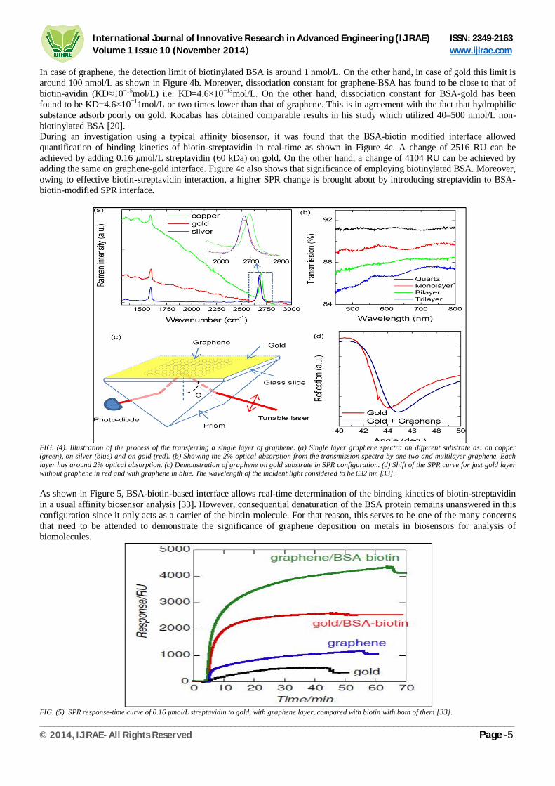
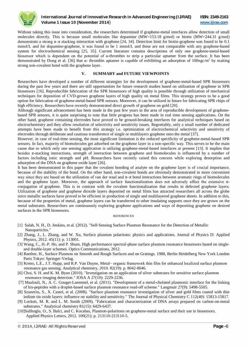
In case of graphene, the detection limit of biotinylated BSA is around 1 nmol/L. On the other hand, in case of gold this limit is around 100 nmol/L as shown in Figure 4b. Moreover, dissociation constant for graphene-BSA has found to be close to that of biotin-avidin (KD≈10−15mol/L) i.e. KD=4.6×10⁻¹³mol/L. On the other hand, dissociation constant for BSA-gold has been found to be KD=4.6×10⁻¹¹mol/L or two times lower than that of graphene. This is in agreement with the fact that hydrophilic substance adsorb poorly on gold. Kocabas has obtained comparable results in his study which utilized 40–500 nmol/L non-biotinylated BSA [20]. During an investigation using a typical affinity biosensor, it was found that the BSA-biotin modified interface allowed quantification of binding kinetics of biotin-streptavidin in real-time as shown in Figure 4c. A change of 2516 RU can be achieved by adding 0.16 µmol/L streptavidin (60 kDa) on gold. On the other hand, a change of 4104 RU can be achieved by adding the same on graphene-gold interface. Figure 4c also shows that significance of employing biotinylated BSA. Moreover, owing to effective biotin-streptavidin interaction, a higher SPR change is brought about by introducing streptavidin to BSA-biotin-modified SPR interface.
FIG. (4). Illustration of the process of the transferring a single layer of graphene. (a) Single layer graphene spectra on different substrate as: on copper (green), on silver (blue) and on gold (red). (b) Showing the 2% optical absorption from the transmission spectra by one two and multilayer graphene. Each layer has around 2% optical absorption. (c) Demonstration of graphene on gold substrate in SPR configuration. (d) Shift of the SPR curve for just gold layer without graphene in red and with graphene in blue. The wavelength of the incident light considered to be 632 nm [33]. As shown in Figure 5, BSA-biotin-based interface allows real-time determination of the binding kinetics of biotin-streptavidin in a usual affinity biosensor analysis [33]. However, consequential denaturation of the BSA protein remains unanswered in this configuration since it only acts as a carrier of the biotin molecule. For that reason, this serves to be one of the many concerns that need to be attended to demonstrate the significance of graphene deposition on metals in biosensors for analysis of biomolecules.
FIG. (5). SPR response-time curve of 0.16 µmol/L streptavidin to gold, with graphene layer, compared with biotin with both of them [33].
International Journal of Innovative Research in Advanced Engineering (IJIRAE) ISSN: 2349-2163 Volume 1 Issue 10 (November 2014) www.ijirae.com
______________________________________________________________________________________________________ © 2014, IJIRAE- All Rights Reserved Page -6
Without taking this issue into consideration, the researchers determined if graphene-metal interfaces allow detection of small molecules directly. This is because small molecules like dopamine (MW=153.18 g/mol) or biotin (MW=244.31 g/mol) demonstrate a strong π–π stacking interaction with graphene [24, 34]. Detection limit for biotin-graphene was found to be 0.1 mmol/L and for dopamine-graphene, it was found to be 1 mmol/L and these are not comparable with any graphene-based system for electrochemical sensing [25, 35]. Current literature contains descriptions of only one graphene-metal-based biosensor which is dependent on the potential of α-thrombin to strip a particular aptamer from the surface. It has been demonstrated by Dong et al. [36] that α- thrombin aptamer is capable of exhibiting an adsorption of 100ngc/m2 by making strong non-covalent bond with the graphene layer.
V. SUMMARY and FUTURE VIEWPOINTS Researchers have developed a number of different strategies for the development of graphene-metal-based SPR biosensors during the past few years and there are still opportunities for future research studies based on utilization of graphene in SPR biosensors [16]. Reproducible fabrication of the SPR biosensors of high quality is possible through utilization of mechanical techniques for deposition of CVD-grown graphene layers of high quality on metal films. This strategy proves to be a good option for fabrication of graphene-metal-based SPR sensors. Moreover, it can be utilized in future for fabricating SPR chips of high efficiency. Researchers have recently demonstrated direct growth of graphene on gold [20]. Although significant advancement has been made in the past few years in the area of reproducible development of graphene-based SPR sensors, it is quite surprising to note that little progress has been made in real time sensing applications. On the other hand, graphene containing electrodes have proved to be ground-breaking interfaces for analytical techniques based on electrochemistry and they allow resolution of selectivity and sensitivity issues. Regrettably, only a small number of dedicated attempts have been made to benefit from this strategy i.e. optimization of electrochemical selectivity and sensitivity of electrodes through deliberate and cautious transference of single or multilayers graphene onto the metal [37]. However, in case of real-time sensing, the most significant limitation is the reduced specificity of graphene-metal-based SPR sensors. In fact, majority of biomolecules get adsorbed on the graphene layer in a non-specific way. This serves to be the main cause due to which only one sensing application is utilizing graphene-metal-based interfaces at present [13]. It implies that besides π-stacking interactions, strength of interaction between graphene and biomolecules is influenced by a number of factors including ionic strength and pH. Researchers have recently raised this concern while exploring desorption and adsorption of the DNA on graphene oxide layer [26]. It has been demonstrated in this paper that the covalent bonding of analyte on the graphene layer is of crucial importance, because of the stability of the bond. On the other hand, non-covalent bonds are obviously demonstrated in more convenient way since they are based on the utilization of van der waal and π–π bond interactions between aromatic rings of biomolecules and the graphene layer. Moreover, the approach of surface functionalization does not adversely affect the extensive π-conjugation of graphene. This is in contrast with the covalent functionalization that results in defected graphene layers. Utilization of graphene and graphene dioxide layers deposited on metal films has attracted researchers all across the globe since metallic surfaces have proved to be efficient in production of single and multilayered graphene sheets. In addition to this, because of the properties of metal, graphene layers can be transferred to other insulating supports once they are grown on the metal substrates. Researchers are continuously exploring graphene applications and ways of depositing graphene on desired surfaces in the SPR biosensors.
REFERENCES [1] Salah, N. H., D. Jenkins, et al. (2012). "Self-Sensing Surface Plasmon Resonance for the Detection of Metallic Nanoparticles." [2] Zhang, J., L. Zhang, and W. Xu, Surface plasmon polaritons: physics and applications. Journal of Physics D: Applied
Physics, 2012. 45(11): p. 113001. [3] Wang, C., H.-P. Ho, and P. Shum, High performance spectral-phase surface plasmon resonance biosensors based on single-
and double-layer schemes. Optics Communications, 2012. [4] Raether, H., Surface Plasmon on Smooth and Rough Surfaces and on Gratings. 1988, Berlin Heidelberg New York London
Paris Tokyo: Springer-Verlag. [5] Kreno, L.E., J.T. Hupp, and R.P. Van Duyne, Metal− organic framework thin film for enhanced localized surface plasmon resonance gas sensing. Analytical chemistry, 2010. 82(19): p. 8042-8046. [6] Choi, S. H. and K. M. Byun (2010). "Investigation on an application of silver substrates for sensitive surface plasmon resonance imaging detection." JOSA A 27(10): 2229-2236. [7] Maalouli, N., A. C. Gouget-Laemmel, et al. (2011). "Development of a metal-chelated plasmonic interface for the linking
of his-peptides with a droplet-based surface plasmon resonance read-off scheme." Langmuir 27(9): 5498-5505. [8] Szunerits, S., X. Castel, et al. (2008). "Surface plasmon resonance investigation of silver and gold films coated with thin
indium tin oxide layers: influence on stability and sensitivity." The Journal of Physical Chemistry C 112(40): 15813-15817. [9] Lockett, M. R. and L. M. Smith (2009). "Fabrication and characterization of DNA arrays prepared on carbon-on-metal
substrates." Analytical chemistry 81(15): 6429-6437. [10]Salihoglu, O., S. Balci, and C. Kocabas, Plasmon-polaritons on graphene-metal surface and their use in biosensors. Applied Physics Letters, 2012. 100(21): p. 213110-213110-5.
International Journal of Innovative Research in Advanced Engineering (IJIRAE) ISSN: 2349-2163 Volume 1 Issue 10 (November 2014) www.ijirae.com
______________________________________________________________________________________________________ © 2014, IJIRAE- All Rights Reserved Page -7
[11]Choi, S. H., Y. L. Kim, et al. (2011). "Graphene-on-silver substrates for sensitive surface plasmon resonance imaging biosensors." Optics express 19(2): 458-466. [12]Song, B., D. Li, et al. (2010). "Graphene on Au (111): A Highly Conductive Material with Excellent Adsorption Properties for High Resolution Bio/Nanodetection and Identification." ChemPhysChem 11(3): 585-589. [13]Szunerits, S., N. Maalouli, et al. (2013). "Recent advances in the development of graphene-based surface plasmon resonance (SPR) interfaces." Analytical and bioanalytical chemistry 405(5): 1435-1443. [14] Kravets, V., et al., Singular phase nano-optics in plasmonic metamaterials for label-free single-molecule detection. Nature materials, 2013. 12(4): p. 304-309. [15] Verma, R., B.D. Gupta, and R. Jha, Sensitivity enhancement of a surface plasmon resonance based biomolecules sensor using graphene and silicon layers. Sensors and Actuators B: Chemical, 2011. 160(1): p. 623-631. [16] Vasic, B., G. Isic, et al. (2013). "Localized surface plasmon resonances in graphene ribbon arrays for sensing of dielectric environment at infrared frequencies." Journal of Applied Physics 113(1): 013110-013110-013117. [17] Ye, Q., et al., Polarization-dependent optical absorption of graphene under total internal reflection. Applied Physics Letters, 2013. 102(2): p. 021912-021912-4. [18] Poland, C.A., et al., Carbon nanotubes introduced into the abdominal cavity of mice show asbestos-like pathogenicity in a pilot study. Nature nanotechnology, 2008. 3(7): p. 423-428. [19] Voloshina, EN and Dedkov, S. (2012) Graphene on Metallic Surfaces: Problems and Perspectives. [20] Salihoglu, O., S. Balci, et al. (2012). "Plasmon-polaritons on graphene-metal surface and their use in biosensors." Applied Physics Letters 100(21): 213110-213110-213115. [21] Guo, S. and S. Dong (2011). "Graphene and its derivative-based sensing materials for analytical devices." Journal of Materials Chemistry 21(46): 18503-18516. [22] Zhang, J. et al (2012) Surface plasmon polaritons: physics and applications. J. Phys. D: Appl. Phys. 45 (2012) 113001. [23] Choi, SH, Kim, YL, and Byun, KM (2012) Graphene-on-silver substrates for sensitive surface plasmon resonance imaging biosensors. 17 January 2011 / Vol. 19, No. 2 / OPTICS EXPRESS. [24] Grigorenko, AN, Polini, M., and Novoselov, KS (2012) Graphene Plasmonics. Focus. 31 Oct 2012. [25] Pillai, S. (2007) Surface plasmon enhanced silicon solar cells. JOURNAL OF APPLIED PHYSICS 101, 093105 [26] Ye, Q. et al (2013) Polarization-dependent optical absorption of graphene under total internal reflection. APPLIED PHYSICS LETTERS 102, 021912 (2013). [27] Dong, B., et al., Large tunable optical absorption of CVD graphene under total internal reflection by strain engineering. Nanotechnology, 2014. 25(45): p. 455707. [28] Wang, J. and H. S. Zhou (2008). "Aptamer-based Au nanoparticles-enhanced surface plasmon resonance detection of small molecules." Analytical chemistry 80(18): 7174-7178. [29] Szunerits, S. (2013) Recent advances in the development of graphene-based surface plasmon resonance (SPR) interfaces. Annal Bioanal Chem (2013) 405:1435-1443. [30] Wu, L., et al., Highly sensitive graphene biosensors based on surface plasmon resonance. Optics express, 2010. 18(14): p. 14395-14400. [31] Voloshina, E. and Y. Dedkov, Graphene on metallic surfaces: problems and perspectives. Physical Chemistry Chemical Physics, 2012. 14(39): p. 13502-13514. [32] Zhang, J., et al., A novel surface plasmon resonance biosensor based on graphene oxide decorated with gold nanorod– antibody conjugates for determination of transferrin. Biosensors and Bioelectronics, 2013. 45: p. 230-236. [33] Voloshina, E.N., et al., Electronic structure and imaging contrast of graphene moire on metals. Scientific reports, 2013. 3. [34] Guo, S. and S. Dong, Graphene and its derivative-based sensing materials for analytical devices. Journal of Materials Chemistry, 2011. 21(46): p. 18503-18516. [35] Bhatt, N., P.-J. J. Huang, et al. (2011). "Dissociation and degradation of thiol-modified DNA on gold nanoparticles in aqueous and organic solvents." Langmuir 27(10): 6132-6137. [36] Fengjun, Z., et al., Surface plasmon resonance induced reduction of high quality Ag/graphene composite at water/toluene phase for reduction of H 2 O 2. 2013. [37] Maharana, P.K. and R. Jha, Chalcogenide prism and graphene multilayer based surface plasmon resonance affinity biosensor for high performance. Sensors and Actuators B: Chemical, 2012.
Related Documents