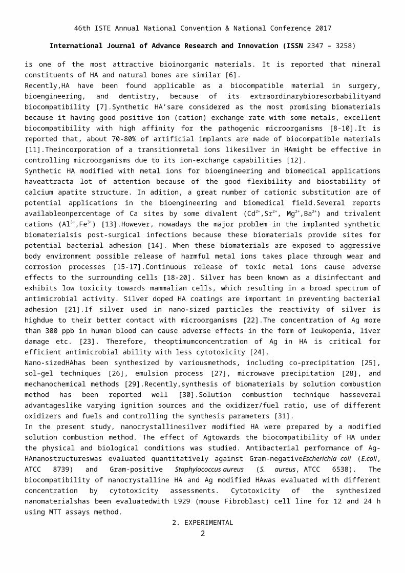
46th ISTE Annual National Convention & National Conference 2017 International Journal of Advance Research and Innovation (ISSN 2347 – 3258) Bactericidal Activity and Biostability Assessment of Silver Containing Hydroxyapatite Nanostructures 1 Sneha S. Bandgar a , 2 Rajaram R Lohar b , 3 Ramesh D Sul b , 4 Tanaji V. Kolekar b* 1 Department of Chemistry, LalBahadurShastri College, Satara, 415002,(MH),India. 2,3 Department of Sciences and Humanities,RajaramBapu institute of Technology,Sakharale,Islampur,416415, (MH),India. 4 Rajaram Bapu institute of Technology, Sakharale, Islampur, 416415, (MH), India 4 Email:[email protected] ABSTRACT: In the present study, silver-modified hydroxyapatite (0.0, 0.5, 1.0, 1.5, 2.0, 2.5 mole %) nanoparticles were synthesized via combustion method using polyvinyl alcohol (PVA) as a fuel. Synthesized powder was studied by thermogravimetric analysis (TGA),X-ray diffraction (XRD), scanning electron microscopy (SEM), and transmission electron microscopy (TEM) techniques. The in-vitro biocompatibility of silver substituted hydroxyapatite (Ag-HA)was studied. The biocompatibility of the Ag-HA nanoparticles was studied using L929 cell lines with MTT assays upto 24 h. MTT assay shows Ag-HA nanoparticles have non-toxic interaction between L929 cell lines and the Ag-HA, found tobe biocompatible for further applications in-vivosystems. According to the obtained results, the synthesized nanocomposite powder confirms biocompatibility of Ag-HA and could be an attractive candidate for biomedical applications. Keywords: Ag-Hydroxyapatite, MTT assay, L929 cells,biomedical applications. 1. INTRODUCTION Scientists and engineers are actively engaged in developing nanocrystallinecalcium orthophosphate to enhance their biological and mechanical properties for biomedical applications. Due to the biocompatibilityof inorganic bioceramic materials based on calcium orthophosphate have a large number of biomedical applications [1].Bioresorbableand bioactive phases of calcium phosphate bioceramic materials are thechoicesfor bone-tissue engineering application because of their similar inorganic composition with the mineral phases of natural bone, excellent biocompatibility and osteoconductivity [2,3]. The first report came in 1920onthesuccessful implication of calcium phosphate in human beings [4].Calcium orthophosphates (CaP) are one of the most widely used bioresorbable and bioactive ceramics. If the Ca/P molar ratio is between 0.5 and 2.0. Calcium orthophosphates (CaP) are known as non-ionssubstituted calcium orthophosphates and it gives organs stability and hardness of the tendons of mammals [5].Among different forms of calcium phosphate, non-ions substituted hydroxyapatite(HA) 1

Welcome message from author
This document is posted to help you gain knowledge. Please leave a comment to let me know what you think about it! Share it to your friends and learn new things together.
Transcript

46th ISTE Annual National Convention & National Conference 2017
International Journal of Advance Research and Innovation (ISSN 2347 – 3258)
Bactericidal Activity and Biostability Assessment of Silver Containing Hydroxyapatite Nanostructures
1Sneha S. Bandgara,2Rajaram R Loharb, 3Ramesh D Sulb, 4Tanaji V. Kolekarb*
1Department of Chemistry, LalBahadurShastri College, Satara, 415002,(MH),India.2,3Department of Sciences and Humanities,RajaramBapu institute of Technology,Sakharale,Islampur,416415, (MH),India.
4Rajaram Bapu institute of Technology, Sakharale, Islampur, 416415, (MH), India4Email:[email protected]
ABSTRACT:In the present study, silver-modified hydroxyapatite (0.0, 0.5, 1.0, 1.5, 2.0, 2.5 mole %) nanoparticles were synthesized via combustion method using polyvinyl alcohol (PVA) as a fuel. Synthesized powder was studied by thermogravimetric analysis (TGA),X-ray diffraction (XRD), scanning electron microscopy (SEM), and transmission electron microscopy (TEM) techniques. The in-vitro biocompatibility of silver substituted hydroxyapatite (Ag-HA)was studied. The biocompatibility of the Ag-HA nanoparticles was studied using L929 cell lines with MTT assays upto 24 h. MTT assay shows Ag-HA nanoparticles have non-toxic interaction between L929 cell lines and the Ag-HA, found tobe biocompatible for further applications in-vivosystems. According to the obtained results, the synthesized nanocomposite powder confirms biocompatibility of Ag-HA and could be an attractive candidate for biomedical applications. Keywords: Ag-Hydroxyapatite, MTT assay, L929 cells,biomedical applications.
1. INTRODUCTIONScientists and engineers are actively engaged in developing nanocrystallinecalcium orthophosphate to enhance their biological and mechanical properties for biomedical applications. Due to the biocompatibilityof inorganic bioceramic materials based on calcium orthophosphate have a large number of biomedical applications [1].Bioresorbableand bioactive phases of calcium phosphate bioceramic materials are thechoicesfor bone-tissue engineering application because of their similar inorganic composition with the mineral phases of natural bone, excellent biocompatibility and osteoconductivity [2,3]. The first report came in 1920onthesuccessful implication of calcium phosphate in human beings [4].Calcium orthophosphates (CaP) are one of the most widely used bioresorbable and bioactive ceramics. If the Ca/P molar ratio is between 0.5 and 2.0. Calcium orthophosphates (CaP) are known as non-ionssubstituted calcium orthophosphates and it gives organs stability and hardness of the tendons of mammals [5].Among different forms of calcium phosphate, non-ions substituted hydroxyapatite(HA) is one of the most attractive bioinorganic materials. It is reported that mineral constituents of HA and natural bones are similar [6].Recently,HA have been found applicable as a biocompatible material in surgery, bioengineering, and dentistry, because of its extraordinarybioresorbabilityand biocompatibility [7].Synthetic HA’sare considered as the most promising biomaterials because it having good positive ion (cation) exchange rate with some metals, excellent biocompatibility with high affinity for the pathogenic microorganisms [8-10].It is reported that, about 70-80% of artificial implants are made of biocompatible materials [11].Theincorporation of a transitionmetal ions likesilver in HAmight be effective in controlling microorganisms due to its ion-exchange capabilities [12].Synthetic HA modified with metal ions for bioengineering and biomedical applications haveattracta lot of attention because of the good flexibility and biostability of calcium apatite structure. In adition, a great number of cationic substitution are of potential applications in the bioengineering and biomedical field.Several reports availableonpercentage of Ca sites by some divalent (Cd2+,Sr2+, Mg2+,Ba2+) and trivalent cations (Al3+,Fe3+) [13].However, nowadays the major problem in the implanted synthetic biomaterialsis post-surgical infections because these biomaterials provide sites for potential bacterial adhesion [14]. When these biomaterials are exposed to aggressive body environment possible release of harmful metal ions takes place through wear and corrosion processes [15-17].Continuous release of toxic metal ions cause adverse effects to the surrounding cells [18-20]. Silver has been known as a disinfectant and exhibits low toxicity towards mammalian cells, which resulting in a broad spectrum of antimicrobial activity. Silver doped HA coatings are important in preventing bacterial adhesion [21].If silver used in nano-sized particles the reactivity of silver is highdue to their better contact with microorganisms [22].The concentration of Ag more than 300 ppb in human blood can cause adverse effects in the form of leukopenia, liver damage etc. [23]. Therefore, theoptimumconcentration of Ag in HA is critical for efficient antimicrobial ability with less cytotoxicity [24].Nano-sizedHAhas been synthesized by variousmethods, including co-precipitation [25], sol–gel techniques [26], emulsion process [27], microwave precipitation [28], and mechanochemical methods [29].Recently,synthesis of biomaterials by solution
1
46th ISTE Annual National Convention & National Conference 2017
International Journal of Advance Research and Innovation (ISSN 2347 – 3258)
combustion method has been reported well [30].Solution combustion technique hasseveral advantageslike varying ignition sources and the oxidizer/fuel ratio, use of different oxidizers and fuels and controlling the synthesis parameters [31].In the present study, nanocrystallinesilver modified HA were prepared by a modified solution combustion method. The effect of Agtowards the biocompatibility of HA under the physical and biological conditions was studied. Antibacterial performance of Ag-HAnanostructureswas evaluated quantitatively against Gram-negativeEscherichia coli (E.coli, ATCC 8739) and Gram-positive Staphylococcus aureus (S. aureus, ATCC 6538). The biocompatibility of nanocrystalline HA and Ag modified HAwas evaluated with different concentration by cytotoxicity assessments. Cytotoxicity of the synthesized nanomaterialshas been evaluatedwith L929 (mouse Fibroblast) cell line for 12 and 24 h using MTT assays method.
2. EXPERIMENTAL2.1 MaterialsSilver nitrate (AgNO3) was purchased from Thomas Baker. Calcium nitrate tetrahydrate (Ca(NO3)2·4H2O), and di-Ammonium hydrogen orthophosphate (NH4)2HPO4 were purchased from Sigma-Aldrich. All chemicals used without any purification and all are analytical grade.2.2 Synthesis of Ag-doped hydroxyapatiteAg modifiedhydroxyapatite (Ag-HA) were prepared by a modified solution combustion method with different concentration (0.0, 0.5, 1.0, 1.5, 2.0, 2.5 mole %).43The stoichiometric amount of the calcium precursor Ca(NO3)2.4H2O, silver precursor AgNO3and phosphate precursor (NH4)2HPO4 were dissolved in double distilled water to get final concentration of solution 0.1 M. Polyvinyl alcohol (PVA) was used as a fuel. In double distilled water the equimolar solution of PVA was prepared. The equimolar mixture of fuel and oxidants was stirred magnetically for 30 minat room temperature.After evaporation this mixtureform a gel of precursors at 100C and then this gel was heated at 300C to obtain a black coloredpowder. The obtained powder of Ag-HA was then annealed at 950 C for 6 h. The dried mixture can be ignited to start combustion reaction using muffle furnace. Various Ag-doped hydroxyapatite samples containing Ag content 0.0, 0.5, 1.0, 1.5, 2.0, 2.5 mole % were denoted as HA, Ag-HA-1, Ag-HA-2, Ag-HA-3, Ag-HA-4, Ag-HA-5, respectively. 2.3 CHARACTERIZATION2.3.1 Structural and morphological studiesStructural analysis of Ag-HA were studied by X-ray diffraction (Philip-3710) with Cu-Kα radiation. X-ray diffraction patterns were analyzed by using an X-pert high score plus software program and obtainedXRD patterns were then compared with standards established by the Joint Committee on Powder Diffraction and Standards (JCDPS) to get correct phase structure . Thermogravimetric analysis (TGA) is a used for thermal analysis in which changes in chemical and physical properties of materials are measured with constant heating rate as a function of increasing temperature. Particle size and morphology was observed by using a transmission electron microscope (TEM, JEOLJEM-2100) with resolution of 2.4 Å.The elemental analysis was carried out by energy dispersive spectroscopy (EDS, JEOL JSM 6360). 2.4 BIOCOMPATIBILITY STUDY2.4.1. Antibacterial activity assayThe antibacterial activity test of HA, Ag-HA-1, Ag-HA-2, Ag-HA-3, Ag-HA-4, and Ag-HA-5 with the concentration of 0.2, 0.4, 0.6, 0.8, 1.0 ppm, respectivelywas carried by bacteriological plate counting methods. The antimicrobial test was performed against two bacterial strains Gram-negativeEscherichia coli (ATCC 8739) and Gram-positive Staphylococcus aureus (ATCC 6538). The bacterial strains were purchased from National Chemical laboratory, Pune, India. Escherichia coli and Staphylococcus aureus were cultured in liquid nutrient broth medium at 35-37 °C for 12 h and concentration adjusted to about 107 CFU/mL.All glassware’swere sterilizedfor 20 min in an autoclave at 121 °C. 0.5 g of each sterile sample was dispersed in a centrifugal tube containing buffered sodium chloride-peptone (BECP, 9 mL) with bacteria suspension (1 mL) and incubated at 30-35 °C for 4, 8, 12, 16, 32, 48, and 72 h. For subsequent bacteria counting, 100 μL of thesuspension was extracted from thecentrifugal tube and inoculated into solid nutrient agar medium followed by 24 h incubation at 35 °C and then the bacterial colonies were counted. 2.4.2. Cell cultureCytotoxicity of Ag-HA samples was carried outon L929 (mouse fibroblast)out by using MTT assay.The L929 Cell lines were obtained from National Centre for Cell Sciences, Pune, India.In-vitro cytotoxicityassay was carried out at National Toxicology Centre,(NTC) Pune, India (ISO 10993/USP 32 NF 27). The L929 (mouse fibroblast) cells were cultured in Dulbecco’s Modified Eagle Medium (DMEM) containing with 10% fetal bovine serum (0.1 mg.mL -1), sodium bicarbonate (1.5 mg.mL-1),and penicillin G (100 U.mL-1) at 37 C in an atmosphere containing 5% CO2. In-vitro cytotoxicityof AG-HA was carried out by MTT assays.2.4.2. MTT assay The MTT assay was used to determine the in-vitro toxicity of the nanomaterials [33]. The L929 cells (2 × 10⁵ cells.mL-1 ) were incubated for 24 h in a 96-well microtiter plate in the respective medium. After 24 h, the old media was replaced by fresh media
2

46th ISTE Annual National Convention & National Conference 2017
International Journal of Advance Research and Innovation (ISSN 2347 – 3258)
and different concentration of sterile Ag-HA (0.0,0.2, 0.4, 0.6, 0.8 and 1 mg.mL -1 of culture media). Then the total medium was incubated in a 5% CO2 atmosphere at 37 C for 12 and 24 h. After 12 and 24 h, 10 µL(5 mg.mL-1) MTT solution was added into each well including control wells and the plates were incubated for 3 h in a 5% CO2 atmosphere at 37 C for metabolization of MTT assay with the Ag-HA nanoparticles and cell media. In the cell wells only anchored cells remained when the total old medium was removed by flicking the plates. Then the cells were washed with phosphate buffer saline (PBS) and the formazan formed was extracted in 200 µL acidic isopropanol in each well and after 1 h, absorbance was recorded at 570 nm.The cell viability was estimated from recorded absorbance. The experiments were repeated three times and the mean data were taken for graph. The relative cell viability (%) compared with control well containing cells without nanoparticles are calculated by the equation:Relative cell viability (%) = [Aabsorbance]tested/ [Aabsorbance]control × 100 (1)
3. RESULTS3.1 Thermogravimetric analysisThermogravimetric analysis (TGA) curves of pure HA and the representative Ag-HA-5 are depicted in Fig.1.Total five different stages were observed during the total weight loss. In between room temperature to 250 C,the first stage of weight loss was observed due to loss of physically adsorbed water. Due to loss of organic groups, chemisorbed water and attached solvent to the sample, the second stage of weight loss is occurs at 250 to 450 C followed by the third stage from 450 to 650 C. The fourth stage is from 650 to 870 C corresponds to decomposition of carbonate into CO2. The fifth stage from 870C onwards shows weight loss less than 0.65%.Therefore, all Ag-HA samples are calcinated at 950 C for further characterization and studies.3.2. XRD analysisThe XRD patterns of pure HA and Ag-HA samples were shown in Fig.2.Compared with pure HA, no new peaks were detected for Ag-HA samples even the doping concentration increased (0.0, 0.5, 1.0, 1.5, 2.0, 2.5 mole %).The XRD characteristic peaks of hydroxyapatite for each sample could be assigned to standard one (JPCDS no. 00-024-0033). No diffraction peaks of elemental silver or silver phosphate were detectable. This revels that the silver is not exists as a separate phase with sufficient crystallinity. However, the crystallinity of HA changes with increasing Ag content due to the formation of silver phosphate (JCPDS no. 01-084-0192).HA showing space group P63/m with a hexagonal structure.Scherrer equation were used to calculate crystallite size (D) from the Gaussian fit of the most intense peak (211).
(2)
Where, λ isthe wavelength of Cu-Kα radiations (λ = 1.5405 Å), β is full width at half maxima of the most intense peak (211),θ is the corresponding Braggs diffraction angle andD is the crystallite size. The average crystallite sizes of HA and Ag-HA were found to be 28.50 nm and 22.12 nm, respectively. The reflection peaks are quite broad, suggesting their nanocrystallineand pure nature. XRD of Ag-HA shows similar peaks with previously reported work [34].3.3 SEM analysisSEM images of all Ag-HA samples are shown in Fig. 3.SEMimages of Ag-HA samplesshows agglomerates that are consisting of small size and fine crystallites. In bone fracture crystal size distribution of bone material plays an important role [35]. To estimate elemental composition of Ag-HA samples EDS analysis wascarried out. Fig.4 shows EDS spectra of all Ag-HA samples. The EDS spectra show expected stoichiometry and elemental signals indicate that samples are consistent and pure. The peaks corresponds to silver (Ag), calcium (Ca), phosphor (P), and oxygen (O) were observed in Ag-HA samples while the peaks corresponds to oxygen (O),calcium (Ca) and phosphor (P) were detected for pure HA sample. The elemental composition of Ag-HAis tabulated in Table 1. Obtained Atomic ratios suggest that samples are stoichiometric.The obtained Ca/P atomic ratios for pure nanocrystalline HA is 1.66 which is close to standard (Ca/P =1.67). The Ca/P atomic ratios (1.51, 1.48, 1.46.1.45, 1.42) for Ag-HA samples are almost equalsto the standard. It can be seen that there is a slightly decrease in Ca/P atomic ratio in the Ag-HA samples. The obtained results reveal that proper substitution of Ca with Ag metal ions during the synthesis process. TheCa/P atomic ratios between 1.33 - 1.55 could be very beneficial to the formation of new bone in-vivo[36]. Thus, in agreement with previous reports, Ag- HA could be good candidature for the formation of new bone in vivo [36].3.4 TEM studies. Fig. 5 displays TEM images of HA and the representative Ag-HA-5 sample. Pure HA nanoparticles were a rod-like shape and around 100 nm in diameter with homogeneous microstructure, most particles seem to be slightly aggregated. Hence particle sizes of Ag-HA-5 about 100 nm and decreased significantly with the existence of Ag element. Crystal sizes obtained from XRD are slightly smaller than observed crystal sizes it’s because of high-temperaturecalcination (950 °C) which cause the grain size growth and due to the presence of nanocrystalline surface layers. The inset Fig. 7(a) and (b) showscorresponding SAEDpatterns of HA and Ag-HA5 samples. Presence of bright ring patterns confirms polycrystalline nature of Ag-nanoparticles.
3

46th ISTE Annual National Convention & National Conference 2017
International Journal of Advance Research and Innovation (ISSN 2347 – 3258)
3.5 Antimicrobial activity Antimicrobial activities of the Ag-HA samples were tested for both microorganisms that can cause implant infections. To evaluate antibacterial effect, both of the bacteria were cultivated with different samples for a series of time.According to the bacteria counting method bacteria re-cultivated on agar. The percentage bacterial survival forE. coli and S. aureus as a function of time are shown in Fig. 6 and Fig.7, respctively.The growth of E. colibacteria reductionfor Ag-HA samples exhibits time-dependent and dose-concentration features. The growth of E. coli reduction for Ag-HA (0.05 ppm) are increased from 40% to 63% when the incubation time extended from 2.5 h to 15 h. Moreover, the antimicrobial activitywas increased from 70% to 98 % after 15 h incubation with the increasing Ag doping concentration from 0.05 ppm to 190 ppm.Antimicrobial results reveals that the antimicrobials rates are 85% and 93% at 0.30 ppm and 2.3 ppm Ag-HAnanocrystalscome incontact with bacteria. The maximum concentration that exhibits effective antibacterial activity is 2.3 ppm. The effective antimicrobial activity of Ag-HA reveals that an appropriate amount of silver doping can also be adopted as an ideal antimicrobial agent, high doping concentration of Agis not necessary to achieve effective antimicrobial activity.The physical and chemical characteristics such as morphology, chemical composition and surface properties of surrounding medium can affect the viability of bacteria [37].The antibacterial activity of Ag-HA demonstrated the release of Ag+ cause structural changes and damage to the membranes [38]. Also, these Ag+will bind to proteins on the membrane [39].It was reported that release Ag+entered into cells and interacts with nucleic acids, causes death of bacteria [40].Thebacteria count reduction for 2.3 ppm and 190 ppm of Ag-HA were 91% and 98%, respectively. This is due to the gathered Ag +
release amount towardsS. aureus.However, the Ag-HA (0.30 ppm) alsounveils 85% bacteria reduction with Ag+ release. This maybe due to the contact antibacterial process that results inS. aureusbacteria deathat the surface of Ag-HA nanoparticles. When the gram positive and gram negative bacteria come in contact with Ag-HA nanoparticles, the bacteria were killed due to the concentration of Ag+ on the surface of Ag-HA nanoparticles.When the Ag concentration was 0.05 ppm accompanied by 63% antibacterial ratio upon further adding the doping concentration2.3 ppm, the antibacterial activity was enhanced unceasingly to 83 %. When Ag concentrationreaches to 190 ppm, 99% of bacteria were killed. The two curves come into an intersection between 0.30 ppm and2.3 ppm of Ag-HA, which exhibited 85% antibacterial activity.The antibacterial properties of Ag were mainly depend on its concentration [41]. Ag-HA-5 shows efficient antibacterial activity against E. coli at low concentration within appropriate time. The antibacterial experiments showsthat the inactivation of bacteria is depends on the concentration of AG-HA along with time. Fig.6 shows the complete inhibition of E. coli bacteria at 7.5 h for AG-HA-5 (190 ppm) and 12.5 h for 2.3 ppm.The similar experiments were performed for the inactivation of S. aureus(Fig.7).After 10 h treatment by AG-HA-5 (190 ppm), almost all cells were destroyed (100 %), whereas the use of AG-HA-5 (2.3 ppm) shows complete inhibition at 15h.These results are in good agreement with previous work by Shi et al. [42].3.6 Cytotoxicity studyThe effect of different concentrations and incubation time of Ag-HA nanoparticles on the cell viability was evaluated against L929 cell lines. Fig. 8 (a-d) shows the obtained data. In vivo L929 cell lines were interact with HA, Ag-HA-1, Ag-HA-3, Ag-HA-5 nanoparticles for 12 h and 24 h, respectively with the concentration of 0.2, 0.4, 0.6, 0.8, and 1 mg.mL−1in a 5% CO2 atmosphere at 37 °C. The relative cell viability (%) compared with a control wellcontaining cells without nanoparticles are calculated by the equation: Relative cell viability (%) = [A]tested/[A]control × 100 (3)As increasing concentration and incubation time of Ag-HA nanoparticles the cell viability gradually decreases of all four Ag-HA samples. Decreasing the grain sizes increasing cell cytotoxicity.As compared to positive control it shows that low extractconcentration of Ag-HA have less cytotoxicity and higher extract concentration accelerateslight toxic effect to L929 cells [43].The obtained results from cytotoxicity experiments reveals better biocompatibility of HA and Ag-HA nanoparticles with a concentration up to 1 mg.mL−1. The cytotoxicity assessment suggest that Ag-HA having very low cytotoxicity towards L929 cell lines.
4. DISCUSSION
The hydroxyapatite is with antibacterial properties are increasingly developed to reduce different gram-positive and gram-negative bacterial infections. Now days HA becomes as alternative to routine antibacterial agent. Forbiomedical applications HA nanoparticles attracted great attention because it’s having ability to kill the wide range of different bacteria and generate reactive oxygen system (ROS). In the present study the attempt is made for the synthesis of Ag doped HA by using modified sol-gel method. The strategies have been used to improve biocompatibility is appropriate amount of Ag doping in HA. XRD analysis.confirms the crystallite sizes and phase purity of the prepared materials (Fig.2). The Ag doped HA Nano powder shows the characteristic peak of Ag is overlapped by most intense peak of HA. To demonstrate its antibacterial applicability, Gram-
4

46th ISTE Annual National Convention & National Conference 2017
International Journal of Advance Research and Innovation (ISSN 2347 – 3258)
positive and Gram-negative bacteria were selected and results are shown in Fig. 6and Fig. 7. This results confirms that complete inhibition of Gram-negativeE. coli and Gram-positive S. aureus was achieved.To inhibition Gram-positive bacteria withAg-HA-5(190 ppm) and Ag-HA-5 (2.3 ppm) required 7h and 12 h, respectively. However,for Gram-negative bacteria it required 10 h and 15 h for Ag-HA-5(190 ppm) and Ag-HA-5 (2.3 ppm), respectively. Gram-negative bacteria is relatively more resistant because of the nature of their cell wall, which avoids absorption of different molecules to movements through the cell membrane. For Gram-positive bacteria, the cell wall made up of peptidoglycan (90%) while for the Gram-negative bacteria, cell wall contains only of the Peptidoglycan (10%). Peptidoglycan layer is very porous in nature which allows nanoparticles of 2 to pass through layer in both Gram positive bacteria and Gram-negative [44]. Peptidoglycan layer pores are enough to pass the oxidative species, hydroxyl radical and likes superoxide due to this the Gram-positive bacterial inhibition take place in short time than Gram-negative bacteria.Ag-HA nanoparticles generates ROS which, causescell perforation and finally leading to cell death [45,46].No major difference in the cytotoxicity of HA and Ag-HA nanoparticles was observed. This is attributed to a relatively small change composition (Ca/P ratio) with difference in the particle size. The cytotoxicity study on L929 cells of HA and Ag-HA nanoparticles synthesized by modified solution combustion technique, here confirmed that the biological effects are not only based on chemical composition but also on size, shape, and surface texture and aggregation state.
CONCLUSIONSModified solution combustion technique has been employed to facile synthesize HA nanoparticles in which in which Ca is partially substituted by Ag. Addition of Ag precursor under controlled condition which leads to Ag-HA with a Ca/P ratio very similar to standard one. The structural, morphological, compositional, cytotoxicity and antimicrobial properties of the HA and Ag-HA nanoparticles with different stoichiometric ratios have been studied in great detail. The physical and chemical properties of the HA have been modifiedby the presence of silver. The HA and Ag-HA nanoparticles having no any secondary phase and almost identical particle sizes. EDS analysis confirmed presence of HA and Ag-HA with theCa/P ratio similar to the standard ratio and this material could be applicable to the formation of new bone in vivo. HA nanoparticles dopedwith Ag possess effective antimicrobial activity.The antibacterial rate increases from 65% to 99% whilethe cell viability decreases from 97% to 75 % when the Ag-doping concentration varies from0.05 to 190 ppm. The optimal propertieswith antibacterial ability and excellent cytocompatibility can be achievedwhenthe Ag-doping concentration is between 0.30 ppmand 2.3 ppm. The antibacterial HAwith Ag is non-toxicity to living cells andtissues even if the particles are internalized by cells. The results of this work demonstrate the applicability of Ag-HA nanoparticles in the biomedical field. Furthermore, the cytotoxicity studies show that the Ag-HA lower cytotoxicity. Hence, it is proved that Ag-HA Nano crystals are potentially applicable as bone substitution materials in biomedical application.Ag-HA is an excellent material in the tissue engineering. The ultra-trace Ag-doped HA nanocrystals may provide new opportunities for anon-cytotoxic implant with antimicrobial ability in tissue engineering.
REFERENCES:[1] A. Bigi, E. Boanini, K. Rubini, Hydroxyapatite gels and nanocrystals prepared through a sol–gel process, J. Solid State
Chem. 177 (2004) 3092–3098.[2] L.L. Hench,Bioceramics J. American ceramic society. 81 (1998) 1705-28.[3] K.D. Groot, C.P.A.T. Klein, J.G.C. Wolke, J.M.A. Blieck-Hogervorst, In: T. Yamamuro, L.L.Hench, J. Wilson,
Handbook of Bioactive Ceramics, Vol. 2, Calcium phosphate and Hydroxyapatite Ceramics, CRC Press, Boca Raton, FL,1990, P-3.
[4] R.Z. LeGeros, International Symposium on New Wave of ceramics for the 21 st Century at the 40th symposium on Basic Science of ceramics, Convention Center, Osaka University, 2002.
[5] Monika Supovan, Substituted hydroxyapatites for biomedical applications: A review, Ceramics International, 41(2015),9203–9231
[6] QiuH J, Yang J, Kodali P, Koh J, Ameer G A. A citric acid based hydroxyapatite composite for orthopaedic implants [J]. Biomaterials, 2006, 27:5845-5854.
[7] Tanaka, Y., Hirata, Y., Yoshinaka, R.,. Synthesis and characteristics of ultrafinehydroxyapatite particles. J. Ceram. Process. Res. 2003,4 (4), 197-201[8] I. Smiciklas, A. Onjia, J. Markovic, S. Raicevic, Comparison of hydroxyapatite sorption properties towards cadmium,
Lead, Zinc and strontium ions , Mater. Sci. forum,494 (2005) 405-410. [9] R.Z. LeGeros, Calcium phosphate-based osteoinductive materials, chem. Rev. 108(2008) 4742-4753.[10] E.D.Berry, G.R. Siragusa, Hydroxyapatite adherence as a means to concentrate bacteria, Appl. Environ. Microbial.
63(1997) 4069-4074.
5

46th ISTE Annual National Convention & National Conference 2017
International Journal of Advance Research and Innovation (ISSN 2347 – 3258)
[11] M. Niinomi, M. Nakai, and J. Hieda, ActaBiomaterialia,Development of new metallic alloys for biomedical applications, 2012, vol. 8, pp. 3888-3903.
[12] Oh, K.S., Kim, K.J., Jeong, Y.K., Choa, Y.H., 2003. Effect of fabrication processes on the antimicrobial properties of silver dopednano-sized Hap. Key Eng. Mater.240-242, 583-586.
[13] M. Wakamura, K. Kandori, T. ishikawa, Colloids surface:A, 164,(2000),297.[14] E. M. Hetrick, M. H. Schoenfisch, Reducing implant-relate dinfections: active release
strategies,Chem.Soc.Rev.35(2006)780–789.[15] U. Türkan, O. Öztürk, and A. E. Eroğlu, Surface and Coatings Technology,Metal ion
release from TiN coated CoCrMo orthopedic implant material, 4/27/ 2006, vol. 200, pp.5020-5027.[16] N. Espallargas, C. Torres, and A. I. Muñoz, Wear,A metal ion release study of Co,Cr,Mo. exposed to corrosion and
tribocorrosion conditions in simulated body fluids,"[17] S.-J. Lee and J.-J. Lai, Journal of Materials Processing Technology,The effects of
electropolishing (EP) process parameters on corrosion resistance of 316L stainless steel,2003,140,206-210.[18] C.-C. Shih, C.-M. Shih, Y.-Y. Su, L. H. J. Su, M.-S. Chang, and S.-J. Lin, Corrosion
Science,"Effect of surface oxide properties on corrosion resistance of 316L stainless steel for biomedical applications," 2// 2004, vol. 46, pp. 427-441.
[19] T. F. Zhang, Q. Y. Deng, B. Liu, B. J. Wu, F. J. Jing, Y. X. Leng, et al., Surface andCoatings Technology,"Wear and Corrosion Properties of diamond like carbon (DLC)Coating on stainless steel, CoCrMo and Ti6Al4V substrate,"
[20] B. Alemón, M. Flores, W. Ramírez, J. C. Huegel, and E. Broitman, TribologyInternational,"Tribocorrosion behavior and ions release of CoCrMo alloy coated with a TiAlVCN/CNx multilayer in simulated body fluid plus bovine serum albumin," 1// 2015,vol. 81, pp. 159-168.
[21] Stanic, V., Janackovic, D., Dimitrijevic, S., Tanaskovic, S.B., Mitrica, M., Pavlovic, M.S.,Krstic, A., Jovanovic, D., Raicevic, S., 2011. Synthesis of antimicrobial monophase silver-doped hydroxyapatite nano powders for bone tissue engineering. Appl.Surf. Sci. 257, 4510-4518.
[22] J. R. Morones, J. L. Elechiguerra, A. Camacho, K. Holt, J. B. Kouri, J.T. Ramirez, M. J. Yacaman, The bactericidal effect of silver nanoparticles, Nanotechnology 16 (2005) 2346–2353.
[24] C. Shi, J. Gao, M. Wang, J. Fu, D. Wang, Y. Zhu, Ultra-trace silver-doped hydroxyapatite with non-cytotoxicity and effective antibacterial activity, Mater. Sci. Eng. C. 55 (2015) 497–505. doi:10.1016/j.msec.2015.05.078.
[25] R. Murugan,S.Ramakrishna, Production of ultra-fine bioresorbable carbonated hydroxyapatite, ActaBiomater.2(2006)201–206.
[26] M.H. Fathi, A.Hanifi, V.Mortazavi, Preparation and bioactivity evaluation of bone-like hydroxyapatite nanopowder, J.Mater.Process. Technol. 202(2008)536–542.
[27] G.C. Koumoulidis, A.P.Katsoulidis, A.K.Ladavos, P.J.Pomonis, C.C. Trapalis, A.T.Sdoukos, T.C.Vaimakis, Preparation of hydroxya- patite via microemulsion route, J.Colloid Interface Sci. 259 (2003) 254–260.
[28] Z. Zou, K.Lin, L.Chen, J.Chang, Ultra fast synthesis and characterization of carbonated hydroxyapatite nanopowders via sonochemistry- assisted micro wave proces, Ultrason.Sonochem.19(2012)1174–1179.
[29] S. Lala, S.Brahmachari, P.K.Das, D.Das, T.Kar, S.K.Pradhan, Biocompatible nanocrystalline natural bone like carbonated hydroxyapa- tite synthesized by mechanical alloying in a record minimum time, Mater. Sci. Eng.C42(2014)647–656.
[30] A.G. Merzhanov, 40 years of SHS: A lucky Star of Scientific Discovery,Bentham Science, e-book, , 2012,112.[31] S.T. Aruna, Solution combustion synthesis – an overview, in: M. Lackner (Ed.),
Combustion Synthesis: Novel Routes to Novel Materials, Bentham Publishers, 2010, 206–221.[32] Chao Shi, Jianyong Gao, MingWang, Jingke Fu, DalinWang , Yingchun Zhu Ultra-trace silver-doped hydroxyapatite
with non-cytotoxicity and effective antibacterial activity Materials Science and Engineering C 55 (2015) 497–505.[33] N.D. Thorat, S. V Otari, R.M. Patil, V.M. Khot, A.I. Prasad, R.S. Ningthoujam, et al., Enhanced colloidal stability of
polymer coated La0.7Sr0.3MnO3 nanoparticles in physiological media for hyperthermia application., Colloids Surf. B. Biointerfaces. 111 (2013) 264–9.
[34] Flavio Augusto Cavadas Andradea, n, Luci Cristinade Oliveira Vercikb, Fernando Jorge Monteiroc, d, Eliana Cristinada Silva Rigoa,Preparation, characterization and antibacterial properties of silver nanoparticles–hydroxyapatite composites by asimple and eco-friendly method, Ceramics International42(2016)2271–2280.
6

46th ISTE Annual National Convention & National Conference 2017
International Journal of Advance Research and Innovation (ISSN 2347 – 3258)
[35] M.J. Phillips, J.A. Darr, Z.B. Luklinska, I. Rehman, Synthesis and characterization of nano-biomaterials with potential osteological applications., J. Mater. Sci. Mater. Med. 14 (2003) 875–82.
[36] S. V. Dorozhkin, A review on the dissolution models of calcium apatites, Prog. Cryst. Growth Charact. Mater. 44 (2002) 45–61.
[37] S. Kang, M. Herzberg, D.F. Rodrigues, M. Elimelech, Antibacterial effects of carbonnanotubes: size does matter, Langmuir 24 (2008) 6409–6413.[38] Z.M. Xiu, Q.B. Zhang, H.L. Puppala, V.L. Colvin, P.J.J. Alvarez, Negligible particle specific antibacterial activity of
silver nanoparticles, Nano Lett. 12 (2012) 4271–4275.[39] W.K. Jung, H.C. Koo, K.W. Kim, S. Shin, S.H. Kim, Y.H. Park, Antibacterial activity and mechanism of action of the
silver ion in Staphylococcus aureus and Escherichia coli,Appl. Environ. Microbiol. 74 (2008) 2171–2178.[40] Q.L. Feng, J.Wu, G.Q. Chen, F.Z. Cui, T.N. Kim, J.O. Kim, A mechanistic study of the antibacterial effect of silver ions
on Escherichia coli and Staphylococcus aureus, J. Biomed. Mater. Res. 52 (2000) 662–668.[41] R.J. Chung,M.F. Hsieh, C.W. Huang, L.H. Perng, H.W. Wen, T.S. Chin, Antimicrobial effects and human gingival
biocompatibility of hydroxyapatite sol–gel coatings, J. Biomed. Mater. Res. 76B (2006) 169–178.[42] Chao shi, Jianyong Gao, Ming Wang, Jingke Fu, Dalin Wang, Yingchun Zhu, Material science and engineering C55(2015),497-50.[43] Tanaji V. Kolekar, Nanasaheb D. Thorat, Hemraj M. Yadav, Veeresh T. Magalad, Mahesh A. Shinde, Sneha S. Bandgar,
Jin H. Kim, Ganesh L. Agawane, Nanocrystalline hydroxyapatite doped with aluminium: A potential carrier for biomedical applications, Ceramic International, 42, 2016, 5304-5311.
[44] Demchick P, Koch a L. The permeability of the wall fabric of Escherichia coli and Bacillus subtilis . The Permeability of the Wall Fabric of Escherichia coli and Bacillus subtilis. J Bacteriol. 1996,178,768–73.
[45] Adams LK, Ã DYL, Ã PJJA. Comparative eco-toxicity of nanoscale TiO 2 , SiO 2 , and ZnO water suspensions.Water Research 2006,40,3527–32.
[46] Tsuang Y-H, Sun J-S, Huang Y-C, Lu C-H, Chang WH-S, Wang C-C. Studies of photokilling of bacteria using titanium dioxide nanoparticles. Artif. Organs. 2008,32,167–74.
Figure Captions:
Fig. 1 TGA pattern of pure HA and Ag-HA-5 nanoparticles.Fig. 2 XRD patterns of pure HA and Ag-HA nanoparticles.Fig. 3 SEM images of (a) pure HA, (b) Ag-HA-1, (c) Ag-HA-2, (d) Ag-HA-3, (e) Ag-HA-4, and (f) Ag-HA-5.Fig. 4 EDS spectrum of (a) pure HA, (b) Ag-HA-1, (c) Ag-HA-2, (d) Ag-HA-3, (e) Ag-HA-4, and (f) Ag-HA-5.Fig. 5 TEM images of (a) HA, (b) Ag-HA-5. (Inset: corresponding SAED pattern).Fig. 6E. colisurvival (%) as a function of time for (a) Ag-HA-1,(b) Ag-HA-3, and (c) Ag-HA-5.Fig. 7S. aureussurvival (%) as a function of time (a) Ag-HA-1,(b) Ag-HA-3, and (c) Ag-HA-5.Fig. 8 Cytotoxicity profiles of (a) pure HA, (b) Ag-HA1, (c) Ag-HA3, (d) Ag-HA5 for 12 and 24 hr.
7

46th ISTE Annual National Convention & National Conference 2017
International Journal of Advance Research and Innovation (ISSN 2347 – 3258)
Fig. 1 TGA pattern of pure HA and Ag-HA-5 nanoparticles.
8

46th ISTE Annual National Convention & National Conference 2017
International Journal of Advance Research and Innovation (ISSN 2347 – 3258)
Fig. 2 XRD patterns of pure HA and Ag-HA nanoparticles.
Fig. 3
9

46th ISTE Annual National Convention & National Conference 2017
International Journal of Advance Research and Innovation (ISSN 2347 – 3258)
Fig. 3 SEM images of (a) pure HA, (b) Ag-HA-1, (c) Ag-HA-2, (d) Ag-HA-3, (e) Ag-HA-4, and (f) Ag-HA-5.
Fig. 4 EDS spectrum of (a) pure HA, (b) Ag-HA-1, (c) Ag-HA-2, (d) Ag-HA-3, (e) Ag-HA-4, and (f) Ag-HA-5.
10

46th ISTE Annual National Convention & National Conference 2017
International Journal of Advance Research and Innovation (ISSN 2347 – 3258)
Fig. 5 TEM images of (a) HA, (b) Ag-HA-5. (Inset: corresponding SAED pattern).
Fig. 6 E. coli survival (%) as a function of time for (a) Ag-HA-1,(b) Ag-HA-3, and (c) Ag-HA-5.
11

46th ISTE Annual National Convention & National Conference 2017
International Journal of Advance Research and Innovation (ISSN 2347 – 3258)
Fig. 7 S. aureus survival (%) as a function of time (a) Ag-HA-1,(b) Ag-HA-3, and (c) Ag-HA-5.
Fig. 8 Cytotoxicity profiles of (a) pure HA, (b) Ag-HA1, (c) Ag-HA3, (d) Ag-HA5 for 12 and 24 hr.
12

46th ISTE Annual National Convention & National Conference 2017
International Journal of Advance Research and Innovation (ISSN 2347 – 3258)
Table caption:Table 1. Elemental composition of HA and Ag-HA nanoparticles.
Sample Atomic % compositionAg Ca P O
HA ---- 32.33 19.52 48.15Ag-HA-1 1.02 28.89 19.03 51.06Ag-HA-2 1.51 28.13 18.97 51.40Ag-HA-3 3.27 26.97 18.40 57.68Ag-HA-4 4.48 23.12 15.93 58.29Ag-HA-5 6.07 21.90 15.33 60.21
13
Related Documents