III. CNS Tumors

III. CNS Tumors. -The majority of CNS tumors (brain and spinal cord are primary) -Only one fourth to one half are metastatic -Tumors of the CNS account.
Dec 24, 2015
Welcome message from author
This document is posted to help you gain knowledge. Please leave a comment to let me know what you think about it! Share it to your friends and learn new things together.
Transcript
III. CNS Tumors

- The majority of CNS tumors (brain and spinal cord are primary)
- Only one fourth to one half are metastatic- Tumors of the CNS account nearly 20% of
all cancers of childhood.

- 70% of all childhood tumors arise infratentorially
- About 70% of all CNS tumors in adults arise supratentorially
- The clinical course of brain tumors is strongly influenced by :
1. Patterns of growth- Some glial tumors with low grade histologic
features may infiltrate large portions of brain and lead to serious clinical deficits and may not be amenable to surgical resection
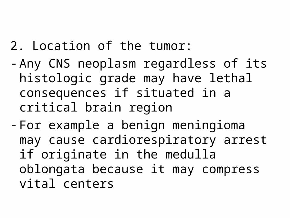
2. Location of the tumor:- Any CNS neoplasm regardless of its histologic
grade may have lethal consequences if situated in a critical brain region
- For example a benign meningioma may cause cardiorespiratory arrest if originate in the medulla oblongata because it may compress vital centers

- The highly malignant gliomas rarely metastasize outside the CNS
- Tumors such as ependymomas and medulloblastomas are able to spread through CSF if they encroach upon the subarachnoid space ; therefore may be associated with implantation along the brain and spinal cord away from the original tumor site

- Treatment protocols of CNS tumors are usually based on WHO classification, which segregates tumors into four grades according to their biologic behavior from grade I to IV

Tumors of he CNS are classified into:I. GliomasII.Neuronal tumorsIII.Poorly differentiated tumorsIV.Germ cell tumorsV.Primary CNS lymphomaVI.Meningiomas

I. Gliomas- Is the most common group of primary brain
tumors- This group include : 1. Astrocytomas, 2. Oligodendrogliomas, 3. Ependymomas
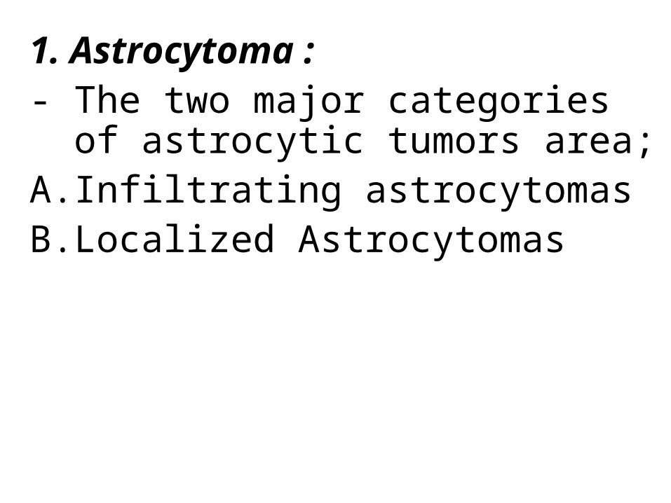
1. Astrocytoma : - The two major categories of astrocytic
tumors area;A. Infiltrating astrocytomasB. Localized Astrocytomas
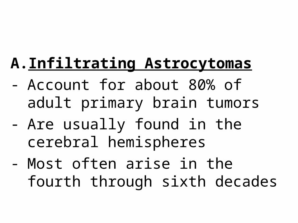
A. Infiltrating Astrocytomas- Account for about 80% of adult primary brain
tumors- Are usually found in the cerebral hemispheres- Most often arise in the fourth through sixth
decades
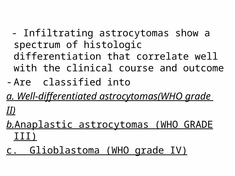
- Infiltrating astrocytomas show a spectrum of histologic differentiation that correlate well with the clinical course and outcome
- Are classified into a. Well-differentiated astrocytomas(WHO grade
II)
b.Anaplastic astrocytomas (WHO GRADE III)c. Glioblastoma (WHO grade IV)

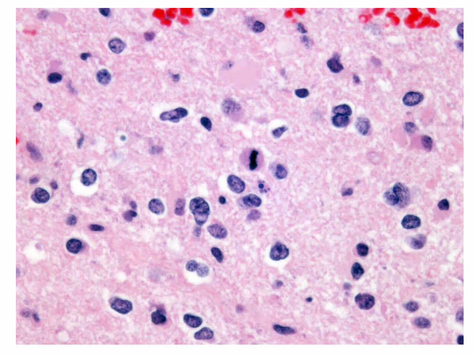
Morphologya. Well-differentiated astrocytomas(WHO grade II)- Have a cellular density that is greater than the
normal white matter- The tumor cells are separated by astrocytic
processes (called fibrillary background)- These astrocytic processes are positive for glial
fibrillary acidic protein immunostain
- Show minimal degree of pleomorphism- The transition between neoplastic and
normal tissue is indistinct, therefore the tumor cells can be seen infiltrating normal tissue at some distance from the main lesion

Glioma: uneven cell distribution


b. Anaplastic astrocytomas (WHO GRADE III) show:
1. Are more densely cellular2. The cells have greater nuclear pleomorphism 3. Increased mitoses are often observed (The
most important feature) that distinguishes this grade from grade II

Anaplastic Astrocytoma
(WHO grade III)
Anaplastic Astrocytoma (WHO grade III)

c. Glioblastoma (WHO grade IV)- It has a histologic appearance similar to
anaplastic astrocytoma with the additional features of:
a. Pseudopalisading necrosis - The neoplastic cells collect along the edges of
the necrotic regionsa. and /or b. Microvasular proliferation

• Radiologically: Shows contrast enhancement
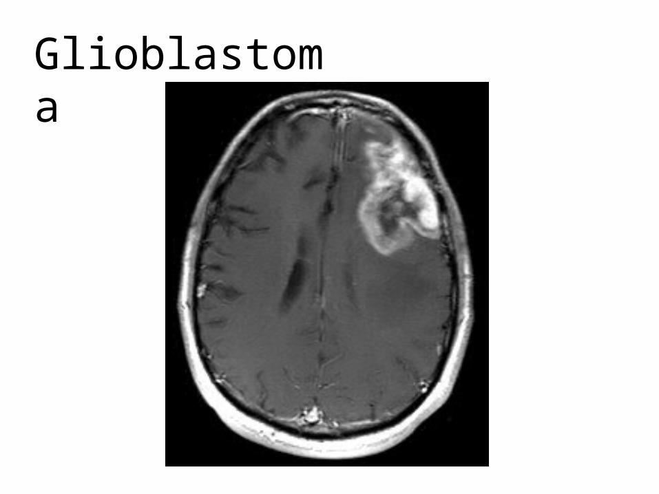
Glioblastoma

Patterns of Contrast EnhancementDemyelinating Pseudotumor
GBM
PCNSL PXA Cavernoma
Abscess
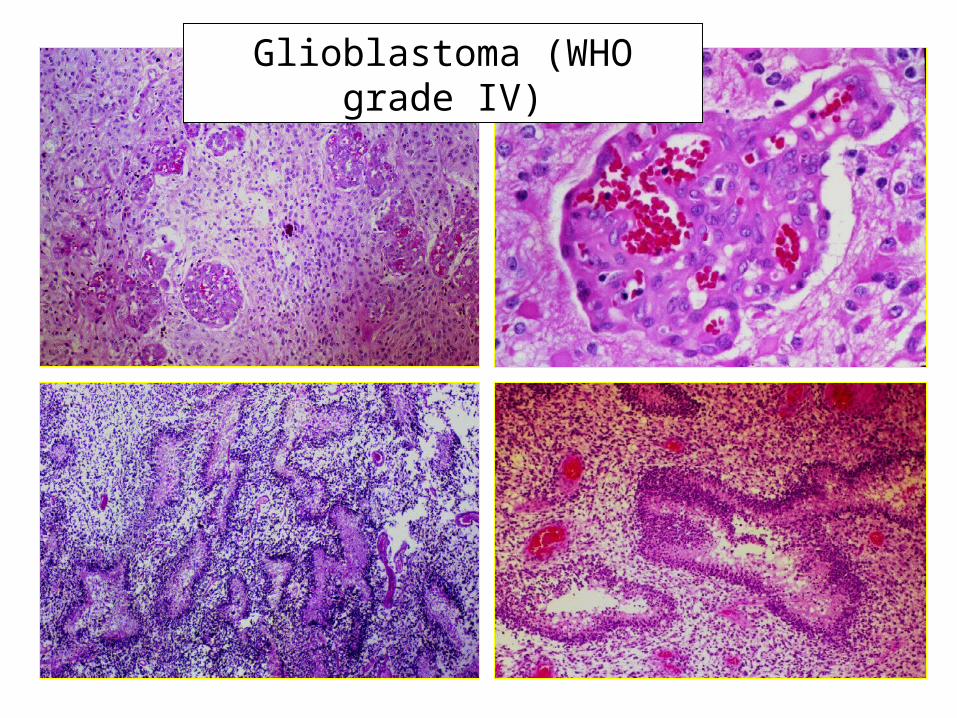
Glioblastoma (WHO grade IV)

Clinical features of astrocytomas:- The most common presenting signs and
symptoms of infiltrating astrocytomas are:i. Seizures,ii. Headaches ii. Focal neurologic deficits related to the anatomic
site of involvement

Well differentiated grade II astrocytoma - May remain stable or progress only slowly
over a number of years- The mean survival rate is more than 5 years- Eventually , clinical deterioration occurs
Glioblastoma
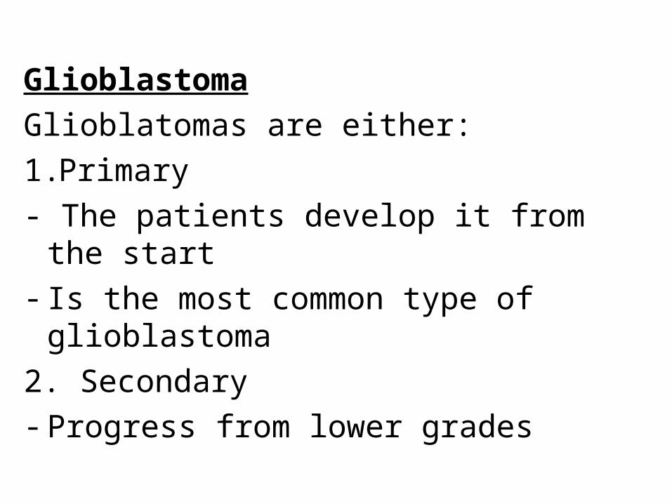
Glioblatomas are either:1.Primary- The patients develop it from the start- Is the most common type of glioblastoma2. Secondary- Progress from lower grades
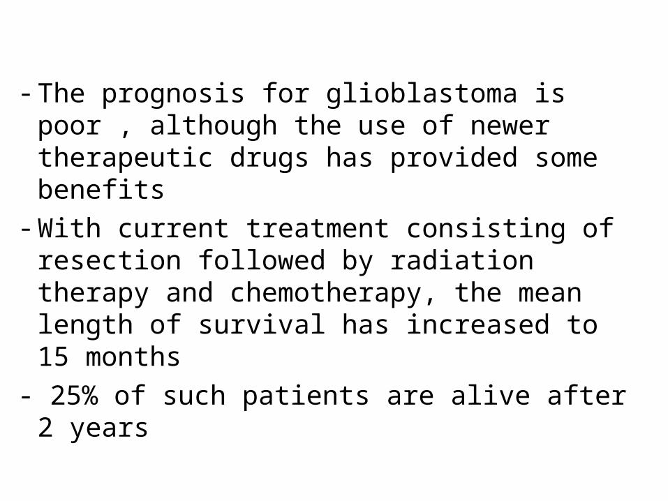
- The prognosis for glioblastoma is poor , although the use of newer therapeutic drugs has provided some benefits
- With current treatment consisting of resection followed by radiation therapy and chemotherapy, the mean length of survival has increased to 15 months
- 25% of such patients are alive after 2 years

• Genetic Changes in astrocytomas
1. In low grade astrocytomas(grade II)- Mutations that alter the enzymatic activity of
two isoforms of the metabolic enzyme isocitrate dehydrogenases(IDH1 and IDH2)

2. In glioblastomas - Loss of function mutation in p53 and Rb
tumor suppressor pathway
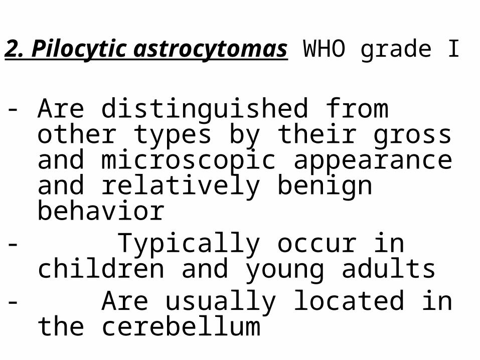
2. Pilocytic astrocytomas WHO grade I
- Are distinguished from other types by their gross and microscopic appearance and relatively benign behavior
- Typically occur in children and young adults
- Are usually located in the cerebellum
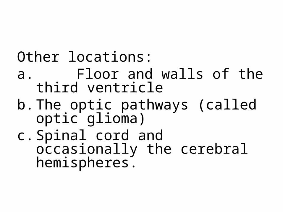
Other locations:a. Floor and walls of the third ventricleb. The optic pathways (called optic glioma) c. Spinal cord and occasionally the cerebral
hemispheres.
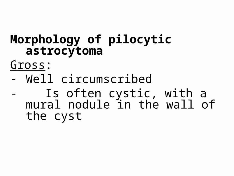
Morphology of pilocytic astrocytomaGross:- Well circumscribed- Is often cystic, with a mural nodule in the wall
of the cyst

Pilocytic astrocytoma

Pilocytic astrocytoma

Microscopically, - The tumor is composed ofa. Hypercellular areas composed of
bipolar astrocytes with with long, thin "hairlike " processes that are GFAP positive
b. Hypocellular areas formed of microcysts.
c. Rosenthal fibers d. No necrosis or mitoses
Clinical features of pilocytic astrocytoma
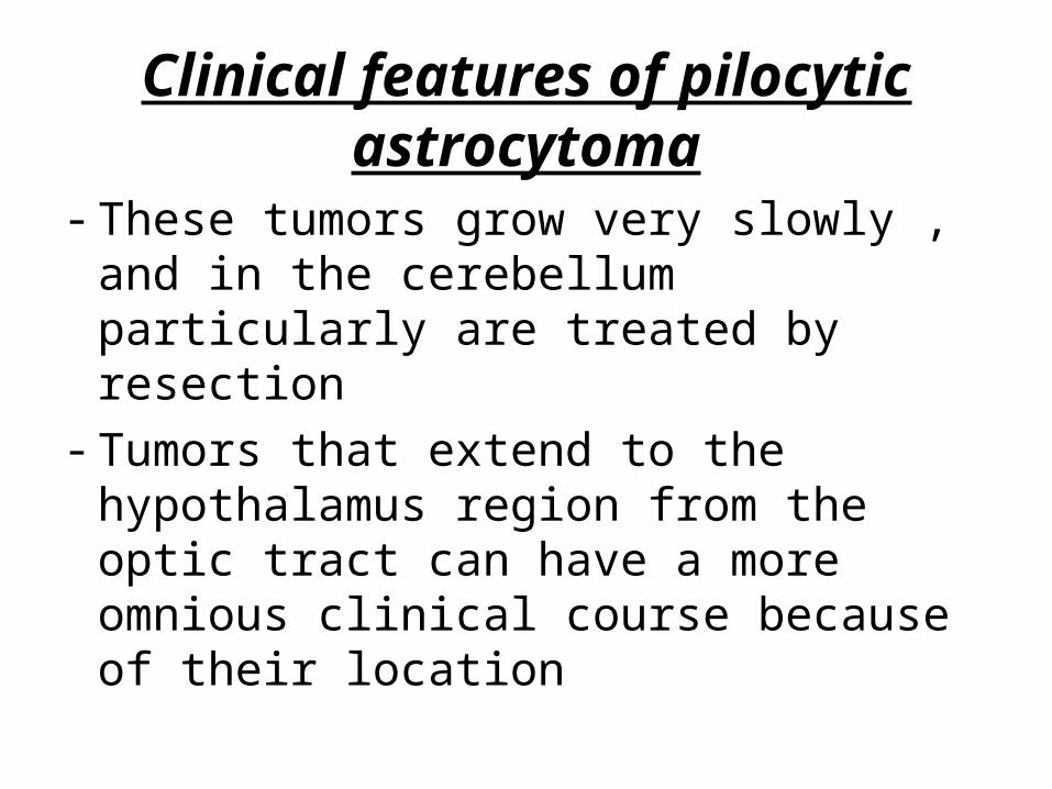
- These tumors grow very slowly , and in the cerebellum particularly are treated by resection
- Tumors that extend to the hypothalamus region from the optic tract can have a more omnious clinical course because of their location

B. Oligodendroglioma - These are infiltrating gliomas comprised of
cells that resemble oligodendrocytes- These tumors constitute about 5% to 15%
of gliomas- Are most common in the fourth and fifth
decades.

MorphologyGross- Are infiltrative tumors - may show cysts, hemorrhage, and
calcification.Note: Is the most common CNS tumor
showing calcification

B.OligodendrogliomaOn microscopic examination, 1. Grade II oligodendroglioma: - Is composed of sheets of regular cells with
spherical nuclei - The nuclei are surrounded by a clear halo of
cytoplasm - The cells are separated by a delicate network
anastomosing capillaries
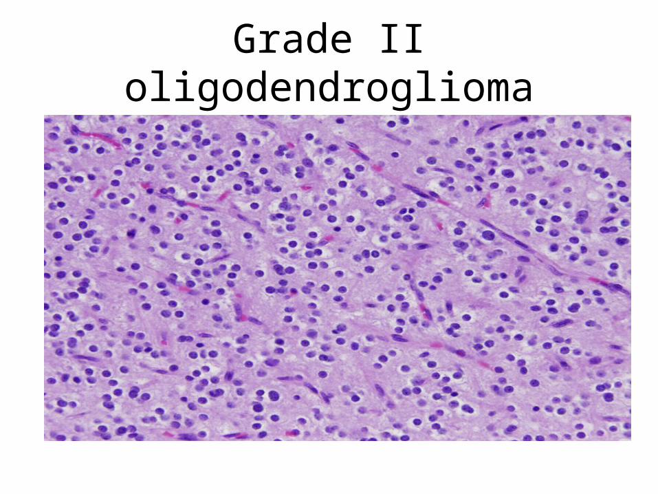
Grade II oligodendroglioma

2. Grade III anaplastic oligodendroglioma- Characterized a. And/or microvascular proliferationb. And/ or necrosis

Clinical features- Patients may have had several years of
neurologic complaints, often including seizures.
- The lesions are found mostly in the cerebral hemispheres, mainly in the frontal or temporal lobes
- Patients have better prognosis than astrocytomas of similar grade

- Individuals with anaplastic oligodendrogliomas have an overall worse prognosis
- Progression from low to higher grade lesion occurs , typically over about 6 years
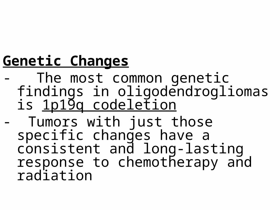
Genetic Changes- The most common genetic findings in
oligodendrogliomas is 1p19q codeletion- Tumors with just those specific changes have a
consistent and long-lasting response to chemotherapy and radiation

C. Ependymoma - Ependymomas are tumors that most
often arise next to the ependyma-lined ventricular system, including the oft-obliterated central canal of the spinal cord

In the first 2 decades of life - Typically arise from the floor of the fourth
ventricle and constitutes 5%-10% of all primary brain tumors in this age group
In adults, - The spinal cord is the most common location

Microscopically:1. Grade II epenymomas- Are well circumscribed gliomas - Composed of cells with regular, round to oval
nuclei - Between the nuclei there is a variably dense
fibrillary background

- Tumor cells may a. Form gland –like round or elongated structiure
(canals, rosettes) that resemble the embryologic ependymal canal (true rosettes)
- Are specific to ependymomas

a. Form pseudovascular rosettes- In which tumor cells are arranged around
blood vessels with an intervening zone consisting of ependyma rosettes directed toward the wall of the blood vessels
- More frequently seen- Not specific to ependymoma

Ependymoma

True Rosettes of ependymoma

Perivascular pseudo rosettes

2. Anaplastic ependymomas (grade III) show :3. Myxopapilllary ependymoma ( grade I ) - that arise in the filum terminale - and has good prognosis but tends to recur if
not completely excised

- Although fourth ventricular tumors are relatively well circumscribed, their proximity to vital pontine and medullary nuclei make complete resection is impossible
- Spinal cord ependymomas have better prognosis than ventricular ependymoma because can be resected

Molecular genetics
In the spinal cord ependymomas
- NF2 gene on chromosome 22 is commonly mutated

II. Neuronal Tumors 1. Central neurocytoma (grade II)- Is a low-grade neuronal neoplasm - Found within and adjacent to the ventricular
system (most commonly at the level of foramen of Munro)
2. Gangliogliomas- Are tumors with a mixture of glial elements
(looking like a low-grade astrocytoma) and mature-appearing neurons.
- Most of these tumors are slow growing, - These lesions often present with seizures

III. Embryonal primitive neoplasms A. MedulloblastomaB. Cerebral primitive neuroectodermal tumor
common is the medulloblastoma,

A. Medulloblastoma (WHO grade IV)- Accounts for 20% of pediatric brain tumors. - Occurs predominantly in children - and exclusively in the cerebellum. - The tumor is highly malignant,

Morphology- In children, medulloblastomas are located in
the midline of the cerebellum (vermis) but may extend to the surface of the cerebellar folia and involving the leptomeninges
- Lateral tumors (in cerebellar hemispheres) occur more often in adults.

Microscopicallya. Are extremely cellularb. Sheets of anaplastic ("small blue") cells c. The tumor cells are small with little cytoplasm
and hyperchromatic nucleic. Mitoses are abundant. d. May show Homer-wright rosettes as evidence of
neuronal differentiation

- The prognosis for untreated patients is dismal
- It is radiosensitive- With total excision and radiation, the 5-
year survival rate may be as high as 75%.

Medulloblastoma
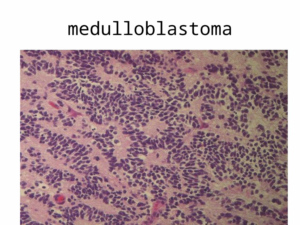
medulloblastoma

.
B. Tumors of similar histology can be found elsewhere in the nervous system (called CNS primitive neuroectodermal tumor, or CNS PNET )

VI. Meningiomas : (extraxial tumor)- They occur in adults - Are predominantly benign tumors that arise from
arachnoid cap cells and attached to the dura.- May be found along any of the external surfaces of
the brain as well as within the ventricular system, where they arise from the arachnoid cells of the choroid plexus.

- Clinically they either present with vague non-localizing symptoms, or with focal findings referable to compression of adjacent brain.
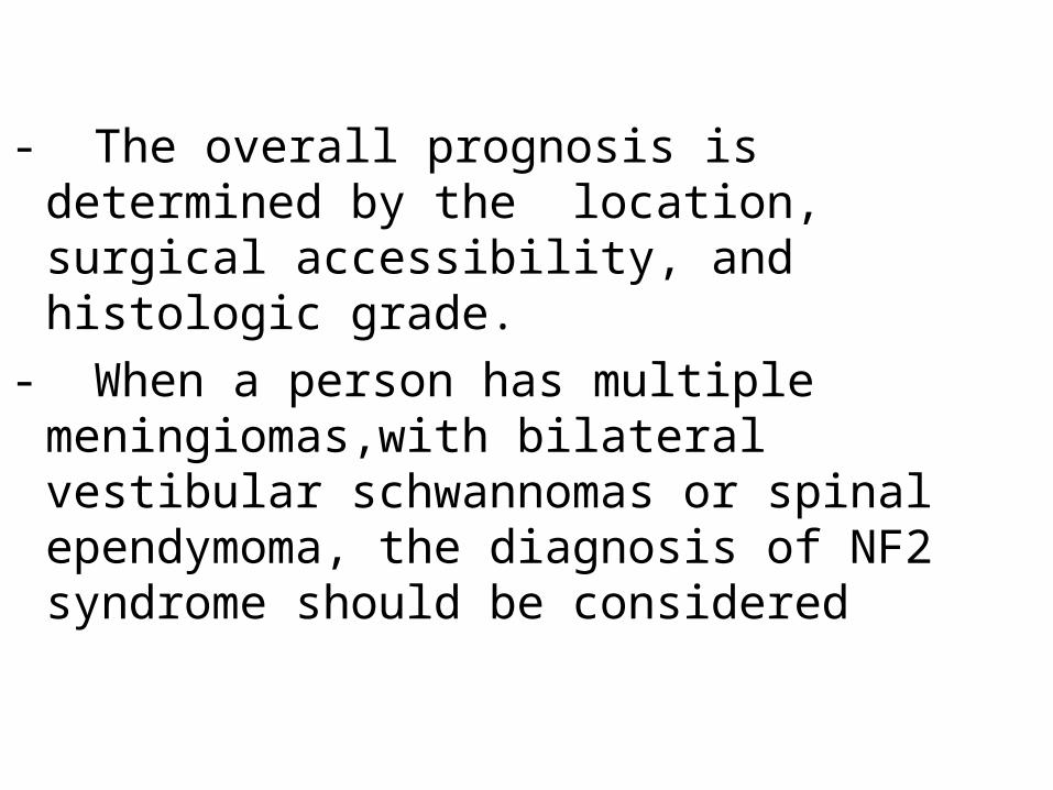
- The overall prognosis is determined by the location, surgical accessibility, and histologic grade.
- When a person has multiple meningiomas,with bilateral vestibular schwannomas or spinal ependymoma, the diagnosis of NF2 syndrome should be considered

- Menigiomas are classified into 3 grades- The majority of meningiomas are WHO grade 1Gross-: WHO grade I grow as well-defined dura-based
masses that may compress the brain but do not invade it and extension into the overlying bone may be present and does not upgrade the tumor to grade II.
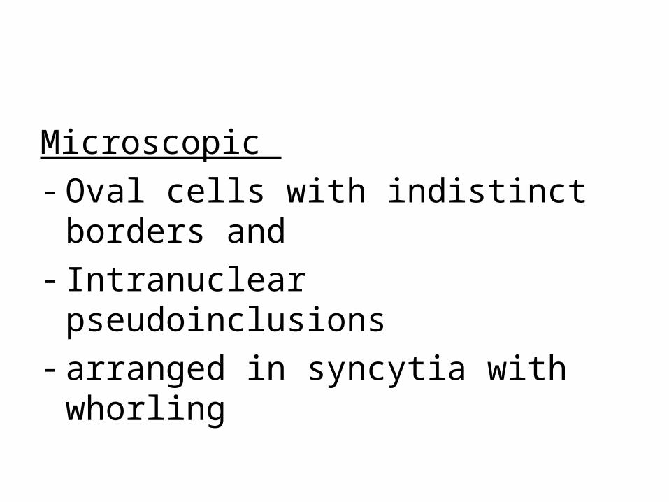
Microscopic - Oval cells with indistinct borders and - Intranuclear pseudoinclusions - arranged in syncytia with whorling

Meningioma

Meningeoma
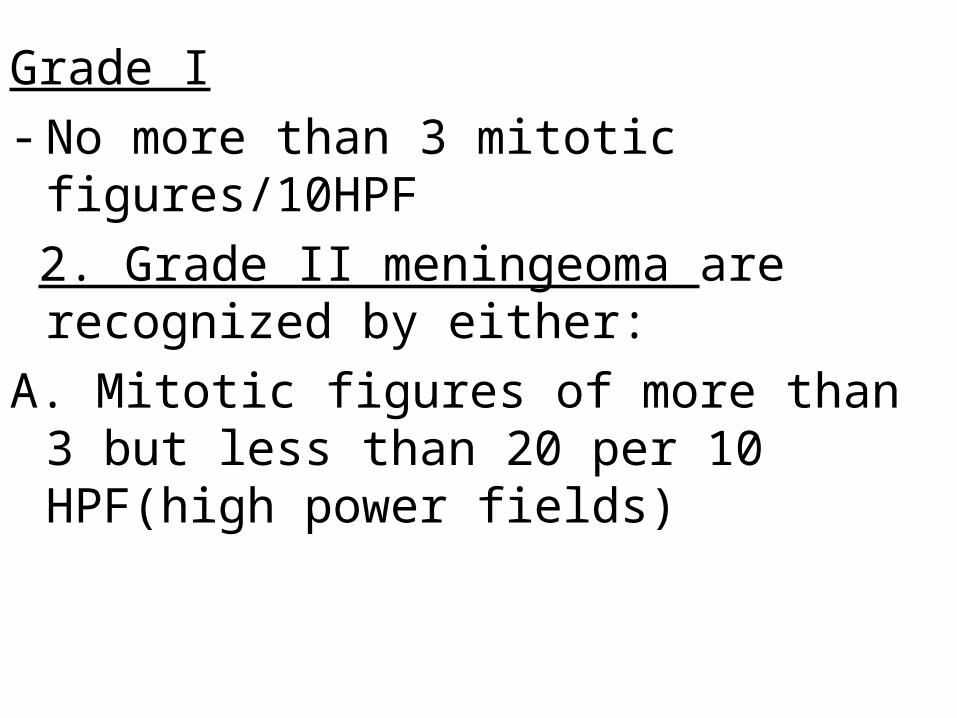
Grade I- No more than 3 mitotic figures/10HPF 2. Grade II meningeoma are recognized by either:A. Mitotic figures of more than 3 but less than 20 per
10 HPF(high power fields)

B. Or presence of 3 of the following 5 atypical features1. Small cells, 2. Prominent nucleoli ,3. Sheeting4. Hypercellularity, 5. Spontaneous necrosis not induced by embolization
Related Documents