I.G. Shibi et al / IJRAP 3(3), May – Jun 2012 433 Research Article www.ijrap.net PHYSICO-CHEMICAL ANALYSIS OF ARUMUGA CHENDOORAM I.G. Shibi 1 *, G. Shalu 1 , K. Jagannathan 2 1 PG Department of Chemistry, Sree Narayana College, Chempazhanthy, Thiruvananthapuram, 695587, Kerala, India 2 Santhigiri Siddha Medical College, Pothencode, Thiruvananthapuram, Kerala, India Received on: 06/03/12 Revised on: 20/04/12 Accepted on: 09/05/12 *Corresponding author I.G. Shibi, Associate Professor, PG Department of Chemistry, Sree Narayana College, Chempazhanthy, Thiruvananthapuram, 695587, Kerala, India Email: [email protected] ABSTRACT The present study is aimed at the characterization of the physico-chemical traits of the traditional Indian Siddha medicine, Arumuga Chendooram. The surface area, pHzpc and density of the chendooram sample were analyzed. The ICP-OES analysis revealed that the main metals present in Arumuga Chendooram are iron (20.9%), tin (8.5%) and mercury (3.1%). FT-IR spectroscopy has been used to study the presence of organic substances, if any, in the sample. Powder XRD technique was used to identify the crystalline phases, chemical nature and crystalline dimensions of the sample. The average crystalline size calculated using Debye-Scherrer equation corresponding to different diffraction planes is 37.277 nm. Measurements by DLS technique and SEM analyses were also carried out. The study highlights the appropriate application of modern scientific methods for developing new insights into metal based siddha drugs. Key words: Siddha, Physico-chemical analysis, Arumuga Chendooram INTRODUCTION The Siddha system is an Indian system of medicine which is developed and mainly practised in Tamil Nadu and other parts of Southern India since ancient times 1 . The materia medica of Siddha system encompasses around 500 medicinal plants, 150 metals and minerals, 150 animals and their derivatives. It depends largely on drugs of metallic origin in contrast to Ayurveda, which mainly depends on drugs of plant origin. The advantages of metal and mineral based medicines in Siddha are as follows. 1. Mild dose is enough 2. They are very effective for treating dreadful diseases 3. The action is very speedy 4. Wide range of diseases could be treated with the same medicine by changing or without changing the vehicle 5. Shelf life is very high 6. Unlike herbal drugs, no problem of chemical changes occurs due to geo-climatic conditions 7. They are less expensive. Even if a metal based Siddha medicine causes ill effects due to improper administration, diet habits and medical advice, appropriate antidotes are available to nullify the effect. Generally, the method of preparation of metal based Siddha medicines involves conversion of minerals or metals into the oxide or sulphide form by various herbal treatment followed by repeated high temperature calcination and grinding cycles. The chendooras thus obtained constitute ultra small particles and are taken along with vehicles such as milk, honey, butter, ghee etc according to the disease. This makes these drugs easily assimilable, eliminating their harmful effects and enhancing their biocompatibility 2 . The rigidity of the methods of preparation for a particular chendooram makes the drug, unique. However very few studies have been carried out to understand the physico-chemical nature of these type of traditional medicines 3,4 . Though metal based Siddha medicines are time tested drugs, extensive research works should be carried out to explore its effectiveness and to bring all Indian Traditional Systems into the limelight. There is a general public apprehension regarding the toxicity of these medicines due to westerners’ apathy to the traditional systems of medicine as evidenced by some of the studies 5-8 . For metal and mineral based medicinal preparations, it becomes imperative that these drugs should be characterized with the help of modern instrumental techniques like Inductively Coupled Plasma Optical Emission Spectrometry (ICP-OES), X-ray diffraction (XRD), Scanning Electron Microscopy (SEM), Dynamic Light Scattering (DLS), Infrared Spectroscopy (IR) and surface area measurement. Based on these, the specifications of metal based drugs can be well standardized on a scientific basis. In the present era of globalization, for the development of a world market for traditional medicines, research & development is very essential. The present study investigated the physico- chemical properties of the traditional Indian Siddha medicine, Arumuga chendooram, which is widely used for treating anaemic conditions, scrofula, headache and rheumatic diseases. It is also used for the treatment of piles, uterine disorders, orchitis, gastric ulcer, syphilis and tuberculosis etc. MATERIALS AND METHODS Materials The Siddha medicine Arumuga chendooram was procured at its different stages of preparation from Santhigiri Ayurveda & Siddha Vaidhyasala, Pothencode, Thiruvananthapuram. Preparation of Arumuga chendooram In the first stage of the preparation of Arumuga chendooram (Figure 1), five parts of purified mercury (suththi seitha rasam), nine parts of purified sulphur (suththi seitha kanthakam), seven parts of purified lode stone (suththi seitha kantham), twelve parts of purified iron filings (suththi seitha ayapodi), four parts of rock salt (induppu) and eight parts of desicated borax (poriththa venkaram) were ground with sufficient quantity of aloe juice (kumari charu) for five days continuously (Figure

Welcome message from author
This document is posted to help you gain knowledge. Please leave a comment to let me know what you think about it! Share it to your friends and learn new things together.
Transcript

I.G. Shibi et al / IJRAP 3(3), May – Jun 2012
433
Research Article www.ijrap.net
PHYSICO-CHEMICAL ANALYSIS OF ARUMUGA CHENDOORAM I.G. Shibi1*, G. Shalu1, K. Jagannathan2 1PG Department of Chemistry, Sree Narayana College, Chempazhanthy, Thiruvananthapuram, 695587, Kerala, India 2Santhigiri Siddha Medical College, Pothencode, Thiruvananthapuram, Kerala, India Received on: 06/03/12 Revised on: 20/04/12 Accepted on: 09/05/12 *Corresponding author I.G. Shibi, Associate Professor, PG Department of Chemistry, Sree Narayana College, Chempazhanthy, Thiruvananthapuram, 695587, Kerala, India Email: [email protected] ABSTRACT The present study is aimed at the characterization of the physico-chemical traits of the traditional Indian Siddha medicine, Arumuga Chendooram. The surface area, pHzpc and density of the chendooram sample were analyzed. The ICP-OES analysis revealed that the main metals present in Arumuga Chendooram are iron (20.9%), tin (8.5%) and mercury (3.1%). FT-IR spectroscopy has been used to study the presence of organic substances, if any, in the sample. Powder XRD technique was used to identify the crystalline phases, chemical nature and crystalline dimensions of the sample. The average crystalline size calculated using Debye-Scherrer equation corresponding to different diffraction planes is 37.277 nm. Measurements by DLS technique and SEM analyses were also carried out. The study highlights the appropriate application of modern scientific methods for developing new insights into metal based siddha drugs. Key words: Siddha, Physico-chemical analysis, Arumuga Chendooram INTRODUCTION The Siddha system is an Indian system of medicine which is developed and mainly practised in Tamil Nadu and other parts of Southern India since ancient times1. The materia medica of Siddha system encompasses around 500 medicinal plants, 150 metals and minerals, 150 animals and their derivatives. It depends largely on drugs of metallic origin in contrast to Ayurveda, which mainly depends on drugs of plant origin. The advantages of metal and mineral based medicines in Siddha are as follows. 1. Mild dose is enough 2. They are very effective for treating dreadful diseases 3. The action is very speedy 4. Wide range of diseases could be treated with the same medicine by changing or without changing the vehicle 5. Shelf life is very high 6. Unlike herbal drugs, no problem of chemical changes occurs due to geo-climatic conditions 7. They are less expensive. Even if a metal based Siddha medicine causes ill effects due to improper administration, diet habits and medical advice, appropriate antidotes are available to nullify the effect. Generally, the method of preparation of metal based Siddha medicines involves conversion of minerals or metals into the oxide or sulphide form by various herbal treatment followed by repeated high temperature calcination and grinding cycles. The chendooras thus obtained constitute ultra small particles and are taken along with vehicles such as milk, honey, butter, ghee etc according to the disease. This makes these drugs easily assimilable, eliminating their harmful effects and enhancing their biocompatibility2. The rigidity of the methods of preparation for a particular chendooram makes the drug, unique. However very few studies have been carried out to understand the physico-chemical nature of these type of traditional medicines3,4. Though metal based Siddha medicines are time tested drugs, extensive research works should be carried out to explore its effectiveness and to bring all Indian Traditional Systems into the limelight.
There is a general public apprehension regarding the toxicity of these medicines due to westerners’ apathy to the traditional systems of medicine as evidenced by some of the studies5-8. For metal and mineral based medicinal preparations, it becomes imperative that these drugs should be characterized with the help of modern instrumental techniques like Inductively Coupled Plasma Optical Emission Spectrometry (ICP-OES), X-ray diffraction (XRD), Scanning Electron Microscopy (SEM), Dynamic Light Scattering (DLS), Infrared Spectroscopy (IR) and surface area measurement. Based on these, the specifications of metal based drugs can be well standardized on a scientific basis. In the present era of globalization, for the development of a world market for traditional medicines, research & development is very essential. The present study investigated the physico-chemical properties of the traditional Indian Siddha medicine, Arumuga chendooram, which is widely used for treating anaemic conditions, scrofula, headache and rheumatic diseases. It is also used for the treatment of piles, uterine disorders, orchitis, gastric ulcer, syphilis and tuberculosis etc. MATERIALS AND METHODS Materials The Siddha medicine Arumuga chendooram was procured at its different stages of preparation from Santhigiri Ayurveda & Siddha Vaidhyasala, Pothencode, Thiruvananthapuram. Preparation of Arumuga chendooram In the first stage of the preparation of Arumuga chendooram (Figure 1), five parts of purified mercury (suththi seitha rasam), nine parts of purified sulphur (suththi seitha kanthakam), seven parts of purified lode stone (suththi seitha kantham), twelve parts of purified iron filings (suththi seitha ayapodi), four parts of rock salt (induppu) and eight parts of desicated borax (poriththa venkaram) were ground with sufficient quantity of aloe juice (kumari charu) for five days continuously (Figure
I.G. Shibi et al / IJRAP 3(3), May – Jun 2012
434
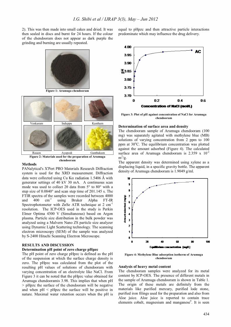
2). This was then made into small cakes and dried. It was then sealed in discs and burnt for 24 hours. If the colour of the chendooram does not appear as dark purple the grinding and burning are usually repeated.
Figure 1: Arumuga chendooram
Venkaram Induppu Kantham
Rasam Ayapodi Ganthakam
Figure 2: Materials used for the preparation of Arumuga chendooram
Methods PANalytical's X'Pert PRO Materials Research Diffraction system is used for the XRD measurement. Diffraction data were collected using Cu Ka radiation 1.5406 Ǻ with generator settings of 40 kV 30 mA. A continuous scan mode was used to collect 2θ data from 5° to 80° with a step size of 0.0840° and scan step time of 201.145 s. The FTIR spectra of the samples were recorded between 4000 and 400 cm-1 using Bruker Alpha FT-IR Spectrophotometer with ZnSe ATR technique at 2 cm-1 resolution. The ICP-OES used in the study is Perkin Elmer Optima 4300 V (Simultaneous) based on Argon plasma. Particle size distribution in the bulk powder was analyzed using a Malvern Nano ZS particle size analyzer using Dynamic Light Scattering technology. The scanning electron microscopy (SEM) of the sample was analyzed by S-2400 Hitachi Scanning Electron Microscope. RESULTS AND DISCUSSION Determination pH point of zero charge pHpzc The pH point of zero charge pHpzc is defined as the pH of the suspension at which the surface charge density is zero. The pHpzc was calculated from the plot of the resulting pH values of solutions of chendooram with varying concentration of an electrolyte like NaCl. From Figure 3 it can be noted that the pHpzc value obtained for Arumuga chendooramis 3.98. This implies that when pH > pHpzc the surface of the chendooram will be negative and when pH < pHpzc the surface will be positive in nature. Maximal water retention occurs when the pH is
equal to pHpzc and then attractive particle interactions predominate which may influence the drug delivery.
Figure 3: Plot of pH against concentration of NaCl for Arumuga
chendooram Determination of surface area and density The chendooram sample of Arumuga chendooram (100 mg) was separately agitated with methylene blue (MB) solutions of varying concentration from 2 ppm to 100 ppm at 30oC. The equilibrium concentration was plotted against the amount adsorbed (Figure 4). The calculated surface area of Arumuga chendooram is 2.359 x 10⁻² m2/g. The apparent density was determined using xylene as a displacing liquid, in a specific gravity bottle. The apparent density of Arumuga chendooram is 1.9049 g/ml.
Figure 4: Methylene Blue adsorption isotherm of Arumuga
chendooram Analysis of heavy metal content The chendooram samples were analyzed for its metal content by ICP-OES. The presence of different metals in the sample of Arumuga chendooram is shown in Table 1. The origin of these metals are definitely from the materials like purified mercury, purified lode stone, purified iron filings used for the preparation and also from Aloe juice. Aloe juice is reported to contain trace elements cobalt, magnesium and manganese9. It is seen
I.G. Shibi et al / IJRAP 3(3), May – Jun 2012
435
that iron, mercury and tin which are the prominent metals present, constitute 20.9, 8.5 and 3.1% respectively. The presence of arsenic (0.35%) is important since it is regarded as toxic with high health risk. The fact is that arsenicals have a long and remarkable history of utility in pharmacology. Inorganic arsenic is now accepted in western medicine as a first line chemotherapeutic agent against certain hematopoietic cancers as well10. Mercury compounds have been widely used as therapeutic agents in Ayurveda and Siddha medicines from time immemorial.
Table 1: Metallic composition of Arumuga chendooram from ICP-
OES analysis Metal Amount (%) Mn 0.19 Ni 0.02 Mg 0.09 Co 0.007 Zn 0.028 Cu 0.03 Fe 20.9 Ca 0.37 Sn 8.5 As 0.35 Hg 3.1 Pb 0.04
The finding is significant in the light of several studies appearing in the literature regarding the heavy metal contents of herbal medicines. The biological or therapeutic role of these metals is to be understood scientifically in order to establish the efficacy of these medicines. The herbo-metallic preparations used in the traditional system of medicine which contain heavy metals as therapeutic agents after proper de-toxification process have no significant adverse drug reactions so far reported. Even though the total mass percentage of these metals constitutes 33.625 %, only very minute quantities of it are administered as a medicine and that too for minimum number of days, together with vehicles like honey. Elemental analysis The carbon, hydrogen, nitrogen and sulphur (CHNS) analysis revealed that the sample of Arumuga chendooram has no nitrogen present in it. It is also observed that the carbon, hydrogen and sulphur present in the sample are 1.28, 0.32 and 10.20 % respectively. It is expected that the juice of Aloe serves as a medium for the reaction for the formation of the chendooram initially and will be burnt out during the repeated incineration process, resulting in the low % of carbon.
Figure 5: FTIR Spectra of Arumuga chendooram
Figure 6: XRD Spectra of Arumuga chendooram
I.G. Shibi et al / IJRAP 3(3), May – Jun 2012
436
Figure 7: Particle size distribution by volume of Arumuga chendooram
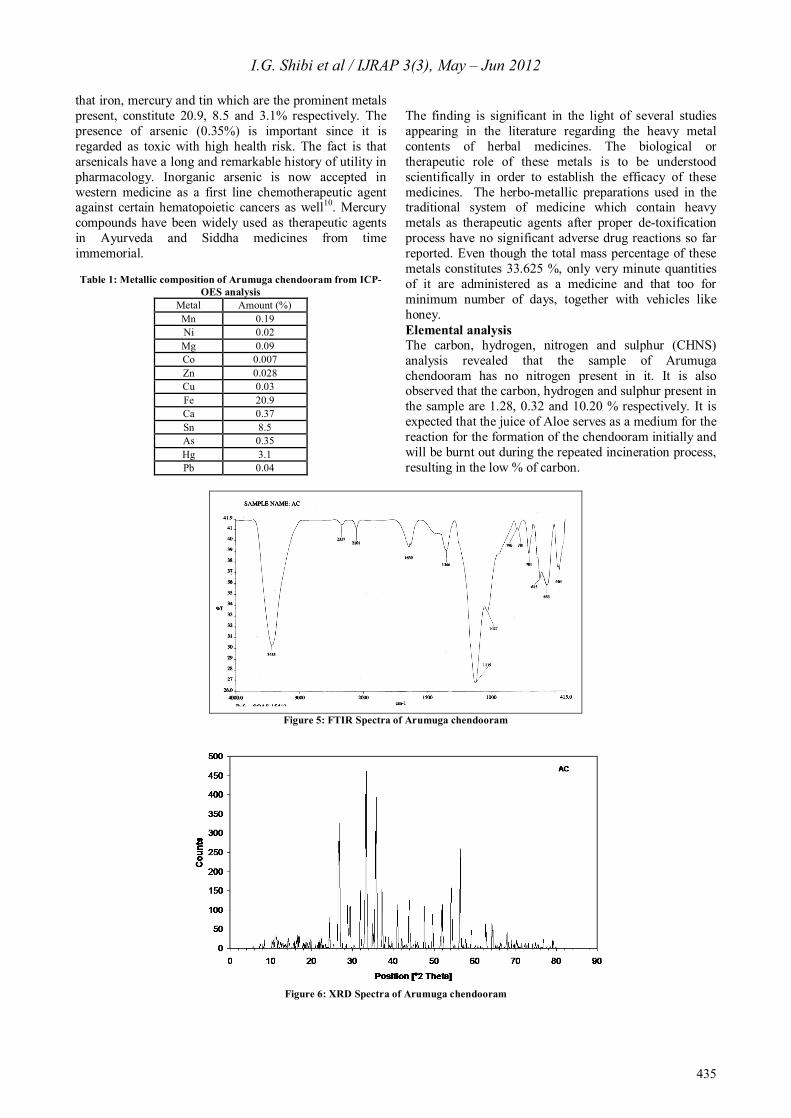
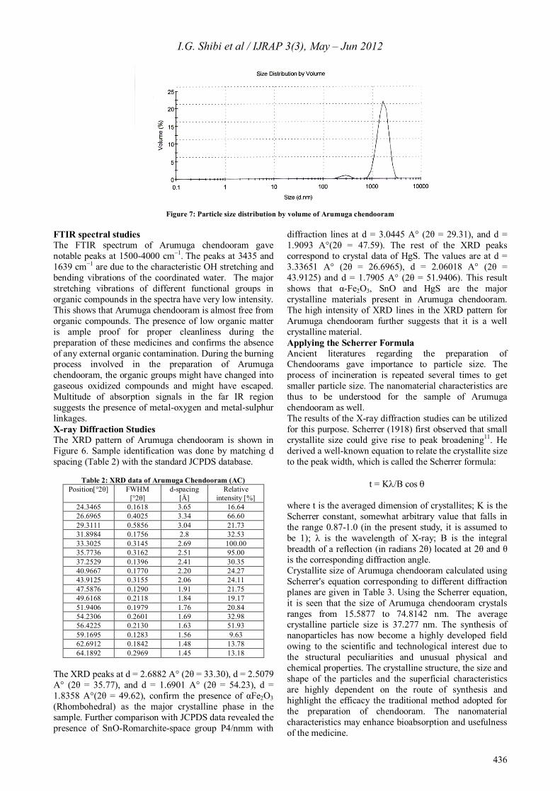
FTIR spectral studies The FTIR spectrum of Arumuga chendooram gave notable peaks at 1500-4000 cm−1. The peaks at 3435 and 1639 cm−1 are due to the characteristic OH stretching and bending vibrations of the coordinated water. The major stretching vibrations of different functional groups in organic compounds in the spectra have very low intensity. This shows that Arumuga chendooram is almost free from organic compounds. The presence of low organic matter is ample proof for proper cleanliness during the preparation of these medicines and confirms the absence of any external organic contamination. During the burning process involved in the preparation of Arumuga chendooram, the organic groups might have changed into gaseous oxidized compounds and might have escaped. Multitude of absorption signals in the far IR region suggests the presence of metal-oxygen and metal-sulphur linkages. X-ray Diffraction Studies The XRD pattern of Arumuga chendooram is shown in Figure 6. Sample identification was done by matching d spacing (Table 2) with the standard JCPDS database.
Table 2: XRD data of Arumuga Chendooram (AC)
Position[°2θ] FWHM [°2θ]
d-spacing [Å]
Relative intensity [%]
24.3465 0.1618 3.65 16.64 26.6965 0.4025 3.34 66.60 29.3111 0.5856 3.04 21.73 31.8984 0.1756 2.8 32.53 33.3025 0.3145 2.69 100.00 35.7736 0.3162 2.51 95.00 37.2529 0.1396 2.41 30.35 40.9667 0.1770 2.20 24.27 43.9125 0.3155 2.06 24.11 47.5876 0.1290 1.91 21.75 49.6168 0.2118 1.84 19.17 51.9406 0.1979 1.76 20.84 54.2306 0.2601 1.69 32.98 56.4225 0.2130 1.63 51.93 59.1695 0.1283 1.56 9.63 62.6912 0.1842 1.48 13.78 64.1892 0.2969 1.45 13.18
The XRD peaks at d = 2.6882 A° (2θ = 33.30), d = 2.5079 A° (2θ = 35.77), and d = 1.6901 A° (2θ = 54.23), d = 1.8358 A°(2θ = 49.62), confirm the presence of αFe2O3 (Rhombohedral) as the major crystalline phase in the sample. Further comparison with JCPDS data revealed the presence of SnO-Romarchite-space group P4/nmm with
diffraction lines at d = 3.0445 A° (2θ = 29.31), and d = 1.9093 A°(2θ = 47.59). The rest of the XRD peaks correspond to crystal data of HgS. The values are at d = 3.33651 A° (2θ = 26.6965), d = 2.06018 A° (2θ = 43.9125) and d = 1.7905 A° (2θ = 51.9406). This result shows that α-Fe2O3, SnO and HgS are the major crystalline materials present in Arumuga chendooram. The high intensity of XRD lines in the XRD pattern for Arumuga chendooram further suggests that it is a well crystalline material. Applying the Scherrer Formula Ancient literatures regarding the preparation of Chendoorams gave importance to particle size. The process of incineration is repeated several times to get smaller particle size. The nanomaterial characteristics are thus to be understood for the sample of Arumuga chendooram as well. The results of the X-ray diffraction studies can be utilized for this purpose. Scherrer (1918) first observed that small crystallite size could give rise to peak broadening11. He derived a well-known equation to relate the crystallite size to the peak width, which is called the Scherrer formula:
t = Kl/B cos q
where t is the averaged dimension of crystallites; K is the Scherrer constant, somewhat arbitrary value that falls in the range 0.87-1.0 (in the present study, it is assumed to be 1); λ is the wavelength of X-ray; B is the integral breadth of a reflection (in radians 2θ) located at 2θ and θ is the corresponding diffraction angle. Crystallite size of Arumuga chendooram calculated using Scherrer's equation corresponding to different diffraction planes are given in Table 3. Using the Scherrer equation, it is seen that the size of Arumuga chendooram crystals ranges from 15.5877 to 74.8142 nm. The average crystalline particle size is 37.277 nm. The synthesis of nanoparticles has now become a highly developed field owing to the scientific and technological interest due to the structural peculiarities and unusual physical and chemical properties. The crystalline structure, the size and shape of the particles and the superficial characteristics are highly dependent on the route of synthesis and highlight the efficacy the traditional method adopted for the preparation of chendooram. The nanomaterial characteristics may enhance bioabsorption and usefulness of the medicine.
I.G. Shibi et al / IJRAP 3(3), May – Jun 2012
437
More experiments using TEM could only confirm the nonmaterial nature of the Chendooram sample.
Table 3: Crystal size data of Arumuga Chendooram
2qqqq Crystalline size (nm) 33.3025 29.3087 35.7736 29.3471 54.2306 38.1421 49.6168 45.9317
29.31 15.5877 47.59 74.8142
26.6965 22.5501 43.9125 30.1786 51.9406 49.6328
Table 4: Details of elemental analysis by EDAX
Element (keV) Mass% Atom% K Na 1.041 8.79 20.01 5.9193 Si 1.739 1.62 3.03 1.3285 S 2.307 22.85 37.29 24.2197 Cl 2.621 6.27 9.25 4.8971 Fe 6.398 21.67 20.3 22.6158 Hg 2.195 38.79 10.12 36.8079
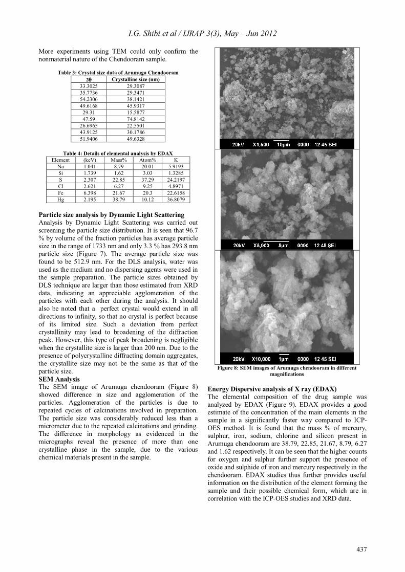
Particle size analysis by Dynamic Light Scattering Analysis by Dynamic Light Scattering was carried out screening the particle size distribution. It is seen that 96.7 % by volume of the fraction particles has average particle size in the range of 1733 nm and only 3.3 % has 293.8 nm particle size (Figure 7). The average particle size was found to be 512.9 nm. For the DLS analysis, water was used as the medium and no dispersing agents were used in the sample preparation. The particle sizes obtained by DLS technique are larger than those estimated from XRD data, indicating an appreciable agglomeration of the particles with each other during the analysis. It should also be noted that a perfect crystal would extend in all directions to infinity, so that no crystal is perfect because of its limited size. Such a deviation from perfect crystallinity may lead to broadening of the diffraction peak. However, this type of peak broadening is negligible when the crystallite size is larger than 200 nm. Due to the presence of polycrystalline diffracting domain aggregates, the crystallite size may not be the same as that of the particle size. SEM Analysis The SEM image of Arumuga chendooram (Figure 8) showed difference in size and agglomeration of the particles. Agglomeration of the particles is due to repeated cycles of calcinations involved in preparation. The particle size was considerably reduced less than a micrometer due to the repeated calcinations and grinding. The difference in morphology as evidenced in the micrographs reveal the presence of more than one crystalline phase in the sample, due to the various chemical materials present in the sample.
Figure 8: SEM images of Arumuga chendooram in different
magnifications Energy Dispersive analysis of X ray (EDAX) The elemental composition of the drug sample was analyzed by EDAX (Figure 9). EDAX provides a good estimate of the concentration of the main elements in the sample in a significantly faster way compared to ICP-OES method. It is found that the mass % of mercury, sulphur, iron, sodium, chlorine and silicon present in Arumuga chendooram are 38.79, 22.85, 21.67, 8.79, 6.27 and 1.62 respectively. It can be seen that the higher counts for oxygen and sulphur further support the presence of oxide and sulphide of iron and mercury respectively in the chendooram. EDAX studies thus further provides useful information on the distribution of the element forming the sample and their possible chemical form, which are in correlation with the ICP-OES studies and XRD data.

I.G. Shibi et al / IJRAP 3(3), May – Jun 2012
438
Figure 9: SEM EDAX of Arumuga chendooram
CONCLUSION The present study evaluated the physico-chemical properties of the traditional Indian medicine Arumuga chendooram. The results of XRD, ICP, FTIR and SEM studies can be used as excellent physico-chemical fingerprints for the validation of the medicine. The near-nano size of Arumuga chendooram may enable better bioabsorption. There is an intense competition from other countries in the trading of traditional medicinal products. India’s share in the world market is negligible as proper standardization techniques for checking the quality are inadequate. This study is an earnest attempt at making appropriate scientific validation of metal based traditional Indian medicines.
ACKNOWLEDGEMENT The authors IGS and SG gratefully acknowledge the University Grants Commission, New Delhi for awarding a Major Research Project and also express their sincere gratitude to Santhigiri Ayurveda and Siddha Vaidyasala, Pothencode, Thiruvananthapuram for their continuous support. REFERENCES 1. Scott Hajicek-Dobberstein. Soma siddhas and alchemical
enlightenment: psychedelic mushrooms in Buddhist tradition. J Ethnopharmacol 1995; 48: 99-118.
2. Suoboda RE. Prakriti: your ayurvedic constitution. Motilal Banarsidass Publ, India 1998.
3. Kumar A, Nair AGC, Reddy AVR, Garg AN. Bhasmas: unique ayurvedic metallic-herbal preparations, chemical characterization. Biol Trace Elem Res 2006; 109: 231-254.
4. Wadekar MP, Rode CV, Bendale YN, Patil KR, Gaikwad AB, Prabhune A. Effect of calcinations cycles on the preparation of tin oxide based traditional drug: Studies on its formation and characterization. J Pharmaceu Biomed Anal 2006; 41:1473-1478.
5. Saper RB, Kales SN, Paquim J, Burns MJ, Eisenberg DM, Davis RB, Phillips RS. Heavy Metal Content of Ayurvedic Herbal Medicine Products. J AM MED ASSOC 2004; 292: 2868-2873.
6. Grippo AA, Hamilton B, Hannigan R, Gurley BJ. Metal content of ephedra-containing dietary supplements and select botanicals. Am J Health Syst Pharm 2006; 63: 635-644.
7. Hardy AD, Sutherland HH, Vaishnav R, Worthing MA. A report on the composition of mercurials used in traditional medicines in Oman. J Ethnopharmacol 1995; 49:17-22.
8. Ernst E. Heavy metals in traditional Indian remedies. Eur J Clin Pharmacol 2002; 57: 891-896.
9. Rajasekara S, Sivagnanam K, Subramanian S. Mineral contents of aloe vera leaf gel and their role on streptozotocin-induced diabetic rats. Biol Trace Elem Res 2005; 108:185-195.
10. Beauchamp EM, Uren A. A new era for an ancient drug: arsenic trioxide and hedgehog signaling. Vitam Horm 2012; 88:333-354.
11. Langford JI, Wilson AJC. Scherrer after sixty years: A survey and some new results in the determination of crystallite size. J. Appl. Cryst. 1978; 11:102-113.
Source of support: University Grants Commission, New Delhi, Conflict of interest: None Declared
Related Documents