This journal is © The Royal Society of Chemistry 2020 Mol. Omics, 2020, 16, 287--304 | 287 Cite this: Mol. Omics, 2020, 16, 287 Identifying the targets and functions of N-linked protein glycosylation in Campylobacter jejuni Joel A. Cain, ab Ashleigh L. Dale, ab Zeynep Sumer-Bayraktar, ab Nestor Solis† a and Stuart J. Cordwell * abcd Campylobacter jejuni is a major cause of bacterial gastroenteritis in humans that is primarily associated with the consumption of inadequately prepared poultry products, since the organism is generally thought to be asymptomatic in avian species. Unlike many other microorganisms, C. jejuni is capable of performing extensive post-translational modification (PTM) of proteins by N- and O-linked glycosylation, both of which are required for optimal chicken colonization and human virulence. The biosynthesis and attachment of N-glycans to C. jejuni proteins is encoded by the pgl (protein glycosylation) locus, with the PglB oligosaccharyltransferase (OST) enabling en bloc transfer of a heptasaccharide N-glycan from a lipid carrier in the inner membrane to proteins exposed within the periplasm. Seventy-eight C. jejuni glycoproteins (represented by 134 sites of experimentally verified N-glycosylation) have now been identified, and include inner and outer membrane proteins, periplasmic proteins and lipoproteins, which are generally of poorly defined or unknown function. Despite our extensive knowledge of the targets of this apparently widespread process, we still do not fully understand the role N-glycosylation plays biologically, although several phenotypes, including wild-type stress resistance, biofilm formation, motility and chemotaxis have been related to a functional pgl system. Recent work has described enzymatic processes (nitrate reductase NapAB) and antibiotic efflux (CmeABC) as major targets requiring N-glycan attachment for optimal function, and experimental evidence also points to roles in cell binding via glycan–glycan interactions, protein complex formation and protein stability by conferring protection against host and bacterial proteolytic activity. Here we examine the biochemistry of the N-linked glycosylation system, define its currently known protein targets and discuss evidence for the structural and functional roles of this PTM in individual proteins and globally in C. jejuni pathogenesis. Introduction Campylobacter jejuni is a Gram negative enteric pathogen with helical cell morphology. C. jejuni is also microaerophilic and typically requires oxygen levels to be no greater than 10% for growth. 1 Gastrointestinal infection caused by this organism was first characterized in the late 1970s 2 and C. jejuni is now considered the most common causative agent of gastroenteritis in the developed world, with an estimated 400 million people infected worldwide annually. 3 Infection in humans is acquired though consumption of contaminated water or food, particu- larly under-cooked or inappropriately handled poultry products (with estimates suggesting between 75–90% of supermarket chicken is contaminated with the organism 4 ), since C. jejuni is generally considered an asymptomatic commensal in avian species. 5 While the differences in human and avian response to C. jejuni infection are largely unknown, there is evolving evidence that differences in host mucin O-glycan composition, particularly sulfated O-glycans, may play a role in colonization. 6 Furthermore, chicken mucins from different regions of the gastrointestinal tract can inhibit human epithelial cell virulence, 7 providing further evidence for glycan recognition in the establishment of host-specific niches. 8 Human disease is generally self-limiting and symptoms present as fever and abdominal cramping that progress from mild to, in some cases, severe diarrhoea. 2,3 Relapse is possible in the absence of medical intervention, and is likely due to gut persistence for up to 3 weeks. 9 C. jejuni infection is also an established antecedent for an increasing number of debilitating conditions including Guillain–Barre ´ Syndrome (GBS), a School of Life and Environmental Sciences, The University of Sydney, 2006, Australia b Charles Perkins Centre, The University of Sydney, Level 4 East, The Hub Building (D17), 2006, Australia. E-mail: [email protected]; Tel: +612-9351-6050 c Discipline of Pathology, School of Medical Sciences, The University of Sydney, 2006, Australia d Sydney Mass Spectrometry, The University of Sydney, 2006, Australia † Current address: Centre for Blood Research, Department of Oral Biological and Medical Sciences, University of British Columbia, Vancouver, Canada. Received 16th March 2020, Accepted 20th April 2020 DOI: 10.1039/d0mo00032a rsc.li/molomics Molecular Omics REVIEW Published on 22 April 2020. Downloaded on 10/17/2021 2:15:15 AM. View Article Online View Journal | View Issue

Welcome message from author
This document is posted to help you gain knowledge. Please leave a comment to let me know what you think about it! Share it to your friends and learn new things together.
Transcript
This journal is©The Royal Society of Chemistry 2020 Mol. Omics, 2020, 16, 287--304 | 287
Cite this:Mol. Omics, 2020,
16, 287
Identifying the targets and functions of N-linkedprotein glycosylation in Campylobacter jejuni
Joel A. Cain,ab Ashleigh L. Dale, ab Zeynep Sumer-Bayraktar,ab Nestor Solis†a andStuart J. Cordwell *abcd
Campylobacter jejuni is a major cause of bacterial gastroenteritis in humans that is primarily associated
with the consumption of inadequately prepared poultry products, since the organism is generally
thought to be asymptomatic in avian species. Unlike many other microorganisms, C. jejuni is capable of
performing extensive post-translational modification (PTM) of proteins by N- and O-linked glycosylation,
both of which are required for optimal chicken colonization and human virulence. The biosynthesis and
attachment of N-glycans to C. jejuni proteins is encoded by the pgl (protein glycosylation) locus, with
the PglB oligosaccharyltransferase (OST) enabling en bloc transfer of a heptasaccharide N-glycan from a
lipid carrier in the inner membrane to proteins exposed within the periplasm. Seventy-eight C. jejuni
glycoproteins (represented by 134 sites of experimentally verified N-glycosylation) have now been
identified, and include inner and outer membrane proteins, periplasmic proteins and lipoproteins, which
are generally of poorly defined or unknown function. Despite our extensive knowledge of the targets of
this apparently widespread process, we still do not fully understand the role N-glycosylation plays
biologically, although several phenotypes, including wild-type stress resistance, biofilm formation,
motility and chemotaxis have been related to a functional pgl system. Recent work has described
enzymatic processes (nitrate reductase NapAB) and antibiotic efflux (CmeABC) as major targets requiring
N-glycan attachment for optimal function, and experimental evidence also points to roles in cell binding
via glycan–glycan interactions, protein complex formation and protein stability by conferring protection
against host and bacterial proteolytic activity. Here we examine the biochemistry of the N-linked
glycosylation system, define its currently known protein targets and discuss evidence for the structural
and functional roles of this PTM in individual proteins and globally in C. jejuni pathogenesis.
Introduction
Campylobacter jejuni is a Gram negative enteric pathogen withhelical cell morphology. C. jejuni is also microaerophilic andtypically requires oxygen levels to be no greater than 10% forgrowth.1 Gastrointestinal infection caused by this organismwas first characterized in the late 1970s2 and C. jejuni is nowconsidered the most common causative agent of gastroenteritisin the developed world, with an estimated 400 million peopleinfected worldwide annually.3 Infection in humans is acquired
though consumption of contaminated water or food, particu-larly under-cooked or inappropriately handled poultry products(with estimates suggesting between 75–90% of supermarketchicken is contaminated with the organism4), since C. jejuni isgenerally considered an asymptomatic commensal in avianspecies.5 While the differences in human and avian responseto C. jejuni infection are largely unknown, there is evolvingevidence that differences in host mucin O-glycan composition,particularly sulfated O-glycans, may play a role in colonization.6
Furthermore, chicken mucins from different regions of thegastrointestinal tract can inhibit human epithelial cellvirulence,7 providing further evidence for glycan recognitionin the establishment of host-specific niches.8
Human disease is generally self-limiting and symptomspresent as fever and abdominal cramping that progress frommild to, in some cases, severe diarrhoea.2,3 Relapse is possiblein the absence of medical intervention, and is likely due to gutpersistence for up to 3 weeks.9 C. jejuni infection is also anestablished antecedent for an increasing number of debilitatingconditions including Guillain–Barre Syndrome (GBS),
a School of Life and Environmental Sciences, The University of Sydney, 2006,
Australiab Charles Perkins Centre, The University of Sydney, Level 4 East, The Hub
Building (D17), 2006, Australia. E-mail: [email protected];
Tel: +612-9351-6050c Discipline of Pathology, School of Medical Sciences, The University of Sydney,
2006, Australiad Sydney Mass Spectrometry, The University of Sydney, 2006, Australia
† Current address: Centre for Blood Research, Department of Oral Biological andMedical Sciences, University of British Columbia, Vancouver, Canada.
Received 16th March 2020,Accepted 20th April 2020
DOI: 10.1039/d0mo00032a
rsc.li/molomics
MolecularOmics
REVIEW
Publ
ishe
d on
22
Apr
il 20
20. D
ownl
oade
d on
10/
17/2
021
2:15
:15
AM
.
View Article OnlineView Journal | View Issue

288 | Mol. Omics, 2020, 16, 287--304 This journal is©The Royal Society of Chemistry 2020
Miller–Fisher Syndrome (MFS), immunoproliferative smallintestine disease, reactive arthritis and Sweet’s syndrome.10,11
The basis for these post-acute immune-mediated disorders isthought to be largely based on cross-reactivity between anti-bodies directed against C. jejuni surface lipooligosaccharide(LOS) and human cell surface gangliosides, and this relation-ship has been reviewed extensively.12–14
Several C. jejuni genomes have been sequenced fromlaboratory-adapted and clinical strains and several featuresremain consistent; the organism encodes B1620–1650 genes,a large proportion of which encode membrane-associatedproteins that are poorly functionally annotated.15–17 Humaninfection is not completely understood but involves bacterialadherence to gut epithelial cells, followed by invasion and subse-quent toxin production. Several factors are critical in C. jejunihost colonization, including flagellar-based motility, cell shape,chemosensing and chemotaxis mediated by transducer-like
proteins (Tlps), as well as a number of adhesins includingthe fibronectin-binding proteins Campylobacter adherencefactor CadF and fibronectin-like protein FlpA, the surface-exposed lipoprotein JlpA and the PEB antigens [reviewed in18–20].The ability to survive in the hostile environment encounteredduring gut infection, consisting of for example low pH, presenceof bile salts and competitive factors from established microflora, isparamount to establishing disease and C. jejuni is adapted toutilize nutrients, such as amino and organic acids as primarycarbon sources, that are in rich supply in the gut micro-environment (e.g. serine and proline from mucins, organic acidsproduced as a by-product of metabolism by residentmicroorganisms).21,22 C. jejuni lacks typical virulence-associatedtype III/IV secretion systems (T3SS/T4SS) employed by many otherenteric bacteria to secrete toxins and proteases that directlyinteract with host cells, although it is now well-established thatextracellular virulence factors (e.g. the Campylobacter invasion
Joel A. Cain
Joel Cain received a Bachelor ofScience with Honours in 2012and is currently completing aPhD degree at The University ofSydney, Australia. He wasawarded an Australian Post-graduate Award for thesestudies. He has also worked as aResearch Assistant in SydneyMass Spectrometry. His currentwork focuses on the contri-butions of post-translationalmodifications, such as targetedprotein degradation and
N-glycosylation, towards the pathogenic mechanisms of theenteric pathogen Campylobacter jejuni.
Ashleigh L. Dale
Ashleigh Dale received a Bachelorof Science with Honours at theUniversity of Sydney at the end of2018 and commenced her PhDcandidature in biochemistry thefollowing year. Her current workinvolves implementing cross-linking mass spectrometryapproaches to define theinteractome of C. jejuni andcharacterize protein–proteininteractions in the hope of eluci-dating mechanisms of pathogen-esis in this organism.
Zeynep Sumer-Bayraktar
Zeynep Sumer-Bayraktar receiveda PhD in biochemistry fromMacquarie University, SydneyAustralia in 2016. Her researchfocuses on protein post-translational modifications,specifically the glycan-components of N- and O-linkedglycosylation using liquidchromatography and massspectrometry-based approaches.In her current work, Zeynepinvestigates the role of proteinglycosylation in Campylobacter
jejuni human gut colonization and the glyco/proteomic responsesof the human host against C. jejuni infection.
Nestor Solis
Nestor Solis obtained his PhD inmicrobial proteomics in 2014from The University of Sydney,Australia, where he explored theidentification of cell-surfaceproteins from Staphylococcusspecies using cell-shavingproteomics. He joined thelaboratory of ProfessorChristopher Overall in 2014 tofurther expand on proteomicmethods to study N- and C-terminomes in macrophages. Hewas awarded two fellowships in
2016: a Michael Smith Foundation for Health Researchpostdoctoral fellowship from British Columbia, Canada, and a CJMartin Early Career Fellowship from the National Health andMedical Research Council of Australia.
Review Molecular Omics
Publ
ishe
d on
22
Apr
il 20
20. D
ownl
oade
d on
10/
17/2
021
2:15
:15
AM
. View Article Online

This journal is©The Royal Society of Chemistry 2020 Mol. Omics, 2020, 16, 287--304 | 289
antigens [Cia] and cytolethal distending toxin [CDT]) aresecreted via the flagellar export apparatus that acts as apseudo-T3SS.20,23 Another mechanism by which C. jejunivirulence determinants can interact with host cells is via theirpackaging into outer membrane vesicles (OMVs;24–26). Finally,despite the somewhat small size of the genome, C. jejunidevotes considerable resources to post-translational modifica-tion (PTM) of proteins by N- and O-linked glycosylation, both ofwhich are considered established virulence determinants.
Protein glycosylation in C. jejuni
Despite their ability to synthesize large polysaccharides, bac-teria were long thought to be enzymatically incapable ofmodifying proteins with glycans. In the past two decadeshowever, this opinion has largely been overturned with theidentification of conserved bacterial N- and O-linked glycosyla-tion systems in many microorganisms.27–30 Such systems arealmost universally biologically important and associated withpathogenic processes including cell–cell recognition andbinding,31 however their ultimate purpose and functionsremain to be determined. C. jejuni was the first bacterium tobe recognized as containing a ‘general glycosylation system’that could widely modify proteins.32–34 Since then, our under-standing of the biochemistry, targets and putative functions ofthese PTMs has increased rapidly. While our knowledge remainsincomplete, recent advances in glycoproteomics-focused massspectrometry (MS) have generated large-scale site identificationsin C. jejuni35 and other organisms, which have enabled directedfunctional studies to elucidate the roles of these modifications inbacterial phenotypes associated with pathogenicity.
O-Glycosylation in C. jejuni
C. jejuni modifies its flagella by O-glycosylation of the flagellinstructural protein with derivatives of the bacterial-specific sialicacid-like monosaccharides, pseudaminic acid or the closely
related legionaminic acid.36–41 At least 19–23 serine/threoninesites are modified on the FlaA flagellin depending on the strainemployed,42–46 and O-glycan attachment is essential for bothchicken and human infection phenotypes, including motility,autoagglutination, chicken colonization, and human epithelialcell adherence and invasion.47–49 Unlike N-glycosylation (seebelow), there is considerably more structural heterogeneity withrespect to the attached glycan in individual strains, includingboth chemical and steriometric differences. Synthesis of pseu-daminic and legionaminic acid occur independently of oneanother, beginning with nucleotide-linked precursors; pseuda-minic acid as a uridine diphosphate (UDP)-linked and legiona-minic acid as a guanosine diphosphate (GDP)-linked precursor.Enzymatic affinities for specific nucleotide precursors arecritical for differentiation of the two pathways and this preventscompetition for intermediates.38 Synthesis of pseudaminic acidfrom UDP-N-acetylglucosamine (UDP-GlcNAc) is performed bythe actions of PseB, PseC and PseH, which act sequentiallyto form UDP-2,4-diNAc-6-deoxy-altropyranose.50–52 Followingrelease of UDP by PseG, this sugar serves as the substrate ofPseI to form pseudaminic acid.50,52–54 Synthesis of legionaminicacid and derivatives mirrors pseudaminic acid in many respects;from a GDP-GlcNAc precursor, formation of UDP-2,4-diNAc-6-deoxy-glucose is catalyzed by the sequential activities of LegB,LegC and LegH.38 In principal, the lack of initial LegB epimeraseactivity creates the structural distinction between pseudaminicand legionaminic acids – instead this activity is performed by thehydrolysing 2-epimerase LegG alongside nucleotide release toform 2,4-diNAc-6-deoxy-mannose, which is then used by thelegionaminic acid synthase LegI to form legionaminic acid.38,47
Both pseudaminic and legionaminic acid are subsequently con-jugated onto cytodine monophosphate (CMP) nucleotides by theCMP-sugar synthetases PseF and LegF, respectively, prior toattachment onto FlaA by an undefined glycosyltransfersase.38,50
Unlike other Gram negative bacteria,55–57 in C. jejuni thereappears to be no other substrates of O-glycosylation (at leastthose modified with the flagellar glycan; see below) beyond FlaA.There is conjecture that given the role of the flagellar apparatusas a T3SS-like export apparatus that proteins secreted via thispathway may also be O-glycosylated in a similar manner to theFlaA flagellin. No studies however, have been able to globallyidentify extracellular proteins from this organism, mostly due tothe very specific requirements (presence of confounding serumor host cells) needed to induce secretion in C. jejuni.
It has been suggested that the major outer membraneprotein (MOMP), which accounts for B40–50% of the totalmembrane protein in C. jejuni,17 can also be O-glycosylated58
with a glycan unrelated to the flagellin modification describedabove. MOMP may be modified at Thr-268 with the tetrasac-charide Gal-b1,3-GalNAc-b1,4-GalNAc-b1,4-GalNAc-a1, althoughintact glycan-peptide MS validation is yet to be generated.MOMP modification was further indicated by Whitworth andcolleagues in C. jejuni strain 81–176 by galactose oxidase(GalO)-mediated selective biotinylation and subsequent enrich-ment of GalNAc containing cell surface glycoconjugates.59 Site-directed mutagenesis of Thr-268 indicated that this residue is
Stuart J. Cordwell
Stuart Cordwell is Professor ofAnalytical Biochemistry in theSchool of Life and Environ-mental Sciences and theDiscipline of Pathology, Schoolof Medical Sciences at theUniversity of Sydney. He is alsothe Director of Sydney MassSpectrometry. His researchfocusses on post-translationalmodifications of proteins andtheir roles in the virulencemechanisms of pathogenicbacteria. His group is also
increasingly using multi-omics strategies to decipher thesemechanisms to better understand the proteome-phenotype nexus.
Molecular Omics Review
Publ
ishe
d on
22
Apr
il 20
20. D
ownl
oade
d on
10/
17/2
021
2:15
:15
AM
. View Article Online
290 | Mol. Omics, 2020, 16, 287--304 This journal is©The Royal Society of Chemistry 2020
important for autoagglutination, biofilm formation andcolonization of both human Caco-2 cells and chickens,58 aphenotype also consistent with observations on the roles ofFlaA glycosylation. It remains to be seen whether additionalO-glycoproteins are present in C. jejuni and whether this PTMoccurs as a widespread presence on proteins secreted by theorganism during infection.
The pgl-encoded N-glycosylationsystem in C. jejuni
C. jejuni was the first bacterium demonstrated to possessthe ability to N-glycosylate proteins. Proteins are modified bythe N-linked addition of a heptasaccharide glycan (GalNAc-a1,4-GalNAc-a1,4-[Glcb1,3]-GalNAc-a1,4-GalNAc-a1,4-GalNAc-a1,3-diNAcBac-b1; where diNAcBac is N 0,N 0-diacetylbacillosamine[2,4-diacetamido-2,4,6 trideoxyglucopyranose])33 at the con-sensus sequon Asp (D)/Glu (E)-X1-Asn (N)-X2-Ser (S)/Thr (T)(where X1,2 a Pro), with Asn (N) being the attachmentsite.60,61 The synthesis of the N-glycan and attachment toproteins are encoded by the 16 kb pgl (protein glycosylation)gene cluster;32,34,62 the pgl cluster is highly conserved amongmembers of the order Campylobacteriales,63,64 although N-glycancomposition and structure, as well as the genomic location of thepgl locus (including being split into multiple loci), can differbetween species.63 Gene transfer of the complete pgl cluster intootherwise N-glycosylation incompetent bacterial species (e.g. mostcommonly E. coli) is sufficient to confer the ability to N-glycosylateco-expressed acceptor proteins.62 Therefore, the pgl system hasbecome a model for the production of glycoconjugate vaccines inrecombinant expression procedures [reviewed in65–67].
Biosynthesis and transfer of the N-glycan to proteins (Fig. 1)involves the actions of 10 Pgl proteins (an eleventh member ofthe locus, pglG, does not appear to contribute to the processand remains functionally undefined68) and begins withthe cytoplasmic synthesis of nucleotide-activated (uridinediphosphate; UDP) UDP-diNAcBac from UDP-GlcNAc, which iscatalyzed by the activities of (in order) the PglF dehydratase(conferring the rate-limiting step in the Pgl pathway), PglEaminotransferase and PglD acetyltransferase.69–74 Synthesis ofdiNAcBac has been reviewed extensively elsewhere.75,76 Thepotential for cross-talk between the N- and O-linked pathwaysis evidenced by shared nucleotide-activated precursors and bythe activity of PglD, which can form intermediates from withinthe legionaminic acid biosynthetic pathway, albeit at substan-tially reduced catalysis compared with LegH.38 DiNAcBac isattached to the cytoplasmic side of an inner membrane span-ning lipid carrier (undecaprenyl-pyrophosphate [Und-P]) by thePglC glycosyl-1-phosphate transferase, and Und-P then servesas the carrier for the nascent N-glycan.77,78 Continued synthesisof the glycan on Und-P-diNAcBac involves the sequentialaddition of 5 N-acetylgalactosamine (GalNAc) residues by threepgl-encoded glycosyltransferases (the first by PglA, the secondby PglJ and the final three by PglH).79 Glycan length is con-trolled by increased competitive inhibition of the PglH active
site relative to the number of GalNAc residues, and is consid-ered limited by the final GalNAc(x5) product.80 The PglHtertiary structure also contains a novel ‘ruler helix’ that bindsthe pyrophosphate of Und-P and limits PglH catalysis to 3GalNAc.81 Glycan synthesis is completed by the PglI glucosyl-transferase, which adds a single glucose (Glc) branch to thethird GalNAc in the N-glycan.68 This last Glc residue is not astrict requirement for N-glycosylation as, unlike all previoussteps, addition of the complete N-glycan (without the Glcbranch) to proteins still occurs in the absence of pglI,77,82 albeitat lower catalytic efficiency. Deletion of other pgl genes resultsin either complete loss of the N-glycan or the presenceof significantly truncated N-glycans (e.g. pglD82), as well ascompromised protein transfer efficiency.77 Once the hepta-saccharide has been completed, the PglK flippase translocatesthe Und-P-linked glycan from the cytoplasm into the periplas-mic space utilizing a mechanism dependent on the hydrolysisof two molecules of ATP.83,84 The mature glycan is thentransferred en bloc from Und-P onto target proteins by thePglB oligosaccharyltransferase (OST),68,85 which recognizesboth the Und-P-N-glycan complex and peptide acceptor assubstrates.86
The PglB OST is also capable of releasing the N-glycan fromUnd-P into the periplasm as a ‘free oligosaccharide’ (fOS),82,87,88
although the exact proportion of N-glycan as Asn-bound:fOSremains a point of contention. Nothaft et al. reported a ratiofavouring high fOS at B1 : 10, while Scott et al. reported adistribution of 4.5 : 1 in favour of protein-bound N-glycan.82,89
While there are a number of technical considerations that mayhelp explain this discrepancy,89,90 there may also be dynamiccontrol of fOS production based on environmental conditionsand the kinetics of PglB. The fOS itself has been shown toprovide protection against osmotic stress, further supportingthe notion that the cellular fate of the N-glycan may be deter-mined to some degree by environmental sensing.82 Unlikeprotein N-glycosylation, the free N-glycan is highly dependenton the synthesis of the complete heptasaccharide, as pglIdeficient strains produce B55% less fOS.90 The N-glycan itselfcan also be further modified with a phosphoethanolamine(pEtN) group, which is added to the terminal GalNAc of theheptasaccharide at a small number of glycosites by the soleC. jejuni pEtN transferase, EptC.91 An inability to detect pEtN-modified fOS suggests that variation of the glycan by EptC occurspost-attachment to protein targets.91
Attachment of the N-glycan toproteins in C. jejuni
Modification of protein substrates by PglB at the C. jejuniN-glycosylation consensus motif is driven by a tryptophan–tryptophan–aspartic acid (WWD) motif, which is common toeukaryotic OST STT3 homologs.85,92 The OST WWD (WWDYGin C. jejuni PglB) motif interacts by hydrogen bonding withresidues at the +2 position (Ser/Thr) in the N-glycosylationsequon, while isoleucine 572 (Ile-572) of PglB also contacts
Review Molecular Omics
Publ
ishe
d on
22
Apr
il 20
20. D
ownl
oade
d on
10/
17/2
021
2:15
:15
AM
. View Article Online

This journal is©The Royal Society of Chemistry 2020 Mol. Omics, 2020, 16, 287--304 | 291
Fig. 1 Schematic of the biosynthesis and attachment of the heptasaccharide N-glycan to C. jejuni proteins. (lower) The pgl gene cluster colour coded byfunctional class; (middle) glycan biosynthesis begins with the ordered actions of PglFED that convert UDP-GlcNAc to diNAcBac. The PglC glycosyl-1-phosphate transferase adds diNAcBac-P to the membrane-bound lipid carrier undecaprenylphosphate (Und-P). Glycan assembly continues in thecytoplasm with the sequential addition of 5 GalNAc (1 by PglA, a second by PglJ and the final 3 by PglH). PglI adds a glucose (Glc) to the third GalNAc.Following assembly, the PglK flippase flips the N-glycan into the periplasm; (upper) the PglB OST transfers the N-glycan to proteins predominantly at theconsensus sequon D/E-X1-�N-X2-S/T (where X1,2 cannot be proline); structures of known glycoproteins PEB3, JlpA and CmeB are shown with the glycanpositioned at known glycosites; the pEtN transferase EptC can further modify some glycoproteins with pEtN at the terminal GalNAc of theheptasaccharide. PglB can also liberate a free glycan (fOS).
Molecular Omics Review
Publ
ishe
d on
22
Apr
il 20
20. D
ownl
oade
d on
10/
17/2
021
2:15
:15
AM
. View Article Online

292 | Mol. Omics, 2020, 16, 287--304 This journal is©The Royal Society of Chemistry 2020
the Thr methyl group in target sequons and Ile-572 pointmutants have reduced activity.92 The additional specificity ofthe C. jejuni sequon (requiring D/E at the�2 position comparedto N-X-S/T in eukaryotic N-glycosylation61) is conferred at leastpartly by PglB Arg-331, which interacts with these acidicresidues.85 In yeast OST STT3, this residue is an Asp, whichexplains the shorter eukaryotic-like N-X-S/T glycosylation motifbased on charge repulsion. The distorted conformation of thepeptide in the PglB OST active site is not capable of accom-modating Pro at either the �1 or +1 positions,85 and thisconformational constraint may also contribute to the prefer-ence of PglB for targeting unstructured regions of its proteinsubstrates.93 Mechanistically, it is thought that the carboxa-mide group of the modified Asn is twisted through hydrogenbond interactions with PglB residues Asp-56 and Glu-319,which prime it for nucleophilic attack of the lipid-linkedheptasaccharide.94 A large, partially disordered periplasmicexternal loop region (known as ‘EL5’) was also identified thatcontains a C-terminal portion (including Glu-319) involved insequon binding, and disengagement of EL5 allows release ofthe glycosylated substrate from PglB.85 A conserved Tyr residue(Tyr-293) in the N-terminal region of EL5 was found to beessential for PglB catalysis but did not influence sequon bind-ing, yet rather was associated with the interaction with thelipid-linked N-glycan donor.95 Additional studies have sug-gested a further conserved motif (475DGGK478) in PglB maycontribute to Und-P binding, and that this region could beessential for function, as PglB orthologs that do not containthis motif are unable to glycosylate proteins or produce fOS.96
Despite our knowledge of the structure and function of PglBand other OSTs, a number of elements still remain poorlyunderstood. Firstly, observations of N-glycosylation at non-canonical sequons89,97 are not consistent with the above model,particularly considering that it has previously been demon-strated that such substitutions are catalytically unfavourable.92
It has been suggested that these atypical or non-canonicaloccupied sequons may reflect observations that peptide bind-ing is not necessarily the rate-limiting step in the PglBreaction.95 Evidence for this can be seen in similar turnoverrates between sequons containing Thr or Ser at the +2 positiondespite an apparent 4-fold reduced affinity of PglB for Ser,92
and this is further supported by similar propensities for the twoamino acids at this position in vivo.89 Additionally, while PglBcan modify glutamine (Gln) at very low rates in in vitro peptide-based assays,94 no glycosite at Gln has been demonstrated inany C. jejuni glycopeptide identified thus far, with the verysmall number of non-canonical sequons limited to differencesat the �2 and +2 positions.89 Additionally, the exquisite sensi-tivity of MS-based approaches for glycopeptide identificationmay mean that even experimentally verified non-canonicalsequons could occur at extremely low occupancy and havepotentially little biological value. Finally, the current modeldoes not address how PglB is able to perform fOS release giventhe catalytic importance of also binding a peptide substrate. Inyeast, purified STT3 can generate fOS by hydrolyzing the lipid(dolichol rather than Und-P) linked oligosaccharide irrespective
of peptide binding,98 however an observation that the WWDmotif is required for PglB-mediated fOS release82 suggests apeptide substrate is necessary in Campylobacter.
Addition of the N-glycan does not appear to be coupled toany particular membrane translocation pathway as CmeA wasmodified when shuttled into the periplasm via either thesecretory (Sec) or twin-arginine translocation (Tat) bacterialtranslocation systems.93 Kowarik et al. however, did demon-strate differences in N-glycosylation site occupancy when pro-teins were transported via these different systems in E. coli,93
and consistent with their findings that showed lowerN-glycosylation efficiency with Tat-translocated proteins, only1 identified C. jejuni N-glycoprotein is predicted (by SignalP99)to be translocated using this system.100
Identification of proteins modified byN-glycosylation in C. jejuni
In early studies, lectin affinity approaches employing soybeanagglutinin (SBA) specific for the GalNAc residues of theC. jejuni N-linked glycan were undertaken in combinationwith gel electrophoresis for separation of SBA-bound proteins.Gel-separated proteins could also be highlighted by Westernblotting using glycan-specific antisera.33,34 In each case,significant problems were encountered, including the lack ofseparation of very hydrophobic membrane-associated pro-teins on gels, the difficulty in performing post-separationanalysis and confirmation of glycosylation sites for blottedproteins; and finally, even for those proteins that could begel separated, the incompatibility of the Asn-N-glycan bond(most likely due to the structure of diNAcBac) with protein-N-glycosidase F (PNGase F) digestion and chemical b-eliminationthat meant intact glycopeptide analysis was needed for siteverification.35
Site-specific glycopeptide analysis firstly relied on collision-induced dissociation (CID) MS-based fragmentation, howeverthe highly labile nature of the glycosidic bonds resulted in verypoor peptide backbone sequence coverage and therefore aninability to identify the modified sites. The advent of higherenergy collisional dissociation (HCD) fragmentation enabledswitching between CID (for glycan confirmation) and HCD forpeptide fragmentation and sequencing,35 while concurrentadvances in hydrophilic interaction liquid chromatography(HILIC) facilitated better enrichment and separation of glyco-sylated C. jejuni peptides compared to previous studiesemploying SBA affinity and gel electrophoresis. An optimizedworkflow employing HCD tandem MS (MS/MS) provides glycan-derived diagnostic oxonium ions from the C. jejuni N-glycan(e.g. GalNAc, 204.08 mass:charge [m/z]) and peptide sequence.89,91
In addition to improvements in MS-based glycan site identifi-cation, glycoprotein analysis can also be coupled to a multi-protease digestion strategy (e.g. employing alternatives totrypsin, including pepsin and chymotrypsin) that improveN-glycosite coverage and provide independent site verificationin many cases.35
Review Molecular Omics
Publ
ishe
d on
22
Apr
il 20
20. D
ownl
oade
d on
10/
17/2
021
2:15
:15
AM
. View Article Online

This journal is©The Royal Society of Chemistry 2020 Mol. Omics, 2020, 16, 287--304 | 293
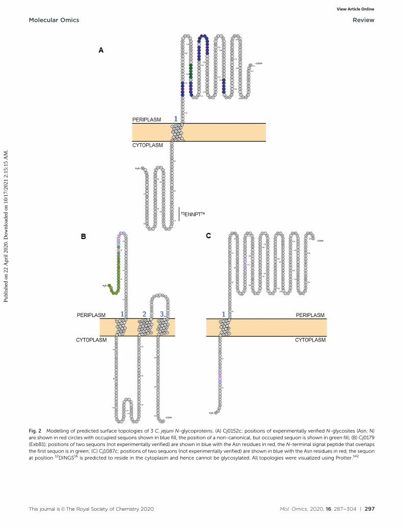
This approach has now yielded the identification of 134 sitesof C. jejuni N-glycosylation from 78 membrane-associated pro-teins that have been experimentally confirmed (predominantlyby MS), including periplasmic proteins, lipoproteins, innermembrane proteins and at least one protein that is thoughtto be surface-exposed (the lipoprotein JlpA101) across 5 C. jejunistrains,33,35,61,63,89 meaning that C. jejuni is likely to be the mostcomplete glycoproteome yet described in the literature(Table 1). Some glycoproteins are modified at multiple sites;for example, the Cj0152c putative membrane protein (whichshares significant sequence similarity with the Helicobacterpylori neuraminidase/sialidase) contains 6 occupied canonicalsites, as well as a single atypical site (Fig. 2A). Cj0152c alsocontains an additional pseudo-sequon (70ENNPT74) that is notoccupied, and that is predicted to be located in the cytoplasmicregion of the protein. Cj0610c (encoding the peptidoglycanO-acetyltransferase PatB) is potentially the most ‘modified’protein in C. jejuni since it contains 5 confirmed N-glycositesand 10 N-sequons in total, all of which are predicted to belocated within the periplasm; structural elucidation of thisprotein could be particularly useful in determining the three-dimensional constraints involved in N-glycan site occupancy(see below). A further 5 proteins (Cj0114, Cj0592c, Cj0843c,Cj1013c and Cj1670c) each contain 4 verified N-glycosites(Table 1). Additionally, eight proteins have been identifiedwith the pEtN-modified N-glycan attached.91 Although thefunction of the pEtN-glycan remains completely unknown,the proteins displaying this modification are amongst themost immunogenic in C. jejuni, including the major antigenPEB3 (Cj0289c), and the previously identified immunogensCjaC (Cj0734c), CjaA (Cj0982c) and JlpA (Cj0983c).17,34,101–103
Despite this, deletion of the eptC pEtN transferase responsiblefor pEtN modification of the N-glycan did not influence thereactivity of these proteins with human serum.91 Further workis required to better understand the occupancy levels of non-to pEtN-modified N-glycan on these glycosites and thus toassist in determining the biological role of the pEtN group inthis context. It is also important to note that a secondCampylobacter species, C. gracilis, exclusively modifies pro-teins with an N-glycan displaying a terminal pEtN group,63
however again, the role of this modification remains to beelucidated.
As discussed above, despite PglB showing a preference forThr at the +2 position,92 there is no obvious bias towards Thr inthe identified N-glycosites; in fact only 60 of 134 identified sitescontain sequons with a Thr in this position (44.8%), with 73containing Ser (54.5%) and the final sequon displaying alanine(Ala) in a non-canonical sequon (Table 1).89 Conversely, there isclear preference for Asp at the �2 position with 84 sequonsdisplaying this amino acid (62.7%) compared with only 47displaying Glu (35.1%). The final 3 sequons were non-canonical (Table 1). These data align with previous studies thathave tested various sequon compositions and their glycosyla-tion efficiency by the PglB OST and found DQNAT to be theoptimal sequon, as well as an B5-fold preference for Asp,rather than Glu, at the �2 position.104
Structural constraints ofN-glycosylation
C. jejuni contains B500 N-glycosylation sequons within thetranslated genome sequence, depending on the strain exam-ined (for example, 510 sequons are found in strain HB93-1335),and B370 of these are found in 4260 predicted membrane-associated proteins (or proteins of unknown localization)suggesting that there are evolutionary constraints associatedwith maintenance of the sequon in proteins connected withthis sub-cellular localization; similar sequon bias (albeitagainst the presence of the sequon) has been observed forthe HMW system in Haemophilus influenzae.105 Realistically,the modifiable N-glycoproteome is likely to be considerablysmaller, given that both topological and structural constraintslikely play a crucial role in the ability of the PglB OST to modifya given sequon. While tools such as PSORTb106 provide pre-dicted sub-cellular localization for a given protein sequence, itis absolutely critical to understand that the topology of theprotein defines PglB sequon accessibility. This is particularlyimportant for proteins associated with the cytoplasmic/innermembrane in Gram negative organisms, since regions withinthese proteins can be cytoplasmic, and thus any sequonscontained within those regions will not be amenable to thecatalytic activity of PglB. For the 134 N-glycosites shown inTable 1, we employed a variety of localization and topologytools that show 133 (99.2%) are predicted to localize to theperiplasm (Fig. 2A and Table 1). Only a single site, 3EI�NKT7
from DsbI (Cj0017c), is predicted to localize to the cytoplasm.This site is challenging to accurately predict given its proximityto the N-terminus and the difficulty in orienting termini intothe inner membrane (inside or outside). Furthermore, the sitewas identified only in a single study35 and was based on a lowscoring, manually validated and very short glycopeptidesequence; an approach no longer valid due to many improve-ments in computational intact glycopeptide analysis (anothersite identified in a similar manner is the non-canonical50AM�NVS54 from Cj0864). Despite the overwhelming associa-tion of experimentally verified N-glycosites with periplasmiclocalization, many sequons in membrane-associated proteinsthat have not been experimentally identified are predicted tolocalize to the cytoplasm (or be located within cleaved signalpeptides) and therefore cannot be glycosylated (for examples,see Fig. 2B and C). Fundamentally, this means that the theore-tical N-glycoproteome of C. jejuni may only comprise between200–250 possible sites.
Beyond localization and topology, the next major influenceon sequon occupancy is the tertiary conformation of theprotein, with the three-dimensional structure of both the targetprotein and PglB itself dictating site accessibility.85,93,107 Unlikein eukaryotes, where N-glycosylation occurs in the endoplasmicreticulum (with further processing in the Golgi apparatus) priorto or during folding (and hence partially dictates the finalconformation), the prevailing viewpoint is that the C. jejuniN-glycan is added to already, or at least partially, foldedsubstrates,93,100 meaning that existing tertiary structural
Molecular Omics Review
Publ
ishe
d on
22
Apr
il 20
20. D
ownl
oade
d on
10/
17/2
021
2:15
:15
AM
. View Article Online

294 | Mol. Omics, 2020, 16, 287--304 This journal is©The Royal Society of Chemistry 2020
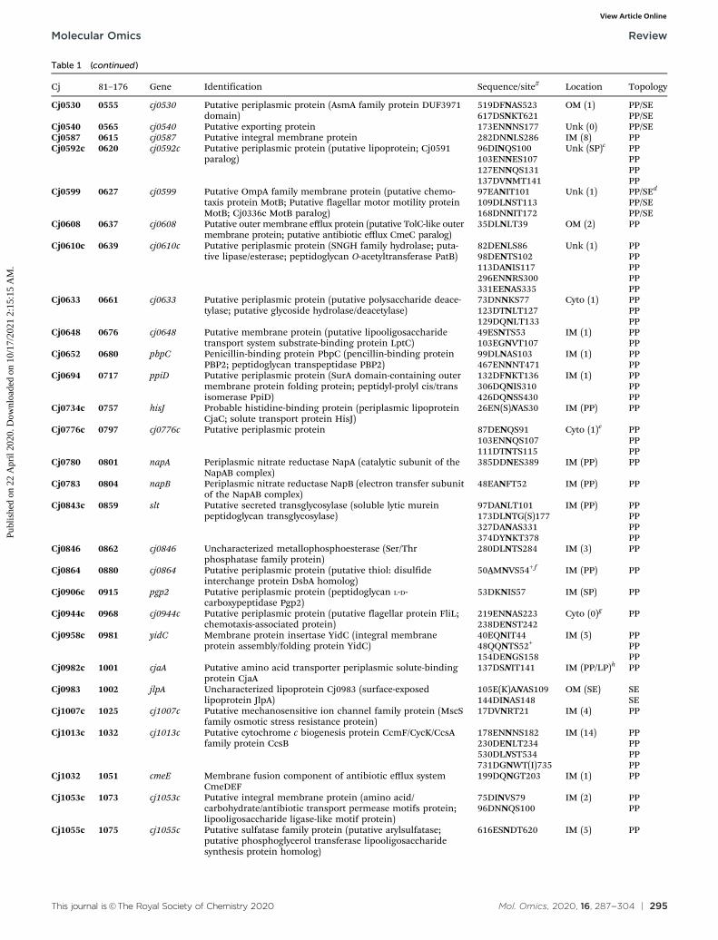
Table 1 Experimentally validated C. jejuni proteins modified by N-glycosylation. Cj, gene identifier from C. jejuni NCTC11168 genome sequence;81–176, gene identifier from C. jejuni 81–176 genome sequence; gene, gene name if known; identification, protein identification taken from NCTC11168UniProt entry (additional information added by BLAST and literature search); sequence/site, sequence is shown for NCTC11168
Cj 81–176 Gene Identification Sequence/site# Location Topology
Cj0011c 0037 cj0011c Putative non-specific DNA-binding protein (competenceComEA-like; natural transformation protein)
49EANFT53 IM (1) PP
Cj0017c 0044 dsbI Disulfide bond formation protein DsbI 3EINKT7 IM (5) Cytoa
Cj0081 0118 cydA Cytochrome bd oxidase subunit I 283DNNES287 IM (9) PP351EN(S)NDT355 PP
Cj0089 0124 cj0089 Putative lipoprotein (TPR tetricopeptide repeat-like helicaldomain protein)
73DFNKS77 LP/IM (SP) PP
Cj0114 0149 cj0114 Putative periplasmic protein (TPR tetricopeptide repeat-likehelical domain protein; putative Tol-Pal system protein YbgF/putative cell division coordinator CpoB)
99ENNFT103 OM PP153DA(V)NLS157 PP171DSNST175 PP177ENNNT181 PP
Cj0131 0166 cj0131 Putative peptidase M23 family protein/putative zinc metallo-peptidase (putative Gly–Gly endopeptidase)
73DDNTS75 Unk (1) PP
Cj0143c 0179 znuA Putative periplasmic ABC transport solute-binding protein(zinc-binding ABC transporter ZnuA)
26E(D)QNTS30 PP PP
Cj0152c 0188 cj0152c Putative membrane protein (45.3% similarity to H. pylorisialidase A/neuraminidase)
126EQNNT130 Unk (1) PP157DNNK�A161+ PP163ETNRT167 PP182DKNIS186 PP188ENNIS192 PP193ENNTT197 PP250DFNIS254 PP
Cj0158c 0194 cj0158c Putative haem-binding lipoprotein (cytochrome c oxidaseCbb3-like protein)
119DKNHS123 LP/OM (SP) PP
Cj0168c 0204 cj0168c Putative periplasmic protein 26DVNQT30 PP (SP) PPCj0176c 0212 cj0176c Putative lipoprotein 29DLNKT33 LP/OM (SP) PPCj0177 ND ctuA/chaN Putative iron transport protein (putative iron-regulated lipoprotein) 83EGNLS87^ IM (1) PPCj0182 0213 cj0182 Putative transmembrane transport protein (ABC transporter
transmembrane family; long chain fatty acid ABC transportprotein; peptide antibiotic transport protein SbmA)
58DSNST62 IM PP70ENNAT74 PP
Cj0199c 0230 Putative periplasmic protein 126DINLS130 Unk (1) PPCj0200c 0231 cj0200c Putative periplasmic protein 33DNNKT37 Unk (SP) PPCj0235c 0260 secG Uncharacterized protein (preprotein translocase subunit
SecG)87ENNNT91 IM (2) PP118DVNSS122 PP
Cj0238 0263 cj0238 Putative mechanosensitive ion channel family protein (MscSfamily membrane integrity protein)
24DANIS28 IM (5) PP56DENSS60 PP
Cj0256 0283 eptC Putative sulfatase family protein (phosphoethanolaminetransferase EptC; lipid A/lipooligosaccharide pEtN transferaseEptC)
213ENNHT217 IM (5) PP
Cj0268c 0295 cj0268c Putative transmembrane protein (SPFH domain/band 7family protein; FtsH protease regulator HflC)
274EANAT278 Unk (1) PP
Cj0277 0304 mreC Homolog of E. coli rod-shape determining protein MreC 91DQNST95 Unk (1) PPCj0289c 0315 peb3 Major antigenic peptide PEB3 (thiosulfate/sulfate-binding
protein)88DFNVS92 Unk (SP) PP
Cj0313 0335 cj0313 Putative integral membrane protein (putative lipooligo-saccharide export ABC transporter permease LptG)
173DLNLS177 IM (6) PP196DGNIT200 PP
Cj0365c 0388 cmeC Outer membrane channel protein CmeC (multi-drugantibiotic efflux system CmeABC protein)
30EANYS34 OM (SP) PP47ENNSS51 PP
Cj0366c 0389 cmeB Efflux pump membrane transporter CmeB (Multi-drugantibiotic efflux system CmeABC protein)
634DRNVS638 IM (12) PP
Cj0367c 0390 cmeA Periplasmic fusion protein CmeA (multi-drug antibiotic effluxsystem CmeABC protein)
121DFNRS125 IM (1) PP271DNNNS275 PP
Cj0371 0395 cj0371 UPF0323 lipoprotein Cj0371 (putative secreted proteininvolved in flagellar motility)
75DLNGT79 LP/OM (SP) PP/SEb
Cj0376 0400 cj0376 Putative periplasmic protein 50DKNQT54 Cyto PPCj0397c 0420 cj0397c Uncharacterized protein 105DFNNT109 Unk (1) PPCj0399 0422 cvpA Colicin V production protein homolog CvpA 179DLNNT183 IM (4) PPCj0404 0428 dedD Putative transmembrane protein (SPOR sporulation domain-
containing protein; putative cell division protein DedD)101EQNNT105 Unk (1) PP
Cj0454c 0479 cj0454c Putative membrane protein 91ENNKS95 IM (1) PPCj0455c 0480 cj0455c Putative membrane protein 60�LQNQT64+ IM (1) PPCj0494 0515 cj0494 Putative exporting protein 26DNNIT30 Unk PP/SECj0508 0536 pbpA Penicillin-binding protein PbpA (penicillin-binding protein
1A; peptidoglycan transpeptidase PBP1A)312DANLS316 IM (1) PP
Cj0511 0539 ctpA Putative secreted protease (protease family S41;carboxy-terminal protease CtpA)
67DQNIS71 IM (1) PP
Cj0515 0543 cj0515 Putative periplasmic protein 207ELNAT211 IM (3) PP234DFNAS238 PP
Review Molecular Omics
Publ
ishe
d on
22
Apr
il 20
20. D
ownl
oade
d on
10/
17/2
021
2:15
:15
AM
. View Article Online
This journal is©The Royal Society of Chemistry 2020 Mol. Omics, 2020, 16, 287--304 | 295
Table 1 (continued )
Cj 81–176 Gene Identification Sequence/site# Location Topology
Cj0530 0555 cj0530 Putative periplasmic protein (AsmA family protein DUF3971domain)
519DFNAS523 OM (1) PP/SE617DSNKT621 PP/SE
Cj0540 0565 cj0540 Putative exporting protein 173ENNNS177 Unk (0) PP/SECj0587 0615 cj0587 Putative integral membrane protein 282DNNLS286 IM (8) PPCj0592c 0620 cj0592c Putative periplasmic protein (putative lipoprotein; Cj0591
paralog)96DINQS100 Unk (SP)c PP103ENNES107 PP127ENNQS131 PP137DVNMT141 PP
Cj0599 0627 cj0599 Putative OmpA family membrane protein (putative chemo-taxis protein MotB; Putative flagellar motor motility proteinMotB; Cj0336c MotB paralog)
97EANIT101 Unk (1) PP/SEd
109DLNST113 PP/SE168DNNIT172 PP/SE
Cj0608 0637 cj0608 Putative outer membrane efflux protein (putative TolC-like outermembrane protein; putative antibiotic efflux CmeC paralog)
35DLNLT39 OM (2) PP
Cj0610c 0639 cj0610c Putative periplasmic protein (SNGH family hydrolase; puta-tive lipase/esterase; peptidoglycan O-acetyltransferase PatB)
82DENLS86 Unk (1) PP98DENTS102 PP113DANIS117 PP296ENNRS300 PP331EENAS335 PP
Cj0633 0661 cj0633 Putative periplasmic protein (putative polysaccharide deace-tylase; putative glycoside hydrolase/deacetylase)
73DNNKS77 Cyto (1) PP123DTNLT127 PP129DQNLT133 PP
Cj0648 0676 cj0648 Putative membrane protein (putative lipooligosaccharidetransport system substrate-binding protein LptC)
49ESNTS53 IM (1) PP103EGNVT107 PP
Cj0652 0680 pbpC Penicillin-binding protein PbpC (pencillin-binding proteinPBP2; peptidoglycan transpeptidase PBP2)
99DLNAS103 IM (1) PP467ENNNT471 PP
Cj0694 0717 ppiD Putative periplasmic protein (SurA domain-containing outermembrane protein folding protein; peptidyl-prolyl cis/transisomerase PpiD)
132DFNKT136 IM (1) PP306DQNIS310 PP426DQNSS430 PP
Cj0734c 0757 hisJ Probable histidine-binding protein (periplasmic lipoproteinCjaC; solute transport protein HisJ)
26EN(S)NAS30 IM (PP) PP
Cj0776c 0797 cj0776c Putative periplasmic protein 87DENQS91 Cyto (1)e PP103ENNQS107 PP111DTNTS115 PP
Cj0780 0801 napA Periplasmic nitrate reductase NapA (catalytic subunit of theNapAB complex)
385DDNES389 IM (PP) PP
Cj0783 0804 napB Periplasmic nitrate reductase NapB (electron transfer subunitof the NapAB complex)
48EANFT52 IM (PP) PP
Cj0843c 0859 slt Putative secreted transglycosylase (soluble lytic mureinpeptidoglycan transglycosylase)
97DANLT101 IM (PP) PP173DLNTG(S)177 PP327DANAS331 PP374DYNKT378 PP
Cj0846 0862 cj0846 Uncharacterized metallophosphoesterase (Ser/Thrphosphatase family protein)
280DLNTS284 IM (3) PP
Cj0864 0880 cj0864 Putative periplasmic protein (putative thiol: disulfideinterchange protein DsbA homolog)
50�AMNVS54+ f IM (PP) PP
Cj0906c 0915 pgp2 Putative periplasmic protein (peptidoglycan L-D-carboxypeptidase Pgp2)
53DKNIS57 IM (SP) PP
Cj0944c 0968 cj0944c Putative periplasmic protein (putative flagellar protein FliL;chemotaxis-associated protein)
219ENNAS223 Cyto (0)g PP238DENST242
Cj0958c 0981 yidC Membrane protein insertase YidC (integral membraneprotein assembly/folding protein YidC)
40EQNIT44 IM (5) PP48�QQNTS52+ PP154DENGS158 PP
Cj0982c 1001 cjaA Putative amino acid transporter periplasmic solute-bindingprotein CjaA
137DSNIT141 IM (PP/LP)h PP
Cj0983 1002 jlpA Uncharacterized lipoprotein Cj0983 (surface-exposedlipoprotein JlpA)
105E(K)ANAS109 OM (SE) SE144DINAS148 SE
Cj1007c 1025 cj1007c Putative mechanosensitive ion channel family protein (MscSfamily osmotic stress resistance protein)
17DVNRT21 IM (4) PP
Cj1013c 1032 cj1013c Putative cytochrome c biogenesis protein CcmF/CycK/CcsAfamily protein CcsB
178ENNNS182 IM (14) PP230DENLT234 PP530DLNST534 PP731DGNWT(I)735 PP
Cj1032 1051 cmeE Membrane fusion component of antibiotic efflux systemCmeDEF
199DQNGT203 IM (1) PP
Cj1053c 1073 cj1053c Putative integral membrane protein (amino acid/carbohydrate/antibiotic transport permease motifs protein;lipooligosaccharide ligase-like motif protein)
75DINVS79 IM (2) PP96DNNQS100 PP
Cj1055c 1075 cj1055c Putative sulfatase family protein (putative arylsulfatase;putative phosphoglycerol transferase lipooligosaccharidesynthesis protein homolog)
616ESNDT620 IM (5) PP
Molecular Omics Review
Publ
ishe
d on
22
Apr
il 20
20. D
ownl
oade
d on
10/
17/2
021
2:15
:15
AM
. View Article Online

296 | Mol. Omics, 2020, 16, 287--304 This journal is©The Royal Society of Chemistry 2020
constraints are a major factor in the final attachment andkinetics of the modification. Sequons buried within the tertiarystructure are therefore inaccessible to PglB and cannot bemodified, irrespective of their sub-cellular location. The earlieststructural consideration of C. jejuni N-glycosylation was basedon the crystal structure of the major antigen PEB3 (Cj0289c),108
which showed the N-glycosite at 88DF�NVS92 occurs in a flexibleexposed loop region readily accessible to the PglB OST. There-fore, without determining structures of glycoproteins itremains difficult to predict which sequons will be occupiedand the likely level of site occupancy, and there are only veryfew N-glycoproteins for which three-dimensional structures arecurrently available. In addition to PEB3, and PglB itself,85
structures of components of the tripartite antibiotic efflux systemCmeABC (Cj0365c–Cj0367c) have also been elucidated,109,110
and all 3 are N-glycoproteins (Table 1). CmeA is the periplasmicmembrane fusion family protein, with 2 N-glycosites bothpredicted to be located within the periplasm (Table 1). CmeCis the outer membrane channel and examination of the crystalstructure109 shows that both experimentally verified glycosy-lated sequons (30EA�NYS34 and 47EN�NSS51) are located in aperiplasmic disordered exposed loop region that leadsfrom the membrane-embedded N-terminal lipidated cysteine(following removal of the signal peptide) to the first structuredpart of the protein. Therefore both sequons are consistent withthe known structural requirements for N-glycosylation.93,99,108
CmeB, which is the inner membrane efflux pump, containsone well characterized N-glycosite (634DR�NVS638). A secondsequon (653DRNAS657) is located proximal to this confirmedsite, but no experimental evidence exists for this site being
Table 1 (continued )
Cj 81–176 Gene Identification Sequence/site# Location Topology
Cj1126c 1143 pglB Undecaprenyl-diphosphooligosaccharide-protein glycosyl-transferase (PglB oligosaccharyltransferase)
532DYNQS536 IM (12) PP
Cj1219c 1232 cj1219c Putative periplasmic protein (uncharacterized proteininvolved in outer membrane biogenesis assembly)
47DVNIT51 OM (1) PP
Cj1345c 1344 pgp1 Putative periplasmic protein (peptidoglycanD-L-carboxypeptidase Pbp1)
59DYNIT63 Cyto (1) PP159EINAS163 PP348DGNET352 PP
Cj1373 1376 cj1373 Putative integral membrane protein (antibiotic resistancesterol-sensing domain protein; RND superfamily exportprotein MmpL family)
134DINRT138 IM (12) PP497DQNTS501 PP
Cj1444c 1438 kpsD Capsule polysaccharide export system periplasmic proteinKpsD
37DQNLS41 IM (PP) PP50ENNLT54 PP
Cj1496c 1488 cj1496c Putative periplasmic protein (putative magnesium transpor-ter MgtE-like protein; putative motility chaperone MotE;putative flagellar protein FliG)
71EVNAT75 Cyto (PP) PP167DNNAS171 PP
Cj1565c 1550 pflA Paralysed flagellar motility protein A PflA 456DNNAS460 Cyto (PP) PP495EGNFS499 PP
Cj1621 1608 cj1621 Putative periplasmic protein 197DLNKT201 E (1) PPCj1661 1652 cj1661 Putative ABC transport system permease (putative antibiotic
macrolide export protein MacB; putative cell division proteinFtsX)
188ENNQS192 IM (4) PP
Cj1670c 1666 cgpA Putative periplasmic protein (campylobacter glycoprotein A;AMIN-domain containing protein, membrane proteinassembly protein)
26DQNIT30 Unk (0) PP71DVNKS75 PP104EKNSS108 PP111ESNST115 PP
ND 0063 sirA Dissimilatory sulfite reductase SirA/MccA 213DGNLS217 IM (1) PPND 0701 kdpC Potassium-transporting ATPase KdpC subunit 83DTNES87 IM (1) PPND 1263 1263 Uncharacterized proteini 26EQNGS30 Unk (SP) PPVirB10 pVir0003 virB10 Type IV secretion system protein VirB10 30EENVS34 OM (SP) PP
95DNNIT99 PP
#Where glycosylation site was identified only in another strain this sequence is shown in (brackets), +non-canonical sequon denoted by underliningof atypical amino acid at �2 or +2 position, Asn (N) highlighted in bold and shaded in italicized bold is also modified by pEtN-modified N-glycan;location, predicted subcellular localization as determined by PSORTb (vers. 3.0.2.)106 and Lipo P 1.0,139 (x) number of predicted transmembraneregions (TMR), or presence of signal peptide (SP), unless experimentally proven all lipoproteins were considered anchored to OM or IM (dependenton Lipo P use of the ‘+2 rule’, Asp at +2 from lipo-Cys predicts IM anchoring, all other amino acids predict OM anchoring) with protein facing intothe periplasm; topology, predicted location of the N-glycosylation site as determined by TmPred (https://embnet.vital-it.ch/software/TMPRED) andTOPCONS.140 Cyto, cytoplasm; E, extracellular; IM, inner membrane; LP, lipoprotein; OM, outer membrane; PP, periplasm; SE, surface exposed;Unk, unknown. ^Site identified by expression in E. coli containing the pgl cluster and over-expression in C. jejuni [H. M. Frost, PhD Thesis,University of Manchester, 2015], not seen in any wild-type C. jejuni glycoproteome studies. a Cj0017c localization depends on correct prediction oforientation for N- and C-terminus of protein. b Cj0371 co-localises to the poles of C. jejuni cells and thus co-localises with flagella.134 Thus, theprotein is potentially surface-exposed (SE). c Cj0592c PSORT b predicts unknown localization; Lipo P predicts a lipoprotein signal peptide with OManchor (Asp at +2 position to SpII cleavage site); protein is described as ‘putative periplasmic protein’. d Cj0599 PSORT b predicts unknownlocalization; protein contains C-terminal OmpA domain suggesting OM localization and therefore topology could be PP or SE. e Cj0776c PSORT bpredicts cytoplasmic localization; 1 predicted TMR; TOPCONS predicts 1 TMR with the majority of the protein localized to the periplasm. f Cj0864Ding et al.141 reported this sequence as DMoxNVS (where the methionine is methionine sulfoxide), however the NCTC11168 sequence indicates the�2 position is an alanine. This sequence was also low scoring as discussed in the text. g Cj0944c PSORT b predicts cytoplasmic localization; Lipo Pand TOPCONS predict 1 SP and periplasmic location. h Cj0982c PSORT b predicts periplasmic localization; TOPCONS and Lipo P predictlipoprotein with IM anchoring. Experimental evidence in ref. 103. i Cjj81176_1263 was originally described in ref. 89 and 91 as CJE1384.
Review Molecular Omics
Publ
ishe
d on
22
Apr
il 20
20. D
ownl
oade
d on
10/
17/2
021
2:15
:15
AM
. View Article Online
This journal is©The Royal Society of Chemistry 2020 Mol. Omics, 2020, 16, 287--304 | 297
Fig. 2 Modelling of predicted surface topologies of 3 C. jejuni N-glycoproteins. (A) Cj0152c; positions of experimentally verified N-glycosites (Asn; N)are shown in red circles with occupied sequons shown in blue fill, the position of a non-canonical, but occupied sequon is shown in green fill; (B) Cj0179(ExbB1); positions of two sequons (not experimentally verified) are shown in blue with the Asn residues in red, the N-terminal signal peptide that overlapsthe first sequon is in green; (C) Cj1087c; positions of two sequons (not experimentally verified) are shown in blue with the Asn residues in red, the sequonat position 12DINGS16 is predicted to reside in the cytoplasm and hence cannot be glycosylated. All topologies were visualized using Protter.142
Molecular Omics Review
Publ
ishe
d on
22
Apr
il 20
20. D
ownl
oade
d on
10/
17/2
021
2:15
:15
AM
. View Article Online
298 | Mol. Omics, 2020, 16, 287--304 This journal is©The Royal Society of Chemistry 2020
occupied in any C. jejuni strain, and hence CmeB is the onlyprotein with both an occupied and unoccupied glycosite forwhich structural information can currently be determined.These two sites are also of interest since their sequons are nearidentical and hence, any effects of differences at the �2 and +2positions, as described above, are likely to be negligible (indeedthe arginine [Arg] at the �1 position is shared, while the +1position is a semi-conservative substitution from valine [Val] toalanine [Ala], which are both aliphatic amino acids) and mostlikely do not influence site occupancy. Interrogation of theCmeB tertiary structure shows that both sequons are located inthe large periplasmic section of the protein located between thesixth and seventh transmembrane-spanning regions (TMR;residues 554–867, with CmeB predicted to contain 11 TMR,excluding the N-terminal signal peptide) and are found inshort disordered exposed loop regions separated by a smallalpha-helix (Fig. 3A). Tertiary structure modelling showsthat 634DR�NVS638 is located close to the membrane and themodified Asn is highly solvent accessible, while 653DRNAS657 islocated further into the periplasm. Although solvent accessible,Asn-655 is partially occluded by Arg-654 (Fig. 3A). The CmeBstructure was next modelled in protein complex with the PglBOST, using the model sequon DQNAT104 to provide the PglBbinding conformation. CmeB/PglB docking clearly demon-strated a preference for the Asn-636 site, consistent with theidentification of this site in several MS-based studies (Fig. 3Band C), while the Asn-655 site does not appear to readilyinteract with the PglB model, and hence therefore is likely toeither not be glycosylated or glycosylated at only very low sitestoichiometry.
Further evidence for structural constraints determiningoptimal glycosylation have been shown for the doubly glycosy-lated surface-exposed glycoprotein JlpA.101 Scott et al. showedthat JlpA must be glycosylated at one site (144DI�NAS148) before asecond site (105EA�NAS109) can be glycosylated, inferring thatstructural modifications to JlpA conferred by Asn-146 glycosyla-tion open the protein conformation and allow PglB to add theN-glycan to the second site. These structural constraints havesince been confirmed using structural predictions andcrystallography.111 Finally, nuclear magnetic resonance (NMR)analysis of a recombinant C. jejuni CmeA domain indicates thatthe N-glycan itself adopts a rigid rod conformation112 thatappears to fold back over the exposed protein (thus suggestinga role in protection from proteolysis), although it remains to beseen how well conserved this is in vivo. Although no exampleshave been shown in the literature, the converse may also betrue in that the N-glycan itself may hinder accessibility of asecond site in a given protein to the PglB OST. Despite thispossibility, proteins such as Cj0152c (Fig. 2A) have multiplesites in close sequence space; occupied sequons are found at7 sites, with 3 (Asn-184, Asn-190 and Asn-195) located within20 amino acids. To determine if N-glycan steric hindrance ofPglB occurs, better understanding of individual site occupancy,in the context of tertiary structures, is needed.
A final structural/topological consideration is the role ofN-glycosylation in OMVs that have been associated with
C. jejuni virulence.24–26 OMVs package cytoplasmic, periplas-mic, outer membrane-associated and N-glycoproteins in a‘bleb’-like structure.24 PglB is located in the cytoplasmic/innermembrane, which is not typically associated with OMVs. It ispossible however, that inner membrane fragments may also bepackaged into OMVs, and all C. jejuni OMV proteomics studieshave demonstrated the identification of integral cytoplasmicmembrane proteins (e.g. CmeB24). Packaging of PglB into OMVsmay enable glycosylation of sites not typically found in themembrane; however despite this, we and others have observedno such cytoplasmic N-glycosites, even at low levels, which mayimply that PglB does not occur in OMVs, or that OMVs are notinduced (or collected) under the culture conditions employedin the N-glycosite discovery studies conducted thus far. C. jejuniOMV composition is however, altered in pgl-negative comparedwith wild-type C. jejuni,113 suggesting N-glycosylation doesimpact protein packaging, although no differences wereobserved in the ability of OMVs from either pgl positive ornegative bacteria to induce an immune response.113
Putative functions of C. jejuni proteinN-glycosylation
Deletion of genes from the pgl cluster (except pglI) results inC. jejuni that are poorly able to colonize chickens and displayreduced adherence to, and invasion of, human epithelialcells.114,115 Additional recent modelling of C. jejuni virulencein a human small intestine-like gut-immune co-culture modelalso revealed that pgl-negative C. jejuni (in this case, pglEdeletion) were vastly deficient (B100 times less) in adherenceand invasion.113 Therefore, N-glycosylation is considered afundamental virulence determinant in this organism. Despitethis, until recently116,117 there have been very few studies thathave broadly characterized pgl-associated phenotypes, and thefunction(s) of the N-glycosylation system in general, and evenmore so the role of the N-glycan on individual proteins,remains almost completely unknown. Genome-wide and func-tional screens have shown an association between glycosylationand biofilm formation, amongst other traits, and pgl-negativeC. jejuni are attenuated in some, but not all, models ofbiofilm growth.116,118 C. jejuni pgl deletion strains incapableof N-glycosylating proteins display a number of additionalaltered traits including a reduced capacity for natural trans-formation,119 altered antibiotic resistance,117 greater suscepti-bility to host-derived proteases,120 impaired motility116 andmodified binding to host cell surface lectins.121 Proteomics-based approaches have been employed in an attempt to globallycharacterize phenotypes associated with N-glycosylation.116,117
pgl negative C. jejuni displayed evidence of induction of thestress response and were attenuated for survival at temperatureextremes,82,116 particularly survival at lower temperatures,which has further implications for the pgl system acting as aninterventional target to limit the presence of the organism onsupermarket chicken for human consumption. Loss of glyco-sylation influenced metabolism and nutrient transport, as well
Review Molecular Omics
Publ
ishe
d on
22
Apr
il 20
20. D
ownl
oade
d on
10/
17/2
021
2:15
:15
AM
. View Article Online

This journal is©The Royal Society of Chemistry 2020 Mol. Omics, 2020, 16, 287--304 | 299
Fig. 3 CmeB modeling with PglB highlighting N-glycosylation sequons. (A) CmeB trimer (Protein Data Bank [PDB]: 5LQ3) has a transmembrane domain(highlighted in blue with 11 TMR) and periplasmic domain. The experimentally validated sequon (634DR�NVS638) is labelled red and the non-identifiedsequon (653DRNAS657) is labelled green, with both Asn labelled cyan. Both Asn are located on the periplasmic side on exposed loops and are solvent-accessible with Asn-636 more accessible than Asn-655; (B) The PglB OST (PDB: 3RCE) shown in yellow has a transmembrane spanning domain(highlighted in blue with 12 TMR) and a larger periplasmic region where the catalytic domain is located. The sequon recognition site is highlighted inorange and facing towards CmeB with the glycan-binding site located behind. Sequon 634DR�NVS638 is in closer proximity and has better accessibility tothe PglB catalytic site; (C) (left) PglB viewed from the front (901 counter-clockwise rotation to upper panels) reveals the sequon-binding surface inorange, (Middle) PglB fitted with the model peptide mimic DQNAT, (right) alignment of CmeB to PglB (901 counter-clockwise rotation to panel B) revealsthat sequon 634DR�NVS638 is more spatially likely to fit into the active site of PglB suggesting this sequon is more readily glycosylated than 653DRNAS657.Analysis was performed in UCSF Chimera 1.14 (build 42094).
Molecular Omics Review
Publ
ishe
d on
22
Apr
il 20
20. D
ownl
oade
d on
10/
17/2
021
2:15
:15
AM
. View Article Online

300 | Mol. Omics, 2020, 16, 287--304 This journal is©The Royal Society of Chemistry 2020
as chemoattraction towards some of these substrates.116
Finally, pgl-negative C. jejuni displayed very strongly reducedabundance of proteins involved in respiration using alternativeelectron acceptors under low oxygen conditions; a phenotypeparamount to C. jejuni colonization of all hosts, and potentiallyassociated with a loss of proton motive force.116 This phenotypecould be linked to reduced nitrate reductase (NapA/NapB)activity, which was shown in both PglB OST (DpglB) and glycanbiosynthesis (DpglFED) negative C. jejuni.116 Since both NapAand NapB are experimentally proven glycoproteins (Table 1),this loss of activity may be associated with a requirement forglycosylation in formation of the NapAB complex, in generatinga structural conformation that maximizes Nap catalysis, or inproviding stability against protein degradation.
The membrane-associated targets of the pgl N-glycosylationsystem are largely functionally uncharacterized ‘putative’ proteins.The remaining proteins share some degree of sequence identitywith well characterized proteins from other organisms, while onlya very small number have been experimentally validated. Exam-ination of the relationships between glycoprotein identifications(Table 1) highlight several clusters of potentially functionallyrelated classes of protein, including those involved in antibioticresistance (all 3 members of the CmeABC antibiotic efflux systemare glycosylated, as is CmeE of the CmeDEF efflux system), andantibiotic resistance has been strongly associated with the pglsystem.117 Additionally, proteins with putative functions, orsequence similarity to proteins, involved in peptidoglycan bio-synthesis, modification and C. jejuni helical cell morphology(Pgp1, Pgp2, MreC, PatB [Cj0610c], Cj0843c and the penicillin-binding proteins PbpA and PbpC), LOS and capsular polysacchar-ide (CPS) transport and assembly (Cj0313/LptG, Cj0648/LptC,Cj1053c, Cj1055c and KpsD), and membrane protein transloca-tion and assembly (SecG, Cj0238, PpiD, YidC, Cj1219c, CgpA) arealso enriched in the 78 identified N-glycoproteins, however thesephenotypes have not yet been tested in pgl negative C. jejuni orN-glycosite mutants.
While several of the above studies have examined pheno-types from the perspective of pgl negative and positive C. jejuni,comparatively fewer studies have attempted to exploit site-directed mutagenesis to understand the role of the N-glycanin individual proteins. This is mainly due to the difficulty ingenerating site mutants in C. jejuni, which is considered poorlytractable and somewhat recalcitrant to molecular biologyapproaches considered standard in species such as E. coli. Despitethis, a limited number of studies have been performed.122–124
N-Glycosite point mutants in cmeA (CmeA contains 2 glycosites;Table 1) have increased susceptibility to several antimicrobialsincluding bile salts and ciprofloxacin, and are attenuated forchicken colonization.125 The PglB OST is also capable oftransferring the N-glycan to itself,61 however recombinant PglBexpressed in otherwise non-glycosylating E. coli remains cap-able of catalyzing the transfer of N-glycans to proteins,62
suggesting PglB does not strictly require modification withthe heptasaccharide to maintain function. Plasmid encodedVirB10 (as well as CmeA, discussed above) was reported torequire N-glycosylation to perform its function (in natural
transformation) at wild-type levels.123 VirB10 is not universallydistributed among strains of C. jejuni, however observationsof impaired natural transformation in the absence ofN-glycosylation have also been observed in studies of theCj0011c N-glycoprotein.126 Several confirmed N-glycoproteins(including DsbI, JlpA, PEB3, EptC, Cj0268c, Cj0371, Cj0454c,Cj0511c/CtpA, Cj0587 and Pgp1/Pgp2) have been associatedwith host colonization;127–134 however, these focused studiesof individual glycoproteins have only rarely attempted to pro-vide evidence of a contribution from the N-glycan, rather thantesting gene-specific deletion mutants. In vitro expression andfunctional analysis of C. jejuni N-glycoproteins in non-pgl-containing E. coli suggest that N-glycosylation is not requiredfor the function of a number of glycoproteins,128,130,135,136
however, without site mutants or comparative expression inpgl-positive expression systems, it is not possible to comparethe functional efficiency of these proteins when glycosylated.
While evidence that C. jejuni protein N-glycosylation occurson folded substrates indicates that the modification is not adriver of protein folding, there is a mounting body of evidenceto suggest that the N-glycan may be important for proteinstability. Mansell et al. demonstrated that the glycoproteinsPEB3, CjaA and PatB/Cj0610c displayed differences in proteinstability in an N-glycosylation competent, pgl system-containingE. coli.137 These proteins also showed altered folding whenglycosylated, further supporting the JlpA evidence thatindicates glycan attachment can alter conformational state.111
Similarly, Min et al. showed an increase in thermostability forrecombinant expressed PEB3 engineered to have an additionalN-glycosylation site in comparison to an unmodified variant.138
Finally, Alemka et al. showed that a pgl-negative strain dis-played reduced viability when cultured under physiologicallevels of human- and chicken-derived proteases,120 which alsosupports the notion that N-glycosylation is involved in confer-ring protein stability.
Conclusions
N- and O-linked glycosylation in C. jejuni are fundamentalrequirements for virulence. Interventions targeting the bio-synthesis of unique bacterial sugars may be useful in the futureto reduce severity of human infection, and in particular to limitserious immune-mediated complications. Additionally, knowl-edge of the biochemistry, structural biology and the manypeptide targets of these pathways provides a unique opportu-nity to better understand the functional roles of these PTM inconferring organism-wide phenotypes, and in specific proteinfunctions. The overall lack of data regarding the function ofN-glycosylation on C. jejuni proteins means that the associationwith virulence remains poorly understood, and could reflect ageneral requirement for glycosylation in a global, C. jejuni-specific process (such as protection against proteolytic degra-dation) and/or that the effect is protein-specific. For the latter,comprehensive knowledge of glycosylation sites is still requireddespite our advances in understanding the glycoproteome in
Review Molecular Omics
Publ
ishe
d on
22
Apr
il 20
20. D
ownl
oade
d on
10/
17/2
021
2:15
:15
AM
. View Article Online

This journal is©The Royal Society of Chemistry 2020 Mol. Omics, 2020, 16, 287--304 | 301
this organism. Therefore, several approaches are needed; (i) afull phenotypic characterization of different pgl mutants that areattenuated for virulence, coupled with multi-omics approachesto determine affected pathways; (ii) a comprehensive analysis ofoccupied and unoccupied N-glycosylation sequons, and theiroccupancy, that can be quantified across many changes inenvironmental or host-specific conditions to create a knowledgebank of sites suitable for mutational analysis; and (iii) testablehypotheses regarding the role of the N-glycan that can beexamined by interventional approaches. Ultimately, it remainslikely that the pgl N-glycosylation system plays a multi-factorialrole in C. jejuni biology that is imperative in environmental,avian and human niches occupied by the organism.
Conflicts of interest
There are no conflicts to disclose.
Acknowledgements
This work was supported in part by the National Health andMedical Research Council (NHMRC) of Australia (Project GrantAPP1106878 to S. J. C.). J. A. C. and A. L. D. are supportedby Australian Government Research Training Program (RTP)Stipends. A. L. D. is additionally supported by the William G.Murrell Postgraduate Scholarship in Microbiology and aUniversity of Sydney Merit Award Supplementary Scholarship.N. S. is supported by an NHMRC Early Career PostdoctoralFellowship.
References
1 P. L. Griffiths and R. W. Park, J. Appl. Bacteriol., 1990, 69,281–301.
2 M. B. Skirrow, Br. Med. J., 1977, 2, 9–11.3 J. P. Butzler, Clin. Microbiol. Infect., 2004, 10, 868–876.4 P. D. Allan, C. Palmer, F. Chan, R. Lyons, O. Nicholson,
M. Rose, S. Hales and M. G. Baker, BMC Public Health,2018, 18, 414.
5 W. A. Awad, C. Hess and M. Hess, Avian Pathol., 2018, 47,352–363.
6 W. B. Sruwe, R. Gough, M. E. Gallagher, D. T. Kenny,S. D. Carrington, N. G. Karlsson and P. M. Rudd, Mol. Cell.Proteomics, 2015, 14, 1464–1477.
7 A. Alemka, S. Whelan, R. Gough, M. Clyne, M. E. Gallagher,S. D. Carrington and B. Bourke, J. Med. Microbiol., 2010, 59,898–903.
8 R. Janssen, K. A. Krogfelt, S. A. Cawthraw, W. van Pelt,J. A. Wagenaar and R. J. Owen, Clin. Microbiol. Rev., 2008,21, 505–518.
9 M. J. Blaser, D. N. Taylor and R. A. Feldman, Epidemiol.Rev., 1983, 5, 157–176.
10 M. Koga, M. Kishi, T. Fukusako, N. Ikuta, M. Kato andT. Kanda, J. Neurol., 2019, 266, 1655–1662.
11 E. F. Wijdicks and C. J. Klein, Mayo Clin. Proc., 2017, 92,467–479.
12 V. Phongsisay, Immunobiology, 2016, 221, 535–543.13 J. A. Goodfellow and H. J. Willison, Nat. Rev. Neurol., 2016,
12, 723–731.14 F. Yoshida, H. Yoshinaka, H. Tanaka, S. Hanashima,
Y. Yamaguchi, M. Ishihara, M. Saburomaru, Y. Kato,R. Saito, H. Ando, M. Kiso, A. Imamura and H. Ishida,Chem. – Eur. J., 2019, 25, 796–805.
15 J. Parkhill, B. W. Wren, K. Mungall, J. M. Ketley,C. Churcher, D. Basham, T. Chillingworth, R. M. Davies,T. Feltwell, S. Holroyd, K. Jagels, A. V. Karlyshev, S. Moule,M. J. Pallen, C. W. Penn, M. A. Quail, M. A. Rajandream,K. M. Rutherford, A. H. M. van Vliet, S. Whitehead andB. G. Barrell, Nature, 2000, 403, 665–668.
16 C. P. Skarp, O. Akinrinade, A. J. Nilsson, P. Ellstrom,S. Myllykangas and H. Rautelin, Sci. Rep., 2015, 5, 17300.
17 S. J. Cordwell, A. C. Len, R. G. Touma, N. E. Scott,L. Falconer, D. Jones, A. Connolly, B. Crossett andS. P. Djordjevic, Proteomics, 2008, 8, 122–139.
18 V. Korolik, Curr. Opin. Microbiol., 2019, 47, 32–37.19 D. J. Bolton, Food Microbiol., 2015, 48, 99–108.20 P. M. Burnham and D. R. Hendrixson, Nat. Rev. Microbiol.,
2018, 16, 551–565.21 M. Stahl, J. Butcher and A. Stintzi, Front. Cell. Infect.
Microbiol., 2012, 2, 5.22 D. Hofreuter, Front. Cell. Infect. Microbiol., 2014, 4, 137.23 M. E. Konkel, J. D. Klena, V. Rivera-Amill, M. R. Monteville,
D. Biswas, B. Raphael and J. Mickelson, J. Bacteriol., 2004,186, 3296–3303.
24 A. Elmi, E. Watson, P. Sandu, O. Gundogdu, D. C. Mills,N. F. Inglis, E. Manson, L. Imrie, M. Bajaj-Elliott,B. W. Wren, D. G. Smith and N. Dorrell, Infect. Immun.,2012, 80, 4089–4098.
25 A. Elmi, A. Dorey, E. Watson, H. Jagatia, N. F. Inglis,O. Gundogdu, M. Bajaj-Elliott, B. W. Wren, D. G. E. Smithand N. Dorrell, Cell. Microbiol., 2018, 20, 3.
26 N. Taheri, M. Fallman, S. N. Wai and A. Fahlgren,J. Proteomics, 2019, 195, 33–40.
27 C. M. Szymanski and B. W. Wren, Nat. Rev. Microbiol.,2005, 3, 225–237.
28 H. Nothaft and C. M. Szymanski, Nat. Rev. Microbiol., 2010,8, 765–778.
29 M. Koomey, Curr. Opin. Struct. Biol., 2019, 56, 198–203.30 A. H. Bhat, S. Maity, K. Giri and K. Ambatipudi, Crit. Rev.
Microbiol., 2019, 45, 82–102.31 J. Poole, C. J. Day, M. von Itzstein, J. C. Paton and
M. P. Jennings, Nat. Rev. Microbiol., 2018, 16, 440–452.32 C. M. Szymanski, R. Yao, C. P. Ewing, T. J. Trust and
P. Guerry, Mol. Microbiol., 1999, 32, 1022–1030.33 N. M. Young, J. R. Brisson, J. Kelly, D. C. Watson,
L. Tessier, P. H. Lanthier, H. C. Jarrell, N. Cadotte, F. St.Michel, E. Aberg and C. M. Szymanski, J. Biol. Chem., 2002,277, 42530–42539.
34 D. Linton, E. Allan, A. V. Karlyshev, A. D. Cronshaw andB. W. Wren, Mol. Microbiol., 2002, 43, 497–508.
Molecular Omics Review
Publ
ishe
d on
22
Apr
il 20
20. D
ownl
oade
d on
10/
17/2
021
2:15
:15
AM
. View Article Online

302 | Mol. Omics, 2020, 16, 287--304 This journal is©The Royal Society of Chemistry 2020
35 N. E. Scott, B. L. Parker, A. M. Connolly, J. Paulech,A. V. Edwards, B. Crossett, L. Falconer, D. Kolarich,S. P. Djordjevic, P. Højrup, N. H. Packer, M. R. Larsen andS. J. Cordwell, Mol. Cell. Proteomics, 2011, 10, M000031.
36 P. Thibault, S. M. Logan, J. F. Kelly, J. R. Brisson,C. P. Ewing, T. J. Trust and P. Guerry, J. Biol. Chem.,2001, 276, 34862–34870.
37 D. J. McNally, A. J. Aubry, J. P. Hui, N. H. Khieu, D. Whitfield,C. P. Ewing, P. Guerry, J. R. Brisson, S. M. Logan andE. C. Soo, J. Biol. Chem., 2007, 282, 14463–14475.
38 I. C. Schoenhofen, E. Vinogradov, D. M. Whitfield, J. R.Brisson and S. M. Logan, Glycobiology, 2009, 19, 715–725.
39 A. I. M. Salah Ud-Din and A. Roujeinikova, Cell. Mol. LifeSci., 2018, 75, 1163–1178.
40 H. S. Chidwick and M. A. Fascione, Org. Biomol. Chem.,2020, 18, 799–809.
41 N. Zebian, A. Merkx-Jacques, P. P. Pittock, S. Houle,C. M. Dozois, G. A. Lajoie and C. Creuzenet, Glycobiology,2016, 26, 386–397.
42 M. Schirm, I. C. Schoenhofen, S. M. Logan, K. C. Waldronand P. Thibault, Anal. Chem., 2005, 77, 7774–7782.
43 G. N. Ulasi, A. J. Creese, S. X. Hui, C. W. Penn andH. J. Cooper, Proteomics, 2015, 15, 2733–2745.
44 C. G. Zampronio, G. Blackwell, C. W. Penn andH. J. Cooper, J. Proteome Res., 2011, 10, 1238–1245.
45 C. P. Ewing, E. Andreishcheva and P. Guerry, J. Bacteriol.,2009, 191, 7086–7093.
46 S. M. Logan, J. P. Hui, E. Vinogradov, A. J. Aubry,J. E. Melanson, J. F. Kelly, H. Nothaft and E. C. Soo, FEBSJ., 2009, 276, 1014–1023.
47 S. L. Howard, A. Jagannathan, E. C. Soo, J. P. Hui,A. J. Aubry, I. Ahmed, A. Karlyshev, J. F. Kelly,M. A. Jones, M. P. Stevens, S. M. Logan and B. W. Wren,Infect. Immun., 2009, 77, 2544–2556.
48 P. Guerry, C. P. Ewing, M. Schirm, M. Lorenzo, J. Kelly,D. Pattarini, G. Majam, P. Thibault and S. Logan, Mol.Microbiol., 2006, 60, 299–311.
49 C. D. Carrillo, E. Taboada, J. H. Nash, P. Lanthier, J. Kelly,P. C. Lau, R. Verhulp, O. Mykytczuk, J. Sy, W. A. Findlay,K. Amoako, S. Gomis, P. Willson, J. W. Austin, A. Potter,L. Babiuk, B. Allan and C. M. Szymanski, J. Biol. Chem.,2004, 279, 20327–20338.
50 I. C. Schoenhofen, D. J. McNally, E. Vinogradov, D. Whitfield,N. M. Young, S. Dick, W. W. Wakarchuk, J. R. Brisson andS. M. Logan, J. Biol. Chem., 2006, 281, 723–732.
51 W. S. Song, M. S. Nam, B. Namgung and S. I. Yoon,Biochem. Biophys. Res. Commun., 2015, 458, 843–848.
52 D. J. McNally, J. P. Hui, A. J. Aubry, K. K. Mui, P. Guerry,J. R. Brisson, S. M. Logan and E. C. Soo, J. Biol. Chem.,2006, 281, 18489–18498.
53 E. S. Rangarajan, A. Proteau, Q. Cui, S. M. Logan,Z. Potetinova, D. Whitfield, E. O. Purisima, M. Cygler,A. Matte, T. Sulea and I. C. Schoenhofen, J. Biol. Chem.,2009, 284, 20989–21000.
54 W. K. Chou, S. Dick, W. W. Wakarchuk and M. E. Tanner,J. Biol. Chem., 2005, 280, 35922–35928.
55 D. Vorkapic, F. Mitterer, K. Pressler, D. R. Leitner, J. H.Anonsen, L. Liesinger, L. M. Mauerhofer, T. Kuehnast,M. Toeglhofer, A. Schulze, F. G. Zingl, M. F. Feldman,J. Reidl, R. Birner-Gruenberger, M. Koomey and S. Schild,Front. Microbiol., 2019, 10, 2780.
56 Y. Fathy Mohamed, N. E. Scott, A. Molinaro, C. Creuzenet,X. Ortega, G. Lertmemongkolchai, M. M. Tunney, H. Green,A. M. Jones, D. DeShazer, B. J. Currie, L. J. Foster, R. Ingram,C. De Castro and M. A. Valvano, J. Biol. Chem., 2019, 294,13248–13268.
57 J. A. Iwashkiw, A. Seper, B. S. Weber, N. E. Scott, E. Vinogradov,C. Stratilo, B. Reiz, S. J. Cordwell, R. Whittal, S. Schild andM. F. Feldman, PLoS Pathog., 2012, 8, e1002758.
58 J. Mahdavi, N. Pirinccioglu, N. J. Oldfield, E. Carlsohn,J. Stoof, A. Aslam, T. Self, S. A. Cawthraw, L. Petrovska,N. Colborne, C. Sihlbom, T. Boren, K. G. Wooldridge andD. A. Ala’Aldeen, Open Biol., 2014, 4, 130202.
59 G. E. Whitworth and B. Imperiali, Glycobiology, 2015, 25,756–766.
60 M. Nita-Lazar, M. Wacker, B. Schegg, S. Amber andM. Aebi, Glycobiology, 2005, 15, 361–367.
61 M. Kowarik, N. M. Young, S. Numao, B. L. Schulz, I. Hug,N. Callewaert, D. C. Mills, D. C. Watson, M. Hernandez,J. F. Kelly, M. Wacker and M. Aebi, EMBO J., 2006, 25,1957–1966.
62 M. Wacker, D. Linton, P. G. Hitchen, M. Nita-Lazar,S. M. Haslam, S. J. North, M. Panico, H. R. Morris, A. Dell,B. W. Wren and M. Aebi, Science, 2002, 298, 1790–1793.
63 H. Nothaft, N. E. Scott, E. Vinogradov, X. Liu, R. Hu,B. Beadle, C. Fodor, W. G. Miller, J. Li, S. J. Cordwell andC. M. Szymanski, Mol. Cell. Proteomics, 2012, 11, 1203–1219.
64 A. J. Jervis, A. G. Wood, J. A. Cain, J. A. Butler, H. Frost,E. Lord, R. Langdon, S. J. Cordwell, B. W. Wren andD. Linton, Glycobiology, 2018, 28, 233–244.
65 J. D. Valderrama-Rincon, A. C. Fisher, J. H. Merritt,Y. Y. Fan, C. A. Reading, K. Chhiba, C. Heiss, P. Azadi,M. Aebi and M. P. DeLisa, Nat. Chem. Biol., 2012, 8, 434–436.
66 V. S. Terra, D. C. Mills, L. E. Yates, S. Abouelhadid,J. Cuccui and B. W. Wren, J. Med. Microbiol., 2012, 61,919–926.
67 H. Nothaft and C. M. Szymanski, Curr. Opin. Chem. Biol.,2019, 53, 16–24.
68 J. Kelly, H. Jarrell, L. Millar, L. Tessier, L. M. Fiori,P. C. Lau, B. Allan and C. M. Szymanski, J. Bacteriol.,2006, 188, 2427–2434.
69 N. B. Oliver, M. M. Chen, J. R. Behr and B. Imperiali,Biochemistry, 2006, 45, 13659–13669.
70 M. Demendi and C. Creuzenet, Biochem. Cell Biol., 2009,87, 469–483.
71 M. J. Morrison and B. Imperiali, J. Biol. Chem., 2013, 288,32248–32260.
72 N. B. Olivier and B. Imperiali, J. Biol. Chem., 2008, 283,27937–27946.
73 A. S. Riegert, J. B. Thoden, I. C. Schoenhofen, D. C. Watson,N. M. Young, P. A. Tipton and H. M. Holden, Biochemistry,2017, 56, 6030–6040.
Review Molecular Omics
Publ
ishe
d on
22
Apr
il 20
20. D
ownl
oade
d on
10/
17/2
021
2:15
:15
AM
. View Article Online
This journal is©The Royal Society of Chemistry 2020 Mol. Omics, 2020, 16, 287--304 | 303
74 A. S. Riegert, N. M. Young, D. C. Watson, J. B. Thoden andH. M. Holden, Protein Sci., 2015, 24, 1609–1616.
75 M. J. Morrison and B. Imperiali, Biochemistry, 2014, 53,624–638.
76 N. B. Olivier, M. M. Chen, J. R. Behr and B. Imperiali,Biochemistry, 2006, 45, 13659–13669.
77 D. Linton, N. Dorrell, P. G. Hitchen, S. Amber, A. V. Karlyshev,H. R. Morris, A. Dell, M. A. Valvano, M. Aebi and B. W. Wren,Mol. Microbiol., 2005, 55, 1695–1703.
78 K. J. Glover, E. Weerapana, M. M. Chen and B. Imperiali,Biochemistry, 2006, 45, 5343–5350.
79 K. J. Glover, E. Weerapana and B. Imperiali, Proc. Natl.Acad. Sci. U. S. A., 2005, 102, 14255–14259.
80 J. M. Troutman and B. Imperiali, Biochemistry, 2009, 48,2807–2816.
81 A. S. Ramırez, J. Boilevin, A. R. Mehdipour, G. Hummer,T. Darbre, J. L. Reymond and K. P. Locher, Nat. Commun.,2018, 9, 445.
82 H. Nothaft, X. Liu, D. J. McNally, J. Li and C. M. Szymanski,Proc. Natl. Acad. Sci. U. S. A., 2009, 106, 15019–15024.
83 C. Perez, S. Gerber, J. Boilevin, M. Bucher, T. Darbre,M. Aebi, J. L. Reymond and K. P. Locher, Nature, 2015,524, 433–438.
84 C. Perez, A. R. Mehdipour, G. Hummer and K. P. Locher,Structure, 2019, 27, 669–678.
85 C. Lizak, S. Gerber, S. Numao, M. Aebi and K. P. Locher,Nature, 2011, 474, 350–355.
86 M. Napiorkowska, J. Boilevin, T. Darbre, J. L. Reymond andK. P. Locher, Sci. Rep., 2018, 8, 16297.
87 X. Liu, D. J. McNally, H. Nothaft, C. M. Szymanski,J. R. Brisson and J. Li, Anal. Chem., 2006, 78, 6081–6087.
88 H. Nothaft, X. Liu, J. Li and C. M. Szymanski, Virulence,2010, 1, 546–550.
89 N. E. Scott, N. B. Marzook, J. A. Cain, N. Solis,M. Thaysen-Andersen, S. P. Djordjevic, N. H. Packer,M. R. Larsen and S. J. Cordwell, J. Proteome Res., 2014,13, 5136–5150.
90 R. Dwivedi, H. Nothaft, B. Reiz, R. M. Whittal andC. M. Szymanski, Biopolymers, 2013, 99, 772–783.
91 N. E. Scott, H. Nothaft, A. V. Edwards, M. Labbate,S. P. Djordjevic, M. R. Larsen, C. M. Szymanski andS. J. Cordwell, J. Biol. Chem., 2012, 287, 29384–29396.
92 S. Gerber, C. Lizak, G. Michaud, M. Bucher, T. Darbre,M. Aebi, J. L. Reymond and K. P. Locher, J. Biol. Chem.,2013, 288, 8849–8861.
93 M. Kowarik, S. Numao, M. F. Feldman, B. L. Schulz,N. Callewaert, E. Kiermaier, I. Catrein and M. Aebi, Science,2006, 314, 1148–1150.
94 C. Lizak, S. Gerber, G. Michaud, M. Schubert, Y. Y. Fan,M. Bucher, T. Darbre, M. Aebi, J. L. Reymond andK. P. Locher, Nat. Commun., 2013, 4, 2627.
95 C. Lizak, S. Gerber, D. Zinne, G. Michaud, M. Schubert,F. Chen, M. Bucher, T. Darbre, R. Zenobi, J. L. Reymondand M. Aebi, J. Biol. Chem., 2014, 289, 735–746.
96 Y. Barre, H. Nothaft, C. Thomas, X. Liu, J. Li, K. K. S. Ngand C. M. Szymanski, Glycobiology, 2017, 27, 978–989.
97 F. Schwarz, C. Lizak, Y. Y. Fan, S. Fleurkens, M. Kowarikand M. Aebi, Glycobiology, 2011, 21, 45–54.
98 Y. Harada, R. Buser, E. M. Ngwa, H. Hirayama, M. Aebi andT. Suzuki, J. Biol. Chem., 2013, 288, 32673–32684.
99 J. J. A. Armenteros, K. D. Tsirigos, C. K. Sønderby,T. N. Petersen, O. Winther, S. Brunak, G. von Heijne andH. Nielsen, Nat. Biotechnol., 2019, 37, 420–423.
100 J. M. Silverman and B. Imperiali, J. Biol. Chem., 2016, 291,22001–22010.
101 N. E. Scott, D. R. Bogema, A. M. Connolly, L. Falconer,S. P. Djordjevic and S. J. Cordwell, J. Proteome Res., 2009, 8,4654–4664.
102 A. Wyszynska, K. Tomczyk and E. K. Jagusztyn-Krynicka,Acta Biochim. Pol., 2007, 54, 143–150.
103 A. Wyszynska, J. Zycka, R. Godlewska and E. K. Jagusztyn-Krynicka, Curr. Microbiol., 2008, 57, 181–188.
104 M. M. Chen, K. J. Glover and B. Imperiali, Biochemistry,2007, 46, 5579–5585.
105 J. A. Gawthorne, N. Y. Tan, U. M. Bailey, M. R. Davis, L. W.Wong, R. Naidu, K. L. Fox, M. P. Jennings and B. L. Schulz,Biochem. Biophys. Res. Commun., 2014, 445, 633–638.
106 N. Y. Yu, J. R. Wagner, M. R. Laird, G. Melli, S. Rey, R. Lo,P. Dao, S. C. Sahinalp, M. Ester, L. J. Foster andF. S. L. Brinkman, Bioinformatics, 2010, 26, 1608–1615.
107 M. Wacker, M. F. Feldman, N. Callewaert, M. Kowarik,B. R. Clarke, N. L. Pohl, M. Hernandez, E. D. Vines,M. A. Valvano, C. Whitfield and M. Aebi, Proc. Natl. Acad.Sci. U. S. A., 2006, 103, 7088–7093.
108 E. S. Rangarajan, S. Bhatia, D. C. Watson, C. Munger,M. Cygler, A. Matte and N. M. Young, Protein Sci., 2007,16, 990–995.
109 C. C. Su, A. Radhakrishnan, N. Kumar, F. Long, J. R. Bolla,H. T. Lei, J. A. Delmar, S. V. Do, T. H. Chou, K. R. Rajashankar,Q. Zhang and E. W. Yu, Protein Sci., 2014, 23, 954–961.
110 C. C. Su, L. Yin, N. Kumar, L. Dai, A. Radhakrishnan,J. R. Bolla, H. T. Lei, T. H. Chou, J. A. Delmar,K. R. Rajashankar, Q. Zhang, Y. K. Shin and E. W. Yu,Nat. Commun., 2017, 8, 171.
111 F. Kawai, S. Paek, K. J. Choi, M. Prouty, M. I. Kanipes,P. Guerry and H. J. Yeo, J. Struct. Biol., 2012, 177, 583–588.
112 V. Slynko, M. Schubert, S. Numao, M. Kowarik, M. Aebiand F. H. Allain, J. Am. Chem. Soc., 2009, 131, 1274–1281.
113 C. Y. Zamora, E. M. Ward, J. C. Kester, W. L. K. Chen,J. G. Velazquez, L. G. Griffith and B. Imperiali, Glycobiol-ogy, 2020, DOI: 10.1093/glycob/cwz105.
114 C. M. Szymanski, D. H. Burr and P. Guerry, Infect. Immun.,2002, 70, 2242–2244.
115 A. V. Karlyshev, P. Everest, D. Linton, S. Cawthraw,D. G. Newell and B. W. Wren, Microbiology, 2004, 150,1957–1964.
116 J. A. Cain, A. L. Dale, P. Niewold, W. P. Klare, L. Man,M. Y. White, N. E. Scott and S. J. Cordwell, Mol. Cell.Proteomics, 2019, 18, 715–734.
117 S. Abouelhadid, S. J. North, P. Hitchen, P. Vohra,C. Chintoan-Uta, M. Stevens, A. Dell, J. Cuccui andB. W. Wren, mBio, 2019, 10, e00297.
Molecular Omics Review
Publ
ishe
d on
22
Apr
il 20
20. D
ownl
oade
d on
10/
17/2
021
2:15
:15
AM
. View Article Online

304 | Mol. Omics, 2020, 16, 287--304 This journal is©The Royal Society of Chemistry 2020
118 B. Pascoe, G. Meric, S. Murray, K. Yahara, L. Mageiros,R. Bowen, N. H. Jones, R. E. Jeeves, H. M. Lappin-Scott,H. Asakura and S. K. Sheppard, Environ. Microbiol., 2015,17, 4779–4789.
119 C. S. Vegge, L. Brøndsted, M. Ligowska-Marzeta andH. Ingmer, PLoS One, 2012, 7, e45467.
120 A. Alemka, H. Nothaft, J. Zheng and C. M. Szymanski,Infect. Immun., 2013, 81, 1674–1682.
121 N. M. van Sorge, N. M. Bleumink, S. J. van Vliet, E. Saeland,W. L. van der Pol, Y. van Kooyk and J. P. van Putten, Cell.Microbiol., 2009, 11, 1768–1781.
122 T. Kakuda and V. J. DiRita, Infect. Immun., 2006, 74,4715–4723.
123 J. C. Larsen, C. Szymanski and P. Guerry, J. Bacteriol., 2004,186, 6508–6514.
124 L. M. Davis, T. Kakuda and V. J. DiRita, J. Bacteriol., 2009,191, 1631–1640.
125 R. K. Dubb, H. Nothaft, B. Beadle, M. R. Richards andC. M. Szymanski, Glycobiology, 2020, 30, 105–119.
126 B. Jeon and Q. Zhang, J. Bacteriol., 2007, 189, 7399–7407.127 S. Jin, A. Joe, J. Lynett, E. K. Hani, P. Sherman and
V. L. Chan, Mol. Microbiol., 2001, 39, 1225–1236.128 E. Frirdich, J. Biboy, C. Adams, J. Lee, J. Ellermeier,
L. D. Gielda, V. J. DiRita, S. E. Girardin, W. Vollmer andE. C. Gaynor, PLoS Pathog., 2012, 8, e1002602.
129 E. Frirdich, J. Vermeulen, J. Biboy, F. Soares,M. E. Taveirne, J. G. Johnson, V. J. DiRita, S. E. Girardin,W. Vollmer and E. C. Gaynor, J. Biol. Chem., 2014, 289,8007–8018.
130 A. V. Karlyshev, G. Thacker, M. A. Jones, M. O. Clementsand B. W. Wren, FEBS Open Bio, 2014, 4, 468–472.
131 V. Novik, D. Hofreuter and J. E. Galan, Infect. Immun., 2008,78, 3540–3553.
132 A. M. Lasica, A. Wyszynska, K. Szymanek, P. Majewski andE. K. Jagusztyn-Krynicka, J. Appl. Genet., 2010, 51, 383–393.
133 A. M. Tareen, C. G. Luder, A. E. Zautner, U. Groß,M. M. Heimesaat, S. Bereswill and R. Lugert, PLoS One,2013, 9, e107076.
134 X. Du, N. Wang, F. Ren, H. Tang, X. Jiao and J. Huang,Front. Microbiol., 2016, 7, 1094.
135 T. W. Cullen and M. S. Trent, Proc. Natl. Acad. Sci. U. S. A.,2010, 107, 5160–5165.
136 X. Zeng, B. Gillespie and J. Lin, Front. Microbiol., 2015,6, 1292.
137 T. J. Mansell, C. Guarino and M. P. DeLisa, Biotechnol. J.,2013, 8, 1445–1451.
138 T. Min, M. Vedadi, D. C. Watson, G. A. Wasney, C. Munger,M. Cygler, A. Matte and N. M. Young, Biochemistry, 2009,48, 3057–3067.
139 O. Rahman, S. P. Cummings, D. J. Harrington andI. C. Sutcliffe, World J. Microbiol. Biotechnol., 2008, 24,2377–2382.
140 K. D. Tsirigos, C. Peters, N. Shu, L. Kall and A. Elofsson,Nucleic Acids Res., 2015, 43, W401–W407.
141 W. Ding, H. Nothaft, C. M. Szymanski and J. Kelly, Mol.Cell. Proteomics, 2009, 8, 2170–2185.
142 U. Omasits, C. H. Ahrens, S. Muller and B. Wollscheid,Bioinformatics, 2014, 30, 884–886.
Review Molecular Omics
Publ
ishe
d on
22
Apr
il 20
20. D
ownl
oade
d on
10/
17/2
021
2:15
:15
AM
. View Article Online
Related Documents