IDENTIFYING PROTEIN PARTNERS OF THE NEURONAL TRANSMEMBRANE PROTEIN NETO2 BY VIVEK MAHADEVAN A thesis submitted in conformity with the requirements for the degree of Master of Science, Department of Molecular Genetics, University of Toronto © Copyright by Vivek Mahadevan 2010

Welcome message from author
This document is posted to help you gain knowledge. Please leave a comment to let me know what you think about it! Share it to your friends and learn new things together.
Transcript
IDENTIFYING PROTEIN PARTNERS OF THE NEURONAL
TRANSMEMBRANE PROTEIN NETO2
BY
VIVEK MAHADEVAN
A thesis submitted in conformity with the requirements for the degree of
Master of Science,
Department of Molecular Genetics,
University of Toronto
© Copyright by Vivek Mahadevan 2010

ii
Abstract
IDENTIFYING PROTEIN PARTNERS OF THE NEURONAL
TRANSMEMBRANE PROTEIN NETO2
Vivek Mahadevan
Master of Science, Department of Molecular Genetics
University of Toronto, Copyright by Vivek Mahadevan 2010
The neuronal transmembrane proteins Neto1 and Neto2, along with different protein
partners, are known to perform multiple roles in diverse processes including axon guidance in
the developing nervous system and synaptic plasticity in the adult brain. My project focuses on
identifying the membrane bound interacting partners of Neto2 using Membrane Yeast Two
Hybrid (MYTH). By performing MYTH screens for the Neto2 molecule using human adult and
embryonic whole brain cDNA libraries, I have identified several novel membrane bound putative
interacting partners including VAMP associated protein B (VAPB) and Glutamate transporter
EAAT3-associated protein (GTRAP3-18), which play diverse functions during the glutamatergic
neurotransmission. Initial studies to validate these interactions in vivo are currently underway by
co-immunoprecipitation approach using mouse brain tissue. If these two candidate proteins are
confirmed to be true interactors, it will open important avenues of research for the Neto2 protein
during excitatory neurotransmission in the mammalian central nervous system.

iii
Acknowledgements
First and foremost, my deepest gratitude goes to my supervisor Dr. Roderick McInnes,
for giving me a wonderful opportunity to be a part of an exciting project in his exciting lab!
Thanks Rod, for sharing your experience, motivation and the quest for excellence. I am going to
go a long way with whatever I learned out of this short stay in your lab. I would like to thank Dr.
Sabine Cordes and Dr. Michael Salter - my committee members, for encouraging me in my baby
steps towards critical thinking and always pushing me further in my efforts. Also, I am grateful
to Dr. Igor Stagljar, for his collaboration and positive suggestions rendered during the whole
project.
This work would not have been possible, if not for the generous help and constructive
criticism from many people from the McInnes lab and also from the Stagljar lab. Foremost in the
list is Zhenya - a post-doc from the McInnes lab, who was an invaluable companion and mentor
during every step of this project. Your immense patience and wealth of knowledge has got me to
where I am right now Zhenya. Thanks so much for all your encouragement!!! I am immensely
grateful to Victoria Wong from the Stagljar lab, for her tremendous patience and timely
suggestions throughout the project. Thank you so much!!!
My heart-felt thank to Cristina, for being such a wonderful support during my stay. Your
spirit of perseverance is worth emulating. Also, all the little discussions we had gave more
colours to the time I spent in the lab. A special thank goes to Jeff, for silently sharing his
sincerity and passion for science. I would also like to thank you for your critical reading and
corrections with the manuscript. I am very much grateful to Lynda, Coco, Alexa, Denize and
Diana for all your unwavering support and words of encouragement. They always mean a lot to
me! I am very grateful to previous McInnes lab members; particularly David, who we fondly call
the ‘Father of the Netos’. You were instrumental in developing the Neto projects to where they
are now. My sincere thank to Dorothy for making the impossible happen every three months!
Without you it would have been impossible to set up the dates for the committee meetings.

I would also like to acknowledge Dr. Shiv Kumar Sharma and his lab at the National Brain Research Centre, India, for getting me started in this intriguing field of Neuroscience.
I am deeply indebted to all my friends; particularly, Meera, Vijay, Santhosh, Navin and Arvind. Your mere presence and comradeship will give me the courage and strength to move mountains!!!
My Family!!! How can I ever thank you all? Or even think of it? My dearest Amma, Meera, Radha, Govind and Surabhi, you all hold the key to my life and everything in it. Love you all immeasurably and unconditionally.
Lead us from untruth to the truth. Lead us from ignorance to knowledge.
Lead us from death to immortality.
And let there be peace, peace and peace!
(An ancient Indian aphorism)
iv

v
Table of contents
Abstract .................................................................................................................................... ii
Acknowledgements.................................................................................................................. iii
Table of contents....................................................................................................................... v
List of figures............................................................................................................................ x
List of tables............................................................................................................................. xi
Foreword................................................................................................................................. xii
Chapter 1: Introduction........................................................... 1
Part 1 - Introduction to the synapse ........................................................................2
1.1.1 The structure of the chemical synapse .......................................................................... 4
1.1.2 Ion channels and receptors at the synapse ..................................................................... 8
1.1.3 Additional regulatory protein partners for the ion channels and receptors ................... 13
Part 2 - Introduction to the Neto proteins..............................................................16
1.2.1 Neto1 and Neto2 ........................................................................................................ 16
1.2.2 Neto proteins and the glutamate receptors................................................................... 20
1.2.3 Known interacting partners of the Neto proteins......................................................... 24
Part 3 - Introduction to Membrane Yeast Two Hybrid (MYTH)...........................27
1.3.1 Split-ubiquitin based MYTH – A novel approach to identify the interactome of the
neuronal transmembrane protein Neto2 ...................................................................... 27
1.3.2 Rationale to the use of MYTH strategy....................................................................... 30
1.3.3 Specific objectives and rationale of the thesis ............................................................. 31
1.3.4 Advantages and limitations of MYTH ........................................................................ 33

vi
Chapter 2: Materials and methods ....................................... 36
Part 1 - Summary of materials ..............................................................................37
Part 2 - Construction of the Neto2 bait plasmid ....................................................38
2.2.1 Primer design for the Neto2- MYTH .......................................................................... 38
2.2.2 PCR amplification of the Neto2 cDNA using MYTH primers..................................... 38
2.2.3 pNeto2-Cub-fusion bait construction by in vivo homologous recombination strategy . 39
2.2.4 Identification of putative Neto2-Cub-fusion clones, sequencing, and verification......... 41
Part 3 - Verification of the bait protein expression and localization......................43
2.3.1 Preparation of total protein extracts and verification of the Neto2-fusion protein
expression .................................................................................................................. 43
2.3.2 Immunoprecipitation of the bait protein using Neto2-ectodomain specific antibody.... 45
2.3.3 Preparation of membrane protein extracts and verification of the Neto2-fusion
protein localization..................................................................................................... 46
Part 4 - Verification of the Neto2-bait protein folding using a functional assay....47
2.4.1 NubG / NubI test for the Neto2 bait-bearing yeast......................................................... 47
Part 5 - Pilot screening and optimization of the screening conditions ...................48
2.5.1 Large scale transformation of the empty prey plasmid into Neto2 bait-bearing yeast
and 3-aminotriazole titration....................................................................................... 48
Part 6 - cDNA library screening using the Neto2 bait ...........................................50
2.6.1 Large scale transformation of a human adult / human embryonic, whole brain............ 50
cDNA library into the Neto2 bait-bearing yeast .......................................................... 50
2.6.2 Selection of clones in the growth assay (white-pink selection).................................... 51
2.6.3 Selection of clones in the lacz assay (blue-white selection)......................................... 51

vii
Part 7 - Identification of the positive clones from the reporter assays ...................52
2.7.1 Colony PCR for the reporter assay-positive clones ..................................................... 52
2.7.2 Sequencing of the clones and BLAST analysis ........................................................... 53
2.7.3 Prey plasmid recovery from the putative interacting bait:prey-bearing yeast and
retransformation in E. Coli ......................................................................................... 54
Part 8 - Controls for the Neto2-MYTH screen ......................................................54
2.8.1 Construction of a positive control prey plasmid (Human Neto1) for the Neto2 bait..... 54
2.8.2 Construction of a negative control bait plasmid (Human CD4) for the cDNA prey ..... 55
Part 9 - Identification of true positives in the Neto2 MYTH screen ......................56
2.9.1 Bait dependency test................................................................................................... 56
2.9.2 Re-sequencing and verification of the prey plasmids .................................................. 57
Part 10 - Possible outcomes of the Neto2 MYTH screen based on the choice of
cDNA libraries.......................................................................................58
Chapter 3: Results and discussion ........................................ 59
Part 1 - The Neto2-Cub bait plasmid was generated by yeast homologous
recombination .........................................................................................60
Part 2 - The Neto2-Cub bait protein is expressed within the yeast membrane ........62
Part 3 - The Neto2-Cub-fusion protein is folded and functional in the
NubG / NubI test .......................................................................................66
Part 4 - 25 mM of 3-aminotriazole reduces the background colonies in the ..........69
pilot screen .............................................................................................69
Part 5 - cDNA library screens using the Neto2-bait yielded several positive

viii
clones......................................................................................................71
Part 6 - True Neto2-bait-dependent prey interactions were identified ...................73
Part 7 - Multiple false positives were eliminated at different stages during the
Neto2-MYTH screen ...............................................................................77
Part 8 - MYTH identifies 13 novel putative Neto2 interacting proteins ................82
3.8.1 Results from the Neto2-adult cDNA library screen..................................................... 82
3.8.2 Results from the Neto2-embryonic cDNA library screen ............................................ 88
Part 9 - Criteria used to choose molecules from the MYTH screen for
subsequent analyses ................................................................................90
Chapter 4: Conclusions and future directions ..................... 91
Part 1 - A brief summary of the project and major findings from the Neto2-
MYTH screen ..........................................................................................92
Part 2 - The most promising putative-Neto2 interactors for follow-up studies ......94
4.2.1 VAMP-associated protein B (VAPB) ......................................................................... 94
4.2.2 Current knowledge about VAPB ................................................................................ 95
4.2.3 Glutamate transporter EAAT3-associated protein (GTRAP3-18)................................ 99
4.2.4 Current knowledge about GTRAP3-18..................................................................... 100
Part 3 - Proposed experiments for Neto2 and VAPB .......................................... 103
Part 4 - Proposed experiments for Neto2 and GTRAP3-18................................. 106
Part 5 - Conclusions ........................................................................................... 107

ix
Appendices............................................................................ 109
Appendix I - List of strains, antibodies, plasmids and primers............................ 110
Appendix II – Additional details about the MYTH bait vector pAMBV and
prey vector pPR3-N..................................................................... 112
Appendix III - Brief description of the MYTH cDNA libraries .......................... 115
Appendix IV - Standard reagent recipies ............................................................ 116
References ............................................................................. 121

x
List of figures
Figure 1.1- An electron micrograph of an excitatory synapse 6
Figure 1.2- A schematic representation for the mechanism of nerve impulse transmission at
excitatory chemical synapse 7
Figure 1.3- Different types of membrane proteins at the neuronal membrane 11 Figure 1.4- Known AMPAR associated proteins at the PSD 15
Figure 1.5- Domain organisation of the Neto and its related proteins in vertebrates and
Invertebrates 18
Figure 1.6- Association of the known CUB domain proteins with components of ionotropic
Receptors 23 Figure 1.7- Some of the known interacting partners of Neto1 and Neto 2 26
Figure 1.8- Overview of the Membrane Yeast Two Hybrid technology 29
Figure 3.1- Generation of pNeto2- Cub fusion plasmid 61
Figure 3.2- Verification of expression and localization of Neto2- Cub fusion protein 64
Figure 3.3- Verification of folding and functionality of Neto2- Cub fusion protein 68
Figure 3.4- 3-aminotriazole titration for reducing the background colonies 70
Figure 3.5- Identification of putative-Neto2 interacting clones from the MYTH screen 75
Figure 4.1- Domain organisation of VAPB 98
Figure 4.2- Domain organisation of GTRAP3-18 102
Figure 4.3- Possible novel functions for Neto2 during the fine tuning of glutamatergic
neurotransmission 108

xi
List of tables
Table 1.1 Distinguishing properties of electrical and chemical synapses 3
Table 1.2 Important synaptic ionotropic receptors and ion channels that are known
to facilitate synaptic plasticity 12
Table 3.1 Putative Neto2-interactors from the adult library 83
Table 3.2 Putative Neto2-interactors from the embryonic library 88

xii
Foreword
Thesis organisation: This thesis reports the interacting partners of the neuronal transmembrane
protein Neto2, by applying an approach called Membrane Yeast Two Hybrid (MYTH), and
describes briefly these novel findings. For ease of understanding, the Introduction (Chapter 1) is
split into three parts, beginning with a brief sketch and discussion of the structural and molecular
organization of synapse. The next part introduces the neuronal protein Neto2 and the current
knowledge about its biology. The third part delineates the MYTH approach and the rationale for
applying this approach towards the study of Neto2. The Materials and methods section (Chapter
2) details the experimental techniques used in the thesis. The Results and discussion section
(Chapter3) briefly reports the result of the MYTH screens performed, and discusses these
findings. Also, this chapter gives a particular emphasis on the important controls that are critical
for the different experiments used during the MYTH technique. Finally, the Future directions
section (Chapter 4) reviews the two important Neto2-putative interactors identified by the
MYTH screen, namely VAMP associated protein B (VAPB) and Glutamate transporter3-18
(GTRAP3-18), and the functional relevance of such interactions with Neto2

1
Chapter 1: Introduction

2
Part 1 - Introduction to the synapse
The human brain is the most complex biological entity known. The complexity and
diversity of the nervous system is made possible by numerous interconnections between the
nerve cells (neurons). Synapses are the functional connections between neurons, or between
neurons and other cell types. The human brain has about 1011 neurons and each has on average
1,000 – 10,000 synaptic connections to other neurons. It has been estimated that the total number
of synapses in an adult brain is about 1014 to 1015 (Kandel and Schwartz 1981). Most synapses
connect the axon of one neuron to the dendrite of another. There are also other types of synapses
including the connections between axon : axon, axon : cell body, and dendrite : dendrite of
adjacent neurons (Noback 2005).
There are two types of synapses distinguished on the basis of their mechanism of
transmission - electrical and chemical (Table 1.1). At electrical synapses, ions flow through gap
junctions which are specialized membrane channels that connect two cells and they are
composed of connexin proteins. Connexins form clusters of hemi-receptors and associate with
the hemi-receptors of adjacent neurons, forming a continuous link allowing the rapid exchange
of ions and thus electrical signals (Connors and Long 2004).
In contrast, chemical synapses enable cell-to-cell communication via the release of
chemical agents called neurotransmitters; neurotransmitters released by the presynaptic neuron
produce a corresponding effect on the postsynaptic neuron (Purves 2008). The chemical synapse
is referred to as excitatory when synaptic activity drives the postsynaptic neuron above its firing
threshold. This mode of chemical transmission is mediated through neurotransmitters such as
glutamate and dopamine. In contrast, chemical synapses are called inhibitory whe n synaptic

3
activity drives the postsynaptic neuron below its firing potential. This mode of chemical
transmission is mediated through neurotransmitters such as γ-aminobutyric acid (GABA) and
glycine (Kandel and Schwartz 1981).
Table 1.1 Distinguishing properties of electrical and chemical synapses
Type of
synapse
Distance between pre
and post synaptic
membranes
Ultrastructural
components
Synaptic delay Agent /
Direction of
Transmission
Electrical 3.5 nm Gap-junction channels Virtually absent Ion current/
Bidirectional
Chemical 20 – 40 nm Presynaptic vesicles and
active zones; postsynaptic
receptors
Significant:
0.3 - 5 ms long
Chemical
transmitter/
Unidirectional
Modified from (Kandel and Schwartz 1981)

4
1.1.1 The structure of the chemical synapse
The majority of the synapses in the nervous system follow the chemical mode of nerve
impulse transmission (Greengard 2001) and thus, chemical transmission between neurons is the
first level of complexity towards the understanding of higher-order brain functions like learning,
memory, and other complex behaviour (Kandel 2001). A chemical synapse is characterized by
unidirectional transmission of information directly from the presynaptic cell to the postsynaptic
cell. In an axodendritic chemical synapse, the presynaptic terminal or the synaptic boutons are
specialized regions of the termination of the presynaptic axon that contain chemical messengers
called neurotransmitters enclosed within membrane-bound synaptic vesicles (SV). These vesicles
are docked at the presynaptic plasma membrane at regions called active zones (AZ) (Rettig and
Neher 2002). Directly opposite to the synaptic bouton is the dendritic membrane of the
postsynaptic cell that contains neurotransmitter receptors. Immediately behind the postsynaptic
membrane is an electron dense region called the postsynaptic density (PSD), which contains
several multi-protein complexes (Sheng and Kim 2002) found in specialized protrusions from the
main dendritic shaft called dendritic spines (Kennedy 2000). The presynaptic and the
postsynaptic membranes are separated by a synaptic cleft that is 20nm wide (Figure 1.1).
Nerve impulse transmission at the chemical synapse is based on a series of sequential
events as described in Figure 1.2. The process begins when an action potential reaches the
presynaptic axon terminal and alters the local resting membrane potential. This change results in
the opening of a type of voltage-sensing ion channels present on the presynaptic membrane that
allow a huge influx of Ca2+ ions into the presynaptic axon terminal, and leads to a transient
increase in the Ca2+ concentration in the cytoplasm. The elevated Ca2+ concentration triggers the

5
fusion of synaptic vesicles with the plasma membrane of the presynaptic neuron. The Ca2+-
dependent fusion of the synaptic vesicles with the terminal membrane causes the release of
neurotransmitters into the synaptic cleft and marks the conversion of the electrical signal into a
chemical signal at the presynaptic terminal - a hallmark of the chemical mode of synaptic
transmission (Purves 2008).
Following exocytosis, the neurotransmitters diffuse across the synaptic cleft to bind to
specific receptors on the membrane of the postsynaptic terminal of the neuron. Following the
neurotransmitter binding, a complex interplay between multiple ion channels, transporters,
pumps and several other proteins lead to ion fluxes within the postsynaptic terminal. This ion
flux alters the membrane potential in such a way that it increases or decreases the probability for
that neuron to fire an action potential. For example at an excitatory synapse, the binding of
excitatory neurotransmitters to their cognate receptors at the postsynaptic membrane leads to a
net increase of Na+ ions within the postsynaptic terminal to produce an excitatory postsynaptic
potential (EPSP). Conversely, at an inhibitory synapse, the binding of inhibitory
neurotransmitters to their cognate receptors at the postsynaptic membrane leads to a net increase
of Cl- ions within the postsynaptic terminal to produce an inhibitory postsynaptic potential
(IPSP). Thus, the chemical signal is converted back into an electrical signal at the postsynaptic
terminal and re-establishes the propagation of nerve impulse transmission (Purves 2008).

6

7

8
1.1.2 Ion channels and receptors at the synapse
Apart from mediating neural communication, chemical synapses are also involved in
altering the strength of connection between adjacent neurons, a phenomenon termed synaptic
plasticity. More than 50 years ago, Donald Hebb postulated that the modulation of synaptic
strength underlies complex cognitive functions like learning and memory. He proposed that the
synapses between neurons are strengthened during concerted pre-and postsynaptic neuronal
activity, and they are weakened by non-coincidental firing (Turrigiano and Nelson 2000; Purves
2008). It is now clear that the repeated neural activity which is the basis for plasticity as
postulated by Hebb is mediated through the different types of neurotransmitter receptors, ion
channels, pumps and other proteins present on the neuronal membrane (Figure 1.3) (Malenka
and Nicoll 1999; Greengard 2001; Sheng and Kim 2002; Voglis and Tavernarakis 2006).
There exist different types of synaptic membrane proteins that mediate the two categories
of communication between the neurons, referred to as slow and fast synaptic transmission. Slow
neurotransmission employs G-protein coupled (metabotropic) receptors, and occurs over periods
of hundreds of milliseconds to minutes. Activation of metabotropic receptors leads to the
activation of downstream signalling cascades, which in turn results in multiple cellular changes
including the transcription of genes required for plasticity and alteration of the properties of other
molecules involved in neurotransmission. (Greengard 2001). On the other hand, rapid
neurotransmission employs ionotropic receptors to mediate neural communication within
1/1000th of a second. Ionotropic receptors are specialised synaptic ion channels that are gated by
the binding of specific neurotransmitters. Upon activation these ionotropic receptors together
with other ion channels cause ion fluxes within the postsynaptic terminal leading to a fast

9
excitation or inhibition (depending upon the neurotransmitter employed) (Voglis and
Tavernarakis 2006). Some important synaptic ion channels and receptors which are known to
directly facilitate synaptic plasticity are reviewed in Table 1.2 (Voglis and Tavernarakis 2006).
Additionally, there are other classes of membrane proteins like neurotransmitter transporters and
cell adhesion molecules that indirectly regulate synaptic plasticity by modulating the dynamics
of the released neurotransmitters within the synapse (Amara and Fontana 2002; Tzingounis and
Wadiche 2007) and by regulating the activities of neurotransmitter receptors (Dityatev, Bukalo et
al. 2008)
Of particular relevance to this thesis is glutamatergic neurotransmission and the synaptic
membrane proteins involved in this particular neurotransmission at excitatory synapses. The
ionotropic glutamate receptors are the principal class of molecules that directly mediate
glutamatergic neurotransmission. These receptors are multimeric assemblies of individual
subunits. The ionotropic receptors are subdivided into three groups, the N-methyl-D-aspartate
(NMDA) receptors, α-amino-3-hydroxyl-5-methyl-4-isoxazole-propionic acid (AMPA) receptors
and the kainate receptors, each named after their ability to be activated by their respective
synthetic glutamate analogs. These receptors are ligand-gated cation channels that allow the flow
of K+, Na+ and Ca2+ in response to glutamate binding (Ozawa, Kamiya et al. 1998). These
receptors are associated with multiple scaffolding proteins like PSD 95, SAP 97, GRIP and
PICK1 that anchor the receptors within the PSD. They are also associated with several signalling
molecules that perform various cellular processes leading to synaptic plasticity at excitatory
synapses (Sheng and Kim 2002).
In addition to the ionotropic glutamate receptors, the other important class of synaptic
membrane proteins that regulate glutamatergic neurotransmission are the glutamate transporters

10
belonging to the family of excitatory amino acid transporters (EAAT). The 5 subtypes of EAAT
(1-5) are Na+-dependent, high-affinity glutamate transporters that clear glutamate from synapses
released during neurotransmission. Glutamate mediates normal neurotransmission only when its
concentration is regulated at the synapse. Excess glutamate can excessively activate glutamate
receptors leading to neuronal death, a phenomenon termed excitotoxicity (Sheldon and
Robinson 2007). Hence, the EAATs act as molecular buffers for synaptic glutamate, thereby
preventing neuronal cell death by glutamate excitotoxicity (Amara and Fontana 2002;
Tzingounis and Wadiche 2007). Recent studies also indicate that a subtype of EAAT indirectly
modulates synaptic plasticity by regulating the recruitment of extrasynaptic and perisynaptic
NMDARs in hippocampal CA1 neurons (Scimemi, Tian et al. 2009).
The defective individual functions of ionotropic glutamate receptor complexes or their
individual components, along with the defective function of glutamate transporters are important
causes of several central nervous system disorders including dementia, psychiatric illnesss, and
epilepsy (Lau and Zukin 2007; Bowie 2008; Vincent and Mulle 2009), as well as
neurodegenerative diseases (Sheldon and Robinson 2007) . It is likely that a broader
understanding of the biology of glutamate-mediated neurotransmission and its individual
components, will contribute to understanding pathobiology.

11

12

13
1.1.3 Additional regulatory protein partners for the ion channels and receptors
Ionotropic receptor function at synapses is modulated by multiple factors, including
alternate splicing of the individual subunits, subunit composition of the heteromer, and post-
translational modification of the receptor (Wilding and Huettner 2001; Cull-Candy and
Leszkiewicz 2004; Greger, Ziff et al. 2007). Also, receptor trafficking to the surface, timing of
expression at the neuronal membrane, endocytosis, and abundance within the PSD are additional
factors that further fine tune receptor functions (Shepherd and Huganir 2007; Coussen 2009;
Groc, Bard et al. 2009). The emerging theme in this field is that the core subunit of these ligand-
gated synaptic ion channels and receptors associate with additional transmembrane protein
partners to regulate receptor function (Gally, Eimer et al. 2004; Zheng, Mellem et al. 2004;
Milstein and Nicoll 2008; Coombs and Cull-Candy 2009; Ng, Pitcher et al. 2009; Sager, Tapken
et al. 2009; Zhang, St-Gelais et al. 2009).
One widely studied example of this association occurs with AMPARs and the
transmembrane AMPA receptor regulatory proteins (TARPs). Directly interacting with the
AMPAR subunits, TARPs are thought to act like chaperones for AMPARs, assisting their proper
folding and assembly within the endoplasmic reticulum. More importantly, the TARPs
participate in delivery and synaptic localisation of AMPARs, facilitate stabilisation within the
PSD, as well as their lateral diffusion between the extrasynaptic and synaptic regions. Also,
TARPs alter the biophysical properties of AMPARs by increasing the glutamate-evoked current
and by reducing their desensitization following glutamate binding (Collingridge, Isaac et al.
2004; Sager, Tapken et al. 2009). Therefore the AMPAR and TARP complex, along with several

14
other AMPAR associated proteins, perform various cellular processes leading to synaptic
plasticity (Figure 1.4).
Although critical interacting partners for the core subunit of other ionotropic glutamate
receptors have been identified (Zheng, Brockie et al. 2006; Ng, Pitcher et al. 2009; Zhang, St-
Gelais et al. 2009), a much more elaborate study to understand the precise functional role of such
interactions is required in order to have a clearer picture of their precise roles during
glutamatergic neurotransmission. These studies will not only broaden our understanding of
normal brain function, but also increase knowledge about different pathological states.

15

16
Part 2 - Introduction to the Neto proteins
1.2.1 Neto1 and Neto2
Previously, in an approach to utilize the existing expressed sequence tag (EST) data to
attribute potential functions for novel proteins, an in silico screen of human retinal ESTs was
performed. Two molecules encoding the CUB domain protein:protein interaction motif were
identified in the screen. These proteins were designated neuropilin-1/tolloid-like (Neto), based
on their sequence similarity with the known axon guidance protein neuropilin, (He and Tessier-
Lavigne 1997) and the dorso-ventral patterning protein tolloid (Shimell, Ferguson et al. 1991).
The Neto1 and Neto2 genes encode two closely related, evolutionarily conserved type I
transmembrane neuronal proteins with their C-terminus facing the cytoplasm and N-terminus
facing the extracellular space at the neuronal membrane (Stohr, Berger et al. 2002; Michishita,
Ikeda et al. 2003; Michishita, Ikeda et al. 2004). Neto1 (533 amino acids) and Neto2 (525 amino
acids) contain two extracellular CUB domains (for Complement C1r/C1s, Uegf, and Bmp 1), one
LDLa motif (for low-density lipoprotein receptor domain class A), a single pass transmembrane
domain, and a cytoplasmic tail (Figure 1.5). The cytoplasmic tails of Neto1and Neto2 are 168
and 157 amino acids long respectively.
The CUB1, CUB2 and LDLa domains (ectodomain) of vertebrate Neto1 and Neto2 share
63%, 71%, and 83% amino acid identity respectively, while their cytoplasmic domain
(endodomain) shares only 39% identity. Both Neto1 and Neto2 have C-terminal PDZ ligands.
While Neto1 has a class I PDZ tripeptide, Neto2 terminates in a putative class II PDZ tripeptide.
This similarity suggests that Neto1 and Neto2 may bind to similar proteins on their extracellular
side but may associate with different intracellular partners. There are also Neto-like proteins in

17
invertebrates that have similar domain organisation to the Neto proteins such as Q9XUU2 in C.
elegans and Q9VYC7 in Drosophila melanogaster (Zhang, St-Gelais et al. 2009) and other CUB
domain proteins with less similar domain organisation to the Neto proteins like SOL-1 and LEV-
10 in C. elegans (Gally, Eimer et al. 2004; Zheng, Mellem et al. 2004).
In the mature mouse brain, Neto1 and Neto2 display both overlapping and distinct
patterns of expression. Both are strongly expressed in the cerebral cortex, olfactory bulb,
olfactory tubercle, pons and hippocampus. However, in cerebellum Neto1 is only expressed in
Purkinje cells, whereas Neto2 is expressed in both Purkinje and granule cell layers. In the
hippocampus Neto1 mRNA is seen throughout the pyramidal and granule cell layer with strong
expression in CA3 pyramidal cells. In contrast, Neto2 is uniformly expressed in the pyramidal
cells and weakly expressed in the granule cells (Michishita, Ikeda et al. 2003; Michishita, Ikeda
et al. 2004; Ng, Pitcher et al. 2009; Zhang, St-Gelais et al. 2009).

18

19
To elucidate the biological functions of the Neto proteins, a previous graduate student in
the lab (David Ng) generated mice lacking each of the Neto1 and Neto2 genes (Neto1-/- mice and
Neto2-/- mice) and observed several biochemical changes and behavioral phenotypes in the
knockout animals. Both Neto1-/- and Neto2-/- mice showed axon-guidance defects during
development (Ng 2006), synaptic plasticity defects (Ng, Pitcher et al. 2009) and neuronal
excitability defects in adults (Ng 2006).
The developmental defects in Neto-null mice can be ascribed to the axon guidance
functions of the Neto proteins, similar to the phenotypes observed in loss of function mouse
mutants of the molecules of the Neuropilin family. The more surprising adult phenotypes such as
learning and memory impairments and myoclonic seizures seen in Neto-null mice further
indicated that the Neto proteins might be involved in a wide variety of CNS functions, both
during early development and in adults. Several biochemical fractionation tests were performed
to identify the precise subcellular localisation of the Neto proteins in vivo, and the results
indicated that both Neto1 and Neto2 are present at the postsynaptic membrane and within the
PSD of excitatory synapses in the mouse brain, and that each interacts with multiple components
of the PSD (Ng, Pitcher et al. 2009; Zhang, St-Gelais et al. 2009). These fractionation tests also
revealed that Neto2 is a component of the presynapse (Tang, Unpublished), but the presynaptic
protein partners of Neto2 and its precise role at the presynapse are currently unknown.
20
1.2.2 Neto proteins and the glutamate receptors
In a recent study published from our lab (Ng, Pitcher et al. 2009), it was shown that Neto1
physically interacts with the NR2A and NR2B subunits of the NMDAR, and that this interaction
is mediated by the ectodomain of Neto1. In addition, Neto1 interacts with PSD-95 through its
PDZ ligand, in both hippocampal crude synaptosome fractions and in transfected HEK293 cells.
The NR2A and NR2B subunits of the NMDAR and PSD-95 are important components of the
NMDAR complex. The NMDAR complex is the principal ionotropic receptor complex that is
critical for glutamatergic neurotransmission at excitatory synapses. They are heterotetrameric
assemblies between the glycine-binding NR1 subunits and the glutamate-binding NR2 subunits.
Upon glutamate binding and a concurrent voltage-dependent activation, the ion channel of the
NMDAR opens leading to the influx of Na+ and Ca2+ and an efflux of K+. This in turn leads to
downstream signalling cascades that regulate several cellular functions. The NR2A and NR2B
subunits are therefore critical core components of the NMDAR which modulate receptor activity.
PSD-95 is an important scaffolding protein of the NMDAR complex which anchors several ion
channels and receptors in the PSD (including the NMDAR) with cytoskeletal and signalling
proteins. It is well known that these components of the NMDAR complex are critical molecular
players for hippocampal-dependent synaptic plasticity as well as spatial learning and memory
(Cull-Candy and Leszkiewicz 2004; Nakazawa, McHugh et al. 2004) (Fig 1.6a).
While the Neto1 interaction with the NMDAR complex through NR2 subunits and PSD-95
has important physiological and behavioural consequences in mice, the molecular mechanisms
remain unknown. The absence of Neto1 results in a reduction in the abundance of NR2A but not
NR2B subunits in the hippocampal PSD of Neto1-null mice. However, no change was observed

21
in the overall abundance of both NR2A and NR2B subunits in whole brain extracts or in their
surface expression in the hippocampus (Ng, Pitcher et al. 2009). These findings imply that Neto1
is critical for the delivery or stability of NR2A-NMDARs at the synapse. As a consequence of
the reduction of NR2A subunits in hippocampal PSDs, Neto1-null mice showed a decrease in
NR2A-mediated currents and an overall reduction in long term potentiation (LTP) at Schaffer
collateral-CA1 synapses of the hippocampus (Ng, Pitcher et al. 2009). LTP is an important
phenomena underlying synaptic plasticity that is considered one of the major cellular
mechanisms underlying learning and memory (Malenka and Nicoll 1999). Correspondingly,
Neto1-null mice also showed impairment in hippocampus-dependent spatial learning in Morris
water maze tasks. Comparable to the TARPs that control AMPAR targeting to the synapse,
Neto1 thus represents a new protein that functions to maintain synaptic NMDARs and NMDAR-
mediated synaptic transmission and learning.
Another group recently published that Neto2 directly interacts with the kainate type of
ionotropic glutamate receptor (KAR) subunit GluR6 in rat cerebellar lysates (Zhang, St-Gelais et
al. 2009). They showed that Neto2 modulates the functional properties of kainate receptors
including glutamate-evoked currents (mediated by the LDLa domain of Neto2) and glutamate
sensitivity but not the surface trafficking of GluR6 subunits in Xenopus laevis oocytes injected
with Neto2 and GluR6 cRNA. They also showed that Neto2 modulates kainate-receptor-
mediated synaptic transmission in cerebellar granule cell cultures cotransfected with Neto2 and
GluR6 cDNA. Although the investigations did not extend to Neto2 null mice, additional
experiments from our lab clearly highlight the association of Neto2 and KAR subunits in
multiple brain regions in vivo (Tang, unpublished) (Figure 1.6A).

22
While the ionotropic glutamate receptors that interact with vertebrate Neto1 and Neto2 are
known, the binding partners of Neto-like molecules in invertebrates are not well characterized.
However, other invertebrate CUB domain proteins like SOL-1 and LEV-10 in C. elegans, are
known to physically interact with ionotropic receptors in the nervous system. SOL-1 was
identified as an auxiliary subunit that modulates the gating of C. elegans GLR-1-type
glutamate receptors (Zheng, Mellem et al. 2004; Zheng, Brockie et al. 2006), while LEV-10 is
involved in clustering of acetylcholine receptors (AchRs) at the C. elegans neuromuscular
junction (Figure 1.6B).
A clear picture emerging out of these studies is that ionotropic receptors, particularly
glutamate-binding ionotropic receptors in the CNS, require additional regulatory binding
partners for their proper function, and that CUB domain proteins are an important class of
proteins that mediate protein:protein interactions with ionotropic receptors and contribute to
multiple aspects of their function. It is well known that the ionotropic glutamate receptors are
involved in several important cellular and physiological processes, and aberration in their
function leads to pathological states including psychiatric illness, epilepsy, or dementia (Lau and
Zukin 2007; Bowie 2008; Vincent and Mulle 2009). Therefore it is important to gain more
understanding about the precise molecular mechanisms by which Neto1 and Neto2 modulate
neurotransmission.

23

24
1.2.3 Known interacting partners of the Neto proteins
Several studies were conducted in the McInnes lab to identify the interacting protein
partners of the Neto proteins to gain a better understanding of their biological function. A
candidate approach was undertaken to identify the protein partners of the Neto proteins during
axon-guidance in the developing brain, based on the knowledge of known interacting partners of
the neuropilins. This approach identified PlexinD1 as a strong interactor of Neto1 in the presence
of the semaphorin3F-axon-guidance ligand (Gingrich, unpublished). In parallel, a candidate
approach to identify the interacting partners of the Neto-C-terminal PDZ ligand with known PDZ
domain proteins was also attempted. These experiments identified Glutamate Receptor
Interacting Protein (GRIP) as an interacting partner of Neto2 through its class II PDZ ligand
(Tang, unpublished) (Figure 1.7).
Unbiased approaches like yeast two-hybrid (YTH) and glutathione-S-transferase (GST)
pull down were also applied to identify the interactome of the Neto proteins (Tang, Ivakine,
unpublished) (Figure 1.7). However, these approaches relied on the soluble cytoplasmic domain
of the Neto proteins. Since it is well established from the literature that CUB domains also
mediate protein:protein interactions, and that several CUB domain proteins including Neto1 and
Neto2 regulate the function of some important ion channel receptors, it remained possible that
other membrane-bound interactors of the Neto proteins may not have been identified by these
approaches (unless the cytoplasmic region of the Neto proteins was exclusively required for an
interaction, as in the K+Cl- -cotransporter KCC2-Neto2 interaction identified through the GST
pull down approach (Ivakine, unpublished)).

25
Supporting this idea, there are other phenotypes in the Neto2-null mice that are not clearly
explained by the known interacting partners to date. For example, an interesting phenotype
observed in the Neto2-null mice is the presence of recurrent myoclonic seizures. These mice also
show abnormal brain discharges as observed in electroencephalogram (EEG) recordings, which
is a characteristic of aberrant neuronal excitability. This phenotype suggests that Neto2 might
associate with additional molecules that are involved directly or indirectly in the maintenance of
neuronal membrane excitability. If these additional protein interactions were mediated through
the Neto2-ectodomain, they may not have been identified by the previous approaches.
Thus I undertook to identify the membrane-bound and possibly, the ectodomain mediated
interacting partners of the Neto proteins to understand their full range of biological functions.
My project focused on identifying the membrane-bound, interacting partners of the Neto2
protein using a novel approach called Membrane Yeast Two Hybrid (MYTH).

26

27
Part 3 - Introduction to Membrane Yeast Two Hybrid (MYTH)
In the traditional yeast two-hybrid (YTH) system, the bait and the prey molecules are
fused to split-transcription factor components. Therefore, the interacting partner fusions must be
relocated into the nucleus to activate the reporter genes. This system poses a considerable
challenge for proteins that contain high hydrophobicity and/or transmembrane domains.
Therefore, the only way to study membrane-anchored proteins using traditional YTH is to limit
the baits to individual cytosolic and extracellular domains (Auerbach, Thaminy et al. 2002;
Stagljar and Fields 2002). This separation of peptides from their natural context may complicate
or detract from functions. To circumvent this problem, a split-ubiquitin based Membrane Yeast
Two Hybrid system was developed for identifying interacting partners of integral membrane
proteins and membrane associated proteins (Fetchko and Stagljar 2004; Miller and Stagljar 2004;
Thaminy, Miller et al. 2004).
1.3.1 Split-ubiquitin based MYTH – A novel approach to identify the interactome of the
neuronal transmembrane protein Neto2
The principle behind MYTH is based on the observation that ubiquitin can be separated
into two fragments and that ubiquitin can functionally reconstitute when both ubiquitin parts are
presented in close proximity. Ubiquitin is an evolutionarily conserved protein which marks
proteins for degradation by the proteasome system. In the MYTH approach, the membrane
protein of interest (Neto2), called the “bait protein” was fused to the C-terminal half of split-
ubiquitin (Cub), along with an artificial transcription factor that consists of the bacterial LexA-
DNA binding domain and the Herpes simplex VP16 transactivator protein. The putative
interacting proteins, called “prey-proteins” are fused with the N-terminal half of split-ubiquitin

28
(Nub) and can be either membrane-bound or cytosolic protens (Fetchko and Stagljar 2004; Miller
and Stagljar 2004; Thaminy, Miller et al. 2004).
Due to a high affinity between the two halves of split-ubiquitin, they naturally
reconstitute and are recognized by ubiquitin binding proteases (UBPs). In order to prevent a
rapid reconstitution, glycine (G) is substituted for an isoleucine (I) at position 13 in the wildtype
Nub fragment to produce NubG. Only upon the interaction between the split-ubiquitin-associated
bait and the prey protein, Cub and NubG are brought into close proximity resulting in the
reconstitution of split-ubiquitin. The reconstituted ubiquitin is then recognized by UBPs resulting
in the cleavage of the linker-polypeptide chain between Cub and the LexA-VP16 part of the
fusion-bait protein. As a result, the artificial transcription factor is released from the membrane
bound-bait and translocates into the nucleus where it binds to the LexA operator region situated
upstream of the reporter genes HIS3, ADE2 and to an additional LexA operator region situated
upstream of the reporter gene lacZ. The VP16 transactivator domain then recruits the RNA
polymerase II complex to the transcriptional start of the reporter gene and results in its
transcriptional activation and gene expression. A common promoter activates the reporter genes
HIS3 and ADE2 while a seperate promoter activates the lacz reporter gene. The HIS3 and ADE2
genes are two auxotrophic growth markers, which encode enzymes for the histidine and adenine
biosynthetic pathways, while the lacZ gene encodes the enzyme β-galactosidase. Thus, upon the
interaction between two proteins at the membrane of yeast, the activation of the reporter genes
are translated into a transcriptional readout resulting in growth of yeast on selective media and
color development in a β-galactosidase assay (Thaminy, Miller et al. 2004; Iyer, Burkle et al.
2005) Figure 1.8.

29

30
1.3.2 Rationale to the use of MYTH strategy
It is well known that several synaptic proteins; particularly the ion channels and
receptors, form multi-protein complexes to carry out specific cellular functions (Kennedy 2000;
Scannevin and Huganir 2000). Traditionally, biochemical methods such as co-purification or co-
immunoprecipitation from tissue samples and heterologous cell systems have been used to study
the composition of protein complexes and identify direct protein interactions. However, to
characterise the direct interacting partners of a specific protein of interest these methods
generally require extensive optimization for each interaction, making them unsuitable for
unbiased screens. These biochemical approaches are best suited to validate putative interactors
obtained from other large-scale, unbiased mass-spectrometry (MS) and YTH screens – the two
most popular approaches available to study protein: protein interactions occurring in the nervous
system (Husi and Grant 2001). While the traditional YTH approach cannot be used to identify
the protein interactions occurring at the membrane (as described in the Part 3 introduction), a
limitation of MS screens is that these screens require elaborate isolation and purification of
native protein complexes from the membrane (Iyer, Burkle et al. 2005; Anderson and Grant
2006).
MYTH is a recently developed approach to identify protein: protein interactions and is a
modification of the traditional YTH, where split-ubiquitin with a cleavable transcription factor is
tagged with the protein of interest. This single modification results in an important advantage -
integral membrane proteins can be directly tagged with split-ubiquitin, thus allowing the
identification of their interacting protein partners at the membrane (Stagljar and Fields 2002;
Fetchko and Stagljar 2004; Iyer, Burkle et al. 2005). The previous approaches used in the lab to

31
identify Neto2 interacting partners did not allow for such an advantage, MYTH is one of the best
approaches available to perform an unbiased protein: protein interaction screen for Neto2.
1.3.3 Specific objectives and rationale of the thesis
Objective
My overall objective was to identify novel membrane-bound interacting partners of the
Neto2 protein in order to acquire a better understanding of the biology of Neto2.
Strategy
I used a recently developed yeast-based screen called MYTH that allows the
identification of putative interacting partners of membrane proteins (Fetchko and Stagljar 2003;
Gisler et al, 2008). I used a human adult whole brain cDNA library (purchased from Dual system
Biotech) and a human embryonic whole brain cDNA library (obtained from our collaborator Dr.
Igor Stagljar) to identify putative interacting partners. The rationale behind choosing these
particular cDNA libraries is based on:
- My interest in understanding the role of Neto proteins in the embryonic and adult
brain.
- Availability of libraries specifically for type I and type II transmembrane proteins.
Both type I and type II membrane proteins have single-pass transmembrane domains,
but differ in the orientation of their COO- and NH2+ termini. While type I proteins
have their N-terminus facing the extra cellular space (or ER lumen) and C-terminus
facing the cytoplasm, type II proteins have their have C-terminus facing the

32
extracellular space (or ER lumen) and the N-terminus facing the cytoplasm. The
specificity of each library for these two types of membrane proteins allow the
identification of putative Neto2-interacting partners specifically from these two
libraries (explained in discussion and appendix III).
- Reliability of the cDNA libraries as observed from previous publications (Thaminy,
Auerbach et al. 2003; Matsuda, Giliberto et al. 2005).
Rationale
It will be informative to identify additional interacting partners of Neto proteins other
than those reported because, certain complex phenotypes in the Neto2 null mice cannot be
explained by the known interactions alone. Furthermore, the inclusion of the CUB and/or LDL
domains of Neto2 will enable elucidation of a broader interactome of the Neto2, and the
anticipated partners may include components of neurotransmitter receptor complexes, ion
channels, channel binding proteins, neurotransmitter transporter complexes, or other classes of
proteins that could not be identified with the traditional Y2H methodology. It might provide
more clues about the observed phenotypes in Neto2 null mice (like the aberrant neuronal
excitability phenotype). Additionally, this study could also open new avenues in understanding
the broader biological functions of Neto2 with the identification of additional primary
phenotypes that have not been observed so far.

33
1.3.4 Advantages and limitations of MYTH
The most important advantage of the MYTH system is that it allows for the identification
of the interacting partners of membrane proteins in their natural cellular location at the
membrane. Therefore by using this approach, novel membrane-bound interacting partners for
Neto2 can be identified. However, the ectopic expression of human Neto2 in the yeast system
has its own limitations.
I. The strong exogenous promoter directing the Neto2-bait fusion expression in yeast may
result in Neto2-bait protein over-expression. Self-activation of the over-expressed bait
protein may lead to a high number of false positive interactions in the MYTH system.
Another similar limitation in the MYTH system is the over-expression of prey proteins
due to the use of an exogenous promoter for prey plasmid expression. Therefore,
appropriate measures need to be taken to verify the expression of bait protein and to
recognize self-activating bait complications.
II. The over-expression of bait/prey proteins might lead to false positive results due to non-
specific interactions. This limitation should be avoided by using appropriate experiments
and carefully designed control evaluations.
III. Some of the Neto2 interactions that might be mediated by post-translational
modifications, such as glycosylation, cannot be identified by the MYTH system because
yeast do not have the elaborate post-translational mechanisms of higher eukaryotes.
IV. The Cub-LexA-VP16 tag on the bait protein could cause steric hindrance for certain
interactions, which thus cannot be identified through this system. Also, because Neto2

34
has a C-terminal class II PDZ ligand, and because this PDZ ligand is completely masked
by the Cub-fusion tag, those interactions mediated through the Neto2-PDZ ligand cannot
be identified in the MYTH system. However, this limitation is not anticipated to be a
major issue as the traditional YTH has already led to the detection of C-terminal class II
PDZ ligand-mediated interactions.
V. Additionally, there are certain limitations that are associated with the cDNA libraries
used for the MYTH screen. As in most cDNA libraries, the cDNA clonal representations
in the MYTH library might not be the actual representations of mRNA levels in the brain
tissue from which they were produced. As a result, certain clones might be over-
represented and others could be under-represented. Therefore, putative interactors
identified using the MYTH system should be well validated by other biochemical
approaches in vivo.
VI. Other limitations of this yeast-based approach are inherent to the model system. For
example, mutations within the reporter genes could cause false positive results in the
growth and lacZ assay. This can be prevented using fresh yeast strains from time to time.
Also, leaky reporter genes might cause false positive results in the growth assay. This can
be minimized by the use of appropriate chemical inhibitors that inhibit the leaky gene
product.
Despite these limitations, MYTH allows the identification of protein partners of
membrane proteins by not requiring the nuclear translocation of the bait-protein, but only a
cleavage product from it. For the Neto2-MYTH screen, well-validated human whole brain-
embryonic and adult cDNA libraries are available separately for type I and type II membrane

35
proteins. Additionally, these cDNA libraries will allow the identification of novel Neto2-
interacting partners from multiple brain regions simultaneously. Therefore, coupled with
additional biochemical approaches like co-immunoprecipitation from heterologous cell systems
and tissue samples, MYTH is a powerful approach to identify putative membrane-bound Neto2-
interactors.

36
Chapter 2: Materials and methods

37
Part 1 - Summary of materials
Appendix I is a compiled list of bacterial and yeast strains, plasmids, oligonucleotide
primers and antibodies used; Appendix II is a compiled list of additional information on the
MYTH bait and prey plasmids; Appendix III provides details about the cDNA libraries used for
the MYTH screen; and Appendix IV contains all the standard protocols used.
The yeast strain THYAP4, MYTH empty plasmids, MYTH control plasmids, and human
embryonic whole brain cDNA library used in the project was obtained from Dr. Stagljar’s lab.
The plasmids has been constructed and verified by Victoria Wang, Dawn Edmonds and Jamie
Snider from their lab. The Human adult whole brain cDNA library was purchased from
Dualsystems Biotech, Switzerland. The Neto2-bait plasmid and Neto1-positive control prey
plasmid were constructed using base vectors obtained from Dr. Stagljar’s lab as described in this
chapter.

38
Part 2 - Construction of the Neto2 bait plasmid
2.2.1 Primer design for the Neto2- MYTH
Forward and reverse long primers were designed such that they contained 40 nucleotide
(nt) homologous to the bait vector pAMBV, in-frame with the 20 nt homologous to the human
Neto2 cDNA reading frame. In the forward primer, the homologous region within the bait vector
was flanked with an MF–alpha signal sequence at the 5’end and the NcoI restriction site at the 3’
end, while the homologous region within the Neto2 cDNA was chosen from the beginning of the
first CUB domain, omitting the human signal sequence. In the reverse primer, the homologous
region within the bait vector was flanked with the NcoI restriction site at the 5’end and Cub -
VP16 fusion tag at the 3’end, whereas the homology chosen in the Neto2 cDNA was at its 5’end,
omitting the human stop-codon. Thus the forward primer (5’ - CCG AAC CAG TGG CTG CAG
GGC CGC CTC GGC CAA AGG CCT CAA AAA ACC CAA GAT GGA CAA – 3’) and the
reverse primer (5’ -GAT CAA TCT TTG TTG ATC TGG AGG GAT CCC CCC CGA CAT
GAA GTC AAT GGA TAT GGA TGC – 3’) were designed.
2.2.2 PCR amplification of the Neto2 cDNA using MYTH primers
Full length human Neto2 was initially PCR amplified from total retinal cDNA, cloned
into pBluescript vector and sequenced by Zhenya Ivakine. Using the MYTH long primers, the
human Neto2 cDNA was amplified such that its native signal sequence and stop codon were
removed and a 40 nt stretch of homologous arms of the bait plasmid were included at its 5’ and
3’ ends. The PCR reaction was performed as described in (Iyer, Burkle et al. 2005). Briefly, the

39
reaction contained 10 µM of each primer, 25 mM of each dNTP, 5 ng of Neto2 cDNA template,
1 µl of high-fidelity Herculase polymerase (Stratagene) and 10 µl of 5X PCR buffer, made to a
final volume of 50 µl with nucleic acid-free sterile ddH2O. After a 2 min denaturation of the
sample at 95˚C, PCR was performed for 30 thermal cycles consisting of a short denaturation for
30 sec at 95˚C, annealing for 1 min at 48˚C and extension for 4 min at 72˚C. A final extension
was performed at 72˚C for 4 min before the PCR products were incubated at 4˚C. PCR products
were analyzed using a 0.8 % agarose gel and they were gel-purified using a QIAquick Gel
Extraction kit from Qiagen according to the manufacturer’s instructions. Purified PCR products
were stored at -20˚C prior to transformation
2.2.3 pNeto2-Cub-fusion bait construction by in vivo homologous recombination strategy
1 µg of empty bait vector pAMBV was restriction digested using 5U/µl of NcoI
restriction enzyme prepared in NEB 3 buffer (New England Biolabs), along with 2.5 µl of 10X
NEB 3 buffer and made to a final volume of 25 µl with nucleic acid-free sterile ddH2O. The
reaction mixture was incubated at 37˚C for 1 hr after which the enzyme was heat-inactivated at
75 ˚C for 3 min. 2 µl of the digested DNA was run on a 0.8% agarose gel along with an
undigested empty vector control to verify complete digestion of the empty bait vector. The
reaction mixture was purified using a QIAgen purification kit from Qiagen according to the
manufacturer’s instructions. Purified linearized vector was stored at -20˚C prior to
transformation.
Before the day of transformation, a single colony of fresh THYAP4 yeast strain was
inoculated into 5 ml of YPAD media. The culture was grown overnight at 30˚C, shaking at 225

40
rpm. The OD600 of the overnight culture was measured and the culture was diluted in YPAD
media to a final OD600 of 0.3. For each transformation 2 ml of yeast culture was required.
Therefore the overnight culture was diluted in an appropriate volume of YPAD media. The
diluted culture was again incubated at 30˚C with shaking to a final OD600 of 0.8 (3-5 hrs). 2 ml of
yeast cells per transformation reaction were then pelleted at 700g for 5 min at 4˚C; resuspended
and washed in 1 ml of sterile chilled-1X TAE buffer in a sterile 1.5 ml eppendorf tube. The cells
were again pelleted at 700g for 5 min at 4˚C and resuspended in 100 µl of sterile, chilled ddH2O.
The cells were incubated on ice while preparing the other reagents.
2mg/ml ssDNA was thawed in a boiling water bath for 5 min prior to use and the
PEG/LiOAc master mix was freshly prepared. For each transformation reaction, 326 µl of the
master mix, 500 ng of the purified Neto2 PCR product and 100 ng of linearized empty bait vector
were added and the volume was made to 360 µl / reaction with sterile chilled ddH2O in the tube
containing 100 µl of yeast cells. Additionally, a similar reaction with 100 ng of linearized empty
bait (without the PCR product); and a third reaction with 100 ng of undigested bait vector
(without the PCR product) served as a negative control and transformation control respectively.
All the transformation reactions were incubated in a 42˚C water bath for 15 min.
All transformation reactions were pelleted at 700 g for 5 min at room temperature, and
they were resuspended in 200 µl of ddH2O. 25 µl and 50 µl of each transformation reaction were
diluted in 0.9% NaCl to a final volume of 100 µl, which were plated onto SD-Leu plates. All the
plates were sealed with parafilm and incubated at 30˚C for 2 – 3 days. After growth, the colonies
were counted in the control plates and the master plates to assess transformation efficiency of
homologous recombination. After a successful transformation, eight isolated colonies from each
plate containing putative recombinant Neto2-Cub-fusion vector and 23 isolated colonies from the

41
negative control transformation plate were restreaked onto fresh SD-Leu plates; they were sealed
and incubated at 30˚C for 2 days. After growth, colony PCR was performed on each of these
restreaked colonies to assess a successful transformation.
2.2.4 Identification of putative Neto2-Cub-fusion clones, sequencing, and verification
Colony PCR - For colony PCR, a forward primer was designed from within the first CUB
domain of human Neto2 and a reverse primer was designed within the LexA domain of pAMBV;
forward (5’ - ATT TGG GTT CGA ACC AGC AAT GGA – 3’) and reverse (5’ - TTT TCT
GGC AAC AGT TCG ACT TTA TT – 3’) respectively. 10 µl of 2X Qiagen multiplex PCR
buffer (containing DNA polymerase, dNTPs and reaction buffer) was mixed with 0.5 µl each of
the 10 µM forward and reverse primers; and were made to a final volume of 20 µl with nucleic
acid-free sterile ddH2O in PCR tubes. The tubes were incubated on ice until the start of the
reaction. Each fresh colony of the putative Neto2-Cub-fusion clone was picked using a sterile
micropipette tip and inoculated into the individual PCR tubes and mixed. Only miniscule
amounts of the colony were sufficient for a successful PCR reaction. A clone from yeast bearing
an empty bait plasmid was considered as a negative control.
Before the actual reaction, the reaction mixtures were heated in the PCR plate for 95˚C
for 15 min to break the yeast cell wall. 30 thermal cycles consisting of a short denaturation for 30
sec at 95˚C, annealing for 1 min at 57˚C and extension for 2 min at 72˚C was performed. This
was followed by a final extension for 4 min at 72˚C, before the PCR products were incubated at
4˚C. PCR products were analyzed using a 0.8 % agarose gel. Putative Neto2-Cub-fusion
plasmids were rescued from the clones corresponding to a DNA band size of 1.6 kb.

42
Plasmid rescue from yeast and bacterial transformation – Putative Neto2-Cub-fusion
plasmids were isolated using a QIAprep -Miniprep Kit from Qiagen. The manufacturer’s
bacterial miniprep protocol was slightly modified and applied for yeast plasmid rescue. Fresh
colonies were inoculated into 5 ml of SD-Leu media each. The cultures were grown for 20 – 24
hrs at 30˚C, shaking at 225 rpm. The cells were harvested and resuspended in P1 buffer
containing RNaseA. 200 µl of glass beads were added and the cells were vortexed 5 times for 1
min each, on ice. After spinning, the clear supernatant was incubated with P2 buffer and the
regular bacterial miniprep protocol was applied subsequently, to extract the yeast plasmid using
QIAprep spin columns. At the final step DNA was eluted in 30 µl TE buffer. Upon isolating the
yeast plasmid DNA, the quality and concentration of the yield were verified by running the
samples on a 0.8% agarose gel and by spectrophotometry.
Since the yeast plasmid isolation procedures may not yield good quality plasmids
sufficient in quantity for subsequent sequencing analyses, the isolated yeast plasmids were
transformed into E. coli to obtain high quality pure plasmid in sufficient quantity for subsequent
sequencing and analyses. 5 µl of the yeast plasmid from the above procedure was used to
transform 25 µl of the bacterial E. coli strain DH5α by the heat-shock method. After a recovery
of the transformants in 970 µl of LB media in 37 ˚C, 100 µl of each transformed culture was
plated onto a single LB plate containing 30 µg/ml of kanamycin. After growth of the colonies,
the plasmids were isolated using a standard QIAprep -Miniprep Kit from Qiagen according to the
manufacturer’s instructions.
Sequencing of the clones containing the Neto2-fusion bait insert - A forward primer and a
reverse primer were designed for sequencing and verifying that the Neto2 cDNA was in-frame
with the pAMBV bait vector; forward (5’ – TTT CCT CGT CAT TGT TCT CGT TCC CTT

43
TCT – 3’) and reverse (5’ - TTT TCT GGC AAC AGT TCG ACT TTA TT – 3’). 250 ng of the
bacterial miniprep along with 35 ng of the custom primers made to 7.7 µl with TE buffer was
sent for sequencing at the TCAG (The Centre for Applied Genomics)-DNA sequencing facility
at The Hospital for Sick Children. After verifying that the Neto2 cDNA sequence was in-frame at
its 5’ and 3’ end along with the pAMBV, the whole fusion plasmid was sequenced with four
forward and four reverse primers spanning throughout the ORF of the fusion plasmid (refer to
appendix I for the primer information). Upon obtaining the sequences, each was carefully
examined for point mutations that might alter the reading frame of the Neto2-Cub-fusion bait
plasmid. One clone containing the Neto2-Cub-fusion plasmid was identified with proper insert
orientation devoid of any change in the nucleotides. This clone was used for the experiments
performed to verify proper expression, localization, and functionality.
Part 3 - Verification of the bait protein expression and localization
2.3.1 Preparation of total protein extracts and verification of the Neto2-fusion protein
expression
To identify whether the Neto2-fusion protein is expressed in the yeast transformants
bearing the Neto2-fusion plasmid, total protein extracts were prepared and tested with different
antibodies after the samples were resolved by SDS-PAGE and transferred onto nitrocellulose
membrane. A fresh Neto2-Cub bait-bearing yeast clone was inoculated into 5 ml of SD-Leu
media and grown overnight at 30˚C with shaking. In parallel, a fresh culture of empty bait-
bearing yeast was grown as a negative control. The overnight culture was diluted into 20 ml of
fresh SD-Leu media to an OD600 of 0.2. Cells were grown at 30˚C with shaking until an OD600 of

44
0.6 – 0.8 was reached. 9 ml of the fresh culture was then fixed with 1ml of 100 % trichloroacetic
acid (TCA). The culture tube was rotated for 15 min at room temperature. Cells were pelleted at
4000 rpm for 10 min at 4˚C and they were resuspended in 5 ml of 1M Tris-HCl, pH 7.5 and
washed. Cells were pelleted at 13,000 rpm for 1 min at 4˚C and the supernatant was discarded.
Cells at this stage were quickly frozen in liquid nitrogen and stored at -80˚C until used. Before
loading, the pellets were thawed on ice and resuspended in 50 µl 6X SDS-gel loading buffer
(containing fresh DTT) along with 25 µl of glass beads. Cells were vortexed 6 times for 30 sec
each, alternating with 30 sec on ice. Another 50 µl of 6X SDS-gel loading buffer was added.
Samples were denatured in a boiling water bath for 5 min. The mixture was again vortexed 2
times for 30 sec. 10 µl each of the fusion protein extract and the negative control along with 10
µg of (positive control) crude synaptosomal extract from wild type mice were resolved using 8%
SDS-PAGE as duplicates. The proteins were transferred onto a nitrocellulose membrane
overnight at 4˚C. After confirming the transfer of proteins onto the membranes using Ponceau-S
staining, the membranes were washed in 1X TBST thoroughly before adding the blocking buffer.
After 1 hr of blocking in 5 % blocking milk buffer, the duplicate blots were incubated with
primary antibodies in 3% milk buffer (Rabbit anti-Neto2 – 1:1000; Rabbit anti-VP16 – 1:2000)
for 2 hrs at room temperature. Following incubation, blots were washed with 1X TBST thrice, 10
min each, and then blots were incubated with HRP tagged anti-rabbit secondary antibody
(1:5000) in 5 % milk buffer for 1 hr at room temperature. Following incubation, blots were
washed with 1X TBST thrice, 10 min each and the blots were developed using the enhanced
chemiluminesence system from Amersham.

45
2.3.2 Immunoprecipitation of the bait protein using Neto2-ectodomain specific antibody
To identify whether the ectodomain of the Neto2-bait protein is intact in the bait-bearing
yeast, total protein extracts were prepared and the samples were immunoprecipitated with Neto2-
ectodomain specific antibody, resolved by SDS-PAGE and blotted against the Neto2 intracellular
region and the VP16 fusion tag of the Neto2-bait. Fresh cultures of the Neto2-Cub bait-bearing
yeast clone and empty bait-bearing yeast were inoculated into 10 ml of SD-Leu media and grown
overnight at 30˚C with shaking. The yeast cells were grown overnight to an OD600 of 0.5-1.0.
The volume of culture corresponding to 20-OD600 units was pelleted at 3000 rpm for 3 min.
After washing the pellets in 2 ml of TAE, the cells were pelleted down at 3000 rpm for 3 min
and spheroplasted with 60 µl of 10 mg/ml zymolyase in 2 ml SK buffer for 1 h at 30°C.
Spheroplasts were pelleted at 6500 rpm for 30 sec and washed 3 times with SK buffer.
Spheroplasts were resuspended in 1 ml of immunoprecipitation-lysis buffer in the presence of
protease inhibitors (appendix IV). Cells were homogenized 5 times in a tight glass-potter. At
4°C, the cells were vortexed vigorously for 30 sec followed by chilling on ice for 30 sec, which
was repeated 10 times. The supernatant was transferred to an eppendorf tube. The homogenates
were centrifuged at 2000 rpm for 5 min at 4°C to remove the broken cells. After removing 20 µl
of the homogenate, the rest was incubated overnight at 4°C with 5 µl of Neto2 ectodomain-
specific antibody. These samples were then incubated with 30 µl of a 50 % slurry of Sepharose-
gamma bind beads for 30 min at 4°C. Later, the mixture was centrifuged at 2500 rpm for 1 min
at 4°C to collect the beads and the unbound fraction was stored. The beads were washed 3 times,
10 min each with cold IP lysis buffer (without SDS) containing protease inhibitors. The
homogenate, bound, and unbound fractions for the Neto2-bait bearing sample and the negative
control sample were denatured with 6X SDS loading buffer and resolved using 8% SDS-PAGE.

46
The proteins were transferred onto a nitrocellulose membrane overnight at 4˚C. The blots were
probed with Neto2-cytosolic and VP 16 primary antibodies as described above and processed.
2.3.3 Preparation of membrane protein extracts and verification of the Neto2-fusion
protein localization
A fresh Neto2-Cub bait-bearing yeast clone was inoculated into 10 ml of SD-Leu media
and grown overnight at 30˚C with shaking. In parallel, a fresh culture of empty bait-bearing yeast
was grown for negative control and a fresh culture of artificial bait CD4 as a positive control for
membrane localization. The yeast cells were grown overnight to an OD600 of 0.5-1.0. The yeast
homogenates were prepared as described above. After removing the broken cells, cleared
homogenate was transferred carefully to a new eppendorf tube. 400 µl was saved as H fraction
and 500 µl was centrifuged at 13000 rpm for 10 min at 4°C to obtain the S13 and P13 fractions.
The S13 fraction was then centrifuged at 50000 rpm for 20 min at 4°C in a Beckman table top
centrifuge using a TLA100.3 rotor to obtain the S200 (cytosolic proteins) and the P200 fraction
(membrane proteins). P13 and P200 fractions were resuspended in 500 µl and 400 µl
respectively with lysis buffer containing protease inhibitors.
50 µl of 6X SDS-gel loading buffer was added to the samples. While the H fraction and
S200 fractions were denatured in a boiling water bath for 5 min, the P200 fraction was incubated
at 37°C for 20 min for denaturation. The mixtures were again vortexed 2 times for 30 sec. 50 µl
each of the fusion protein extract, the positive control, and the negative control extracts were
resolved using 8% SDS-PAGE. The proteins were transferred onto a nitrocellulose membrane
overnight at 4˚C. The blots were probed with Neto2-cytosolic and VP 16 primary antibodies as
described above and processed.

47
Part 4 - Verification of the Neto2-bait protein folding using a functional assay
2.4.1 NubG / NubI test for the Neto2 bait-bearing yeast
Several 100 mm diameter SD-trp-leu, SD-trp-leu-his and SD-trp-leu-his-ade selection
plates were initially prepared for the assay. Colonies of fresh, Neto2 bait-bearing yeast strain
were inoculated in 50 ml YPAD and grown overnight with shaking at 30˚C. The OD600 of the
overnight culture was between 0.6 – 0.8. Cells were pelleted for 5 min at 2500x g and
resuspended in 5 ml ddH2O and the whole transformation procedure was performed on ice.
PEG/LiOAc master mix sufficient for 10 transformations was prepared and 300 µl added to each
tube containing 1.5 µg of the control plasmids pOst1- NubI, PFur4- NubI, pOst1- NubG and PFur4-
NubG along with the empty prey vector pPR3N (obtained from Dr. Stagljar’s lab). 100 µl of the
resuspended yeast cells was added to each tube, followed by incubating at 42˚C water bath for 45
min. The cells were then pelleted for 5 min at 700x g. Each pellet was dissolved in 150 µl of
0.9% NaCl and 50 µl of each transformation was plated onto 100mm diameter selection plates –
SD-trp-leu, SD-trp-leu-his and SD-trp-leu-his-ade. The plates were sealed with parafilm and
incubated at 30˚C for 4 days. After growth, the transformation efficiency was calculated.
In this co-transformation experiment, growth of the transformants on SD-trp-leu plates
indicate that the clones have both bait and the prey plasmids reflected by their ability to
synthesize tryptophan (using the prey plasmid) and leucine (using the bait plasmid) on medium
lacking these amino acids. Additionally growth of these clones on SD-trp-leu-his-ade plates and
a white colour of the colony indicates the activation of the reporter genes HIS3 and ADE2

48
following bait:prey interaction whereas absence of growth on this selection plate or appearance
of a red colony indicates the absence of a bait:prey interaction (discussed elaborately in 2.6.2).
Spot dilution test - Isolated colonies from the different control co-transformations were picked
and resuspended in 20 µl of 0.9% NaCl. 5 µl of this resuspension were plated at different
dilutions ranging from 1:10 to 1:10000 on SD-trp-leu-his-ade to verify the NubG / NubI test
results. The plates were sealed with parafilm and incubated at 30˚C for 2 days and verified for
growth.
Part 5 - Pilot screening and optimization of the screening conditions
2.5.1 Large scale transformation of the empty prey plasmid into Neto2 bait-bearing yeast
and 3-aminotriazole titration
Several 150 mm diameter SD-trp-leu-his and SD-trp-leu-his-ade selection plates supplemented
with 5, 10, 25, 50 or 100 mM 3-AT were initially prepared for the pilot screen. Colonies of fresh, Neto2
bait-bearing yeast strain were inoculated in 10 ml SD-leu medium and grown overnight with shaking at
30˚C. The OD600 of the overnight culture was measured and the amount required for 22.5 OD600 units
was calculated. This amount of the overnight culture was transferred into a 50 ml Falcon tube and the
cells were pelleted at 700x g for 5 min. The pellet was resuspended in 10 ml of prewarmed (30˚C) 2X
YPAD medium and transferred into a 1-liter conical flask. 150 ml of prewarmed 2X YPAD was added
into the flask, and the OD600 of the resulting culture was measured to ensure that it was 0.15. The cells
were grown with vigorous shaking (225rpm) to an OD600 of 0.6 – 0.7 at 30˚C. After appropriate growth,
the culture was split into three 50 ml falcon tubes and the cells were pelleted for 5 min at 700x g at 4˚C.

49
The pellets were resuspended in 30 ml sterile ddH2O and again pelleted for 5 min at 700x g at 4˚C and
placed on ice. Fresh ssDNA, LiOAc/TAE master mix and PEG/LiOAc master mixes were prepared and
each pellet was resuspended in 600 µl of LiOAc/TAE master mix. To each reaction 7 µg of empty prey
plasmid pPR3N, 100 µl of ssDNA and 2.5 ml of PEG/LiOAc master mixes were added and the mixture
was thoroughly vortexed for 1 min.
The tubes were incubated in a 30 ˚C water bath for 45 min, briefly vortexing them every 15 min.
Then, 160 µl of DMSO was added to each tube and incubated in a 42˚C water bath for 20 min. After
heat-shock, the cells were pelleted at 700x g for 5 min, and each pellet was resuspended in 3 ml of 2X
YPAD medium. All the resuspended cells were then pooled and were allowed to recover at 30 ˚C for 90
min with slow shaking (150 rpm). Finally the cells were pelleted at 700x g for 5 min and resuspended in
3.6 ml of 0.9% NaCl. 300 µl of the cells were spread onto each of the selection plates (150 mm SD-trp-
leu-his and SD-trp-leu-his-ade). 1:100, 1:1000, 1:10000 dilutions in 0.9% NaCl were prepared from the
remaining cell suspension and 100 µl of each dilution was plated onto 100mm SD-trp-leu plates to
calculate the transformation efficiency. All plates were sealed with parafilm and incubated at 30 ˚C for 2
days for the small plates and 5 -7 days for the large plates. After growth, the transformation efficiency
was calculated from the number of colonies on the SD-trp-leu plates:
Transformation efficiency (clones/g DNA) = Total number of transformants
21 µg
Total number of transformants = (no of colonies on SD-trp-leu plates) X (dilution factor) X (10) X (3.6)

50
Part 6 - cDNA library screening using the Neto2 bait
2.6.1 Large scale transformation of a human adult / human embryonic, whole brain
cDNA library into the Neto2 bait-bearing yeast
Large-scale transformation of the cDNA library into Neto2 bait-bearing yeast was
performed using a similar protocol as described above for the pilot screen. The important
changes that were made from that protocol were that overnight culture corresponding to 30
OD600 units were grown in 300 ml of 2X YPAD medium at 30˚C, until they reach an OD 600 of
0.6 - 0.7. After appropriate growth, the culture was split into six 50 ml Falcon tubes and they
were prepared for transformation as described above. The entire transformation reaction was
scaled up for six reactions, each using 7 µg of human adult, whole brain-pPR3N cDNA library
(or human embryonic, whole brain- pPR3N cDNA library). After the transformation reaction,
cells from the six reactions were resuspended in 7.2 ml of 0.9% NaCl. 300 µl of the cells were
spread onto each of the 22 selection plates (150 mm SD-trp-leu-his-ade). ). 1:100, 1:1000, and
1:10000 dilutions in 0.9% NaCl were prepared from the remaining cell suspension and plated
100 µl of each dilution onto 100mm SD-trp-leu plates to calculate the transformation efficiency.
All plates were sealed with parafilm and incubated at 30 ˚C for 2 days for the small plates and 5 -
7 days for the large plates. After growth, the transformation efficiency was calculated from the
number of colonies on the SD-trp-leu plates.
Transformation efficiency (clones/g DNA) = Total number of transformants
42 µg
Total number of transformants = (no of colonies on SD-trp-leu plates) X (dilution factor) X (10) X (7.2)

51
2.6.2 Selection of clones in the growth assay (white-pink selection)
Due to the presence of the ADE2 reporter gene in the THYAP4 yeast strain, yeast
colonies carrying this reporter will display different colors ranging from dark red to white
depending on whether an interacting protein pair is present or not. When the ADE2 reporter gene
is not transcribed (i.e. when no protein interaction takes place between the Neto2- bait and a
prey), the adenine synthesis pathway is blocked and a red colored intermediate accumulates,
turning the cells red. This may be initially visible as a slightly pink color and can turn into dark
red upon longer storage of the yeast. If the Neto2-bait and prey interact, the ADE2 reporter gene
is activated and the adenine synthesis pathway is unblocked, and there is no accumulation of the
red colored intermediate. Thus, the color of yeast clones containing a protein-protein interaction
can appear from a color range of faintly pink (a weak interaction corresponding to slow growth
on a SD-trp-leu-his-ade selection plate) to white (a strong interaction corresponding to fast
growth on a SD-trp-leu-his-ade selection plate). After 7 days of incubation, faintly pink to white
colonies were picked from the master SD-trp-leu-his-ade plate and resuspended in 25 µl of 0.9%
NaCl. 5 µl of each of these resuspended clones was spotted onto fresh 100 mm SD-trp, SD-trp-
leu, SD-trp-leu-his and SD-trp-leu-his-ade selection plates. All plates were sealed with parafilm
and incubated at 30 ˚C for 2 days. The size and colours of the clones were observed and
tabulated.
2.6.3 Selection of clones in the lacz assay (blue-white selection)
In addition to the growth reporter genes HIS3 and ADE2 the THYAP4 yeast strain also
contains the color reporter lacZ gene. The lacZ gene encodes the bacterial enzyme β-

52
galactosidase, which converts the substrate X-gal into a blue compound. Yeast cells expressing
the enzyme β-galactosidase therefore turn blue when incubated with X-gal reagent. Colonies that
show a rapid and intense blue coloration indicates the presence of a strong interaction while a
slow and faint blue coloration indicates a weak interaction. The white and faint-pink colonies
from the above selection assay were again resuspended in 15 µl of 0.9% NaCl. 5 µl of each of
these clones were spotted onto fresh 100 mm SD-trp-leu-his-ade selection plates containing 100
mg/ml X-Gal as duplicates. All plates were sealed with parafilm and incubated at 30 ˚C for 2
days. The rate of appearance of blue colouration and the strength of blue colouration were
observed and tabulated. All clones positive in the lacZ assay were further restreaked and
maintained on fresh SD-trp-leu and SD-trp-leu-his-ade selection plates.
Part 7 - Identification of the positive clones from the reporter assays
2.7.1 Colony PCR for the reporter assay-positive clones
For colony PCR, forward and reverse primers were designed such that they were flanking
the insert region in the NubG and HA tag regions within the pPR3-N library vector; forward (5’ –
GTC GAA AAT TCA AGA CAA GG – 3’) and reverse (5’ – AAG CGT GAC ATA ACT AAT
TAC – 3’) respectively. 10 µl of 2X Qiagen multiplex PCR buffer (containing DNA polymerase,
dNTPs and reaction buffer) was mixed with 0.5 µl each of the 10 µM forward and reverse
primers; and were made to a final volume of 20 µl with nucleic acid-free sterile ddH2O in PCR
tubes. The tubes were incubated on ice until the start of the reaction. The fresh clones positive
for both selection assays (containing the putative-interacting protein pairs) were picked using a
sterile micropipette tip and inoculated into individual PCR tubes and mixed.

53
Before the actual reaction, the reaction mixtures were heated in the PCR plate for 95˚C
for 15 min, to break the yeast cell wall. 30 thermal cycles consisting of a short denaturation for
30 sec at 95˚C, annealing for 1 min at 42˚C and extension for 2 min at 72˚C was performed. This
was followed by a final extension for 4 min at 72˚C, before the PCR products were incubated at
4˚C. PCR products were analyzed using 0.8 % agarose gel. Putative prey plasmids
corresponding to DNA band size of 0.9 – 1.9 kb were excised and gel-purified using a QIAquick
Gel Extraction kit from Qiagen, according to the manufacturer’s instructions.
2.7.2 Sequencing of the clones and BLAST analysis
The purified PCR products correspond to the putative interactors of the Neto2-bait. A
forward primer and a reverse primer were designed for sequencing and identifying the putative
Neto2-interacting prey plasmid; forward (5’ – AAT GTA AGC GTG ACA TAA CTA ATT
ACA TGA C – 3’) and reverse (5’ – ATC GAC AAC GTT AAG TCG AAA ATT CAA GAC –
3’). 50 - 100 ng of the purified PCR product along with 35 ng of the custom primers made to 7.7
µl with TE buffer was sent for sequencing at the TCAG (The Centre for Applied Genomics)-
DNA sequencing facility at The Hospital for Sick Children. The sequences obtained were
matched against the Human Genome using Basic Local Alignment Search Tool (BLAST) from
National Center for Biotechnology Information (NCBI) and the University of California Santa
Cruz (UCSC) Genome browser and the results were tabulated. The frequent false positives from
both MYTH libraries, as reported from previous screens, were identified and they were not
analyzed further. For all the remaining clones, the sequences were analyzed to check whether the
cDNA insert was in-frame with the reading frame of the MYTH prey vector.

54
2.7.3 Prey plasmid recovery from the putative interacting bait:prey-bearing yeast and
retransformation in E. Coli
Putative Neto2-interacting prey plasmids (after disregarding the frequent false positives)
were rescued from yeast by using modified Qiagen bacterial miniprep protocol as described
earlier. After isolation, they were transformed into DH5α cells and plated onto LB plate
containing 100 µg/ml of ampicillin. After growth of the colonies, the plasmids were isolated
using a standard QIAprep -Miniprep Kit from Qiagen according to the manufacturer’s
instructions.
Part 8 - Controls for the Neto2-MYTH screen
2.8.1 Construction of a positive control prey plasmid (Human Neto1) for the Neto2 bait
Earlier, other experiments performed in our lab indicated that Neto2 heterodimerizes with
Neto1 in both HEK293 cells and in vivo (Tang, unpublished). Therefore Neto1 integrated into
the MYTH-prey plasmid pPR3C (MYTH prey plasmid for type I membrane proteins) was
chosen as a positive control for the Neto2-bait.
Forward and reverse long primers were designed such that they contained 40 nt
homologous to the prey vector pPR3C, in-frame with the 20 nt homologous to the human Neto1
cDNA reading frame as described previously. Thus the forward primer (5’ – GAG TGG CCA
TTA CGG CCC GGG AAA AAA CAT GTC GGC CGC ATG ATC CAT GGG CGC AGC
GTG – 3’) and the reverse primer (5’ –AGC GTA ATC TGG AAC ATC GTA TGG GTA CAT
55
ATC GAT AAG GAC CCT AGT TGT GTT GTA TTC – 3’) were designed to insert human
Neto1 (devoid of native signal sequence and stop codon) between the NubG tag and HA tag in the
prey vector pPR3C.
Full length human Neto1 was initially PCR amplified from total retinal cDNA, cloned
into pBluescript vector and sequenced (using standard protocols, not described here). The PCR
reaction was performed as described earlier during the Neto2-bait construction, with the
exception of annealing the PCR products at 61˚C. After the reaction, products were purified as
described earlier. Empty prey vector pPR3C was restriction digested using EcoRI restriction
enzyme (New England Biolabs); and the linearized prey vector was purified as described earlier.
Homologous recombination in yeast was applied to integrate the linearized empty pPR3C
with the human Neto1-purified PCR product. The transformants were plated onto SD-Trp plates
and incubated. After growth of colonies, putative recombinant pNeto1- NubG - fusion clones
were identified using a colony PCR approach and the positive clones were verified using
sequencing as described earlier. The following primers were designed and used for sequencing;
forward (5’ – TAT CTC GAA GCA CAC GAA ACT TTT TCC TTC CTT – 3’) and reverse (5’
– TAT AAT GTT ACA TGC GTA CAG GCG TCT GTA CAG – 3’). This verified pNeto1-
NubG clone inserted into the pPR3C vector was used as a positive control for the Neto2-Cub bait.
2.8.2 Construction of a negative control bait plasmid (Human CD4) for the cDNA prey
An artificial bait comprising the transmembrane segment of Human CD4 - integrated
with pAMBV was constructed and transformed into THYAP4 by Jamie Snider from Dr.

56
Stagljar’s lab. This served as a negative control bait to detect the non-specifically interacting
prey proteins obtained from the MYTH screen.
Part 9 - Identification of true positives in the Neto2 MYTH screen
2.9.1 Bait dependency test
This test was performed to identify the genuinely interacting prey isolated from the
MYTH screen from the non-specifically interacting prey. For this, the isolated prey plasmids
from the MYTH screen were reintroduced into fresh yeast bearing Neto2-bait or CD4 artificial
bait. The clones showing positive in the growth assay and in the lacZ assay following
transformation into negative control bait-bearing yeast were considered to be non-specifically
interacting prey and they were ignored. The clones showing positive in the growth assay and in
the lacZ assay following retransformation with the Neto2-control bait-bearing yeast were
considered as genuine Neto2-putative interactors in the MYTH system.
Several 100 mm diameter SD-trp-leu and SD-trp-leu-his-ade (containing 25 mM 3-AT
and 100 mg/ml X-Gal) selection plates were initially prepared for the test. Colonies of fresh,
Neto2 bait-bearing yeast and Alg3 bait-bearing yeast were separately inoculated as two cultures
into 50 ml of YPAD and they were grown overnight with shaking at 30˚C. The OD600 of the
overnight cultures were between 0.6 – 0.8. Cells were pelleted for 5 min at 700x g and
resuspended each in 5 ml ddH2O and the whole transformation procedure was performed on ice.
1.5 µg of isolated prey plasmids, including the related prey plasmid Neto1- NubG and the
unrelated prey plasmid pFUR4- NubG were added as duplicates in separate eppendorf tubes.
PEG/LiOAc master mix sufficient for 40 transformations was prepared and 300 µl was added to

57
each tube containing the prey plasmids to be transformed. 100 µl of the resuspended Neto2 bait-
bearing cells were added to the tubes containing prey plasmids. 100 µl of the resuspended CD4
artificial bait-bearing cells were added to the duplicate tubes containing prey plasmids. Tubes
were thoroughly vortexed for 30 sec and all transformation reactions were incubated in a 42˚C
water bath for 45 min. The cells were then pelleted for 5 min at 700x g. Each pellet was
dissolved in 200 µl of 0.9% NaCl and each transformation reaction was plated onto 100mm SD-
trp-leu and SD-trp-leu-his-ade (containing 25 mM 3-AT and 100 mg/ml X-Gal) selection plates,
100 µl each. The plates were sealed with parafilm and incubated at 30˚C for 4 days. Growth on
high stringency SD-trp-leu-his-ade with 25mM 3-AT, and appearance of blue colouration was
observed for all the clones and tabulated.
2.9.2 Re-sequencing and verification of the prey plasmids
Putative Neto2 bait-interating prey plasmids were recovered from the clones that were
positive in the bait-dependency test. The plasmids were retransformed into DH5α cells and the
plasmids were extracted by standard Qiagen bacterial miniprep protocols, as described earlier.
The recovered plasmids were sent for re-sequencing using the following primer set; forward (5’
– AAT GTA AGC GTG ACA TAA CTA ATT ACA TGA C – 3’) and reverse (5’ – ATC GAC
AAC GTT AAG TCG AAA ATT CAA GAC – 3’) as described above. The sequences were
matched against the Human Genome using BLAST software from NCBI and the UCSC Genome
browser and the results were tabulated.

58
Part 10 - Possible outcomes of the Neto2 MYTH screen based on the choice of
cDNA libraries
An important feature of the oligo dT primed NubG-X, (adult and embryonic) cDNA
libraries are that they contain predominantly full length genes, increasing the chances of
identifying interactions mediated through a large portion of the protein. Another feature of the
two libraries employed is that the oligo dT priming of the total RNAs allows the cDNA library to
be biased towards the 3’end of the coding sequence (which is the C-terminus of the encoded
protein). This is advantageous for membrane proteins that have internal membrane-anchor
sequences near the C-terminus of the protein, like type II membrane proteins. These proteins are
oriented in such a way that their N-terminus faces the cytosol due to the absence of an N-
terminal ER signal sequence. Therefore, the specific orientation of NubG-HA fused with the N-
terminal portion of the prey protein will allow the NubG tag to face the cytoplasm, similar to the
Cub-tagged Neto2 bait. (Recollect that it is important in the split-ubiquitin system for the Nub and
Cub tags to face the same membrane compartment for them to successfully reconstitute upon bait:
prey interaction). This method of cDNA synthesis and the orientation of the NubG tag do not
affect cytoplasmic proteins, but affects the type I membrane protein enrichment in this cDNA
library. This is because the 5' ends of very long transcripts and hence the amino terminus of the
type I membrane proteins (including their ER signal sequences) may not be covered during the
oligo dT primed - cDNA synthesis and thus they may be underrepresented. Therefore, this
particular pPR3N - NubG-X cDNA library is more suitable for finding integral membrane
proteins that does not have a N-terminal signal sequence (like the type II membrane and other
multi-pass membrane proteins topologically similar to the type II membrane proteins) as well as
the cytosolic interacting partners of Neto2.

59
Chapter 3: Results and discussion

60
Part 1 - The Neto2-Cub bait plasmid was generated by yeast homologous
recombination
Yeast homologous recombination was applied to integrate the linearized MYTH bait-
vector with a Neto2 PCR product containing homologous arms (Figure 3.1a). Since the MYTH
bait plasmid contains the LEU2 gene for auxotrophic selection in media lacking leucine,
successful transformation was revealed by the growth of transformants on SD-leu selection
plates. This was further confirmed by colony PCR for the putative Neto2-Cub-fusion clones, in
which the forward and reverse primers were designed to match the insert and vector regions
respectively. DNA bands corresponding to the amplified insert and vector regions indicated that
the bait vector contains the Neto2 reading frame insert (Figure 3.1b). As a final confirmation, the
putative clones were sequenced and confirmed to posses the Neto2 coding region in-frame with
the ORF of the Cub-fusion plasmid. It was also confirmed through sequencing that the Neto2-Cub-
fusion bait plasmid did not have any mutations that would affect function (Figure 3.1c). This
sequence-verified bait clone was then further verified for expression, localisation, and
functionality of fusion protein before conducting the MYTH screen.

61

62
Part 2 - The Neto2-Cub bait protein is expressed within the yeast membrane
After confirming the sequence of the Neto2-fusion plasmid, the expression and
localization of the fusion protein were confirmed by multiple approaches. Initially, the
expression of the Neto2 fusion protein in total yeast lysates was examined using an antibody
directed against the VP16 portion of the tag and an antibody directed against the terminal 70
amino acids at the carboxyl cytoplasmic tail of Neto2. Extracts from yeast bearing empty bait
plasmid were used as a negative control, while whole brain extract from wild type mice was used
as a positive control for the Neto2 antibody. While the expected molecular weight of the fusion
protein was around 93kD, the prominent band observed in the fusion protein lane was around 70
kD. A shift in the molecular weight of the fusion protein was observed from the actual molecular
weight of 60kD for wildtype Neto2, while the fusion protein was absent in the negative control
lysates (Figure 3.2a). These results indicated that the Neto2-Cub-fusion protein was expressed in
the yeast cells bearing the Neto2-Cub-fusion plasmid and corresponding antibodies could detect
the endodomain of the fusion protein. Since the human Neto2 was ectopically expressed in yeast,
the fusion protein may have been cleaved within the yeast cells by proteases and may no longer
be intact. This could have also happened during the sample preparation due to the action of yeast
proteases that were not adequately inhibited by the protease-inhibitor cocktail used in the
extraction buffer.
To detect whether the Neto2-fusion protein has an intact ectodomain as well, the yeast
lysates were immunoprecipitated with Neto2-ectodomain recognizing antibody and probed with
the endodomain recognizing VP16 and Neto2-cytoplasmic antibodies. Both the VP16 tag and
Neto2 endodomain were detected in the yeast extracts from the Neto2-bearing clone, while they

63
were absent in the empty plasmid bearing negative control clone (Figure 3.2b). This indicated
that the fusion protein has both the ectodomain and endodomain of Neto2 including the C-
terminal VP16 tag. A faint band of around 100kD was visible slightly above the expected
molecular weight of 93kD, and this band may correspond to the full-length Neto2-fusion protein.
It is also possible that the degradation products might have associated together to produce a band
of this molecular weight. However, the prominent band observed in this experiment was around
70kD, and it is again likely that the ectodomain of the fusion protein at the N-terminus got
cleaved. Even if this was the case and if the CUB1 domain was cleaved, the fusion protein
would still be able to mediate protein:protein interactions through its CUB2 domain and LDLa
motif. Therefore the same Neto2-bait clone was used for further experiments.
To determine whether the expressed fusion protein was localized to the membrane, a
different approach was undertaken. Here, subcellullar fractionation of yeast homogenate was
performed to separate the soluble cytoplasmic and membrane components of the extracts
followed by probing using VP16 antibody. The Neto2-fusion bait indicated the presence of VP16
tag in the membrane extract fraction but not in the cytoplasmic extract fraction. This result
indicated that the Neto2-fusion protein is localized to the membrane (Figure 3.2c). A parallel
approach was undertaken to determine the localization of the Neto2-fusion protein by
immunofluorescence, but the results were inconclusive and the data are not shown.

64

65

66
Part 3 - The Neto2-Cub-fusion protein is folded and functional in the
NubG / NubI test
To test whether the membrane localized fusion protein is correctly folded and functional,
the Neto2-bait bearing yeast strain was co-transformed individually with each of the 4 control
prey plasmids pFur4-NubG, pFur4-NubI, pOst1-NubG, or pOst1-NubI encoding the yeast plasma
membrane prey protein Fur4 and the yeast endoplasmic reticulum prey protein Ost1. Co-
expression of the Neto2-bait with NubI plasmids should result in the reconstitution of split-
ubiquitin due to the strong affinity of wildtype NubI for the Cub, and a subsequent activation of
reporter genes followed by growth on SD-trp-leu-his-ade selective plates. On the other hand, co-
expression of bait with NubG plasmids should not result in reconstitution of split-ubiquitin and no
activation of reporter genes since NubG has no affinity for Cub. Thus, NubI serves as a positive
control and NubG serves as a negative control in this experiment. Since the Neto 2 fusion protein
is ectopically expressed and tagged, it may not be properly folded. However, detection of an
interaction in a NubG / NubI test would indicate that atleast a potion of fusion protein is correctly
folded and functional (Fetchko and Stagljar 2004; Iyer, Burkle et al. 2005) and usable in a library
screen.
Growth of red colonies was evident in the baits transformed with negative control NubG
plasmids, indicating that the Neto2- Cub bait protein does not non-specifically interact with the
yeast membrane proteins Fur4 and Ost1 due to bait over-expression. Also, red colonies were
observed in the baits transformed with the, empty prey plasmid pPR3N, indicating that the bait-
protein does not self-activate. Growth of white colonies was observed in the baits transformed
with positive control NubI plasmids, indicating that the bait has passed through the secretory

67
pathway, where the Neto2-Cub-fusion protein has interacted with NubI of the control proteins
Ost1 and Fur4 at the ER and plasma membrane (the basis for the red/pink – white selection
explained in Part 5 of this chapter). This experiment clearly demonstrates that the Cub tag of the
fusion protein is properly folded and functional (Figure 3.3a).
Additionally, the Neto2-bait bearing clone was co-transformed with positive control
Neto1-prey plasmid and growth of white colonies was observed in SD-trp-leu-his-ade selective
plate indicating that the Neto2-bait and the Neto1-prey interact. This experiment clearly
demonstrates that the ectodomain of the Neto2-fusion protein is also properly folded and
functional (Figure 3.3b).

68

69
Part 4 - 25 mM of 3-aminotriazole reduces the background colonies in the
pilot screen
After ensuring that the Neto2-Cub bait was functional, the basic screening conditions were
optimized in a pilot screen. Here, the actual screening conditions were simulated except that the
empty library vector pPR3N was used instead of the cDNA library. After a large scale
transformation of the empty prey plasmid into the bait-bearing yeast cells, growth of colonies
were observed on selective plates of increasing stringency (SD-leu-trp > SD-leu-trp-his > SD-
leu-trp-his-ade.). Since the bait is co-expressed with empty NubG, the colonies seen in the pilot
screen are background colonies due to leaky HIS3 gene expression. To eliminate false positives
due to leaky HIS3 gene expression, increasing concentrations of 3-aminotriazole (3-AT, a
competitive inhibitor of the HIS3 gene product) were added to the SD-leu-trp-his-ade plates.
While 50 mM 3-AT in the selection plate completely abolished all growth, 25 mM 3-AT
considerably reduced the number of background colonies when compared to the plates without
any 3-AT. These finding indicate that the 50mM 3-AT concentration is too stringent to allow any
histidine synthesis even during true bait: prey interaction. Therefore a level of 25 mM 3-AT was
chosen to be optimal for library screening (Figure 4).
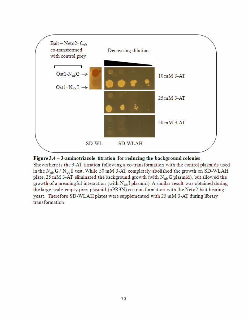
70

71
Part 5 - cDNA library screens using the Neto2-bait yielded several positive
clones
After the screening conditions were optimised, a human adult and a human embryonic
whole-brain cDNA library were transformed into the Neto2 bait-bearing yeast in separate
experiments. A transformation efficiency of approximately 4 million -trp-leu co-transformants
was achieved for both libraries as observed from their respective SD-trp-leu plates. This
transformation efficiency was comparable to previous split-ubiquitin screens with other baits
performed in the Stagljar lab and indicated that all individual clones in the cDNA library were
evaluated at least 2 times considering that the libraries had approximately 2 million independent
clones.
First reporter assay - After library transformation, visible colonies started to appear as
early as the third day and they were allowed to grow for at least a week before picking them for
further analyses. These colonies growing on SD-trp-leu-his-ade indicated that they were able to
synthesize histidine and adenine due to the activation of the reporter genes HIS3 and ADE2. The
HIS3 marker is known to be a very sensitive reporter even for low level of transcriptional
activation driven by a low number of cleaved VP16 transactivators as may occur with weak
interactions. On the other hand, it is also a leaky reporter gene as a bait with a very low level of
self-activation might also show auxotrophic growth leading to a most common concern for false
positives. Therefore background growth due to leaky HIS3 expression was suppressed by the
addition of 25 mM 3-aminotriazole (3-AT) to the SD-trp-leu-his-ade plates. Along with HIS
activation, a second ADE reporter gene (activated by the same ADH promoter element as the
HIS3 gene), permit clone selection based on colony colour. The absence of bait: prey interaction

72
results in the accumulation of a colour absorbing intermediate due tothe blockade of adenine
synthesis and hence the colonies appear red upon storage. In contrast, a potentially strong
interaction would remove the adenine synthesis blockade resulting in white colony colour even
upon storage; and a weak interacting pair would result in a whitish-pink colony colour upon
storage. This white pink selection along with auxotrophic growth on the SD-trp-leu-his-ade
plates was used to select the colonies from the master screening plates. A total of 215 colonies
for the adult library and 125 colonies for the embryonic library (white to faint pink) were picked
from the master plates. They represented the putative strong and intermediate Neto2-interactors
from the first reporter assay (Figure 3.5a).
Second reporter assay – A second reporter assay was applied for further increasing the
stringency above that used for white-pink selection. This assay used the lacZ reporter gene,
which was activated by a second ADH promoter element (at a position different from the HIS3,
ADE2 reporter genes). This lacZ reporter gene is not as sensitive as HIS3. Hence, lacZ
transcription requires stronger activation of VP16 transactivator expression driven by a
meaningful interaction than in the absence of a bait:prey interaction. The blue colouration
shown by the clones bearing an interaction pair (in the presence of X-gal reagent) is thus a direct
transcriptional read-out of the strength of interaction. Therefore the lacZ assay fine-tunes the
distinction between potential interacting pair-bearing clones and the clones that do not have an
interaction pair or a very weak interaction by the presence or absence of blue coloration
respectively. Out of 215 adult clones that were positive in the growth assay, only 69 were
positive during the X-gal assay; and out of 125 embryonic clones that were positive in the
growth assay, only 46 were positive during the X-gal assay ranging in colour from faint to dark

73
blue (Figure 3.5a). This indicated that a substantial number of clones were false positives in the
white-pink selection. All the lacZ positive clones were further analysed.
Part 6 - True Neto2-bait-dependent prey interactions were identified
Identification of HIS3+-ADE2+-lacZ+ clones - Colony PCR was performed to amplify the
cDNA insert within the prey vector for all HIS3+-ADE2+-lacZ+ clones. This yielded products
corresponding to DNA band sizes of 0.9 – 1.9Kb (Figure 3.5b). Upon sequencing of the PCR
products and subsequent analysis using BLAST software, the results revealed that there were
many clones which were identified as frequent false positives as reported by Dualsystems
Biotech (including proteolipid protein and ubiquitin). Further literature searches revealed that a
few clones corresponded to hypothetical proteins. With the exception of these clones, a bait-
dependency test was performed on the remaining positive clones to confirm the final putative
Neto2-interactors identified through the MYTH system.
Bait-dependency test – This was the final confirmatory test to identify the true Neto2-
bait-dependent-preys as opposed to potentially sticky-preys that may non-specifically interact
with unrelated bait. After plasmid rescue and the reintroduction into fresh Neto2-bearing bait,
only 21 clones from the adult library and 7 clones from the embryonic library were positive in
the white-pink and blue-white assays. The remaining clones were negative in the lacZ assay
indicating that the lacZ gene may have been mutated to give false positive results in the same
assay previously performed for these clones. In parallel, the Neto1-NubG - positive control prey
showed strong blue coloration (and thus a true interaction), and the pFUR4-NubG – negative

74
control prey did not grow on the stringent plate (Figure 3.5c) indicating that the assay was
robust.
In parallel experiments the rescued plasmids of what were considered positive clones
were reintroduced into fresh cultures of yeast with artificial bait CD4- bearing negative control
bait. Of these clones, only one clone corresponding to the Nogo receptor 3-prey from the
embryonic library was positive in the lacZ assay. This result indicated that this particular prey
interaction occurred non-specifically with the unrelated bait (Figure 3.5c). The negative control
artificial bait contained only the transmembrane domain (TMD) of the human CD4 molecule
fused with the Cub tag. Therefore any prey protein interacting with this artificial prey might be
due to prey over-expression (a sticky prey), or a non-specific hydrophobic TMD – mediated
interaction, or a physiologically irrelevant interaction. In this case, it was found that the NogoR3
prey clone rescued from the cDNA library corresponded to a segment outside the ORF of human
Nogo3 cDNA (within the 3’UTR) and thus interpreted as not corresponding to a physiologically
meaningful product. Therefore this clone was considered to be a false positive and was
disregarded.

75

76

77
Part 7 - Multiple false positives were eliminated at different stages during the
Neto2-MYTH screen
False positives are proteins that behave as spurious interactors with the Neto2 bait protein
in the MYTH screen, but do not correspond to a physiologically meaningful interaction. Due to
different reasons these interactions will lead to a His+ and lacZ+ result, independent of a true
interaction in vivo. This section details the multiple reasons that might lead to a false positive
interaction in a MYTH screen, and the different approaches taken to eliminate them. The most
common causes for false positives in the MYTH system are described below. All of these are
common to the traditional YTH as well.
• Bait over-expression
This is the most common cause for false positive results. Since the MYTH system
uses an exogenous ADH promoter within the pAMBV-bait plasmid to drive the expression
of the Neto2-Cub-fusion protein, the expression of the fusion protein might not be as
tightly regulated as if it were driven by an endogenous promoter. This might lead to an
over-expression of Neto2, which in turn could lead to non-specific interactions. This type
of false positive was identified in the Neto2 MYTH screen by co-transforming the Neto2-
bait bearing strain with yeast proteins fused with NubG as negative controls. The yeast
plasma membrane protein Fur4 and the ER protein Ost1 are not expected to have an
affinity for the Neto2 bait. Also, the NubG fusion tags do not interact with the bait- Cub
tag, unless the bait and the prey show affinity towards one another. In an ideal scenario,
where the bait protein is not over-expressed, the transformants should not grow on a SD-
trp-leu-his-ade selection plate whereas the transformants will activate the reporter genes

78
if the bait were over-expressed. During the screen, the transformants did not grow on the
selection plate after co-transformation, indicating that the screen was not affected by
Neto2-bait protein over-expression.
An additional line of evidence to show that the Neto2-fusion protein was not
over-expressed came from another co-transformation experiment with the empty prey
plasmid pPR3N. Over-expressed bait also may have the property of self-activation and
expression of reporter genes. In this experiment, even in the absence of prey protein,
there was no growth on the selection plates indicating that the Neto2 bait did not self
activate.
• Bait folding and functionality
Another important point to consider is due to the ectopic expression of the human
protein in yeast. If the human Neto2-fusion protein is not properly folded in yeast, it
might not only be non-functional, but it might also lead to non-specific interactions that
are not physiologically relevant. Also, the native Neto2 protein is fused with an artificial
tag that might also lead to misfolding. Therefore, during the Neto2 MYTH screen, I
tested control prey proteins to assess whether the Neto2 fusion protein is properly folded
and functional. From other experiments conducted in our lab, it was demonstrated that
Neto2 heteromerizes with Neto1 in both a heterologous mammalian cell expression
system (HEK293 cells) and endogenously in the mouse brain. Therefore, a pNeto1-NubG -
fusion prey plasmid was constructed to serve as a positive control for the interaction with
the Neto2 bait. During co-transformation of the pNeto1- NubG prey plasmid with the
Neto2- Cub bearing yeast, HIS+ and ADE+ transformants were observed. Also, these

79
transformants developed a deep blue colouration indicating a strong interaction consistent
with our lab’s findings of interaction. These results further indicate that the ectodomain
of the fusion protein was properly folded and functional since the Neto1: Neto2
interaction is ectodomain-mediated (Tang, unpublished).
Additionally, growth of white colonies was observed in the baits transformed with
positive control NubI plasmids, indicating that the bait has passed through the secretory
pathway, where the Neto2-Cub-fusion protein has interacted with NubI of the control
proteins Ost1 and Fur4 at the ER and plasma membrane. This experiment further
demonstrated that the Cub tag of the fusion protein is also properly folded and functional.
• Leaky HIS reporter gene
The HIS3 reporter gene always shows a basal level expression in yeast strain
THYAP4. This allows the growth of even wildtype colonies on the selection plates
lacking histidine thus increasing the cooncern of false positive results. Therefore during
the pilot screen with empty prey plasmid, the selection plates SD-trp-leu-his-ade were
supplemented with different concentrations of 3-aminotriazole, a competitive inhibitor
for the HIS3 gene product (Imidazoleglycerol-phosphate dehydratase) that plays a vital
role in yeast histidine biosynthesis. Increasing concentrations of 3AT reduced the number
of background colonies during the pilot screen. While 50 mM 3AT completely abolished
all background colonies, a level of 25mM 3AT substantially reduced the background
colonies, at the same time allowing sufficient HIS3 gene expression for growth assays. 25
mM 3AT containing SD-trp-leu-his-ade plates were thus used during the cDNA library
screening.

80
• Mutations in the reporter genes HIS, ADE, lacZ
Because the Neto2 bait-bearing yeast strain was subjected to repeated plating and
growth cycles during the MYTH screen, it was also possible that reversion mutations
within the reporter genes might trigger their expression even during the absence of
meaningful bait: prey interactions. In order to negate this sort of false positive sitiation,
the putative Neto2-interacting prey plasmids were rescued from the yeast and
reintroduced back into fresh Neto2 bait-bearing yeast (restreaked from the glycerol stock)
to assess reporter gene activation in independent bait-dependency tests. Only 21 clones
(corresponding to 9 putative interactors) out of 69 clones from the adult library and 6
clones (corresponding to 4 putative interactors) out of the 46 clones from the embryonic
library were positive in the lacZ assays during the bait-dependency test, from a total of
115 clones that originally passed the reporter assays. This indicated that several bait: prey
interactions identified through the reporter assays were caused due to reversion mutations
in the reporter genes.
• Prey over-expression
Just like bait-over-expression, the exogenous promoter in the prey plasmid can
also drive prey over-expression, resulting in a non-specific interaction of a prey protein.
To test whether the 14 rescued prey plasmids were positive in the reporter assays due to
prey-over-expression; they were reintroduced into yeast cells bearing the artificial bait
plasmid pCD4- Cub. This bait has been previously tested and is currently used in the
Stagljar lab as a standard negative control-bait plasmid. While 13 clones failed to grow
upon the prey plasmid co-transformation into CD4-bait bearing cells, one prey

81
corresponding to human NogoR3 was positive for the reporter assays. While the other 13
clones did not non-specifically interact with the negative control bait, the clone
corresponding to the human NogoR3 showed a suspicious non-specific interaction.
Further verification of the false positive result of this clone revealed that the
cDNA insert of this clone contained sequences outside the open reading frame (ORF) of
the human NogoR3 gene but within its 3’UTR. Thus, the corresponding fusion-prey
product did not appear to be physiologically meaningful and therefore it was concluded
that this false positive result was not due to prey over-expression, but due to the
physiologically irrelevant prey. Other prey-insert sequences were also matched with the
human cDNA sequences to identify similar results for the other clones. It was found that
the prey-insert corresponding to the remaining 13 clones included the ORF of their
corresponding human genes and that they were in-frame with the ORF of the prey vector.
Although several false positive results were eliminated during the Neto2 MYTH screen,
it should again be emphasised that the screen only identifies putative interactors, which may or
may not turn out to be true interactors under physiological conditions. This is largely because the
interactions identified in the yeast system are entirely due to ectopic expression of the human
proteins that do not directly reflect correct cell type expression in the same sub-cellular location
under physiological conditions. Therefore it becomes extremely important to validate any
putative interactions with both in vitro and in vivo methods by using other biochemical tools
such as co-immunoprecipitation following the heterologous expression of the interacting proteins
in mammalian cells, and co-immunoprecipitation from endogenously expressed proteins from
the appropriate mammalian tissues where they might physiologically interact.

82
Part 8 - MYTH identifies 13 novel putative Neto2 interacting proteins
3.8.1 Results from the Neto2-adult cDNA library screen
Nine putative Neto2-interactors (corresponding to 21 clones) that were positive in all the
MYTH selection assays (white-pink assay, blue-white assay and bait-dependency test) were
obtained from the human adult, whole brain cDNA library (Table 3.1). Clones corresponding to
three proteins namely, the VAMP-associated protein B (VAPB), glutamate transporter EAAT3-
associated protein (GTRAP3-18) and emopamil binding protein (EBP) were considered to be
putative strong Neto2-interactors. Clones corresponding to VAPB were the strongest hits in
terms of the growth of the clones and intensity of blue colour in the lacZ assay. Also, 10 identical
clones of VAPB were identified in two separate library screens. The clone determined to encode
GTRAP3-18 also showed rapid growth in the growth assay and rapid appearance of blue
coloration in the lacZ assay. Sequencing results revealed that a full length human GTRAP3-18-
cDNA was integrated into the NubG prey vector. The clone corresponding to EBP also showed
rapid growth in the growth assay and rapid appearance of blue coloration in the lacZ assay. Two
identical clones of EBP were identified from two independent library screens and the sequencing
results revealed that full length human EBP-cDNA was integrated into the NubG prey vector.
The remaining clones were considered to be putative, weaker Neto2 interactors as
indicated by the strength of their interaction in the growth and lacZ assays. These were chimerin-
isoform-b, protein phosphatase 1B, BCL2/adenovirus E1B 19kD-interacting protein 3-like,
transmembrane protein 59-like 1, rhomboid domain containing 2 isoform-a, and zinc finger
protein with UFM1-specific peptidase domain.

83

84
A brief overview of the putative interactors is given here. Detailed descriptions of the two
most interesting hits VAPB and GTRAP3-18 and the rationale for choosing these molecules for
initial studies are given in the next chapter:
• VAMP-associated protein B (VAPB)
The protein encoded by this gene is a 27.2 kDa, conserved type II membrane protein
found in the plasma membrane and intracellular vesicle membranes of neurons. VAPB
mRNA is expressed in multiple adult-mouse brain regions like the hippocampus,
cerebellum and spinal cord motor neurons, where Neto2 mRNA is also expressed. This
neuronal protein is known be present at the presynapse (where Neto2 is also present),
particularly at the spinal cord motor neuron presynapse in mouse and at the glutamatergic
neuromuscular junction presynapse in flies, and plays multiple roles in glutamatergic
neurotransmission. A single missense mutation within the human VAPB gene, involving a
conserved proline residue at position 56 (P56S), within the protein – protein interacting
MSP domain was identified to be associated with three forms of familial motor neuron
disease (Nishimura, Mitne-Neto et al. 2004; Kanekura, Nishimoto et al. 2006). The VAPB
clones identified from the adult library correspond to amino acids 183 – 243 (comprising
the coiled-coil and the transmembrane domains) of the human VAPB gene product.
• Glutamate transporter EAAT3 – associated protein (GTRAP3-18)
The protein encoded by this gene is a 21.6 kDa, four-transmembrane domain protein
found in the plasma membrane and intracellular vesicle membranes of neurons. GTRAP3-
18 mRNA is expressed abundantly in different regions of the developing and adult-mouse
brain regions. The mRNA expression of GTRAP3-18 in the adult mouse brain closely

85
resembles Neto2 mRNA expression in the cortex, hippocampus and cerebellum, and this
neuronal protein is present at the postsynaptic membrane of glutamatergic and
GABAergic neurons in these regions. GTRAP3-18 is known to regulate glutamatergic
neurotransmission by modulating the trafficking and substrate affinity of the neuronal
glutamate transporter EAAT3, an important excitatory amino acid-transporter that is
present at the postsynaptic neuronal membrane (Lin, Orlov et al. 2001; Ruggiero, Liu et
al. 2008). The GTRAP3-18 clone identified from the adult library corresponds to the full-
length human GTRAP3-18 gene product (amino acids 1 – 188).
• Chimerin isoform1 b (Chn1b)
The protein encoded by this gene is a 53.2 kDa, neuronal GTPase-activating protein. This
cytoplasmic protein is known to interact with NR2A-NMDAR subunits and to play an
important role in regulating dendritic spine morphology. Also this protein interacts with
EphrinA4 receptor and plays an important role in EphA4-mediated axon pathfinding.
Heterozygous missense mutations in this gene have been identified in Duane's retraction
syndrome 2, a congenital oculomotor disorder (Miyake, Chilton et al. 2008). The Chn1
clone identified from the adult library corresponds to amino acids 194 – 459 (comprising
the protein kinase-c-like and the Rho-GTPase activating protein (RhoGAP) domains) of
the human Chn1 gene product.
• Protein phosphatase, magnesium dependent, 1B (PPM1B)
The protein encoded by this gene is a 52.6 kDa, Ser/Thr protein phosphatase that is
present in many cell types including neurons. This phosphatase has been shown to
dephosphorylate cyclin-dependent kinases (CDKs) and also regulates NF-kappa B

86
activity and thus may be involved in cell cycle control (Cheng, Kaldis et al. 2000;
Prajapati, Verma et al. 2004). Other proteins of this family are known to be negative
regulators of cell stress response pathways. The PPM1B clone identified from the adult
library corresponds to amino acids 379 – 479 (uncharacterised region) of the human
PPM1B gene product.
• Emopamil binding protein (EBP)
The protein encoded by this gene is an 18.3 kDa, four-transmembrane protein found in
the ER membranes of multiple cell types including neurons and astrocytes. This protein is
known to play a crucial role in cholesterol biosynthesis by catalyzing the sterol-isomerise
reaction. Missense mutations in this gene have been identified in X-linked dominant
Conradi-Hunermann syndrome which is characterised by defective cholesterol
biosynthesis (Ikegawa, Ohashi et al. 2000). The EBP clone identified from the adult
library corresponds to the full-length human EBP gene product (amino acids 1 – 160).
• BCL2/adenovirus E1B 19kD-interacting protein 3-like (BNIP3)
The protein encoded by this gene is a 21.5 kDa, type II membrane protein that plays a
role during oxidative stress and other proapoptotic cellular functions along with Bcl2.
Other BH3 domain family proteins are known to be intracellular death-ligands critical for
initiating apoptosis (Lomonosova and Chinnadurai 2008; Burton and Gibson 2009). The
BNIP3 clone identified from the adult library corresponds to amino acids 19 – 194
(comprises the BH3 and the transmembrane domains) of the human BNIP3 gene product.

87
• Transmembrane protein 59 – like 1 (TMEM59L)
The protein encoded by this gene is a 37.6 kDa, predicted membrane glycoprotein protein
of uncharacterised function. The TMEM59L clone identified from the adult library
corresponds to amino acids 187 – 342 (uncharacterised region) of the human TMEM59L
gene product.
• Rhomboid domain containing 2 isoform a (RHBDD2)
The protein encoded by this gene is a 39.2 kDa, predicted multi-pass membrane protein
of uncharacterised function. The RHBDD2 clone identified from the adult library
corresponds to amino acids 1 – 230 (uncharacterised, predicted transmembrane region) of
the human RHBDD2 gene product. In a recent study the RHBDD2 mRNA and protein
expression were found to be significantly elevated in breast carcinomas (Abba, Lacunza
et al. 2009). Other rhomboid family members are widely conserved proteins that are
known to function as serine proteases and are also known to regulate molecules involved
in apoptosis.
• Zinc finger protein with UFM1-specific peptidase domain (ZUFSP)
The protein encoded by this gene is a 65.9 kDa protein of uncharacterised function. The
ZUFSP clone identified from the adult library corresponds to amino acids 311 – 579
(comprises the uncharacterised peptidase domain) of the human ZUFSP gene product.
Other members of this family are known to be cysteine peptidases required for the
processing and activation of ubiquitin fold modifier 1 (UFM1) and their zinc-finger
domains have been shown to be involved in protein: protein interactions (Kang, Kim et
al. 2007)

88
3.8.2 Results from the Neto2-embryonic cDNA library screen
Four putative unique Neto2-interactors (corresponding to 6clones) that were positive in
all the MYTH selection assays were obtained from the human embryonic whole brain cDNA
library. Three clones corresponding to transmembrane protein 14A (TMEM14A) led to it being
considered as a putative strong Neto2-interactor. The three identical clones corresponding to the
TMEM14A were amongst the strongest hits in terms of the rapid growth of the clone and rapid
appearance of blue colour in the lacZ assay. The remaining clones were considered as putative
weak-Neto2 interactors as indicated by their relatively weaker interaction based on the growth
and lacZ assays. These were transmembrane protein 53, protein di-sulphide isomerasae and the
BCL2/adenovirus E1B 19kD-interacting protein 3-like protein.
A brief overview of the positive hits is given here:

89
• Transmembrane protein 14A (TMEM14A)
The protein encoded by this gene is a 10.7 kDa, predicted membrane protein of
uncharacterised function. The TMEM14A clone identified from the embryonic library
corresponds to the full length human TMEM14A gene product (amino acids 1 – 99).
• Transmembrane protein 53 (TMEM53)
The protein encoded by this gene is a 31.6 kDa, predicted membrane protein of
uncharacterised function. The TMEM53 clone identified from the embryonic library
corresponds to amino acids 62 – 277 of the human TMEM53 gene product.
• Protein di-sulphide isomerasae, family A, member 6 (PDIA6)
The protein encoded by this gene is a 48.1 kDa, ER resident proteins that catalyzes the
formation, reduction, and isomerization of disulfide bonds in proteins and play a role in
folding of disulfide-bonded proteins (Hayano and Kikuchi 1995). The PDIA6 clone
identified from the embryonic library corresponds to amino acids 1 – 226 (comprises the
di-sulphide isomerase domain) of the human PDIA6 gene product.
• BCL2/adenovirus E1B 19kD-interacting protein 3-like (BNIP3)
Refer to above section from the adult screen

90
Part 9 - Criteria used to choose molecules from the MYTH screen for
subsequent analyses
It should be remembered that MYTH screening results suggest only a possible interaction
in vivo, and systematic studies must subsequently be conducted to determine which putative
interactors actually associate with Neto2 in vivo. The strong and multiclone interactors in the
screen may not necessarily be real interactors in vivo and weak, infrequent interactors should not
necessarily be ignored. However, strong and multiclone interactors should be the subjects of
initial work.
Initial studies will be focused on those molecules that are known to have well-established
neuronal functions, particularly at the synapse. Particularly, those transmembrane proteins that
are known to play cruicial roles during glutamate-mediated neurotransmission at the synapse will
be of immediate interest. Additionally, the molecules that are strongly associated with human
neurological disease conditions / mouse phenotypes will be of particular interest. I will later
focus on those molecules with less established neuronal functions, particularly the subsets that
are not neurospecific. I will give less attention to those molecules with no established neuronal
function or unknown function. I will not study molecules that have been reported to be frequent
non-specific interactors in the MYTH screening system.

91
Chapter 4: Conclusions and future directions

92
Part 1 - A brief summary of the project and major findings from the Neto2-
MYTH screen
Neto2, a novel type I neuronal transmembrane protein was recently reported as an
auxiliary partner for the kainate-type glutamate receptor subunits (Zhang, St-Gelais et al. 2009).
Other unpublished results from our lab (reviewed earlier) indicated that Neto2 can interact with
multiple protein partners during the regulation of different neuronal functions including neuronal
excitability and synaptic transmission. In order to acquire a broader understanding of the
biological roles of Neto2 in neurons, and to gain additional knowledge about its currently known
functions, Membrane Yeast Two Hybrid screens were performed to identify putative membrane-
bound interacting partners of Neto2.
The Neto2-MYTH screens were conducted using first a human adult, and then a human
embryonic cDNA library. Thirteen novel putative Neto2 interactors were identified. Of the
thirteen putative Neto2 interactors six proteins have a defined biological function, while seven
proteins are uncharacterised. All six proteins with known functions participate in different roles
including protein folding, trafficking, and post-translational protein modification. Four proteins
namely, VAPB, GTRAP3-18, Chn1b, and PPM1b have neuron-specific functions whereas the
proteins EBP and PDIA6 have functions in multiple cell types.
Of the 13 putative Neto2 interactors, clones corresponding to five molecules occured
more than once during the screen. Ten identical clones of VAPB, three identical cones of ZUSFP
and two identical clones of EBP were identified in two seperate screens from the adult library,
while three identical clones of TMEM14A were identified in the screen of the embryonic library.
Interestingly, two different clones corresponding to overlapping regions of BNIP3 were

93
identified independently from both the adult and embryonic cDNA library screens indicating that
this molecule may be a true Neto2-interactor. Apart from the three soluble proteins Chn1b,
PDIA6 and PPM1B, the remaining ten proteins identified in the screens were transmembrane
proteins; of these three clones correspond to full-length transmembrane proteins (GTRAP3-18,
EBP and TMEM14A).
In view of the specific rationale of the project - to identify novel membrane-bound
Neto2-interacting partners in the nervous system, two proteins generate particular interest;
VAPB and GTRAP3-18. While VAPB is a type II membrane protein, GTRAP3-18 is a four-
transmembrane domain protein found in the plasma membrane, ER membrane, and other
vesicular membrane compartments of neurons. In particular, VAPB and GTRAP3-18 are both
known to be present at the synapse and to interact with multiple synaptic proteins. Both proteins
are involved in the trafficking of specific proteins between the ER and other secretory
compartments and also to the plasma membrane. While the Drosophila homologue of VAPB is
known to regulate postsynaptic glutamate receptor clustering, murine GTRAP3-18 is known to
alter glutamate reuptake by the glutamate transporter EAAT3 from the synapse. Thus both
molecules are known to regulate glutamatergic neurotransmission. Since Neto1 and Neto2 are
already known to play important roles in regulating the function of NMDA and kainate type
glutamate receptors, the study of Neto2-VAPB and Neto2-GTRAP3-18 interactions, once
validated in vivo, might lead to a deeper understanding of how the Neto proteins (particularly
Neto2) fine tune glutamatergic neurotransmission. Literature reviews of VAPB and GTRAP3-18
are discussed in the next section.

94
Part 2 - The most promising putative-Neto2 interactors for follow-up studies
In this section I will review the features of VAPB and GTRAP3-18 and their functional
relevance with Neto2 that makes them interesting candidates for further study.
4.2.1 VAMP-associated protein B (VAPB)
VAPB is a potential candidate for further studies for the following reasons:
It is the strongest, multiclone interactor identified in 2 independent MYTH screens conducted
for the Neto2 bait using the type II adult cDNA library. VAPB is an evolutionarily conserved
protein, which is abundantly present in multiple brain regions. VAPB mRNA is widely expressed
in the mouse nervous system, in the spinal cord, cortex, hippocampus, and cerebellum
(Kanekura, Nishimoto et al. 2006; Teuling, Ahmed et al. 2007), where Neto2 is also highly
expressed. The protein encoded by this gene is reported to be abundant at the presynapses (like
Neto2); particularly at the presynapses of murine spinal-motor neurons and also at the
presynaptic boutons of the Drosophila glutamatergic neuromuscular junction, and VAPB has
also been reported only once at the postsynapse (Collins, Husi et al. 2006; Chai, Withers et al.
2008).
Mutations in the human VAPB gene have been identified in three forms of familial Motor
neuron disease including late onset spinal muscular atrophy (SMA) as well as typical and
atypical amyotrophic lateral sclerosis (ALS) (Nishimura, Mitne-Neto et al. 2004; Kanekura,
Nishimoto et al. 2006). Moreover, the Drosophila homologue of VAPB has been shown to
regulate glutamatergic neurotransmission by different mechanisms. It regulates glutamate release

95
at the presynapse through its interactions with the vesicle associated membrane protein
/synaptobrevin (VAMP) (Skehel, Martin et al. 1995). VAPB also regulates the presynaptic
bouton size by modulating microtubule assembly, postsynaptic glutamate receptor clustering and
the postsynaptic abundance of specific glutamate receptor subunits at the fly neuromuscular
junction (Pennetta, Hiesinger et al. 2002; Chai, Withers et al. 2008). While the presynaptic
function of Neto2 has not been studied before, it has been shown to interact with the GluR6
subunit of the kainate-type glutamate receptor in the cerebellum and to modulate glutamatergic
neurotransmission by modulating channel function of the kainate receptor subunits (Zhang, St-
Gelais et al. 2009). Additionally, initial studies from our lab also suggest that Neto2 along with
Neto1 might regulate the abundance of postsynaptic kainate receptor subunits within the PSDs in
the hippocampus and cortex (Tang, Unpublished). These studies indicate that Neto2 might also
play multiple roles during glutamatergic neurotransmission.
4.2.2 Current knowledge about VAPB
Human VAPB is a 27kD, type II integral membrane protein that belongs to a family of
proteins found across species from yeast to mammals. It shares 63% amino acid identity and
hetero dimerizes with a closely related protein, VAPA, in mammals. VAPB has a cytoplasmic N-
terminal major sperm protein (MSP) domain, a central coiled-coil domain, and a C-terminal,
membrane anchored transmembrane domain (Figure 4.1 a, b) (Weir, Klip et al. 1998; Lev, Ben
Halevy et al. 2008). In both humans and mice, VAPB is highly expressed in the motor neurons of
the spinal cord and is moderately abundant in the cortex, hippocampus and cerebellum (Figure
4.1 c) as well as in heart, kidney and liver. It is predominantly localized to the ER, golgi
membrane and neuromuscular junctions in motor neurons. It has been mostly shown to be a

96
component of the presynapse and in one study as a component of the postsynaptic density (Weir,
Klip et al. 1998; Collins, Husi et al. 2006; Teuling, Ahmed et al. 2007; Chai, Withers et al. 2008;
Emes, Pocklington et al. 2008; Lev, Ben Halevy et al. 2008).
VAPB is known to play important roles in many cellular processes including
glutamatergic neurotransmission through specific interacting partners. It is known to play
important roles in vesicular protein transport between the ER and golgi that is mediated by the
coat-protein (COPI) (Soussan, Burakov et al. 1999). Also, it plays an important role in
maintaining the lipid compositions of the ER and golgi membranes by interacting with the lipid
binding and lipid transfer proteins Nir2, oxysterol binding protein (OSBP), and ceramide transfer
protein (CERT) (Peretti, Dahan et al. 2008). The VAPB homologue in Aplysia was originally
identified as a binding partner of the vesicle associated membrane protein (VAMP /
synaptobrevin), a protein known to be involved in neurotransmitter release (Skehel, Martin et al.
1995). In Drosophila, loss-of-function mutations of the VAPB homologue result in an increase of
presynaptic-bouton size associated with an increased clustering of postsynaptic glutamate
receptor subunit-IIA (Pennetta, Hiesinger et al. 2002; Chai, Withers et al. 2008). Moreover, over-
expression of the VAPB homologue leads to a decrease in presynaptic-bouton size and a
reduction in cluster volume of several glutamate receptor subunits by altering microtubule
assembly (Pennetta, Hiesinger et al. 2002; Chai, Withers et al. 2008). These studies indicate that
VAPB plays a crucial role in synaptic homeostasis by coordinating changes between both the pre
and post synapse.
A single missense mutation within the human VAPB gene, substituting a conserved
proline residue at position 56 with a serine (P56S) within the protein:protein interacting MSP
domain, has been identified in three forms of familial motor neuron disease (Nishimura, Mitne-

97
Neto et al. 2004; Kanekura, Nishimoto et al. 2006). Studies from animal models and cultured
motor neurons reveal that it is a dominant negative mutation and that it depletes wild type VAPB
protein from its normal cellular localization, forming insoluble aggregates (Kanekura, Nishimoto
et al. 2006; Teuling, Ahmed et al. 2007). P56S VAPB has been shown to affect lipid metabolism
and lipid transport between the ER and golgi compartments (Peretti, Dahan et al. 2008). It is also
known that this mutation attenuates the activity of ATF6, a resident ER stress signaling
transcription factor, thereby leading to an increased unfolded protein response (UPR) (Gkogkas,
Middleton et al. 2008). These studies demonstrate that VAPB participates in multiple cellular
processes and the mechanism responsible for motor neuron degeneration may be due to a
complex interplay between different cellular pathways.

98

99
4.2.3 Glutamate transporter EAAT3-associated protein (GTRAP3-18)
GTRAP3-18 is a potential candidate for further studies for the following reasons:
It is a strong, full-length putative interactor identified during the Neto2-MYTH screen using
the human adult cDNA library. GTRAP3-18 is an evolutionarily conserved neuronal protein that
is abundantly present in multiple brain regions that have high densities of glutamatergic and
GABAergic neurons, particularly at their postsynaptic membrane. GTRAP3-18 mRNA is widely
expressed in the mouse nervous system particularly in the cortex, hippocampus, cerebellum, and
basal ganglia (Butchbach, Lai et al. 2002; Inoue, Akiduki et al. 2005), and it is expressed both
during development and in the adult brain. Neto2 mRNA is also expressed both in the developing
and in adult brain, in similar brain regions as GTRAP3-18 mRNA. (Figure 4.2 a, b, c)
GTRAP3-18 is known to directly associate with the neuronal protein of the excitatory amino
acid transporter family, EAAT3 (also called as excitatory amino acid carrier-1, EAAC1 in
humans) to regulate its trafficking and glutamate affinity (Lin, Orlov et al. 2001; Ruggiero, Liu
et al. 2008). EAAT3 is an important neuronal glutamate transporter that buffers synaptic-
glutamate levels following the release of glutamate from the presynapse (Nieoullon, Canolle et
al. 2006), thus modulating glutamate-mediated neurotransmsission. Additionally, EAAT3
regulates GABA synthesis and also functions as a glutamate-gated Cl- channel (Eskandari,
Kreman et al. 2000; Koch, Brown et al. 2007), thus regulating GABAergic-inhibitory
neurotransmission. Therefore GTRAP3-18 might play important roles during EAAT3- mediated
glutamatergic and GABAergic neurotransmission. While the role of the Neto2:GluR6 interaction
modulating glutamate-mediated neurotransmission is reviewed earlier (section 1.2.2), an
interaction of Neto2 with KCC2 (a major ion pump required for Cl- homeostasis in neurons)

100
regulates the chloride equilibrium and membrane excitability in cultured hippocampal neurons
(Ivakine et al, Unpublished). Therefore Neto2 is also thought to perform multiple functions
during excitatory and inhibitory neurotransmission like GTRAP3-18.
4.2.4 Current knowledge about GTRAP3-18
GTRAP3-18 is a 21.6 kDa, four-transmembrane protein expressed in the neuronal
membrane and ER membrane in mice and humans (Lin, Orlov et al. 2001; Inoue, Akiduki et al.
2005; Schweneker, Bachmann et al. 2005; Akiduki, Ochiishi et al. 2007; Ruggiero, Liu et al.
2008). This molecule was identified as a negative regulator of the glutamate-uptake activity of
the excitatory amino acid transporter 3 (EAAT3) (Lin, Orlov et al. 2001) that acts by decreasing
the glutamate affinity of EAAT3. Further studies indicated that GTRAP3-18 delays the ER-exit
of EAAT3 and other members of the excitatory amino acid transporter family (Ruggiero, Liu et
al. 2008), thus playing an important modulatory role in glutamatergic neurotransmission.
The time point and expression of GTRAP3-18 in the brain is similar with the expression
patterns of EAAT3. Both EAAT3 and GTRAP3-18 mRNA is expressed early during development
(peaks at around P5) and also in the adult brain across multiple regions, particularly regions of
high densities of glutamatergic and GABAergic neurons in the hippocampus, cerebellum,
caudate putamen, and amygdala (Inoue, Akiduki et al. 2005; Akiduki, Ochiishi et al. 2007;
Tzingounis and Wadiche 2007; Ruggiero, Liu et al. 2008). It has also been shown that EAAT3 is
the earliest expressed neuronal glutamate transporter and thus GTRAP3-18, along with EAAT3,
is thought to play important roles in the developing and in the adult brain (Nieoullon, Canolle et
al. 2006; Tzingounis and Wadiche 2007).

101
Apart from regulating glutamate concentration at synapses, EAAT3 is also known to
regulate GABA synthesis. Homo-trimeric and homo-pentameric EAAT3 also functions as
aglutamate-gated chloride channel (Eskandari, Kreman et al. 2000; Amara and Fontana 2002;
Koch, Brown et al. 2007). While the precise role of GTRAP3-18 in EAAT3-mediated inhibitory
neurotransmission has not been studied, it might indirectly participate during this process
because these functions are also dependent on glutamate binding to EAAT3.

102

103
Part 3 - Proposed experiments for Neto2 and VAPB
The following experimental plan would help us understand the biological relevance of
Neto2-VAPB interaction. Initial studies would focus on characterizing the interaction between
Neto2 and VAPB, and the following questions could be addressed:
a) Do VAPB and Neto2 physically interact in vivo?
Co-immunoprecipitation (CoIP) experiments using wildtype and Neto2 null mice tissues,
particularly from the spinal cord, hippocampus and cerebellum (where both proteins are
expressed) would help us find whether the interaction is physiologically relevant. Also, CoIP
experiments using a heterologous cell system (HEK293 cells) by transfecting those with Neto2
and VAPB cDNA could be performed to confirm whether the interaction is direct. Deletion
constructs could be generated for VAPB, to determine the minimal domains required for an
interaction with Neto2. Previously validated Neto1 association with Neto2 or GluR6 association
with Neto2 would serve as a positive control for the Neto2 interaction in these experiments.
b) In which sub-cellular location do they co-localize?
Using primary neuronal cultures of spinal cord motor neurons / hippocampus / cerebellum
obtained from Neto2 null and WT mice, co-immunostaining could be performed to identify
whether Neto2 and VAPB co-localise, using markers specific for different sub-cellular locations.
Using the same approach, it could also be determined whether the two proteins co-localize at the
presynapse or at the postsynapse. These results could be confirmed by performing CoIP from
crude synaptosomes, presynaptic vesicles and postsynaptic density components from Neto2 null
mice and WT mice. In these experiments VAMP-2 could be used as a marker for synaptic

104
vesicle at the presynapse, PSD-95 for postsynaptic density, microtubule associated protein1B for
microtubule association, and calnexin for ER membrane.
Depending upon in which sub-cellular location the association between the two molecules is
observed, the following questions could be addressed:
c) Does the absence of Neto2 affect any of the known cellular functions of VAPB?
Since VAPB regulates the presynaptic microtubule assembly and glutamate release,
disruption of microtubule assembly along with changes in the size and number of synaptic
vesicles could be visualized using electron microscopy approach in multiple brain regions like
spinal cord, hippocampus and cerebellum of the Neto2 null animals. Additionally, glutamate
release assay could be performed in the motor neurons of Neto2 null animals to confirm whether
the presynaptic glutamate release is aberrant.
d) Does the loss of VAPB function affect the trafficking / surface expression of Neto2 and /
or kainate receptor subunits? Does the loss of both VAPB and Neto2 function affect the
kainate receptor trafficking?
The above questions could be addressed by using primary neuronal cultures of spinal cord
motor neurons / hippocampus / cerebellum obtained from Neto2 null and WT mice. By applying
RNAi approach VAPB mRNA in both Neto2 null and WT cultures could be knocked down and
subsequently the surface expression, sub-cellular location of Neto2 and different subunits of
KAR could be studied using immunostaining. Additional experiments could be performed to
study whether VAPB sub-cellular location is altered in the Neto2 nulls.

105
If the kainate receptor trafficking / abundance / surface expression is disrupted in the VAPB
knockdown experiments in motor neuronal cultures, then its biological consequences and
corresponding neurophysiological changes (changes in glutamate mediated currents) could be
studied. Our lab collaborates with Dr. Michael Salter, and Dr. Melanie Woodin to study the
electrophysiology of these neurons following such changes in the receptor density at the
neuronal surface.
Additionally, due to a significant amino acid identity between Neto1 and Neto2, it would be
important to determine whether Neto1 could also interact with VAPB. Neto1 has been shown to
associate with different NMDAR subunits and loss of Neto 1 leads to a decrease in the NR2A
level (a subunit of NMDA receptor) (Ng et al., 2009) and also a decrease in the GluR6 (a subunit
of kainate receptor) (Tang, Unpublished) in the hippocampal PSD fraction. It would be
interesting to study the consequence of Neto1-VAPB interaction with respect to glutamate
receptor trafficking, surface expression and abundance.

106
Part 4 - Proposed experiments for Neto2 and GTRAP3-18
The following experimental plan would help us understand the biological relevance of
Neto2-GTRAP3 interaction. Initial studies would focus on characterizing the interaction between
Neto2 and GTRAP3-18, and the following questions could be addressed:
a) Does GTRAP3-18 and Neto2 physically interact in vivo?
b) In which sub-cellular location do they co-localize?
Similar experimental approaches could be used to address these questions as described for the
characterization of the Neto2-VAPB interaction. After validating the Neto2-GTRAP3
interactions in vivo the other important questions to be addressed are:
a) Does the loss of Neto2 affect the surface expression GTRAP3-18 and EAAT3?
HEK293 cells expressing GTRAP3-18 / EAAT3 with or without the Neto2 co-expression,
followed by biotinlation studies would reveal whether the absence of Neto2 affect the surface
expression of GTRAP318 / EAAT3 in these non-neuronal cells. Additionally, subcellular
fractionation of the hippocampus / cerebellar lysates from the Neto2 null and wildtype mice,
could be used to study whether the surface abundance of GTRAP3/18 / EAAT3 in the neuronal
membrane is altered using specific antibodies against them.
b) Does the loss of Neto2 affect the glutamate-uptake function of GTRAP3-18 and EAAT3?
The glutamate affinity for EAAT3 can be measured during the presence and absence of
Neto2, co-expressed in HEK293 cells. Additionally the glutamate affinity can be measured in the
presence of both Neto2 and GTRAP3-18 in HEK293 cells.

107
c) Does the loss of Neto2 affect the anion conductance function of GTRAP3-18 and
EAAT3?
The anion conductance of the EAAT3 can be electrophysiologically measured co-
injecting Neto2 cRNA and with EAAT3 cRNA in Xenopus laevis oocytes.
Part 5 - Conclusions
The Neto2-MYTH screen was successfully completed leading to the identification of
several novel, putative-Neto2 interactors. Particularly, if the Neto2-VAPB and Neto2-GTRAP3-
18 interactions are confirmed in vivo, this would open new avenues in the current understanding
about the biology of Neto2 particularly during synaptic transmission in the central nervous
system (Figure 4.3)

108

109
Appendices

110
Appendix I - List of strains, antibodies, plasmids and primers
List of yeast and bacterial strains used
Yeast strain THYAP4 was obtained from Dr. Igor Stagljar
Bacterial strains DH5α, XL1-Blue were purchased from Stratagene Inc.
List of antibodies used
1. Rabbit Anti-Neto2; in-house antibody raised against the intracellular carboxyl 70 amino acids of mouse Neto2
2. Mouse Anti-Neto2; raised against the ectododmain of mouse Neto2
3. Rabbit Anti-VP16; Raised against synthetic peptide corresponding to amino acids 464-476 of the herpes simplex virus VP16 protein (Sigma)
4. HRP conjugated mouse anti-rabbit secondary antibody (Sigma)
5. Alexa fluor 488 conjugated goat anti-rabbit secondary antibody (Invitrogen)
List of plasmids and cDNA used
All MYTH plasmids were obtained from Dr. Igor Stagljar
Human Neto2 cloned into pBluescript was obtained from Zhenya Ivakine
1. pAMBV (bait vector)
2. pPR3-N (prey/ type II library vector)
3. pPR3-C (prey/type I library vector)
4. pOst1-NubI (unrelated prey, positive control for bait over-expression, ER)
5. pOst1-NubG(unrelated prey, negative control for bait over-expression , ER)
6. pFur4-NubI(unrelated prey, positive control for bait over-expression, PM)
7. pFur4-NubG(unrelated prey, negative control for bait over-expression, PM)
8. pCD4 (unrelated artificial bait, control for prey over-expression)

111
9. Human Neto2 cDNA (ensembl gene ID ENSG00000171208)
10. Human Neto1 cDNA (ensembl gene ID ENSG00000166342)
11. Human adult, whole brain-cDNA library – pPR3N (specific for type II membrane proteins and cytosolic proteins) was purchased from Dual Systems Biotech
12. Human embryonic, whole brain-cDNA library – pPR3N (specific for type II membrane proteins and cytosolic proteins) was originally from Dual Systems Biotech, was obtained from Dr. Igor Stagljar.
List of additional primers used for Neto2- Cub-fusion plasmid sequencing*
1. Forward primer 1 - (5’ – TTT CCT CGT CAT TGT TCT CGT TCC CTT TCT – 3’)
2. Forward primer 2 - (5’ – ATG AAG AGC TTG AAG GAC TGG GAT TTC GAG – 3’)
3. Forward primer 3 - (5’ – TTC AGG GAT TGT CTT GGT CCT TCT CAT TA – 3’)
4. Forward primer 4 - (5’ – TTT GAT CTC ATC CGT GAT CAC ATC AGC CAG – 3’)
5. Reverse primer 1 - (5’ – TTC TAC CTG ACT AGA GCG CAC TAT TCC ATC – 3’)
6. Reverse primer 2 - (5’ – ATG GAA CTG AGG TTG GTC CTG CTT T – 3’)
7. Reverse primer 3 - (5’ – AAC CCT TCT TCC TCT TCC TGC AAC AGA CGG AT – 3’)
8. Reverse primer 4 - (5’ – ATA AAT AGG GAC CTA GAC TTC AGG TTG TCT – 3’)
*Vector NTI software (version 10) was used for designing primers and construction of the fusion plasmids

112
Appendix II – Additional details about the MYTH bait vector pAMBV and
prey vector pPR3-N
The Bait vector pAMBV (ADH1-MFα-Bait-Cub-LexA-VP16) contains a weak ADH1
promoter, which drives low levels of heterologous protein expression. Specific for type I
transmembrane proteins, pAMBV contain the yeast MFα signal sequence at the N terminus,
resulting in an improved targeting of the heterologous bait protein to the yeast membrane.
Following the signal sequence is the multiple cloning site (MCS) for introducing the gene of
interest (Neto2). Following the MCS is the C-terminus of Ubiquitin (Cub) and the artifitial
transcriprion factors LexA-VP16. pAMBV is a centromeric plasmid containing an autonomously
replicating sequence (ARS) origin of replication and one centromeric locus (CEN), which results
in one to two copies of the plasmid per cell. These low-copy-number plasmids autonomously
replicate in both E. coli and S. cerevisiae and contain the KanR and LEU2 genes for selection of
plasmid-bearing cells on medium containing kanamycin in bacteria) or lacking leucine in yeast
respectively.
The prey vector pPR3-N (ADH -NubG-HA- prey) allows cloning of a library of prey genes
in frame with NubG at the N - terminus of the protein (for type II membrane and cytoplasmic
proteins). It expresses the fusion protein from the strong constitutive promoter ADH1 and is
replicated by the high-copy-number origin of replication. They are selected for by the AmpR and
TRP1 genes, allowing growth on medium containing ampicillin (bacteria) or lacking tryptophan
(yeast), respectively. The use of the AmpR marker for E. coli facilitates the re-isolation of these
vectors after screening with respect to the bait vector, which has a KanR marker.

113
Bait vector features (Dualsystems biotech)
Position Feature
Start: 107 End: 1581 ADH1 promoter
Start: 1709 End: 1844 Cub, ubiquitin (amino acids 34-76)
Start: 1868 End: 2474 LexA DNA binding domain (amino acids 1-200)
Start: 2491 End: 2729 Herpes simplex VP16 transactivator (amino acids 464-476)
Start: 2730 End: 2983 CYC1 terminator
Start: 3701 End: 5929 LEU2 auxotrophic marker
Start: 6078 End: 7110 KanR resistance gene
Start: 8193 End: 8917 CEN/ARS - Origin of replication

114
Prey vector features (Dualsystems biotech)
Position Feature
Start: 62 End: 352 CYC1 promoter
Start: 364 End: 480 NubG, ubiquitin amino acids 1-38
Start: 481 End: 510 HA epitope tag
Start: 622 End: 883 CYC1 terminator
Start: 1598 End: 2791 TRP1 auxotrophic marker
Start: 2855 End: 4202 2micron origin of replication
Start: 4335 End: 5193 AmpR resistance gene
Start: 5328 End: 5995 pBluescript origin of replication

115
Appendix III - Brief description of the MYTH cDNA libraries
Human adult and Human fetal whole- brain cDNA-library (Dualsystems biotech)
The employed human adult brain NubG-X split-ubiquitin cDNA library was purchased
from Dual systems Biotech and the human fetal brain NubG-X split-ubiquitin cDNA library was
kindly provided by Dr. Igor Stagljar, and they were constructed as follows: Split-ubiquitin prey
expression vectors are based on plasmid pPR3N that contain the NubG fused with HA coding
sequence. Total RNA was isolated from the brain of a Caucasian-female donor aged 59 years
(who died of tracheal cancer). Total RNA was isolated from normal fetal brains pooled from 21
spontaneously aborted male/female Caucasian fetuses that were aged between 26-40 weeks.
cDNA synthesis was performed using oligo dT priming during which Sfi I restriction sites were
introduced. The cDNA were directionally cloned into the pPR3N vector using Sfi I site such that,
the NubG-HA coding sequence is oriented in-frame with the 5' end of the cDNA. These libraries
have a complexity of 2 x 10⁶ independent clones and average insert sizes of 1.6 kb. These
pPR3N - NubG-X cDNA libraries are more suitable for finding integral membrane proteins that
does not have a N-terminal signal sequence (like the type II membrane proteins as well as other
multi-pass membrane proteins topologically similar to the type II membrane proteins) as well as
the cytosolic interacting partners of Neto2.

116
Appendix IV - Standard reagent recipies
10X TAE buffer, pH 7.5
Mix 20 ml 1M Tris-HCl (pH 7.5) and 4 ml of 0.5M EDTA (pH 8.0), add ddH2O to a final volume of 200 ml. Autoclave at 121°C for 15 min, store at room temperature.
10X PBS, pH 7.5
Dissolve 80 g of NaCl, 2 g of KCl, 14.4 g of NaH2PO4 and 2.4 g of KH2PO4 in 800 ml of ddH2O. Adjust the pH to 7.5 with HCl and adjust the volume to 1 liter with ddH2O. Autoclave at 121°C for 15 min, and store at room temperature.
YPAD plates and media
Mix 10 g Bacto Yeast Extract, 20 g Peptone, 20 g glucose, 40 mg Adenine Sulphate (add 20 g of Bacto Agar for plates). Add ddH2O to a final volume of 1000 ml, mix, and autoclave at 121°C for 15 min. Pour plates while the agar is still warm (≈50°C). Store the media at room temperature and the plates at 4°C.
2X YPAD media
Mix 20 g Bacto Yeast Extract, 40 g Peptone, 40 g glucose, 40 mg Adenine Sulphate. Add ddH2O to a final volume of 1000 ml, mix, autoclave at 121°C for 15 min and store at room temperature.
LB plates and media
Mix 10g Tryptone, 5g Bacto Yeast Extract, 10g NaCl (add 20 g of Bacto Agar for plates). Add ddH2O to a final volume of to 1000 ml, mix, autoclave at 121°C for 15 min. Pour plates while the agar is still warm (≈50°C). Store the media at room temperature and the plates at 4°C.
Lysis buffer for yeast immunoprecipitation
Mix 0.5ml of 1M Tris-HCl, pH 7.4, 0.3 ml of 5M NaCl, 20 µl of 0.5M EDTA, 100 µl of 1% triton-X-100, 100 µl 100mM of NaVO4, 50 µl of 20% SDS. Adjust the final volume to 10 ml with ddH2O and store at -4˚C. Dissolve 1 tablet of 10X Complete protease inhibitor cocktail tablets (inhibits serine proteases, cysteine proteases and metalloproteases) from Roche prior to use.

117
6X gel-loading buffer for SDS-PAGE
Mix 7.2 ml of 1M Tris-HCl, pH 6.8, 2.4 g of SDS, 2.82 ml of 87 % glycerol, 6 mg of bromophenol blue and 2.4 ml of β-mercapto ethanol. Adjust the final volume to 20 ml with ddH2O and store at -20˚C. Add 20 µl of 1M DTT to 80 µl of 6X buffer prior to gel loading.
10% acrylamide SDS-PAGE gel
For resolving gel, mix 2.5 ml of 1.5 M Tris-HCl buffer, pH, 8.8, 2.5 ml 40 % acrylamide-bisacrylamide mix (29:1), 100 µl of 10 % SDS, 100 µl of 10 % APS and 4 µl of TEMED. Mix it well with 4.8 ml of ddH2O. For stacking gel, mix 1.815 ml of water, 750 µl of 1M Tris-HCl buffer, pH, 6.8, 375 µl 40 % acrylamide-bisacrylamide mix, 30 µl of 10 % SDS, 30 µl of 10 % APS and 3 µl of TEMED.
10 X SDS-PAGE running buffer
Dissolve 30.3 g Tris base, 144.4 g of glycine, 10 g of SDS in 900 ml of ddH2Oand adjust the volume to 1 liter.
5X Transfer Buffer for western blotting
Dissolve 14.5 g of glycine, 29 g of tris base and 1.5 g of SDS in 900 ml ddH2O and make up the volume to 1 liter. During western transfer, mix 200 ml of 5X TB with 200 ml of methanol and adjust the volume to 1 liter with ddH2O.
5X TBST (for 2 liters)
Dissolve 80 g of NaCl, 2 g of KCl, 30 g of Trizma base and 10 ml of Tween-20 in 1800 ml ddH2O and make up the volume to 2 liters.
Blocking buffer for western blotting
Dissolve 0.5 g of skim milk powder in 10 ml of 1X TBST to make 5 % blocking buffer for pre-incubation of blot prior to addition of primary antibody. Dissolve 0.3 g of milk powder in 10 ml of 1X TBST to prepare 3 % buffer for incubations with primary antibody. Use 5 % buffer for secondary antibody incubations.

118
10X General Yeast dropout amino acid mix
Amino acid In mg (for 1 liter)
L-Isoleucine 300
L-Valine 1500
L-Lysine 300
L-Methionine 1500
L-Phenylalanine 500
L-Threonine 2000
L-Tyrosine 300
L-Uracil 200
L-Arginine 200
After dissolving, make the final volume to 1000 ml with ddH2O, autoclave at 121°C for 15 min and store the media at 4°C.
Yeast Synthetic Dropout plates and media (SD-)
Mix 6.5 g Bacto Yeast Nitrogen Base and 20 g D-Glucose (add 20 g of Bacto Agar for plates).
To make a dropout media / plate, include the specific amino acid (from below) along with 100ml of amino acid dropout mix (from above)
L-Histidine 200
L-Leucine 1000
Adenine Sulfate Dihydrate 400
L-Tryptophan 400
After dissolving, make the final volume to 1000 ml with ddH2O, autoclave at 121°C for 15 min. Pour plates while the agar is still warm ≈50°C. Store the media at room temperature and plates at 4°C.

119
Yeast Synthetic Dropout plates with X-Gal
Mix SD media with agar and dropout mix as indicated; and make the final volume to 900 ml with ddH2O, autoclave at 121°C for 15 min, cool to ≈50°C. In a separate bottle autoclave 7 g of Sodium Phosphate (dibasic) and 3 g of Sodium Phosphate (monobasic) in 100 ml ddH2O and cool to ≈50°C. Mix the two solutions and add 0.8 ml of 100 mg/ml X-Gal (in N,N-dimethyl formamide). Pour plates and store the plates at 4°C.
Yeast Synthetic Dropout plates with 3-aminotriazole (3-AT)
Mix SD media with agar and dropout mix as indicated; and bring the final volume to 1000 ml with ddH2O, autoclave at 121°C for 15 min, cool to ≈50°C. Add 3-AT according to the concentrations of 1, 2.5, 5, 10, 25, 50, 75 and 100 mM. Prepare in small volumes (five - 90mm plates for each concentration) for the initial titration and later in large – 150 mm plates for pilot and library screening.
1M Lithium Acetate, pH 7.5
Dissolve 20.4g of LiOAc in ddH2O, adjust pH to 7.5 with 5% acetic acid and make up the final volume to 200 ml. Mix, autoclave at 121°C for 15 min and store at room temperature.
0.9 % NaCl
Dissolve 0.9 g NaCl in 100 ml ddH2O. Sterlize by autoclaving at 121°C for 15 min; store at room temperature.
50% Poly Ethylene Glycol (PEG) 3350
Dissolve 50 g of PEG 4000 in ddH2O to a final volume of 100 ml. Mix, autoclave at 121°C for 15 min. Aliquot in sterile 15 ml tubes, parafilm the cap, and store at 4°C.
Single-stranded DNA from salmon sperm (2mg/ml)
Dissolve 200 mg salmon sperm DNA sodium salt in 100 ml ddH2O. Dissolve the chunks of DNA by drawing up and down a few times with a 25ml sterile plastic pipette. Stir for 2–3 h to completely dissolve the DNA and sterlize by autoclaving at 121°C for 15 min; aliquot and store at -20°C. Thaw required volumes of ssDNA for 5 min in a 100°C just before use.

120
PEG/LiOAc/ssDNA master mix (small-scale transformation)
Mix 240 µl 50% PEG 3350, 36 µl 1M LiOAc and 50 µl 2mg/ml ssDNA in a 1.5 ml eppendorf tube. This 326 µl total volume is used for a single transformation. Freshly prepare before use.
TAE/LiOAc master mix (large-scale transformation)
Mix 1.1 ml of 1M LiOAc, 1.1 ml of 10X TAE buffer and 7.8 ml of sterile ddH2O. Freshly prepare before use.
PEG/LiOAc/ssDNA master mix (large-scale transformation)
Mix 1.5 ml of 1M LiOAc, 1.5 ml of 10X TAE buffer and 12 ml of 50% PEG in a 50 ml falcon tube. This 15 ml total volume is used for a single transformation. Freshly prepare before use.
Sorbitol/Phosphate (SK) buffer
Mix 30 ml of 2M sorbitol, with 6.4 ml of 0.2 M of K2HPO4 and 6.4 ml of KH2PO4 in 12.525 ml of ddH2O. Autoclave at 121°C for 15 min and store at 4°C.
Zymolyase solution (25mg/ml in S/K buffer)
Mix 50 mg Zymolyase powder in 1.2 ml S/K buffer, 20 µl β-mercapto ethanol; and bring the volume to 2 ml with ddH2O. Aliquot and store at -20˚C.
DAPI reagent
10 mg/ml of DAPI was dissolved in 1X PBS; aliquoted and stored in -20°C. 1:5000 dilution of stock DAPI in 1X PBS was used as a working reagent.

121
References

122
Abba, M. C., E. Lacunza, et al. (2009). "Rhomboid domain containing 2 (RHBDD2): a novel
cancer-related gene over-expressed in breast cancer." Biochim Biophys Acta 1792(10):
988-97.
Akiduki, S., T. Ochiishi, et al. (2007). "Neural localization of addicsin in mouse brain." Neurosci
Lett 426(3): 149-54.
Amara, S. G. and A. C. Fontana (2002). "Excitatory amino acid transporters: keeping up with
glutamate." Neurochem Int 41(5): 313-8.
Anderson, C. N. and S. G. Grant (2006). "High throughput protein expression screening in the
nervous system--needs and limitations." J Physiol 575(Pt 2): 367-72.
Auerbach, D., S. Thaminy, et al. (2002). "The post-genomic era of interactive proteomics: facts
and perspectives." Proteomics 2(6): 611-23.
Bowie, D. (2008). "Ionotropic glutamate receptors & CNS disorders." CNS Neurol Disord Drug
Targets 7(2): 129-43.
Burton, T. R. and S. B. Gibson (2009). "The role of Bcl-2 family member BNIP3 in cell death
and disease: NIPping at the heels of cell death." Cell Death Differ 16(4): 515-23.
Butchbach, M. E., L. Lai, et al. (2002). "Molecular cloning, gene structure, expression profile
and functional characterization of the mouse glutamate transporter (EAAT3) interacting
protein GTRAP3-18." Gene 292(1-2): 81-90.
Chai, A., J. Withers, et al. (2008). "hVAPB, the causative gene of a heterogeneous group of
motor neuron diseases in humans, is functionally interchangeable with its Drosophila
homologue DVAP-33A at the neuromuscular junction." Hum Mol Genet 17(2): 266-80.
Cheng, A., P. Kaldis, et al. (2000). "Dephosphorylation of human cyclin-dependent kinases by
protein phosphatase type 2C alpha and beta 2 isoforms." J Biol Chem 275(44): 34744-9.

123
Collingridge, G. L., J. T. Isaac, et al. (2004). "Receptor trafficking and synaptic plasticity." Nat
Rev Neurosci 5(12): 952-62.
Collins, M. O., H. Husi, et al. (2006). "Molecular characterization and comparison of the
components and multiprotein complexes in the postsynaptic proteome." J Neurochem 97
Suppl 1: 16-23.
Connors, B. W. and M. A. Long (2004). "Electrical synapses in the mammalian brain." Annu
Rev Neurosci. 27: 393-418.
Coombs, I. D. and S. G. Cull-Candy (2009). "Transmembrane AMPA receptor regulatory
proteins and AMPA receptor function in the cerebellum." Neuroscience 162(3): 656-65.
Coussen, F. (2009). "Molecular determinants of kainate receptor trafficking." Neuroscience
158(1): 25-35.
Cull-Candy, S. G. and D. N. Leszkiewicz (2004). "Role of distinct NMDA receptor subtypes at
central synapses." Sci STKE 2004 (255): re16.
Dityatev, A., O. Bukalo, et al. (2008). "Modulation of synaptic transmission and plasticity by cell
adhesion and repulsion molecules." Neuron Glia Biol 4(3): 197-209.
Emes, R. D., A. J. Pocklington, et al. (2008). "Evolutionary expansion and anatomical
specialization of synapse proteome complexity." Nat Neurosci 11(7): 799-806.
Eskandari, S., M. Kreman, et al. (2000). "Pentameric assembly of a neuronal glutamate
transporter." Proc Natl Acad Sci U S A 97(15): 8641-6.
Fetchko, M. and I. Stagljar (2004). "Application of the split-ubiquitin membrane yeast two-
hybrid system to investigate membrane protein interactions." Methods 32(4): 349-62.
Foster, L. J., M. L. Weir, et al. (2000). "A functional role for VAP-33 in insulin-stimulated
GLUT4 traffic." Traffic 1(6): 512-21.

124
Gally, C., S. Eimer, et al. (2004). "A transmembrane protein required for acetylcholine receptor
clustering in Caenorhabditis elegans." Nature 431(7008): 578-82.
Gkogkas, C., S. Middleton, et al. (2008). "VAPB interacts with and modulates the activity of
ATF6." Hum Mol Genet 17(11): 1517-26.
Greengard, P. (2001). "The neurobiology of slow synaptic transmission." Science 294(5544):
1024-30.
Greger, I. H., E. B. Ziff, et al. (2007). "Molecular determinants of AMPA receptor subunit
assembly." Trends Neurosci 30(8): 407-16.
Groc, L., L. Bard, et al. (2009). "Surface trafficking of N-methyl-D-aspartate receptors:
physiological and pathological perspectives." Neuroscience 158(1): 4-18.
He, Z. and M. Tessier-Lavigne (1997). "Neuropilin is a receptor for the axonal chemorepellent
Semaphorin III." Cell 90(4): 739-51.
Husi, H. and S. G. Grant (2001). "Proteomics of the nervous system." Trends Neurosci 24(5):
259-66.
Ikegawa, S., H. Ohashi, et al. (2000). "Novel and recurrent EBP mutations in X-linked dominant
chondrodysplasia punctata." Am J Med Genet 94(4): 300-5.
Inoue, K., S. Akiduki, et al. (2005). "Expression profile of addicsin/GTRAP3-18 mRNA in
mouse brain." Neurosci Lett 386(3): 184-8.
Iyer, K., L. Burkle, et al. (2005). "Utilizing the split-ubiquitin membrane yeast two-hybrid
system to identify protein-protein interactions of integral membrane proteins." Sci STKE
2005(275): pl3.
Kandel, E. R. (2001). "The molecular biology of memory storage: a dialogue between genes and
synapses." Science 294(5544): 1030-8.

125
Kandel, E. R. and J. H. Schwartz (1981). Principles of neural science. New York, Elsevier North
Holland.
Kanekura, K., I. Nishimoto, et al. (2006). "Characterization of amyotrophic lateral sclerosis-
linked P56S mutation of vesicle-associated membrane protein-associated protein B
(VAPB/ALS8)." J Biol Chem 281(40): 30223-33.
Kang, S. H., G. R. Kim, et al. (2007). "Two novel ubiquitin-fold modifier 1 (Ufm1)-specific
proteases, UfSP1 and UfSP2." J Biol Chem 282(8): 5256-62.
Kennedy, M. B. (2000). "Signal-processing machines at the postsynaptic density." Science
290(5492): 750-4.
Koch, H. P., R. L. Brown, et al. (2007). "The glutamate-activated anion conductance in
excitatory amino acid transporters is gated independently by the individual subunits." J
Neurosci 27(11): 2943-7.
Lau, C. G. and R. S. Zukin (2007). "NMDA receptor trafficking in synaptic plasticity and
neuropsychiatric disorders." Nat Rev Neurosci 8(6): 413-26.
Lev, S., D. Ben Halevy, et al. (2008). "The VAP protein family: from cellular functions to motor
neuron disease." Trends Cell Biol 18(6): 282-90.
Lin, C. I., I. Orlov, et al. (2001). "Modulation of the neuronal glutamate transporter EAAC1 by
the interacting protein GTRAP3-18." Nature 410(6824): 84-8.
Lomonosova, E. and G. Chinnadurai (2008). "BH3-only proteins in apoptosis and beyond: an
overview." Oncogene 27 Suppl 1: S2-19.
Maier, S., V. Reiterer, et al. (2009). "GTRAP3-18 serves as a negative regulator of Rab1 in
protein transport and neuronal differentiation." J Cell Mol Med 13(1): 114-24.

126
Malenka, R. C. and R. A. Nicoll (1999). "Long-term potentiation--a decade of progress?"
Science 285 (5435): 1870-4.
Matsuda, S., L. Giliberto, et al. (2005). "The familial dementia BRI2 gene binds the Alzheimer
gene amyloid-beta precursor protein and inhibits amyloid-beta production." J Biol Chem
280(32): 28912-6.
Michishita, M., T. Ikeda, et al. (2003). "A novel gene, Btcl1, encoding CUB and LDLa domains
is expressed in restricted areas of mouse brain." Biochem Biophys Res Commun 306(3):
680-6.
Michishita, M., T. Ikeda, et al. (2004). "Expression of Btcl2, a novel member of Btcl gene
family, during development of the central nervous system." Brain Res Dev Brain Res
153(1): 135-42.
Miller, J. and I. Stagljar (2004). "Using the yeast two-hybrid system to identify interacting
proteins." Methods Mol Biol 261: 247-62.
Milstein, A. D. and R. A. Nicoll (2008). "Regulation of AMPA receptor gating and
pharmacology by TARP auxiliary subunits." Trends Pharmacol Sci 29(7): 333-9.
Miyake, N., J. Chilton, et al. (2008). "Human CHN1 mutations hyperactivate alpha2-chimaerin
and cause Duane's retraction syndrome." Science 321(5890): 839-43.
Nakazawa, K., T. J. McHugh, et al. (2004). "NMDA receptors, place cells and hippocampal
spatial memory." Nat Rev Neurosci 5(5): 361-72.
Ng, D. (2006). Wiring the brain with Neto1: A multivalent NMDA receptor interacting CUB
domain protein with essential roles in axon guidance, synaptic plasticity, and
hippocampal-dependant spatial learning and memory. |c2006.

127
Ng, D., G. M. Pitcher, et al. (2009). "Neto1 is a novel CUB-domain NMDA receptor-interacting
protein required for synaptic plasticity and learning." PLoS Biol 7(2): e41.
Nieoullon, A., B. Canolle, et al. (2006). "The neuronal excitatory amino acid transporter
EAAC1/EAAT3: does it represent a major actor at the brain excitatory synapse?" J
Neurochem 98(4): 1007-18.
Nishimura, A. L., M. Mitne-Neto, et al. (2004). "A mutation in the vesicle-trafficking protein
VAPB causes late-onset spinal muscular atrophy and amyotrophic lateral sclerosis." Am
J Hum Genet 75(5): 822-31.
Noback, C. R. (2005). The human nervous system : structure and function. Totowa, N.J.,
Humana Press.
Ozawa, S., H. Kamiya, et al. (1998). "Glutamate receptors in the mammalian central nervous
system." Prog Neurobiol 54(5): 581-618.
Pennetta, G., P. R. Hiesinger, et al. (2002). "Drosophila VAP-33A directs bouton formation at
neuromuscular junctions in a dosage-dependent manner." Neuron 35(2): 291-306.
Peretti, D., N. Dahan, et al. (2008). "Coordinated lipid transfer between the endoplasmic
reticulum and the Golgi complex requires the VAP proteins and is essential for Golgi-
mediated transport." Mol Biol Cell 19(9): 3871-84.
Prajapati, S., U. Verma, et al. (2004). "Protein phosphatase 2Cbeta association with the IkappaB
kinase complex is involved in regulating NF-kappaB activity." J Biol Chem 279(3):
1739-46.
Purves, D. (2008). Principles of cognitive neuroscience. Sunderland, Mass., Sinauer Associates.
Rettig, J. and E. Neher (2002). "Emerging roles of presynaptic proteins in Ca++-triggered
exocytosis." Science 298(5594): 781-5.

128
Ruggiero, A. M., Y. Liu, et al. (2008). "The endoplasmic reticulum exit of glutamate transporter
is regulated by the inducible mammalian Yip6b/GTRAP3-18 protein." J Biol Chem
283(10): 6175-83.
Sager, C., D. Tapken, et al. (2009). "Functional modulation of AMPA receptors by
transmembrane AMPA receptor regulatory proteins." Neuroscience 158(1): 45-54.
Scannevin, R. H. and R. L. Huganir (2000). "Postsynaptic organization and regulation of
excitatory synapses." Nat Rev Neurosci 1(2): 133-41.
Schweneker, M., A. S. Bachmann, et al. (2005). "JM4 is a four-transmembrane protein binding
to the CCR5 receptor." FEBS Lett 579(7): 1751-8.
Scimemi, A., H. Tian, et al. (2009). "Neuronal transporters regulate glutamate clearance, NMDA
receptor activation, and synaptic plasticity in the hippocampus." J Neurosci 29(46):
14581-95.
Sheldon, A. L. and M. B. Robinson (2007). "The role of glutamate transporters in
neurodegenerative diseases and potential opportunities for intervention." Neurochem Int
51(6-7): 333-55.
Sheng, M. and M. J. Kim (2002). "Postsynaptic signaling and plasticity mechanisms." Science
298(5594): 776-80.
Shepherd, J. D. and R. L. Huganir (2007). "The cell biology of synaptic plasticity: AMPA
receptor trafficking." Annu Rev Cell Dev Biol 23: 613-43.
Shimell, M. J., E. L. Ferguson, et al. (1991). "The Drosophila dorsal-ventral patterning gene
tolloid is related to human bone morphogenetic protein 1." Cell 67(3): 469-81.
Skehel, P. A., K. C. Martin, et al. (1995). "A VAMP-binding protein from Aplysia required for
neurotransmitter release." Science 269(5230): 1580-3.

129
Soussan, L., D. Burakov, et al. (1999). "ERG30, a VAP-33-related protein, functions in protein
transport mediated by COPI vesicles." J Cell Biol 146(2): 301-11.
Stagljar, I. and S. Fields (2002). "Analysis of membrane protein interactions using yeast-based
technologies." Trends Biochem Sci 27(11): 559-63.
Stohr, H., C. Berger, et al. (2002). "A novel gene encoding a putative transmembrane protein
with two extracellular CUB domains and a low-density lipoprotein class A module:
isolation of alternatively spliced isoforms in retina and brain." Gene 286(2): 223-31.
Teuling, E., S. Ahmed, et al. (2007). "Motor neuron disease-associated mutant vesicle-associated
membrane protein-associated protein (VAP) B recruits wild-type VAPs into endoplasmic
reticulum-derived tubular aggregates." J Neurosci 27(36): 9801-15.
Thaminy, S., D. Auerbach, et al. (2003). "Identification of novel ErbB3-interacting factors using
the split-ubiquitin membrane yeast two-hybrid system." Genome Res 13(7): 1744-53.
Thaminy, S., J. Miller, et al. (2004). "The split-ubiquitin membrane-based yeast two-hybrid
system." Methods Mol Biol 261: 297-312.
Turrigiano, G. G. and S. B. Nelson (2000). "Hebb and homeostasis in neuronal plasticity." Curr
Opin Neurobiol 10(3): 358-64.
Tzingounis, A. V. and J. I. Wadiche (2007). "Glutamate transporters: confining runaway
excitation by shaping synaptic transmission." Nat Rev Neurosci 8(12): 935-47.
Vincent, P. and C. Mulle (2009). "Kainate receptors in epilepsy and excitotoxicity."
Neuroscience 158(1): 309-23.
Voglis, G. and N. Tavernarakis (2006). "The role of synaptic ion channels in synaptic plasticity."
EMBO Rep 7(11): 1104-10.

130
Weir, M. L., A. Klip, et al. (1998). "Identification of a human homologue of the vesicle-
associated membrane protein (VAMP)-associated protein of 33 kDa (VAP-33): a broadly
expressed protein that binds to VAMP." Biochem J 333 ( Pt 2): 247-51.
Wilding, T. J. and J. E. Huettner (2001). "Functional diversity and developmental changes in rat
neuronal kainate receptors." J Physiol 532(Pt 2): 411-21.
Zhang, W., F. St-Gelais, et al. (2009). "A transmembrane accessory subunit that modulates
kainate-type glutamate receptors." Neuron 61(3): 385-96.
Zheng, Y., P. J. Brockie, et al. (2006). "SOL-1 is an auxiliary subunit that modulates the gating
of GLR-1 glutamate receptors in Caenorhabditis elegans." Proc Natl Acad Sci U S A
103(4): 1100-5.
Zheng, Y., J. E. Mellem, et al. (2004). "SOL-1 is a CUB-domain protein required for GLR-1
glutamate receptor function in C. elegans." Nature 427(6973): 451-7.
Related Documents


