Identifying Organelles from an Electron Micrograph

Welcome message from author
This document is posted to help you gain knowledge. Please leave a comment to let me know what you think about it! Share it to your friends and learn new things together.
Transcript

Identifying Organelles from an
Electron Micrograph
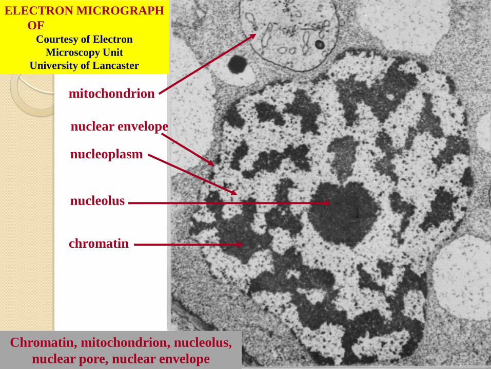
ELECTRON MICROGRAPH
OF CELL NUCLEUS Courtesy of Electron
Microscopy Unit
University of Lancaster
mitochondrion
nuclear envelope
nucleoplasm
nucleolus
chromatin
Chromatin, mitochondrion, nucleolus,
nuclear pore, nuclear envelope

Endoplasmic reticulum ELECTRON MICROGRAPH OF
ROUGH ENDOPLASMIC RETICULUM
Ribosome Cavity of cisterna

Courtesy of Dr. Julian
Thorpe – EM & FACS Lab,
Biological Sciences
University Of Sussex
ELECTRON MICROGRAPH
OF GOLGI APPARATUS
Golgi
vesicles
stacked
membranes

ELECTRON MICROGRAPH
OF MITOCHONDRION Courtesy of Electron
Microscopy Unit
University of Lancaster
endoplasmic
reticulum fluid matrix cristae outer
membrane
ER, cristae, fluid matrix, ribosome,
outer membrane

Courtesy of Dr. Julian Thorpe – EM & FACS Lab,
Biological Sciences University Of Sussex
The electron micrograph displayed below illustrates many of the
plant cell characteristics discussed
The cell wall, large central vacuole and chloroplasts are clearly visible
Also visible
is the clearly
defined nucleus
containing
chromatin
Nucleus
Chromatin
The vacuole
in this mature
plant cell from
a leaf is large,
and occupies
about 80% of
the cell volume

The photograph shown below details chloroplast structure
as viewed with a transmission electron microscope
Courtesy of Dr. Julian Thorpe – EM & FACS Lab,
Biological Sciences University Of Sussex
A single
Granum
Chloroplast envelope visible as two membranes Stroma containing numerous
small ribosomes
Lamellae connecting
different grana
Lipid
droplets
Related Documents


![The Relativistic Electron Density [1ex] and Electron ... · PDF fileThe Relativistic Electron Density and Electron Correlation Markus Reiher ... Electron density distributions for](https://static.cupdf.com/doc/110x72/5ab2020e7f8b9aea528d15ec/the-relativistic-electron-density-1ex-and-electron-relativistic-electron-density.jpg)