The Plant Cell, Vol. 12, 787–801, May 2000, www.plantcell.org © 2000 American Society of Plant Physiologists Identification, Purification, and Molecular Cloning of a Putative Plastidic Glucose Translocator Andreas Weber, a,1,2 Jerome C. Servaites, b,1 Donald R. Geiger, b Heike Kofler, a Diana Hille, a Ferdi Gröner, a Ulrike Hebbeker, a and Ulf-Ingo Flügge a a Universität zu Köln, Lehrstuhl Botanik II, Gyrhofstrasse 15, D-50931 Cologne, Germany b Department of Biology, University of Dayton, Dayton, Ohio 45469-2320 During photosynthesis, part of the fixed carbon is directed into the synthesis of transitory starch, which serves as an in- termediate carbon storage facility in chloroplasts. This transitory starch is mobilized during the night. Increasing evi- dence indicates that the main route of starch breakdown proceeds by way of hydrolytic enzymes and results in glucose formation. This pathway requires a glucose translocator to mediate the export of glucose from the chloroplasts. We have reexamined the kinetic properties of the plastidic glucose translocator and, using a differential labeling proce- dure, have identified the glucose translocator as a component of the inner envelope membrane. Peptide sequence in- formation derived from this protein was used to isolate cDNA clones encoding a putative plastidic glucose translocator from spinach, potato, tobacco, Arabidopsis, and maize. We also present the molecular characterization of a candidate for a hexose transporter of the plastid envelope membrane. This transporter, initially characterized more than 20 years ago, is closely related to the mammalian glucose transporter GLUT family and differs from all other plant hexose trans- porters that have been characterized to date. INTRODUCTION In plants, carbon fixed during the day is exported from the chloroplasts in the form of triose phosphate (trioseP), which is converted in the cytosol to sucrose. Sucrose often serves as the predominant photoassimilate being allocated to sink tissues. The export of trioseP from the chloroplasts is medi- ated by the trioseP/3-phosphoglycerate/phosphate translo- cator (TPT; Fliege et al., 1978; Flügge et al., 1989). Rather than being exported, a considerable amount of the fixed carbon is maintained within the chloroplasts and is involved in the biosynthesis of transitory starch, which could amount to approximately one-half of the carbon assimilated by pho- tosynthesis during the day. During the next dark period, transitory starch is mobilized to sustain a continuous supply of carbon (i.e., sucrose) for export to growing sinks as well as for energy metabolism in leaves. Mutants lacking the abil- ity to synthesize (Caspar et al., 1985; Hanson and McHale, 1988; Huber and Hanson, 1992; Geiger et al., 1995) or de- grade transitory starch (Zeeman et al., 1998a, 1998b; Caspar et al., 1991) show reduced growth under conditions in which photosynthesis is restricted. Starch degradation could follow either the phosphorolytic pathway, yielding trioseP, or the amylolytic pathway, leading to free sugars, glucose (Glc), and maltose. There is evidence that the dominant pathway for the degradation of transitory starch is the amylolytic one. First, trioseP, the end product of the phosphorolytic pathway, must be exported from the chloroplasts and subsequently be converted to hexose phosphate (hexoseP) in the cytosol. This reaction is con- trolled by the regulatory metabolite fructose 2,6-bisphos- phate, which is a strong inhibitor of the cytosolic fructose– bisphosphate phosphatase (Stitt, 1990). During the transi- tion from light to dark, the fructose 2,6-bisphosphate con- centration in leaves increases markedly (Servaites et al., 1989a, 1989b), and cytosolic and stromal concentrations of fructose 1,6-bisphosphate and trioseP decrease to zero (Gerhardt et al., 1987), indicating that the synthesis of tri- oseP and its conversion to hexoseP in the cytosol have ceased. In the dark, however, sucrose synthesis continues, and cytosolic concentrations of hexoseP remain high (Gerhardt et al., 1987) because of starch degradation, trans- port of these degradation products to the cytosol, and con- version of these products into sucrose. Second, evidence has been presented that transgenic po- tato and tobacco plants that have decreased TPT activity di- rect most of the fixed carbon into the biosynthesis of starch at the expense of sucrose synthesis. The diminished TPT activity in these transformants is compensated for by mobi- lizing transitory starch by way of the amylolytic pathway and 1 These authors contributed equally to this article. 2 To whom correspondence should be addressed. E-mail andr.weber@ uni-koeln.de; fax 49-221-470-5039.

Welcome message from author
This document is posted to help you gain knowledge. Please leave a comment to let me know what you think about it! Share it to your friends and learn new things together.
Transcript

The Plant Cell, Vol. 12, 787–801, May 2000, www.plantcell.org © 2000 American Society of Plant Physiologists
Identification, Purification, and Molecular Cloning of a Putative Plastidic Glucose Translocator
Andreas Weber,
a,1,2
Jerome C. Servaites,
b,1
Donald R. Geiger,
b
Heike Kofler,
a
Diana Hille,
a
Ferdi Gröner,
a
Ulrike Hebbeker,
a
and Ulf-Ingo Flügge
a
a
Universität zu Köln, Lehrstuhl Botanik II, Gyrhofstrasse 15, D-50931 Cologne, Germany
b
Department of Biology, University of Dayton, Dayton, Ohio 45469-2320
During photosynthesis, part of the fixed carbon is directed into the synthesis of transitory starch, which serves as an in-termediate carbon storage facility in chloroplasts. This transitory starch is mobilized during the night. Increasing evi-dence indicates that the main route of starch breakdown proceeds by way of hydrolytic enzymes and results in glucoseformation. This pathway requires a glucose translocator to mediate the export of glucose from the chloroplasts. Wehave reexamined the kinetic properties of the plastidic glucose translocator and, using a differential labeling proce-dure, have identified the glucose translocator as a component of the inner envelope membrane. Peptide sequence in-formation derived from this protein was used to isolate cDNA clones encoding a putative plastidic glucose translocatorfrom spinach, potato, tobacco, Arabidopsis, and maize. We also
present the molecular characterization of a candidatefor a hexose transporter of the plastid envelope membrane. This transporter, initially characterized more than 20 yearsago, is closely related to the mammalian glucose transporter GLUT family and differs from all other plant hexose trans-porters that have been characterized to date.
INTRODUCTION
In plants, carbon fixed during the day is exported from thechloroplasts in the form of triose phosphate (trioseP), whichis converted in the cytosol to sucrose. Sucrose often servesas the predominant photoassimilate being allocated to sinktissues. The export of trioseP from the chloroplasts is medi-ated by the trioseP/3-phosphoglycerate/phosphate translo-cator (TPT; Fliege et al., 1978; Flügge et al., 1989). Ratherthan being exported, a considerable amount of the fixedcarbon is maintained within the chloroplasts and is involvedin the biosynthesis of transitory starch, which could amountto approximately one-half of the carbon assimilated by pho-tosynthesis during the day. During the next dark period,transitory starch is mobilized to sustain a continuous supplyof carbon (i.e., sucrose) for export to growing sinks as wellas for energy metabolism in leaves. Mutants lacking the abil-ity to synthesize (Caspar et al., 1985; Hanson and McHale,1988; Huber and Hanson, 1992; Geiger et al., 1995) or de-grade transitory starch (Zeeman et al., 1998a, 1998b; Casparet al., 1991) show reduced growth under conditions in whichphotosynthesis is restricted.
Starch degradation could follow either the phosphorolytic
pathway, yielding trioseP, or the amylolytic pathway, leadingto free sugars, glucose (Glc), and maltose. There is evidencethat the dominant pathway for the degradation of transitorystarch is the amylolytic one. First, trioseP, the end productof the phosphorolytic pathway, must be exported from thechloroplasts and subsequently be converted to hexosephosphate (hexoseP) in the cytosol. This reaction is con-trolled by the regulatory metabolite fructose 2,6-bisphos-phate, which is a strong inhibitor of the cytosolic fructose–bisphosphate phosphatase (Stitt, 1990). During the transi-tion from light to dark, the fructose 2,6-bisphosphate con-centration in leaves increases markedly (Servaites et al.,1989a, 1989b), and cytosolic and stromal concentrations offructose 1,6-bisphosphate and trioseP decrease to zero(Gerhardt et al., 1987), indicating that the synthesis of tri-oseP and its conversion to hexoseP in the cytosol haveceased. In the dark, however, sucrose synthesis continues,and cytosolic concentrations of hexoseP remain high(Gerhardt et al., 1987) because of starch degradation, trans-port of these degradation products to the cytosol, and con-version of these products into sucrose.
Second, evidence has been presented that transgenic po-tato and tobacco plants that have decreased TPT activity di-rect most of the fixed carbon into the biosynthesis of starchat the expense of sucrose synthesis. The diminished TPTactivity in these transformants is compensated for by mobi-lizing transitory starch by way of the amylolytic pathway and
1
These authors contributed equally to this article.
2
To whom correspondence should be addressed. E-mail [email protected]; fax 49-221-470-5039.
788 The Plant Cell
exporting the products of starch breakdown in the form ofGlc (Heineke et al., 1994; Häusler et al., 1998).
Third, a study of a high-starch mutant of Arabidopsis,designated
sex1
(for starch excess; Caspar et al., 1991),which is unable to degrade starch, supports the notion thatthe products of starch breakdown are exported from thechloroplast by a translocator different from the TPT. Chloro-plasts isolated from the
sex1
mutant were shown to possessa functional TPT but were unable to transport Glc (Tretheweyand ap Rees, 1994a). Finally, using nuclear magnetic resonanceto determine the intramolecular distribution of deuterium-labeled Glc liberated during the breakdown of transitorystarch showed that most carbon was exported as hexosesat night (Gleixner et al., 1998; Schleucher et al., 1998).
The amylolytic pathway of starch breakdown requires thepresence of a transport capacity for the resulting hexoses.Herold et al. (1981), Beck (1985), and Rost et al. (1996)reported that the chloroplast envelope is permeable for mal-tose but not for maltodextrins. Rost et al. (1996) demon-strated that the uptake of
D
-Glc does not compete withmaltose transport. Schäfer et al. (1977) reported that severalpentoses and hexoses, including Glc, were taken up by iso-lated chloroplasts by way of facilitated diffusion. The signifi-cance of this discovery was obscured largely by the fact thatthe reported rate of Glc transport was much lower than therate of Pi transport mediated by the TPT. This made it seemunlikely that, during nighttime, the plastidic Glc translocator(pGlcT) could export all the Glc made from starch synthe-sized during the day.
Our recent investigations of Glc transport into chloro-plasts, using a differential affinity labeling strategy, have al-lowed us to identify the pGlcT as a component of the innerenvelope membrane. We obtained peptide sequence infor-mation for this protein and isolated the correspondingcDNA. Here, we identify a candidate for the Glc transporterof the plastid inner envelope membrane and report the re-sults of cloning the corresponding cDNAs from spinach, to-bacco, potato, Arabidopsis, and maize.
RESULTS
Measurements of the Transport Capacity of pGlcT and Its Substrate Specificity
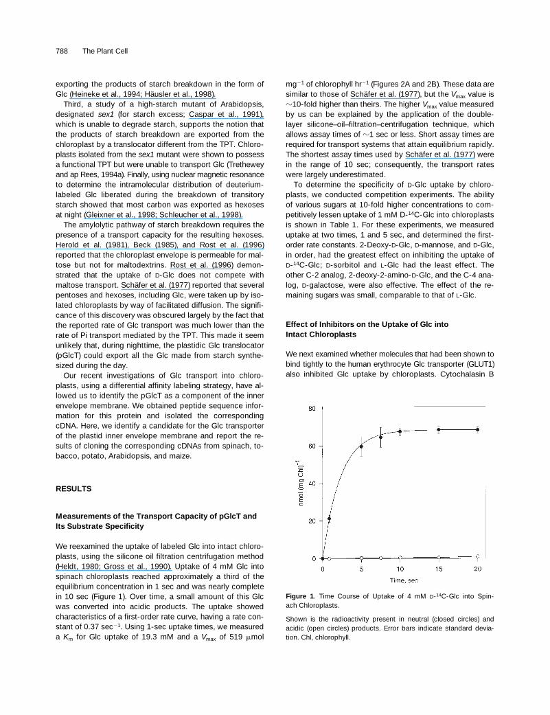
We reexamined the uptake of labeled Glc into intact chloro-plasts, using the silicone oil filtration centrifugation method(Heldt, 1980; Gross et al., 1990). Uptake of 4 mM Glc intospinach chloroplasts reached approximately a third of theequilibrium concentration in 1 sec and was nearly completein 10 sec (Figure 1). Over time, a small amount of this Glcwas converted into acidic products. The uptake showedcharacteristics of a first-order rate curve, having a rate con-stant of 0.37 sec
2
1
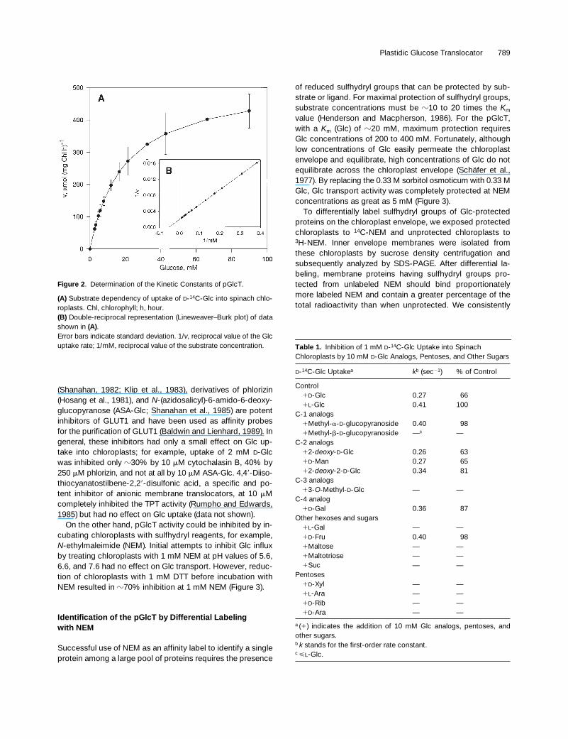
. Using 1-sec uptake times, we measureda
K
m
for Glc uptake of 19.3 mM and a
V
max
of 519
m
mol
mg
2
1
of chlorophyll hr
2
1
(Figures 2A and 2B). These data aresimilar to those of Schäfer et al. (1977), but the
V
max
value is
z
10-fold higher than theirs. The higher
V
max
value measuredby us can be explained by the application of the double-layer silicone–oil–filtration–centrifugation technique, whichallows assay times of
z
1 sec or less. Short assay times arerequired for transport systems that attain equilibrium rapidly.The shortest assay times used by Schäfer et al. (1977) werein the range of 10 sec; consequently, the transport rateswere largely underestimated.
To determine the specificity of
D
-Glc uptake by chloro-plasts, we conducted competition experiments. The abilityof various sugars at 10-fold higher concentrations to com-petitively lessen uptake of 1 mM D-
14
C-Glc into chloroplastsis shown in Table 1. For these experiments, we measureduptake at two times, 1 and 5 sec, and determined the first-order rate constants. 2-Deoxy-
D
-Glc,
D
-mannose, and
D
-Glc,in order, had the greatest effect on inhibiting the uptake of
D
-
14
C-Glc;
D
-sorbitol and
L
-Glc had the least effect. Theother C-2 analog, 2-deoxy-2-amino-
D
-Glc, and the C-4 ana-log,
D
-galactose, were also effective. The effect of the re-maining sugars was small, comparable to that of
L
-Glc.
Effect of Inhibitors on the Uptake of Glc intoIntact Chloroplasts
We next examined whether molecules that had been shown tobind tightly to the human erythrocyte Glc transporter (GLUT1)also inhibited Glc uptake by chloroplasts. Cytochalasin B
Figure 1. Time Course of Uptake of 4 mM D-14C-Glc into Spin-ach Chloroplasts.
Shown is the radioactivity present in neutral (closed circles) andacidic (open circles) products. Error bars indicate standard devia-tion. Chl, chlorophyll.
Plastidic Glucose Translocator 789
(Shanahan, 1982; Klip et al., 1983), derivatives of phlorizin(Hosang et al., 1981), and
N
-(azidosalicyl)-6-amido-6-deoxy-glucopyranose (ASA-Glc; Shanahan et al., 1985) are potentinhibitors of GLUT1 and have been used as affinity probesfor the purification of GLUT1 (Baldwin and Lienhard, 1989). Ingeneral, these inhibitors had only a small effect on Glc up-take into chloroplasts; for example, uptake of 2 mM
D
-Glcwas inhibited only
z
30% by 10
m
M cytochalasin B, 40% by250
m
M phlorizin, and not at all by 10
m
M ASA-Glc. 4,4
9
-Diiso-thiocyanatostilbene-2,2
9
-disulfonic acid, a specific and po-tent inhibitor of anionic membrane translocators, at 10
m
Mcompletely inhibited the TPT activity (Rumpho and Edwards,1985) but had no effect on Glc uptake (data not shown).
On the other hand, pGlcT activity could be inhibited by in-cubating chloroplasts with sulfhydryl reagents, for example,
N
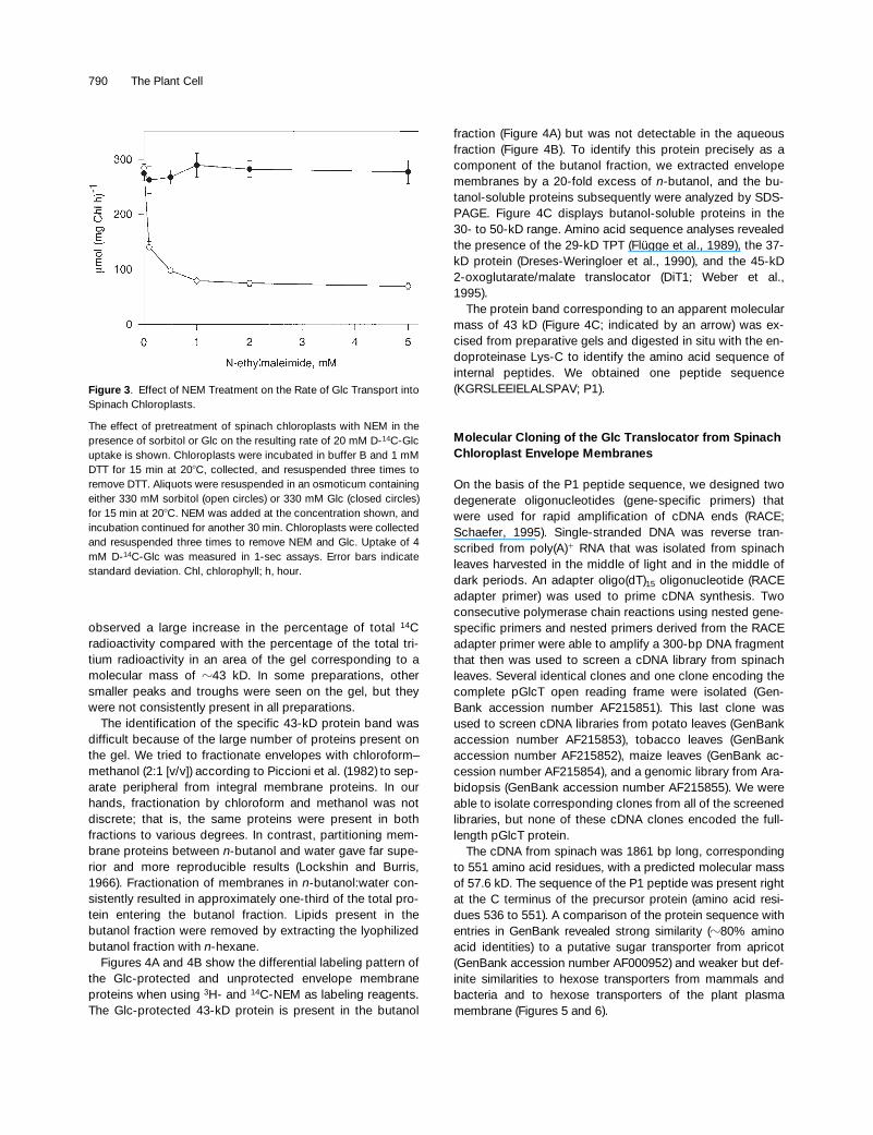
-ethylmaleimide (NEM). Initial attempts to inhibit Glc influxby treating chloroplasts with 1 mM NEM at pH values of 5.6,6.6, and 7.6 had no effect on Glc transport. However, reduc-tion of chloroplasts with 1 mM DTT before incubation withNEM resulted in
z
70% inhibition at 1 mM NEM (Figure 3).
Identification of the pGlcT by Differential Labelingwith NEM
Successful use of NEM as an affinity label to identify a singleprotein among a large pool of proteins requires the presence
of reduced sulfhydryl groups that can be protected by sub-strate or ligand. For maximal protection of sulfhydryl groups,substrate concentrations must be
z
10 to 20 times the
K
m
value (Henderson and Macpherson, 1986). For the pGlcT,with a
K
m
(Glc) of
z
20 mM, maximum protection requiresGlc concentrations of 200 to 400 mM. Fortunately, althoughlow concentrations of Glc easily permeate the chloroplastenvelope and equilibrate, high concentrations of Glc do notequilibrate across the chloroplast envelope (Schäfer et al.,1977). By replacing the 0.33 M sorbitol osmoticum with 0.33 MGlc, Glc transport activity was completely protected at NEMconcentrations as great as 5 mM (Figure 3).
To differentially label sulfhydryl groups of Glc-protectedproteins on the chloroplast envelope, we exposed protectedchloroplasts to
14
C-NEM and unprotected chloroplasts to
3
H-NEM. Inner envelope membranes were isolated fromthese chloroplasts by sucrose density centrifugation andsubsequently analyzed by SDS-PAGE. After differential la-beling, membrane proteins having sulfhydryl groups pro-tected from unlabeled NEM should bind proportionatelymore labeled NEM and contain a greater percentage of thetotal radioactivity than when unprotected. We consistently
Figure 2. Determination of the Kinetic Constants of pGlcT.
(A) Substrate dependency of uptake of D-14C-Glc into spinach chlo-roplasts. Chl, chlorophyll; h, hour.(B) Double-reciprocal representation (Lineweaver–Burk plot) of datashown in (A).Error bars indicate standard deviation. 1/v, reciprocal value of the Glcuptake rate; 1/mM, reciprocal value of the substrate concentration.
Table 1.
Inhibition of 1 mM
D
-
14
C-Glc Uptake into Spinach Chloroplasts by 10 mM
D
-Glc Analogs, Pentoses, and Other Sugars
D
-
14
C-Glc Uptake
a
k
b
(sec
2
1
) % of Control
Control
1
D
-Glc 0.27 66
1
L
-Glc 0.41 100C-1 analogs
1
Methyl-
a
-
D
-glucopyranoside 0.40 98
1
Methyl-
b
-
D
-glucopyranoside —
c
—C-2 analogs
1
2-
deoxy
-
D
-Glc 0.26 63
1
D
-Man 0.27 65
1
2-
deoxy
-2-
D
-Glc 0.34 81C-3 analogs
1
3-
O
-Methyl-
D
-Glc — —C-4 analog
1
D
-Gal 0.36 87Other hexoses and sugars
1
L
-Gal — —
1
D
-Fru 0.40 98
1
Maltose — —
1
Maltotriose — —
1
Suc — —Pentoses
1
D
-Xyl — —
1
L
-Ara — —
1
D
-Rib — —
1
D
-Ara — —
a
(
1
) indicates the addition of 10 mM Glc analogs, pentoses, andother sugars.
b
k
stands for the first-order rate constant.
c
<
L
-Glc.
790 The Plant Cell
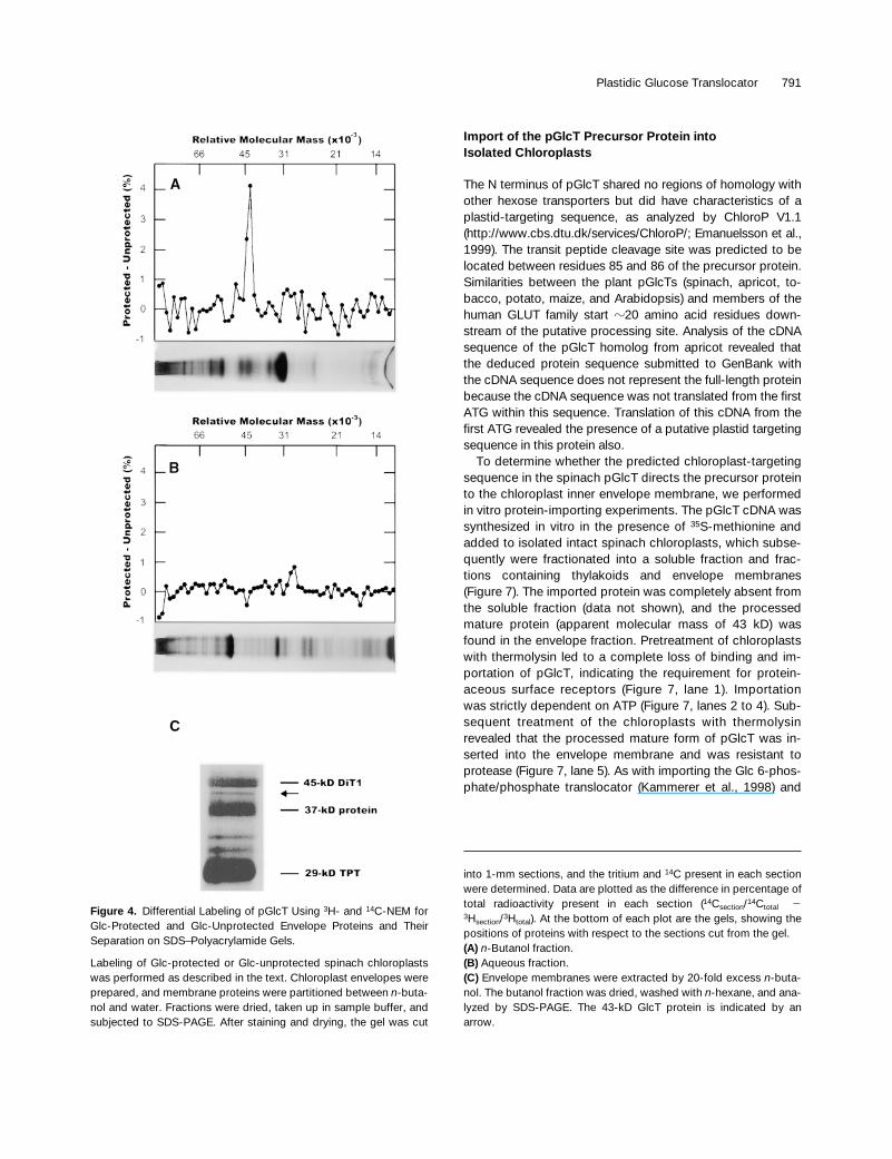
observed a large increase in the percentage of total
14
Cradioactivity compared with the percentage of the total tri-tium radioactivity in an area of the gel corresponding to amolecular mass of
z
43 kD. In some preparations, othersmaller peaks and troughs were seen on the gel, but theywere not consistently present in all preparations.
The identification of the specific 43-kD protein band wasdifficult because of the large number of proteins present onthe gel. We tried to fractionate envelopes with chloroform–methanol (2:1 [v/v]) according to Piccioni et al. (1982)
to sep-arate peripheral from integral membrane proteins. In ourhands, fractionation by chloroform and methanol was notdiscrete; that is, the same proteins were present in bothfractions to various degrees. In contrast, partitioning mem-brane proteins between
n
-butanol and water gave far supe-rior and more reproducible results
(Lockshin and Burris,1966). Fractionation of membranes in
n-
butanol:water con-sistently resulted in approximately one-third of the total pro-tein entering the butanol fraction. Lipids present in thebutanol fraction were removed by extracting the lyophilizedbutanol fraction with
n
-hexane.Figures 4A and 4B show the differential labeling pattern of
the Glc-protected and unprotected envelope membraneproteins when using
3
H- and
14
C-NEM as labeling reagents.The Glc-protected 43-kD protein is present in the butanol
fraction (Figure 4A) but was not detectable in the aqueousfraction (Figure 4B). To identify this protein precisely as acomponent of the butanol fraction, we extracted envelopemembranes by a 20-fold excess of
n-
butanol, and the bu-tanol-soluble proteins subsequently were analyzed by SDS-PAGE. Figure 4C displays butanol-soluble proteins in the30- to 50-kD range. Amino acid sequence analyses revealedthe presence of the 29-kD TPT (Flügge et al., 1989), the 37-kD protein (Dreses-Weringloer et al., 1990), and the 45-kD2-oxoglutarate/malate translocator (DiT1; Weber et al.,1995).
The protein band corresponding to an apparent molecularmass of 43 kD (Figure 4C; indicated by an arrow) was ex-cised from preparative gels and digested in situ with the en-doproteinase Lys-C to identify the amino acid sequence ofinternal peptides. We obtained one peptide sequence(KGRSLEEIELALSPAV; P1).
Molecular Cloning of the Glc Translocator from Spinach Chloroplast Envelope Membranes
On the basis of the P1 peptide sequence, we designed twodegenerate oligonucleotides (gene-specific primers) thatwere used for rapid amplification of cDNA ends (RACE;Schaefer, 1995). Single-stranded DNA was reverse tran-scribed from poly(A)
1
RNA that was isolated from spinachleaves harvested in the middle of light and in the middle ofdark periods. An adapter oligo(dT)
15
oligonucleotide (RACEadapter primer) was used to prime cDNA synthesis. Twoconsecutive polymerase chain reactions using nested gene-specific primers and nested primers derived from the RACEadapter primer were able to amplify a 300-bp DNA fragmentthat then was used to screen a cDNA library from spinachleaves. Several identical clones and one clone encoding thecomplete pGlcT open reading frame were isolated (Gen-Bank accession number AF215851). This last clone wasused to screen cDNA libraries from potato leaves (GenBankaccession number AF215853), tobacco leaves (GenBankaccession number AF215852), maize leaves (GenBank ac-cession number AF215854), and a genomic library from Ara-bidopsis (GenBank accession number AF215855). We wereable to isolate corresponding clones from all of the screenedlibraries, but none of these cDNA clones encoded the full-length pGlcT protein.
The cDNA from spinach was 1861 bp long, correspondingto 551 amino acid residues, with a predicted molecular massof 57.6 kD. The sequence of the P1 peptide was present rightat the C terminus of the precursor protein (amino acid resi-dues 536 to 551). A comparison of the protein sequence withentries in GenBank revealed strong similarity (
z80% aminoacid identities) to a putative sugar transporter from apricot(GenBank accession number AF000952) and weaker but def-inite similarities to hexose transporters from mammals andbacteria and to hexose transporters of the plant plasmamembrane (Figures 5 and 6).
Figure 3. Effect of NEM Treatment on the Rate of Glc Transport intoSpinach Chloroplasts.
The effect of pretreatment of spinach chloroplasts with NEM in thepresence of sorbitol or Glc on the resulting rate of 20 mM D-14C-Glcuptake is shown. Chloroplasts were incubated in buffer B and 1 mMDTT for 15 min at 208C, collected, and resuspended three times toremove DTT. Aliquots were resuspended in an osmoticum containingeither 330 mM sorbitol (open circles) or 330 mM Glc (closed circles)for 15 min at 208C. NEM was added at the concentration shown, andincubation continued for another 30 min. Chloroplasts were collectedand resuspended three times to remove NEM and Glc. Uptake of 4mM D-14C-Glc was measured in 1-sec assays. Error bars indicatestandard deviation. Chl, chlorophyll; h, hour.
Plastidic Glucose Translocator 791
Import of the pGlcT Precursor Protein intoIsolated Chloroplasts
The N terminus of pGlcT shared no regions of homology withother hexose transporters but did have characteristics of aplastid-targeting sequence, as analyzed by ChloroP V1.1(http://www.cbs.dtu.dk/services/ChloroP/; Emanuelsson et al.,1999). The transit peptide cleavage site was predicted to belocated between residues 85 and 86 of the precursor protein.Similarities between the plant pGlcTs (spinach, apricot, to-bacco, potato, maize, and Arabidopsis) and members of thehuman GLUT family start z20 amino acid residues down-stream of the putative processing site. Analysis of the cDNAsequence of the pGlcT homolog from apricot revealed thatthe deduced protein sequence submitted to GenBank withthe cDNA sequence does not represent the full-length proteinbecause the cDNA sequence was not translated from the firstATG within this sequence. Translation of this cDNA from thefirst ATG revealed the presence of a putative plastid targetingsequence in this protein also.
To determine whether the predicted chloroplast-targetingsequence in the spinach pGlcT directs the precursor proteinto the chloroplast inner envelope membrane, we performedin vitro protein-importing experiments. The pGlcT cDNA wassynthesized in vitro in the presence of 35S-methionine andadded to isolated intact spinach chloroplasts, which subse-quently were fractionated into a soluble fraction and frac-tions containing thylakoids and envelope membranes(Figure 7). The imported protein was completely absent fromthe soluble fraction (data not shown), and the processedmature protein (apparent molecular mass of 43 kD) wasfound in the envelope fraction. Pretreatment of chloroplastswith thermolysin led to a complete loss of binding and im-portation of pGlcT, indicating the requirement for protein-aceous surface receptors (Figure 7, lane 1). Importationwas strictly dependent on ATP (Figure 7, lanes 2 to 4). Sub-sequent treatment of the chloroplasts with thermolysinrevealed that the processed mature form of pGlcT was in-serted into the envelope membrane and was resistant toprotease (Figure 7, lane 5). As with importing the Glc 6-phos-phate/phosphate translocator (Kammerer et al., 1998) and
Figure 4. Differential Labeling of pGlcT Using 3H- and 14C-NEM forGlc-Protected and Glc-Unprotected Envelope Proteins and TheirSeparation on SDS–Polyacrylamide Gels.
Labeling of Glc-protected or Glc-unprotected spinach chloroplastswas performed as described in the text. Chloroplast envelopes wereprepared, and membrane proteins were partitioned between n-buta-nol and water. Fractions were dried, taken up in sample buffer, andsubjected to SDS-PAGE. After staining and drying, the gel was cut
into 1-mm sections, and the tritium and 14C present in each sectionwere determined. Data are plotted as the difference in percentage oftotal radioactivity present in each section (14Csection/14Ctotal 23Hsection/3Htotal). At the bottom of each plot are the gels, showing thepositions of proteins with respect to the sections cut from the gel.(A) n-Butanol fraction.(B) Aqueous fraction.(C) Envelope membranes were extracted by 20-fold excess n-buta-nol. The butanol fraction was dried, washed with n-hexane, and ana-lyzed by SDS-PAGE. The 43-kD GlcT protein is indicated by anarrow.
792 The Plant Cell
the putative plastidic adenylate translocator of maize amylo-plasts (Bt1; Li et al., 1992), processing of the spinach pGlcTprecursor protein obviously proceeded through an inter-mediate (apparent molecular mass of 46 kD; Figure 7). Insummary, the in vitro protein-importing experiments demon-strate that the N terminus of pGlcT has the characteristics ofa plastid-targeting sequence and is able to direct the spin-ach pGlcT protein correctly to the plastid inner envelopemembrane where it is inserted, resistant to protease treat-ment, and processed to its mature form.
Expression Analysis of pGlcT
We analyzed the steady state amounts of pGlcT mRNA indifferent tissues and during a light/dark cycle in tobacco
plants. The greatest amounts of pGlcT mRNA were found insource/sink transition leaves, followed by source leaves andgreen flower buds. Expression was low in sink tissues suchas stems, roots, and petioles (Figure 8A); no strong varia-tions in the pGlcT mRNA amounts were observed through-out cycles of light and dark. Expression was lowest at theends of the dark and light periods and greatest at the begin-nings of the dark and light periods (Figure 8B). Using Arabi-dopsis leaves, we analyzed the amounts of pGlcT mRNA inthe middle of the light and dark periods by semiquantitativereverse transcription–polymerase chain reaction. No signifi-cant differences in the steady state mRNA levels could bedetected (data not shown).
In tobacco lines that show a strong antisense repressionof the plastidic TPT (Häusler et al., 1998), we were not ableto detect an increase in the pGlcT mRNA (Figure 8C), al-
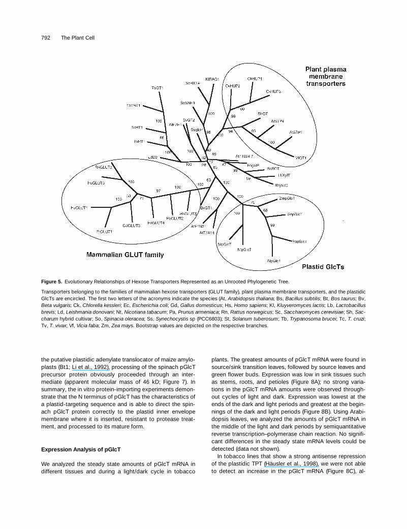
Figure 5. Evolutionary Relationships of Hexose Transporters Represented as an Unrooted Phylogenetic Tree.
Transporters belonging to the families of mammalian hexose transporters (GLUT family), plant plasma membrane transporters, and the plastidicGlcTs are encircled. The first two letters of the acronyms indicate the species (At, Arabidopsis thaliana; Bs, Bacillus subtilis; Bt, Bos taurus; Bv,Beta vulgaris; Ck, Chlorella kessleri; Ec, Escherichia coli; Gd, Gallus domesticus; Hs, Homo sapiens; Kl, Kluyveromyces lactis; Lb, Lactobacillusbrevis; Ld, Leishmania donovani; Nt, Nicotiana tabacum; Pa, Prunus armeniaca; Rn, Rattus norwegicus; Sc, Saccharomyces cerevisiae; Sh, Sac-charum hybrid cultivar; So, Spinacia oleracea; Ss, Synechocystis sp (PCC6803); St, Solanum tuberosum; Tb, Trypanosoma brucei; Tc, T. cruzi;Tv, T. vivax; Vf, Vicia faba; Zm, Zea mays. Bootstrap values are depicted on the respective branches.
Plastidic Glucose Translocator 793
though Häusler et al. (1998) have demonstrated an in-creased transport capacity for Glc in plastids isolated fromthese plants. That might indicate a control on plastidic Glctransport capacity at the post-transcriptional or post-trans-lational level.
Expression of the Spinach pGlcT in the Arabidopsis Mutant sex1 Does Not Complement theMutant Phenotype
Trethewey and ap Rees (1994a, 1994b) have analyzed inmore detail the starch excess mutant sex1 that originallywas isolated by Caspar et al. (1991). Their results indicatethat sex1 is deficient in the pGlcT. To determine whether themutation in sex1 can be complemented by the pGlcT cDNA,we transformed the Arabidopsis mutant with the corre-sponding cDNA from spinach by an in-the-plant transforma-tion protocol (Bechtold et al., 1993). Having obtained 70independent transgenic plant lines, we tested all transfor-mants for their ability to mobilize transitory starch during thedark period. In contrast to the wild type, none of the trans-
formants was able to mobilize starch, even after prolongedincubation in the dark. Obviously, the cDNA that encodesthe pGlcT from spinach is not able to complement the mu-tant phenotype of sex1.
To further determine whether sex1 actually carries a muta-tion in the pGlcT, we mapped the Arabidopsis pGlcT genewith the help of yeast artificial chromosome (YAC) contigsimmobilized on nylon filters. We were able to localize the Ar-abidopsis pGlcT gene on YACs CIC3F11, CIC8E12, andCIC8H6. This places the gene between markers ve029 andnga106 on chromosome 5 (Schmidt et al., 1997; http://nasc.nott.ac.uk/new_ri_map.html).
Using cleaved amplified polymorphic sequence mark-ers (Konieczny and Ausubel, 1993), we localized the sex1mutation on chromosome 1 between the markers phyAand NCC1. No homology with sugar transporters wasfound in this region. From the complementation assay de-scribed above and the genetic data, sex1 appears un-likely to be deficient in the pGlcT. A detailed descriptionof the genetic location of the sex1 mutation will be givenin a forthcoming paper (H. Kofler et al., manuscript inpreparation).
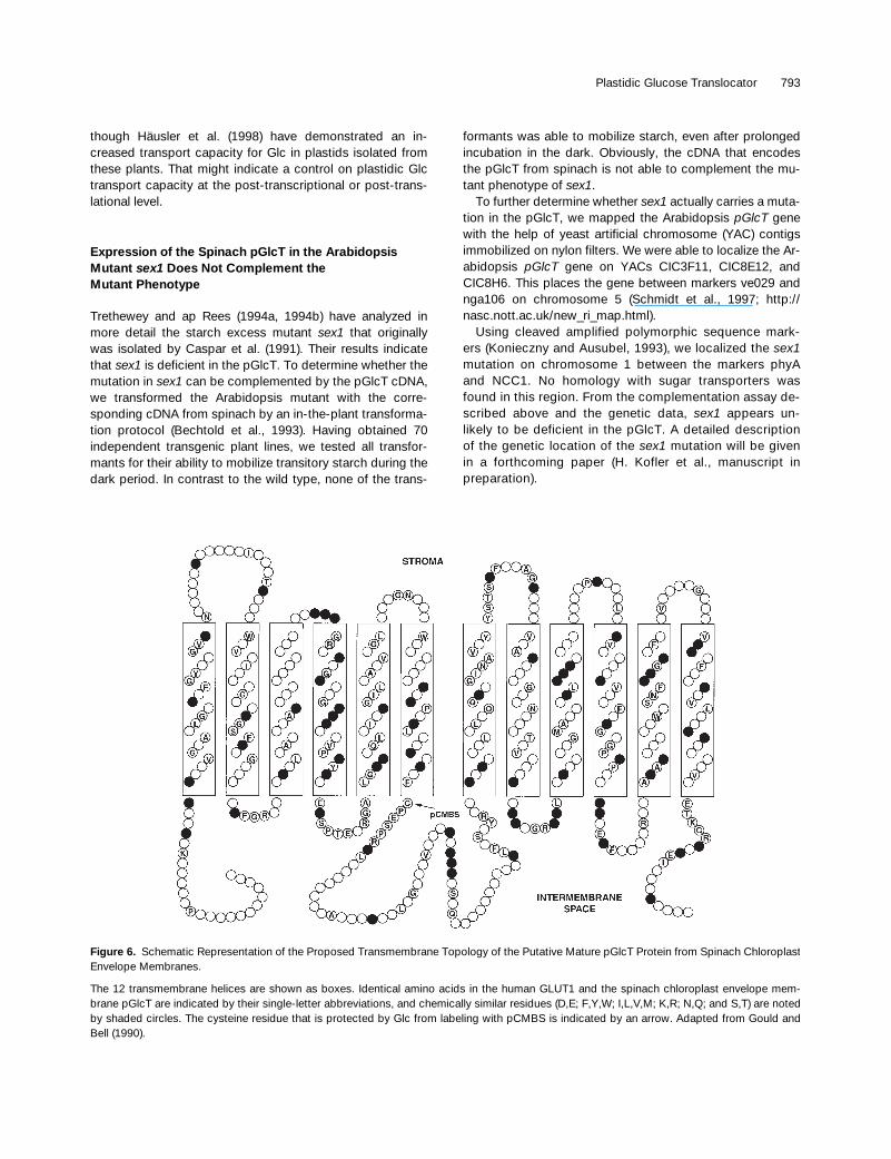
Figure 6. Schematic Representation of the Proposed Transmembrane Topology of the Putative Mature pGlcT Protein from Spinach ChloroplastEnvelope Membranes.
The 12 transmembrane helices are shown as boxes. Identical amino acids in the human GLUT1 and the spinach chloroplast envelope mem-brane pGlcT are indicated by their single-letter abbreviations, and chemically similar residues (D,E; F,Y,W; I,L,V,M; K,R; N,Q; and S,T) are notedby shaded circles. The cysteine residue that is protected by Glc from labeling with pCMBS is indicated by an arrow. Adapted from Gould andBell (1990).
794 The Plant Cell
DISCUSSION
Using a differential labeling strategy, we identified a 43-kDprotein of the chloroplast inner envelope membrane as acandidate for the pGlcT. A peptide sequence derived fromthis protein was used to isolate the corresponding cDNAfrom spinach. This cDNA then was used to clone the homol-ogous cDNAs from different plant species. In vitro proteinimportation experiments demonstrated that pGlcT is effi-ciently imported into the chloroplast inner envelope mem-brane, where it is processed to its mature form.
The mature proteins of the pGlcTs show a strong aminoacid sequence identity (80 to 86%). They share z40% iden-tical amino acid residues with two uncharacterized putativesugar transporters from Arabidopsis that have been identi-fied on bacterial artificial chromosome sequences in the Ar-abidopsis genome project (F1N21 and T7A41). The putativesugar transporter found on bacterial artificial chromosomeF1N21 also possesses an N-terminal extension that mightrepresent a targeting signal; however, this sequence wasnot classified as a chloroplast targeting signal by ChloroP.
A weaker but definite similarity was observed to hexosetransporters from mammals and bacteria (amino acid identi-ties of z34%). Approximately 30% of the amino acids wereidentical to those of hexose transporters of the plant plasmamembrane (Figure 5).
Analysis of the pGlcT Sequence
Henderson (1991) has reported that a repeated [RK]XGR[RK]motif in the hydrophilic loops connecting helices 2 and 3 aswell as helices 8 and 9 is a common feature shared by sugar
facilitators of eukaryotes and bacteria. In the pGlcTs, thismotif is conserved in the loop connecting helices 8 and 9,whereas the second motif includes a threonine residue in-serted in front of the last basic residue: [RK]XGRT[RK] (Fig-ure 6).
The binding of cytochalasin B to the mammalian GLUT1transporter requires the presence of two tryptophan resi-dues (Trp-388 and Trp-412) and an Asn-Trp motif in helix 11(reviewed in Walmsley et al., 1998). In the plant pGlcT family,the residue corresponding to Trp-388 is replaced by an ala-nine (Ala-467; spinach), and a histidine residue (His-490;spinach) replaces the asparagine of the GLUT1 Asn-Trp mo-tif. These modifications are probably the reason why cy-
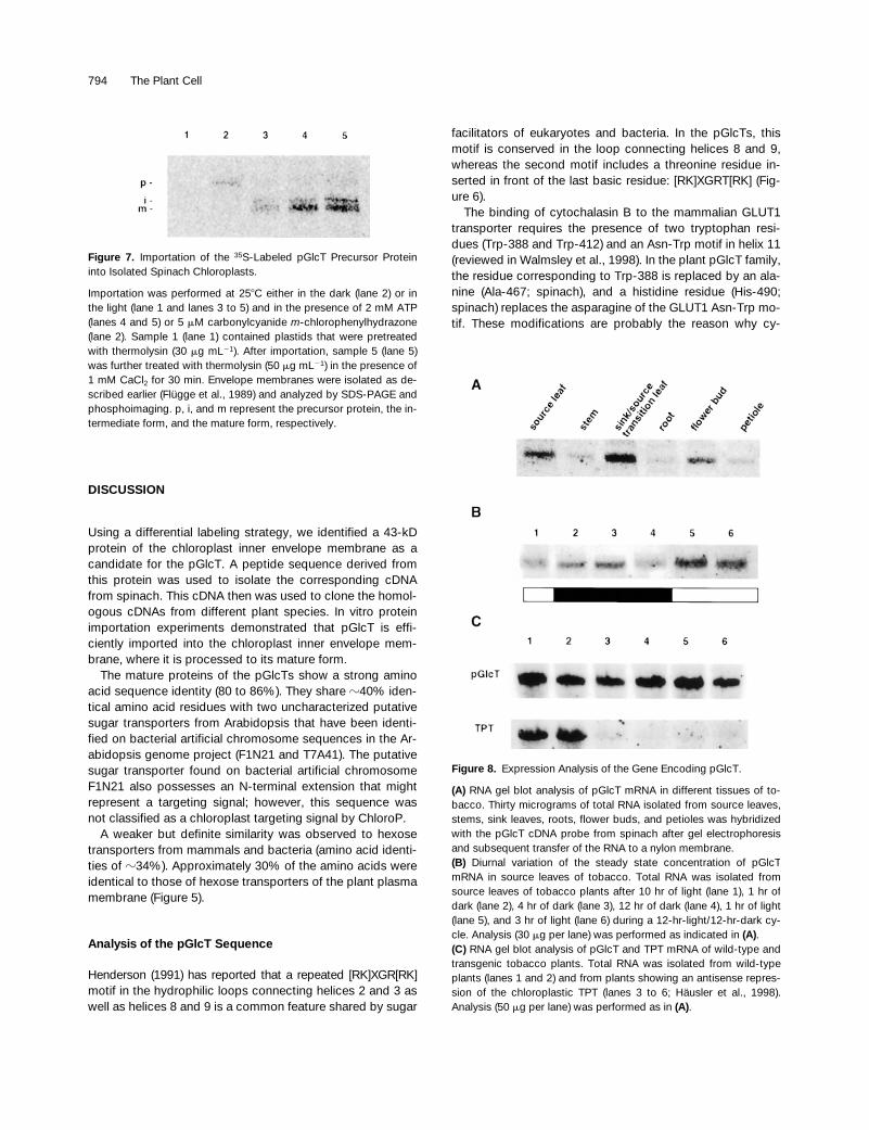
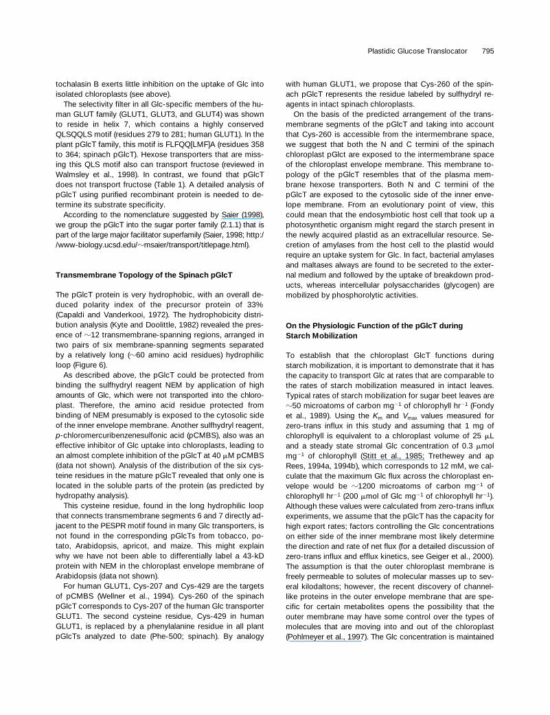
Figure 7. Importation of the 35S-Labeled pGlcT Precursor Proteininto Isolated Spinach Chloroplasts.
Importation was performed at 258C either in the dark (lane 2) or inthe light (lane 1 and lanes 3 to 5) and in the presence of 2 mM ATP(lanes 4 and 5) or 5 mM carbonylcyanide m-chlorophenylhydrazone(lane 2). Sample 1 (lane 1) contained plastids that were pretreatedwith thermolysin (30 mg mL21). After importation, sample 5 (lane 5)was further treated with thermolysin (50 mg mL21) in the presence of1 mM CaCl2 for 30 min. Envelope membranes were isolated as de-scribed earlier (Flügge et al., 1989) and analyzed by SDS-PAGE andphosphoimaging. p, i, and m represent the precursor protein, the in-termediate form, and the mature form, respectively.
Figure 8. Expression Analysis of the Gene Encoding pGlcT.
(A) RNA gel blot analysis of pGlcT mRNA in different tissues of to-bacco. Thirty micrograms of total RNA isolated from source leaves,stems, sink leaves, roots, flower buds, and petioles was hybridizedwith the pGlcT cDNA probe from spinach after gel electrophoresisand subsequent transfer of the RNA to a nylon membrane.(B) Diurnal variation of the steady state concentration of pGlcTmRNA in source leaves of tobacco. Total RNA was isolated fromsource leaves of tobacco plants after 10 hr of light (lane 1), 1 hr ofdark (lane 2), 4 hr of dark (lane 3), 12 hr of dark (lane 4), 1 hr of light(lane 5), and 3 hr of light (lane 6) during a 12-hr-light/12-hr-dark cy-cle. Analysis (30 mg per lane) was performed as indicated in (A).(C) RNA gel blot analysis of pGlcT and TPT mRNA of wild-type andtransgenic tobacco plants. Total RNA was isolated from wild-typeplants (lanes 1 and 2) and from plants showing an antisense repres-sion of the chloroplastic TPT (lanes 3 to 6; Häusler et al., 1998).Analysis (50 mg per lane) was performed as in (A).
Plastidic Glucose Translocator 795
tochalasin B exerts little inhibition on the uptake of Glc intoisolated chloroplasts (see above).
The selectivity filter in all Glc-specific members of the hu-man GLUT family (GLUT1, GLUT3, and GLUT4) was shownto reside in helix 7, which contains a highly conservedQLSQQLS motif (residues 279 to 281; human GLUT1). In theplant pGlcT family, this motif is FLFQQ[LMF]A (residues 358to 364; spinach pGlcT). Hexose transporters that are miss-ing this QLS motif also can transport fructose (reviewed inWalmsley et al., 1998). In contrast, we found that pGlcTdoes not transport fructose (Table 1). A detailed analysis ofpGlcT using purified recombinant protein is needed to de-termine its substrate specificity.
According to the nomenclature suggested by Saier (1998),we group the pGlcT into the sugar porter family (2.1.1) that ispart of the large major facilitator superfamily (Saier, 1998; http://www-biology.ucsd.edu/zmsaier/transport/titlepage.html).
Transmembrane Topology of the Spinach pGlcT
The pGlcT protein is very hydrophobic, with an overall de-duced polarity index of the precursor protein of 33%(Capaldi and Vanderkooi, 1972). The hydrophobicity distri-bution analysis (Kyte and Doolittle, 1982) revealed the pres-ence of z12 transmembrane-spanning regions, arranged intwo pairs of six membrane-spanning segments separatedby a relatively long (z60 amino acid residues) hydrophilicloop (Figure 6).
As described above, the pGlcT could be protected frombinding the sulfhydryl reagent NEM by application of highamounts of Glc, which were not transported into the chloro-plast. Therefore, the amino acid residue protected frombinding of NEM presumably is exposed to the cytosolic sideof the inner envelope membrane. Another sulfhydryl reagent,p-chloromercuribenzenesulfonic acid (pCMBS), also was aneffective inhibitor of Glc uptake into chloroplasts, leading toan almost complete inhibition of the pGlcT at 40 mM pCMBS(data not shown). Analysis of the distribution of the six cys-teine residues in the mature pGlcT revealed that only one islocated in the soluble parts of the protein (as predicted byhydropathy analysis).
This cysteine residue, found in the long hydrophilic loopthat connects transmembrane segments 6 and 7 directly ad-jacent to the PESPR motif found in many Glc transporters, isnot found in the corresponding pGlcTs from tobacco, po-tato, Arabidopsis, apricot, and maize. This might explainwhy we have not been able to differentially label a 43-kDprotein with NEM in the chloroplast envelope membrane ofArabidopsis (data not shown).
For human GLUT1, Cys-207 and Cys-429 are the targetsof pCMBS (Wellner et al., 1994). Cys-260 of the spinachpGlcT corresponds to Cys-207 of the human Glc transporterGLUT1. The second cysteine residue, Cys-429 in humanGLUT1, is replaced by a phenylalanine residue in all plantpGlcTs analyzed to date (Phe-500; spinach). By analogy
with human GLUT1, we propose that Cys-260 of the spin-ach pGlcT represents the residue labeled by sulfhydryl re-agents in intact spinach chloroplasts.
On the basis of the predicted arrangement of the trans-membrane segments of the pGlcT and taking into accountthat Cys-260 is accessible from the intermembrane space,we suggest that both the N and C termini of the spinachchloroplast pGlct are exposed to the intermembrane spaceof the chloroplast envelope membrane. This membrane to-pology of the pGlcT resembles that of the plasma mem-brane hexose transporters. Both N and C termini of thepGlcT are exposed to the cytosolic side of the inner enve-lope membrane. From an evolutionary point of view, thiscould mean that the endosymbiotic host cell that took up aphotosynthetic organism might regard the starch present inthe newly acquired plastid as an extracellular resource. Se-cretion of amylases from the host cell to the plastid wouldrequire an uptake system for Glc. In fact, bacterial amylasesand maltases always are found to be secreted to the exter-nal medium and followed by the uptake of breakdown prod-ucts, whereas intercellular polysaccharides (glycogen) aremobilized by phosphorolytic activities.
On the Physiologic Function of the pGlcT duringStarch Mobilization
To establish that the chloroplast GlcT functions duringstarch mobilization, it is important to demonstrate that it hasthe capacity to transport Glc at rates that are comparable tothe rates of starch mobilization measured in intact leaves.Typical rates of starch mobilization for sugar beet leaves arez50 microatoms of carbon mg21 of chlorophyll hr21 (Fondyet al., 1989). Using the Km and Vmax values measured forzero-trans influx in this study and assuming that 1 mg ofchlorophyll is equivalent to a chloroplast volume of 25 mLand a steady state stromal Glc concentration of 0.3 mmolmg21 of chlorophyll (Stitt et al., 1985; Trethewey and apRees, 1994a, 1994b), which corresponds to 12 mM, we cal-culate that the maximum Glc flux across the chloroplast en-velope would be z1200 microatoms of carbon mg21 ofchlorophyll hr21 (200 mmol of Glc mg21 of chlorophyll hr21).Although these values were calculated from zero-trans influxexperiments, we assume that the pGlcT has the capacity forhigh export rates; factors controlling the Glc concentrationson either side of the inner membrane most likely determinethe direction and rate of net flux (for a detailed discussion ofzero-trans influx and efflux kinetics, see Geiger et al., 2000).The assumption is that the outer chloroplast membrane isfreely permeable to solutes of molecular masses up to sev-eral kilodaltons; however, the recent discovery of channel-like proteins in the outer envelope membrane that are spe-cific for certain metabolites opens the possibility that theouter membrane may have some control over the types ofmolecules that are moving into and out of the chloroplast(Pohlmeyer et al., 1997). The Glc concentration is maintained
796 The Plant Cell
at z100 mM in the cytosol of numerous plants during day-time (Moore et al., 1997) and might be low also during thenight, whereas the stromal Glc concentration at night ismuch higher (z12 mM; Stitt et al., 1985; Trethewey and apRees, 1994a, 1994b). Under such conditions, Glc flux fromthe chloroplast to the cytosol would be favored.
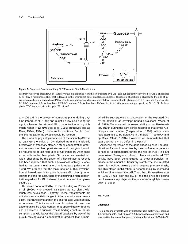
The probable physiologic function of the spinach pGlcT isto catalyze the efflux of Glc derived from the amylolyticbreakdown of transitory starch. A steep concentration gradi-ent between the chloroplast stroma and the cytosol wouldbe required to obtain high rates of Glc transport. After beingexported from the chloroplasts, Glc has to be converted intoGlc 6-phosphate by the action of a hexokinase. It recentlyhas been reported that such a hexokinase activity is local-ized to the outer membrane of chloroplasts (Wiese et al.,1999). We propose that the main function of this envelope-bound hexokinase is to phosphorylate Glc directly whenleaving the chloroplasts, thereby maintaining a high concen-tration gradient for Glc between the stroma and the cytosol(Figure 9).
This idea is corroborated by the recent findings of Veramendiet al. (1999), who created transgenic potato plants withmuch less hexokinase 1 activity. These transformants didnot show substantial changes in tuber carbohydrate metab-olism, but transitory starch in the chloroplasts was markedlyaccumulated. This increase in starch content at dawn wasaccompanied by a Glc content that approximately doubledand a decrease in sucrose. These findings confirm the as-sumption that Glc leaves the plastid passively by way of thepGlcT, moving along a concentration gradient that is main-
tained by subsequent phosphorylation of the exported Glcby the action of an envelope-bound hexokinase (Wiese etal., 1999). The observed decreased ability to mobilize transi-tory starch during the dark period resembles that of the Ara-bidopsis sex1 mutant (Caspar et al., 1991), which somehave assumed to be defective in the pGlcT (Trethewey andap Rees, 1994a, 1994b). However, we demonstrated thatsex1 does not carry a defect in the pGlcT.
Antisense repression of the gene encoding pGlcT or iden-tification of a knockout mutant by means of reverse geneticsis needed to characterize further the role of pGlcT in plantmetabolism. Transgenic tobacco plants with reduced TPTactivity have been demonstrated to show a transient in-crease in the amount of transitory starch. The accumulatedstarch is mobilized already during ongoing photosynthesis,and this starch mobilization is accompanied by increasedactivities of amylases, the pGlcT, and hexokinase (Häusler etal., 1998). Thus, both the pGlcT and the envelope-boundhexokinase are key players in the process of amylolytic break-down of starch.
METHODS
Chemicals
14C-3-phosphoglycerate was synthesized from NaH14CO3, ribulose1,5-bisphosphate, and ribulose 1,5-bisphosphate/carboxylase andwas purified by ion exchange chromatography with an AG50X8-Cl2
Figure 9. Proposed Function of the pGlcT Protein in Starch Mobilization.
Glc from hydrolytic breakdown of transitory starch is exported from the chloroplasts by pGlcT and subsequently converted to Glc 6-phosphate(G-6-P) by a hexokinase (HxK) that is located in the chloroplast outer envelope membrane. Glucose 6-phosphate is shuttled to the site of su-crose biosynthesis, whereas trioseP that results from phosphorolytic starch breakdown is subjected to glycolysis. F-6-P, fructose 6-phosphate;F-1,6-bP, fructose 1,6-bisphosphate; F-2,6-bP, fructose 2,6-bisphosphate; FbPase, fructose 1,6-bisphosphate phosphatase; G-1-P, Glc 1-phos-phate; TCC, tricarboxylic acid cycle; TP, trioseP.
Plastidic Glucose Translocator 797
column (Bio-Rad) and a 0 to 400 mM LiCl gradient; its specific activ-ity was determined by enzymatic analysis (Lowry and Passonneau,1972) and scintillation counting. Other radiochemicals were pur-chased from New England Nuclear Life Science (Boston, MA) andICN (Eschwege, Germany). ASA-Glc (N-[azidosalicyl]-6-amido-6-deoxyglucopyranose) was a gift of W. Hitz (DuPont, Wilmington, DE).Sorbitol and other chemicals were purchased from Sigma. Sorbitol(S-7547) was found by enzymatic analysis (Lowry and Passonneau,1972) to contain 0.5 mg/g glucose (Glc) and was used without furtherpurification.
Reagents and enzymes for recombinant DNA techniques were ob-tained from Promega. U. Sonnewald (Institut für Pflanzengenetik undKulturpflanzenforschung, Gatersleben, Germany) kindly provided thecDNA libraries from potato and tobacco leaves. The cDNA librariesfrom spinach, Arabidopsis, and maize leaves were obtained fromStratagene.
Plant Material
Spinach (Spinacia oleracea cv Melody) plants were grown in 6-litercontainers in a mixture of Metromix 360 (Scotts Sierra, Marysville,OH):sand (1:1 [v/v]) under a 10-hr photoperiod (258C day and 208Cnight) and a photosynthetic photon flux of 600 mmol m22 sec21 pro-vided by sodium vapor and metal halide lamps. Plants were irrigatedat 6-hr intervals with a balanced nutrient solution.
Chloroplast Isolation
Spinach leaves from 6-week-old plants were harvested 1 hr beforethe beginning of the light period. Chloroplasts were isolated and pu-rified according to Robinson (1983) and resuspended in buffer B (330mM sorbitol, 50 mM Hepes-KOH, pH 7.6, 1 mM MgCl2, 1 mM MnCl2,and 2 mM EDTA).
Uptake Assays
Uptake assays were initiated by exposing chloroplasts to D-14C-Glcat 208C or 14C-3-phosphoglycerate at 48C in room light; they wereterminated by centrifugation of chloroplasts through silicone oil(Heldt, 1980). In addition to labeled substrates, L-3H-Glc or D-3H sor-bitol were included to measure the sorbitol-permeable space. Forassay times .5 sec, uptake was measured by using assay tubescontaining a single oil layer (Heldt, 1980). One-second uptake assayswere measured by modification of the double oil-layer method ofGross et al. (1990). Microcentrifuge tubes contained five layers:50 mL of glycerol–methanol–water (2:1:1 [v/v]), 50 mL of polyphenyl-methylsiloxane (silicone oil, AR200; Fluka, Deisenhofen, Germany),100 mL of assay buffer (buffer B containing 0.22 M sorbitol, 0.11 Msucrose, and radiolabeled substrates), 50 mL of polyphenylmethylsi-loxane, and 100 mL of chloroplast suspension in buffer B containing50 mg of chlorophyll. From extrapolation of time-course measure-ments of 14C-3-phosphoglycerate uptake at 48C by using a combina-tion of single and double oil-layer tubes, we measured an uptaketime of 0.85 sec for the double oil-layer method used. After centrifu-gation, the sample-containing tubes were frozen in liquid N2, and thelower part of the tubes containing the chloroplast pellet and a small
part of the oil layer were removed with a tubing cutter and placed in1.5-mL tubes containing 450 mL of 80% (v/v) methanol and 450 mL ofTris-HCl–washed CHCl3. The chloroplast pellet was dispersed, andthe cut tip was discarded. Water (450 mL) then was added, and thesample was mixed on a vortex-type mixer. The two phases wereseparated by centrifugation for 1 min at 12,000g. The bottom organiclayer was removed and diluted to 5 mL with ethanol. A649 and A665
were measured, from which the chlorophyll concentration was deter-mined (Wintermans and DeMots, 1965). The aqueous phase thenwas diluted to 1 mL with water. Radioactivity was separated into ba-sic, acidic, and neutral fractions by filtering the aqueous extract se-quentially through 0.5-mL columns containing ion exchange resinforms AG50X8-H1 and AG1X8-formate (Bio-Rad). The radioactivityin each fraction was determined by scintillation counting.
Analysis of Radiolabeled Products
Chloroplasts were incubated in tubes containing 3H2O and 4 mM ei-ther D-14C-Glc or 14C-sorbitol for various times. Uptake was termi-nated by centrifugation through silicone oil. Tubes containing D-14C-Glc were analyzed to identify the nature of the radioactive products.The lower HClO4 layer was removed and neutralized with KOH. Thechloroplast pellet was extracted in 0.2 mL of chloroform–methanol(1:3 [v/v]), and the extract was separated into organic and aqueousphases by adding 0.2 mL of water. Extracts were combined and di-luted to 1 mL with water. An aliquot was removed for determining to-tal radioactivity, and the remainder was passed successively through0.5-mL ion exchange columns containing AG50X8-H1 and AG1X8-formate to bind basic and acidic products, respectively. Ion ex-change columns were washed twice with 1 mL of water. Radioactiv-ity passing through both columns was considered to represent labelin neutral products. The columns were eluted with 2 mL of 2 N HCl,and aliquots were removed for determining radioactivity. The con-centrations of labeled Glc and acidic products present in the sorbi-tol-impermeable space were calculated from measurements of 14C-sorbitol and 3H2O at the same times.
Differential Labeling and Membrane Isolation
Differential labeling of chloroplasts with 3H-N-ethylmaleimide(3H-NEM) and 14C-NEM was based on the procedure of Hendersonand Macpherson (1986). Chloroplasts containing z20 mg of chloro-phyll were incubated in 10 mL of buffer B and 1 mM DTT for 15 minat 208C, divided equally between two tubes, and collected by centrif-ugation for 5 min at 2000g. Chloroplasts in one tube were resus-pended in 10 mL of buffer B (unprotected); those in the other wereresuspended in buffer B in which Glc replaced sorbitol (protected).After 15 min at 208C, freshly prepared 5 mM NEM was added toeach. After 30 min, chloroplasts were collected by centrifugation andresuspended in buffer B three times to remove traces of NEM andGlc. Unprotected chloroplasts were incubated for 30 min at 208Cwith 0.5 mM 3H-NEM (500 Ci/mol) and protected chloroplasts with0.5 mM 14C-NEM (100 Ci/mol). Both sets of chloroplasts were col-lected by centrifugation, and envelope membranes were prepared asdescribed by Keegstra and Yousif (1986). Purified inner envelopevesicles were removed from the 1 and 0.8 M sucrose interface, di-luted to 0.2 M sucrose, collected by ultracentrifugation at 90,000g for1 hr, and resuspended in 0.1 mL of 0.2 M sucrose, 10 mM Hepes,and 1 mM MgCl2.
798 The Plant Cell
Sample Preparation and SDS-PAGE
Inner membrane vesicles from protected and unprotected chloro-plasts were mixed and placed on ice. An equal volume of ice-coldn-butanol was added and mixed by vortex mixing for 10 sec. Layerswere separated by centrifugation at 20,000g for 1 min in a microcen-trifuge. The butanol and aqueous layers were removed, leaving theinsoluble residue that appeared at the interface. The insoluble resi-due was extracted two more times with equal amounts of water andn-butanol, and these were added to the initial fractions and dried in arotary evaporator (Bachhofer, Reutlingen, Germany). The smallamount of insoluble residue remaining was added to the aqueousfraction. Lipids were removed from the dried butanol fraction by ex-traction with n-hexane. The hexane-insoluble residue was collectedby centrifugation at 20,000g in a microcentrifuge. The hexane wasremoved, and the pellet was dried under N2. Proteins were solubi-lized in sample buffer (50 mM Tris-HCl, pH 6.8, 2% [w/v] SDS, 20%[v/v] glycerol, and 5 mM DTT), heated at 608C for 30 min, and sub-jected to SDS-PAGE on a 12.5% acrylamide running gel and a 5%stacking gel at 108C (Henderson and Macpherson, 1986). From gelsthat had been stained with Coomassie Brilliant Blue R 250, the lanecontaining the sample of interest was removed from the gel. Sections1 mm thick were removed in order, and each was digested in 0.1 mLof 80% H2O2 and 70% HClO4 (6:1 [v/v]) for 1 hr at 708C. After decayof chemiluminescence, the radioactivity present was determined byscintillation counting using a dual-labeling format. The percentage oftotal 14C and tritium radioactivity present in each section was deter-mined. Data are plotted as the difference in percentage of total radio-activity present in each section (14Csection/14Ctotal 2 3Hsection/3Htotal).
Peptide Sequencing of the Glc Translocator and Cloning ofIts cDNA
Envelope membranes (5 mg/mL) were extracted by a 20-fold excessof n-butanol at pH 3.0. The butanol fraction was dried, washed withn-hexane, and analyzed by SDS-PAGE (Laemmli, 1970). The proteinwith an apparent molecular mass of 43 kD was excised from theCoomassie blue–stained gel and digested with endoproteinase Lys-Cwhile still in the polyacrylamide matrix. The resulting peptides wereeluted, purified by reverse phase HPLC (Eckerskorn and Lottspeich,1989), and sequenced in a gas-phase sequencer (Eckerskorn et al.,1988).
From the 43-kD protein, we obtained one peptide sequence:KGRSLEEIELALSPAV (P1). Two degenerate oligonucleotides weredesigned on the basis of the peptide sequence: 59-AARGGIMGIWII-YTIGARGARATIGA-39 (01) and 59-GARATIGARYTIGCIYTIWIICCIGC-39
(02), where R is an A or G residue, I is inosine, Y is C or T, W is A orT, and M is A or C.
A first-strand cDNA was prepared by using poly(A)1 RNA fromlight- and dark-harvested spinach leaves, an oligo(dT)15 anchorprimer (rapid amplification of cDNA ends [RACE] adapter primer)designated 59-d(CCACGAGTCGACTCTAGAGCTCGGATCCTT[T]15)-39, and a cDNA preamplification system, according to the instruc-tions given by the manufacturer (Gibco BRL Life Technologies,Gaithersburg, MD). The resulting single-stranded cDNA was the tem-plate for the RACE procedure as described previously (Kammerer etal., 1998). The generated DNA fragment (210 bp) was subcloned intothe pGEM-Teasy vector according to the instructions given by themanufacturer (Promega) and sequenced. The fragment was used fora plaque hybridization screening of a cDNA library from spinach
leaves. Positive plaques were purified, and the pBluescript SK1 phage-mid containing the cDNA insert was excised from the purified phagesin vivo according to the instructions given by the manufacturer (Strat-agene). The full-length cDNA was sequenced on both strands andthen used for plaque hybridization screenings of cDNA libraries frommaize, tobacco, potato, and Arabidopsis. Phagemids were preparedas described above, and the longest inserts were sequenced on bothstrands.
Protein Import Assays
Spinach chloroplasts were isolated from spinach leaves as de-scribed previously (Douce and Joyard, 1982). The cloned cDNA ofthe plastidic Glc translocator (pGlcT) protein from spinach leaveswas transcribed and translated by the TNT lysate system, accordingto the instructions of the manufacturer (Promega); 35S-methioninewas the labeled amino acid. Protein import assays were performed inimport buffer (250 mM sorbitol, 10 mM methionine, 25 mM potas-sium gluconate, 2 mM MgSO4, 50 mM Hepes-KOH, pH 8.0, and0.2% BSA) containing radiolabel, in vitro–synthesized precursor pro-tein, and purified organelles equivalent to 200 mg of chlorophyll in afinal volume of 300 mL. Other additions are indicated in the legend toFigure 7. The import reaction was allowed to proceed for 20 min. En-velope membranes were isolated as previously described (Flügge etal., 1989). All fractions were analyzed by SDS-PAGE and a Phospho-Imager (Molecular Dynamics, Krefeld, Germany).
Isolation of RNA and RNA Gel Blot Hybridization
Total RNA was isolated from different tissues of tobacco accordingto Logemann et al. (1987). Equal amounts of RNA were size-fraction-ated on agarose–formaldehyde gels, and RNA was transferred to PallBiodyne B nylon membranes (Pall GmbH, Dreieich, Germany) bydownward capillary blot in 10 3 SSC (1 3 SSC is 0.15 M NaCl and0.015 M sodium citrate). Hybridization of immobilized RNA to cDNAprobes was performed overnight in 7% SDS (w/v), 500 mM NaPi, pH7.2, and 1 mM EDTA (Church and Gilbert, 1984) at 628C. Nonspecif-ically bound probe was removed by washing the filter with 6 3 SSCand 0.2% SDS for 5 min at room temperature, with 4 3 SSC and0.2% SDS for 5 min at room temperature, and with 2 3 SSC and0.2% SDS at room temperature for 5 min, followed by two subse-quent stringent washes with 1 3 SSC and 0.2% SDS at 658C for 5min each.
Hybridization Analysis of Yeast Artificial Chromosome Clones
Mapping of pGlcT in the Arabidopsis genome with the help of yeastartificial chromosome (YAC) contigs immobilized on nylon filters wasperformed as described by Schmidt and Dean (1995).
Construction of Sequence Alignments and Phylogenetic Trees
Multiple alignment of protein sequences was performed with the helpof the ClustalX program (Higgins et al., 1996; Thompson et al., 1997).From all sequences that showed N- or C-terminal extensions (e.g.,targeting signals) in comparison with the related hexose transporterproteins, we removed these extensions to improve the quality of thealignments. We then constructed an unrooted phylogenetic tree from
Plastidic Glucose Translocator 799
the aligned sequences, using the distance matrix neighbor-joiningmethod (Saitou and Nei, 1987) from within the ClustalX program. Thetree was displayed with the help of the TREEVIEW program (Page, 1996).
ACKNOWLEDGMENTS
This work was supported in part by grants from the Monsanto Com-pany (to D.R.G); Grant No. 9501219 from the U.S. Department ofAgriculture, National Research Initiative Competitive Grants Program(to J.C.S); Grant No. IBN-9205966 from the National Science Foun-dation (to D.R.G); and grants from the Deutsche Forschungs-gemeinschaft (to U.-I.F. and A.W.) and the Fonds der ChemischenIndustrie (to U.-I.F.). We are grateful to Dr. Renate Schmidt (MaxDelbrück Labor, Cologne, Germany) for help with YAC filter hybrid-izations and mapping the pGlcT.
Received January 10, 2000; accepted March 20, 2000.
REFERENCES
Baldwin, S.A., and Lienhard, G.E. (1989). Purification and reconsti-tution of glucose transporter from human erythrocytes. MethodsEnzymol. 174, 39–50.
Bechtold, N., Ellis, G., and Pelletier, G. (1993). In planta Agrobac-terium-mediated gene transfer by infiltration of adult Arabidopsisthaliana plants. C. R. Acad. Sci. 316, 1194–1199.
Beck, E. (1985). The degradation of transitory starch in chloroplasts.In Regulation of Carbon Partitioning in Photosynthetic Tissue, R.L.Heath and J. Preiss, eds (Baltimore, MD: Waverly), pp. 27–44.
Capaldi, R.A., and Vanderkooi, G. (1972). The low polarity of manymembrane proteins. Proc. Natl. Acad. Sci. USA 69, 930–932.
Caspar, T., Huber, S.C., and Somerville, C. (1985). Alterations ingrowth, photosynthesis, and respiration in a starchless mutant ofArabidopsis thaliana (L) deficient in chloroplast phosphoglucomu-tase activity. Plant Physiol. 79, 11–17.
Caspar, T., Lin, T.-P., Kakefuda, G., Benbow, L., Preiss, J., andSomerville, C. (1991). Mutants of Arabidopsis with altered regula-tion of starch degradation. Plant Physiol. 95, 1181–1188.
Church, G.M., and Gilbert, W. (1984). Genomic sequencing. Proc.Natl. Acad. Sci. USA 81, 1991–1995.
Douce, R., and Joyard, J. (1982). Purification of the chloroplast envel-ope. In Methods of Chloroplast Molecular Biology, M. Edelman,R.B. Hallick, and N.-H. Chua, eds (Amsterdam, The Netherlands:Elsevier), pp. 239–256.
Dreses-Weringloer, U., Fischer, K., Wachter, E., Link, T.A., andFlügge, U.I. (1990). cDNA sequence of the precursor of the 37kDa inner envelope membrane polypeptide from spinach chloro-plasts: Its transit peptide contains an amphiphilic a-helix as theonly detectable structural element. Eur. J. Biochem. 195, 361–368.
Eckerskorn, C., and Lottspeich, F. (1989). Internal amino acidsequence analysis of proteins separated by gel electrophoresisafter tryptic digestion in polyacrylamide matrix. Chromatographia28, 92–94.
Eckerskorn, C., Mewes, W., Goretzki, H., and Lottspeich, F.(1988). A new siliconized-glass fiber as support for protein chemi-cal analysis of electroblotted proteins. Eur. J. Biochem. 176, 509–519.
Emanuelsson, O., Nielsen, H., and von Heijne, G. (1999). ChloroP,a neural network-based method for predicting chloroplast transitpeptides and their cleavage sites. Protein Sci. 8, 978–984.
Fliege, R., Flügge, U.-I., Werdan, K., and Heldt, H.W. (1978). Spe-cific transport of inorganic phosphate, 3-phosphoglycerate andtriosephosphates across the inner membrane of the envelope inspinach chloroplasts. Biochim. Biophys. Acta 502, 232–247.
Flügge, U.-I., Fischer, K., Gross, A., Sebald, W., Lottspeich, F.,and Eckerskorn, C. (1989). The triose phosphate-3-phospho-glycerate-phosphate translocator from spinach chloroplasts:Nucleotide sequence of a full-length cDNA clone and import ofthe in vitro synthesized precursor protein into chloroplasts. EMBOJ. 8, 39–46.
Fondy, B.R., Geiger, D.R., and Servaites, J.C. (1989). Photosyn-thesis, carbohydrate metabolism and export in Beta vulgaris L.and Phaseolus vulgaris L. during square and sinusoidal lightregimes. Plant Physiol. 89, 396–402.
Geiger, D.R., Shieh, W.-J., and Yu, X.-M. (1995). Photosyntheticcarbon metabolism and translocation in wild-type and starch-defi-cient mutant Nicotiana sylvestris L. Plant Physiol. 107, 507–514.
Geiger, D.R., Servaites, J.C., and Fuchs, M.A. (2000). Role ofstarch in carbon translocation and partitioning at the plant level.Aust. J. Plant Physiol., in press.
Gerhardt, R., Stitt, M., and Heldt, H.W. (1987). Subcellular metabo-lite levels in spinach leaves. Plant Physiol. 83, 399–407.
Gleixner, G., Scrimgeour, C., Schmidt, H.-L., and Viola, R. (1998).Stable isotope distribution in the major metabolites of source andsink organs of Solanum tuberosum L.: A powerful tool in the studyof metabolite partitioning in intact plants. Planta 207, 241–245.
Gould, G.W., and Bell, G.I. (1990). Facilitative glucose transporters:An expanding family. Trends Biochem. Sci. 15, 18–23.
Gross, A., Brückner, G., Heldt, H.W., and Flügge, U.-I. (1990).Comparison of the kinetic properties, inhibition and labelling ofthe phosphate translocators from maize and spinach mesophyllchloroplasts. Planta 180, 262–271.
Hanson, K.R., and McHale, N.A. (1988). A starchless mutant ofNicotiana sylvestris containing a modified plastid phosphogluco-mutase. Plant Physiol. 88, 838–844.
Häusler, R.E., Schlieben, N.H., Schulz, B., and Flügge, U.-I.(1998). Compensation of decreased triose phosphate/phosphatetranslocator activity by accelerated starch turnover and glucosetransport in transgenic tobacco. Planta 204, 366–376.
Heineke, D., Kruse, A., Flügge, U.I., Frommer, W.B., Riesmeier,J.W., Willmitzer, L., and Heldt, H.W. (1994). Effect of antisenserepression of the chloroplast triose-phosphate translocator onphotosynthetic metabolism in transgenic potato plants. Planta193, 174–180.
Heldt, H.W. (1980). Measurement of metabolite movement acrossthe envelope and of the pH in the stroma and the thylakoid spacein intact chloroplasts. Methods Enzymol. 69, 604–613.
Henderson, P.J.F. (1991). Sugar transport proteins. Curr. Opin.Struct. Biol. 1, 590–601.
800 The Plant Cell
Henderson, P.J.F., and Macpherson, A.J.S. (1986). Assay, genet-ics, proteins, and reconstitution of proton-linked galactose, arabi-nose, and xylose transport systems of Escherichia coli. MethodsEnzymol. 125, 387–429.
Herold, A., Leegood, R.C., McNeil, P.H., and Robinson, S.P.(1981). Accumulation of maltose during photosynthesis in proto-plasts isolated from spinach leaves treated with mannose. PlantPhysiol. 67, 85–88.
Higgins, D.G., Thompson, J.D., and Gibson, T.J. (1996). UsingCLUSTAL for multiple sequence alignments. Methods Enzymol.266, 383–402.
Hosang, M., Gibbs, E.M., Deidrich, D.F., and Semenza, G. (1981).Photoaffinity labeling and identification of (a component of) thesmall-intestinal Na1, D-glucose transporter using 4-azidophlorizin.FEBS Lett. 130, 244–248.
Huber, S.C., and Hanson, K.R. (1992). Carbon partitioning andgrowth of a starchless mutant of Nicotiana sylvestris. Plant Phys-iol. 99, 1449–1454.
Kammerer, B., Fischer, K., Hilpert, B., Schubert, S., Gutensohn,M., Weber, A., and Flügge, U.-I. (1998). Molecular characteriza-tion of a carbon transporter in plastids of heterotrophic tissues:The glucose 6-phosphate/phosphate antiporter. Plant Cell 10,105–117.
Keegstra, K., and Yousif, A.E. (1986). Isolation and characteriza-tion of chloroplast envelope membranes. Methods Enzymol. 118,316–325.
Klip, A., Walker, D., Ransome, K.J., Schroer, D.W., and Lienhard,G.E. (1983). Identification of the glucose transporter in rate skele-tal muscle. Arch. Biochem. Biophys. 226, 198–205.
Konieczny, A., and Ausubel, F.M. (1993). A procedure for mappingArabidopsis mutations using co-dominant ecotype-specific PCR-based markers. Plant J. 4, 403–410.
Kyte, J., and Doolittle, R.F. (1982). A simple method for displayingthe hydrophobic character of a protein. J. Mol. Biol. 157, 105–132.
Laemmli, U.K. (1970). Cleavage of structural proteins during theassembly of the head of bacteriophage T4. Nature 227, 680–685.
Li, H., Sullivan, T.D., and Keegstra, K. (1992). Information for tar-geting to the chloroplastic inner envelope membrane is containedin the mature region of the maize Bt1-encoded protein. J. Biol.Chem. 267, 18999–19004.
Lockshin, A., and Burris, R.H. (1966). Solubilization and propertiesof chloroplast lamellar protein. Proc. Natl. Acad. Sci. USA 56,1564–1570.
Logemann, J., Schell, J., and Willmitzer, L. (1987). Improvedmethod for the isolation of RNA from plant tissues. Anal. Bio-chem. 163, 16–20.
Lowry, O.H., and Passonneau, J.V. (1972). A Flexible System ofEnzymatic Analysis. (New York: Academic Press).
Moore, B.D., Palmquist, D.E., and Seemann, J.R. (1997). Influenceof plant growth at high CO2 concentrations on leaf content of ribu-lose-1,5-bisphosphate carboxylase/oxygenase and intracellulardistribution of soluble carbohydrates in tobacco, snapdragon, andparsley. Plant Physiol. 115, 241–248.
Page, R.D.M. (1996). TREEVIEW: An application to display phyloge-netic trees on personal computers. Comp. Appl. Biosci. 12, 357–358.
Piccioni, R., Bellemare, G., and Chua, N.-H. (1982). Methods of
polyacrylamide gel electrophoresis in the analysis and preparationof plant polypeptides. In Methods of Chloroplast Molecular Biol-ogy, M. Edelman, R.B. Hallick, and N.-H. Chua, eds (Amsterdam,The Netherlands: Elsevier), pp. 985–1014.
Pohlmeyer, K., Soll, J., Steinkamp, T., Hinnah, S., and Wagner,R. (1997). Isolation and characterization of an amino acid–selec-tive channel protein present in the chloroplastic outer envelopemembrane. Proc. Natl. Acad. Sci. USA 94, 9504–9509.
Robinson, S.P. (1983). Isolation of intact chloroplasts with high CO2
fixation from sugar beet leaves containing calcium oxalate. Photo-synth. Res. 4, 281–287.
Rost, S., Frank, C., and Beck, E. (1996). The chloroplast envelopeis permeable for maltose but not for maltodextrins. Biochim. Bio-phys. Acta 1291, 221–227.
Rumpho, M.E., and Edwards, G.E. (1985). Characterization of 4,49-diisothiocyano-2,29-disulfonic acid stilbene inhibition of 3-phos-phoglycerate–dependent O2 evolution in isolated chloroplasts.Plant Physiol. 78, 537–544.
Saier, M.H., Jr. (1998). Molecular phylogeny as a basis for the clas-sification of transport proteins from bacteria, archaea andeukarya. Adv. Microbiol. Physiol. 40, 81–136.
Saitou, N., and Nei, M. (1987). The neighbor-joining method: A newmethod for reconstructing phylogenetic trees. Mol. Biol. Evol. 4,406–425.
Schaefer, B.C. (1995). Revolutions in the rapid amplification ofcDNA ends: New strategies for polymerase chain reaction cloningof full-length cDNA ends. Anal. Biochem. 227, 255–273.
Schäfer, G., Heber, U., and Heldt, H.W. (1977). Glucose transportinto spinach chloroplasts. Plant Physiol. 60, 286–289.
Schleucher, J., Vanderveer, P.J., and Sharkey, T.D. (1998). Exportof carbon from chloroplasts at night. Plant Physiol. 118, 1439–1445.
Schmidt, R., and Dean, C. (1995). Hybridization analysis of YACclones. Methods Mol. Cell. Biol. 5, 309–318.
Schmidt, R., Love, K., West, J., Lenehan, Z., and Dean, C. (1997).Description of 31 YAC contigs spanning the majority of Arabidop-sis thaliana chromosome 5. Plant J. 11, 563–572.
Servaites, J.C., Geiger, D.R., Tucci, M.A., and Fondy, B.R.(1989a). Leaf carbon metabolism and metabolite levels during aperiod of sinusoidal light. Plant Physiol. 89, 403–408.
Servaites, J.C., Fondy, B.R., Li, B., and Geiger, D.R. (1989b).Sources of carbon for export from spinach leaves throughout theday. Plant Physiol. 90, 1168–1174.
Shanahan, M.F. (1982). Cytochalasin B. A natural photoaffinityligand for labeling the human erythrocyte glucose transporter. J.Biol. Chem. 257, 7290–7293.
Shanahan, M.F., Wadzinski, B.E., Lowndes, J.M., and Ruoho,A.E. (1985). Photoaffinity labeling of the human erythrocytemonosaccharide transporter with an aryl azide derivative of D-glu-cose. J. Biol. Chem. 260, 10897–10900.
Stitt, M. (1990). Fructose-2,6-bisphosphate as a regulatory metabo-lite in plants. Annu. Rev. Plant Physiol. Plant Mol. Biol. 41, 153–185.
Stitt, M., Wirtz, W., Gerhardt, R., Heldt, H.W., Spencer, C.,Walker, D., and Foyer, C. (1985). A comparative study of metab-olite levels in plant leaf material in the dark. Planta 166, 354–364.
Thompson, J.D., Gibson, T.J., Plewniak, F., Jeanmougin, F., and
Plastidic Glucose Translocator 801
Higgins, D.G. (1997). The ClustalX windows interface: Flexiblestrategies for multiple sequence alignment aided by quality analy-sis tools. Nucleic Acids Res. 25, 4876–4882.
Trethewey, R.N., and ap Rees, T. (1994a). A mutant of Arabidopsisthaliana lacking the ability to transport glucose across the chloro-plast envelope. Biochem J. 301, 449–454.
Trethewey, R.N., and ap Rees, T. (1994b). The role of the hexosetransporter in the chloroplasts of Arabidopsis thaliana L. Planta195, 168–174.
Veramendi, J., Roessner, U., Renz, A., Willmitzer, L., andTrethewey, R.N. (1999). Antisense repression of hexokinase 1leads to an overaccumulation of starch in leaves of transgenicpotato plants but not to significant changes in tuber carbohydratemetabolism. Plant Physiol. 121, 123–133.
Walmsley, A.R., Barett, M.P., Bringaud, F., and Gould, G.W. (1998).Sugar transporters from bacteria, parasites and mammals: Struc-ture–activity relationships. Trends Biochem. Sci. 23, 476–481.
Weber, A., Menzlaff, E., Arbinger, B., Gutensohn, M., Eckerskorn,C., and Flügge, U.-I. (1995). The 2-oxoglutarate/malate translo-cator of chloroplast envelope membranes: Molecular cloning of a
transporter protein containing a 12-helix motif and expression ofthe functional protein in yeast cells. Biochemistry 34, 2621–2627.
Wellner, M., Monden, I., and Keller, K. (1994). The role of cysteineresidues in glucose-transporter-GLUT1–mediated transport andtransport inhibition. Biochem. J. 299, 813–817.
Wiese, A., Gröner, F., Sonnewald, U., Deppner, H., Lerchl, J.,Hebbeker, U., Flügge, U.I., and Weber, A. (1999). Spinach hex-okinase I is located in the outer envelope membrane of plastids.FEBS Lett. 461, 13–18.
Wintermans, J.F., and DeMots, A. (1965). Spectrophotometriccharacteristics of chlorophyll a and b and their pheophytins in eth-anol. Biochim. Biophys. Acta 109, 448–453.
Zeeman, S.C., Northorp, F., Smith, A.M., and ap Rees, T. (1998a).A starch-accumulating mutant of Arabidopsis thaliana deficient ina chloroplastic starch-hydrolysing enzyme. Plant J. 15, 357–365.
Zeeman, S.C., Umemoto, T., Lue, W.L., Au-Yeung, P., Martin, C.,Smith, A.M., and Chen, J. (1998b). A mutant of Arabidopsis lack-ing a chloroplastic isoamylase accumulates both starch and phy-toglycogen. Plant Cell 10, 1699–1711.
Related Documents


