of June 13, 2013. This information is current as Immune Responses Negative Regulator of Basophil-Mediated Identification of Semaphorin 4B as a Katayama and Atsushi Kumanogoh Kataoka, Masahito Ikawa, Toshihiko Toyofuku, Ichiro Sujin Kang, Satoshi Nojima, Tetsuya Kimura, Tatsuki R. Yukinobu Nakagawa, Hyota Takamatsu, Tatsusada Okuno, http://www.jimmunol.org/content/186/5/2881 doi: 10.4049/jimmunol.1003485 January 2011; 2011; 186:2881-2888; Prepublished online 26 J Immunol Material Supplementary 5.DC1.html http://www.jimmunol.org/content/suppl/2011/01/26/jimmunol.100348 References http://www.jimmunol.org/content/186/5/2881.full#ref-list-1 , 12 of which you can access for free at: cites 47 articles This article Subscriptions http://jimmunol.org/subscriptions is online at: The Journal of Immunology Information about subscribing to Permissions http://www.aai.org/ji/copyright.html Submit copyright permission requests at: Email Alerts http://jimmunol.org/cgi/alerts/etoc Receive free email-alerts when new articles cite this article. Sign up at: Print ISSN: 0022-1767 Online ISSN: 1550-6606. Immunologists, Inc. All rights reserved. Copyright © 2011 by The American Association of 9650 Rockville Pike, Bethesda, MD 20814-3994. The American Association of Immunologists, Inc., is published twice each month by The Journal of Immunology by guest on June 13, 2013 http://www.jimmunol.org/ Downloaded from

Welcome message from author
This document is posted to help you gain knowledge. Please leave a comment to let me know what you think about it! Share it to your friends and learn new things together.
Transcript
of June 13, 2013.This information is current as
Immune ResponsesNegative Regulator of Basophil-Mediated Identification of Semaphorin 4B as a
Katayama and Atsushi KumanogohKataoka, Masahito Ikawa, Toshihiko Toyofuku, IchiroSujin Kang, Satoshi Nojima, Tetsuya Kimura, Tatsuki R. Yukinobu Nakagawa, Hyota Takamatsu, Tatsusada Okuno,
http://www.jimmunol.org/content/186/5/2881doi: 10.4049/jimmunol.1003485January 2011;
2011; 186:2881-2888; Prepublished online 26J Immunol
MaterialSupplementary
5.DC1.htmlhttp://www.jimmunol.org/content/suppl/2011/01/26/jimmunol.100348
Referenceshttp://www.jimmunol.org/content/186/5/2881.full#ref-list-1
, 12 of which you can access for free at: cites 47 articlesThis article
Subscriptionshttp://jimmunol.org/subscriptions
is online at: The Journal of ImmunologyInformation about subscribing to
Permissionshttp://www.aai.org/ji/copyright.htmlSubmit copyright permission requests at:
Email Alertshttp://jimmunol.org/cgi/alerts/etocReceive free email-alerts when new articles cite this article. Sign up at:
Print ISSN: 0022-1767 Online ISSN: 1550-6606. Immunologists, Inc. All rights reserved.Copyright © 2011 by The American Association of9650 Rockville Pike, Bethesda, MD 20814-3994.The American Association of Immunologists, Inc.,
is published twice each month byThe Journal of Immunology
by guest on June 13, 2013http://w
ww
.jimm
unol.org/D
ownloaded from
The Journal of Immunology
Identification of Semaphorin 4B as a Negative Regulator ofBasophil-Mediated Immune Responses
Yukinobu Nakagawa,*,†,1 Hyota Takamatsu,*,1 Tatsusada Okuno,*,‡ Sujin Kang,*
Satoshi Nojima,*,x Tetsuya Kimura,* Tatsuki R. Kataoka,{ Masahito Ikawa,‖
Toshihiko Toyofuku,* Ichiro Katayama,† and Atsushi Kumanogoh*,#
Basophils are strong mediators of Th2 responses during helminthic infections. Recently, basophils were shown to function as APCs
and promote both Th2 skewing and humoral memory responses. However, the mechanisms that regulate basophils are still
unclear. In this article, we show that a class IV semaphorin, Sema4B, negatively regulates basophil functions through T cell–
basophil contacts. In a screen to identify semaphorins that function in the immune system, we determined that Sema4B is
expressed in T and B cells. Interestingly, Sema4B2/2 mice had considerably increased serum IgE levels despite normal lympho-
cyte and dendritic cell functions. Recombinant Sema4B significantly inhibited IL-4 and IL-6 production from basophils in
response to various stimuli, including IL-3, papain, and Fc«RI cross-linking. In addition, T cell-derived Sema4B, which accu-
mulated at contact sites between basophils and CD4+ T cells, suppressed basophil-mediated Th2 skewing, suggesting that Sema4B
regulates basophil responses through cognate cell–cell contacts. Furthermore, Sema4B2/2 mice had enhanced basophil-mediated
memory IgE production, which was abolished by treating with an anti-Fc«RIa Ab. Collectively, these results indicate that Sema4B
negatively regulates basophil-mediated Th2 and humoral memory responses. The Journal of Immunology, 2011, 186: 2881–2888.
Basophils are rare granulocytes that are found in the cir-culation and are effector cells of the innate immune systemthat are associated with allergic inflammation and infec-
tions with helminth parasites (1–4). Recent studies indicated thatbasophils regulate Th1/Th2 homeostasis (5, 6) and humoral im-munity (7). Basophils produce large amounts of IL-4, a key cy-tokine in Th2 skewing, in response to various stimuli, includingIL-3, the protease allergen papain, and cross-linking of surface-
bound IgE molecules (8, 9). In addition, basophils have beenshown to express MHC class II and costimulatory molecules andto function as APCs (10–12), although the role of basophils asAPCs in vivo is still controversial (13–16). Previous studies haveshown that basophil-mediated Th2 skewing can be promoted whenthese cells are activated by papain (11) or internalize Ags throughAg-specific IgE on their cell surface (12). In addition, basophilshave been implicated in humoral memory responses; on re-exposure to Ags, Ag and Ag-specific IgE complexes activatebasophils to release IL-4 and IL-6, resulting in enhanced humoralimmune responses (7). However, the mechanisms that regulatebasophil-mediated Th2 responses are still unknown.Semaphorins were originally identified as axon guidance mol-
ecules during neuronal development (17). However, cumulativefindings indicate that semaphorins have diverse functions in manyphysiological processes (18–22). Semaphorins have been shown tobe involved in various phases of immune responses, including theactivation of B cells (23), T cells (24), and dendritic cells (DCs)(25); the regulation of Th differentiation (26); and the navigationof immune cell trafficking (27). In particular, membrane-boundclass IV semaphorins, Sema4A and Sema4D/CD100, have beenextensively investigated. Sema4D has been shown to be importantfor B cell and DC activation (23). In addition, Sema4A has beendemonstrated to be critical for Th differentiation (26). However,the impact of other class IV semaphorins on other immunefunctions has not been determined.In this study, we searched for semaphorins that function in the
immune system and identified a class IV semaphorin, Sema4B, asa novel immune semaphorin. To determine the physiological rolesof Sema4B, we generated Sema4B2/2 mice and determined thatSema4B negatively regulates IL-4 and IL-6 production by baso-phils. We also determined that T cell-derived Sema4B suppressesbasophil-mediated Th2 skewing. In addition, Sema4B2/2 micenot only had increased serum IgE levels under steady-state con-ditions but also enhanced memory IgE responses caused bydefects in Sema4B-mediated negative regulation of basophils.
*Department of Immunopathology, World Premier International Immunology Fron-tier Research Center, Research Institute for Microbial Diseases, Osaka University,Suita, Osaka 565-0871, Japan; †Department of Dermatology, Osaka University Grad-uate School of Medicine, Suita, Osaka 565-0871, Japan; ‡Department of Neurology,Osaka University Graduate School of Medicine, Suita, Osaka 565-0871, Japan; xDe-partment of Pathology, Osaka University Graduate School of Medicine, Suita, Osaka565-0871, Japan; {Department of Diagnostic Pathology, Kyoto University Hospital,Kyoto 606-8507, Japan; ‖Animal Resource Center for Infectious Diseases, ResearchInstitute for Microbial Diseases, Osaka University, Suita, Osaka 565-0871, Japan;and #Department of Respiratory Medicine, Allergy and Rheumatic Diseases, OsakaUniversity Graduate School of Medicine, Suita, Osaka 565-0871, Japan
1Y.N. and H.T. contributed equally to this study.
Received for publication October 25, 2010. Accepted for publication December 19,2010.
This work was supported by research grants from the Ministry of Education, Culture,Sports, Science and Technology of Japan; grants-in-aid from the Ministry of Health,Labour and Welfare; the program for Promotion of Fundamental Studies in HealthSciences of the National Institute of Biomedical Innovation (to A.K.); and the TargetProtein Research Program of the Japan Science and Technology Agency (to T.T. andA.K.).
Address correspondence and reprint requests to Prof. Atsushi Kumanogoh, Depart-ment of Immunopathology, World Premier International Immunology Frontier Re-search Center, Research Institute for Microbial Diseases, Osaka University, 3-1Yamada-oka, Suita, Osaka 565-0871, Japan. E-mail address: [email protected]
The online version of this article contains supplemental material.
Abbreviations used in this article: BM, bone marrow; BMDC, bone marrow-deriveddendritic cell; DC, dendritic cell; hIgG, human IgG; HSA, human serum albumin;KLH, keyhole limpet hemocyanin; NP, 4-hydroxy-3-nitrophenyl acetyl; NP-CGG, 4-hydroxy-3-nitrophenylacetyl-chicken-g-globulin conjugate; PSD, postsynaptic den-sity; rSema4B, recombinant Sema4B; Tg, transgenic; WT, wild-type.
Copyright� 2011 by The American Association of Immunologists, Inc. 0022-1767/11/$16.00
www.jimmunol.org/cgi/doi/10.4049/jimmunol.1003485
by guest on June 13, 2013http://w
ww
.jimm
unol.org/D
ownloaded from
Materials and MethodsMice
C57BL/6 and BALB/c mice were purchased from CLEA Japan. Sema4B2/2
mice on C57BL/6 and BALB/c backgrounds were generated as describedlater. Sema4B2/2 mice were backcrossed .63 onto the C57BL/6 orBALB/c background. Transgenic (Tg) mice expressing the TCR specificfor OVA (323–339; BALB/c background) were kindly provided by S. Habu(28). OVA-TCR Tg mice on a Sema4B2/2 background were established bycrossing the OVA-TCR Tg mice with Sema4B2/2 BALB/c mice. All micewere bred in specific pathogen-free conditions. All animal experimentalprotocols were reviewed and approved by our institutional animal carecommittees.
Generation of Sema4B2/2 mice
A targeting vector was designed to replace the fifth to eighth exons of theSema4B gene with a Neo resistance cassette. In addition, the HSV thy-midine kinase gene was inserted to select against random integration ofthis targeting vector. The linearized targeting plasmid DNAwas transfectedinto embryonic stem cells by electroporation. After double selection withG418 and ganciclovir, we screened for homologous recombination of theSema4B allele by PCR and FACS analysis. PCR was performed using 35cycles at 94˚C for 30 s, 65˚C for 30 s, and 72˚C for 60 s. The followingoligonucleotide primers were used to identify the rearranged Sema4B lo-cus: forward primer (P1): 59-TAGTGGCATATGTGGACCTG-39; reverseprimer (P2): 59-TCCCTGGAAGCTACTGACTGTT-39; and reverse primer(P3) that includes the Neo cassette: 59-TGCTCGACGTTGTCACTGAA-39.
RT-PCR
Sema4B mRNA expression was examined by RT-PCR using a panel ofmultiple mouse tissue cDNAs (Clontech). RT-PCR was performed using35 cycles at 94˚C for 30 s, 60˚C for 30 s, and 72˚C for 30 s with theforward primer (59-AACAGCAACCTCAGCTTCTTGC-39) and reverseprimer (59-GGCCTCATCTTGGGCTAAAGTA-39).
Generation of the anti-Sema4B Ab
Rats were repeatedly immunized with purified mouse recombinantSema4B (rSema4B) and alum, and their spleens were harvested to generatehybridomas using standard methodologies. The hybridomas were screenedby ELISA for the production of Sema4B-specific Abs. To confirm thespecificity of the Ab for Sema4B, we transfected COS7 cells with Sema4B,Sema4A, Sema4D, or the control vector using Lipofectamine (Invitrogen).After 48 h, the transfectants were stained with the biotinylated anti-Sema4B Ab (TK-2) followed by allophycocyanin-conjugated streptavidin.
Reagents and Abs
Anti-CD4 (GK1.5), anti-CD8 (53-6.7), anti-B220 (RA3-6B2), anti-CD49b(DX5), anti-FcεRIa (MAR-1), anti-CD62L (MEL-14), anti–c-Kit (2B8),MHC class II (I-A/I-E; M5/114.15.2), and allophycocyanin-, FITC-, andCy5-conjugated streptavidin were obtained from eBioscience. Anti-CD16/CD32 (2.4G2), anti-CD11c (HL3), anti-CD40 (HM40-3), and anti-CD3ε(145-2c11) were from BD Pharmingen. Anti-T1/ST2 was from MD Bio-sciences. Anti-p44/42 MAPK (SC-154) and anti-STAT5 (SC-835) werefrom Santa Cruz Biotechnology. Anti–phospho-p44/42 MAPK (9101) wasfrom Cell Signaling Technology. Anti–phospho-STAT5 (47) was from BDBiosciences. Papain was purchased from Calbiochem. The monoclonal IgEanti-DNP Ab (SPE-7), OVA, DNP-human serum albumin (HSA), and LPSwere from Sigma. 4-Hydroxy-3-nitrophenylacetyl-chicken-g-globulin con-jugate (NP-CGG), NP-BSA, and DNP-OVA were from Biosearch Tech-nologies.
Cell purification
To prepare bone marrow (BM)-derived basophils, BM cells were culturedfor 14 d with IL-3 (30 ng/ml, conditioned medium from a mouse IL-3–producing cell line) in RPMI 1640 medium supplemented with 10% (v/v)FBS, 50 mM 2-ME, 100 U/ml penicillin, and 100 mg/ml streptomycin (29).Samples were enriched for basophils by single-cell sorting on CD11c2c-Kit2FcεRIa+ cells or by positively selecting CD11c2c-Kit2DX5+ cellswith MACS (Miltenyi Biotech). Otherwise, BM-derived basophils wereenriched by depleting mast cells using anti–c-Kit–biotin and streptavidin-coupled Dynabeads (Invitrogen) (30). T cells, B cells, and DCs wereisolated from the spleen by MACS. The resulting purity was .95% foreach experiment. BM-derived DCs (BMDCs) were generated by stimu-lating with GM-CSF as previously described (31).
Flow cytometry
For the rSema4B binding assay, BM-derived basophils were incubated witheither biotinylated rSema4B or biotinylated human IgG (hIgG), followed bystreptavidin-allophycocyanin. For in vivo Th2 skewing, single-cell sus-pensions from the spleens of adoptively transferred mice were stained withallophycocyanin–anti-CD4 and FITC–anti-T1/ST2. To evaluate Sema4Bexpression in specific immune cell populations, we identified CD4+ T cells(CD4+CD82), CD8+ T cells (CD42CD8+), B cells (B220+CD11c2), DCs(CD11c+B2202), and basophils (FcεRIa+DX5+) using the indicatedmarkers.
In vitro stimulation assays
c-Kit–depleted BM-derived basophils were stimulated with IL-3 (30 ng/ml)after being starved for 8 h, or stimulated with papain (20 mg/ml) or IgEcross-linking without starvation. Basophils were activated with IgE cross-linking by incubating with IgE anti-DNP (1 mg/ml), followed by variousconcentrations of DNP-HSA for 16 h. Basophils were stimulated on platescoated with rSema4B (10mg/ml) or hIgG (10mg/ml). The concentrations ofIL-4 and IL-6 in the culture supernatants were measured by ELISA (R&DSystems). B cells were cultured with anti-CD40 (0.2 mg/ml) and variousconcentrations of IL-4 for 7 d (32). The IgM, IgG1, and IgE levels in theculture supernatants were measured by ELISA (Bethyl Laboratories).
In vitro and in vivo basophil-mediated Th2 skewing assays
For the in vitro basophil-mediated Th2 skewing assay, BM-derived baso-phils (CD11c2c-Kit2FcεRIa+) were enriched by flow cytometry sorting.Naive CD4+CD62L+ T cells from OVA-TCR Tg mice (4 3 105 cells/ml)and irradiated basophils (5 3 105 cells/ml) were cultured with the OVApeptide (1 mM), OVA (100 mg/ml) and papain (20 mg/ml), or IgE anti-DNP(1 mg/ml) and DNP-OVA (100 mg/ml) (11, 12) in plates coated withSema4B (10 mg/ml) or hIgG (10 mg/ml). In some experiments, naive CD4+
CD62L+ T cells from wild-type (WT) or Sema4B2/2 OVA-TCR Tg micewere used instead of rSema4B. After 5 d of coculture, CD4+ T cells iso-lated by anti-CD4–conjugated magnetic beads (autoMACS) wererestimulated with immobilized anti-CD3 for 24 h. For the in vivo basophil-mediated Th2 skewing assay, naive CD4+CD62L+ T cells (5 3 106 cells/mouse) from WT or Sema4B2/2 OVA-TCR Tg mice were i.v. transferredinto nude mice. The next day, basophils (CD11c2c-Kit2FcεRIa+, 5 3 106
cells/mouse) pulsed with IgE anti-DNP (2 mg/ml) and DNP-OVA (100 mg/ml) were transferred through the tail vein into these recipient nude mice.After 4 d, CD4+ T cells (1 3 105 cells/well) isolated from the spleen byflow cytometry sorting were cultured with BMDC (1 3 105 cells/well) andDNP-OVA (100 mg/ml) in 96-well plates for 36 h. Cytokine concentrationsin the supernatants were measured by ELISA (R&D Systems).
In vivo T cell priming and Ab production assays
Micewere immunized in the hind footpads with keyhole limpet hemocyanin(KLH; 100 mg) in CFA or alum. Five days after priming, cells were pu-rified from the draining lymph nodes. Lymph node cells (1 3 105 cells/well) were stimulated for 48 h with various concentrations of KLH. Forproliferation assays, the cells were pulsed with 2 mCi [3H]thymidine forthe last 14 h (26). To induce Ab responses to T cell-dependent Ags, weimmunized mice i.p. with 50 mg NP-CGG as an alum-precipitated complexon day 0 and then boosted on day 14. Serum was collected on days 0, 14,and 21. NP-specific Abs were detected with NP-BSA–coated ELISA platesand quantified using isotype-specific Abs as previously described (33).
Measurement of serum Igs
The concentrations of serum Igs were measured using a mouse IgE ELISAquantitation kit (Bethyl Laboratories).
In vivo memory response assays
WT and Sema4B2/2 mice were immunized i.p. with 100 mg OVAwithoutadjuvant. Four weeks after the primary immunization, the mice were ad-ministered 10 mg OVA i.v. (7). Serum was collected on days 0, 5, 7, and 9after rechallenging with the Ag. To deplete basophils, we injected primaryimmunized mice twice daily for 3 d with 5 mg anti- FcεRIa. The micewere allowed to rest for 2 d and then were i.v. injected with 10 mg OVA.OVA-specific serum IgE was measured with a mouse OVA-IgE ELISA kit(Dainippon Sumitomo Pharma). OVA-specific serum IgG1 was measuredwith a mouse OVA-IgG1 ELISA kit (Shibayagi).
Immunohistochemistry
Sema4B2/2 BM-derived basophils (CD11c2c-Kit2DX5+) and OVA-TCRTg-derived CD4+ T cells were cultured with OVA peptide for 1 h. Then the
2882 Sema4B NEGATIVELY REGULATES BASOPHIL FUNCTIONS
by guest on June 13, 2013http://w
ww
.jimm
unol.org/D
ownloaded from
cells were fixed with 4% paraformaldehyde in phosphate buffer for 15 min,attached to coverslips by cytospin, stained with FITC–anti-CD3ε, PE–anti–I-A/I-E, and anti-Sema4B–biotin plus streptavidin-Cy5, and then examinedby confocal microscopy (Zeiss Exciter).
Western blot analysis
BM-derived basophils were starved for 16 h and then stimulated withIL-3 (30 ng/ml) in the presence of rSema4B- or hIgG-coated Dynabeads(Invitrogen). The cells were lysed at the indicated times with lysis buffercontaining 1%Nonidet P40, 10 mMTris-HCl, 150 mMNaCl, 1 mM EDTA,10 mM Na3Vo4, 0.5 mM PMSF, 5 mg/ml leupeptin, 5 mg/ml aprotinin, 1mM sodium orthovanadate, and a protease inhibitor mixture (Roche).Whole-cell lysates were separated by SDS-PAGE and then electrophoreti-cally transferred to nitrocellulose membranes. The membranes were immu-noblotted with various Abs.
Statistical analysis
Data are presented as mean 6 SD. The p values were calculated with thetwo-tailed Student t test after the data were confirmed to fulfill the criteria.Otherwise, Mann–Whitney U test was performed.
ResultsSema4B is expressed in T and B cells
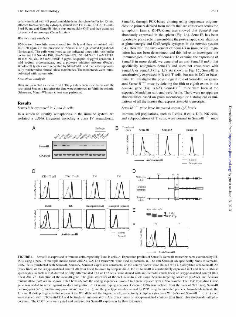
In a screen to identify semaphorins in the immune system, weisolated a cDNA fragment encoding a class IV semaphorin,
Sema4B, through PCR-based cloning using degenerate oligonu-cleotide primers derived from motifs that are conserved across thesemaphorin family. RT-PCR analyses showed that Sema4B wasabundantly expressed in the spleen (Fig. 1A). Sema4B has beenreported to play a role in assembling the postsynaptic specializationat glutamatergic and GABAergic synapses in the nervous system(34). However, the involvement of Sema4B in immune cell regu-lation has not been determined, and this led us to investigate theimmunological function of Sema4B. To examine the expression ofSema4B in more detail, we generated an anti-Sema4B mAb thatspecifically recognizes Sema4B and does not cross-react withSema4A or Sema4D (Fig. 1B). As shown in Fig. 1C, Sema4B isconstitutively expressed in B and T cells, but not in DCs or baso-phils. To investigate the physiological role of Sema4B, we gener-ated Sema4B2/2 mice by deleting the fifth to eighth exons in theSema4B gene (Fig. 1D–F). Sema4B2/2 mice were born at theexpected Mendelian ratio and were fertile. There were no apparentabnormalities based on gross macroscopic or histological exami-nations of all the tissues that express Sema4B transcripts.
Sema4B2/2 mice have increased serum IgE levels
Immune cell populations, such as T cells, B cells, DCs, NK cells,and subpopulations of T cells, were normal in Sema4B2/2 mice
FIGURE 1. Sema4B is expressed in immune cells, especially T and B cells. A, Expression profiles of Sema4B. Sema4B transcripts were examined by RT-
PCR using a panel of multiple mouse tissue cDNAs. GAPDH transcripts were used as controls. B, The anti-Sema4B Ab specifically binds to Sema4B.
COS7 cells transfected with Sema4B, Sema4A, Sema4D expression constructs, or the control vector were stained with a biotinylated anti-Sema4B Ab
(thick lines) or the isotype-matched control Ab (thin lines) followed by streptavidin-FITC. C, Sema4B is constitutively expressed in T and B cells. Mouse
splenocytes, as well as BM-derived or fully differentiated Th1 or Th2 cells, were stained with anti-Sema4B (thick lines) or isotype-matched control (thin
lines) Abs. D, Disruption of the Sema4B gene. The gene structures of the WT Sema4B allele (top), Sema4B-targeting construct (middle), and Sema4B
mutant allele (bottom) are shown. Filled boxes denote the coding sequences. Exons 5 to 8 were replaced with a Neo cassette. The HSV thymidine kinase
gene was added to select against random integration. E, Genomic typing analyses. Genomic DNA was isolated from the tails of WT (+/+), Sema4B
heterozygous (+/2), and homozygous mutant mice (2/2), and the genotype was determined by PCR using the indicated primers. Arrowheads indicate the
1.1- and 0.85-kbp fragments that represent the WT allele and the targeted allele, respectively. F, Splenocytes from WT (+/+) and Sema4B2/2 (2/2) mice
were stained with FITC–anti-CD3 and biotinylated anti-Sema4B mAbs (thick lines) or isotype-matched controls (thin lines) plus streptavidin-allophy-
cocyanin. The CD3+ cells were gated and analyzed for Sema4B expression by flow cytometry.
The Journal of Immunology 2883
by guest on June 13, 2013http://w
ww
.jimm
unol.org/D
ownloaded from
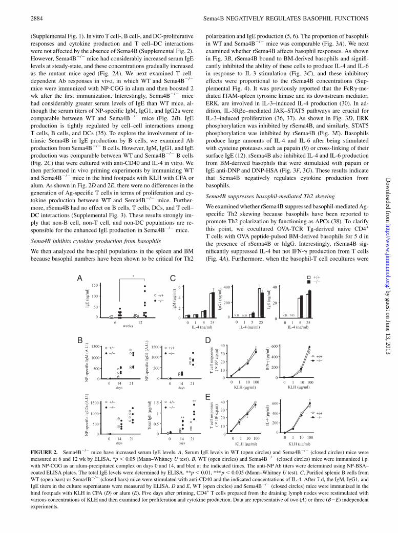
(Supplemental Fig. 1). In vitro T cell-, B cell-, and DC-proliferativeresponses and cytokine production and T cell–DC interactionswere not affected by the absence of Sema4B (Supplemental Fig. 2).However, Sema4B2/2 mice had considerably increased serum IgElevels at steady-state, and these concentrations gradually increasedas the mutant mice aged (Fig. 2A). We next examined T cell-dependent Ab responses in vivo, in which WT and Sema4B2/2
mice were immunized with NP-CGG in alum and then boosted 2wk after the first immunization. Interestingly, Sema4B2/2 micehad considerably greater serum levels of IgE than WT mice, al-though the serum titers of NP-specific IgM, IgG1, and IgG2a werecomparable between WT and Sema4B2/2 mice (Fig. 2B). IgEproduction is tightly regulated by cell–cell interactions amongT cells, B cells, and DCs (35). To explore the involvement of in-trinsic Sema4B in IgE production by B cells, we examined Abproduction from Sema4B2/2B cells. However, IgM, IgG1, and IgEproduction was comparable between WT and Sema4B2/2 B cells(Fig. 2C) that were cultured with anti-CD40 and IL-4 in vitro. Wethen performed in vivo priming experiments by immunizing WTand Sema4B2/2 mice in the hind footpads with KLH with CFA oralum. As shown in Fig. 2D and 2E, there were no differences in thegeneration of Ag-specific T cells in terms of proliferation and cy-tokine production between WT and Sema4B2/2 mice. Further-more, rSema4B had no effect on B cells, T cells, DCs, and T cell–DC interactions (Supplemental Fig. 3). These results strongly im-ply that non-B cell, non-T cell, and non-DC populations are re-sponsible for the enhanced IgE production in Sema4B2/2 mice.
Sema4B inhibits cytokine production from basophils
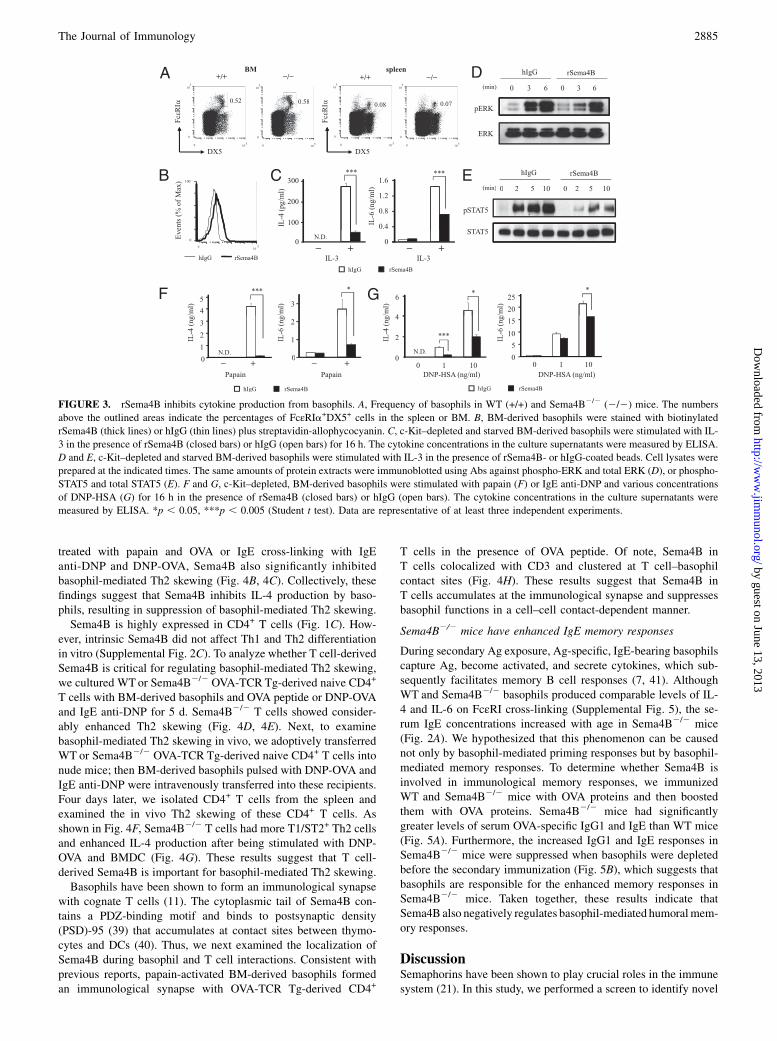
We then analyzed the basophil populations in the spleen and BMbecause basophil numbers have been shown to be critical for Th2
polarization and IgE production (5, 6). The proportion of basophilsin WT and Sema4B2/2 mice was comparable (Fig. 3A). We nextexamined whether rSema4B affects basophil responses. As shownin Fig. 3B, rSema4B bound to BM-derived basophils and signifi-cantly inhibited the ability of these cells to produce IL-4 and IL-6in response to IL-3 stimulation (Fig. 3C), and these inhibitoryeffects were proportional to the rSema4B concentrations (Sup-plemental Fig. 4). It was previously reported that the FcRg-me-diated ITAM-spleen tyrosine kinase and its downstream mediator,ERK, are involved in IL-3–induced IL-4 production (30). In ad-dition, IL-3Rbc–mediated JAK–STAT5 pathways are crucial forIL-3–induced proliferation (36, 37). As shown in Fig. 3D, ERKphosphorylation was inhibited by rSema4B, and similarly, STAT5phosphorylation was inhibited by rSema4B (Fig. 3E). Basophilsproduce large amounts of IL-4 and IL-6 after being stimulatedwith cysteine proteases such as papain (9) or cross-linking of theirsurface IgE (12). rSema4B also inhibited IL-4 and IL-6 productionfrom BM-derived basophils that were stimulated with papain orIgE anti-DNP and DNP-HSA (Fig. 3F, 3G). These results indicatethat Sema4B negatively regulates cytokine production frombasophils.
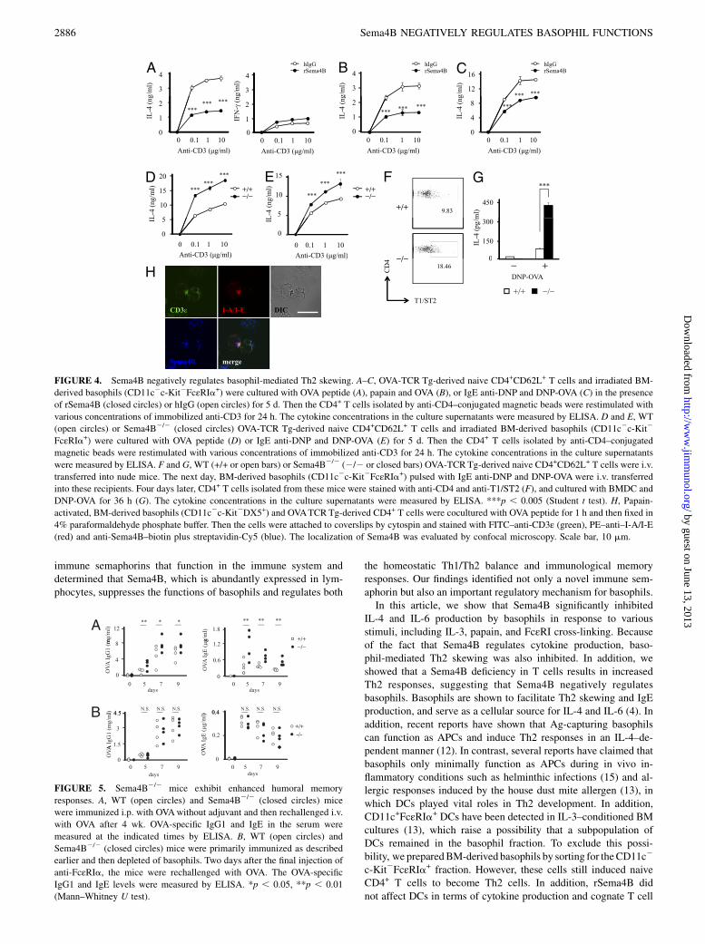
Sema4B suppresses basophil-mediated Th2 skewing
We examined whether rSema4B suppressed basophil-mediated Ag-specific Th2 skewing because basophils have been reported topromote Th2 polarization by functioning as APCs (38). To clarifythis point, we cocultured OVA-TCR Tg-derived naive CD4+
T cells with OVA peptide-pulsed BM-derived basophils for 5 d inthe presence of rSema4B or hIgG. Interestingly, rSema4B sig-nificantly suppressed IL-4 but not IFN-g production from T cells(Fig. 4A). Furthermore, when the basophil-T cell cocultures were
FIGURE 2. Sema4B2/2 mice have increased serum IgE levels. A, Serum IgE levels in WT (open circles) and Sema4B2/2 (closed circles) mice were
measured at 6 and 12 wk by ELISA. *p , 0.05 (Mann–Whitney U test). B, WT (open circles) and Sema4B2/2 (closed circles) mice were immunized i.p.
with NP-CGG as an alum-precipitated complex on days 0 and 14, and bled at the indicated times. The anti-NPAb titers were determined using NP-BSA–
coated ELISA plates. The total IgE levels were determined by ELISA. **p , 0.01, ***p , 0.005 (Mann–Whitney U test). C, Purified splenic B cells from
WT (open bars) or Sema4B2/2 (closed bars) mice were stimulated with anti-CD40 and the indicated concentrations of IL-4. After 7 d, the IgM, IgG1, and
IgE titers in the culture supernatants were measured by ELISA. D and E, WT (open circles) and Sema4B2/2 (closed circles) mice were immunized in the
hind footpads with KLH in CFA (D) or alum (E). Five days after priming, CD4+ T cells prepared from the draining lymph nodes were restimulated with
various concentrations of KLH and then examined for proliferation and cytokine production. Data are representative of two (A) or three (B2E) independent
experiments.
2884 Sema4B NEGATIVELY REGULATES BASOPHIL FUNCTIONS
by guest on June 13, 2013http://w
ww
.jimm
unol.org/D
ownloaded from
treated with papain and OVA or IgE cross-linking with IgEanti-DNP and DNP-OVA, Sema4B also significantly inhibitedbasophil-mediated Th2 skewing (Fig. 4B, 4C). Collectively, thesefindings suggest that Sema4B inhibits IL-4 production by baso-phils, resulting in suppression of basophil-mediated Th2 skewing.Sema4B is highly expressed in CD4+ T cells (Fig. 1C). How-
ever, intrinsic Sema4B did not affect Th1 and Th2 differentiationin vitro (Supplemental Fig. 2C). To analyze whether T cell-derivedSema4B is critical for regulating basophil-mediated Th2 skewing,we cultured WTor Sema4B2/2 OVA-TCR Tg-derived naive CD4+
T cells with BM-derived basophils and OVA peptide or DNP-OVAand IgE anti-DNP for 5 d. Sema4B2/2 T cells showed consider-ably enhanced Th2 skewing (Fig. 4D, 4E). Next, to examinebasophil-mediated Th2 skewing in vivo, we adoptively transferredWT or Sema4B2/2 OVA-TCR Tg-derived naive CD4+ T cells intonude mice; then BM-derived basophils pulsed with DNP-OVA andIgE anti-DNP were intravenously transferred into these recipients.Four days later, we isolated CD4+ T cells from the spleen andexamined the in vivo Th2 skewing of these CD4+ T cells. Asshown in Fig. 4F, Sema4B2/2 T cells had more T1/ST2+ Th2 cellsand enhanced IL-4 production after being stimulated with DNP-OVA and BMDC (Fig. 4G). These results suggest that T cell-derived Sema4B is important for basophil-mediated Th2 skewing.Basophils have been shown to form an immunological synapse
with cognate T cells (11). The cytoplasmic tail of Sema4B con-tains a PDZ-binding motif and binds to postsynaptic density(PSD)-95 (39) that accumulates at contact sites between thymo-cytes and DCs (40). Thus, we next examined the localization ofSema4B during basophil and T cell interactions. Consistent withprevious reports, papain-activated BM-derived basophils formedan immunological synapse with OVA-TCR Tg-derived CD4+
T cells in the presence of OVA peptide. Of note, Sema4B inT cells colocalized with CD3 and clustered at T cell–basophilcontact sites (Fig. 4H). These results suggest that Sema4B inT cells accumulates at the immunological synapse and suppressesbasophil functions in a cell–cell contact-dependent manner.
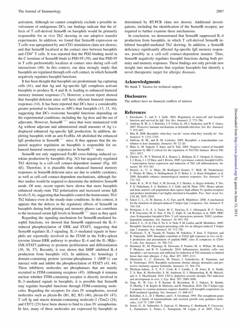
Sema4B2/2 mice have enhanced IgE memory responses
During secondary Ag exposure, Ag-specific, IgE-bearing basophilscapture Ag, become activated, and secrete cytokines, which sub-sequently facilitates memory B cell responses (7, 41). AlthoughWT and Sema4B2/2 basophils produced comparable levels of IL-4 and IL-6 on FcεRI cross-linking (Supplemental Fig. 5), the se-rum IgE concentrations increased with age in Sema4B2/2 mice(Fig. 2A). We hypothesized that this phenomenon can be causednot only by basophil-mediated priming responses but by basophil-mediated memory responses. To determine whether Sema4B isinvolved in immunological memory responses, we immunizedWT and Sema4B2/2 mice with OVA proteins and then boostedthem with OVA proteins. Sema4B2/2 mice had significantlygreater levels of serum OVA-specific IgG1 and IgE than WT mice(Fig. 5A). Furthermore, the increased IgG1 and IgE responses inSema4B2/2 mice were suppressed when basophils were depletedbefore the secondary immunization (Fig. 5B), which suggests thatbasophils are responsible for the enhanced memory responses inSema4B2/2 mice. Taken together, these results indicate thatSema4B also negatively regulates basophil-mediated humoralmem-ory responses.
DiscussionSemaphorins have been shown to play crucial roles in the immunesystem (21). In this study, we performed a screen to identify novel
FIGURE 3. rSema4B inhibits cytokine production from basophils. A, Frequency of basophils in WT (+/+) and Sema4B2/2 (2/2) mice. The numbers
above the outlined areas indicate the percentages of FcεRIa+DX5+ cells in the spleen or BM. B, BM-derived basophils were stained with biotinylated
rSema4B (thick lines) or hIgG (thin lines) plus streptavidin-allophycocyanin. C, c-Kit–depleted and starved BM-derived basophils were stimulated with IL-
3 in the presence of rSema4B (closed bars) or hIgG (open bars) for 16 h. The cytokine concentrations in the culture supernatants were measured by ELISA.
D and E, c-Kit–depleted and starved BM-derived basophils were stimulated with IL-3 in the presence of rSema4B- or hIgG-coated beads. Cell lysates were
prepared at the indicated times. The same amounts of protein extracts were immunoblotted using Abs against phospho-ERK and total ERK (D), or phospho-
STAT5 and total STAT5 (E). F and G, c-Kit–depleted, BM-derived basophils were stimulated with papain (F) or IgE anti-DNP and various concentrations
of DNP-HSA (G) for 16 h in the presence of rSema4B (closed bars) or hIgG (open bars). The cytokine concentrations in the culture supernatants were
measured by ELISA. *p , 0.05, ***p , 0.005 (Student t test). Data are representative of at least three independent experiments.
The Journal of Immunology 2885
by guest on June 13, 2013http://w
ww
.jimm
unol.org/D
ownloaded from
immune semaphorins that function in the immune system anddetermined that Sema4B, which is abundantly expressed in lym-phocytes, suppresses the functions of basophils and regulates both
the homeostatic Th1/Th2 balance and immunological memoryresponses. Our findings identified not only a novel immune sem-aphorin but also an important regulatory mechanism for basophils.In this article, we show that Sema4B significantly inhibited
IL-4 and IL-6 production by basophils in response to variousstimuli, including IL-3, papain, and FcεRI cross-linking. Becauseof the fact that Sema4B regulates cytokine production, baso-phil-mediated Th2 skewing was also inhibited. In addition, weshowed that a Sema4B deficiency in T cells results in increasedTh2 responses, suggesting that Sema4B negatively regulatesbasophils. Basophils are shown to facilitate Th2 skewing and IgEproduction, and serve as a cellular source for IL-4 and IL-6 (4). Inaddition, recent reports have shown that Ag-capturing basophilscan function as APCs and induce Th2 responses in an IL-4–de-pendent manner (12). In contrast, several reports have claimed thatbasophils only minimally function as APCs during in vivo in-flammatory conditions such as helminthic infections (15) and al-lergic responses induced by the house dust mite allergen (13), inwhich DCs played vital roles in Th2 development. In addition,CD11c+FcεRIa+ DCs have been detected in IL-3–conditioned BMcultures (13), which raise a possibility that a subpopulation ofDCs remained in the basophil fraction. To exclude this possi-bility, we preparedBM-derived basophils by sorting for theCD11c2
c-Kit2FcεRIa+ fraction. However, these cells still induced naiveCD4+ T cells to become Th2 cells. In addition, rSema4B didnot affect DCs in terms of cytokine production and cognate T cell
A
g/m
l)
3
4B
3
4rSema4BhIgG
g/m
l)
C
g/m
l)
12
16
*** ***
rSema4BhIgG
rSema4BhIgG
g/m
l)
3
4
IL-4
(ng
****** ***
0
1
2
0
1
2
IL-4
(ng
****** ***
IL-4
(ng
0
4
8***
IFN
-γ(n
g
0
1
2
D
Anti-CD3 (µg/ml)
0 0.1 1 10
Anti-CD3 (µg/ml)
0 0.1 1 10
Anti-CD3 (µg/ml)
0 0.1 1 10
20 *** F GE15 ***
Anti-CD3 (µg/ml)
0 0.1 1 10
+/+−/−
5
10
15
20
L-4
(n
g/m
l) ******
+/+ 9.83
450
ml)
***+/+−/−
5
10
15
L-4
(n
g/m
l)
***
***
0
5
0 0.1 1 10
Anti-CD3 (µg/ml)
IL
−/− 0
150
300
IL-4
(pg/m
0
IL
0 0.1 1 10
Anti-CD3 (µg/ml)
H−/−
18.46
T1/ST2
CD
4 0
+−DNP-OVA
+/+ −/−
(µg )
T1/ST2
CD3ε I-A/I-E DIC
Sema4B merge
FIGURE 4. Sema4B negatively regulates basophil-mediated Th2 skewing. A–C, OVA-TCR Tg-derived naive CD4+CD62L+ T cells and irradiated BM-
derived basophils (CD11c2c-Kit2FcεRIa+) were cultured with OVA peptide (A), papain and OVA (B), or IgE anti-DNP and DNP-OVA (C) in the presence
of rSema4B (closed circles) or hIgG (open circles) for 5 d. Then the CD4+ T cells isolated by anti-CD4–conjugated magnetic beads were restimulated with
various concentrations of immobilized anti-CD3 for 24 h. The cytokine concentrations in the culture supernatants were measured by ELISA. D and E, WT
(open circles) or Sema4B2/2 (closed circles) OVA-TCR Tg-derived naive CD4+CD62L+ T cells and irradiated BM-derived basophils (CD11c2c-Kit2
FcεRIa+) were cultured with OVA peptide (D) or IgE anti-DNP and DNP-OVA (E) for 5 d. Then the CD4+ T cells isolated by anti-CD4–conjugated
magnetic beads were restimulated with various concentrations of immobilized anti-CD3 for 24 h. The cytokine concentrations in the culture supernatants
were measured by ELISA. F and G, WT (+/+ or open bars) or Sema4B2/2 (2/2 or closed bars) OVA-TCRTg-derived naive CD4+CD62L+ T cells were i.v.
transferred into nude mice. The next day, BM-derived basophils (CD11c2c-Kit2FcεRIa+) pulsed with IgE anti-DNP and DNP-OVA were i.v. transferred
into these recipients. Four days later, CD4+ T cells isolated from these mice were stained with anti-CD4 and anti-T1/ST2 (F), and cultured with BMDC and
DNP-OVA for 36 h (G). The cytokine concentrations in the culture supernatants were measured by ELISA. ***p , 0.005 (Student t test). H, Papain-
activated, BM-derived basophils (CD11c2c-Kit2DX5+) and OVATCRTg-derived CD4+ T cells were cocultured with OVA peptide for 1 h and then fixed in
4% paraformaldehyde phosphate buffer. Then the cells were attached to coverslips by cytospin and stained with FITC–anti-CD3ε (green), PE–anti–I-A/I-E(red) and anti-Sema4B–biotin plus streptavidin-Cy5 (blue). The localization of Sema4B was evaluated by confocal microscopy. Scale bar, 10 mm.
FIGURE 5. Sema4B2/2 mice exhibit enhanced humoral memory
responses. A, WT (open circles) and Sema4B2/2 (closed circles) mice
were immunized i.p. with OVAwithout adjuvant and then rechallenged i.v.
with OVA after 4 wk. OVA-specific IgG1 and IgE in the serum were
measured at the indicated times by ELISA. B, WT (open circles) and
Sema4B2/2 (closed circles) mice were primarily immunized as described
earlier and then depleted of basophils. Two days after the final injection of
anti-FcεRIa, the mice were rechallenged with OVA. The OVA-specific
IgG1 and IgE levels were measured by ELISA. *p , 0.05, **p , 0.01
(Mann–Whitney U test).
2886 Sema4B NEGATIVELY REGULATES BASOPHIL FUNCTIONS
by guest on June 13, 2013http://w
ww
.jimm
unol.org/D
ownloaded from
activation. Although we cannot completely exclude a possible in-volvement of endogenous DCs, our findings indicate that the ef-fects of T cell-derived Sema4B on basophils would be primarilyresponsible for in vivo Th2 skewing in our adoptive transferexperiments. In addition, we found that Sema4B expression onT cells was upregulated by anti-CD3 stimulation (data not shown),and that Sema4B localized at the contact sites between basophilsand CD4+ T cells. It was reported that the PDZ-binding motif inthe C terminus of Sema4B binds to PSD-95 (39), and that PSD-95in T cells preferentially localizes at contact sites during cell–cellinteractions (40). In this context, our data strongly imply thatbasophils are regulated through cell–cell contact, in which Sema4Bnegatively regulates basophil functions.It has been thought that basophils are predominant Ag-capturing
cells (41), and that Ag and Ag-specific IgE complexes activatebasophils to produce IL-4 and IL-6, leading to enhanced humoralmemory immune responses (7). However, a recent report showedthat basophil-deficient mice still have efficient humoral immuneresponses (14). It has been reported that DCs have a considerablygreater potential to function as APCs than basophils (13, 15, 16),suggesting that DCs overcome basophil functions depending onthe experimental conditions, including the Ag dose and the use ofadjuvants. However, Sema4B2/2 mice that were immunized withAg without adjuvant and administered small amounts of Ag i.v.displayed enhanced Ag-specific IgE production. In addition, de-pleting basophils with an anti-FcεRIa Ab abolished the enhancedIgE production in Sema4B2/2 mice. It thus appears that the im-paired negative regulation on basophils is responsible for en-hanced humoral memory responses in Sema4B2/2 mice.Sema4B not only suppressed FcεRI cross-linking–induced cy-
tokine production by basophils (Fig. 3G) but negatively regulatedTh2 skewing in a cell–cell contact-dependent manner (Fig. 4D,4E). Therefore, it is plausible that enhanced humoral memoryresponses in Sema4B-deficient mice are due to soluble cytokines,as well as cell–cell contact-dependent mechanisms, although fur-ther studies would be required to determine the definitive Sema4Bmode. Of note, recent reports have shown that more basophilsenhanced steady-state Th2 polarization and increased serum IgElevels (5, 6), suggesting that basophils control the homeostatic Th1/Th2 balance even in the steady-state conditions. In this context, itappears that the defects in the regulatory effects of Sema4B onbasophils during both priming and memory phases can contributeto the increased serum IgE levels in Sema4B2/2mice as they aged.Regarding the signaling mechanism for Sema4B-mediated ba-
sophil functions, we showed that Sema4B suppressed the IL-3–induced phosphorylation of ERK and STAT5, suggesting thatSema4B regulates IL-3 signaling. IL-3–mediated signals in baso-phils are reportedly involved in the ITAM in the FcRg-spleentyrosine kinase-ERK pathway to produce IL-4 and the IL-3Rbc–JAK-STAT5 pathway to promote proliferation and differentiation(30, 36, 37). Recently, it was shown that SHIP inhibited IL-4production from basophils (42). In addition, Src homology 2domain-containing protein tyrosine-phosphatase 1 (SHP-1) caninteract with and inhibit the phosphorylation of STAT5 (43, 44).These inhibitory molecules are phosphatases that are mainlyrecruited to ITIM-containing receptors (45). Although it remainsunclear whether ITIM-containing receptor is directly involved inIL-3–mediated signals in basophils, it is possible that Sema4Bmay regulate basophil functions through ITIM-containing mole-cules. Regarding the receptors for class IV semaphorins, severalmolecules such as plexin-Bs (B1, B2, B3) (46), plexin-D1 (47),T cell Ig and mucin domain-containing molecule-2 (Tim2) (24),and CD72 (23) have been shown to bind to class IV semaphorins.In fact, many of these molecules are expressed by basophils as
determined by RT-PCR (data not shown). Additional investi-gations, including the identification of the Sema4B receptor, arerequired to further examine these mechanisms.In conclusion, we demonstrated that Sema4B suppressed IL-4
production from basophils, in which T cell-derived Sema4B in-hibited basophil-mediated Th2 skewing. In addition, a Sema4Bdeficiency significantly affected Ag-specific IgE memory respon-ses, possibly in a cell–cell contact-dependent manner. Thus,Sema4B negatively regulates basophil functions during both pri-mary and memory responses. These findings not only provide newinsight into mechanisms that regulate basophils but identify anovel therapeutic target for allergic diseases.
AcknowledgmentsWe thank T. Yazawa for technical support.
DisclosuresThe authors have no financial conflicts of interest.
References1. Kawakami, T., and S. J. Galli. 2002. Regulation of mast-cell and basophil
function and survival by IgE. Nat. Rev. Immunol. 2: 773–786.2. Anthony, R. M., L. I. Rutitzky, J. F. Urban, Jr., M. J. Stadecker, and W. C. Gause.
2007. Protective immune mechanisms in helminth infection. Nat. Rev. Immunol.7: 975–987.
3. Min, B. 2008. Basophils: what they ‘can do’ versus what they ‘actually do’. Nat.Immunol. 9: 1333–1339.
4. Sullivan, B. M., and R. M. Locksley. 2009. Basophils: a nonredundant con-tributor to host immunity. Immunity 30: 12–20.
5. Hida, S., M. Tadachi, T. Saito, and S. Taki. 2005. Negative control of basophilexpansion by IRF-2 critical for the regulation of Th1/Th2 balance. Blood 106:2011–2017.
6. Charles, N., W. T. Watford, H. L. Ramos, L. Hellman, H. C. Oettgen, G. Gomez,J. J. Ryan, J. J. O’Shea, and J. Rivera. 2009. Lyn kinase controls basophil GATA-3 transcription factor expression and induction of Th2 cell differentiation. Im-munity 30: 533–543.
7. Denzel, A., U. A. Maus, M. Rodriguez Gomez, C. Moll, M. Niedermeier,C. Winter, R. Maus, S. Hollingshead, D. E. Briles, L. A. Kunz-Schughart, et al.2008. Basophils enhance immunological memory responses. Nat. Immunol. 9:733–742.
8. Seder, R. A., W. E. Paul, A. M. Dvorak, S. J. Sharkis, A. Kagey-Sobotka, Y. Niv,F. D. Finkelman, S. A. Barbieri, S. J. Galli, and M. Plaut. 1991. Mouse splenicand bone marrow cell populations that express high-affinity Fc epsilon receptorsand produce interleukin 4 are highly enriched in basophils. Proc. Natl. Acad. Sci.USA 88: 2835–2839.
9. Sokol, C. L., G. M. Barton, A. G. Farr, and R. Medzhitov. 2008. A mechanismfor the initiation of allergen-induced T helper type 2 responses. Nat. Immunol. 9:310–318.
10. Perrigoue, J. G., S. A. Saenz, M. C. Siracusa, E. J. Allenspach, B. C. Taylor,P. R. Giacomin, M. G. Nair, Y. Du, C. Zaph, N. van Rooijen, et al. 2009. MHCclass II-dependent basophil-CD4+ T cell interactions promote T(H)2 cytokine-dependent immunity. Nat. Immunol. 10: 697–705.
11. Sokol, C. L., N. Q. Chu, S. Yu, S. A. Nish, T. M. Laufer, and R. Medzhitov. 2009.Basophils function as antigen-presenting cells for an allergen-induced T helpertype 2 response. Nat. Immunol. 10: 713–720.
12. Yoshimoto, T., K. Yasuda, H. Tanaka, M. Nakahira, Y. Imai, Y. Fujimori, andK. Nakanishi. 2009. Basophils contribute to T(H)2-IgE responses in vivo via IL-4 production and presentation of peptide-MHC class II complexes to CD4+T cells. Nat. Immunol. 10: 706–712.
13. Hammad, H., M. Plantinga, K. Deswarte, P. Pouliot, M. A. Willart, M. Kool,F. Muskens, and B. N. Lambrecht. 2010. Inflammatory dendritic cells—notbasophils—are necessary and sufficient for induction of Th2 immunity to inhaledhouse dust mite allergen. J. Exp. Med. 207: 2097–2111.
14. Ohnmacht, C., C. Schwartz, M. Panzer, I. Schiedewitz, R. Naumann, andD. Voehringer. 2010. Basophils orchestrate chronic allergic dermatitis and pro-tective immunity against helminths. Immunity 33: 364–374.
15. Phythian-Adams, A. T., P. C. Cook, R. J. Lundie, L. H. Jones, K. A. Smith,T. A. Barr, K. Hochweller, S. M. Anderton, G. J. Hammerling, R. M. Maizels,and A. S. MacDonald. 2010. CD11c depletion severely disrupts Th2 inductionand development in vivo. J. Exp. Med. 207: 2089–2096.
16. Tang, H., W. Cao, S. P. Kasturi, R. Ravindran, H. I. Nakaya, K. Kundu,N. Murthy, T. B. Kepler, B. Malissen, and B. Pulendran. 2010. The T helper type2 response to cysteine proteases requires dendritic cell-basophil cooperation viaROS-mediated signaling. Nat. Immunol. 11: 608–617.
17. Kolodkin, A. L., D. J. Matthes, and C. S. Goodman. 1993. The semaphorin genesencode a family of transmembrane and secreted growth cone guidance mole-cules. Cell 75: 1389–1399.
18. Serini, G., D. Valdembri, S. Zanivan, G. Morterra, C. Burkhardt, F. Caccavari,L. Zammataro, L. Primo, L. Tamagnone, M. Logan, et al. 2003. Class 3
The Journal of Immunology 2887
by guest on June 13, 2013http://w
ww
.jimm
unol.org/D
ownloaded from
semaphorins control vascular morphogenesis by inhibiting integrin function.Nature 424: 391–397.
19. Toyofuku, T., H. Zhang, A. Kumanogoh, N. Takegahara, F. Suto, J. Kamei,K. Aoki, M. Yabuki, M. Hori, H. Fujisawa, and H. Kikutani. 2004. Dual roles ofSema6D in cardiac morphogenesis through region-specific association of itsreceptor, Plexin-A1, with off-track and vascular endothelial growth factor re-ceptor type 2. Genes Dev. 18: 435–447.
20. Neufeld, G., and O. Kessler. 2008. The semaphorins: versatile regulators oftumour progression and tumour angiogenesis. Nat. Rev. Cancer 8: 632–645.
21. Suzuki, K., A. Kumanogoh, and H. Kikutani. 2008. Semaphorins and theirreceptors in immune cell interactions. Nat. Immunol. 9: 17–23.
22. Capparuccia, L., and L. Tamagnone. 2009. Semaphorin signaling in cancer cellsand in cells of the tumor microenvironment—two sides of a coin. J. Cell Sci.122: 1723–1736.
23. Kumanogoh, A., C. Watanabe, I. Lee, X. Wang, W. Shi, H. Araki, H. Hirata,K. Iwahori, J. Uchida, T. Yasui, et al. 2000. Identification of CD72 asa lymphocyte receptor for the class IV semaphorin CD100: a novel mechanismfor regulating B cell signaling. Immunity 13: 621–631.
24. Kumanogoh, A., S. Marukawa, K. Suzuki, N. Takegahara, C. Watanabe,E. Ch’ng, I. Ishida, H. Fujimura, S. Sakoda, K. Yoshida, and H. Kikutani. 2002.Class IV semaphorin Sema4A enhances T-cell activation and interacts with Tim-2. Nature 419: 629–633.
25. Takegahara, N., H. Takamatsu, T. Toyofuku, T. Tsujimura, T. Okuno,K. Yukawa, M. Mizui, M. Yamamoto, D. V. Prasad, K. Suzuki, et al. 2006.Plexin-A1 and its interaction with DAP12 in immune responses and bone ho-meostasis. Nat. Cell Biol. 8: 615–622.
26. Kumanogoh, A., T. Shikina, K. Suzuki, S. Uematsu, K. Yukawa,S. Kashiwamura, H. Tsutsui, M. Yamamoto, H. Takamatsu, E. P. Ko-Mitamura,et al. 2005. Nonredundant roles of Sema4A in the immune system: defectiveT cell priming and Th1/Th2 regulation in Sema4A-deficient mice. Immunity 22:305–316.
27. Takamatsu, H., N. Takegahara, Y. Nakagawa, M. Tomura, M. Taniguchi,R. H. Friedel, H. Rayburn, M. Tessier-Lavigne, Y. Yoshida, T. Okuno, et al.2010. Semaphorins guide the entry of dendritic cells into the lymphatics byactivating myosin II. Nat. Immunol. 11: 594–600.
28. Sato, T., T. Sasahara, Y. Nakamura, T. Osaki, T. Hasegawa, T. Tadakuma,Y. Arata, Y. Kumagai, M. Katsuki, and S. Habu. 1994. Naive T cells can mediatedelayed-type hypersensitivity response in T cell receptor transgenic mice. Eur. J.Immunol. 24: 1512–1516.
29. Yoshimoto, T., H. Tsutsui, K. Tominaga, K. Hoshino, H. Okamura, S. Akira,W. E. Paul, and K. Nakanishi. 1999. IL-18, although antiallergic when admin-istered with IL-12, stimulates IL-4 and histamine release by basophils. Proc.Natl. Acad. Sci. USA 96: 13962–13966.
30. Hida, S., S. Yamasaki, Y. Sakamoto, M. Takamoto, K. Obata, T. Takai,H. Karasuyama, K. Sugane, T. Saito, and S. Taki. 2009. Fc receptor gamma-chain, a constitutive component of the IL-3 receptor, is required for IL-3-inducedIL-4 production in basophils. Nat. Immunol. 10: 214–222.
31. Inaba, K., R. M. Steinman, M. W. Pack, H. Aya, M. Inaba, T. Sudo, S. Wolpe,and G. Schuler. 1992. Identification of proliferating dendritic cell precursors inmouse blood. J. Exp. Med. 175: 1157–1167.
32. Doi, T., K. Obayashi, T. Kadowaki, H. Fujii, and S. Koyasu. 2008. PI3K isa negative regulator of IgE production. Int. Immunol. 20: 499–508.
33. Shi, W., A. Kumanogoh, C. Watanabe, J. Uchida, X. Wang, T. Yasui, K. Yukawa,M. Ikawa, M. Okabe, J. R. Parnes, et al. 2000. The class IV semaphorin CD100plays nonredundant roles in the immune system: defective B and T cell acti-vation in CD100-deficient mice. Immunity 13: 633–642.
34. Paradis, S., D. B. Harrar, Y. Lin, A. C. Koon, J. L. Hauser, E. C. Griffith, L. Zhu,L. F. Brass, C. Chen, and M. E. Greenberg. 2007. An RNAi-based approachidentifies molecules required for glutamatergic and GABAergic synapse de-velopment. Neuron 53: 217–232.
35. Geha, R. S., H. H. Jabara, and S. R. Brodeur. 2003. The regulation of immu-noglobulin E class-switch recombination. Nat. Rev. Immunol. 3: 721–732.
36. Reddy, E. P., A. Korapati, P. Chaturvedi, and S. Rane. 2000. IL-3 signaling andthe role of Src kinases, JAKs and STATs: a covert liaison unveiled. Oncogene 19:2532–2547.
37. Martinez-Moczygemba, M., and D. P. Huston. 2003. Biology of common betareceptor-signaling cytokines: IL-3, IL-5, and GM-CSF. J. Allergy Clin. Immunol.112: 653–665, quiz 666.
38. Sokol, C. L., and R. Medzhitov. 2010. Emerging functions of basophils inprotective and allergic immune responses. Mucosal Immunol. 3: 129–137.
39. Burkhardt, C., M. Muller, A. Badde, C. C. Garner, E. D. Gundelfinger, andA. W. Puschel. 2005. Semaphorin 4B interacts with the post-synaptic densityprotein PSD-95/SAP90 and is recruited to synapses through a C-terminal PDZ-binding motif. FEBS Lett. 579: 3821–3828.
40. Affaticati, P., O. Mignen, F. Jambou, M. C. Potier, I. Klingel-Schmitt,J. Degrouard, S. Peineau, E. Gouadon, G. L. Collingridge, R. Liblau, et al. 2011.Sustained calcium signalling and caspase-3 activation involve NMDA receptorsin thymocytes in contact with dendritic cells. Cell Death Differ. 18: 99–108.
41. Mack, M., M. A. Schneider, C. Moll, J. Cihak, H. Bruhl, J. W. Ellwart,M. P. Hogarth, M. Stangassinger, and D. Schlondorff. 2005. Identification ofantigen-capturing cells as basophils. J. Immunol. 174: 735–741.
42. Kuroda, E., V. Ho, J. Ruschmann, F. Antignano, M. Hamilton, M. J. Rauh,A. Antov, R. A. Flavell, L. M. Sly, and G. Krystal. 2009. SHIP represses thegeneration of IL-3-induced M2 macrophages by inhibiting IL-4 production frombasophils. J. Immunol. 183: 3652–3660.
43. Paling, N. R., and M. J. Welham. 2002. Role of the protein tyrosine phosphataseSHP-1 (Src homology phosphatase-1) in the regulation of interleukin-3-inducedsurvival, proliferation and signalling. Biochem. J. 368: 885–894.
44. Minoo, P., M. M. Zadeh, R. Rottapel, J. J. Lebrun, and S. Ali. 2004. A novelSHP-1/Grb2-dependent mechanism of negative regulation of cytokine-receptorsignaling: contribution of SHP-1 C-terminal tyrosines in cytokine signaling.Blood 103: 1398–1407.
45. Kraft, S., and J. P. Kinet. 2007. New developments in FcepsilonRI regulation,function and inhibition. Nat. Rev. Immunol. 7: 365–378.
46. Pasterkamp, R. J., and A. L. Kolodkin. 2003. Semaphorin junction: makingtracks toward neural connectivity. Curr. Opin. Neurobiol. 13: 79–89.
47. Toyofuku, T., M. Yabuki, J. Kamei, M. Kamei, N. Makino, A. Kumanogoh, andM. Hori. 2007. Semaphorin-4A, an activator for T-cell-mediated immunity,suppresses angiogenesis via Plexin-D1. EMBO J. 26: 1373–1384.
2888 Sema4B NEGATIVELY REGULATES BASOPHIL FUNCTIONS
by guest on June 13, 2013http://w
ww
.jimm
unol.org/D
ownloaded from
Related Documents