Sports Medicine Research Laboratory The University of North Carolina at Chapel Hill Identification of Risk Factors for ACL Injury and Re-Injury: Implications for Prevention and Rehabilitation Darin A. Padua, PhD, ATC Director, Sports Medicine Research Laboratory College of Arts & Sciences Department of Exercise & Sport Science 2012 E.A.T.A. Convention

Welcome message from author
This document is posted to help you gain knowledge. Please leave a comment to let me know what you think about it! Share it to your friends and learn new things together.
Transcript
Sports Medicine Research Laboratory
The University of North Carolina at Chapel Hill
Identification of Risk Factors for ACL
Injury and Re-Injury: Implications for
Prevention and Rehabilitation
Darin A. Padua, PhD, ATC Director, Sports Medicine Research Laboratory
College of Arts & Sciences
Department of Exercise & Sport Science
2012 E.A.T.A. Convention

Overview
• Prospective risk factors for ACL injury
– JUMP-ACL study findings
• Potential risk factors for ACL re-injury
– Same as incident ACL injury?
• Implications for ACL injury prevention and
return to participation decision making

The ACL Injury Problem
• Disability:
– 77% sports disability in 5 yrs
– 44% disability with ADL’s in
5 yrs
– Increase Risk of Knee
Osteoarthritis
• No Surgery: >90% in 20 years
• “Good” Surgery: >90% in 20
years

How can we avoid
ACL injury / re-injury?
3 Keys to Improving:
1. Understand risk factors
for injury / re-injury
2. Systematic exercise
progression – Focus on modifying risk factors
3. Systematic return to play
criteria – Based on successful
modification of risk factors

• 5 year trial at each academy
• 400 / academy / year
– ~40% female
– ~6,000 subjects
– 15,000 man-years
• Goal = Capture primary ACL
injuries

JUMP - ACL Research Team
UNC Chapel Hill
• Steve Marshall, PhD
• Darin Padua, PhD, ATC
• Sue Wolf, RN
• Shrikant Bangdiwala, PhD
• Bing Yu, PhD
• Charles Thigpen, PhD, PT, ATC
• Michelle Boling, PhD, ATC
• Ben Goerger, MS, ATC
• Sarah Knowles, PhD
Collaborators:
• William Garrett, MD, PhD (Duke)
• Barry Boden, MD (Ortho Cntr)
• Marjorie King, PhD, ATC, PT (PSU)
• Brent Arnold / Scott Ross, PhD, ATC
(VCU)
USUHS
• Anthony Beutler, MD
USNA
• Marlene DeMaio, MD
• Scott Pyne, MD
• Greg Calhoon, ATC
USAFA
• John Tokish, MD
• Keith Odegard, MD
USMA
• Dean Taylor, MD
• Paul DeBeradino, MD
• Steve Slovoda, MD
• Kenneth Cameron, PhD, ATC
• Sally Mountcastle, PhD
• Jennifer Jones, Med, ATC
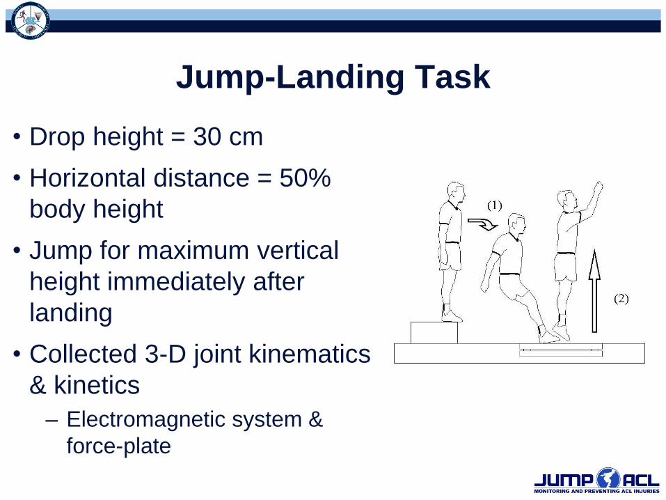
Jump-Landing Task
• Drop height = 30 cm
• Horizontal distance = 50%
body height
• Jump for maximum vertical
height immediately after
landing
• Collected 3-D joint kinematics
& kinetics
– Electromagnetic system &
force-plate

Hip Extension Hip Abduction Hip External Rotation
Hip Internal Rotation Knee Flexion Knee Extension
Strength Testing

Postural Alignment Testing
Q-Angle Navicular Drop
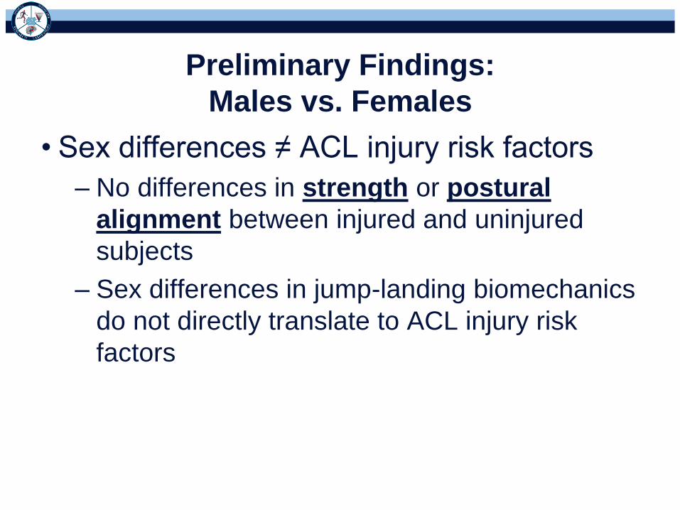
Preliminary Findings:
Males vs. Females
• Sex differences ≠ ACL injury risk factors
– No differences in strength or postural
alignment between injured and uninjured
subjects
– Sex differences in jump-landing biomechanics
do not directly translate to ACL injury risk
factors
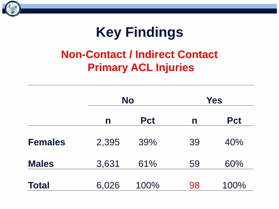
No Yes
n Pct n Pct
Females 2,395 39% 39 40%
Males 3,631 61% 59 60%
Total 6,026 100% 98 100%
Non-Contact / Indirect Contact
Primary ACL Injuries
Key Findings
Males and Females, NonAndIndirectContact_ACL_Injury
Males and Females, NonAndIndirectContact_ACL_Injury
Males and Females, NonAndIndirectContact_ACL_Injury
Kn
ee
Fle
xio
n A
ng
le
Knee V
aru
s(+
) / V
alg
us(-
) K
ne
e IR
(+)
/ E
R(-
)
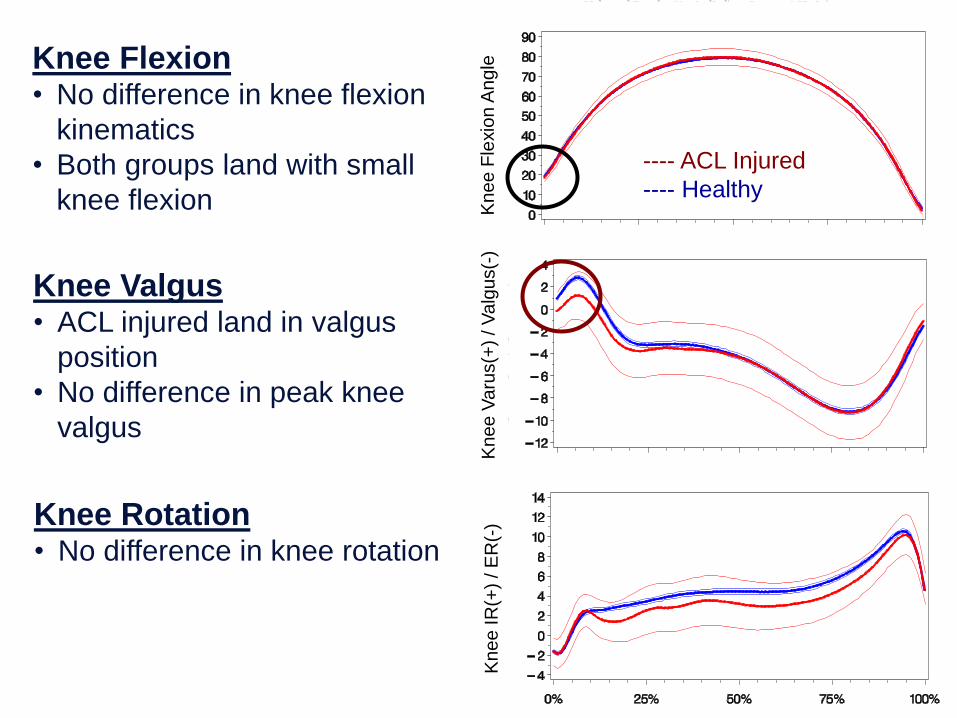
Knee Flexion • No difference in knee flexion
kinematics
• Both groups land with small
knee flexion
Knee Valgus • ACL injured land in valgus
position
• No difference in peak knee
valgus
Knee Rotation • No difference in knee rotation
---- ACL Injured
---- Healthy

Males and Females, NonAndIndirectContact_ACL_Injury
Males and Females, NonAndIndirectContact_ACL_Injury
Males and Females, NonAndIndirectContact_ACL_Injury
Hip
Fle
xio
n (
-)
Hip
IR
(+)
/ E
R(-
) H
ip A
dd
uctio
n(+
) / A
bd
uctio
n(-
)
Hip Flexion • Both groups land with small
knee flexion
• ACL injured demonstrate
greater peak flexion
Hip Adduction • ACL injured land in more
adducted position
Hip Rotation • ACL injured land in more
externally rotated position
---- ACL Injured
---- Healthy

“Prospective Profile” of ACL Injured
↓ Knee Flexion
Knee Valgus
(Initial Contact)
↑ Hip External
Rotation
↑ Hip ADDuction
↑ Hip Flexion
(Displacement)
↓ Hip Flexion ↓ Flexion at
Initial Contact
Valgus Alignment
at Initial Contact
Altered Hip
Neuromuscular
Control

NOTE
• This is NOT a study of injury mechanisms
– No-one tore their ACL during testing
• This is a study that helps us:
– Identify and screen-out individuals with high-
risk movement patterns
– Many years prior to injury

Some findings agree with non-contact ACL
injury mechanisms -- some do not
↓ Knee Flexion
↑ Knee Valgus
(Initial Contact)
↑ Hip External
Rotation
↑ Hip ADDuction
↑ Hip Flexion
(Displacement)
↓ Hip Flexion
Ireland, 1998
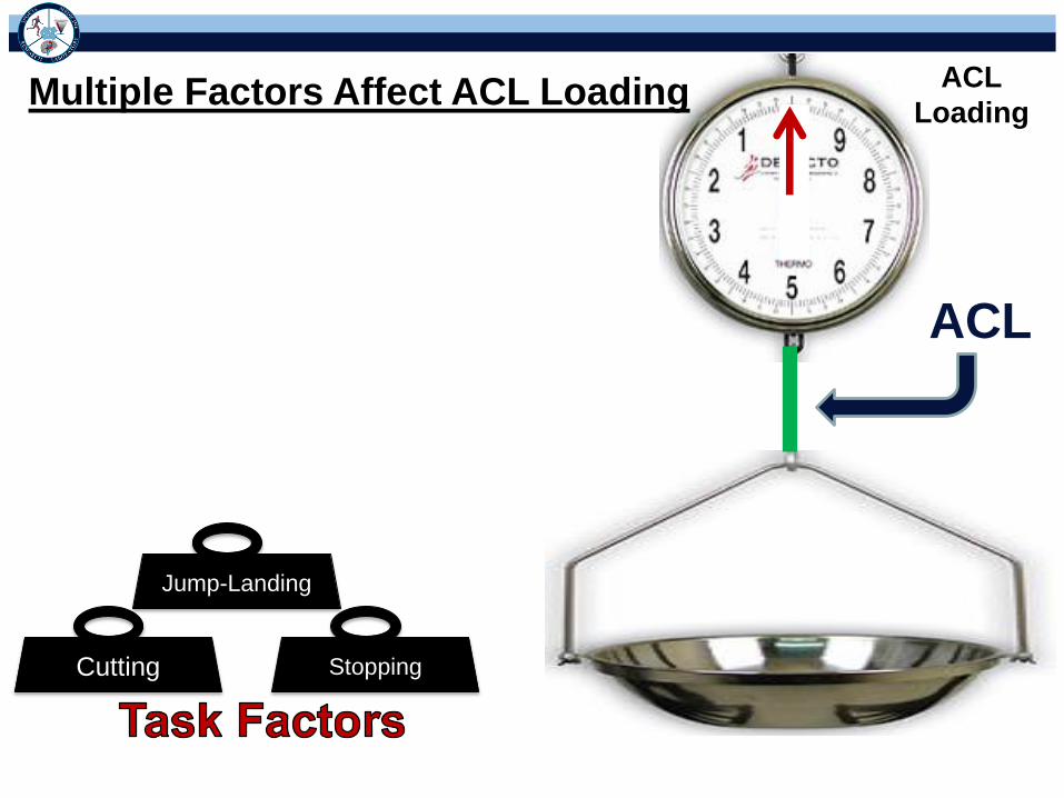
ACL
Loading
ACL
Stopping Cutting
Jump-Landing
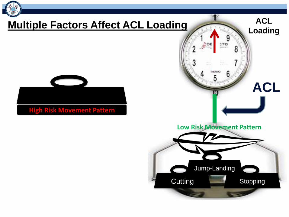
Multiple Factors Affect ACL Loading

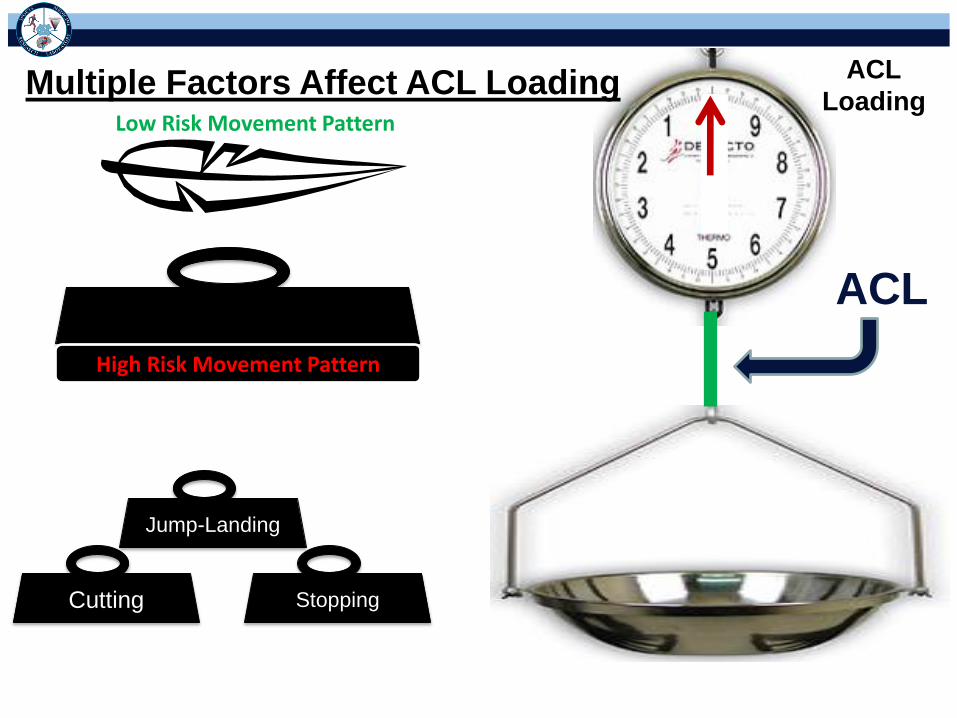
High Risk Movement Pattern
Low Risk Movement Pattern
ACL
ACL
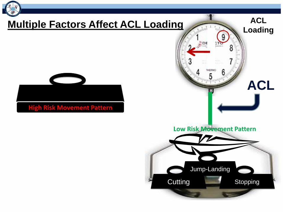
Loading Multiple Factors Affect ACL Loading
High Risk Movement Pattern
Low Risk Movement Pattern
ACL
ACL
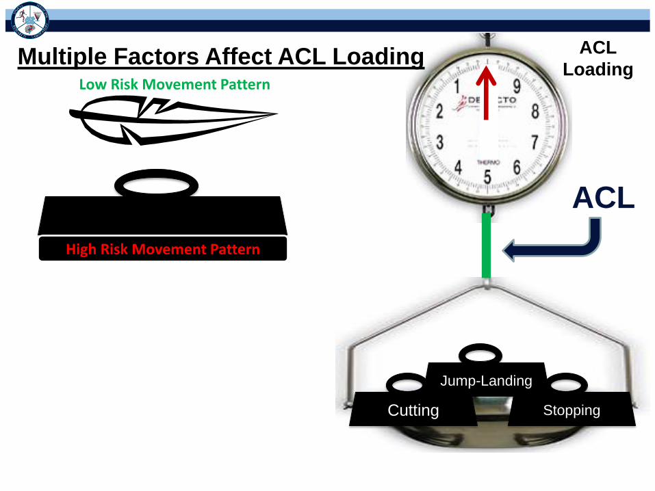
Loading Multiple Factors Affect ACL Loading
Stopping Cutting
Jump-Landing

High Risk Movement Pattern
Low Risk Movement Pattern
ACL
ACL
Loading Multiple Factors Affect ACL Loading
Stopping Cutting
Jump-Landing
High Risk Movement Pattern
Low Risk Movement Pattern
ACL
ACL
Loading Multiple Factors Affect ACL Loading
Stopping Cutting
Jump-Landing
High Risk Movement Pattern
Low Risk Movement Pattern
ACL
ACL
Loading Multiple Factors Affect ACL Loading
Stopping Cutting
Jump-Landing
High Risk Movement Pattern
Low Risk Movement Pattern
ACL
ACL
Loading Multiple Factors Affect ACL Loading
Stopping Cutting
Jump-Landing

Low Risk Movement Pattern
ACL
ACL
Loading Multiple Factors Affect ACL Loading
Stopping Cutting
Jump-Landing
High Risk Movement Pattern

Low Risk Movement Pattern
ACL
ACL
Loading Multiple Factors Affect ACL Loading
Stopping Cutting
Jump-Landing
High Risk Movement Pattern

ACL
ACL
Loading Multiple Factors Affect ACL Loading
Stopping Cutting
Jump-Landing
Misjudgement
Distracted
Defender
Moves
Foot Slips
Pushed
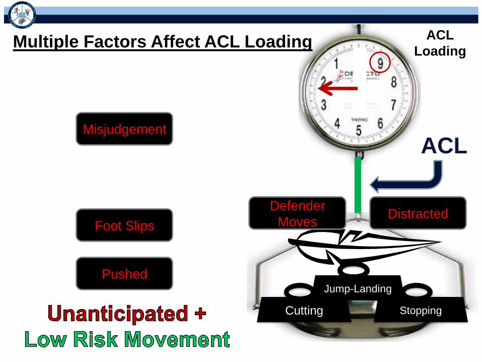
ACL
ACL
Loading Multiple Factors Affect ACL Loading
Stopping Cutting
Jump-Landing
Misjudgement
Distracted Defender
Moves Foot Slips
Pushed

ACL
ACL
Loading Multiple Factors Affect ACL Loading
Misjudgement
Foot Slips
Pushed
Stopping Cutting
Jump-Landing
Distracted Defender
Moves

ACL
ACL
Loading Multiple Factors Affect ACL Loading
Stopping Cutting
Jump-Landing
High Risk Movement Pattern
Misjudgement
Distracted
Defender
Moves
Foot Slips
Pushed

ACL
ACL
Loading Multiple Factors Affect ACL Loading
Stopping Cutting
Jump-Landing
High Risk Movement Pattern
Misjudgement
Distracted Defender
Moves Foot Slips
Pushed

ACL
Injury
ACL
Loading Multiple Factors Affect ACL Loading
Stopping Cutting
Jump-Landing
High Risk Movement Pattern
Misjudgement
Distracted Defender
Moves
Foot Slips
Pushed
• How can this information provide insight
into rehabilitation and return to participation
decisions in ACL injured?

Previous History of ACL Surgery
Non-Contact / Indirect Contact ACL
Injury(excluded Direct Contact)
4.8
33.4
0.0
10.0
20.0
30.0
40.0
50.0
60.0
ACL Hx -ve ACL Hx +ve
Inc
ide
nc
e o
f N
ew
NC
IC In
jury
(pe
r 1
,00
0 p
ers
on
-ye
ars
)N=150 Prior ACL Inj.
13 re-injuries (8.7%)
N=5,758 No ACL Inj.
78 primary injuries
(1.4%) No ACL Inj. Prior ACL Inj.
Rate Ratio= 6.9; 95%CI: 3.8, 12.4; p<0.01
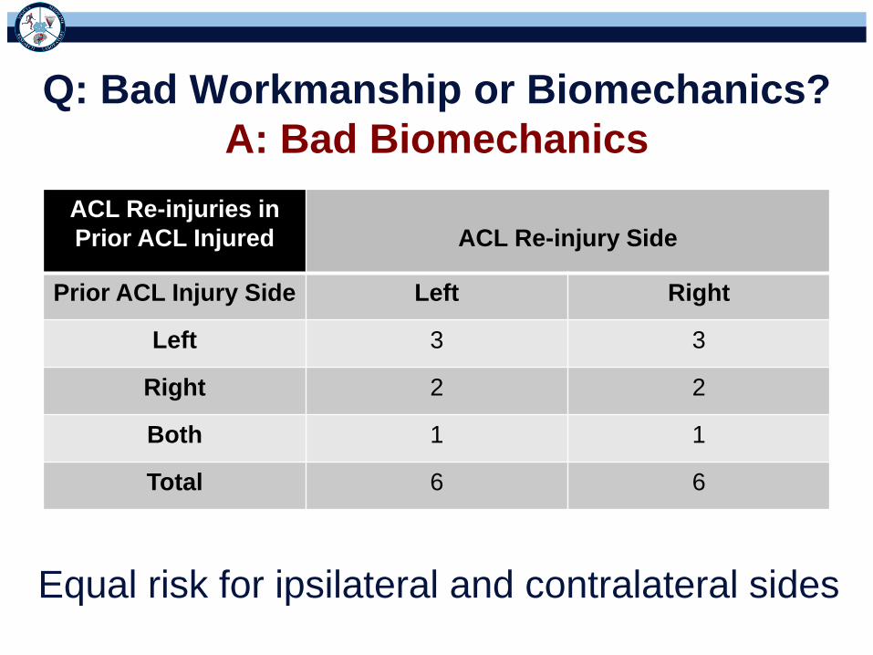
Q: Bad Workmanship or Biomechanics?
A: Bad Biomechanics
ACL Re-injuries in
Prior ACL Injured
ACL Re-injury Side
Prior ACL Injury Side Left Right
Left 3 3
Right 2 2
Both 1 1
Total 6 6
Equal risk for ipsilateral and contralateral sides
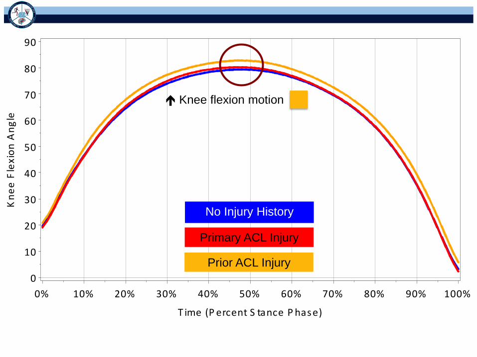
Kn
ee
Fle
xio
n A
ng
le
0
10
20
30
40
50
60
70
80
90
T ime (P ercent S tance P hase)
0% 10% 20% 30% 40% 50% 60% 70% 80% 90% 100%
No Injury History
Primary ACL Injury
Prior ACL Injury
Knee flexion motion
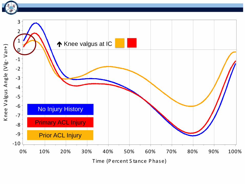
Kn
ee
Va
lgu
s A
ng
le (
Vlg
- V
ar+
)
-10
-9
-8
-7
-6
-5
-4
-3
-2
-1
0
1
2
3
T ime (P ercent S tance P hase)
0% 10% 20% 30% 40% 50% 60% 70% 80% 90% 100%
Knee valgus at IC
No Injury History
Primary ACL Injury
Prior ACL Injury

Hip
Fle
xio
n A
ng
le (
Flx
- E
xt+
)
-80
-70
-60
-50
-40
-30
-20
-10
0
T ime (P ercent S tance P hase)
0% 10% 20% 30% 40% 50% 60% 70% 80% 90% 100%
Hip Flexion at IC
Hip Flexion Motion
No Injury History
Primary ACL Injury
Prior ACL Injury
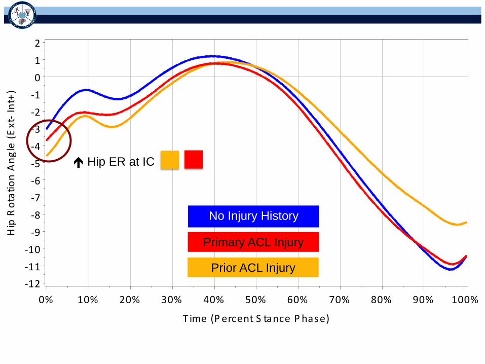
Hip
Ro
tati
on
An
gle
(E
xt-
Int+
)
-12
-11
-10
-9
-8
-7
-6
-5
-4
-3
-2
-1
0
1
2
T ime (P ercent S tance P hase)
0% 10% 20% 30% 40% 50% 60% 70% 80% 90% 100%
Hip ER at IC
No Injury History
Primary ACL Injury
Prior ACL Injury
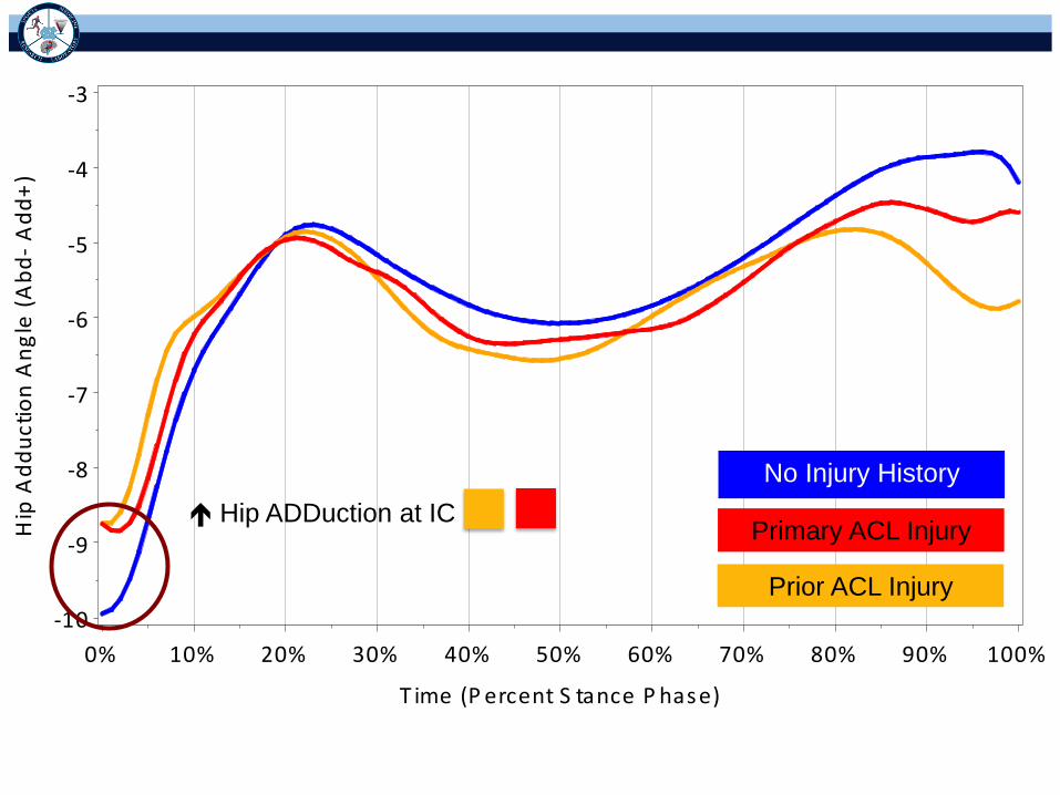
Hip
Ad
du
cti
on
An
gle
(A
bd
- A
dd
+)
-10
-9
-8
-7
-6
-5
-4
-3
T ime (P ercent S tance P hase)
0% 10% 20% 30% 40% 50% 60% 70% 80% 90% 100%
Hip ADDuction at IC
No Injury History
Primary ACL Injury
Prior ACL Injury
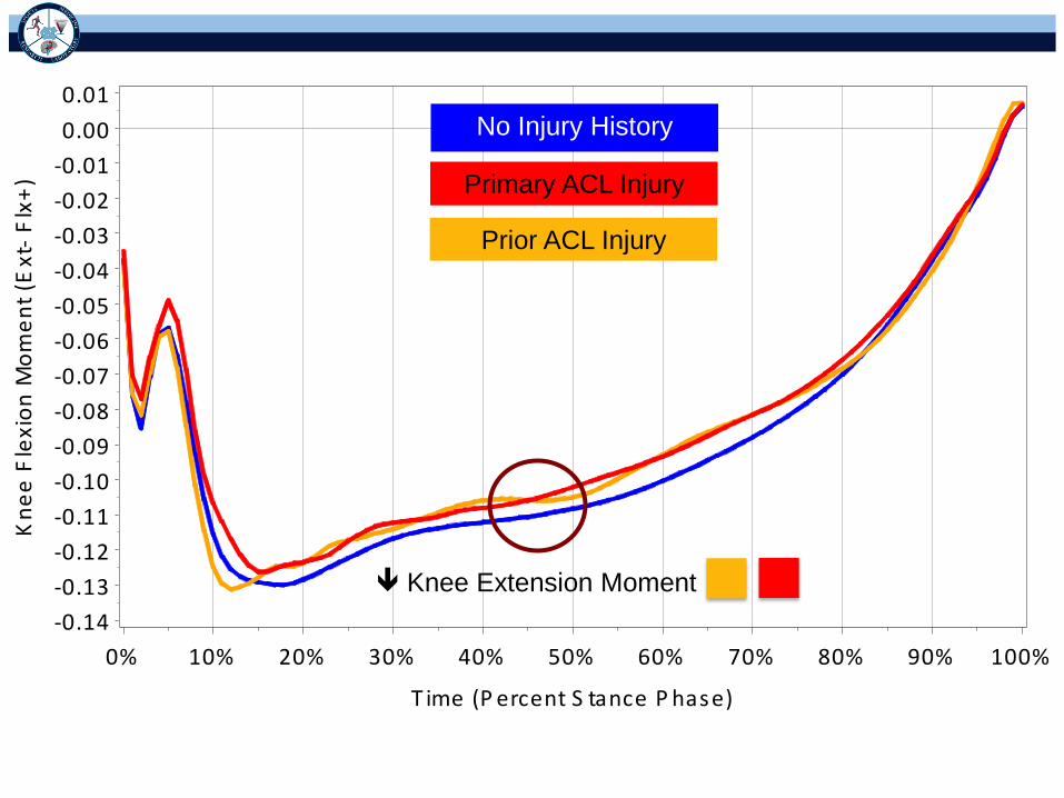
Kn
ee
Fle
xio
n M
om
en
t (E
xt-
Flx
+)
-0.14
-0.13
-0.12
-0.11
-0.10
-0.09
-0.08
-0.07
-0.06
-0.05
-0.04
-0.03
-0.02
-0.01
0.00
0.01
T ime (P ercent S tance P hase)
0% 10% 20% 30% 40% 50% 60% 70% 80% 90% 100%
Knee Extension Moment
No Injury History
Primary ACL Injury
Prior ACL Injury

↓ Knee & Hip
Flexion IC
↑ Knee Valgus IC
↑ Hip Adduction
↑ Hip ER
↑ Hip Flexion
Motion
Movement Patterns
•Healthy ≠ ACL injured
(primary & prior)
•Primary ACL Injury =
Prior ACL Injury
No Injury
History
Primary
ACL Injury
Prior
ACL Injury
↑ Knee
Flexion
Motion
↑ Hip
Flexion
IC
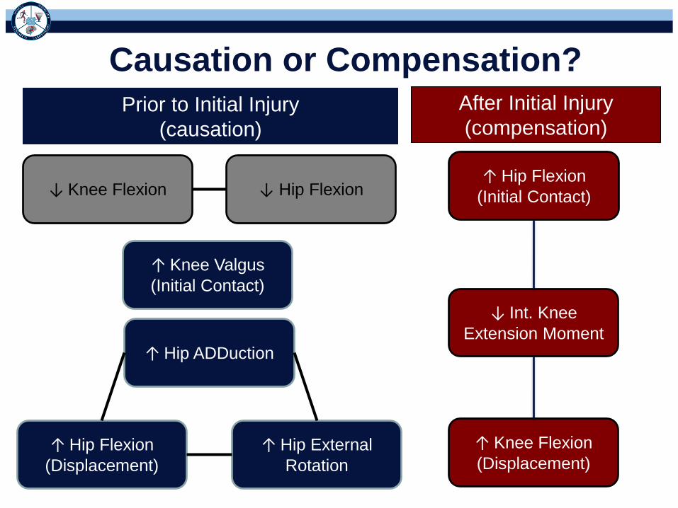
Causation or Compensation?
↓ Knee Flexion
↑ Knee Valgus
(Initial Contact)
↑ Hip External
Rotation
↑ Hip ADDuction
↑ Hip Flexion
(Displacement)
↓ Hip Flexion ↑ Hip Flexion
(Initial Contact)
↑ Knee Flexion
(Displacement)
↓ Int. Knee
Extension Moment
Prior to Initial Injury
(causation)
After Initial Injury
(compensation)
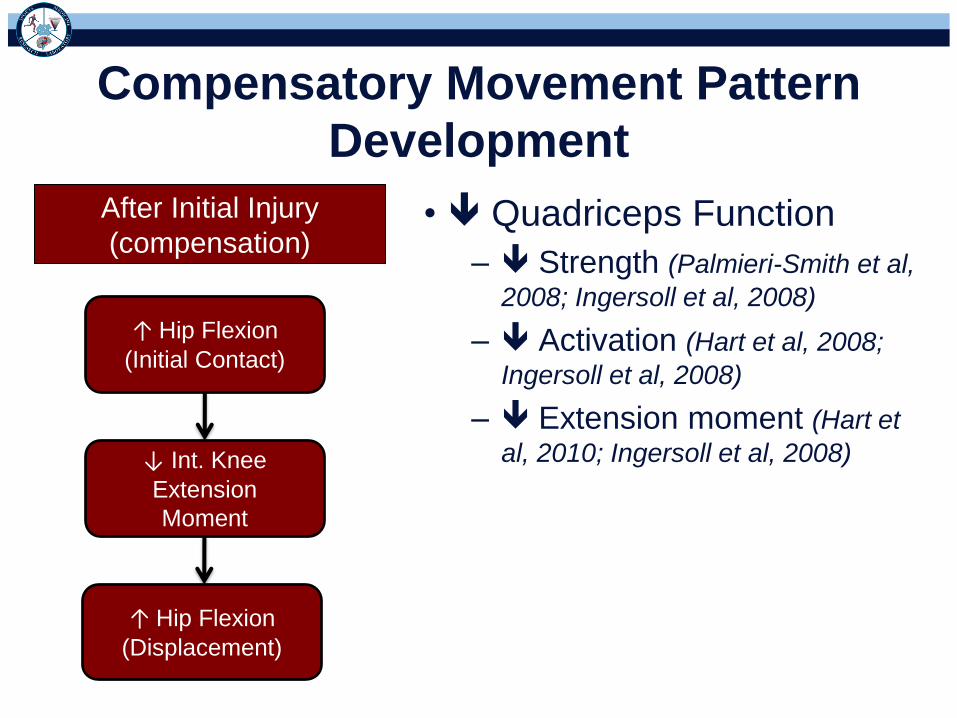
Compensatory Movement Pattern
Development
• Quadriceps Function
– Strength (Palmieri-Smith et al,
2008; Ingersoll et al, 2008)
– Activation (Hart et al, 2008;
Ingersoll et al, 2008)
– Extension moment (Hart et
al, 2010; Ingersoll et al, 2008)
↑ Hip Flexion
(Displacement)
↓ Int. Knee
Extension
Moment
↑ Hip Flexion
(Initial Contact)
After Initial Injury
(compensation)

Normal Quadriceps
Function
Quadriceps Dysfunction
Compensation
↑ Knee Flexion
(Displacement)
↓ Int. Knee
Extension
Moment
↑ Hip Flexion
(Initial Contact)

Quadriceps Dysfunction
• Exacerbate faulty movement patterns
associated with initial injury risk
– Hip Flexion (Anterior Pelvic Tilt)
↑ Knee Valgus
(Initial Contact)
↑ Hip External
Rotation ↑ Hip ADDuction
↑ Hip Flexion
(Displacement)
↑ Int. Knee Varus
Moment
(Ext. Valgus)

↑ Pelvo-Femoral Flexion
(Anterior Pelvic Tilt)
Superior Migration of
Posterior Pelvis
↑ Length of GMAX
& Hamstrings
↓ Force Production
↑ Reliance on
synergistic hip
extensor muscles
to decelerate hip
flexion
Alters
mechanical
function of hip
musculature
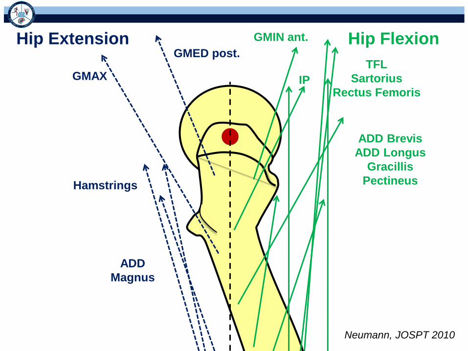
Hip Flexion Hip Extension GMED post.
GMAX
Hamstrings
ADD
Magnus
GMIN ant.
IP
TFL
Sartorius
Rectus Femoris
ADD Brevis
ADD Longus
Gracillis
Pectineus
Neumann, JOSPT 2010

↑ Pelvo-Femoral Flexion
Reversal of Lever Arm (direction of pull)
• Hip ADDuctors (except adductor magnus, already
hip extensor)
– ↑ Hip Flexion → Extension lever arm
Neumann, JOSPT 2010

Hip Flexion Hip Extension GMED post.
GMAX
Hamstrings
ADD
Magnus
GMIN ant.
IP
TFL
Sartorius
Rectus Femoris
ADD Brevis
ADD Longus
Gracillis
Pectineus
Neumann, JOSPT 2010
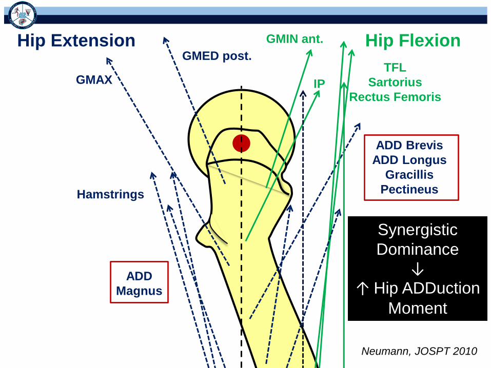
Hip Flexion Hip Extension GMED post.
GMAX
Hamstrings
ADD
Magnus
GMIN ant.
IP
TFL
Sartorius
Rectus Femoris
ADD Brevis
ADD Longus
Gracillis
Pectineus
Synergistic
Dominance ↓
↑ Hip ADDuction
Moment
Neumann, JOSPT 2010

↑ Pelvo-Femoral Flexion
Reversal of Lever Arm (direction of pull)
• Hip External Rotators (piriformis, gluteus medius
– post, gluteus maximus – ant)
– ↑ Hip Flexion → Internal Rotation lever arm
Increase of Lever Arm (M = F * d)
• Dramatically increases the lever arm of hip
internal rotators
– ↑ Hip Flexion → ↑ Internal Rotation lever arm

Hip External Rotation
Hip Internal Rotation
GMIN ant. GMED ant. Pectineus
ADD Longus
ADD Brevis
Obturator ext.
GMED post.
GMIN post.
Quadratus Femoris
Gemellus sup.
Obturatur int.
Gemellus inf.
Piriformis
GMAX
Neumann, JOSPT 2010

Hip External Rotation
Hip Internal Rotation
GMIN ant. GMED ant. Pectineus
ADD Longus
ADD Brevis
Obturator ext.
GMED post.
GMIN post.
Quadratus Femoris
Gemellus sup.
Obturatur int.
Gemellus inf.
Piriformis
GMAX
Neumann, JOSPT 2010

Hip External Rotation
Hip Internal Rotation
GMIN ant. GMED ant. Pectineus
ADD Longus
ADD Brevis
Obturator ext.
GMED post.
GMIN post.
Quadratus Femoris
Gemellus sup.
Obturatur int.
Gemellus inf.
Piriformis
GMAX
↑ Hip Internal
Rotation
Moment

Quadriceps Dysfunction
Post ACL Injury
↑ Hip Flexion (IC) (Ant. Pelvic Tilt)
↑ Length of GMAX & HAMS
Synergistic Dominance
Alters Mechanical Function
Alters Length-Tension → ↓ Force
Hip ADD
Direction Change &
↑ Leverage
Hip ER
Hip IR
↑ Knee Valgus
(Initial Contact)
↑ Hip External
Rotation
↑ Hip
ADDuction
↑ Hip Flexion
(Displacement)
↑ Ext. Knee
Valgus Moment
Compensatory
Movement Patterns
Exacerbate Faulty
Movement Patterns
↓ Int. Knee
Extension Moment ↓
↑ Knee Flexion Displacement

Implications
Prevention
• Prevention of ACL injury / re-injury may be
possible by modifying high risk movements
Rehabilitation
• Movement quality should be part of exercise
progression & return to participation criteria
– Returning to pre-injury status is NOT sufficient
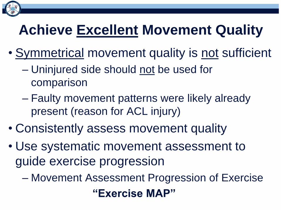
Achieve Excellent Movement Quality
• Symmetrical movement quality is not sufficient
– Uninjured side should not be used for
comparison
– Faulty movement patterns were likely already
present (reason for ACL injury)
• Consistently assess movement quality
• Use systematic movement assessment to
guide exercise progression
– Movement Assessment Progression of Exercise
“Exercise MAP”
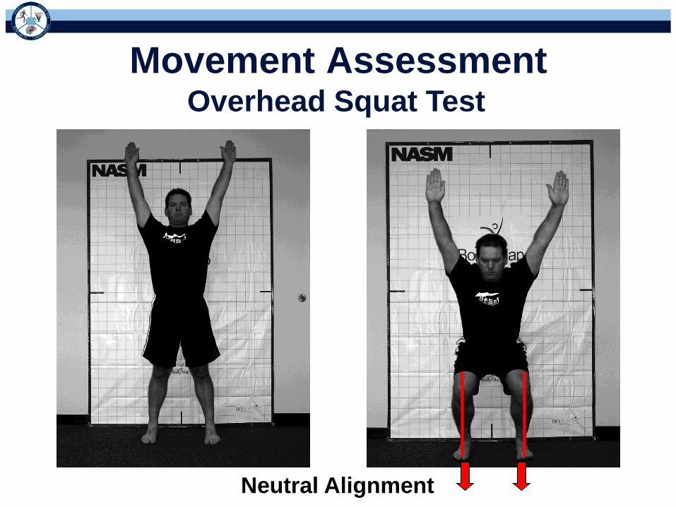
Movement Assessment Overhead Squat Test
Neutral Alignment

Movement Assessment Overhead Squat Test
Neutral Alignment

Movement Impairments Associated
with ACL Injury
Common Movement Impairments:
• Foot external rotation (toe out)
• Foot flat (pronation)
• Limited ankle dorsiflexion (heel lift)
• Knee valgus (knee moves inward)
• Excessive trunk / lumbopelvic flexion
• Asymmetric weight shift
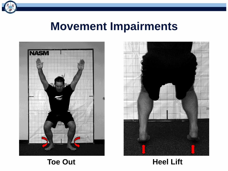
Movement Impairments
Toe Out Heel Lift

Movement Impairments
Foot Flat Knee Valgus
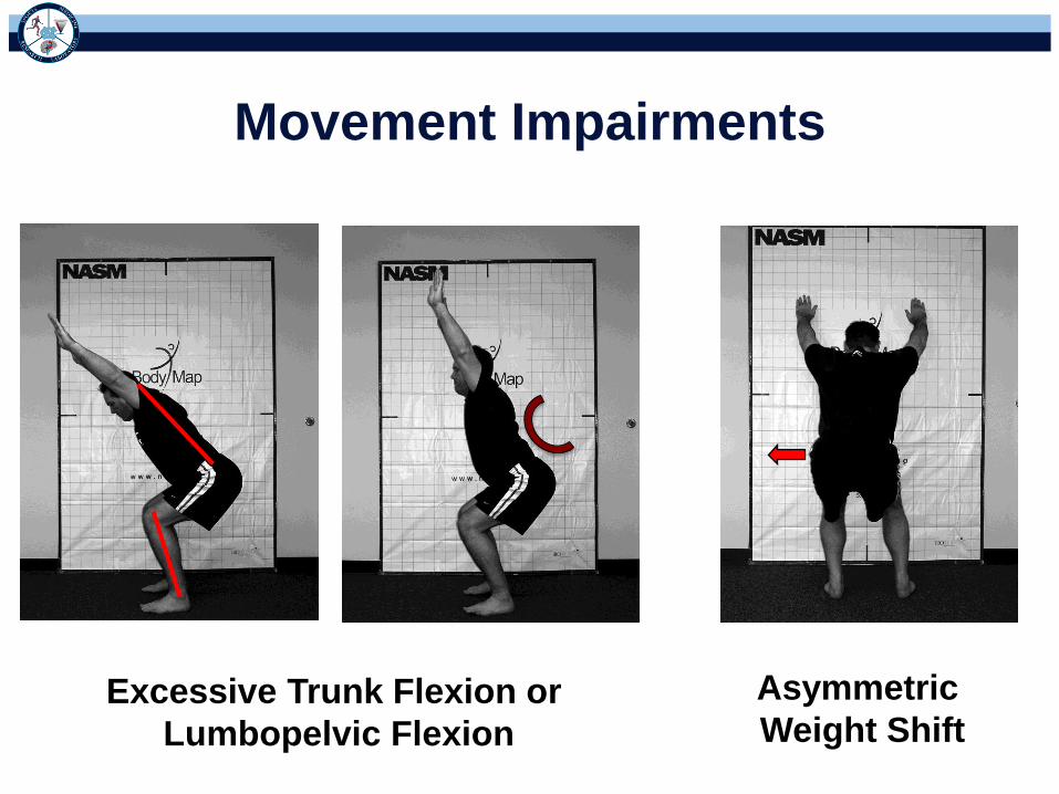
Movement Impairments
Excessive Trunk Flexion or
Lumbopelvic Flexion
Asymmetric
Weight Shift

Landing Error Scoring System (LESS)
• Drop height = 30 cm
• Horizontal distance = 50% body height
• Jump for maximum vertical height after landing
• Focus on initial landing and max knee flexion
• Quantify the number of movement errors
1 2 3 4 5

Knee Flexion Knee Valgus Hip/Trunk Flexion Lat. Trunk Flexion
Narrow Stance Wide Stance Toe In Toe Out

2. Knee Valgus @ Initial Contact: Knees over midfoot
____ Yes (0)
____ No (+1)
12. Knee Flexion Displacement: > 45 degrees
____ Yes (0)
____ No (+1)
13. Knee Valgus Displacement: > great toe
____ Yes (+1)
____ No (0)
4. Trunk Flexion @ Initial Contact: Trunk is flexed
____ Yes (0)
____ No (+1)
5. Lateral Trunk Flexion @ Initial Contact: Trunk is vertical
____ Sternum centered over hips (0)
____ Lateral deviation of sternum over hips (+1)
6. Ankle Plantar Flexion @ Initial Contact: Toe to heel
____ Yes (0)
____ No (+1)
7. Foot Position @ Initial Contact: Toes > 30 of ER
____ Yes (+1)
____ No (0)
9. Stance Width @ Initial Contact:< Shoulder width
____ Yes (+1)
____ No (0)
11. Initial Foot Contact: Symmetric
____ Yes (+0)
____ No (+1)
16. Joint Displacement (Sagittal Plane)
____ Soft (0)
____ Average (+1)
____ Stiff (+2)
17. Overall Impression
____ Excellent (0)
____ Average (+1)
____ Poor (+2)
8. Foot Position @ Initial Contact: Toes > 30 of IR
____ Yes (+1)
____ No (0)
15. Trunk Flexion Displacement: Trunk flexes more
than at initial contact
____ Yes (0)
____ No (1)
3. Hip Flexion @ Initial Contact: Hips are flexed
____ Yes (0)
____ No (+1)
14. Hip Flexion Displacement: Hips flex more than at
initial contact
____ Yes (0)
____ No (1)
1. Knee Flexion @ Initial Contact: > 30 degrees
____ Yes (0)
____ No (+1)
10. Stance Width @ Initial Contact:> Shoulder width
____ Yes (+1)
____ No (0)

Pre-season LESS scores are higher
in those who go on to tear their ACL
0
1
2
3
4
5
6
7
8
ACL Injured Non Injured
LE
SS
Sco
re
F = 5.19, P = 0.002
*
5.4 ± 2.1 4.0 ± 1.7
• 761 youth soccer athletes
• 6 suffered ACL injury Padua et al, J Ath Train, 2010
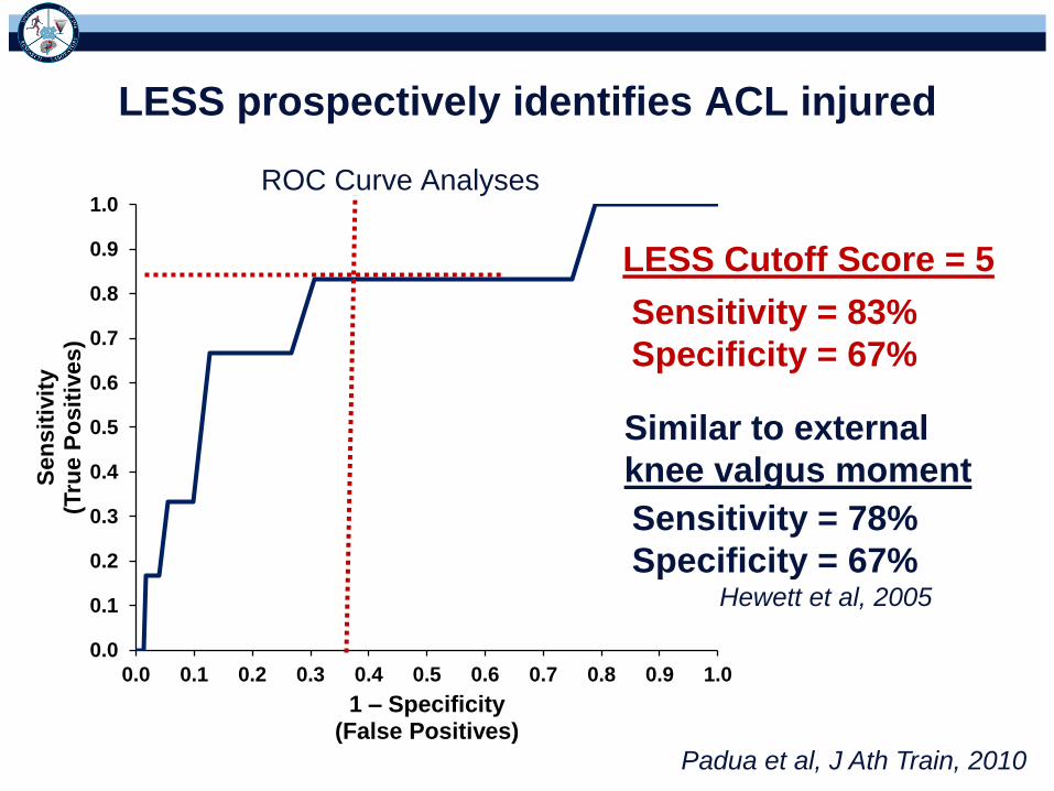
ROC Curve Analyses
0.0
0.1
0.2
0.3
0.4
0.5
0.6
0.7
0.8
0.9
1.0
0.0 0.1 0.2 0.3 0.4 0.5 0.6 0.7 0.8 0.9 1.0
Se
ns
itiv
ity
(Tru
e P
osit
ives)
1 – Specificity (False Positives)
LESS Cutoff Score = 5
Sensitivity = 83%
Specificity = 67%
LESS prospectively identifies ACL injured
Similar to external
knee valgus moment
Sensitivity = 78%
Specificity = 67% Hewett et al, 2005
Padua et al, J Ath Train, 2010

Muscle Imbalance → Movement Impairments
Muscle Balance Muscle Imbalance
Equivalent force / tension → Equilibrium
Unequal force / tension →
Mal-alignment
Tight or
Overactive
Weak or
Inhibited

Example Strategy for Knee Valgus
Movement Impairment
Corrective Exercise Strategy (CES)
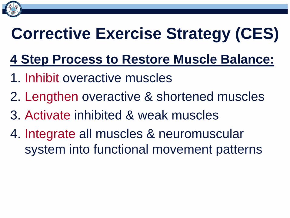
4 Step Process to Restore Muscle Balance:
1. Inhibit overactive muscles
2. Lengthen overactive & shortened muscles
3. Activate inhibited & weak muscles
4. Integrate all muscles & neuromuscular
system into functional movement patterns

Gastrocnemius / Soleus
SMR
Lateral Hamstring
SMR
Peroneals SMR
Hip ADDuctor
SMR
1) Inhibit Overactive / Tight Muscles
Application Guidelines:
• Apply to overactive / tight muscles
• 30 second hold per tender area
• discomfort noted
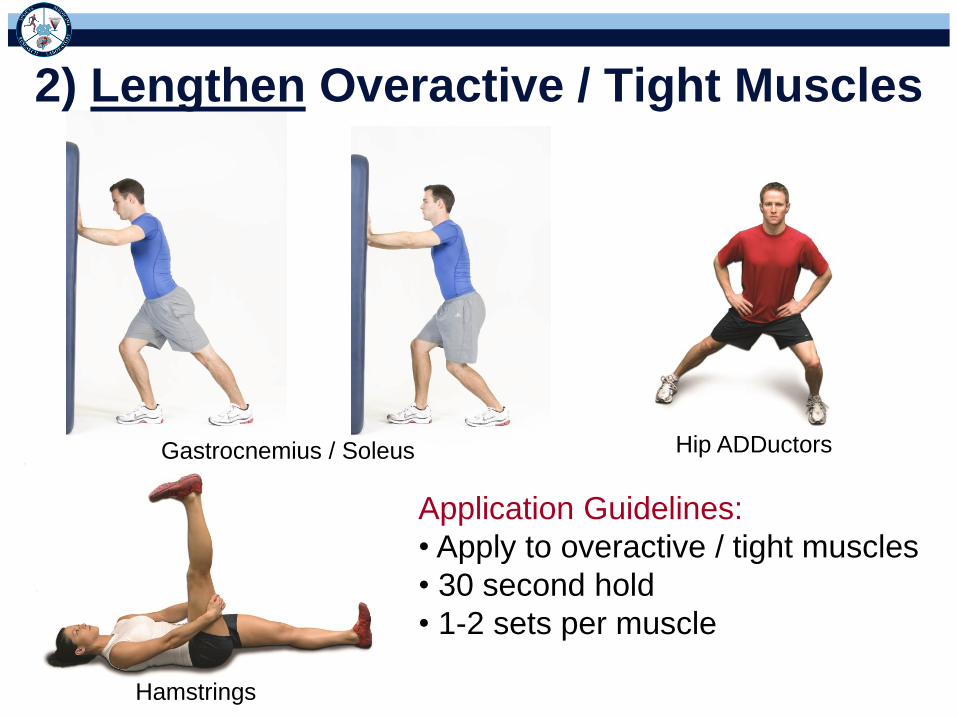
Gastrocnemius / Soleus Hip ADDuctors
2) Lengthen Overactive / Tight Muscles
Hamstrings
Application Guidelines:
• Apply to overactive / tight muscles
• 30 second hold
• 1-2 sets per muscle

Gluteus Medius
Hip Bridge
Gluteus Maximus
3) Activate Weak / Inhibited Muscles
Side Lying Leg Lift
Application Guidelines:
• Apply to weak / inhibited muscles
• 10-15 repetitions
• 2 sec isometric
• 4 sec eccentric
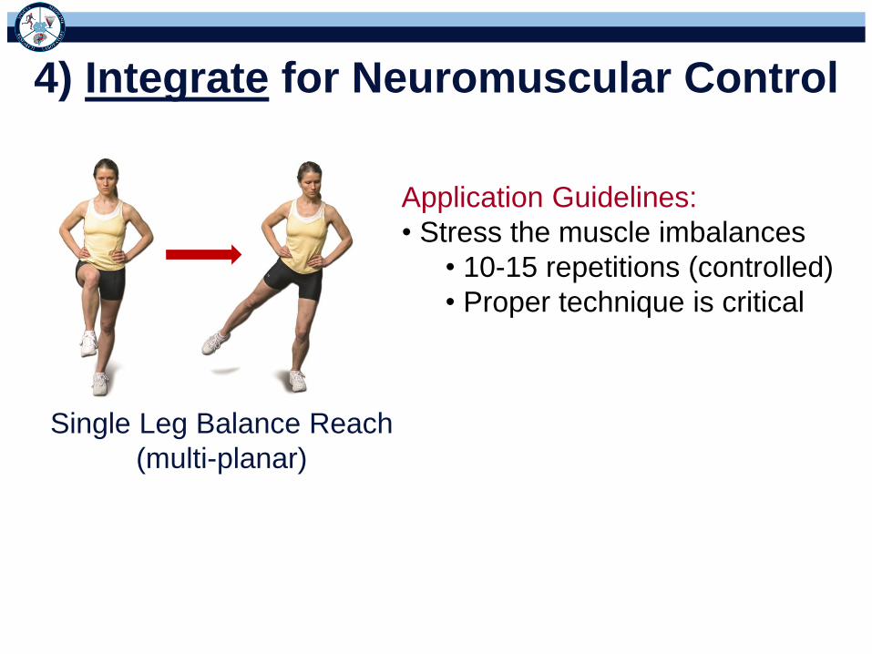
Single Leg Balance Reach
(multi-planar)
4) Integrate for Neuromuscular Control
Application Guidelines:
• Stress the muscle imbalances
• 10-15 repetitions (controlled)
• Proper technique is critical

Sagittal Frontal
Multiplanar Hops to Balance
Transverse
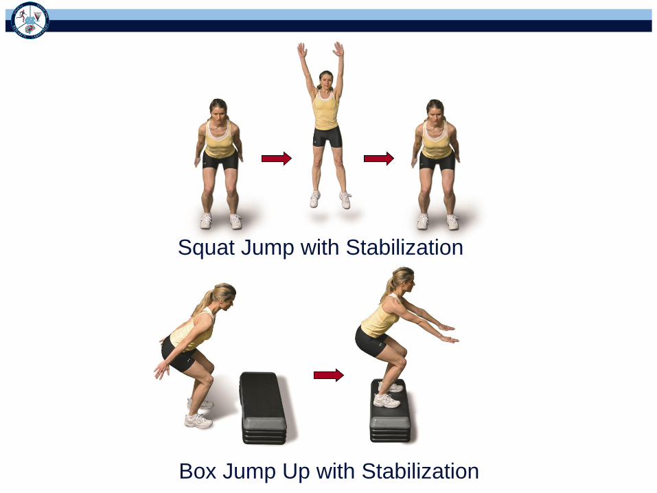
Squat Jump with Stabilization
Box Jump Up with Stabilization
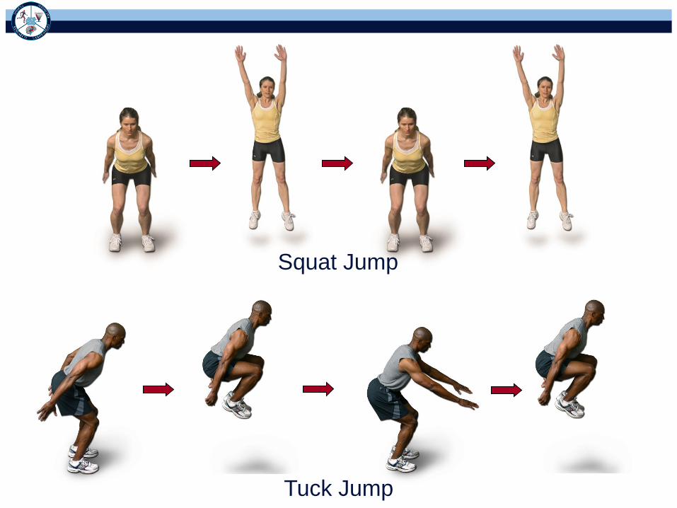
Tuck Jump
Squat Jump
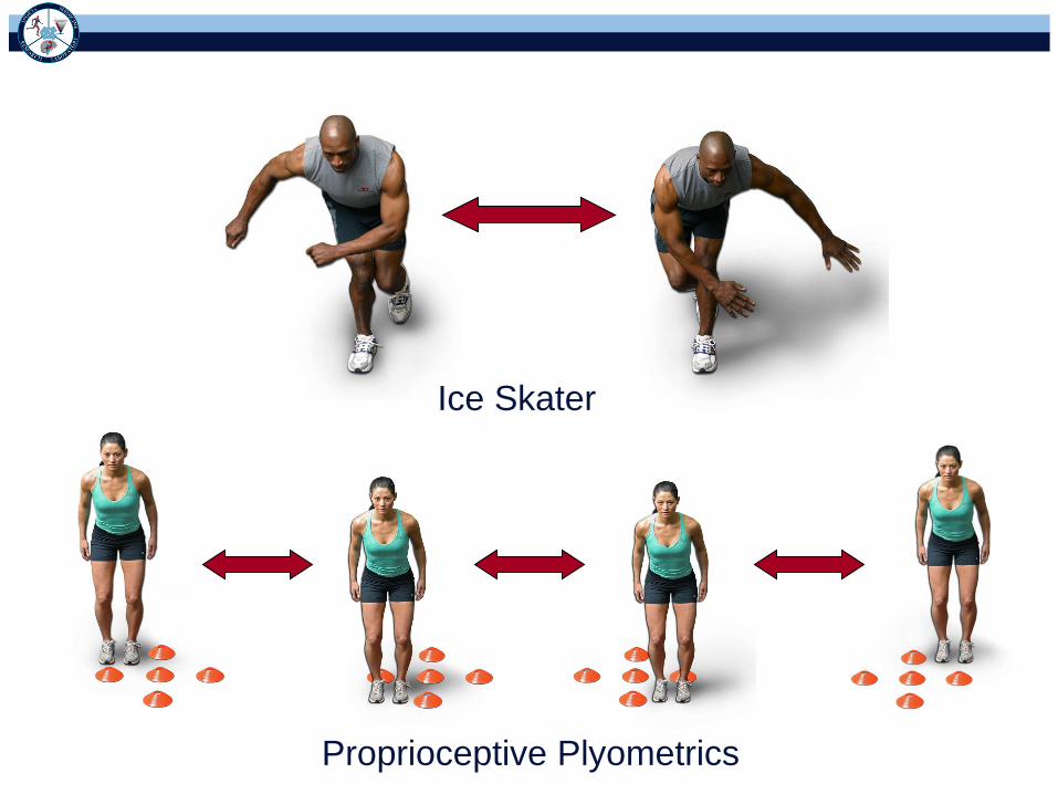
Ice Skater
Proprioceptive Plyometrics

Things to Emphasize
Land “Softly” Bend Knees & Hips
Keep Knees Over Toes &
Toes Straight Ahead

Systematic Progression: Exercise MAP
Phase 1
(0-6 Weeks)
Phase 2
(6-14 Weeks)
Phase 3
(14-20 Weeks) Phase 4
(14-20 Weeks)
Symmetric
AROM
Quad Control
(SLR 3x20)
Single Leg
Balance (firm
surface)
Double Leg
Squat
Symmetric
AROM
Single Leg
Squat (30°)
Single Leg
Balance (foam
surface)
80% Strength
of Uninjured
Side
Double Leg
Jump-Landing
(sagittal)
Single Leg
Squat (60°)
Hop to
Stabilization
(tri-planar)
100% Strength
of Uninjured
Side
Double Leg
Jump-Landing
(tri-planar)
Lunges
(triplanar)
Single Leg
Landing
(sagittal)
Symmetrical
Running

Systematic Progression: Exercise MAP
Single Leg
Landing
(triplanar)
Repeated
Squat Jumps
Double Leg
Jump Landing
to
Sprint
(triplanar)
Ice Skaters
Double Leg
Jump Landing
to
Anticipated Cut
(triplanar)
Single Leg
Landing
to
Sprint
(triplanar)
Double Leg
Jump Landing
to
Unanticipated
Cut (triplanar)
Single Leg
Landing
to
Anticipated Cut
(triplanar)
Phase 5
(20-24 Weeks)
Phase 6
(20-24 Weeks)
Phase 7
(20-24 Weeks) Phase 8
(20-24 Weeks)

Return to Play Considerations
• Able to perform Phase 7 & 8 tasks:
– Under fatigued conditions without movement
compensations
– Under dual task conditions without movement
compensations
• Catching
• Throwing

Quality Movement Matters
• Faulty movement patterns Functional
outcomes & performance (Trulsson et al, 2010)
– Battery of 9 movement tasks:
1) Single leg bridge 6) Stand from half-kneeling
2) Weight shift 7) Forward lunge
3) Single leg squat 8) Backward walking (t-mill)
4) Single leg heel raise 9) Double leg squat
5) Single leg balance on
unstable surface

Quality Movement Matters
• Risk of second ACL injury (Paterno et al, 2010)
– Hip IR moment (uninvolved leg) (8x)
– Frontal plane knee motion (involved leg) (3.5x)
– Asymmetrical knee extension moment (3x)
– Single leg postural stability (involved leg) (2x)
• 3D biomechanical analyses is best
determinant of readiness for return to play
– Sensitivity = 92%
– Specificity = 88%

Problem Solved?
• Barriers exist for widespread injury
prevention efforts
– Compliance
– Supervision
– Retention
– Pediatric population

Compliance Rates Influence
Effectiveness
• Myklebust et al, 2003
– Unchanged ACL injury rate (28% compliant)
– Sub-group analysis of highly compliant players (elite
division) demonstrated ↓ ACL injury risk
• Steffen et al, 2008
– Unchanged ACL and lower extremity injury rates (44%
compliant)
• Soligard et al, 2009
– Divided intervention subjects into tertiles
– 35% lower injury risk in most compliant group

Improving Biomechanics Requires
Supervision & Instruction
Study Direct
Supervision Technique Instruction Outcome % Change Effect Size
Hewett, 1996 Yes Yes ↓ VGRF -18.0% 0.87
Prappavessis, 2003 Yes Yes ↓ VGRF -33.3% 0.79
Irmischer, 2004 Yes Yes ↓ VGRF -26.4% 1.4
Herman, 2008 Yes No No change -3.1% 0.07
Chappell, 2008 No No No change 1.6% 0.07
Lephart, 2005 No No No change -4.2% 0.14
Padua, D.A. & DiStefano. Sagittal Plane Knee Biomechanics & Vertical
Ground Reaction Forces as Modified Following ACL Injury Prevention
Programs: A Systematic Review. Sports Health 1(2):165-173, 2009
0
1
2
3
4
5
6
7
8
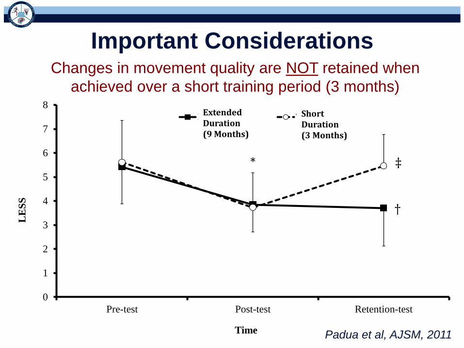
Pre-test Post-test Retention-test
LE
SS
Time
Extended TraditionalShort Duration (3 Months)
Extended Duration (9 Months)
*
†
‡
Important Considerations
Padua et al, AJSM, 2011
Changes in movement quality are NOT retained when
achieved over a short training period (3 months)

Children Require Different Training
• Biomechanics not improved in pediatric populations
• No improvement in:
– Medial knee
displacement
– Vertical jump height
95% CI (-1.0, -0.4) (-1.7, -.98)
∆ Score
0
1
2
3
4
5
6
7
8
Pre-High School High School
LE
SS
To
tal S
co
re
Pre-Training
Post-Training
*
DiStefano, Padua, DiStefano, &
Marshall, Am J Sports Med, 2009
Grandstand et al., J Str Cond Res,
2006
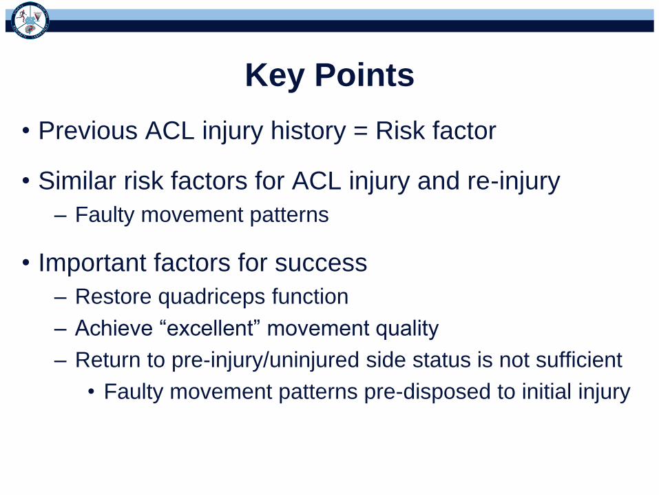
Key Points
• Previous ACL injury history = Risk factor
• Similar risk factors for ACL injury and re-injury
– Faulty movement patterns
• Important factors for success
– Restore quadriceps function
– Achieve “excellent” movement quality
– Return to pre-injury/uninjured side status is not sufficient
• Faulty movement patterns pre-disposed to initial injury

Acknowledgements
Grant #R01-AR050461001

Thank You
Related Documents