RESEARCH ARTICLE Identification of Reprogrammed Myeloid Cell Transcriptomes in NSCLC Anna Durrans 1,2,3☯ , Dingcheng Gao 1,2,3☯ , Ravi Gupta 4 , Kari R. Fischer 1,2,3,5 , Hyejin Choi 1,2,3,5 , Tina El Rayes 1,2,3,5 , Seongho Ryu 1,2,3 , Abu Nasar 1 , Cathy F. Spinelli 1 , Weston Andrews 1 , Olivier Elemento 7 , Daniel Nolan 6 , Brendon Stiles 1,3 , Shahin Rafii 6 , Navneet Narula 8 , Ramana Davuluri 4 , Nasser K. Altorki 1,3 *, Vivek Mittal 1,2,3 * 1 Department of Cardiothoracic Surgery, Weill Cornell Medical College of Cornell University, 1300 York Avenue, New York, New York 10065, United States of America, 2 Department of Cell and Developmental Biology, Weill Cornell Medical College of Cornell University, 1300 York Avenue, New York, New York 10065, United States of America, 3 Neuberger Berman Lung Cancer Center, Weill Cornell Medical College of Cornell University, 1300 York Avenue, New York, New York 10065, United States of America, 4 Molecular and Cellular Oncogenesis Program, The Wistar Institute, 3601 Spruce St, Philadelphia, PA 19104, United States of America, 5 Weill Cornell Graduate School of Medical Sciences, Weill Cornell Medical College of Cornell University, 1300 York Avenue, New York, New York 10065, United States of America, 6 HHMI, Department of Genetic Medicine, Weill Cornell Medical College of Cornell University, 1300 York Avenue, New York, New York 10065, United States of America, 7 Institute for Computational Biomedicine, Department of Physiology and Biophysics, Weill Cornell Medical College of Cornell University, 1300 York Avenue, New York, New York 10065, United States of America, 8 Department of Pathology, Weill Cornell Medical College of Cornell University, 1300 York Avenue, New York, New York 10065, United States of America ☯ These authors contributed equally to this work. * [email protected] (NKA); [email protected] (VM) Abstract Lung cancer is the leading cause of cancer related mortality worldwide, with non-small cell lung cancer (NSCLC) as the most prevalent form. Despite advances in treatment options in- cluding minimally invasive surgery, CT-guided radiation, novel chemotherapeutic regimens, and targeted therapeutics, prognosis remains dismal. Therefore, further molecular analysis of NSCLC is necessary to identify novel molecular targets that impact prognosis and the de- sign of new-targeted therapies. In recent years, tumor “activated/reprogrammed” stromal cells that promote carcinogenesis have emerged as potential therapeutic targets. However, the contribution of stromal cells to NSCLC is poorly understood. Here, we show increased numbers of bone marrow (BM)-derived hematopoietic cells in the tumor parenchyma of NSCLC patients compared with matched adjacent non-neoplastic lung tissue. By sorting specific cellular fractions from lung cancer patients, we compared the transcriptomes of intratumoral myeloid compartments within the tumor bed with their counterparts within adja- cent non-neoplastic tissue from NSCLC patients. The RNA sequencing of specific myeloid compartments (immature monocytic myeloid cells and polymorphonuclear neutrophils) identified differentially regulated genes and mRNA isoforms, which were inconspicuous in whole tumor analysis. Genes encoding secreted factors, including osteopontin (OPN), che- mokine (C-C motif) ligand 7 (CCL7) and thrombospondin 1 (TSP1) were identified, which enhanced tumorigenic properties of lung cancer cells indicative of their potential as targets for therapy. This study demonstrates that analysis of homogeneous stromal populations PLOS ONE | DOI:10.1371/journal.pone.0129123 June 5, 2015 1 / 22 OPEN ACCESS Citation: Durrans A, Gao D, Gupta R, Fischer KR, Choi H, El Rayes T, et al. (2015) Identification of Reprogrammed Myeloid Cell Transcriptomes in NSCLC. PLoS ONE 10(6): e0129123. doi:10.1371/ journal.pone.0129123 Academic Editor: Aamir Ahmed, King's College London, UNITED KINGDOM Received: December 8, 2014 Accepted: May 5, 2015 Published: June 5, 2015 Copyright: © 2015 Durrans et al. This is an open access article distributed under the terms of the Creative Commons Attribution License, which permits unrestricted use, distribution, and reproduction in any medium, provided the original author and source are credited. Data Availability Statement: All relevant data are within the paper and its Supporting Information files. RNA data have been deposited in the Gene Expression Omnibus (GEO) from NCBI with accession number GSE68795 (http://www.ncbi.nlm. nih.gov/geo/query/acc.cgi?token = odqnoygijnoxluz&acc=GSE68795). Funding: This work was supported by funds from The Neuberger Berman Foundation Lung Cancer Center and Weill Cornell Cancer Center Pilot grant. Competing Interests: The authors have declared that no competing interests exist.

Welcome message from author
This document is posted to help you gain knowledge. Please leave a comment to let me know what you think about it! Share it to your friends and learn new things together.
Transcript
RESEARCH ARTICLE
Identification of Reprogrammed Myeloid CellTranscriptomes in NSCLCAnna Durrans1,2,3☯, Dingcheng Gao1,2,3☯, Ravi Gupta4, Kari R. Fischer1,2,3,5,Hyejin Choi1,2,3,5, Tina El Rayes1,2,3,5, Seongho Ryu1,2,3, Abu Nasar1, Cathy F. Spinelli1,Weston Andrews1, Olivier Elemento7, Daniel Nolan6, Brendon Stiles1,3, Shahin Rafii6,Navneet Narula8, Ramana Davuluri4, Nasser K. Altorki1,3*, Vivek Mittal1,2,3*
1 Department of Cardiothoracic Surgery, Weill Cornell Medical College of Cornell University, 1300 YorkAvenue, New York, New York 10065, United States of America, 2 Department of Cell and DevelopmentalBiology, Weill Cornell Medical College of Cornell University, 1300 York Avenue, New York, New York 10065,United States of America, 3 Neuberger Berman Lung Cancer Center, Weill Cornell Medical College ofCornell University, 1300 York Avenue, New York, New York 10065, United States of America, 4 Molecularand Cellular Oncogenesis Program, TheWistar Institute, 3601 Spruce St, Philadelphia, PA 19104, UnitedStates of America, 5 Weill Cornell Graduate School of Medical Sciences, Weill Cornell Medical College ofCornell University, 1300 York Avenue, New York, New York 10065, United States of America, 6 HHMI,Department of Genetic Medicine, Weill Cornell Medical College of Cornell University, 1300 York Avenue,New York, New York 10065, United States of America, 7 Institute for Computational Biomedicine,Department of Physiology and Biophysics, Weill Cornell Medical College of Cornell University, 1300 YorkAvenue, New York, New York 10065, United States of America, 8 Department of Pathology, Weill CornellMedical College of Cornell University, 1300 York Avenue, New York, New York 10065, United States ofAmerica
☯ These authors contributed equally to this work.* [email protected] (NKA); [email protected] (VM)
AbstractLung cancer is the leading cause of cancer related mortality worldwide, with non-small cell
lung cancer (NSCLC) as the most prevalent form. Despite advances in treatment options in-
cluding minimally invasive surgery, CT-guided radiation, novel chemotherapeutic regimens,
and targeted therapeutics, prognosis remains dismal. Therefore, further molecular analysis
of NSCLC is necessary to identify novel molecular targets that impact prognosis and the de-
sign of new-targeted therapies. In recent years, tumor “activated/reprogrammed” stromal
cells that promote carcinogenesis have emerged as potential therapeutic targets. However,
the contribution of stromal cells to NSCLC is poorly understood. Here, we show increased
numbers of bone marrow (BM)-derived hematopoietic cells in the tumor parenchyma of
NSCLC patients compared with matched adjacent non-neoplastic lung tissue. By sorting
specific cellular fractions from lung cancer patients, we compared the transcriptomes of
intratumoral myeloid compartments within the tumor bed with their counterparts within adja-
cent non-neoplastic tissue from NSCLC patients. The RNA sequencing of specific myeloid
compartments (immature monocytic myeloid cells and polymorphonuclear neutrophils)
identified differentially regulated genes and mRNA isoforms, which were inconspicuous in
whole tumor analysis. Genes encoding secreted factors, including osteopontin (OPN), che-
mokine (C-C motif) ligand 7 (CCL7) and thrombospondin 1 (TSP1) were identified, which
enhanced tumorigenic properties of lung cancer cells indicative of their potential as targets
for therapy. This study demonstrates that analysis of homogeneous stromal populations
PLOSONE | DOI:10.1371/journal.pone.0129123 June 5, 2015 1 / 22
OPEN ACCESS
Citation: Durrans A, Gao D, Gupta R, Fischer KR,Choi H, El Rayes T, et al. (2015) Identification ofReprogrammed Myeloid Cell Transcriptomes inNSCLC. PLoS ONE 10(6): e0129123. doi:10.1371/journal.pone.0129123
Academic Editor: Aamir Ahmed, King's CollegeLondon, UNITED KINGDOM
Received: December 8, 2014
Accepted: May 5, 2015
Published: June 5, 2015
Copyright: © 2015 Durrans et al. This is an openaccess article distributed under the terms of theCreative Commons Attribution License, which permitsunrestricted use, distribution, and reproduction in anymedium, provided the original author and source arecredited.
Data Availability Statement: All relevant data arewithin the paper and its Supporting Information files.RNA data have been deposited in the GeneExpression Omnibus (GEO) from NCBI withaccession number GSE68795 (http://www.ncbi.nlm.nih.gov/geo/query/acc.cgi?token =odqnoygijnoxluz&acc=GSE68795).
Funding: This work was supported by funds fromThe Neuberger Berman Foundation Lung CancerCenter and Weill Cornell Cancer Center Pilot grant.
Competing Interests: The authors have declaredthat no competing interests exist.
isolated directly from fresh clinical specimens can detect important stromal genes of
therapeutic value.
IntroductionLung cancer is the leading cause of cancer related mortality worldwide, with an estimated 1.3million new cases each year [1, 2]. Despite improvements in diagnostics and treatment options[3, 4], 5-year survival rate for lung cancer patients only increased from 7 to 14% within the last30 years. Thus, further molecular analysis of NSCLC is necessary to identify novel moleculartargets that impact prognosis and the design of new, targeted therapies.
A major research focus in NSCLC has been directed to cancer cell intrinsic properties [5],which has led to the discovery of important driver mutations, and the development of targetedtherapies such as the receptor tyrosine kinase (RTK) inhibitors gefitinib/erlotinib (EGFR inhib-itors) and crizotinib (EML4-ALK inhibitor) [3]. However, these treatments benefit only thesmall proportion (5–20%) of patients harboring these driver mutations, and acquired resis-tance to these therapies presents a major impediment to the effective treatment of NSCLC pa-tients with these mutations [6–8].
Emerging studies from solid tumors including breast and prostate are beginning to recog-nize that carcinogenesis results from concerted interactions between genetically altered tumorepithelial cells and intratumoral stromal cells, resulting in an “activated/reprogrammed” stro-ma [9]. Consistent with this notion, analysis of enriched stromal compartments derived fromhuman breast cancer revealed gene expression changes associated with cancer progression[10]. Notably, BM-derived hematopoietic cells contribute significantly to the tumor stroma,and are “educated/reprogrammed” by the paracrine activity of tumor epithelial cells to acquirean “activated” protumorigenic phenotype [11]. Examples of tumor-activated stromal cells in-clude macrophages (activated M2 phenotype) [12], neutrophils (N1 to N2 conversion) [13],lymphocytes [14], fibroblasts (cancer activated fibroblasts, CAFs) [15], and endothelial cells[16]. Studies from mouse models have shown that reprogrammed stromal cells promote tumorgrowth by regulating key cancer hallmarks such as angiogenesis, proliferation, migration, andinvasion [11, 17, 18] leading to the inclusion of the tumor microenvironment as an emerginghallmark of cancer [19]. Furthermore, recent studies have demonstrated that stromal cells me-diate innate resistance to therapies in many cancers [20–22]. Importantly, administration ofchemotherapy in combination with a macrophage antagonist (CSFR1 blockade) conferred syn-ergy in breast cancer treatment [23]. These studies, together with the clinical success of theantiangiogenic agent bevacizumab, a humanized monoclonal anti-VEGF antibody, providecompelling rationale for targeting the tumor microenvironment. Intratumoral stromal cellshave thus emerged as attractive targets for anti-cancer therapy [11, 24].
Little is known, however, about the contribution and pathophysiological role of stromalcells in NSCLC. A few clinical studies have shown that activated stromal elements may deter-mine patient prognosis and may play a role in mediating resistance to targeted therapies. Forexample, in patients with stage I NSCLC the presence of CAFs is a poor prognostic indicatortypically associated with nodal metastases and a higher risk of recurrence [25]. Interestingly, aspecific, eleven-gene expression signature in CAFs stratified NSCLC patients into low andhigh-risk groups, and was associated with recurrence free survival [26]. In another study, thetumor-stroma cross talk was implicated in mediating resistance to EGFR-TKIs [27].
Reprogrammed Myeloid Cell Transcriptomes in NSCLC
PLOSONE | DOI:10.1371/journal.pone.0129123 June 5, 2015 2 / 22
In NSCLC, genetic features of tumor epithelial cells continue to be used for both prognosisand the development of targeted therapies; however, another important question is how to ex-ploit the reprogrammed intratumoral stromal compartments, which play pivotal roles in carci-nogenesis. New approaches are required to identify and isolate individual cellular stromalcomponents from the heterogeneous tumor, so that their protumorigenic characteristics can beelucidated and targeted for prognostic and therapeutic purposes. Recognizing that the contri-bution of BM-derived cells to NSCLC progression has not been widely studied, we set out tostudy specific individual populations of BM-derived myeloid cells within the tumor and adja-cent non-neoplastic tissue from fresh clinical material obtained from NSCLC patients, and as-sessed their contribution and function in NSCLC.
Methods
Human samplesHuman tumor and adjacent non-neoplastic lung samples were obtained from the Cardiotho-racic Surgery Department, Weill Cornell Medical College (New York). Specimens were collect-ed after obtaining written informed consent prior to undergoing any study-specific proceduresin accordance with the Declaration of Helsinki. Patient’s identity of pathological specimens re-mained anonymous in the context of this study. Patient sample collection was approved by theInstitutional Review Board of Weill Cornell Medical College, Thoracic Surgery Biobank Proto-col Number 1008011221.
Mice and cell linesAll animal work was conducted in accordance with a protocol approved by the InstitutionalAnimal Care and Use Committee at Weill Cornell Medical College. Wild type C57BL/6J andp53f/f (B6.129P2-Trp53tm1Brn/J) mice were obtained from The Jackson Laboratory (Bar Harbor,Maine), and K-ras (B6.129S4-Krastm4Tyj/J (LSL Kras-G12D)) mice from The National CancerInstitute (Bethesda, Maryland). The p53f/f and K-ras mice were then bred together for 3 gener-ations to get Kras-Het; p53f/f mice.
The HKP-1M cell line was established by culturing cells derived from lung adenocarcino-mas from mice which constitutively express oncogenic K-ras and have loss of function of p53.Cells were cultured in DMEM with 10% fetal bovine serum. The human non-small cell lungcancer cell line (H1650) was obtained from ATCC and cultured in RPMI with 10% fetalbovine serum.
Immunofluorescence and microscopyFor immunofluorescence staining, tissues were fixed in 3.7% formalin overnight, cryoprotectedin 30% sucrose/PBS overnight, and then cryoembedded in a 1:1 solution of 30% sucrose/PBS:Tissue-Tek O.C.T. embedding compound (Electron Microscopy Sciences). To obtain lung tis-sue from animals, perfusion was performed by injecting 5 ml of cold PBS through the rightventricle of the heart before embedding.
Sections (12 μm) were stained following a standard protocol with primary antibodiesagainst EpCAM (anti-human clone 9C4, BioLegend), CD45 (anti-human clone HI30, BioLe-gend), CD11b (anti-human clone ICRF44, eBioscience), OPN (anti-human clone EPR3688,Epitomics), CCL7 (anti-human rabbit polyclonal, Abcam), and TSP1 (anti-human clone Ab-11, ThermoScientific).
Unconjugated primary antibodies were directly conjugated to various Alexa Fluor dyesusing antibody-labeling kits (Invitrogen) performed as per manufacturer’s instructions and
Reprogrammed Myeloid Cell Transcriptomes in NSCLC
PLOSONE | DOI:10.1371/journal.pone.0129123 June 5, 2015 3 / 22
purified over BioSpin P30 Gel (Biorad). Fluorescence images were obtained using a computer-ized Zeiss fluorescence microscope (Axiovert 200M), fitted with an apotome and a HRM cam-era. Images were analyzed using Axiovision 4.6 software (Carl Zeiss Inc.).
For H&E staining, lung sections (5 μm) were stained with hematoxylin and eosin dyes fol-lowing standard protocols. Images were obtained with an Olympus BX51 microscope coupledwith Qcapture software (Olympus).
To obtain single cell suspensions, fresh clinical tissues were minced and then digested in acollagenase/dispase/DNase mix (Roche) for 30 min at 37°C. Cells were washed, strainedthrough a 70-μm strainer (BD Bioscience) and resuspended in FACS staining buffer (PBS,2mM EDTA, 1%BSA). Red blood cells were eliminated by incubation in lysis buffer (5 PRIME)for 10 min at RT. Cell suspensions were pre-blocked with 1% FBS and then incubated with thefollowing primary antibodies from BioLegend: CD45 (anti-human clone HI30), EpCAM (anti-human clone 9C4), CD33 (anti-human clone WM-53), CD11b (anti-human clone ICRF44,anti-mouse clone M1/70), CXCR2 (anti-human clone 5E8), Ly6G (anti-mouse clone 1A8),Ly6C (anti-mouse clone HK1.4), CD4 (anti-mouse clone GK1.5), CD8a (anti-mouse clone 53–6.7), and B220 (anti-mouse clone RA3-6B2). SYTOX Blue (Invitrogen) was added in each cellstaining tube to facilitate the elimination of dead cells in flow cytometry analysis. Single colorstainings were freshly set up with according antibodies and CompBeads (BD Bioscience) ineach experiment for proper calibration of compensations.
Labeled cell populations were measured by LSRII flow cytometer coupled with FACS Divasoftware (BD Bioscience). Flow cytometry analysis was performed using a variety of controlsincluding isotype antibodies, FMO samples [28], and unstained samples for determining ap-propriate gates, voltages, and compensations required in multivariate flow cytometry. For sort-ing, targeted cell populations were gated within FACS Diva software and sorted by Aria IIsorter (BD Bioscience).
RNA isolation, quality assessment and RNA-seq library preparationTotal RNA from flow cytometry sorted cells was extracted using the PicoPure RNA extractionkit (Arcturus) following the manufacturer´s protocol. RNA concentration was measured usinga nanodrop (Thermo Fisher Scientific), and RNA integrity determined using the Agilent 2100Bioanalyzer (Agilent Technologies). cDNA libraries were prepared using 15–35 ng RNA start-ing material (RIN values>6.0), using the TruSeq RNA Sample Preparation Kit (Illumina)according to the manufacturer’s instructions. Libraries were quantified using a Qubit fluorom-eter (Invitrogen) and the size and purity checked on an Agilent 2100 Bioanalyzer (AgilentTechnologies). 19 samples from six patients were prepared for sequencing using the HiSeq2000 (illumina) (S1 Table).
RNA-seq data analysisRNA-seq data analysis was performed according to our published analysis pipeline [29, 30].Differential gene expression analysis was performed using the DEseq method [31] which isbased on a negative binomial distribution model. Approximately 80% of the reads aligned toknown and novel mRNAs in each case, confirming high quality mRNA sequencing data.Uniquely mapped reads were used to identify differentially expressed genes (p� 0.05). Impor-tantly, correlation analysis of these genes showed discrete clustering of stromal cells from tu-mors and adjacent lungs with minimal patient-to-patient variability, suggesting that RNA-seqdata from this patient cohort was sufficiently robust to identify differentially regulated genes.Next, genes differentially regulated in the intratumoral stromal subsets and epithelial cells,compared with the same subsets from adjacent lung tissue, were identified using standard
Reprogrammed Myeloid Cell Transcriptomes in NSCLC
PLOSONE | DOI:10.1371/journal.pone.0129123 June 5, 2015 4 / 22
statistical methods with a false discovery ratio (FDR)<5%. Ingenuity pathway analysis of dif-ferentially regulated genes from stromal compartments showed that a major proportion ofgenes were involved in cancer-specific pathways (data not shown).
An in-house algorithm was used to select genes from the RNA-seq gene list according to thefollowing criteria; adjusted p value<0.05, fold change>2.0, potential for paracrine function asdetermined by GO annotation as secreted, extracellular space, or membrane (except mem-branes of organelles including golgi and endoplasmic reticulum). Of these, genes with func-tions in key tumorigenic pathways including angiogenesis, ECM breakdown, cell migration,proliferation, invasion, cytokine function, chemokine function, and chemotactic functionwere selected.
In addition, a systematic gene set enrichment analysis (GSEA) was carried out using thetumor vs. adjacent comparison to rank genes in each population. Specifically we used negativelog10 DEseq p-values multiplied by the sign of the log2 fold-changes to rank genes. We appliedGSEA to the c5 and c7 gene sets fromMSigDB and used FDR = 5% as threshold.
Quantitative RT-PCR analysisRNA (50 ng, RIN values>6.0) from individual flow-sorted cell populations was pooled from3–5 samples and converted to cDNA using the RT2 First Strand Kit (SABiosciences).QRT-PCR was performed with RT2 SYBR Green Master mix and customized 384 well arraysof 86 specific primers, as well as two housekeeping genes (HPRT1, GAPDH), a control forDNA contamination, a reverse transcription control, and a positive PCR control. The positivePCR control (PPC, SABiosciences) tested the efficiency of the polymerase chain reaction usinga pre-dispensed artificial DNA sequence and the primer set that detects it. Any impurities thataffect the positive control PCR amplification also affect amplification of the gene-specific prod-ucts of interest. The custom PCR array was designed with RT2 primers (SA Biosciences) whichhave an average amplification efficiency of 99% with 95% CI from 90–110% and are optimizedwith the reagents used by SA Biosciences. The array included HPRT1 and GAPDH primers asinternal controls. The geometric mean of both genes was used to normalize cDNA input. PCRdata was analyzed by using established and validated online tools from www.Qiagen.com.
Each sample was duplicated to control pipetting error. A PCR protocol of initial denaturingat 95°C for 10 min, 40 cycles of 95°C for 15 sec, 60°C for 1 min, and 72°C for 30 sec, followedby final extension at 72°C for 5 min and melt curve analysis was applied on an ABI 7900HTFast Real-Time PCR System (Applied Biosystems) coupled with SDS 2.0 software. The relativeabundance of each transcript compared with the control was calculated utilizing the delta-Ct method.
Orthotopic model of lung cancer in mice and bioluminescent imagingTo determine the functional significance of myeloid cells in tumor growth we used the K-rasG12D p53flox/flox mouse model of NSCLC [32]. Tumors were excised from these animals andused for deriving cell lines for generating orthotopic lung adenocarcinoma in mice. Murine,myeloid subsets are defined as CD11b+Gr1+ cells, which are comprised of two major subpopu-lations defined as CD11b+Ly6Chigh (IMMCs) and CD11b+Ly6Ghigh (neutrophils) [18, 33, 34].To assess the effects of depleting specific myeloid cell populations on NSCLC progression, weused a Ly6G-specific monoclonal antibody which has previously been used for depleting neu-trophils in mice [35]. We did not deplete Ly6Chigh cells, as robust reagents for specifically de-pleting these cells are unavailable.
8-week old C57BL/6 mice were injected via tail vein with 1.5 X 10⁵ luciferase-labeled HKP-1M cells. At D7 treatment was begun with retro-orbital administration of anti-Ly6G antibody
Reprogrammed Myeloid Cell Transcriptomes in NSCLC
PLOSONE | DOI:10.1371/journal.pone.0129123 June 5, 2015 5 / 22
or IgG control (2.5 mg/kg, purified rat anti-mouse Ly6G, and rat IgG2a κ isotype control, Bec-ton Dickinson), which was repeated every three days until sacrifice.
To determine the lung tumor burden in vivo, mice were anaesthetized and injected retro-orbitally with 75mg/kg of D-luciferin. Lung growth was monitored every 3 days using biolumi-nescence imaging performed with mice in a supine position by using the Xenogen IVIS systemcoupled to Living Image acquisition and analysis software (Xenogen). For BLI plots, photonflux was calculated for each mouse by using the same rectangle region of interest encompassingthe thorax of the mouse.
Micro-CT imaging and tumor volume measurementsMice were anesthetized with a continuous flow of 2–4% isoflurane/oxygen mixture (2.5 L/min), and imaged two at a time using the Siemens Inveon micro PET/CT scanner (SiemensMedical Solutions USA, Inc). The micro-CT image acquisition consisted of 501 projections col-lected in 203 degree of scan arc. The X-ray exposure time was 1 sec per projection without re-spiratory-gating. The X-ray tube settings were 30 kVp and 500 μA. The resulting raw data werereconstructed to a final image matrix of 1024 x 1024 x 480 slices at 98-μm voxel size. The re-constructed voxel values were scaled into Hounsfield units. The reconstructed images wereviewed and analyzed using the Inveon Research Workspace software (Siemens Medical Solu-tions USA, Inc).
The image analysis method was based on defining the total lung space, excluding the heart,using manual segmentation. Vascular structures around the heart were manually included inthe total lung space. The radiodensity of the combined functional lung, tumor, and vasculaturewas recorded as Hounsfield units (HU) where -1000 is air, -700 is lung, and +100 to +300 issoft tissue. Because tumor tissue and vascular tissue have similar grayscale values, they couldnot be separated during segmentation, however non-tumor vascular volume should remain rel-atively constant between the control and treated groups, and therefore this method provides arelative measure of tumor burden. All data are presented as mean ± SEM.
Cell proliferation, migration and invasion assaysHuman lung cancer cells (H1650) (2×10⁵) were cultured in 6-well plate with or without OPN,CCL7, or TSP1 at a concentration of 100ng/mL for 1 day. For the cell proliferation assay, EdU(10nM) was administered to culture medium to label proliferating cells for 30 min. Cells wereharvested, washed once with PBS, and fixed with 4% paraformaldehyde for 15 min at RT.Fixed cells were permeabilized and stained for EdU incorporation and DNA content using theClick-iT EdU Cell Proliferation Assay kit (Invitrogen Inc) according to the standard protocol.Flow analysis of cell phases was performed using an LSRII coupled with Diva software (BDBioscience).
For the cell migration assay, H1650 cells (2×10⁵) were seeded in 6-well plate (Corning Inc)with or without OPN, CCL7, or TSP1 at different concentrations for 24 hours. A cell migrationvideo was generated by obtaining images every 5 min for 3 hours under a computerized Zeissmicroscope (Axio Observer) equipped with a culture chamber. Cell movement was trackedand analyzed using ImageJ and Manual Tracking Plugin software (NIH).
For the cell invasion assay, H1650 (2×10⁴) cells were seeded in 24-well BD BioCoat Matrigelinvasion chambers (BD Bioscience) in serum free medium with or without OPN, CCL7, orTSP1 for 20 hours. The lower chambers contained 5% serum for attracting tumor cell invasionfor 8 hours. The chambers were fixed and stained with hematoxylin to visualize the invadingcells.
Reprogrammed Myeloid Cell Transcriptomes in NSCLC
PLOSONE | DOI:10.1371/journal.pone.0129123 June 5, 2015 6 / 22

Statistical AnalysisResults are expressed as mean ± SEM, except where indicated otherwise. Analyses of differenttreatment groups were performed using the Mann-Whitney t-test using the GraphPad Prismstatistical program. P values< 0.05 were considered significant.
Results
Increased numbers of bone marrow hematopoietic cells infiltrate lungadenocarcinomaTo evaluate the cellular landscape of tumor-activated BM stromal cells in human NSCLC weused stringent criteria for sample selection, preparation, and analysis. For sample selection thecriteria used were samples from patients who had; 1) surgical resection without preoperativetherapy, 2) stage I/II/III disease, 3) adenocarcinoma cell type, and were, 4) never or formersmokers (to exclude confounding smoking-induced acute inflammatory reactions in activesmokers).
We used surgically resected specimens from NSCLC patients, and performed immunohisto-chemical (IHC) staining to identify regions of adenocarcinoma and adjacent non-neoplasticlung (adjacent lung) (Fig 1A). To determine the relative contribution of BM–derived stromalcells, we stained the adenocarcinoma and adjacent lung with antibodies specific for hematopoi-etic cells (CD45+) and epithelial cells (epithelial cell adhesion molecule, EpCAM+) (Fig 1B andS1 Fig). An increased number of CD45+ hematopoietic cells were commonly observed in theadenocarcinoma compared to adjacent lung tissue (approximately 5 cm away from the tumormargins; Fig 1B and S1 Fig). Consistently, flow cytometry analysis showed an increase inCD45+ hematopoietic cells (>4-folds) in the tumor parenchyma compared to adjacent lung(Fig 1C and 1D).
The CD45+ hematopoietic population is heterogeneous and comprised of various subpopu-lations including T lymphocytes, B cells, and cells of the myeloid lineage [36]. Of the hemato-poietic cells, we were interested in cells of the myeloid lineage as these are globally altered incancer and promote tumor growth by stimulating tumor angiogenesis, suppressing tumor im-munity, and promoting metastasis to distant sites [37]. Of the myeloid cells, we focused on im-mature monocytic myeloid cells (IMMC, CD11b+CD33+) and neutrophils (CD11b+CD33-)[38] (Fig 1E). The identities of these myeloid cells were further confirmed by analyzing the ex-pression of other myeloid cell markers and cell nuclear morphology. The CD11b+CD33+ im-mature myeloid cells were devoid of CXCR2, where as the CD33- cells were CXCR2+
neutrophils (Fig 1F, Left panel). Consistent with flow cytometry data, nuclear morphologyanalysis showed that the CD11b+CD33+CXCR2- cells were mononuclear, whereas theCD11b+CD33-CXCR2+ cells were polymorphonuclear (Fig 1F, Right panel). Further evalua-tion showed that the CD33+ IMMCs expressed low levels of macrophage marker CD68 andshowed a typical mononuclear scatter pattern (S2A and S2B Fig). The increased prevalence ofthese specific BM-derived sub-populations in the adenocarcinoma compared to matched adja-cent lung tissue suggested that these cells may contribute to NSCLC growth.
Specific depletion of myeloid cells, such as neutrophils, impairs NSCLCgrowth in miceMurine, myeloid subsets are defined as CD11b+Gr1+ cells, which are comprised of two majorsubpopulations defined as CD11b+Ly6Chigh (IMMCs) and CD11b+Ly6Ghigh (neutrophils) [18,33, 34]. To assess the effects of depleting specific myeloid cell populations on NSCLC progres-sion, we used a Ly6G-specific monoclonal antibody which has previously been used for
Reprogrammed Myeloid Cell Transcriptomes in NSCLC
PLOSONE | DOI:10.1371/journal.pone.0129123 June 5, 2015 7 / 22

depleting neutrophils in mice [35]. We did not deplete Ly6Chigh cells, as robust reagents forspecifically depleting these cells are unavailable.
Fig 1. Increased number of bonemarrow hematopoietic cells infiltrate lung compared to matched adjacent non-neoplastic lung. (A) H&E staining oflung tissue from an adenocarcinoma patient. The dotted line separates the tumor from the adjacent non-neoplastic lung. Image is at 20X magnification. (B)Representative immunofluorescence image of tumor and matched adjacent non-neoplastic lung of adenocarcinoma patient stained for epithelial cells(EpCAM+, red) and BM-derived hematopoietic cells (CD45+, green). DAPI (blue) was used to label cell nuclei. (C) Flow cytometry scatter plots showingCD45+EpCAM- BM hematopoietic cells and CD45-EpCAM+ epithelial cells in tumor and matched adjacent lung. (D) Quantitation of CD45+EpCAM- BM-derived hematopoietic cells in NSCLC patients (n = 5). Data represents mean ± SEM. (E) Flow cytometry scatter plots showing EpCAM-CD11b+CD33- BM-derived neutrophils and EpCAM-CD11b+CD33+ BM-derived immature myeloid cells in tumor and matched adjacent lung. (F) Flow cytometry scatter plotsshowing CD11b+CD33- neutrophils are CXCR2+ while CD11b+CD33+ immature myeloid cells are CXCR2- (left panel). Microscopy of flow cytometry sortedcells stained with H&E showing nuclear morphology CD11b+CD33- and CD11b+CD33+ cells (right panel).
doi:10.1371/journal.pone.0129123.g001
Reprogrammed Myeloid Cell Transcriptomes in NSCLC
PLOSONE | DOI:10.1371/journal.pone.0129123 June 5, 2015 8 / 22

Using the K-rasG12D p53flox/flox mouse model [39] of orthotopic NSCLC, we found thattreatment with an anti-Ly6G antibody showed impaired NSCLC growth compared to controls,as determined by whole body bioluminescence imaging (BLI) (Fig 2A and 2B). Micro-comput-ed tomography (micro-CT) revealed a significant decrease in the size of lung nodules in anti-Ly6G treated mice compared to that of IgG control antibody treated mice (Fig 2C), as quanti-fied in Fig 2D. Flow cytometry analysis of peripheral blood confirmed efficient and specific de-pletion of Ly6G+ cells while other major hematopoietic populations remained unperturbed(Fig 2E and 2F, and S3 Fig), suggesting that tumor suppression had occurred due to Ly6G+
cell depletion.
RNA-deep sequencing reveals differentially regulated genes and mRNAisoforms in intratumoral BMmyeloid cellsAfter establishing the critical requirement of myeloid cells for lung cancer progression, we nextassessed the activation state of these cells by comparing the transcriptomic profiles of individu-al specific cell populations taken from within the adenocarcinoma, and also from adjacent lungtissue. For sample preparation and analysis we isolated stromal cells directly from fresh clinicalsamples without expanding cells in culture, to minimize aberrant culture-induced alterationsin cellular and molecular phenotypes. To avoid the introduction of potential bias in transcriptdistribution, RNA was not amplified. Finally, in each individual patient we restricted our analy-sis to homogenous stromal populations to unravel cell-specific gene signatures, which could bemissed when heterogeneous populations are used in routine bulk tumor profiling. Thus, we didnot isolate cell types such as fibroblasts for which homogenous populations cannot be isolatedusing existing methods.
We sorted pure populations of IMMCs, neutrophils, and epithelial cells (ranging from14.5K-200K cells per population, per patient) from freshly harvested tumors and adjacent lungof stage I-III lung adenocarcinoma patients. RNA from individual cell populations was used toconstruct cDNA libraries (S4 Fig), which were sequenced using Illumina HiSeq2000 sequenc-ers. To obtain statistically significant gene expression changes and determine patient-to-patientvariability, RNA-seq was performed on the same individual sorted cells from the adenocarcino-ma and adjacent non-neoplastic lung tissue of six patients (Stage I-III) (S1 Table). RNA-seqdata analysis was performed according to our published analysis pipeline [29, 30]. Differentialgene expression analysis was performed using the DEseq method [31] which is based on a neg-ative binomial distribution model (S5 Fig). Approximately 80% of the reads aligned to knownand novel mRNAs in each case, confirming high quality mRNA sequencing data (Fig 3A).Uniquely mapped reads were used to identify differentially expressed genes (p� 0.05). Impor-tantly, correlation analysis of these genes showed discrete clustering of stromal cells from tu-mors and adjacent lungs with minimal patient-to-patient variability (Fig 3B), suggesting thatRNA-seq data from this patient cohort was sufficiently robust to identify differentially regulat-ed genes. Next, genes differentially regulated in the intratumoral stromal subsets and epithelialcells, compared with the same subsets from adjacent lung tissue, were identified using standardstatistical methods with a false discovery ratio (FDR)<5% (Fig 3C). Ingenuity pathway analy-sis of differentially regulated genes from stromal compartments showed that a major propor-tion of genes were involved in cancer-specific pathways (data not shown).
Analysis of individual stromal populations identified potential differentially regulated com-partment-specific genes, which were filtered using specific criteria to select and prioritize genesfor further validation. The criteria used included the selection of genes differentially regulatedin specific tumor stromal cell compartments and not in tumor epithelial cells as our goal was toidentify stroma-specific targets (Fig 3C), and Gene Ontology (GO) enrichment analysis to
Reprogrammed Myeloid Cell Transcriptomes in NSCLC
PLOSONE | DOI:10.1371/journal.pone.0129123 June 5, 2015 9 / 22
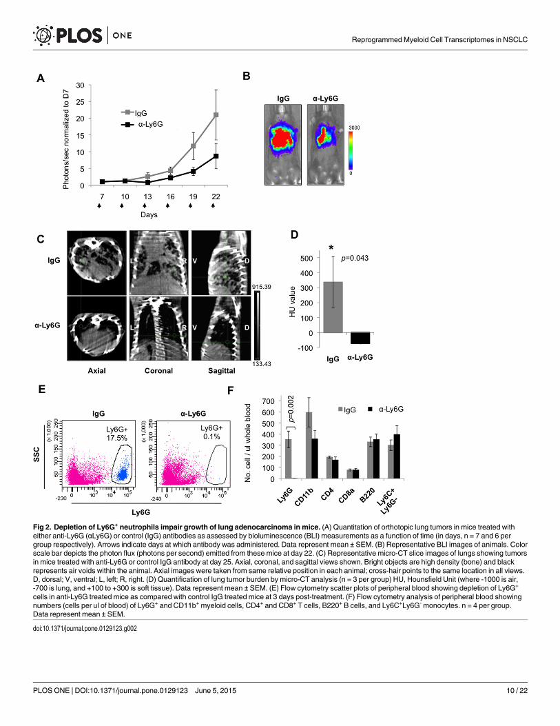
Fig 2. Depletion of Ly6G+ neutrophils impair growth of lung adenocarcinoma in mice. (A) Quantitation of orthotopic lung tumors in mice treated witheither anti-Ly6G (αLy6G) or control (IgG) antibodies as assessed by bioluminescence (BLI) measurements as a function of time (in days, n = 7 and 6 pergroup respectively). Arrows indicate days at which antibody was administered. Data represent mean ± SEM. (B) Representative BLI images of animals. Colorscale bar depicts the photon flux (photons per second) emitted from these mice at day 22. (C) Representative micro-CT slice images of lungs showing tumorsin mice treated with anti-Ly6G or control IgG antibody at day 25. Axial, coronal, and sagittal views shown. Bright objects are high density (bone) and blackrepresents air voids within the animal. Axial images were taken from same relative position in each animal; cross-hair points to the same location in all views.D, dorsal; V, ventral; L, left; R, right. (D) Quantification of lung tumor burden by micro-CT analysis (n = 3 per group) HU, Hounsfield Unit (where -1000 is air,-700 is lung, and +100 to +300 is soft tissue). Data represent mean ± SEM. (E) Flow cytometry scatter plots of peripheral blood showing depletion of Ly6G+
cells in anti-Ly6G treated mice as compared with control IgG treated mice at 3 days post-treatment. (F) Flow cytometry analysis of peripheral blood showingnumbers (cells per ul of blood) of Ly6G+ and CD11b+ myeloid cells, CD4+ and CD8+ T cells, B220+ B cells, and Ly6C+Ly6G- monocytes. n = 4 per group.Data represent mean ± SEM.
doi:10.1371/journal.pone.0129123.g002
Reprogrammed Myeloid Cell Transcriptomes in NSCLC
PLOSONE | DOI:10.1371/journal.pone.0129123 June 5, 2015 10 / 22

Fig 3. RNA-seq analysis of BM immature monocytic myeloid cells and epithelial cells from NSCLC patients and controls. (A) Summary of RNA-seqreads from adenocarcinoma (tumor)- and adjacent lung-derived IMMCs (immature monocytic myeloid cells), neutrophils, and epithelial cells isolated from 6adenocarcinoma patients and mapped to known humanmRNA, genome, and novel mRNA of Aceview gene model. P, unique patient identifier; A, adjacentnon-neoplastic lung tissue; T, neoplastic tumor tissue. (B) Spearman correlation analysis showing clustering of stromal cells derived from IMMCs,neutrophils, and epithelial cells based on global RNA-seq gene expression profiles into distinct tumor and adjacent lung groups. P, unique patient identifier;A, adjacent non-neoplastic lung tissue; T, neoplastic tumor tissue. (C) Venn diagrams showing total number of differentially expressed genes in immatureIMMCs, neutrophils, and epithelial cells from adenocarcinoma compared to non-neoplastic adjacent lung. Cutoff of at least 50 unique mapped reads andFDR <5%. The genes in the list show differential expression with p<0.05, and fold change >2. (D) Differentially regulated stromal genes from (C), enriched forpotential paracrine functions as determined by Gene Ontology annotation as secreted, extracellular space, or membrane (except membranes of organellesincluding golgi and endoplasmic reticulum). Of these, genes with the functions in key tumorigenic pathways including angiogenesis, ECM breakdown, cellmigration, proliferation, invasion, cytokine function, chemokine function, and chemotactic function were selected. Genes selected for analysis are denoted inblue, transmembrane; red, secreted.
doi:10.1371/journal.pone.0129123.g003
Reprogrammed Myeloid Cell Transcriptomes in NSCLC
PLOSONE | DOI:10.1371/journal.pone.0129123 June 5, 2015 11 / 22
identify genes with paracrine activity to capture potential tumor-stroma crosstalk. Using thesecriteria, approximately 80–100 genes were selected for each cell type (Fig 3D and S2 Table).
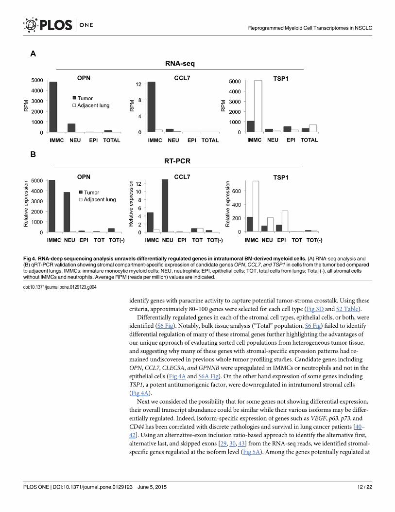
Differentially regulated genes in each of the stromal cell types, epithelial cells, or both, wereidentified (S6 Fig). Notably, bulk tissue analysis (“Total” population, S6 Fig) failed to identifydifferential regulation of many of these stromal genes further highlighting the advantages ofour unique approach of evaluating sorted cell populations from heterogeneous tumor tissue,and suggesting why many of these genes with stromal-specific expression patterns had re-mained undiscovered in previous whole tumor profiling studies. Candidate genes includingOPN, CCL7, CLEC5A, and GPNNB were upregulated in IMMCs or neutrophils and not in theepithelial cells (Fig 4A and S6A Fig). On the other hand expression of some genes includingTSP1, a potent antitumorigenic factor, were downregulated in intratumoral stromal cells(Fig 4A).
Next we considered the possibility that for some genes not showing differential expression,their overall transcript abundance could be similar while their various isoforms may be differ-entially regulated. Indeed, isoform-specific expression of genes such as VEGF, p63, p73, andCD44 has been correlated with discrete pathologies and survival in lung cancer patients [40–42]. Using an alternative-exon inclusion ratio-based approach to identify the alternative first,alternative last, and skipped exons [29, 30, 43] from the RNA-seq reads, we identified stromal-specific genes regulated at the isoform level (Fig 5A). Among the genes potentially regulated at
Fig 4. RNA-deep sequencing analysis unravels differentially regulated genes in intratumoral BM-derived myeloid cells. (A) RNA-seq analysis and(B) qRT-PCR validation showing stromal compartment-specific expression of candidate genesOPN, CCL7, and TSP1 in cells from the tumor bed comparedto adjacent lungs. IMMCs; immature monocytic myeloid cells; NEU, neutrophils; EPI, epithelial cells; TOT, total cells from lungs; Total (-), all stromal cellswithout IMMCs and neutrophils. Average RPM (reads per million) values are indicated.
doi:10.1371/journal.pone.0129123.g004
Reprogrammed Myeloid Cell Transcriptomes in NSCLC
PLOSONE | DOI:10.1371/journal.pone.0129123 June 5, 2015 12 / 22

the isoform level we focused on FLT-1 (VEGFR1) (Fig 5B, 5C and 5D) as the VEGF-FLT-1 axisconstitutes a major regulator of angiogenesis. It has been shown that FLT-1mRNA is alterna-tively spliced to encode both a full-length receptor tyrosine kinase (mFLT-1) that is proangiog-neic, and a soluble isoform (sFLT-1) that binds and sequesters VEGF and PLGF [44] and istherefore antiangiogenic. As determined by the RNA-seq analysis, the overall expression ofFLT-1 or sFLT-1 was similar in the CD33+ IMMCs from tumors and adjacent lung tissue, how-ever themFLT-1 was specifically upregulated in tumor IMMCs (Fig 5C). Importantly, RT-PCRanalysis using isoform-specific primers confirmedmFLT-1 expression levels in the intratu-moral IMMCs (Fig 5D). Notably, FLT-1 expression in cells of the myeloid lineage has beenshown to regulate VEGF/PLGF-oriented migration, survival, and production of angiogenic fac-tors [45, 46], which can promote tumor growth [47], suggesting that it may have a protumori-genic function in NSCLC.
Validation of differentially regulated compartment-specific stromal genesAs our goal was to identify differentially regulated genes in intratumoral stromal cells, we vali-dated a subset of the genes selected from the RNA-seq analysis by quantitative RT-PCR(qRT-PCR) to confirm that their differential expression was confined to specific individualintratumoral cellular compartments (IMMCs, neutrophils, and epithelial cells). Included in theqRT-PCR analysis were sorted samples consisting of all stromal cells without IMMCs and neu-trophils (“Total (-)” population, Fig 4B), to further confirm the lineage specific expression ofthese candidate genes.
qRT-PCR analysis showed significant correlation with RNA-seq data not only in the relativeexpression levels but also in stromal compartment specificity. For example, candidate genes
Fig 5. RNA-deep sequencing analysis unravels differentially regulated mRNA isoforms in intratumoral BM-derivedmyeloid cells. (A) List of stroma-specific genes differentially regulated at the mRNA isoform level. (B) Wiggle plots showing read coverage across the Flt-1 gene in IMMCs fromadenocarcinoma of lung (n = 3 patients) and IMMCs form adjacent lungs (n = 3 patients). The status of sFLT1 and mFLT1 is shown. (C) RNA-seq analysisshowing FLT-1 isoform expression levels of total FLT1, soluble FLT1 (sFLT1), and membrane binding FLT1 (mFLT1) in myeloid cells sorted from adjacentlung and tumor. (D) RT-PCR validation of FLT1 andmFLT-1 isoform expression in myeloid cells sorted from adjacent lung and tumor.
doi:10.1371/journal.pone.0129123.g005
Reprogrammed Myeloid Cell Transcriptomes in NSCLC
PLOSONE | DOI:10.1371/journal.pone.0129123 June 5, 2015 13 / 22

including OPN, CCL7, and TSP1 were differentially expressed in IMMCs or neutrophils andnot in the epithelial cells or other stromal cells (Fig 4B).
We reasoned that differentially regulated genes identified in intratumoral stromal cells maycontribute to carcinogenesis, and went on to select stromal genes including OPN, CCL7, andTSP1 as potential candidates as they are secreted and are likely to influence NSCLC progressionby conferring paracrine functions on tumor epithelial cells. OPN is a secreted phosphoglyco-protein that has been shown to contribute to tumor progression and metastasis [48]. IncreasedOPN expression has been observed in human breast, lung, prostate, colon, ovarian, and gastriccancers [49]. CCL7 is a relatively understudied cytokine, however its expression in tumor stro-ma has been shown to enhance invasion and migration of oral squamous carcinoma cells [50].CCL7 has also been shown to promote neutrophil recruitment in lung inflammation [51].TSP1 is a secreted extracellular matrix protein and one of the most potent inhibitors of tumorangiogenesis and growth [52, 53]. Consistent with this tumor suppressive property, TSP1 ex-pression was found to be downregulated in intratumoral myeloid lineage cells.
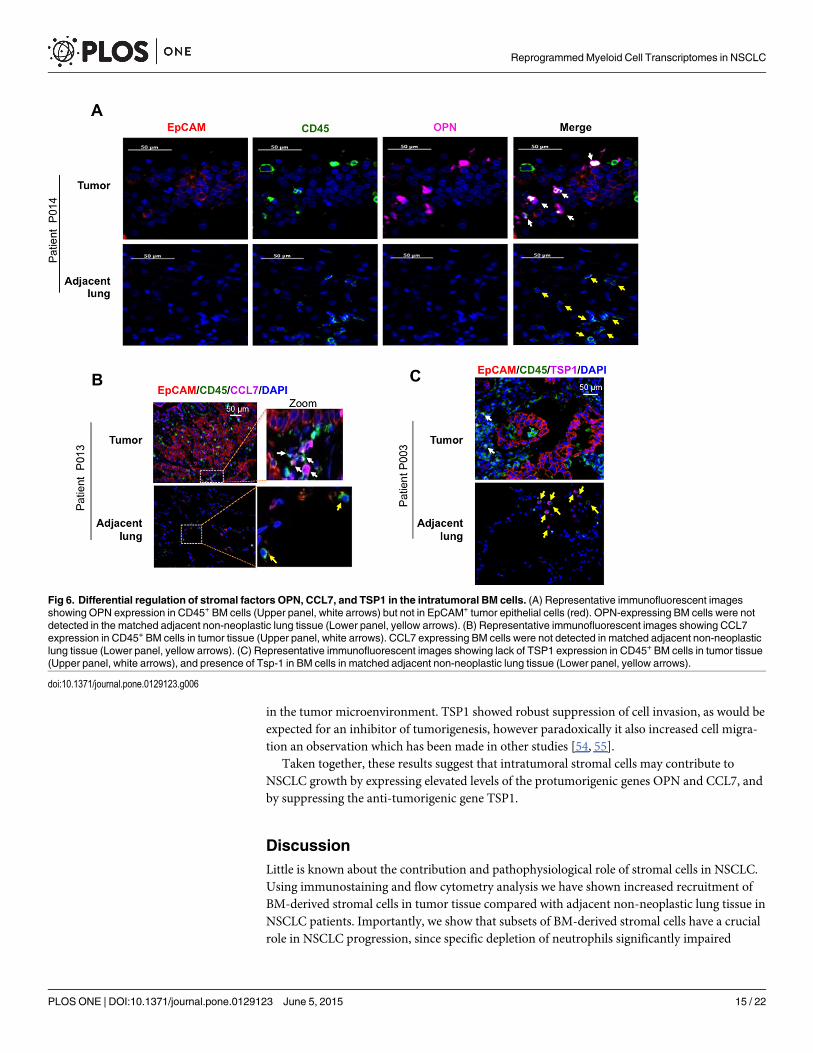
Using immunofluorescence staining of human lung adenocarcinomas, we further assessedthe candidate genes both for expression levels and BM stromal cell specificity. As expectedfrom the RNA-seq and qRT-PCR analysis (Fig 4), OPN staining was confined to a subset ofintratumoral CD45+ BM stromal cells and not the epithelial cells (Fig 6A, Upper panel). Con-sistently, stromal cells in the adjacent non-neoplastic lungs did not exhibit increased expressionof this gene (Fig 6A, Lower panel). Similar staining patterns were observed in tumor tissuefrom other adenocarcinoma patients (S7A Fig). Further analysis showed that, of the CD45+ he-matopoietic cells, the CD11b+ myeloid subset were the main contributors of OPN (S7B Fig).Using a similar approach, we found that CCL7 expression was also confined to the intratu-moral BM cells compared to adjacent lungs (Fig 6B and S7C Fig). In contrast, expression ofTSP1 was suppressed in the intratumoral BM cells compared to adjacent lungs, consistent withthe RNA-seq and RT-PCR analysis (Fig 6C and S7D Fig).
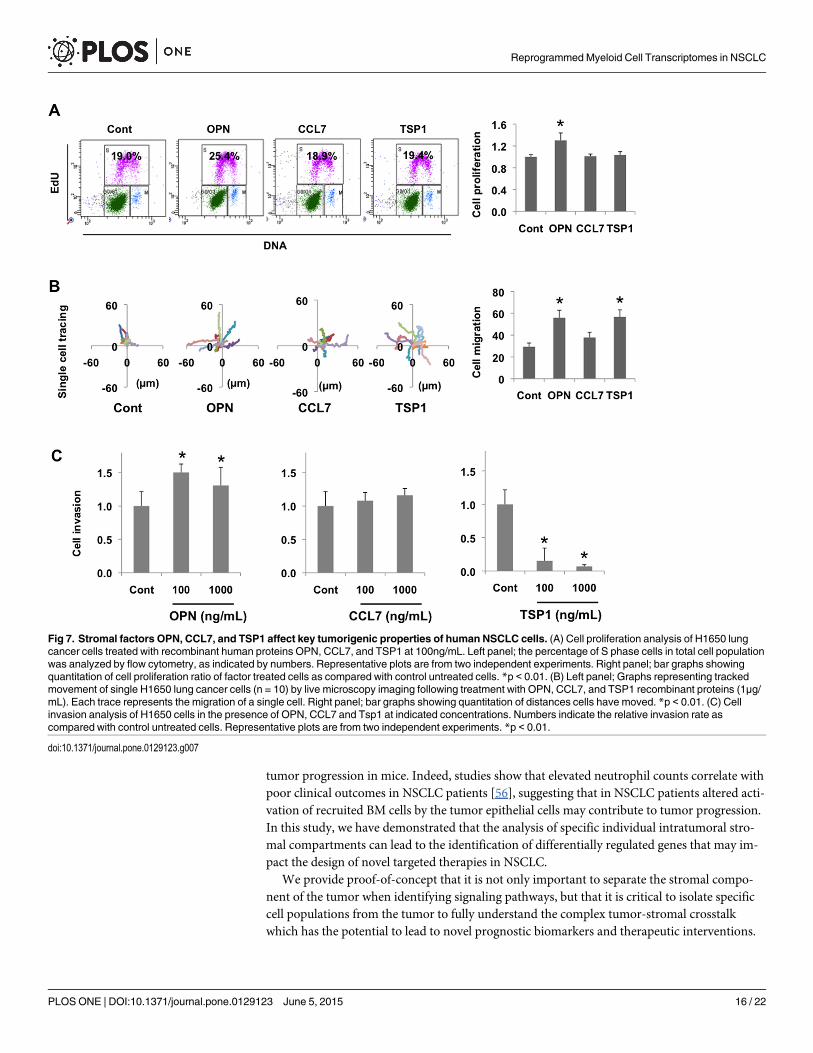
Functional analysis of differentially regulated stromal genes in conferringtumorigenic potential in lung cancer cellsWe next posited that differentially regulated stromal-specific genes identified in this study byproducing secreted factors might contribute to tumor progression in a paracrine fashion byproducing secreted factors. To investigate this, we determined whether these gene productscould directly affect key tumorigeneic properties such as proliferation, migration, or invasionof human lung NSCLC cells. Human lung cancer cells (H1650) were treated with varying con-centrations of gene-specific recombinant proteins. Flow cytometry analysis showed that OPNtreatment increased the percentage of cells in S phase, while CCL7 and TSP1 did not, suggest-ing that OPN stimulates tumor cell proliferation (Fig 7A). Next, we investigated the paracrineeffect of these proteins on cell migration. In cell migration assays using live cell tracking, treat-ment with OPN, CCL7, and TSP1 (1μg/mL 24 hours) enhanced the mobility of H1650 cells, al-beit with different degrees of migration observed (Fig 7B). In a similar way, we determined thepotential of these proteins in influencing cell invasion, and observed that while OPN increasedinvasion, notably, TSP1 significantly inhibited tumor cell invasion through Matrigel-coatedmembranes (Fig 7C). These results suggest that the stromal-derived factors OPN, CCL7, andTSP1 identified from our RNA-seq analysis affect the malignant properties of human lung can-cer cells by promoting either proliferation, migration, or invasion or a combination of these.Clearly, OPN was able to promote invasion, migration, and invasion of lung cancer cells, whileCCL7 showed a marginal increase in cell migration and did not affect proliferation or invasion,suggesting that it may possess other protumorigenic properties such as being a chemoattractant
Reprogrammed Myeloid Cell Transcriptomes in NSCLC
PLOSONE | DOI:10.1371/journal.pone.0129123 June 5, 2015 14 / 22
in the tumor microenvironment. TSP1 showed robust suppression of cell invasion, as would beexpected for an inhibitor of tumorigenesis, however paradoxically it also increased cell migra-tion an observation which has been made in other studies [54, 55].
Taken together, these results suggest that intratumoral stromal cells may contribute toNSCLC growth by expressing elevated levels of the protumorigenic genes OPN and CCL7, andby suppressing the anti-tumorigenic gene TSP1.
DiscussionLittle is known about the contribution and pathophysiological role of stromal cells in NSCLC.Using immunostaining and flow cytometry analysis we have shown increased recruitment ofBM-derived stromal cells in tumor tissue compared with adjacent non-neoplastic lung tissue inNSCLC patients. Importantly, we show that subsets of BM-derived stromal cells have a crucialrole in NSCLC progression, since specific depletion of neutrophils significantly impaired
Fig 6. Differential regulation of stromal factors OPN, CCL7, and TSP1 in the intratumoral BM cells. (A) Representative immunofluorescent imagesshowing OPN expression in CD45+ BM cells (Upper panel, white arrows) but not in EpCAM+ tumor epithelial cells (red). OPN-expressing BM cells were notdetected in the matched adjacent non-neoplastic lung tissue (Lower panel, yellow arrows). (B) Representative immunofluorescent images showing CCL7expression in CD45+ BM cells in tumor tissue (Upper panel, white arrows). CCL7 expressing BM cells were not detected in matched adjacent non-neoplasticlung tissue (Lower panel, yellow arrows). (C) Representative immunofluorescent images showing lack of TSP1 expression in CD45+ BM cells in tumor tissue(Upper panel, white arrows), and presence of Tsp-1 in BM cells in matched adjacent non-neoplastic lung tissue (Lower panel, yellow arrows).
doi:10.1371/journal.pone.0129123.g006
Reprogrammed Myeloid Cell Transcriptomes in NSCLC
PLOSONE | DOI:10.1371/journal.pone.0129123 June 5, 2015 15 / 22
tumor progression in mice. Indeed, studies show that elevated neutrophil counts correlate withpoor clinical outcomes in NSCLC patients [56], suggesting that in NSCLC patients altered acti-vation of recruited BM cells by the tumor epithelial cells may contribute to tumor progression.In this study, we have demonstrated that the analysis of specific individual intratumoral stro-mal compartments can lead to the identification of differentially regulated genes that may im-pact the design of novel targeted therapies in NSCLC.
We provide proof-of-concept that it is not only important to separate the stromal compo-nent of the tumor when identifying signaling pathways, but that it is critical to isolate specificcell populations from the tumor to fully understand the complex tumor-stromal crosstalkwhich has the potential to lead to novel prognostic biomarkers and therapeutic interventions.
Fig 7. Stromal factors OPN, CCL7, and TSP1 affect key tumorigenic properties of human NSCLC cells. (A) Cell proliferation analysis of H1650 lungcancer cells treated with recombinant human proteins OPN, CCL7, and TSP1 at 100ng/mL. Left panel; the percentage of S phase cells in total cell populationwas analyzed by flow cytometry, as indicated by numbers. Representative plots are from two independent experiments. Right panel; bar graphs showingquantitation of cell proliferation ratio of factor treated cells as compared with control untreated cells. *p < 0.01. (B) Left panel; Graphs representing trackedmovement of single H1650 lung cancer cells (n = 10) by live microscopy imaging following treatment with OPN, CCL7, and TSP1 recombinant proteins (1μg/mL). Each trace represents the migration of a single cell. Right panel; bar graphs showing quantitation of distances cells have moved. *p < 0.01. (C) Cellinvasion analysis of H1650 cells in the presence of OPN, CCL7 and Tsp1 at indicated concentrations. Numbers indicate the relative invasion rate ascompared with control untreated cells. Representative plots are from two independent experiments. *p < 0.01.
doi:10.1371/journal.pone.0129123.g007
Reprogrammed Myeloid Cell Transcriptomes in NSCLC
PLOSONE | DOI:10.1371/journal.pone.0129123 June 5, 2015 16 / 22
Analysis of the gene expression profiles of myeloid populations in tumor and adjacent tissuesrevealed approximately 80–100 differentially regulated genes. Among these candidates, expres-sion of OPN was>1000-fold upregulated in intratumoral myeloid cells.
OPN is a secreted phosphoglycoprotein that has been shown to contribute to tumor pro-gression and metastasis [48]. Increased OPN expression has been observed in human breast,lung, prostate, colon, ovarian, and gastric cancers [49]. However, the biological significance ofOPN expression in intratumoral myeloid cells has remained unclear. Our finding that in earlystage NSCLC OPN is upregulated in intratumoral BM cells signifies the importance of evaluat-ing its potential as a prognostic marker for identifying subsets of NSCLC patients with a highrisk of recurrence.
The differentially regulated stromal genes identified in our study have the potential to con-tribute to NSCLC progression by conferring paracrine functions on tumor epithelial cells. Inagreement with this notion, recombinant OPN promoted proliferation, migration, and inva-sion of lung cancer cells in a paracrine fashion, suggesting that OPN has tumor promoting ac-tivity in the microenvironment. Given that multiple and complex mechanisms are involved inthe role of OPN in cancer, much remains to be unraveled about these mechanisms and thefunctional contributions of OPN produced by different cell types in order to pursue anti-OPNtherapeutic strategies. For example, specific loss/gain-of-function of OPN in BM-derived mye-loid cells in mice would provide insights into its role in stroma-mediated tumor progression.To accelerate clinical translation, further studies are necessary to determine a pharmacologicaltargeting strategy for OPN. Recently developed anti-OPN1 neutralizing monoclonal antibod-ies, AOM1 [49] and hu1A12 [57], have effectively blocked OPN and impaired breast andlung tumors in mice. In light of our study it will be important to determine whether this anti-tumorigenic phenotype resulted mainly from stroma-specific suppression of OPN. Similarly,in future studies other stromal-specific genes identified in this study should be assessed forprognostic and therapeutic potential. For example, a potent and highly selective humanized an-tibody inhibitor of MMP14 activity, DX-2400, blocked tumor growth in mouse models ofbreast and liver carcinogenesis, without causing overt toxicity as has been observed with otherMMP inhibitors [58, 59]. Similarly, a CCL7 neutralizing antibody has been shown to inhibitCCL7-induced invasion and migration of oral carcinoma cells [50], and anti-CCL2 and-CCR3antibodies are also being evaluated [50, 60].
TSP1 is a potent anti-tumorigenic factor. Through interaction with its receptor CD36,which is expressed on endothelial cells, TSP1 inhibits angiogenesis by reducing endothelial cellmigration and survival [61]. Therefore, tumors overexpressing TSP1 usually exhibit less angio-genesis, grow slower, and show defects in metastasis. TSP1 is expressed in various cell types in-cluding platelets, endothelial cells, stromal fibroblasts, smooth muscle cells, monocytes, andmacrophages [62–66]. However, in our study we found that TSP1 expression was confined tomyeloid cells in NSCLC. Importantly, TSP1 was downregulated in intratumoral myeloid cellssuggesting that restoration of TSP1 levels in the intratumoral stromal cells constitutes a viablestrategy to block NSCLC growth. Importantly, TSP1 is also being exploited in clinical trials;and a TSP1 mimetic peptide, ABT-510, which retains the tumor suppressive activity of TSP1 isbeing used for the treatment of solid tumors, and was found to inhibit tumor growth in themouse Lewis lung carcinoma model [67].
GSEA revealed downregulated pathways pertaining to immune response and cellulardefense response in intratumoral neutrophils, suggesting that the tumor cells affect the prolifer-ation and differentiation of myeloid cells, orchestrating an immune suppressive microenviron-ment. Further validation and functional analysis of GSEA-identified candidate genes in theNSCLC microenvironment will constitute an area of future investigation.
Reprogrammed Myeloid Cell Transcriptomes in NSCLC
PLOSONE | DOI:10.1371/journal.pone.0129123 June 5, 2015 17 / 22
This study suggests that molecular alterations within the reprogrammed tumor stroma offernovel avenues for therapeutic interventions. Compared to cancer cells, which frequently acquiredrug resistance, stromal cells are genetically more stable and therefore less likely to evolve acquiredresistance to targeted therapy. It is likely that targeted stromal therapeutics for NSCLC patientsmay complement conventional treatments that exclusively target cancer cells. In addition to theidentification of stromal-specific genes with prognostic and therapeutic potential, our approach ofanalyzing intratumoral stromal cells will help to define the basic biological mechanisms of the dis-ease, and will open a new field of research for mechanistic and translational studies of lung cancer.
Supporting InformationS1 Fig. Increased number of bone marrow hematopoietic cells infiltrate the adenocarcino-ma of NSCLC patients.(TIF)
S2 Fig. Validation of sorted IMMCs and neutrophils.(TIF)
S3 Fig. Flow cytometry analysis of peripheral blood in mice treated with anti-IgG and anti-Ly6G antibodies.(TIF)
S4 Fig. Quality control analysis of RNA harvested from flow cytometry sorted cells fromfresh clinical tissue and RNA-seq libraries.(TIF)
S5 Fig. RNA-seq data analysis pipeline.(TIF)
S6 Fig. RNA-deep sequencing analysis unravels differentially regulated genes in specificintratumoral stromal compartments.(TIF)
S7 Fig. OPN, CCL7, and TSP1 are differentially regulated in the intratumoral BM cells.(TIF)
S1 Table. Description of lung cancer patients used in this study.(TIF)
S2 Table. Differentially regulated genes for each cell type.(TIF)
AcknowledgmentsWe thank Jenny Xiang of the Genomics Resources Core Facility of Weill Cornell Medical Col-lege for the RNA sequencing, Bin He and Michael Synan of the Citigroup Biomedical ImagingCenter, Weill Cornell Medical College for micro-CT imaging. We also thank Sharrell Lee fortechnical support. We acknowledge the Thoracic Surgery Biobank, Weill Cornell Medical Col-lege for providing annotated clinical samples.
Author ContributionsConceived and designed the experiments: AD DG NKA S. Rafii VM. Performed the experi-ments: AD DG KRF HC TER DN. Analyzed the data: AD DG RG KRF HC TER S. Ryu AN OE
Reprogrammed Myeloid Cell Transcriptomes in NSCLC
PLOSONE | DOI:10.1371/journal.pone.0129123 June 5, 2015 18 / 22
BS NN RD NKA VM. Contributed reagents/materials/analysis tools: S. Rafii. Inclusion of pa-tients and clinical material: CFS WA BS NKA.
References1. Siegel R, Naishadham D, Jemal A. Cancer statistics, 2012. CA Cancer J Clin. 2012; 62(1):10–29. doi:
10.3322/caac.20138 PMID: 22237781
2. Herbst RS, Heymach JV, Lippman SM. Lung cancer. N Engl J Med. 2008; 359(13):1367–80. doi: 10.1056/NEJMra0802714 PMID: 18815398
3. Haber DA, Bell DW, Sordella R, Kwak EL, Godin-Heymann N, Sharma SV, et al. Molecular targetedtherapy of lung cancer: EGFRmutations and response to EGFR inhibitors. Cold Spring Harb SympQuant Biol. 2005; 70:419–26. PMID: 16869779
4. Kwak EL, Bang YJ, Camidge DR, Shaw AT, Solomon B, Maki RG, et al. Anaplastic lymphoma kinaseinhibition in non-small-cell lung cancer. N Engl J Med. 2010; 363(18):1693–703. doi: 10.1056/NEJMoa1006448 PMID: 20979469
5. Zochbauer-Muller S, Gazdar AF, Minna JD. Molecular pathogenesis of lung cancer. Annu Rev Physiol.2002; 64:681–708. PMID: 11826285
6. Engelman JA, Zejnullahu K, Mitsudomi T, Song Y, Hyland C, Park JO, et al. MET amplification leads togefitinib resistance in lung cancer by activating ERBB3 signaling. Science. 2007; 316(5827):1039–43.PMID: 17463250
7. PaoW, Miller VA, Politi KA, Riely GJ, Somwar R, Zakowski MF, et al. Acquired resistance of lung ade-nocarcinomas to gefitinib or erlotinib is associated with a second mutation in the EGFR kinase domain.PLoS Med. 2005; 2(3):e73. PMID: 15737014
8. Choi YL, Soda M, Yamashita Y, Ueno T, Takashima J, Nakajima T, et al. EML4-ALKmutations in lungcancer that confer resistance to ALK inhibitors. N Engl J Med. 2010; 363(18):1734–9. doi: 10.1056/NEJMoa1007478 PMID: 20979473
9. Hanahan D, Coussens LM. Accessories to the crime: functions of cells recruited to the tumor microenvi-ronment. Cancer Cell. 2012; 21(3):309–22. doi: 10.1016/j.ccr.2012.02.022 PMID: 22439926
10. Allinen M, Beroukhim R, Cai L, Brennan C, Lahti-Domenici J, Huang H, et al. Molecular characterizationof the tumor microenvironment in breast cancer. Cancer Cell. 2004; 6(1):17–32. PMID: 15261139
11. Joyce JA, Pollard JW. Microenvironmental regulation of metastasis. Nat Rev Cancer. 2009; 9(4):239–52. doi: 10.1038/nrc2618 PMID: 19279573
12. Mantovani A, Sica A. Macrophages, innate immunity and cancer: balance, tolerance, and diversity.Curr Opin Immunol. 2010; 22(2):231–7. doi: 10.1016/j.coi.2010.01.009 PMID: 20144856
13. Fridlender ZG, Sun J, Kim S, Kapoor V, Cheng G, Ling L, et al. Polarization of tumor-associated neutro-phil phenotype by TGF-beta: "N1" versus "N2" TAN. Cancer Cell. 2009; 16(3):183–94. doi: 10.1016/j.ccr.2009.06.017 PMID: 19732719
14. Coussens LM, Werb Z. Inflammation and cancer. Nature. 2002; 420(6917):860–7. PMID: 12490959
15. Orimo A, Gupta PB, Sgroi DC, Arenzana-Seisdedos F, Delaunay T, Naeem R, et al. Stromal fibroblastspresent in invasive human breast carcinomas promote tumor growth and angiogenesis through elevat-ed SDF-1/CXCL12 secretion. Cell. 2005; 121(3):335–48. PMID: 15882617
16. Zeng Q, Li S, Chepeha DB, Giordano TJ, Li J, Zhang H, et al. Crosstalk between tumor and endothelialcells promotes tumor angiogenesis by MAPK activation of Notch signaling. Cancer Cell. 2005; 8(1):13–23. PMID: 16023595
17. Gao D, Nolan DJ, Mellick AS, Bambino K, McDonnell K, Mittal V. Endothelial progenitor cells controlthe angiogenic switch in mouse lung metastasis. Science. 2008; 319(5860):195–8. doi: 10.1126/science.1150224 PMID: 18187653
18. Gao D, Joshi N, Choi H, Ryu S, Hahn M, Catena R, et al. Myeloid progenitor cells in the premetastaticlung promote metastases by inducing mesenchymal to epithelial transition. Cancer Res. 2012; 72(6):1384–94. doi: 10.1158/0008-5472.CAN-11-2905 PMID: 22282653
19. Hanahan D, Weinberg RA. Hallmarks of cancer: the next generation. Cell. 2011; 144(5):646–74. doi:10.1016/j.cell.2011.02.013 PMID: 21376230
20. Wilson TR, Fridlyand J, Yan Y, Penuel E, Burton L, Chan E, et al. Widespread potential for growth-fac-tor-driven resistance to anticancer kinase inhibitors. Nature. 2012; 487(7408):505–9. doi: 10.1038/nature11249 PMID: 22763448
21. Straussman R, Morikawa T, Shee K, Barzily-Rokni M, Qian ZR, Du J, et al. Tumour micro-environmentelicits innate resistance to RAF inhibitors through HGF secretion. Nature. 2012; 487(7408):500–4. doi:10.1038/nature11183 PMID: 22763439
Reprogrammed Myeloid Cell Transcriptomes in NSCLC
PLOSONE | DOI:10.1371/journal.pone.0129123 June 5, 2015 19 / 22

22. Nakasone ES, Askautrud HA, Kees T, Park JH, Plaks V, Ewald AJ, et al. Imaging tumor-stroma interac-tions during chemotherapy reveals contributions of the microenvironment to resistance. Cancer Cell.2012; 21(4):488–503. doi: 10.1016/j.ccr.2012.02.017 PMID: 22516258
23. Denardo DG, Brennan DJ, Rexhepaj E, Ruffell B, Shiao SL, Madden SF, et al. Leukocyte ComplexityPredicts Breast Cancer Survival and Functionally Regulates Response to Chemotherapy. Cancer Dis-cov. 2011; 1:54–67. doi: 10.1158/2159-8274.CD-10-0028 PMID: 22039576
24. Albini A, Sporn MB. The tumour microenvironment as a target for chemoprevention. Nat Rev Cancer.2007; 7(2):139–47. PMID: 17218951
25. Ito M, Ishii G, Nagai K, Maeda R, Nakano Y, Ochiai A. Prognostic impact of cancer-associated stromalcells in stage I lung adenocarcinoma patients. Chest. 2012.
26. Navab R, Strumpf D, Bandarchi B, Zhu CQ, Pintilie M, Ramnarine VR, et al. Prognostic gene-expres-sion signature of carcinoma-associated fibroblasts in non-small cell lung cancer. Proc Natl Acad Sci US A. 2011; 108(17):7160–5. doi: 10.1073/pnas.1014506108 PMID: 21474781
27. WangW, Li Q, Yamada T, Matsumoto K, Matsumoto I, Oda M, et al. Crosstalk to stromal fibroblasts in-duces resistance of lung cancer to epidermal growth factor receptor tyrosine kinase inhibitors. Clin Can-cer Res. 2009; 15(21):6630–8. doi: 10.1158/1078-0432.CCR-09-1001 PMID: 19843665
28. Perfetto SP, Chattopadhyay PK, Roederer M. Seventeen-colour flow cytometry: unravelling the im-mune system. Nat Rev Immunol. 2004; 4(8):648–55. PMID: 15286731
29. Pal S, Gupta R, Kim H, Wickramasinghe P, Baubet V, Showe LC, et al. Alternative transcription ex-ceeds alternative splicing in generating the transcriptome diversity of cerebellar development. GenomeRes. 2011; 21(8):1260–72. doi: 10.1101/gr.120535.111 PMID: 21712398
30. Kim H, Bi Y, Pal S, Gupta R, Davuluri RV. IsoformEx: isoform level gene expression estimation usingweighted non-negative least squares frommRNA-Seq data. BMC Bioinformatics. 2011; 12:305. doi:10.1186/1471-2105-12-305 PMID: 21794104
31. Anders S, Huber W. Differential expression analysis for sequence count data. Genome Biol. 2010; 11(10):R106. doi: 10.1186/gb-2010-11-10-r106 PMID: 20979621
32. DuPage M, Dooley AL, Jacks T. Conditional mouse lung cancer models using adenoviral or lentiviraldelivery of Cre recombinase. Nat Protoc. 2009; 4(7):1064–72. doi: 10.1038/nprot.2009.95 PMID:19561589
33. Youn JI, Nagaraj S, Collazo M, Gabrilovich DI. Subsets of myeloid-derived suppressor cells in tumor-bearing mice. J Immunol. 2008; 181(8):5791–802. PMID: 18832739
34. Coffelt SB, Lewis CE, Naldini L, Brown JM, Ferrara N, De Palma M. Elusive identities and overlappingphenotypes of proangiogenic myeloid cells in tumors. Am J Pathol. 2010; 176(4):1564–76. doi: 10.2353/ajpath.2010.090786 PMID: 20167863
35. Daley JM, Thomay AA, Connolly MD, Reichner JS, Albina JE. Use of Ly6G-specific monoclonal anti-body to deplete neutrophils in mice. J Leukoc Biol. 2008; 83(1):64–70. PMID: 17884993
36. Ruffell B, Au A, Rugo HS, Esserman LJ, Hwang ES, Coussens LM. Leukocyte composition of humanbreast cancer. Proc Natl Acad Sci U S A. 2012; 109(8):2796–801. doi: 10.1073/pnas.1104303108PMID: 21825174
37. Schmid MC, Varner JA. Myeloid cells in the tumor microenvironment: modulation of tumor angiogene-sis and tumor inflammation. J Oncol. 2010; 2010:201026. doi: 10.1155/2010/201026 PMID: 20490273
38. Gabrilovich DI, Ostrand-Rosenberg S, Bronte V. Coordinated regulation of myeloid cells by tumours.Nat Rev Immunol. 2012; 12(4):253–68. doi: 10.1038/nri3175 PMID: 22437938
39. Oliver TG, Mercer KL, Sayles LC, Burke JR, Mendus D, Lovejoy KS, et al. Chronic cisplatin treatmentpromotes enhanced damage repair and tumor progression in a mouse model of lung cancer. GenesDev. 2010; 24(8):837–52. doi: 10.1101/gad.1897010 PMID: 20395368
40. Yuan A, Yu CJ, Kuo SH, ChenWJ, Lin FY, Luh KT, et al. Vascular endothelial growth factor 189 mRNAisoform expression specifically correlates with tumor angiogenesis, patient survival, and postoperativerelapse in non-small-cell lung cancer. J Clin Oncol. 2001; 19(2):432–41. PMID: 11208836
41. Goparaju CM, Pass HI, Blasberg JD, Hirsch N, Donington JS. Functional heterogeneity of osteopontinisoforms in non-small cell lung cancer. J Thorac Oncol. 2010; 5(10):1516–23. doi: 10.1097/JTO.0b013e3181eba6bd PMID: 20689445
42. Iacono ML, Monica V, Saviozzi S, Ceppi P, Bracco E, Papotti M, et al. p63 and p73 isoform expressionin non-small cell lung cancer and corresponding morphological normal lung tissue. J Thorac Oncol.2011; 6(3):473–81. doi: 10.1097/JTO.0b013e31820b86b0 PMID: 21289519
43. Wang ET, Sandberg R, Luo S, Khrebtukova I, Zhang L, Mayr C, et al. Alternative isoform regulation inhuman tissue transcriptomes. Nature. 2008; 456(7221):470–6. doi: 10.1038/nature07509 PMID:18978772
Reprogrammed Myeloid Cell Transcriptomes in NSCLC
PLOSONE | DOI:10.1371/journal.pone.0129123 June 5, 2015 20 / 22
44. Kendall RL, Thomas KA. Inhibition of vascular endothelial cell growth factor activity by an endogenous-ly encoded soluble receptor. Proc Natl Acad Sci U S A. 1993; 90(22):10705–9. PMID: 8248162
45. Hattori K, Dias S, Heissig B, Hackett NR, Lyden D, Tateno M, et al. Vascular endothelial growth factorand angiopoietin-1 stimulate postnatal hematopoiesis by recruitment of vasculogenic and hematopoiet-ic stem cells. J Exp Med. 2001; 193(9):1005–14. PMID: 11342585
46. Hattori K, Heissig B, Wu Y, Dias S, Tejada R, Ferris B, et al. Placental growth factor reconstitutes hema-topoiesis by recruiting VEGFR1(+) stem cells from bone-marrow microenvironment. Nat Med. 2002; 8(8):841–9. PMID: 12091880
47. Kerber M, Reiss Y, Wickersheim A, Jugold M, Kiessling F, Heil M, et al. Flt-1 signaling in macrophagespromotes glioma growth in vivo. Cancer Res. 2008; 68(18):7342–51. doi: 10.1158/0008-5472.CAN-07-6241 PMID: 18794121
48. Rittling SR, Chambers AF. Role of osteopontin in tumour progression. Br J Cancer. 2004; 90(10):1877–81. PMID: 15138464
49. Shojaei F, Scott N, Kang X, Lappin PB, Fitzgerald AA, Karlicek S, et al. Osteopontin induces growth ofmetastatic tumors in a preclinical model of non-small lung cancer. J Exp Clin Cancer Res. 2012; 31:26.doi: 10.1186/1756-9966-31-26 PMID: 22444159
50. Jung DW, Che ZM, Kim J, Kim K, Kim KY, Williams D. Tumor-stromal crosstalk in invasion of oral squa-mous cell carcinoma: a pivotal role of CCL7. Int J Cancer. 2010; 127(2):332–44. doi: 10.1002/ijc.25060PMID: 19937793
51. Michalec L, Choudhury BK, Postlethwait E, Wild JS, Alam R, Lett-BrownM, et al. CCL7 and CXCL10orchestrate oxidative stress-induced neutrophilic lung inflammation. J Immunol. 2002; 168(2):846–52.PMID: 11777981
52. Good DJ, Polverini PJ, Rastinejad F, Le Beau MM, Lemons RS, Frazier WA, et al. A tumor suppressor-dependent inhibitor of angiogenesis is immunologically and functionally indistinguishable from a frag-ment of thrombospondin. Proc Natl Acad Sci U S A. 1990; 87(17):6624–8. PMID: 1697685
53. Lawler J. Thrombospondin-1 as an endogenous inhibitor of angiogenesis and tumor growth. J Cell MolMed. 2002; 6(1):1–12. PMID: 12003665
54. Taraboletti G, Roberts DD, Liotta LA. Thrombospondin-induced tumor cell migration: haptotaxis andchemotaxis are mediated by different molecular domains. J Cell Biol. 1987; 105(5):2409–15. PMID:3680388
55. Firlej V, Mathieu JR, Gilbert C, Lemonnier L, Nakhlé J, Gallou-Kabani C, et al. Thrombospondin-1 trig-gers cell migration and development of advanced prostate tumors. Cancer Res. 2011; 71(24):7649–58.doi: 10.1158/0008-5472.CAN-11-0833 PMID: 22037878
56. Tomita M, Shimizu T, Ayabe T, Yonei A, Onitsuka T. Preoperative neutrophil to lymphocyte ratio as aprognostic predictor after curative resection for non-small cell lung cancer. Anticancer Res. 2011; 31(9):2995–8. PMID: 21868550
57. Dai J, Li B, Shi J, Peng L, Zhang D, QianW, et al. A humanized anti-osteopontin antibody inhibits breastcancer growth and metastasis in vivo. Cancer Immunol Immunother. 2010; 59(3):355–66. doi: 10.1007/s00262-009-0754-z PMID: 19690854
58. Devy L, Dransfield DT. New Strategies for the Next Generation of Matrix-Metalloproteinase Inhibitors:Selectively Targeting Membrane-Anchored MMPs with Therapeutic Antibodies. BiochemRes Int.2011; 2011:191670. doi: 10.1155/2011/191670 PMID: 21152183
59. Devy L, Huang L, Naa L, Yanamandra N, Pieters H, Frans N, et al. Selective inhibition of matrix metallo-proteinase-14 blocks tumor growth, invasion, and angiogenesis. Cancer Res. 2009; 69(4):1517–26.doi: 10.1158/0008-5472.CAN-08-3255 PMID: 19208838
60. Zhang J, Patel L, Pienta KJ. Targeting chemokine (C-Cmotif) ligand 2 (CCL2) as an example of transla-tion of cancer molecular biology to the clinic. Prog Mol Biol Transl Sci. 2010; 95:31–53. doi: 10.1016/B978-0-12-385071-3.00003-4 PMID: 21075328
61. Lawler J. Thrombospondin-1 as an endogenous inhibitor of angiogenesis and tumor growth. J Cell MolMed. 2002; 6(1):1–12. PMID: 12003665
62. Baenziger NL, Brodie GN, Majerus PW. A thrombin-sensitive protein of human platelet membranes.Proc Natl Acad Sci U S A. 1971; 68(1):240–3. PMID: 5276296
63. McPherson J, Sage H, Bornstein P. Isolation and characterization of a glycoprotein secreted by aorticendothelial cells in culture. Apparent identity with platelet thrombospondin. J Biol Chem. 1981; 256(21):11330–6. PMID: 7287769
64. Jaffe EA, Ruggiero JT, Leung LK, Doyle MJ, McKeown-Longo PJ, Mosher DF. Cultured human fibro-blasts synthesize and secrete thrombospondin and incorporate it into extracellular matrix. Proc NatlAcad Sci U S A. 1983; 80(4):998–1002. PMID: 6341993
Reprogrammed Myeloid Cell Transcriptomes in NSCLC
PLOSONE | DOI:10.1371/journal.pone.0129123 June 5, 2015 21 / 22
65. Jaffe EA, Ruggiero JT, Falcone DJ. Monocytes and macrophages synthesize and secrete thrombos-pondin. Blood. 1985; 65(1):79–84. PMID: 3965054
66. Raugi GJ, Mumby SM, Abbott-Brown D, Bornstein P. Thrombospondin: synthesis and secretion bycells in culture. J Cell Biol. 1982; 95(1):351–4. PMID: 6754743
67. Haviv F, Bradley MF, Kalvin DM, Schneider AJ, Davidson DJ, Majest SM, et al. Thrombospondin-1 mi-metic peptide inhibitors of angiogenesis and tumor growth: design, synthesis, and optimization of phar-macokinetics and biological activities. J Med Chem. 2005; 48(8):2838–46. PMID: 15828822
Reprogrammed Myeloid Cell Transcriptomes in NSCLC
PLOSONE | DOI:10.1371/journal.pone.0129123 June 5, 2015 22 / 22
Related Documents