Identification of Regulatory Functions for 4-1BB and 4-1BBL in Myelopoiesis and the Development of Dendritic Cells Seung-Woo Lee 1 , Yunji Park 2 , Takanori So 1 , Byoung S. Kwon 3 , Hilde Cheroutre 2 , Robert S. Mittler 4 , and Michael Croft 1 1 Division of Molecular Immunology, La Jolla Institute for Allergy and Immunology, San Diego, CA 92121, USA 2 Division of Developmental Immunology, La Jolla Institute for Allergy and Immunology, San Diego, CA 92121, USA 3 Immunomodulation Research Center, University of Ulsan, Ulsan, Korea, and National Cancer Center, Goyang, Kyonggi-do, Korea 4 Department of Surgery and Emory Vaccine Center, University School of Medicine, Emory University, Atlanta, Georgia, 30329, USA Abstract 4-1BB and 4-1BBL can control adaptive immunity, but we demonstrated that their interaction also suppressed myelopoiesis. 4-1BBL was found to be expressed on hematopoietic stem cells, and differentiating common myeloid (CMPs) and granulocyte-macrophage progenitors (GMPs), and 4-1BB was inducible on activated myeloid progenitors. Steady state numbers of GMPs, myeloid- lineage cells, and mature dendritic cells, were elevated in 4-1BB- and 4-1BBL-deficient mice, indicative of a negative functional role, and this was confirmed in bone marrow chimeras and in vitro where the absence of 4-1BB/4-1BBL interactions led to enhanced differentiation into dendritic cell lineages. The regulatory activity was mediated through 4-1BBL, with binding by 4-1BB inhibiting differentiation of myeloid progenitors. Thus, 4-1BB and 4-1BBL have a novel function in limiting myelopoiesis and dendritic cell development. INTRODUCTION 4-1BB (also known as CD137 and TNFRSF9), a member of the tumor-necrosis factor receptor (TNFR) superfamily, was first identified as an inducible costimulatory molecule on activated T cells 1 . The ligand of 4-1BB (which is variously called 4-1BBL, CD137L or TNFSF9), also a TNF superfamily member, was later found expressed on activated antigen-presenting cells (APCs) such as B cells, macrophages and dendritic cells (DCs) 2,3 . The interaction of 4-1BB with 4-1BBL strongly regulates immunity and has been proposed to preferentially control T cell responses based on studies in various murine models of cancer, infectious disease and autoimmune disease 4,5 . Much of the early literature on 4-1BB–4-1BBL interactions was derived from studies of agonist 4-1BB antibodies and Tnfsf9 −/− mice 5 , demonstrating a positive regulatory role in T cell priming 6,7 , and suggesting that signals from 4-1BB costimulate T Correspondence should be addressed to M.C. ([email protected]). AUTHOR CONTRIBUTIONS S.-W.L., Y.P., T.S. R.S.M. and M.C. designed the research and analyzed the data; S.-W.L., Y.P. and T.S. performed the experiments at LIAI, and S.-W.L. performed experiments as a visiting scientist with R.S.M. at Emory University; R.S.M, B.S.K., and H.C. contributed reagents and to the preparation of the manuscript,; S.-W.L. and M.C. wrote the manuscript. NIH Public Access Author Manuscript Nat Immunol. Author manuscript; available in PMC 2009 August 1. Published in final edited form as: Nat Immunol. 2008 August ; 9(8): 917–926. doi:10.1038/ni.1632. NIH-PA Author Manuscript NIH-PA Author Manuscript NIH-PA Author Manuscript

Welcome message from author
This document is posted to help you gain knowledge. Please leave a comment to let me know what you think about it! Share it to your friends and learn new things together.
Transcript
Identification of Regulatory Functions for 4-1BB and 4-1BBL inMyelopoiesis and the Development of Dendritic Cells
Seung-Woo Lee1, Yunji Park2, Takanori So1, Byoung S. Kwon3, Hilde Cheroutre2, Robert S.Mittler4, and Michael Croft11 Division of Molecular Immunology, La Jolla Institute for Allergy and Immunology, San Diego, CA 92121,USA
2 Division of Developmental Immunology, La Jolla Institute for Allergy and Immunology, San Diego, CA92121, USA
3 Immunomodulation Research Center, University of Ulsan, Ulsan, Korea, and National Cancer Center,Goyang, Kyonggi-do, Korea
4 Department of Surgery and Emory Vaccine Center, University School of Medicine, Emory University,Atlanta, Georgia, 30329, USA
Abstract4-1BB and 4-1BBL can control adaptive immunity, but we demonstrated that their interaction alsosuppressed myelopoiesis. 4-1BBL was found to be expressed on hematopoietic stem cells, anddifferentiating common myeloid (CMPs) and granulocyte-macrophage progenitors (GMPs), and4-1BB was inducible on activated myeloid progenitors. Steady state numbers of GMPs, myeloid-lineage cells, and mature dendritic cells, were elevated in 4-1BB- and 4-1BBL-deficient mice,indicative of a negative functional role, and this was confirmed in bone marrow chimeras and in vitrowhere the absence of 4-1BB/4-1BBL interactions led to enhanced differentiation into dendritic celllineages. The regulatory activity was mediated through 4-1BBL, with binding by 4-1BB inhibitingdifferentiation of myeloid progenitors. Thus, 4-1BB and 4-1BBL have a novel function in limitingmyelopoiesis and dendritic cell development.
INTRODUCTION4-1BB (also known as CD137 and TNFRSF9), a member of the tumor-necrosis factor receptor(TNFR) superfamily, was first identified as an inducible costimulatory molecule on activatedT cells1. The ligand of 4-1BB (which is variously called 4-1BBL, CD137L or TNFSF9), alsoa TNF superfamily member, was later found expressed on activated antigen-presenting cells(APCs) such as B cells, macrophages and dendritic cells (DCs)2,3. The interaction of 4-1BBwith 4-1BBL strongly regulates immunity and has been proposed to preferentially control Tcell responses based on studies in various murine models of cancer, infectious disease andautoimmune disease4,5. Much of the early literature on 4-1BB–4-1BBL interactions wasderived from studies of agonist 4-1BB antibodies and Tnfsf9−/−mice5, demonstrating a positiveregulatory role in T cell priming6,7, and suggesting that signals from 4-1BB costimulate T
Correspondence should be addressed to M.C. ([email protected]).AUTHOR CONTRIBUTIONSS.-W.L., Y.P., T.S. R.S.M. and M.C. designed the research and analyzed the data; S.-W.L., Y.P. and T.S. performed the experiments atLIAI, and S.-W.L. performed experiments as a visiting scientist with R.S.M. at Emory University; R.S.M, B.S.K., and H.C. contributedreagents and to the preparation of the manuscript,; S.-W.L. and M.C. wrote the manuscript.
NIH Public AccessAuthor ManuscriptNat Immunol. Author manuscript; available in PMC 2009 August 1.
Published in final edited form as:Nat Immunol. 2008 August ; 9(8): 917–926. doi:10.1038/ni.1632.
NIH
-PA Author Manuscript
NIH
-PA Author Manuscript
NIH
-PA Author Manuscript
cells. In contrast, more recent data suggest that the biology of 4-1BB is much more complexthan simply positively regulating T cell immunity. Splenocytes from Tnfrsf9−/− mice displayedhyperproliferation to mitogens8 and adoptive transfer experiments with antigen-specific T cellsthat could not express 4-1BB demonstrated enhanced rather than suppressed initial CD4+ andCD8+ T cell responses in vivo9,10. Furthermore, other publications with agonist anti-4-1BBhave demonstrated suppression of inflammation and autoimmunity in multiple settings11, datathat might be explained by promoting regulatory mechanisms or through a bona fidesuppressive activity.
Although originally thought to be exclusive to T cells, expression of 4-1BB in vivo might bequite broad. It has been found on natural killer (NK) cells12 and additionally inducible oncertain myeloid lineage cells such as DCs13–15, granulocytes16 and mast cells17.Furthermore, DCs and mast cells express both 4-1BB and 4-1BBL upon activation, possiblyat the same time, and crosslinking of 4-1BB on these cells can regulate cellular function14,17. Tnfrsf9−/− and Tnfsf9−/− mice show no apparent defects in the development of T and Blymphocytes and lymphoid organs8,18, whereas transgenic expression of 4-1BBL under thecontrol of the major histocompatibility complex (MHC) class II promoter resulted in selectivedepletion of B cells and expansion of Mac-1+ cells19. Intriguingly, Tnfrsf9−/− cells from spleen,blood and bone marrow showed enhanced myeloid colony-forming potential in vitro8.Collectively, these results have suggested the possibility that 4-1BB–4-1BBL interactionsmight play a role in cellular development and differentiation, particularly within the myeloidlineage. Here, we report that 4-1BBL was found to be expressed on hematopoietic stem cells(HSCs), myeloid progenitor cells including common myeloid and granulocyte-macrophageprogenitors (CMPs and GMPs), as well as differentiating myeloid-lineage cells, and 4-1BBwas inducible on the latter cells by cytokines that regulate myelopoiesis. We show that theinteraction of these molecules functionally suppressed early steps of myelopoiesis, and limitedthe differentiation of monocytes and DCs, including conventional and plasmacytoid DClineages. Our results define a novel previously unidentified regulatory role of 4-1BB–4-1BBLinteraction in the development of myeloid-lineage cells and DCs.
RESULTS4-1BBL is expressed on HSCs and progenitors
We assessed the expression of 4-1BB and 4-1BBL in the bone marrow. Recent characterizationof bone marrow cells has enabled identification of the stepwise development of HSCs andprogenitors20–22. Bone marrow cells were grouped into several populations (Fig. 1). Theseincluded LSK cells (Lin−Sca-1+c-Kithi interleukin 7 receptorα−; IL-7Rα−), encompassing longand short-term hematopoietic stem cells (LT- and ST-HSCs) and multipotent progenitors(MPPs); myeloid progenitors (Lin−Sca-1−c-KithiIL-7Rα−) that include common myeloidprogenitors (CMPs), granulocyte-macrophage progenitors (GMPs) and megakaryocyte-erythrocyte progenitors (MEPs); and common lymphoid progenitors (CLPs, Lin−Sca-1loc-KitloIL-7Rα+). Membrane expression of 4-1BBL is regulated upon binding to 4-1BB, acommon phenomenon in the TNF family, likely through cleavage from the cell membrane,and therefore cells from both wild-type and Tnfrsf9−/− mice were analyzed (Fig. 1).Importantly, we detected 4-1BBL on LT- and ST-HSCs and MPPs (Fig. 1a), as well as CLPs(Fig. 1b) and CMPs and GMPs, but not MEPs (Fig. 1c). Upon differentiation, 4-1BBLexpression was lost in mature cells of those specific lineages (B and T cells, granulocytes, andmonocytes) in the bone marrow or periphery (Supplementary Fig. 1 online and data not shown).Thus, 4-1BBL is available on HSCs and progenitor cells within the bone marrow, suggestinga possible role for in early steps of hematopoiesis.
4-1BBL was found on HSCs and MPPs regardless of whether cells were derived from wild-type or Tnfrsf9−/− mice, but expression on CLPs, CMPs, and GMPs was largely confined to
Lee et al. Page 2
Nat Immunol. Author manuscript; available in PMC 2009 August 1.
NIH
-PA Author Manuscript
NIH
-PA Author Manuscript
NIH
-PA Author Manuscript
the situation where 4-1BB was absent (Fig. 1). This result implies that 4-1BBL on CLPs, CMPs,and GMPs is being actively engaged in situ by 4-1BB within the bone marrow, but 4-1BBLon HSCs and MPPs might not be active under steady-state conditions. In line with 4-1BB–4-1BBL playing a role in hematopoiesis, analysis of steady-state numbers of stem cells andprogenitor cells within the bone marrow of adult 4-1BB- and 4-1BBL-deficient mice revealedenhanced percentages of GMPs and CD11b+ myeloid lineage cells (Fig. 2a). This findingpromoted the unexpected notion that these molecules have a suppressive or rate limitingfunction in early steps of myelopoiesis. Further supporting this conclusion, enhancedpercentages of CD11chi dendritic cells were found in the spleen in the absence of 4-1BB–4-1BBL interactions (Fig. 2b), predominantly reflected by increased numbers of CD4+CD8−conventional DCs, although statistically significant differences in numbers were found for allDC subsets including pDCs (Fig. 2c). These data suggest that 4-1BBL on CMPs or GMPs, ordifferentiating progeny of these cells, via engagement of 4-1BB, represents a rate-limiting stepin myelopoiesis leading to differentiation of DCs that accumulate in lymphoid organs.Although CLPs were found to express 4-1BBL, no difference in their steady-state numberswas evident (Fig. 2a), and no difference in peripheral B lineage (follicular, marginal zone,B-1a) cells (Fig. 2c) or T lineage (conventional T, regulatory T, NKT) cells (not shown) wasfound in Tnfrsf9−/−animals, implying that 4-1BBL does not play a major role on CLPs undernormal conditions of lymphopoiesis.
4-1BB on myeloid progenitors limits myelopoiesisThe source(s) of 4-1BB then became of interest. We could not detect 4-1BB on any stem cellor progenitor population in the bone marrow in situ, except for a small percentage (2–3%) ofCLPs (Fig. 1b). Bone marrow NK cells and some CD8+ T cells were found to express 4-1BB(Supplementary Fig. 1 and data not shown), likely reflecting peripherally activated cellstrafficking in and out of this organ. These cells then might represent one potential source of4-1BB to ligate 4-1BBL.
Another potential source might be a differentiating progenitor cell that participates in cellintrinsic or extrinsic regulation and a lack of detection of 4-1BB in the bone marrow in situreflects very transient expression induced by cytokines that promote myelopoiesis. To test thisnotion, we cultured purified total myeloid progenitors (Lin−Sca-1−c-KithiIL-7Rα−; Fig. 3a)with a cytokine cocktail (SCF, IL-11, IL-3, TPO and GM-CSF) known to support in vitromyelopoiesis23,24. The majority of myeloid progenitors downregulated c-Kit expression tointermediate amounts after 1 day and showed a stepwise differentiation into myeloid-lineagecells (Lin+c-Kit−; group III) via a transitional cell population (Lin−c-Kitint; group I andLin+c-Kitint (group II); Fig. 3a and Supplementary Fig. 2a online). We also found asubpopulation of cells that retained myeloid progenitor-like markers, in being Lin−c-Kithi
(group IV), that were visualized from days 2–6, while maintaining their surface phenotype(Fig. 3a and Supplementary Fig. 2a). Further characterization of Lin+ cells showed that 95%were CD11b+ (not shown) and some gained F4/80 or Gr-1 expression at day 4 (Fig. 3b)indicative of differentiating into the monocyte and granulocyte lineages. The majority ofLin−c-Kithi cells expressed immunoglobulin receptor FcεRIα, Sca-1, and integrin α4β7,suggesting that these cells were differentiating precursors of mature mast cells25 (Fig. 3b).
4-1BB was induced on sub-populations of Lin+ cells increasing from days 3–6 (Fig. 3c andSupplementary Fig. 2b,c). Additionally, 4-1BB was prominent on the surface of Lin−c-Kithi
cells at day 3, but expression was more transient. Lin+ cells that expressed 4-1BB wereCD11bhic-Kit+/− and the majority expressed F4/80 with only 3–5% being Gr-1+ (Fig. 3d), andtherefore represented myeloid progenitor cells differentiating into the monocyte lineage.Lin−c-Kithi cells that expressed 4-1BB were FcεRIα+/− and had low surface expression of Sca-1and α4β7 (Fig. 3d), confirming that 4-1BB was also inducible on differentiating mast cell
Lee et al. Page 3
Nat Immunol. Author manuscript; available in PMC 2009 August 1.
NIH
-PA Author Manuscript
NIH
-PA Author Manuscript
NIH
-PA Author Manuscript
precursors. 4-1BBL was expressed on the Lin−c-Kitint population throughout culture, butdown-regulated almost completely on Lin+c-Kitint cells (Fig. 3c and Supplementary Fig. 2b,c).When cells further differentiated into Lin+c-Kit− cells, 4-1BBL was again visualized but atlower abundance. 4-1BBL was also present on the surface of Lin−c-Kithi cells throughoutculture. In most cases, 4-1BBL expression was higher on 4-1BB-deficient cells, suggestingactive ligation with 4-1BB was occurring in these cultures. Interestingly, when myeloidprogenitors were cultured with different cytokine combinations that can stimulate proliferationbut less differentiation, we found that both Lin−c-Kithi and myeloid-lineage cells lacked 4-1BB,and expression depended on GM-CSF (Supplementary Fig. 3 online). In contrast, 4-1BBL wasobserved on the Lin−c-Kitint cells, regardless of cytokines added in culture (data not shown).
Thus, 4-1BB expressed on differentiating myeloid-lineage cells and mast cells precursors,along with 4-1BB on bone marrow resident CD8+ T cells and NK cells, could partner with4-1BBL on CMPs, GMPs and/or cells derived from these progenitors differentiating into themonocyte or DC lineage. Supporting the notion that the source of 4-1BB in vivo is likely to bedifferentiating myeloid and mast cell precursors and confirming a regulatory rather than astimulatory role, we found enhanced numbers of Lin−c-Kithi mast cell precursors (Fig. 4a) andLin+c-Kit− myeloid-lineage cells (Fig. 4b–d) were generated in vitro over time when purifiedTnfrsf9−/− or Tnfsf9−/− myeloid progenitors were differentiated in culture. To further explorethis regulatory event, an agonistic antibody to 4-1BB26, and 4-1BB:Ig delivered in plate-boundform to crosslink 4-1BBL, were added into myeloid progenitor cultures. Agonistic stimulationof 4-1BB had no substantial effect on myelopoiesis. In contrast, 4-1BB:Ig strongly reducedthe accumulation of Tnfrsf9−/− myeloid-lineage cells back to numbers seen with wild-type cells(Fig. 4c), but could not reverse outgrowth of Tnfsf9−/− myeloid progenitors (Fig. 4d). Thus,4-1BB binding to 4-1BBL on differentiating myeloid progenitors (CMPs and GMPs) and/ormyeloid-lineage cells has a suppressive effect on the accumulation of myeloid-lineage cells,brought about by inhibitory signaling through 4-1BBL. This action is analogous to previousresults that showed that crosslinking 4-1BBL suppressed the differentiation and/or outgrowthof osteoclasts from bone marrow macrophages in response to RANKL27,28.
4-1BB negatively regulates myelopoiesis and DC developmentTo further show a suppressive role for 4-1BB–4-1BBL interactions in myelopoiesis in vivo,we performed competitive repopulation experiments with 1:1 mixtures of wild-type(CD45.1+) and Tnfrsf9−/− (CD45.2+) B- and T cell-depleted bone marrow cells transferred intolethally irradiated (CD45.1+) recipients. Eight weeks after reconstitution, the major populationof blood monocytes appeared as CD45.2+ originating from Tnfrsf9−/− bone marrow (Fig. 5a).This population included both Gr-1+ and Gr-1− subsets that have been suggested to possessalternate potential to differentiate into DCs and macrophages29. In parallel, development ofblood granulocytes was favored in the absence of 4-1BB. Analysis of bone marrow, in whichinitial myelopoiesis takes place leading to output into the blood, further demonstrated a greaterproportion of CD11b+Gr-1+ cells of Tnfrsf9−/− origin (Fig. 5b).
Supporting the idea that 4-1BB limits homeostasis of myeloid-lineage cells, analysis ofsplenocytes showed that a substantially greater number of DCs in both conventional (cDCs,CD11chi and high MHC class II expression, I-A&I-Ehi) and plasmacytoid (pDCs,CD11cloPDCA-1+I-A&I-Eint) lineages originated from Tnfrsf9−/− bone marrow (Fig. 5c,d).Parallel data were observed in peripheral lymph nodes (data not shown). The lack of 4-1BBfavored the development of CD4+CD8− cells with an increased percentage within the cDCpopulation (Fig. 5c), although both CD4+CD8− and CD4−CD8+ populations increased innumber (Fig. 5d). No difference in maturation was observed on the DCs, based on expressionof CD80, CD86, CD40, and MHC class II molecules (data not shown). This finding replicatesand confirms the steady-state phenotype in 4-1BB-deficient mice (Fig. 2c), although the
Lee et al. Page 4
Nat Immunol. Author manuscript; available in PMC 2009 August 1.
NIH
-PA Author Manuscript
NIH
-PA Author Manuscript
NIH
-PA Author Manuscript
suppressive effect was more pronounced, likely reflective of the increased rate ofhematopoiesis in the bone marrow chimeras. In lymphoid organs, similar numbers ofgranulocytes (CD11b+Gr-1hi) and NK cells (CD3−NK1.1+DX5+), and CD4+ and CD8+ T cellswere found derived from wild-type and Tnfrsf9−/− bone marrow. Almost the same pattern ofcell development was seen in Rag1−/− recipients of unseparated bone marrow cells(Supplementary Fig. 4a online) and in congenic mice at different time points (SupplementaryFig. 4b and data not shown). Thus, 4-1BB limits the development of DCs that accumulate inlymphoid organs under steady-state conditions and under conditions of strong myelopoiesis.Interestingly, we also observed enhanced numbers of B cells of Tnfrsf9−/− origin in the mixedchimeras in wild-type and Rag1−/− recipients (Fig. 5e and Supplementary Fig. 4a,c), a resultnot replicated in the steady-state in 4-1BB-deficient animals (Fig. 2c). This result shows thatexpression of 4-1BBL on CLPs (Fig. 1), or their progeny, can become active and suppressivefor B cell development in conditions of strong lymphopoiesis.
Expression of 4-1BB on wild-type bone marrow cells did not compensate for the lack of 4-1BBon the knockout cells in the mixed chimeras. This finding suggested that 4-1BB–4-1BBLinteractions are cell intrinsic to differentiating myeloid progenitors and that 4-1BB-expressingT cells and NK cells resident in the bone marrow are unlikely to be the primary mediators ofregulation. To extend this notion, we found that Tnfrsf9−/− CMPs, separated from CLPs andHSCs, still exhibited enhanced differentiation into the myeloid lineage in comparison to wild-type CMPs, when transferred into a T cell-deficient Rag1−/− environment (Fig. 5f).
4-1BBL suppresses myelopoiesis and DC developmentTo verify that the suppressive role of 4-1BB in myelopoiesis and DC development was due tothe interaction with 4-1BBL, and to further support the idea that any interaction might be cellintrinsic in responding and differentiating progenitor cells, we generated mixed chimeras withbone marrow from Tnfsf9−/− mice. Replicating the results with Tnfrsf9−/− bone marrow,approximately 70% of DCs in the spleen, in both conventional and plasmacytoid lineages, werefound to derive from Tnfsf9−/− bone marrow (Fig. 6a and Supplementary Fig. 4d). Similarly,approximately 5 times as many cDCs in the lymph nodes were of Tnfsf9−/−origin (Fig. 6b).Moreover, a greater proportion of CD11b+Gr-1+ cells of Tnfsf9−/− origin were found withinthe bone marrow (Fig. 6c). T cell numbers were similar, however, again we found enhanceddevelopment of B cells from Tnfsf9−/− bone marrow (Fig. 6a). Collectively, these results showthat under conditions of strong hematopoiesis, 4-1BB interacting with 4-1BBL negativelyregulates myelopoiesis and B lymphopoiesis in the bone marrow and development of DC andB cells that populate peripheral lymphoid organs.
4-1BB regulates development of DCs in the lungDCs also reside in many non-lymphoid tissues, such as the lung, liver, gut, kidney andskin30,31, and such DCs have been proposed to arise peripherally from monocytes under thedirective influence of inflammatory cytokines such as tumor necrosis factor (TNF) and GM-CSF32,33. In particular, the lung harbors DCs in both conventional and plasmacytoid lineagesthat regulate pulmonary inflammation34,35. Strikingly, approximately 95% of cDCs(CD11chiI-A&I-Ehi) and 80% of pDCs (CD11cloB220+PDCA-1+) in the lungs of mixed wild-type and Tnfrsf9−/− bone marrow chimeras developed from Tnfrsf9−/− bone marrow (Fig. 7a,b),as well as many granulocytes (Fig. 7b). The lung cDCs exhibited an immature phenotype(CD80loCD86loCD40loI-A&I-Ehi) regardless of origin, although interestingly lung cDCexpressed low amounts of both 4-1BB and 4-1BBL that were not found on splenic cDCs (Fig.7c, and data not shown). These results suggest that 4-1BB and 4-1BBL might also limitperipheral DC development, for example, from monocytes within the lung as well as regulatingmyeloid progenitor development within the bone marrow.
Lee et al. Page 5
Nat Immunol. Author manuscript; available in PMC 2009 August 1.
NIH
-PA Author Manuscript
NIH
-PA Author Manuscript
NIH
-PA Author Manuscript

4-1BB–4-1BBL limits the accumulation of BM-DCsWild-type or Tnfrsf9−/− bone marrow cells were then stimulated with GM-CSF in vitro32. Thisculture system has been proposed to resemble peripheral differentiation of monocytes intoDCs, with bone marrow progenitors transitioning from a CD11b−CD11c−phenotype into aCD11b+CD11c− monocyte-like population, before expressing CD11c32,36. 4-1BBL wasinduced on a proportion of CD11c− and CD11c+ cells at day 2 and expression continued throughto day 6 (Fig. 8a). Again, 4-1BBL was only detected easily when 4-1BB was not available forbinding, providing evidence of active engagement. No 4-1BB could be visualized on day 2,but it was strongly upregulated by day 4 and retained through to day 6 on a small proportionof CD11c− cells and a large proportion of CD11c+ cells. In support of an active regulatory role,greater numbers of CD11chi bone marrow-derived DCs were generated over time fromTnfrsf9−/− bone marrow (Fig. 8b). The absence of 4-1BB again had no effect on maturation(not shown). Thus, both 4-1BB and 4-1BBL are expressed during the differentiation of bonemarrow-derived DCs and their interaction limits DC differentiation or outgrowth.
Lastly, myeloid progenitors were sorted from bone marrow and also stimulated with GM-CSF37. Myeloid progenitors (CD11b−CD11c−) differentiated into a monocyte-like phenotype(CD11b+CD11c−) and then into DCs (CD11b+CD11c+ I-A&I-Ehi) and expressed both 4-1BBand 4-1BBL similar to cultures with unseparated bone marrow cells (Supplementary Fig. 5online and data not shown). Again, accumulation of DCs was enhanced at day 7 inTnfrsf9−/− myeloid progenitor cultures as compared to wild-type controls (Fig. 8c). Notably,crosslinking 4-1BBL with plate-bound 4-1BB:Ig reduced the accumulation of DCs fromTnfrsf9−/− myeloid progenitors to numbers found in wild-type myeloid progenitor cultures(Fig. 8c). Soluble 4-1BB:Ig, which cannot cross-link 4-1BBL, showed no substantial effect.These data confirm a suppressive role for 4-1BBL in controlling DC differentiation.
To further define the mechanism by which 4-1BBL plays an inhibitory role, we tested whetherthe ligation of 4-1BBL through 4-1BB:Ig suppresses intracellular signaling in bone marrowcells differentiating into DC. Plate-bound 4-1BB:Ig inhibited the phosphorylation of ERK andIκBα as compared to hIgG control (Supplementary Fig. 6a online). Casein kinase I is the onlyreported kinase activated by 4-1BBL27, but we found an inhibitor of this molecule could notreverse suppression mediated by 4-1BB:Ig in myeloid-lineage and DC differentiation(Supplementary Fig. 6b and c). Previous work additionally reported that 4-1BBL control ofosteoclast development from bone marrow macrophages involved interferon-β and IL-10production28,38. However, we also failed to alter inhibition through cross-linking 4-1BBL onmyeloid progenitors by blocking these cytokines or TGF-β (Supplementary Fig. 6d and e).
DISCUSSIONIn this report we show that 4-1BBL was expressed on common myeloid and granulocyte-macrophage progenitor cells that are fundamental to myelopoiesis, and that 4-1BB could beinduced on multiple subsets of differentiating myeloid-lineage cells. The interaction of 4-1BBwith 4-1BBL negatively regulated the development of myeloid-lineage cells from CMPs andGMPs and limited the differentiation of both conventional and plasmacytoid lineages of DCs.We also demonstrate that 4-1BBL was expressed on common lymphoid progenitors andsuppressed development of B cells under conditions of strong lymphopoiesis. Collectively, theresults reveal an unappreciated role for these TNF superfamily molecules in controlling highlyorganized pathways of cellular development and provide new insight into the control of distinctlineages of immune cells.
Although 4-1BBL was ubiquitously expressed on HSCs, the fact that steady-state numbers ofthese cells and MPPs were not substantially altered in the 4-1BB- or 4-1BBL-deficient animals,suggests that 4-1BBL is not normally active on HSCs. In contrast, steady-state numbers of
Lee et al. Page 6
Nat Immunol. Author manuscript; available in PMC 2009 August 1.
NIH
-PA Author Manuscript
NIH
-PA Author Manuscript
NIH
-PA Author Manuscript
GMPs and CD11b+ myeloid-lineage cells were elevated in situ in the absence of 4-1BB and4-1BBL. Additionally, in vitro and in vivo studies with total myeloid progenitors, or purifiedCMPs, still revealed the suppressive influence of 4-1BBL on myelopoiesis. Thus, we concludethat 4-1BB–4-1BBL interactions regulate the early steps of myelopoiesis, targeting CMPs orGMPs, and/or cells undergoing transition from these cells into myeloid-lineage cells. However,4-1BBL expression on HSCs might still be highly relevant under certain inflammatoryconditions, a subject which awaits future study.
Importantly, when we mimicked 4-1BB binding to 4-1BBL, by providing 4-1BB:Ig in a formthat crosslinks 4-1BBL, this interaction suppressed the enhanced accumulation of Tnfrsf9−/−
myeloid-lineage cells and DCs derived from unseparated bone marrow cells or from purifiedmyeloid progenitors. In contrast, ligation of 4-1BB with an agonist antibody had no effect.Thus, ‘reverse signaling’ through 4-1BBL is responsible for regulation, providingcomplementary data to other reports describing a similar phenomenon through 4-1BBL, whereimpeded cellular proliferation was found in activated T cells and bone marrow macrophagesdifferentiating into osteoclasts8,27,28,39. We have confirmed suppressive signaling byshowing inhibition of activation of the growth-promoting NF-κB and ERK intracellularpathways in bone marrow cells differentiating into DCs. However, the nature of theintracellular signals transmitted through 4-1BBL that lead to reduced myelopoiesis is not clearand will need further analysis.
Unlike 4-1BBL that is detected in the bone marrow in situ, 4-1BB was highly regulated onprogenitor cells, possibly because in this scenario 4-1BB functions like a typical ligand ratherthan a signaling receptor, fitting with the concept that its expression might be a rate-limitingfactor for myelopoiesis. 4-1BB was found transiently on Lin−c-Kithi differentiating mast cellprecursors and also on differentiating myeloid-lineage cells (Lin+c-Kitint) and expressiondepended on GM-CSF. Similarly, GM-CSF alone strongly promoted 4-1BB in myeloidprogenitors differentiating into DC. Thus, it is likely that this sophisticated and transientregulation might explain why 4-1BB was not detected on bone marrow progenitor cellsimmediately ex vivo.
Our results with differentiating myeloid-lineage cells and DCs derived from unseparated BMcells or purified myeloid progenitors suggest that both 4-1BB and 4-1BBL can be expressedwithin these lineages, either separately or together, leading to the notion of cell intrinsicregulation and/or cross-talk between the same or alternate stages of cells. The spatio-temporalinteractions between 4-1BB and 4-1BBL in the bone marrow might then be quite complex.While 4-1BBL appears to be readily available, 4-1BB could be delivered by multiple sub-populations of cells, either on neighboring cells perhaps in specialized niches, or on the samecell in a cell intrinsic fashion. We found the augmented outgrowth of Tnfrsf9−/−myeloid-lineagecells paralleled that of Tnfsf9−/− myeloid-lineage cells in mixed bone marrow chimeras wherewild-type cells (that could express either molecule) were also responding, which supports thenotion of cell intrinsic regulation. Furthermore, as total myeloid progenitors or purified CMPsfrom Tnfrsf9−/− mice still exhibited enhanced myeloid-lineage and DC differentiation, invitro and in vivo, these results also favor differentiating myeloid-lineage and mast cellprecursors as the primary source of 4-1BB in the bone marrow. However, it is still possiblethat 4-1BB on resident or recirculating mature T and NK cells might modulate myelopoiesis,especially in some inflammatory conditions.
We focused on DCs since these cells are thought largely to develop within the myeloid-lineage30,32,40. DC development is complex and not fully understood. Earlier work identifieda clonogenic progenitor, termed a macrophage-DC progenitor (MDP; Lin−CX3CR1+c-Kit+)41, that is a subset of the GMP population (based on its phenotype of CD34+FcγRII/IIIhi) andgives rise to monocytes, macrophages and cDCs. Further data showed that MDPs can generate
Lee et al. Page 7
Nat Immunol. Author manuscript; available in PMC 2009 August 1.
NIH
-PA Author Manuscript
NIH
-PA Author Manuscript
NIH
-PA Author Manuscript
cDCs that reside in the spleen without transitioning through a monocytic intermediate42,whereas other reports have shown that cDCs in peripheral organs may derive from a monocyteprecursor32. In contrast, both MDPs and monocytes could not give rise to pDCs, suggestingpDCs diverge from cDCs perhaps at the level of CMPs or GMPs32,40. Furthermore, it wasproposed that a Flt3+ cell within either the CMP or CLP populations could develop into bothcDCs and pDCs40,43. In the absence of 4-1BB or 4-1BBL, greater numbers of cDCs and pDCsin lymphoid and non-lymphoid organs were found, and greater numbers of monocytes weregenerated in both bone marrow and blood. Also, enhanced B cell development was found inthe bone marrow chimeras. Collectively, this further supports the conclusion that primaryregulation of myelopoiesis (and B lymphopoiesis) through 4-1BBL will occur in the bonemarrow at the stages of CMPs, GMPs, or precursors of B lineage cells including CLPs.
Peripheral tissue DCs might derive from separate DC precursors that seed these organs beforebeing committed to final differentiation into mature DCs32,40. Monocytes can be DCprecursors and are suggested to give rise to cDCs in non-lymphoid organs42,44,45. It is notclear how this process is regulated, but the cytokine milieu, and specifically GM-CSF, mightbe crucial for monocyte differentiation32,33. In this regard, GM-CSF-induced bone marrowcultures are thought to mimic the stages of in vivo peripheral tissue DC differentiation36,46.We found that 4-1BB and 4-1BBL are expressed during GM-CSF driven differentiation andtheir interaction limited the accumulation of bone marrow derived DCs. Moreover, the majorityof cDCs in the lung of bone marrow chimeras originated from the Tnfrsf9−/− population. Thisresult, in part, might be related to the enhanced number of blood monocytes. However, therelative skewing of lung cDCs in favor of Tnfrsf9−/− derived populations was far greater thanthat of blood monocytes. Therefore, we suggest that 4-1BB and 4-1BBL could also participatein limiting more distal steps of cDC differentiation that potentially occur within the peripheralorgan itself.
In conclusion, we demonstrate an unrecognized role for 4-1BB and 4-1BBL in myelopoiesisand the development of DCs. This work complements previous studies on other TNFsuperfamily members that have shown that CD27 is expressed on HSCs and progenitor cellsand binding of CD70 negatively regulates myeloid colony-forming potential in vitro and Blymphopoiesis in vivo47,48, and that the lymphotoxin α1β249. Thus the TNF superfamilycontrols not only the responsiveness of mature lymphoid populations but also the balancebetween separate cell lineages.
METHODSMice
Tnfrsf9−/− mice and Tnfsf9−/− mice on a C57BL/6 background were bred at LIAI and EmoryUniversity, respectively. C57BL/6J (CD45.2+) and C57BL/6Pep3b/BoyJ (CD45.1+) andRag1−/−mice were purchased from the Jackson Laboratories. All experiments were incompliance with the regulations of the LIAI animal care committee in accordance withguidelines of the Association for the Assessment and Accreditation of Laboratory Animal Care.Studies with Tnfsf9−/− bone marrow cells were performed at Emory University.
Cell suspensionsSpleens and lymph nodes and lung tissue were digested for 30 min at 37 °C with collagenaseD (Roche) and DNase I (Sigma-Aldrich). Bone marrow was extracted from the tibia and femurin HEPES-buffered (pH 7.4) RPMI 1640 medium (Invitrogen) containing 1% FBS (OmegaScientific), and erythrocytes lysed with red blood cell lysing buffer (Sigma-Aldrich). Lungcells underwent density gradient centrifugation (density: 1.0875 ± 0.0010 g/cm3) withLympholyte®-M (Cedarlane) to separate live cells from dead cells and red blood cells.
Lee et al. Page 8
Nat Immunol. Author manuscript; available in PMC 2009 August 1.
NIH
-PA Author Manuscript
NIH
-PA Author Manuscript
NIH
-PA Author Manuscript
ReagentsThe following antibodies and reagents were used; anti-CD16-CD32 (2.4G; 553140; BDBioscience), anti-IL-10R (1B1.3a; 112708), anti-IFN-β (MIB-5E9.1; 508104; fromBioLegend), and anti-TGF-β (1D11.16.8; isolatedmfrom hybridoma purchased from ATCC);biotin-conjugated anti-4-1BB (17B5; 106103) and anti-4-1BBL (TKS-1; 107103; all fromBioLegend); fluorescein isothiocyanate (FITC)-conjugated anti-CD45.1 (A20; 553775), anti-CD45.2 (104; 553772), anti-B220 (RA3-6B2; 553088), anti-CD11b (M1/70; 553310), anti-CD11c (HL3; 553801; all from BD Bioscience), anti-CD3 (145-2C11; 100305), anti-TER119(TER-119; 116205), anti-Gr-1 (RB6-8C5; 108907), anti-TCRβ (H57-597; 109206; all fromBioLegend), anti-CD127 (A7R34; 11-1271), and anti-CD34 (RAM34; 11-0341; all fromeBioscience); phycoerythrin (PE)-conjugated anti-CD4 (RM4-5; 553049), anti-CD80(16-10A1; 553769), anti-CD19 (1D3; 553786), anti-α4β7 (DATK32; 553811; all from BDBioscience), anti-Sca-1 (E13-161.7; 122507), anti-CD49b (DX5; 108907; all fromBioLegend), anti-CD86 (GL1; 12-0862), anti-I-A/E (M5/114.15.2; 11-5321), anti-CD40(1C10; 12-0401), anti-FcεRIα (MAR-1; 12-5898), anti-CD16/32 (FcγRII/III, 93; 12-0161),anti-Flt3 (A2F10; 12-1351; all from eBioscience), and anti-mouse pDC antigen (mPDCA-1,JF05-1C2.4.1; 130-091-962, Miltenyi Biotec); allophycocyanin (APC)-conjugated anti-CD11c (HL3; 550261), anti-Thy1.2 (53-2.1; 553007; all from BD Bioscience), anti-B220(RA3-6B2; 103212), anti-c-Kit (2B8; 105812), anti-NK1.1 (PK136; 108709), anti-Sca-1(E13-161.7; 122511; all from BioLegend), anti-F4/80 (BM8; 17-4801), anti-CD127 (A7R34;17-1271; all from eBioscience), and anti-mPDCA-1 (JF05-1C2.4.1; 130-091-963, MiltenyiBiotec); peridinin chlorophyll protein (PerCP)-conjugated anti-CD45.2 (104; 552950), anti-CD8α (53-6.7; 553036; all from BD Bioscience); PerCP-Cy5.5-conjugated anti-CD11b(M1/70; 550993; BD Bioscience); pacific blue-conjugated anti-Sca-1 (E13-161.7; 122520) andanti-B220 (RA3-6B2; 103227; all from BioLegend); PE-Cy5-conjugated anti-c-Kit (2B8;105809; BioLegend); PE-Cy7-conjugated anti-c-Kit (2B8; 105814), anti-CD11b (M1/70;101216), and anti-CD3 (145-2C11; 100320; all from BioLegend). 7-amino-actinomycin D (7-AAD, 420401) was from BioLegend and anti-c-Kit (130-091-224), and anti-biotin(130-090-485) microbeads were from Miltenyi Biotec. All recombinant cytokines were fromPeprotech. Human IgG:Fc (0160-14) and rat IgG (KLH/G1-2-2; 0116-14) were from R&Dsystems. 4-1BB:Ig and 3H3 mAb were purified as described26. PE-conjugated streptavidinand carboxyfluorescein succinimidyl ester (CFSE) were from Molecular Probes and theinhibitor of casein kinase I (CKI-7) was from Sigma-Aldrich.
Immunofluorescence labeling and sortingBone marrow cells were enriched over MACS LS columns (Miltenyi Biotec) through eitherpositive isolation of c-Kit+ cells by labeling with anti-c-Kit magnetic beads, or negativedepletion of lineage-negative cells by labeling with biotin-conjugated Abs specific for lineagemarkers followed by anti-biotin magnetic beads. Enriched BM cells were collected for furthercell sorting. Cells were gated for flow cytometry or cell sorting as follows: LSK cells,Lin−(B220−, CD11b−, CD3−, TER-119−, Gr-1−, CD11c−) Sca-1+c-KithiIL-7Rα−; LT-HSC,Lin−Sca-1+c-KithiIL-7Rα−Flt-3−Thy1.2+; ST-HSCs, Lin−Sca-1+c-KithiIL-7Rα−Flt-3+Thy1.2+; MPPs, Lin−Sca-1+c-KithiIL-7Rα−Flt-3+Thy1.2−; myeloidprogenitors, Lin−Sca-1−c-KithiIL-7Rα−; CMPs, Lin−Sca-1−c-KithiIL-7Rα−CD34+FcγRII/IIIlo; GMPs, Lin−Sca-1−c-KithiIL-7Rα−CD34+FcγRII/IIIhi; MEPs, Lin−Sca-1−c-KithiIL-7Rα−CD34−FcγRII/IIIlo; CLPs, Lin−Sca-1loc-KitloIL-7Rα+; monocytes,CD11bhiF4/80+Gr-1int; granulocytes, CD11bhiGr-1hiF4/80−; B cells, CD19+B220+; NK cells,CD3−NK1.1+DX5+; T cells, CD3+TCRβ+; CD4+ T cells, CD3+CD4+CD11c−; CD8+ T cells,CD3+CD8+CD11c−; cDCs, CD11c+I-A&I-Ehi; spleen and lymph node pDCs,CD11cloPDCA-1+I-A&I-Eint; lung pDCs, CD11cintPDCA-1+B220+. FACS-VantageSE DiVa(Becton Dickinson), LSR II (Becton Dickinson), and FACS-Calibur (Becton Dickinson) flowcytometers and cell sorters were used with FlowJo software (TreeStar).
Lee et al. Page 9
Nat Immunol. Author manuscript; available in PMC 2009 August 1.
NIH
-PA Author Manuscript
NIH
-PA Author Manuscript
NIH
-PA Author Manuscript

Bone marrow chimerasRecipient mice (CD45.1+ or CD45.2+ Rag1−/− were irradiated with two rounds (separated bya 3 h interval) of irradiation with 5.5 Gy (11 Gy total). Bone marrow cells from wild-type(CD45.1+), Tnfrsf9−/− (CD45.2+), and Tnfsf9−/− (CD45.2+) mice were labeled with biotin-conjugated anti-B220 and anti-CD3 followed by anti-biotin microbeads. Cells were passedover MACS LS columns and the flow-through (B and T cell depleted) fraction was collectedfor adoptive transfer. For CMP transfer, wild-type (CD45.1+) and Tnfrsf9−/− (CD45.2+) CMPwere sorted as described earlier. Mixed bone marrow cells (2.5 × 10⁶ of each strain, total 5 ×10⁶) or CMPs (2.5 × 10⁴ of each strain, total 5 × 10⁴) were transferred i.v. one day afterirradiation. Mice were administered with antibiotics (1.0 mg/ml neomycin and 0.1 mg/ml ofpolymyxin-B sulfate; Sigma-Aldrich) in the drinking water for 2–3 weeks post-reconstitution.Reconstitution of each lineage was analyzed at various time points (at 7–16 weeks forunseparated bone marrow transfers or at day 8 for CMP transfer). In mixed chimeras(CD45.1+ plus CD45.2+), the number of donor bone marrow-derived cells, particularly T cells,having the same congenic marker as host-derived cells (CD45.1+ in most cases) was normalizedby subtracting the average number of host bone marrow-derived cells surviving after irradiation(around 3–5 % of total CD45+ cells in chimera, with > 90% being T cells). The average numberof host bone marrow-derived cells at individual time points was calculated from control mice(n = 3 to 6) that received only one bone marrow population which had a congenic markerdifferent from host. In parallel, the number of donor-derived CD45.2+ myeloid-lineage cellsin mixed CMP chimera in Rag1−/− (CD45.2+) mice was normalized by subtracting the averagenumber of host bone marrow-derived cells from three control Rag1−/− mice that were irradiatedthen injected with PBS.
Myeloid progenitor cultureVarious numbers of cells, typically 2 × 10⁴, were cultured with IMDM (GIBCO) containing20 % FBS in the presence of cytokines; stem cell factor (SCF, 20 ng/ml), IL-3 (10 ng/ml),IL-11 (10 ng/ml), TPO (10 ng/ml), GM-CSF (10 ng/ml). Where indicated, myeloid progenitorswere cultured in the presence of plate-bound human IgG:Fc (20 μg/ml) and 4-1BB:Ig (20 μg/ml), or soluble rat IgG (10 μg/ml) and anti-4-1BB (3H3, 10 μg/ml). Alternatively, myeloidprogenitors were cultured in the presence of plate-bound human IgG:Fc or 4-1BB:Ig togetherwith antibodies (20 μg/ml), such as rat IgG, anti-IL-10R, anti-IFN-β, and anti-TGF-β, or theinhibitor of casein kinase I (CKI-7, 20 μM). Cultures were initiated with 0.5 ml mediumcontaining all reagents, including cytokines, antibodies, and inhibitor, in 24-well plates, and0.5 ml fresh medium with reagents was added at day 3 and day 6.
Bone marrow-derived DCsT and B cell depleted BM cells (3 × 10⁵) were incubated with IMDM containing with 10 %FBS and GM-CSF (20 ng/ml) in 24-well plates. Cultures were initiated with 0.8 ml medium,and 0.8 ml fresh medium with GM-CSF (10 ng/ml) was added at day 3 and day 6. Variousnumbers of sorted myeloid progenitors, typically 1.0 × 10⁵, were cultured with same protocol,and where indicated, plate-bound human IgG:Fc (20 μg/ml), 4-1BB:Ig (20 μg/ml), or soluble4-1BB:Ig (20 μg/ml) were added into culture.
Western blotsLive cells were lysed in ice-cold RIPA Lysis Buffer (20 mM Tris-HCl (pH 7.4), 150 mM NaCl,2 mM EDTA, 1% NP-40, 1% sodium deoxycholate, 0.1% SDS, 50 mM NaF, 1 mM Na3VO4)containing protease inhibitor cocktail for mammalian tissues (P8340, Sigma-Aldrich) for 30min. Insoluble material was removed and lysates used for Western blotting. Protein contentwas determined by bicinchoninic acid protein assay (Thermo scientific). Equal amounts (5–15μg) were loaded onto 4–12% NuPage Bis-Tris precasting gels (SDS-PAGE), transferred
Lee et al. Page 10
Nat Immunol. Author manuscript; available in PMC 2009 August 1.
NIH
-PA Author Manuscript
NIH
-PA Author Manuscript
NIH
-PA Author Manuscript
onto PVDF membrane (Invitrogen), and immunoblotted. All blots were developed with theECL immunodetection system (Immobilon western chemilumiluminescent HRP, Millipore).Anti-p-Erk (#9101) and p-IκBα (#9241) were from Cell Signaling Technology and anti-Erk(K-23, sc-153) and IκBα (C-21, sc-371) from Santa Cruz Biotech.
Statistical analysisStatistical significance was determined by two-tailed Student’s t-test.
Supplementary MaterialRefer to Web version on PubMed Central for supplementary material.
AcknowledgementsWe thank X. Tang, Y. Adams, Y. Kinjo, W. Xiao, C. Hutter and B. Zhang for technical assistance. Supported by NIHgrants AI42944 (to M.C.) and CA85860 and AI059290 (to R.S.M). Additional support from NIH grant AI050265 (toH.C.), NIH grant EY013325 and an “Innovative Research Award” from the Arthritis Foundation (to B.S.K), andfellowships from the Diabetes and Immune Disease National Research Institute (to S.-W.L.) and the Crohn’s andColitis Foundation of America (to Y.P.). This is manuscript 938 from the La Jolla Institute for Allergy andImmunology.
References1. Kwon BS, Weissman SM. cDNA sequences of two inducible T-cell genes. Proc Natl Acad Sci USA
1989;86:1963–1967. [PubMed: 2784565]2. Goodwin RG, et al. Molecular cloning of a ligand for the inducible T cell gene 4-1BB: a member of
an emerging family of cytokines with homology to tumor necrosis factor. Eur J Immunol1993;23:2631–2641. [PubMed: 8405064]
3. Schwarz H. Biological activities of reverse signal transduction through CD137 ligand. J Leukoc Biol2005;77:281–286. [PubMed: 15618293]
4. Croft M. Co-stimulatory members of the TNFR family: keys to effective T-cell immunity? Nat RevImmunol 2003;3:609–620. [PubMed: 12974476]
5. Watts TH. TNF/TNFR family members in costimulation of T cell responses. Annu Rev Immunol2005;23:23–68. [PubMed: 15771565]
6. Tan JT, Whitmire JK, Ahmed R, Pearson TC, Larsen CP. 4-1BB ligand, a member of the TNF family,is important for the generation of antiviral CD8 T cell responses. J Immunol 1999;163:4859–4868.[PubMed: 10528187]
7. Bertram EM, Lau P, Watts TH. Temporal segregation of 4-1BB versus CD28-mediated costimulation:4-1BB ligand influences T cell numbers late in the primary response and regulates the size of the Tcell memory response following influenza infection. J Immunol 2002;168:3777–3785. [PubMed:11937529]
8. Kwon BS, et al. Immune responses in 4-1BB (CD137)-deficient mice. J Immunol 2002;168:5483–5490. [PubMed: 12023342]
9. Lee SW, Vella AT, Kwon BS, Croft M. Enhanced CD4 T cell responsiveness in the absence of 4-1BB.J Immunol 2005;174:6803–6808. [PubMed: 15905521]
10. Lee SW, et al. Functional dichotomy between OX40 and 4-1BB in modulating effector CD8 T cellresponses. J Immunol 2006;177:4464–4472. [PubMed: 16982882]
11. Vinay DS, Cha K, Kwon BS. Dual immunoregulatory pathways of 4-1BB signaling. J Mol Med2006;84:726–736. [PubMed: 16924475]
12. Wilcox RA, Tamada K, Strome SE, Chen L. Signaling through NK cell-associated CD137 promotesboth helper function for CD8+ cytolytic T cells and responsiveness to IL-2 but not cytolytic activity.J Immunol 2002;169:4230–4236. [PubMed: 12370353]
13. Huang Q, et al. The plasticity of dendritic cell responses to pathogens and their components. Science2001;294:870–875. [PubMed: 11679675]
Lee et al. Page 11
Nat Immunol. Author manuscript; available in PMC 2009 August 1.
NIH
-PA Author Manuscript
NIH
-PA Author Manuscript
NIH
-PA Author Manuscript
14. Wilcox RA, et al. Cutting edge: Expression of functional CD137 receptor by dendritic cells. J Immunol2002;168:4262–4267. [PubMed: 11970964]
15. Futagawa T, et al. Expression and function of 4-1BB and 4-1BB ligand on murine dendritic cells. IntImmunol 2002;14:275–286. [PubMed: 11867564]
16. Lee SC, et al. 4-1BB (CD137) is required for rapid clearance of Listeria monocytogenes infection.Infect Immun 2005;73:5144–5151. [PubMed: 16041031]
17. Nishimoto H, et al. Costimulation of mast cells by 4-1BB, a member of the tumor necrosis factorreceptor superfamily, with the high-affinity IgE receptor. Blood 2005;106:4241–4248. [PubMed:16123219]
18. DeBenedette MA, et al. Analysis of 4-1BB ligand (4-1BBL)-deficient mice and of mice lacking both4-1BBL and CD28 reveals a role for 4-1BBL in skin allograft rejection and in the cytotoxic T cellresponse to influenza virus. J Immunol 1999;163:4833–4841. [PubMed: 10528184]
19. Zhu G, et al. Progressive depletion of peripheral B lymphocytes in 4-1BB (CD137) ligand/I-Eα)-transgenic mice. J Immunol 2001;167:2671–2676. [PubMed: 11509610]
20. Iwasaki H, Akashi K. Myeloid lineage commitment from the hematopoietic stem cell. Immunity2007;26:726–740. [PubMed: 17582345]
21. Laiosa CV, Stadtfeld M, Graf T. Determinants of lymphoid-myeloid lineage diversification. AnnuRev Immunol 2006;24:705–738. [PubMed: 16551264]
22. Kondo M, et al. Biology of hematopoietic stem cells and progenitors: implications for clinicalapplication. Annu Rev Immunol 2003;21:759–806. [PubMed: 12615892]
23. Akashi K, Traver D, Miyamoto T, Weissman IL. A clonogenic common myeloid progenitor that givesrise to all myeloid lineages. Nature 2000;404:193–197. [PubMed: 10724173]
24. Kozar K, et al. Mouse development and cell proliferation in the absence of D-cyclins. Cell2004;118:477–491. [PubMed: 15315760]
25. Chen CC, Grimbaldeston MA, Tsai M, Weissman IL, Galli SJ. Identification of mast cell progenitorsin adult mice. Proc Natl Acad Sci USA 2005;102:11408–11413. [PubMed: 16006518]
26. Shuford WW, et al. 4-1BB costimulatory signals preferentially induce CD8+ T cell proliferation andlead to the amplification in vivo of cytotoxic T cell responses. J Exp Med 1997;186:47–55. [PubMed:9206996]
27. Saito K, et al. Infection-induced up-regulation of the costimulatory molecule 4-1BB in osteoblasticcells and its inhibitory effect on M-CSF/RANKL-induced in vitro osteoclastogenesis. J Biol Chem2004;279:13555–13563. [PubMed: 14729681]
28. Shin HH, Lee EA, Kim SJ, Kwon BS, Choi HS. A signal through 4-1BB ligand inhibits receptor foractivation of nuclear factor-κ B ligand (RANKL)-induced osteoclastogenesis by increasing interferon(IFN)-beta production. FEBS Lett 2006;580:1601–1606. [PubMed: 16480981]
29. Geissmann F, Jung S, Littman DR. Blood monocytes consist of two principal subsets with distinctmigratory properties. Immunity 2003;19:71–82. [PubMed: 12871640]
30. Shortman K, Liu YJ. Mouse and human dendritic cell subtypes. Nat Rev Immunol 2002;2:151–161.[PubMed: 11913066]
31. Iwasaki A. Mucosal dendritic cells. Annu Rev Immunol 2007;25:381–418. [PubMed: 17378762]32. Shortman K, Naik SH. Steady-state and inflammatory dendritic-cell evelopment. Nat Rev Immunol
2007;7:19–30. [PubMed: 17170756]33. Naik SH, et al. Intrasplenic steady-state dendritic cell precursors that are distinct from monocytes.
Nat Immunol 2006;7:663–671. [PubMed: 16680143]34. van Rijt LS, et al. In vivo depletion of lung CD11c+ dendritic cells during allergen challenge abrogates
the characteristic features of asthma. J Exp Med 2005;201:981–991. [PubMed: 15781587]35. de Heer HJ, et al. Essential role of lung plasmacytoid dendritic cells in preventing asthmatic reactions
to harmless inhaled antigen. J Exp Med 2004;200:89–98. [PubMed: 15238608]36. Nikolic T, de Bruijn MF, Lutz MB, Leenen PJ. Developmental stages of myeloid dendritic cells in
mouse bone marrow. Int Immunol 2003;15:515–524. [PubMed: 12663681]37. Steptoe RJ, Ritchie JM, Harrison LC. Increased generation of dendritic cells from myeloid progenitors
in autoimmune-prone nonobese diabetic mice. J Immunol 2002;168:5032–5041. [PubMed:11994455]
Lee et al. Page 12
Nat Immunol. Author manuscript; available in PMC 2009 August 1.
NIH
-PA Author Manuscript
NIH
-PA Author Manuscript
NIH
-PA Author Manuscript
38. Shin HH, Lee JE, Choi HS. Absence of 4-1BB increases cell influx into the peritoneal cavity inresponse to LPS stimulation by decreasing macrophage IL-10 levels. FEBS Lett 2007;581:4355–4360. [PubMed: 17716663]
39. Schwarz H, Blanco FJ, von Kempis J, Valbracht J, Lotz M. ILA, a member of the human nerve growthfactor/tumor necrosis factor receptor family, regulates T-lymphocyte proliferation and survival.Blood 1996;87:2839–2845. [PubMed: 8639902]
40. Wu L, Liu YJ. Development of dendritic-cell lineages. Immunity 2007;26:741–750. [PubMed:17582346]
41. Fogg DK, et al. A clonogenic bone marrow progenitor specific for macrophages and dendritic cells.Science 2006;311:83–87. [PubMed: 16322423]
42. Varol C, et al. Monocytes give rise to mucosal, but not splenic, conventional dendritic cells. J ExpMed 2007;204:171–180. [PubMed: 17190836]
43. D’Amico A, Wu L. The early progenitors of mouse dendritic cells and plasmacytoid predendriticcells are within the bone marrow hemopoietic precursors expressing Flt3. J Exp Med 2003;198:293–303. [PubMed: 12874262]
44. Landsman L, Varol C, Jung S. Distinct differentiation potential of blood monocyte subsets in thelung. J Immunol 2007;178:2000–2007. [PubMed: 17277103]
45. Leon B, Lopez-Bravo M, Ardavin C. Monocyte-derived dendritic cells formed at the infection sitecontrol the induction of protective T helper 1 responses against Leishmania. Immunity 2007;26:519–531. [PubMed: 17412618]
46. Inaba K, et al. Generation of large numbers of dendritic cells from mouse bone marrow culturessupplemented with granulocyte/macrophage colony-stimulating factor. J Exp Med 1992;176:1693–1702. [PubMed: 1460426]
47. Wiesmann A, et al. Expression of CD27 on murine hematopoietic stem and progenitor cells. Immunity2000;12:193–199. [PubMed: 10714685]
48. Nolte MA, et al. Immune activation modulates hematopoiesis through interactions between CD27and CD70. Nat Immunol 2005;6:412–418. [PubMed: 15723067]
49. Kabashima K, et al. Intrinsic lymphotoxin-β receptor requirement for homeostasis of lymphoid tissuedendritic cells. Immunity 2005;22:439–450. [PubMed: 15845449]
Lee et al. Page 13
Nat Immunol. Author manuscript; available in PMC 2009 August 1.
NIH
-PA Author Manuscript
NIH
-PA Author Manuscript
NIH
-PA Author Manuscript

Figure 1.4-1BBL is expressed on hematopoietic stem cells and progenitors. Wild-type (WT),Tnfrsf9−/− and Tnfsf9−/− bone marrow cells were analyzed with multi-color staining. Lineagepositive cells were gated out by staining with CD11b, CD3, B220, TER-119, Gr-1, and CD11c.Lineage negative cells were further characterized as described in methods. The emptyhistograms represent the staining of 4-1BB or 4-1BBL and the shaded histograms representisotype controls. Mean fluorescent intensities (MFI) of isotype control and 4-1BB or 4-1BBLare indicated in the top left and right of each histogram, respectively. Data are representativeof three to five independent experiments.
Lee et al. Page 14
Nat Immunol. Author manuscript; available in PMC 2009 August 1.
NIH
-PA Author Manuscript
NIH
-PA Author Manuscript
NIH
-PA Author Manuscript
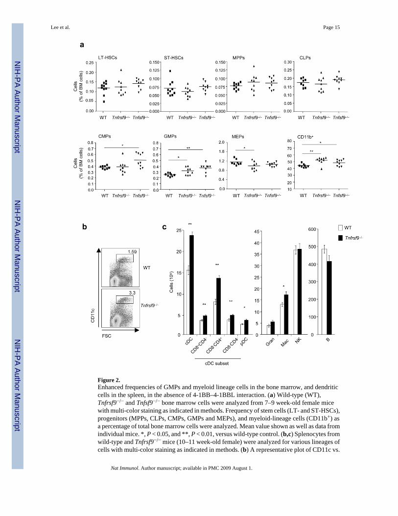
Figure 2.Enhanced frequencies of GMPs and myeloid lineage cells in the bone marrow, and dendriticcells in the spleen, in the absence of 4-1BB–4-1BBL interaction. (a) Wild-type (WT),Tnfrsf9−/− and Tnfsf9−/− bone marrow cells were analyzed from 7–9 week-old female micewith multi-color staining as indicated in methods. Frequency of stem cells (LT- and ST-HSCs),progenitors (MPPs, CLPs, CMPs, GMPs and MEPs), and myeloid-lineage cells (CD11b+) asa percentage of total bone marrow cells were analyzed. Mean value shown as well as data fromindividual mice. *, P < 0.05, and **, P < 0.01, versus wild-type control. (b,c) Splenocytes fromwild-type and Tnfrsf9−/− mice (10–11 week-old female) were analyzed for various lineages ofcells with multi-color staining as indicated in methods. (b) A representative plot of CD11c vs.
Lee et al. Page 15
Nat Immunol. Author manuscript; available in PMC 2009 August 1.
NIH
-PA Author Manuscript
NIH
-PA Author Manuscript
NIH
-PA Author Manuscript

FSC, showing percent CD11chi cells. (c) Total numbers of each cell subset per spleen. Eachbar represents mean value ± S.E.M. from eight mice. *, P < 0.05, and **, P < 0.01, versuscontrol. Data are representative of two to three independent experiments.
Lee et al. Page 16
Nat Immunol. Author manuscript; available in PMC 2009 August 1.
NIH
-PA Author Manuscript
NIH
-PA Author Manuscript
NIH
-PA Author Manuscript
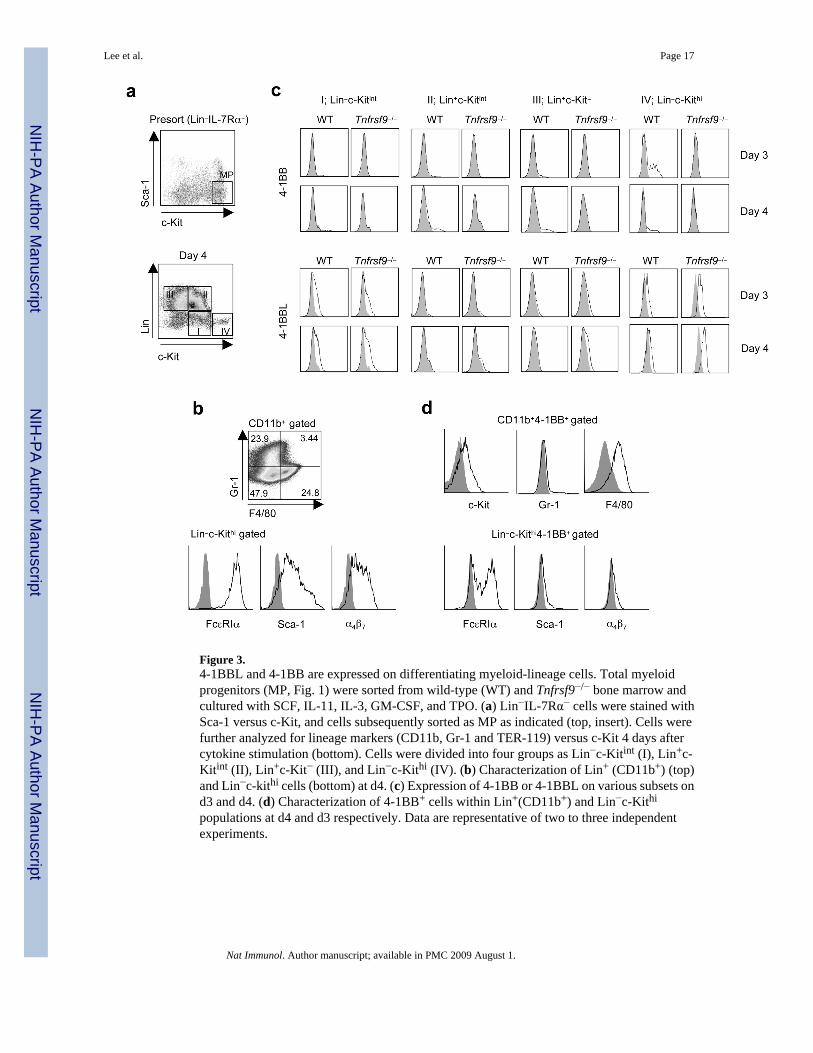
Figure 3.4-1BBL and 4-1BB are expressed on differentiating myeloid-lineage cells. Total myeloidprogenitors (MP, Fig. 1) were sorted from wild-type (WT) and Tnfrsf9−/− bone marrow andcultured with SCF, IL-11, IL-3, GM-CSF, and TPO. (a) Lin−IL-7Rα− cells were stained withSca-1 versus c-Kit, and cells subsequently sorted as MP as indicated (top, insert). Cells werefurther analyzed for lineage markers (CD11b, Gr-1 and TER-119) versus c-Kit 4 days aftercytokine stimulation (bottom). Cells were divided into four groups as Lin−c-Kitint (I), Lin+c-Kitint (II), Lin+c-Kit− (III), and Lin−c-Kithi (IV). (b) Characterization of Lin+ (CD11b+) (top)and Lin−c-kithi cells (bottom) at d4. (c) Expression of 4-1BB or 4-1BBL on various subsets ond3 and d4. (d) Characterization of 4-1BB+ cells within Lin+(CD11b+) and Lin−c-Kithi
populations at d4 and d3 respectively. Data are representative of two to three independentexperiments.
Lee et al. Page 17
Nat Immunol. Author manuscript; available in PMC 2009 August 1.
NIH
-PA Author Manuscript
NIH
-PA Author Manuscript
NIH
-PA Author Manuscript
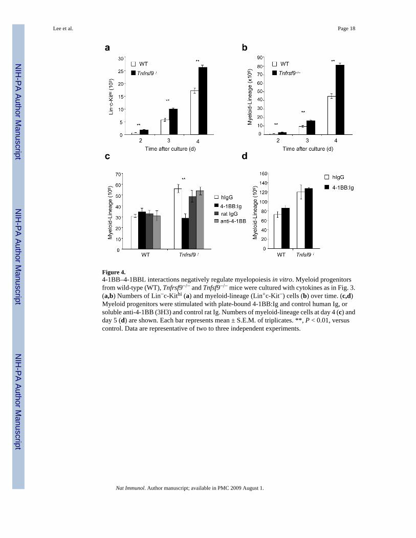
Figure 4.4-1BB–4-1BBL interactions negatively regulate myelopoiesis in vitro. Myeloid progenitorsfrom wild-type (WT), Tnfrsf9−/− and Tnfsf9−/− mice were cultured with cytokines as in Fig. 3.(a,b) Numbers of Lin−c-Kithi (a) and myeloid-lineage (Lin+c-Kit−) cells (b) over time. (c,d)Myeloid progenitors were stimulated with plate-bound 4-1BB:Ig and control human Ig, orsoluble anti-4-1BB (3H3) and control rat Ig. Numbers of myeloid-lineage cells at day 4 (c) andday 5 (d) are shown. Each bar represents mean ± S.E.M. of triplicates. **, P < 0.01, versuscontrol. Data are representative of two to three independent experiments.
Lee et al. Page 18
Nat Immunol. Author manuscript; available in PMC 2009 August 1.
NIH
-PA Author Manuscript
NIH
-PA Author Manuscript
NIH
-PA Author Manuscript
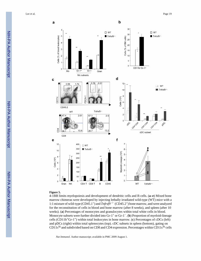
Figure 5.4-1BB limits myelopoiesis and development of dendritic cells and B cells. (a–e) Mixed bonemarrow chimeras were developed by injecting lethally irradiated wild-type (WT) mice with a1:1 mixture of wild-type (CD45.1+) and Tnfrsf9−/− (CD45.2+) bone marrow, and were analyzedfor the reconstitution of cells in blood and bone marrow (after 8 weeks), and spleen (after 10weeks). (a) Percentages of monocytes and granulocytes within total white cells in blood.Monocyte subsets were further divided into Gr-1+ or Gr-1−. (b) Proportion of myeloid-lineagecells (CD11b+Gr-1+) within total leukocytes in bone marrow. (c) Percentages of cDCs (left)and pDCs (right) within total splenocytes (top). cDC subsets in spleen (bottom), gating onCD11chi and subdivided based on CD8 and CD4 expression. Percentages within CD11chi cells
Lee et al. Page 19
Nat Immunol. Author manuscript; available in PMC 2009 August 1.
NIH
-PA Author Manuscript
NIH
-PA Author Manuscript
NIH
-PA Author Manuscript
indicated. (d,e) Actual total numbers of each indicated cell subset in spleen. White and blackbars represent cells originating from wild-type bone marrow (CD45.1+) and Tnfrsf9−/− bonemarrow (CD45.2+), respectively. P value was calculated with paired Student’s t-test. Each barrepresents mean value ± S.E.M. from three to four mice. *, P < 0.05, and **, P < 0.01, versuscontrol. All data are representative of two to three experiments. (f) Common myeloidprogenitors (CMPs) were sorted from wild-type (CD45.1+) and Tnfrsf9−/− (CD45.2+) mice anda 1:1 mixture was injected into lethally irradiated Rag1−/− (CD45.2+) mice. Development ofmyeloid-lineage cells was analyzed at day 8 after transfer. Empty and closed circles,representing numbers of wild-type and Tnfrsf9−/− myeloid-lineage cells, respectively, from thesame recipient mice are linked as lines. P value was calculated with paired Student’s t-test (p= 0.038). Histograms show mean values ± S.E.M. Data are representative of two independentexperiments.
Lee et al. Page 20
Nat Immunol. Author manuscript; available in PMC 2009 August 1.
NIH
-PA Author Manuscript
NIH
-PA Author Manuscript
NIH
-PA Author Manuscript
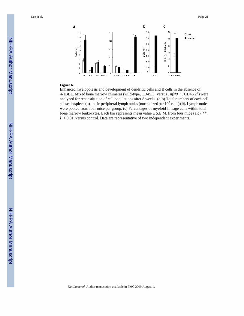
Figure 6.Enhanced myelopoiesis and development of dendritic cells and B cells in the absence of4-1BBL. Mixed bone marrow chimeras (wild-type, CD45.1+ versus Tnfsf9−/−, CD45.2+) wereanalyzed for reconstitution of cell populations after 8 weeks. (a,b) Total numbers of each cellsubset in spleen (a) and in peripheral lymph nodes (normalized per 107 cells) (b). Lymph nodeswere pooled from four mice per group. (c) Percentages of myeloid-lineage cells within totalbone marrow leukocytes. Each bar represents mean value ± S.E.M. from four mice (a,c). **,P < 0.01, versus control. Data are representative of two independent experiments.
Lee et al. Page 21
Nat Immunol. Author manuscript; available in PMC 2009 August 1.
NIH
-PA Author Manuscript
NIH
-PA Author Manuscript
NIH
-PA Author Manuscript
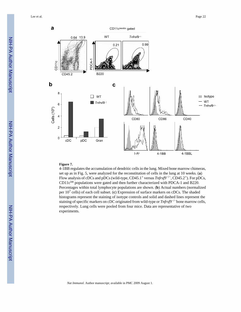
Figure 7.4-1BB regulates the accumulation of dendritic cells in the lung. Mixed bone marrow chimeras,set up as in Fig. 5, were analyzed for the reconstitution of cells in the lung at 10 weeks. (a)Flow analysis of cDCs and pDCs (wild-type, CD45.1+ versus Tnfrsf9−/−, CD45.2+). For pDCs,CD11cint populations were gated and then further characterized with PDCA-1 and B220.Percentages within total lymphocyte populations are shown. (b) Actual numbers (normalizedper 107 cells) of each cell subset. (c) Expression of surface markers on cDCs. The shadedhistograms represent the staining of isotype controls and solid and dashed lines represent thestaining of specific markers on cDC originated from wild-type or Tnfrsf9−/−bone marrow cells,respectively. Lung cells were pooled from four mice. Data are representative of twoexperiments.
Lee et al. Page 22
Nat Immunol. Author manuscript; available in PMC 2009 August 1.
NIH
-PA Author Manuscript
NIH
-PA Author Manuscript
NIH
-PA Author Manuscript
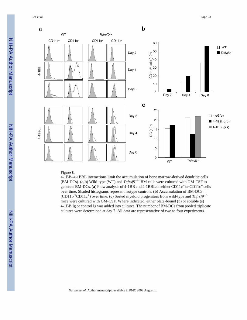
Figure 8.4-1BB–4-1BBL interactions limit the accumulation of bone marrow-derived dendritic cells(BM-DCs). (a,b) Wild-type (WT) and Tnfrsf9−/− BM cells were cultured with GM-CSF togenerate BM-DCs. (a) Flow analysis of 4-1BB and 4-1BBL on either CD11c− or CD11c+ cellsover time. Shaded histograms represent isotype controls. (b) Accumulation of BM-DCs(CD11bhiCD11c+) over time. (c) Sorted myeloid progenitors from wild-type and Tnfrsf9−/−
mice were cultured with GM-CSF. Where indicated, either plate-bound (p) or soluble (s)4-1BB:Ig or control Ig was added into cultures. The number of BM-DCs from pooled triplicatecultures were determined at day 7. All data are representative of two to four experiments.
Lee et al. Page 23
Nat Immunol. Author manuscript; available in PMC 2009 August 1.
NIH
-PA Author Manuscript
NIH
-PA Author Manuscript
NIH
-PA Author Manuscript
Related Documents