This article appeared in a journal published by Elsevier. The attached copy is furnished to the author for internal non-commercial research and education use, including for instruction at the authors institution and sharing with colleagues. Other uses, including reproduction and distribution, or selling or licensing copies, or posting to personal, institutional or third party websites are prohibited. In most cases authors are permitted to post their version of the article (e.g. in Word or Tex form) to their personal website or institutional repository. Authors requiring further information regarding Elsevier’s archiving and manuscript policies are encouraged to visit: http://www.elsevier.com/copyright

Welcome message from author
This document is posted to help you gain knowledge. Please leave a comment to let me know what you think about it! Share it to your friends and learn new things together.
Transcript
This article appeared in a journal published by Elsevier. The attachedcopy is furnished to the author for internal non-commercial researchand education use, including for instruction at the authors institution
and sharing with colleagues.
Other uses, including reproduction and distribution, or selling orlicensing copies, or posting to personal, institutional or third party
websites are prohibited.
In most cases authors are permitted to post their version of thearticle (e.g. in Word or Tex form) to their personal website orinstitutional repository. Authors requiring further information
regarding Elsevier’s archiving and manuscript policies areencouraged to visit:
http://www.elsevier.com/copyright
Author's personal copy
Identification of prostate-specific antigen (PSA) isoforms incomplex biological samples utilizing complementary platforms
Ákos Végvária,⁎, Melinda Rezelia, Charlotte Welinderb, Johan Malmc, Hans Liljac,d,e,f,György Marko-Vargaa, Thomas Laurella
aDiv. Clinical Protein Science & Imaging, Dept. of Measurement Technology and Industrial Electrical Engineering, Lund University, BMC C13,SE-221 84 Lund, SwedenbDept. of Oncology, Clinical Sciences, Lund University, Barngatan 2B, SE-221 85 Lund, SwedencDept. of Laboratory Medicine, Lund University, Malmö University Hospital, SE-205 02 Malmö, SwedendDept. of Clinical Laboratories, Memorial Sloan-Kettering Cancer Center, New York, NY 10021, USAeDept. of Surgery (Urology), Memorial Sloan-Kettering Cancer Center, New York, NY 10021, USAfDept. of Medicine (GU-Oncology), Memorial Sloan-Kettering Cancer Center, New York, NY 10021, USA
A R T I C L E I N F O A B S T R A C T
Article history:Received 21 October 2009Accepted 16 January 2010
Measurements of the prostate-specific antigen (PSA) levels in blood are widely used asdiagnostic, predictive and prognostic marker of prostate disease. The selective detection ofmolecular forms of PSA can contribute clinically to meaningful enhancements of theconventional PSA-test. As it is plausible that an in-depth search for structural variants ofPSA gene products may increase our ability to discriminate distinct patho-biological basisand stages of prostate diseases, we have developed a multi-step protocol comprising gel-based methods followed by mass spectrometric identification.Our current aim was to provide a comprehensive identification of PSA variants occurring inseminal fluid. We provide a proof-of-principle for this multiple step analytical approach toidentify multiple PSA variants from complex biological samples that revealed distinctmolecular characteristics. In addition, sequence-annotated protein bands in SDS–PAGE gelswere compared to those detected by Western blots, and by monitoring the enzymaticactivity in zymogram gels, using gelatin as a substrate. The high accuracy annotations wereobtained by fast turnaround MALDI-Orbitrap analysis from excised and digested gel bands.Multiple PSA forms were identified utilizing a combination of MASCOT and SEQUEST searchengines.
© 2010 Elsevier B.V. All rights reserved.
Keywords:Prostate-specific antigen isoformsSeminal plasmaMass spectrometryMALDI LTQ Orbitrap XL
1. Introduction
Prostate-specific antigen (PSA) is a member of the kallikreinfamily of serine proteinases that is abundantly produced andreleased by epithelial cells of the prostate [1–3]. PSA expres-sion remains exceedingly high also in advanced stages ofprostate adenocarcinomas, and the amount of PSA releasedinto blood can elevate up to 104-fold beyond normal levels in
the blood (which is about six orders of magnitude lower thanthat in seminal fluid) due to increased tumor burden com-bined with histological changes in prostate architecture thataccompany the malignant transformation [4,5].
PSA has restricted chymotrypsin-like endoproteolytic ac-tivity, which beyond the biological substrates, semenogelin 1and 2 (SEMG1 and SEMG2) [1,6], also contributes cleavages offibronectin [6], laminin and gelatin [7]. PSA is secreted as an
J O U R N A L O F P R O T E O M I C S 7 3 ( 2 0 1 0 ) 1 1 3 7 – 1 1 4 7
⁎ Corresponding author. Tel.: +46 46 222 3721; fax: +46 46 222 4527.E-mail address: [email protected] (Á. Végvári).
1874-3919/$ – see front matter © 2010 Elsevier B.V. All rights reserved.doi:10.1016/j.jprot.2010.01.008
ava i l ab l e a t www.sc i enced i r ec t . com
www.e l sev i e r . com/ loca te / j p ro t
Author's personal copy
inactive proenzyme (pro-PSA) into seminal fluid and can (atleast in vitro) be activated by the kallikrein-related peptidase 2(hK2) and other endopeptidases produced by the prostate [3,8–10]. PSA predominantly occurs in a free and active single-chain form, or as inactive (due to internal cleavages) inseminal fluid [1,2,10,11]. A minor proportion (≤5%) of the PSAin the seminal fluid is inactive, due to formation of covalentcomplexes with SERPINA5 (protein C inhibitor), which isabundantly released from the seminal vesicles [12–14]. Incontrast, PSA in blood is non-catalytic and the predominantproportion is covalently linked to SERPINA3 (α-1-antichymo-trypsin) [15,16].
Furthermore, several studies have implicated that consid-erable structural heterogeneity of the free forms of PSA occursin serum, seminal plasma, and hyperplastic-, or cancerous-tissue [2,5,11]. It is suggested that PSA isoforms may appearupon the translation of alternative hKLK3 transcripts [17]. Incontrast to the large number of KLK transcript variantsrecently identified, very few of them have been confirmed atthe protein level (Table 1 in Ref. [18]). Previous studiespresented nicked forms of PSA that were associated withprostatic diseases, linking the Lys145/Lys182 forms as over-represented in benign prostatic hyperplasia (BPH), while thenicked pro-PSA forms to be more abundant in cancer [19–21].
As it is plausible that an in-depth search for structuralvariants of PSA gene products may increase our ability todiscriminate distinct patho-biological basis and stages ofprostate disease, we have therefore developed a multi-stepprotocol comprising gel-based methods followed by massspectrometric identification. We hereby present a proteomicplatform, including gel electrophoretic separations and massspectrometric analyses in order to verify and outline themultitude forms and enzymatic function of PSA. Our goal wasto demonstrate the expression of PSA forms with annotatedsequences and quantities in seminal plasma samples from ahealthy volunteer and from young men undergoing investi-gation for infertility. We present data that identify variousmolecular forms of PSA.
2. Material and methods
2.1. Seminal plasma
Seminal plasma was obtained from a healthy volunteer andfrommen undergoing investigation for infertility prior to finaldiagnosis of any dysfunction. The samples were provided bythe Center for Reproductive Medicine at Malmö UniversityHospital, following the guidelines of the Helsinki Declaration.The collection of seminal plasma was approved by the ethicalboard at Lund University (approval number: LU 532-03), andthe samples were processed according to the WHO guidelines(WHO, 1999). Seminal plasma was obtained by centrifugationat 10000 g for 10 min. Free PSA (fPSA) ranged between 0.505and 1.855 mg/mL (see Table 1) as determined by the DELFIAassay [22] (Perkin Elmer, Turku, Finland). Samples were storedat −20 °C until use.
2.2. Gel electrophoretic techniques
PSA expression was detected by SDS–PAGE gel electrophoresisin 0.75 mm thick, 12%T, 2.67%C polyacrylamide gels underreducing conditions. The sampleswere reducedwith DTT (at afinal concentration of 4 mM) while boiled for 5 min andalkylated with addition of 2 µL of 0.5 M iodoacetamide atroom temperature. Following preparation, 10 µL of samplesand 3 µL of protein molecular weight standards (PageRuler™Prestained Protein Ladder Plus from Fermentas, St. Leon-Rot,Germany) were applied. The gels were run at a constantvoltage of 125 V for 1 h 10 min in a MiniProtean III electropho-resis unit (Bio-Rad, Hercules, CA). Staining was performedwith Coomassie blue R350 (GE Healthcare, Uppsala, Sweden)for 1 h and destaining according to the manufacturer'srecommendations.
The Western blot analysis was performed using 0.75 mmthick, 12%T, 2.67%C polyacrylamide gels run at non-reducingconditions. The separated protein bands were then trans-ferred onto PVDF membranes (0.45 µm pore size Invitrolon™PVDF, Invitrogen, Carlsbad, CA) using a tank blotting (Bio-Rad,Hercules, CA) at constant current of 350 mA for 1.5 h. Themembranes were blocked with 1% of BSA in 50 mM Tris–HCl/150 mM NaCl/0.1% Tween-20, pH 7.4. The primary antibodywas 2E9 diluted to 1:5000 [23], whereas the secondary anti-body (ECL anti-mouse IgG), used in dilution 1:10000, and the
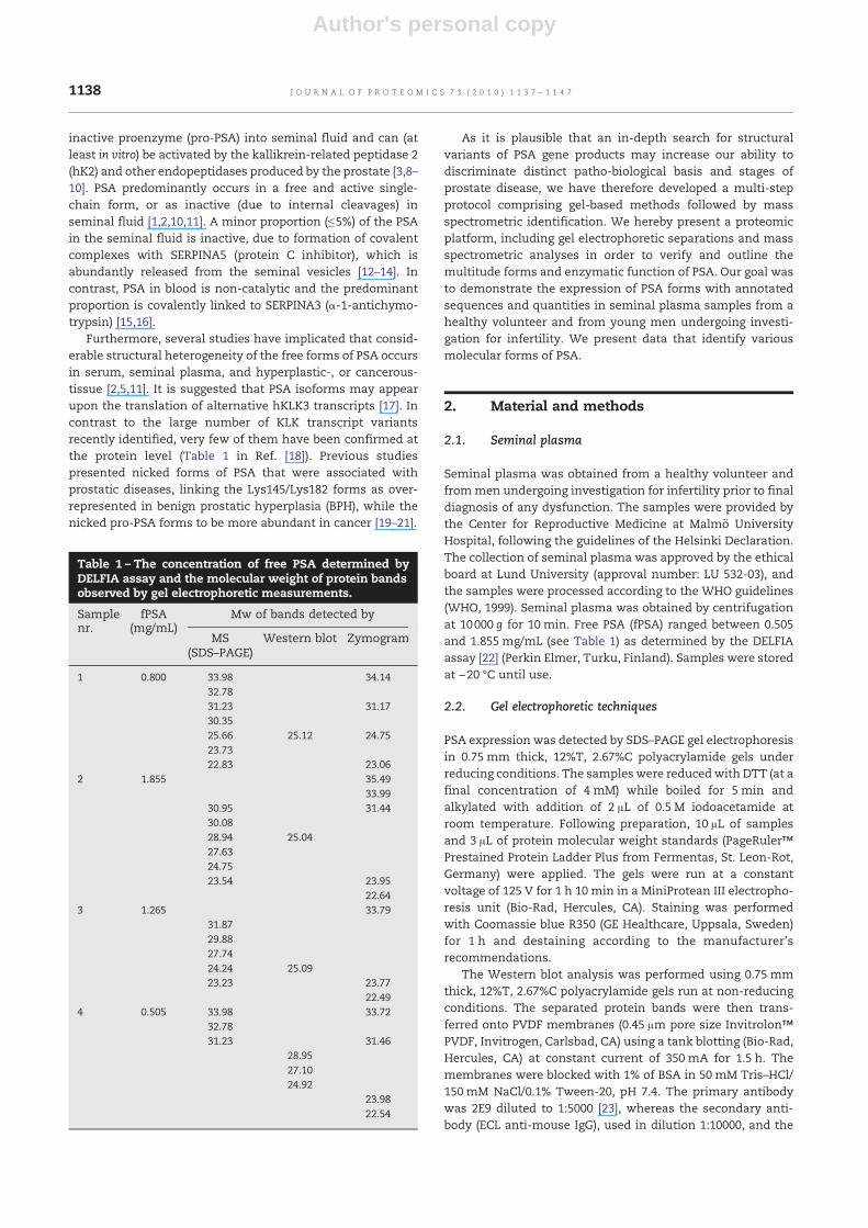
Table 1 – The concentration of free PSA determined byDELFIA assay and the molecular weight of protein bandsobserved by gel electrophoretic measurements.
Sample fPSA Mw of bands detected bynr. (mg/mL)
MS(SDS–PAGE)
Western blot Zymogram
1 0.800 33.98 34.1432.7831.23 31.1730.3525.66 25.12 24.7523.7322.83 23.06
2 1.855 35.4933.99
30.95 31.4430.0828.94 25.0427.6324.7523.54 23.95
22.643 1.265 33.79
31.8729.8827.7424.24 25.0923.23 23.77
22.494 0.505 33.98 33.72
32.7831.23 31.46
28.9527.1024.92
23.9822.54
1138 J O U R N A L O F P R O T E O M I C S 7 3 ( 2 0 1 0 ) 1 1 3 7 – 1 1 4 7
Author's personal copy
horseradish peroxidase reagent system (ECL) were from GEHealthcare (Uppsala, Sweden).
Enzymatic activity of PSA forms was monitored byzymogram gel electrophoresis using non-boiled samplealiquots in 0.75 mm thick, 10%T, 2.67%C polyacrylamidegels containing 0.1% gelatin (from porcine skin; Sigma,Steinheim, Germany) under non-reducing conditions in thepresence of SDS. In order to the proteins refold and their re-gain enzymatic activity, SDS was removed by washing it offwith Triton X-100 and then the zymographic gels wereincubated overnight at 37 °C in Zymogram Developing Buffer(Invitrogen, Carlsbad, CA). Staining was performed withCoomassie blue R350 for 1 h and de-stained according tothe manufacturer's recommendations.
2.3. Quantification
Densitometric quantification of Coomassie blue stainedprotein bands were performed on diluted seminal plasmaproteins separated in SDS–PAGE gels under reducing condi-tions. The molecular weights of proteins were determined bycalculation from the protein standards using Quantity Onesoftware (Bio-Rad, Hercules, CA).
2.4. Mass spectrometry
Following in-gel digestionof themajorproteinbands, observedwithin the size region of 20–36 kDa on SDS–PAGE gels, sampleswere analyzed by MALDI-MS providing both peptide massfingerprint (PMF) and sequencing data. The mass spectromet-ric analysis was performed on a MALDI LTQ Orbitrap XLinstrument (Thermo Scientific, Waltham, MA). Mass spectrawere obtained in positive mode within a mass range of 600–4000 Da, using 60,000 resolution (determined at 400 m/z).Twenty full mass scans (2 microscans/scan) were collectedfrom each spot. The laser, operated at 10 µJ, was movedautomatically after 2 microscans to avoid overburning of thesample. MS/MS data collection was performed on the top 25peaks that matched with the list of masses derived bytheoretical (in silico) digestion of 35 PSA variants by Peptide-Mass (available at the ExPASy Proteomics Server site, http://expasy.org/sprot/), choosing iodoacetamide as alkylationagent, oxidation onmethionine and allowing 1 miss-cleavage.The minimal signal required for MS/MS data acquisition was500 counts. Normalized collision energy was 50% during anactivation time of 30 ms and activation Q of 0.250 was applied.All experiments were performed in triplicates.
Spectra were processed by Xcalibur software v2.0.7 (ThermoScientific, Waltham,MA), averaging 20 fullmass scans followedby quantitative analysis summing the isotope intensities byusing the Xtract function in Xcalibur. The peptide massfingerprint search was performed on the MASCOT server(http://www.matrixscience.com), using the NCBInr database,narrowed down to Homo sapiens (release 20091024, 224815H. sapiens sequences out of 9,937,670 sequences). Side chainmodifications were set to fixed carbamidomethylation atcystein and variable oxidation at methionine residues, respec-tively. Peptide tolerance was kept at 10 ppm. The samples wereconsidered positive PSA identities, if at least 2 specific peptideswere identified in at least two of the triplicates.
The MS/MS search was also performed on the NCBInrdatabase, using the MASCOT search engine and allowing 1missed cleavage. Peptide tolerance was 10 ppm, whereas MS/MS tolerance was chosen to 0.4 Da. The MS/MS search wasrepeated with the SEQUEST search engine, using the Uni-Prot_human database (224,015 sequences), allowing 2 miss-cleavages. The peptide and fragment ion tolerances were setto 2 and 1 amu, respectively. The following filters were usedon the search results: Delta CN≥1; Rsp≤4; Xcorr vs chargestate≥1.5 and protein probability≤0.5. PSA identification wasaccepted if it contained at least one identified peptide.
3. Results
3.1. Concept of the multiple analytical approach
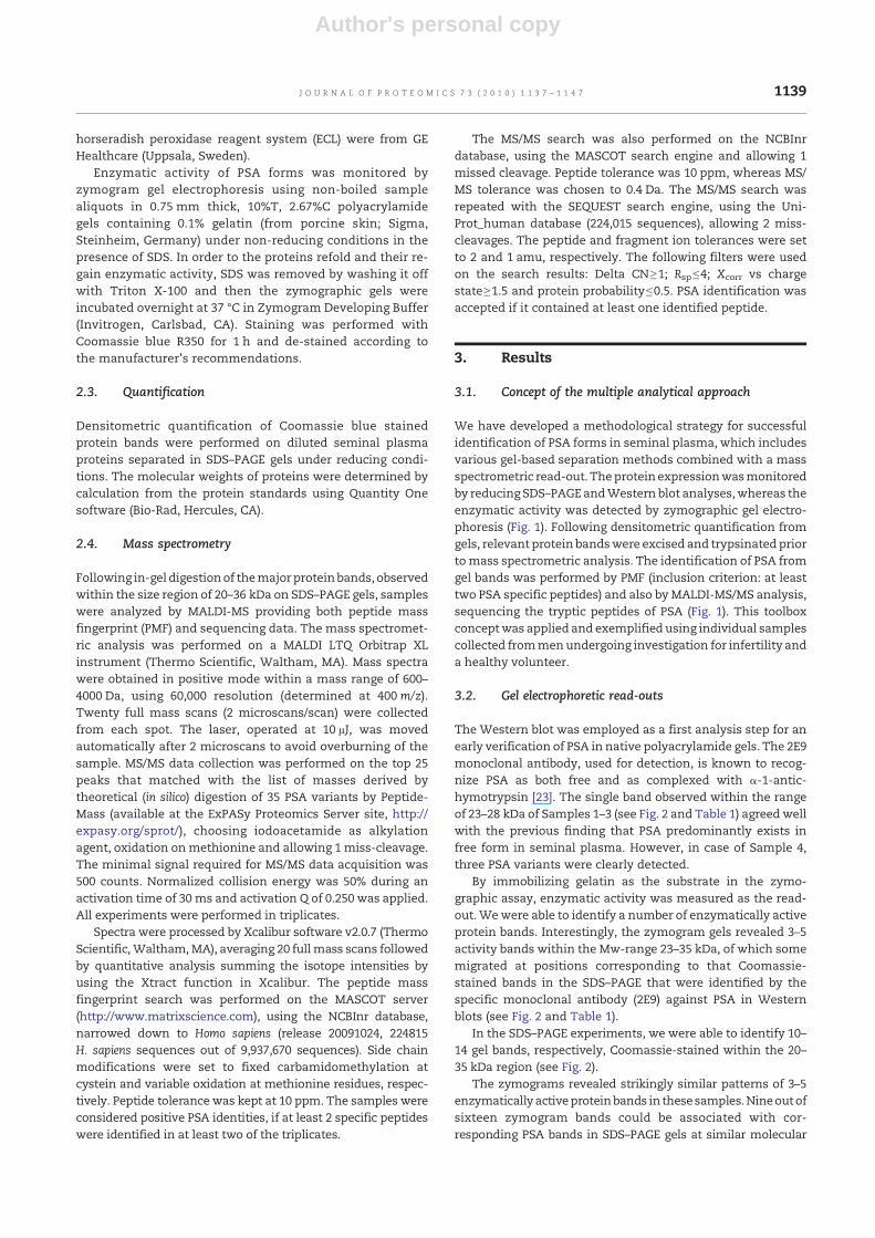
We have developed a methodological strategy for successfulidentification of PSA forms in seminal plasma, which includesvarious gel-based separation methods combined with a massspectrometric read-out. Theprotein expressionwasmonitoredby reducing SDS–PAGEandWestern blot analyses,whereas theenzymatic activity was detected by zymographic gel electro-phoresis (Fig. 1). Following densitometric quantification fromgels, relevant protein bandswere excised and trypsinated priortomass spectrometric analysis. The identification of PSA fromgel bands was performed by PMF (inclusion criterion: at leasttwo PSA specific peptides) and also by MALDI-MS/MS analysis,sequencing the tryptic peptides of PSA (Fig. 1). This toolboxconceptwas applied and exemplified using individual samplescollected frommenundergoing investigation for infertility anda healthy volunteer.
3.2. Gel electrophoretic read-outs
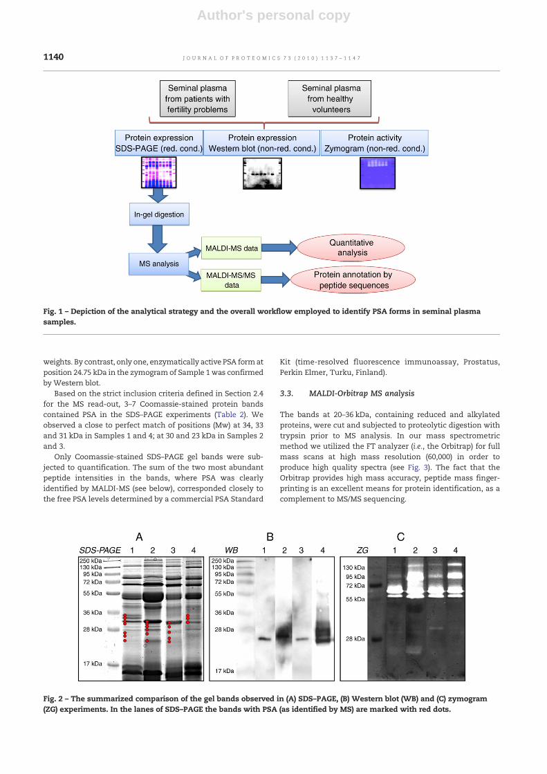
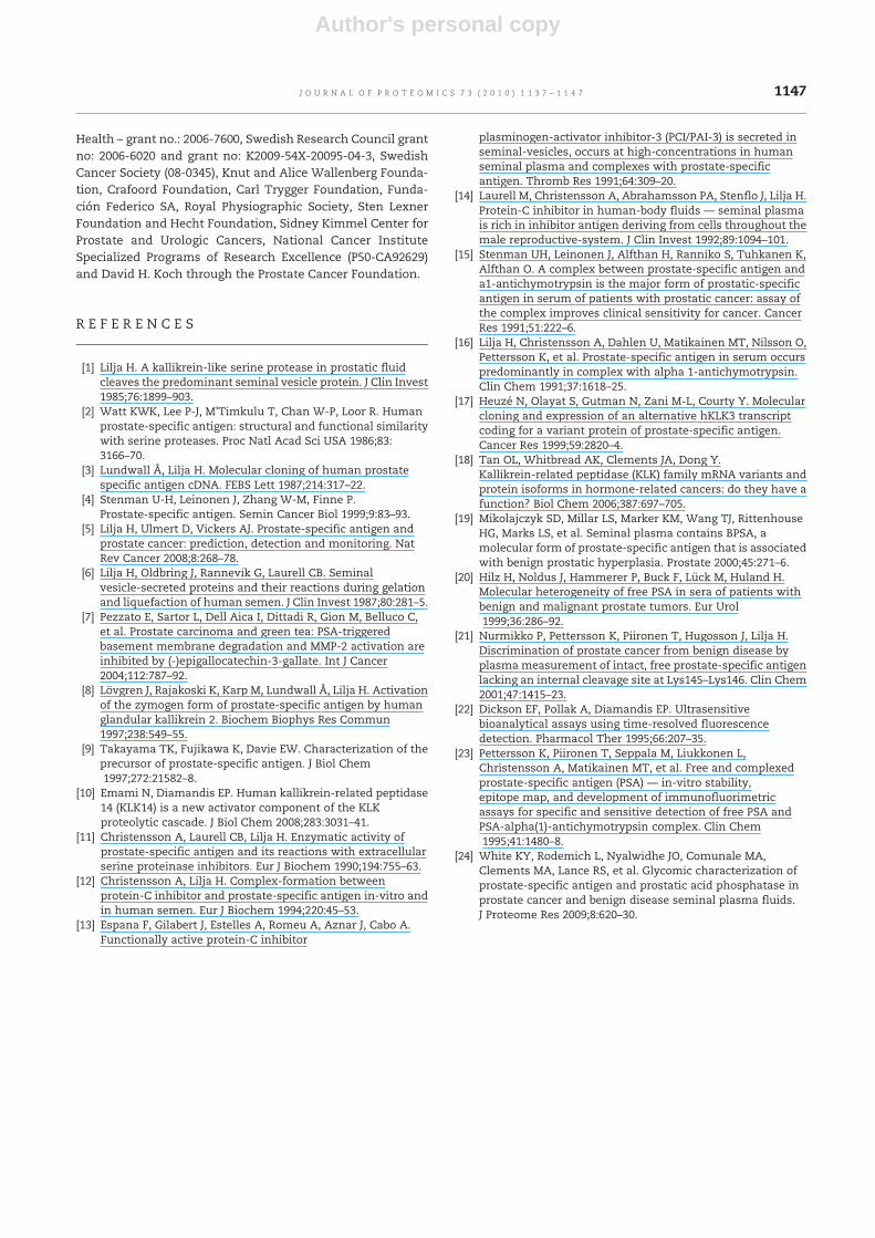
The Western blot was employed as a first analysis step for anearly verification of PSA in native polyacrylamide gels. The 2E9monoclonal antibody, used for detection, is known to recog-nize PSA as both free and as complexed with α-1-antic-hymotrypsin [23]. The single band observed within the rangeof 23–28 kDa of Samples 1–3 (see Fig. 2 and Table 1) agreedwellwith the previous finding that PSA predominantly exists infree form in seminal plasma. However, in case of Sample 4,three PSA variants were clearly detected.
By immobilizing gelatin as the substrate in the zymo-graphic assay, enzymatic activity was measured as the read-out.Wewere able to identify a number of enzymatically activeprotein bands. Interestingly, the zymogram gels revealed 3–5activity bands within the Mw-range 23–35 kDa, of which somemigrated at positions corresponding to that Coomassie-stained bands in the SDS–PAGE that were identified by thespecific monoclonal antibody (2E9) against PSA in Westernblots (see Fig. 2 and Table 1).
In the SDS–PAGE experiments, we were able to identify 10–14 gel bands, respectively, Coomassie-stained within the 20–35 kDa region (see Fig. 2).
The zymograms revealed strikingly similar patterns of 3–5enzymatically activeproteinbands in thesesamples.Nineoutofsixteen zymogram bands could be associated with cor-responding PSA bands in SDS–PAGE gels at similar molecular
1139J O U R N A L O F P R O T E O M I C S 7 3 ( 2 0 1 0 ) 1 1 3 7 – 1 1 4 7
Author's personal copy
weights. By contrast, only one, enzymatically active PSA formatposition 24.75 kDa in the zymogram of Sample 1 was confirmedby Western blot.
Based on the strict inclusion criteria defined in Section 2.4for the MS read-out, 3–7 Coomassie-stained protein bandscontained PSA in the SDS–PAGE experiments (Table 2). Weobserved a close to perfect match of positions (Mw) at 34, 33and 31 kDa in Samples 1 and 4; at 30 and 23 kDa in Samples 2and 3.
Only Coomassie-stained SDS–PAGE gel bands were sub-jected to quantification. The sum of the two most abundantpeptide intensities in the bands, where PSA was clearlyidentified by MALDI-MS (see below), corresponded closely tothe free PSA levels determined by a commercial PSA Standard
Kit (time-resolved fluorescence immunoassay, Prostatus,Perkin Elmer, Turku, Finland).
3.3. MALDI-Orbitrap MS analysis
The bands at 20–36 kDa, containing reduced and alkylatedproteins, were cut and subjected to proteolytic digestion withtrypsin prior to MS analysis. In our mass spectrometricmethod we utilized the FT analyzer (i.e., the Orbitrap) for fullmass scans at high mass resolution (60,000) in order toproduce high quality spectra (see Fig. 3). The fact that theOrbitrap provides high mass accuracy, peptide mass finger-printing is an excellent means for protein identification, as acomplement to MS/MS sequencing.
Fig. 1 – Depiction of the analytical strategy and the overall workflow employed to identify PSA forms in seminal plasmasamples.
Fig. 2 – The summarized comparison of the gel bands observed in (A) SDS–PAGE, (B) Western blot (WB) and (C) zymogram(ZG) experiments. In the lanes of SDS–PAGE the bands with PSA (as identified by MS) are marked with red dots.
1140 J O U R N A L O F P R O T E O M I C S 7 3 ( 2 0 1 0 ) 1 1 3 7 – 1 1 4 7
Author's personal copy
Table 2 – The annotated PSA sequence data from database search by both PMF and MS/MS using MASCOT and SEQUEST.
PMF MASCOT MS/MS MASCOT MS/MS SEQUEST
Sample 1Band 5 673.3764 VVHYRa
757.4915 SVILLGR854.3997870.3945
1077.5023 IVGGWECEK IVGGWECEK1272.66801407.7491 HSQPWQVLVASR HSQPWQVLVASR
FLRPGDDSSHDLMLLR FLRPGDDSSHDLMLLR1887.9388 FLRPGDDSSHDLMLLR FLRPGDDSSHDLMLLR
LQCVDLHVISNDVCAQVHPQK LQCVDLHVISNDVCAQVHPQKKLQCVDLHVISNDVCAQVHPQK
Band 6 673.3760 VVHYR757.4913854.3997 FMLCARG870.3946
1077.5023 IVGGWECEK IVGGWECEK1272.6676 LSEPAELTDAVK LSEPAELTDAVK1407.7492 HSQPWQVLVASR HSQPWQVLVASR
FLRPGDDSSHDLMLLR FLRPGDDSSHDLMLLR1887.9387 FLRPGDDSSHDLMLLR FLRPGDDSSHDLMLLR2460.2116 LQCVDLHVISNDVCAQVHPQK LQCVDLHVISNDVCAQVHPQK
KLQCVDLHVISNDVCAQVHPQK KLQCVDLHVISNDVCAQVHPQKBand 7 673.3761 VVHYR
757.4915854.3997 FMLCARG870.3945
1077.5025 IVGGWECEK IVGGWECEK1407.7499 HSQPWQVLVASR HSQPWQVLVASR
FLRPGDDSSHDLMLLR FLRPGDDSSHDLMLLR1887.9393 FLRPGDDSSHDLMLLR FLRPGDDSSHDLMLLR2460.2132 LQCVDLHVISNDVCAQVHPQK
KLQCVDLHVISNDVCAQVHPQKBand 8 673.3764
757.4919870.3949
1077.5031 IVGGWECEK IVGGWECEK1272.66861407.7503 HSQPWQVLVASR HSQPWQVLVASR1887.9400 FLRPGDDSSHDLMLLR FLRPGDDSSHDLMLLR2460.2123
Band 11 757.49181077.5028 IVGGWECEK IVGGWECEK1272.6685 LSEPAELTDAVK1407.7498 HSQPWQVLVASR HSQPWQVLVASR1887.9397 FLRPGDDSSHDLMLLR FLRPGDDSSHDLMLLR
Band 12 757.49201077.5023 IVGGWECEK IVGGWECEK1272.6687 LSEPAELTDAVK1407.7499 HSQPWQVLVASR HSQPWQVLVASR1887.9404 FLRPGDDSSHDLMLLR FLRPGDDSSHDLMLLR
Band 13 757.49191077.5035 IVGGWECEK1272.66861407.7502 HSQPWQVLVASR HSQPWQVLVASR1887.9397
Sample 2Band 7 673.3768 VVHYR
757.4924 SVILLCG854.4007 FMLCARG FMLCARG870.3959
1077.5039 IVGGWECEK IVGGWECEK
(continued on next page)
1141J O U R N A L O F P R O T E O M I C S 7 3 ( 2 0 1 0 ) 1 1 3 7 – 1 1 4 7
Author's personal copy
Table 2 (continued)
PMF MASCOT MS/MS MASCOT MS/MS SEQUEST
Sample 2Band 7 1272.6697
1407.7518 HSQPWQVLVASR HSQPWQVLVASR1871.9477 FLRPGDDSSHDLMLLR FLRPGDDSSHDLMLLR1887.9430 FLRPGDDSSHDLMLLR FLRPGDDSSHDLMLLR2344.2238 AVCGGVLVHPQWVLTAAHCIR AVCGGVLVHPQWVLTAAHCIR2460.2184 LQCVDLHVISNDVCAQVHPQK LQCVDLHVISNDVCAQVHPQK2588.3118 KLQCVDLHVISNDVCAQVHPQK KLQCVDLHVISNDVCAQVHPQK3493.69303509.6916 HSLFHPEDTGQVFQVSHSFPHPLYDMSLLK HSLFHPEDTGQVFQVSHSFPHPLYDMSLLK
Band 8 673.3771 VVHYR757.4928870.3961
1077.5040 IVGGWECEK IVGGWECEK1272.66941407.7516 HSQPWQVLVASR HSQPWQVLVASR
FLRPGDDSSHDLMLLR FLRPGDDSSHDLMLLR1887.9418 FLRPGDDSSHDLMLLR FLRPGDDSSHDLMLLR
AVCGGVLVHPQWVLTAAHCIRLQCVDLHVISNDVCAQVHPQK LQCVDLHVJSNDVCAQVHPQKKLQCVDLHVISNDVCAQVHPQK KLQCVDLHVISNDVCAQVHPQK
Band 9/10b 757.4920870.3956
1077.50301272.66921407.75101887.9409 FLRPGDDSSHDLMLLR2588.3101
Band 12 IVGGWECEK IVGGWECEK1272.6719 LSEPAELTDAVK1407.7533 HSQPWQVLVASR HSQPWQVLVASR1887.9444 FLRPGDDSSHDLMLLR FLRPGDDSSHDLMLLR
Band 13 757.4927 SVILLCG1077.5040 IVGGWECEK IVGGWECEK1272.6694 LSEPAELTDAVK LSEPAELTDAVK1407.7524 HSQPWQVLVASR HSQPWQVLVASR1887.9430 FLRPGDDSSHDLMLLR FLRPGDDSSHDLMLLR2344.22643509.6918
Sample 3Band 5c 673.3764 VVHYR
757.4920854.4000870.3951
1077.5033 IVGGWECEK IVGGWECEK1272.6693 LSEPAELTDAVK1407.7507 HSQPWQVLVASR HSQPWQVLVASR1887.9403 FLRPGDDSSHDLMLLR FLRPGDDSSHDLMLLR2460.2145 LQCVDLHVISNDVCAQVHPQK2588.3098 KLQCVDLHVJSNDVCAQVHPQK KLQCVDLHVISNDVCAQVHPQK
Band 6c 673.3768 VVHYR757.4923 SVILLGR854.4005870.3956
1077.5034 IVGGWECEK IVGGWECEK1272.6690 LSEPAELTDAVK LSEPAELTDAVK1407.7513 HSQPWQVLVASR HSQPWQVLVASR1887.9413 FLRPGDDSSHDLMLLR FLRPGDDSSHDLMLLR
AVCGGVLVHPQWVLTAAHCIR AVCGGVLVHPQWVLTAAHCIR2460.2154 LQCVDLHVISNDVCAQVHPQK LQCVDLHVISNDVCAQVHPQK2588.3091 KLQCVDLHVISNDVCAQVHPQK KLQCVDLHVISNDVCAQVHPQK3509.6976
Band 7 673.3768 VVHYR757.4922
1142 J O U R N A L O F P R O T E O M I C S 7 3 ( 2 0 1 0 ) 1 1 3 7 – 1 1 4 7
Author's personal copy
The PMF search, using MASCOT search engine, was per-formed on the monoisotopic mass lists obtained by extracting20 averaged full mass scans, resulting in unambiguous PSAidentification with amino acid sequence coverage up to 73%.The delta mass values (accuracy) were in most cases close tozero, but never higher than 3 ppm.
3.4. MS/MS strategy based on “in silico” PSA sequences
A theoretical monoisotopic mass list of possible PSA specificpeptides was generated in silico. The MS/MS fragmentsexpected from all of the available PSA forms were copiedinto theMS data acquisitionmethod and used for the selection
Table 2 (continued)
PMF MASCOT MS/MS MASCOT MS/MS SEQUEST
Sample 3Band 7 870.3953
1077.5031 IVGGWECEK IVGGWECEK1272.66891383.66931407.7503 HSQPWQVLVASR HSQPWQVLVASR1887.9400 FLRPGDDSSHDLMLLR
Band 9 757.4919870.3950
1077.5025 IVGGWECEK IVGGWECEK1272.6685 LSEPAELTDAVK1383.66891407.7497 HSQPWQVLVASR HSQPWQVLVASR1887.9398
Band 10 757.49281077.5027 IVGGWECEK IVGGWECEK1272.6699 LSEPAELTDAVK1383.67071407.7517 HSQPWQVLVASR HSQPWQVLVASR1887.9412 FLRPGDDSSHDLMLLR
Band 11 757.49301077.5046 IVGGWECEK IVGGWECEK1272.6704 LSEPAELTDAVK1407.7520 HSQPWQVLVASR HSQPWQVLVASR1887.9430 FLRPGDDSSHDLMLLR FLRPGDDSSHDLMLLR3509.6958
Sample 4Band 5 673.3770
757.4919854.4009870.3961
1077.5030 IVGGWECEK IVGGWECEK1272.66921407.7512 HSQPWQVLVASR1871.9443 FLRPGDDSSHDLMLLR FLRPGDDSSHDLMLLR
Band 6 673.3769757.4925854.4008 FMLCARG870.3954
1077.5037 IVGGWECEK IVGGWECEK1272.6693 LSEPAELTDAVK LSEPAELTDAVK1407.7510 HSQPWQVLVASR HSQPWQVLVASR1871.9460 FLRPGDDSSHDLMLLR1887.9445 FLRPGDDSSHDLMLLR
Band 7 757.4923854.4007 FMLCARG870.3960
1077.5034 IVGGWECEK IVGGWECEK1272.6694 LSEPAELTDAVK LSEPAELTDAVK1407.7512 HSQPWQVLVASR HSQPWQVLVASR1871.9465 FLRPGDDSSHDLMLLR1887.9427
a Sequences in italics indicate peptides observed only in one of the experiments performed in triplicates.b Bands were cut in a single gel piece.c Bands 5 and 6 appeared as single protein band in gel but cut into half and analyzed separately.
1143J O U R N A L O F P R O T E O M I C S 7 3 ( 2 0 1 0 ) 1 1 3 7 – 1 1 4 7

Author's personal copy
Fig.
3–A
typica
lexa
mpleof
theMSan
dMS/MSsp
ectraob
tained
withth
eMALD
ILTQ
Orb
itrapXLinstru
men
tsh
owingth
reefrag
men
tation
spec
traan
dth
eirco
rres
ponding
sequ
ence
sin
theinse
rts.
1144 J O U R N A L O F P R O T E O M I C S 7 3 ( 2 0 1 0 ) 1 1 3 7 – 1 1 4 7
Author's personal copy
of parent ions. As part of our MS strategy, we selected twenty-two different sequences of PSA variants, collected by searchingthrough the UniProtKB/SwissProt, UniProtKB/TrEMBL and NCBIprotein databases, in which we identified 35 PSA forms. Thisindicates the existence of several duplicates assigned underdifferent names. Due to the fact that many of the resultingpeptides were identical, the total number of unique sequenceswas determined to be 138. We chose to exclude those withm/zless than 500 Da, but peptides with both modified and un-modified cystein and methionine side-chains were included.
Although, fragmentation in the ion trap can be furtheranalyzed in the FT analyzer, we decided to use the ion trap forMS/MS data generation, trading the high mass resolution forhigh sensitivity. In evidence, comparing the MALDI Obritrapresults with those obtained by nanoLC-ESI (on a LTQ XL ETDmass spectrometer, Thermo Scientific) the tryptic peptidepatterns of PSAwere found to overlap, whereby these findingscould be confirmed (data not shown). PSAwas identified basedon fragment (MS/MS) data, using both the MASCOT, as well asthe SEQUEST search engines (Table 2). We were able tosuccessfully identify 12 PSA peptides using these stringentinclusion and exclusion criteria. All of the MS/MS spectra
showed at least 50% complete b- and y-ion series (insets inFig. 3). We identified 14 PSA peptides by PMF, which were inclose agreement with our MS/MS hits.
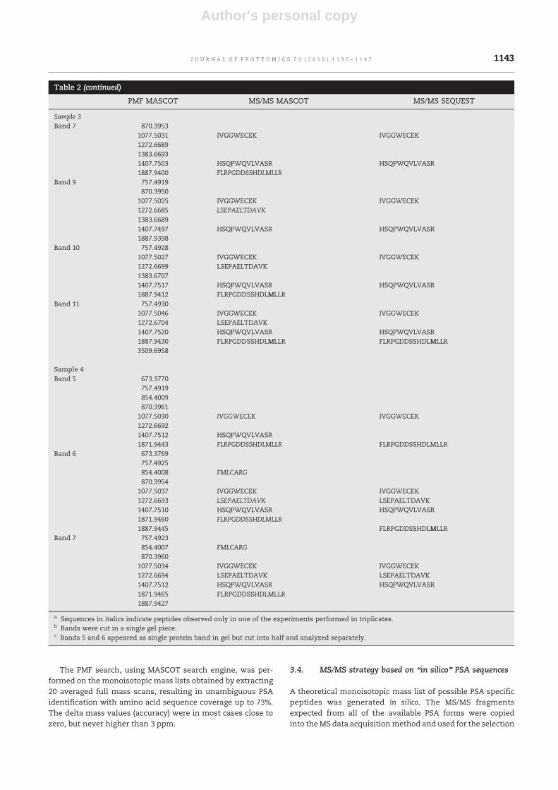
The semi-quantitative analysis of MALDI-MS data, (basedon the sum of isotopic patterns in 20 averaged full massscans), showed good correlation by using HSQPWQVLVASRand FLRPGDDSSHDLM*LLR, as PSA-target sequences (as illus-trated in Fig. 4) when compared with the fPSA valuesmeasured by an ELISA standardized against the WHO-endorsed calibration. In addition, we were able to prove thatthe averaged m/z intensities of the two selected targetpeptides correlate with the densitometric quantification.
4. Discussions
Several PSA isoforms have been described in the literaturethroughout the years, such as the alternative splice variants,and different glycosylations [24], of which some could beassociated with diseases [20]. However, no systematic study hasever been conducted at the protein expression level. One of thereasons is probably the lack of robust and rigorously validated
Fig. 4 – MS identification of PSA bands in gels depicted as it was determined by MALDI-MS data in peptide mass fingerprintsearch in the NCBInr database. The bars show the average of cumulative intensities of the two most abundant peptides incorresponding bands in SDS–PAGE gels. The volumes of the boxes refer to the contribution to the cumulative intensity of thesepeptides HSQPWQVLVASR (m/z=1407.753) and FLRPGDDSSHDLM*LLR (m/z=1887.945), which are shown in red and blue colors,respectively.
1145J O U R N A L O F P R O T E O M I C S 7 3 ( 2 0 1 0 ) 1 1 3 7 – 1 1 4 7
Author's personal copy
analytical protocols that are capable of demonstrating anassociation between expressed PSA forms and disease. Usingonly a minute amount of seminal plasma (3.6 µl), our currentexperience and validation of performance of the presentedtoolbox concept indicates that it will be suited to run large seriesof samples from patients vs controls, e.g., using collection of bio-repositories from large cohorts.
Interestingly, the highly selective and sensitive Westernblot analysis could only determine one protein band with amolecular weight of 25 kDa in three samples. MS data didprovide evidence that the corresponding bands in SDS–PAGEgel at 25 kDa in Samples 1 and 2 contained PSA. However, thePSA level in these bands appeared to be much lower incomparison to other PSA forms (as indicated from the gelbands) identified by the MS. This important observation mayopen up further in-depth studies of additional PSA forms thatcould be related to a given biological function. The zymo-graphic separations could verify an enzymatically activeprotein within the 22.5–35.5 Mw region, but only one band inSample 2 correlated well with the WB findings. Ninecorresponding protein bands could also be localized in theSDS–PAGE gel positions, where PSA was identified by MS in allcases. Consequently, the mass spectrometric analysis plays acentral role in the identification of the protein in semensamples. Hence, MS data appear critical to further under-standing of PSA expression and disease linkage. Furthermore,we expect a good agreement in-between this mass spectrom-etry-based assay and the clinical diagnostic ELISA used as aninternational standard, which measures the affinity of anti-gen-, and antigen-like proteins in the final read-out.
Rapid screening is possible with the new generation ofMALDI instrumentation, providing qualitative MS/MS sequenc-ing by the MALDI LTQ Orbitrap XL, wherein the number ofsamples no longer remain to be a major limitation. Thisplatform also has the advantage that it allows soft laser energyto be applied to the sample within the crystal spot, resulting inhigh sequence coverage and signal intensity in MS/MS peptidesequence peaks. The ion trap principle can provide high signalyields that, from a practical aspect, make it possible to analyzerepeatedly with minimal sample loss. This feature has a par-ticular advantage in runningMS/MS sequencing that is unusualcompared to traditional MALDI instrumentation, where highfrequency lasers will deplete the sample rather quickly.
On an experimental basis, MS/MS searches resulted in hitson several PSA isoforms by blast searching through the NCBIand the UniProt databases. Additional PSA isoforms could berecognized in comprehensive searches through additionaldatabases, such as the US Patent database. Our findings verifythat unique iso-specific PSA forms do exist. However, the iso-annotation with the entire sequence coverage, as well as thepossible modifications is still not sufficiently verified andpresently remains unclear.
We found identical PSA variants that are listed under othernames and/or annotations. One example is the isoform CRA_hon locus EAW71929 (gi:119592335) and the splice variant RP5on locus CAD54617 (gi:24370944) that have identical aminoacid sequences in the NCBI database. Furthermore, many ofthese proposed isoforms were splice variants; some of themwere artificial sequences, which were not yet confirmed at theprotein expression level.
In conclusion, we have shown that our platform enablesthe identification PSA molecular forms by means of peptidemass fingerprinting and MS sequencing. The application ofour platformon seminal plasma samples clearly indicates thatMS annotation can identify more molecular forms of PSAcompared with Western and zymographic analyses. Wepropose that by using our strategy to identify and verifysome of these novel variants at the protein level, which canopen up a better understanding of the disease mechanisms.This may also include whether PSA has a causal influence inthe development or progression of prostate disease orinfertility. We also envision that the proposed analyticalplatform can be applicable to PSA determination in prostatecancer as preceding sample enrichment may overcome priorlimitations due the low abundance of the protein target in theblood.
5. Conclusions
In this study we present a useful combination of techniques,which can be employed for collection of refined informationregarding patterns of PSA isoforms in clinical samples. Aprofound difference was observed at the protein expressionlevel comparing four samples from patients undergoinginvestigation for infertility and a healthy volunteer. TheWestern blot analysis proved that the monoclonal antibody(2E9) used for detection could clearly interact with a single orthreemolecular variants present in samples. However, the MSanalysis proved the existence of 15 PSA forms with differentmolecular weights at various concentration ratios.
We believe that our observation of various isoforms ofPSA in patients reported in this communication maycontribute to the further identification of disease-relevantheterogeneity of PSA, including transcriptional and post-translational modifications present due to various stages andcauses of prostate disease. This may also allow comprehen-sive structural characterization of this important targetprotein in samples with low concentration of PSA, such asblood, in the near future. This is particularly interesting sincecirculating PSA exists predominantly in complexed formand the structurally and functionally detailed relationshipbetween isoform patterns in prostate tissue and blood ispresently unclear.
Conflict of interest
Dr. Hans Lilja holds patents for free PSA and hK2 assays.
Acknowledgments
We would like to thank Gun-Britt Eriksson at the Dept. ofClinical Chemistry, University Hospital, Malmö, Sweden, fortechnical assistance; Martin Hornshaw and Egon Rosén atThermo Fisher Scientific, for mass spectrometry support. Theauthors are grateful for funding support from the SwedishResearch Council, Vinnova and Foundation for StrategicResearch – The Programme: Biomedical Engineering for Better
1146 J O U R N A L O F P R O T E O M I C S 7 3 ( 2 0 1 0 ) 1 1 3 7 – 1 1 4 7
Author's personal copy
Health – grant no.: 2006-7600, Swedish Research Council grantno: 2006-6020 and grant no: K2009-54X-20095-04-3, SwedishCancer Society (08-0345), Knut and Alice Wallenberg Founda-tion, Crafoord Foundation, Carl Trygger Foundation, Funda-ción Federico SA, Royal Physiographic Society, Sten LexnerFoundation and Hecht Foundation, Sidney Kimmel Center forProstate and Urologic Cancers, National Cancer InstituteSpecialized Programs of Research Excellence (P50-CA92629)and David H. Koch through the Prostate Cancer Foundation.
R E F E R E N C E S
[1] Lilja H. A kallikrein-like serine protease in prostatic fluidcleaves the predominant seminal vesicle protein. J Clin Invest1985;76:1899–903.
[2] Watt KWK, Lee P-J, M'Timkulu T, Chan W-P, Loor R. Humanprostate-specific antigen: structural and functional similaritywith serine proteases. Proc Natl Acad Sci USA 1986;83:3166–70.
[3] Lundwall Å, Lilja H. Molecular cloning of human prostatespecific antigen cDNA. FEBS Lett 1987;214:317–22.
[4] Stenman U-H, Leinonen J, Zhang W-M, Finne P.Prostate-specific antigen. Semin Cancer Biol 1999;9:83–93.
[5] Lilja H, Ulmert D, Vickers AJ. Prostate-specific antigen andprostate cancer: prediction, detection and monitoring. NatRev Cancer 2008;8:268–78.
[6] Lilja H, Oldbring J, Rannevik G, Laurell CB. Seminalvesicle-secreted proteins and their reactions during gelationand liquefaction of human semen. J Clin Invest 1987;80:281–5.
[7] Pezzato E, Sartor L, Dell Aica I, Dittadi R, Gion M, Belluco C,et al. Prostate carcinoma and green tea: PSA-triggeredbasement membrane degradation and MMP-2 activation areinhibited by (-)epigallocatechin-3-gallate. Int J Cancer2004;112:787–92.
[8] Lövgren J, Rajakoski K, Karp M, Lundwall Å, Lilja H. Activationof the zymogen form of prostate-specific antigen by humanglandular kallikrein 2. Biochem Biophys Res Commun1997;238:549–55.
[9] Takayama TK, Fujikawa K, Davie EW. Characterization of theprecursor of prostate-specific antigen. J Biol Chem1997;272:21582–8.
[10] Emami N, Diamandis EP. Human kallikrein-related peptidase14 (KLK14) is a new activator component of the KLKproteolytic cascade. J Biol Chem 2008;283:3031–41.
[11] Christensson A, Laurell CB, Lilja H. Enzymatic activity ofprostate-specific antigen and its reactions with extracellularserine proteinase inhibitors. Eur J Biochem 1990;194:755–63.
[12] Christensson A, Lilja H. Complex-formation betweenprotein-C inhibitor and prostate-specific antigen in-vitro andin human semen. Eur J Biochem 1994;220:45–53.
[13] Espana F, Gilabert J, Estelles A, Romeu A, Aznar J, Cabo A.Functionally active protein-C inhibitor
plasminogen-activator inhibitor-3 (PCI/PAI-3) is secreted inseminal-vesicles, occurs at high-concentrations in humanseminal plasma and complexes with prostate-specificantigen. Thromb Res 1991;64:309–20.
[14] Laurell M, Christensson A, Abrahamsson PA, Stenflo J, Lilja H.Protein-C inhibitor in human-body fluids — seminal plasmais rich in inhibitor antigen deriving from cells throughout themale reproductive-system. J Clin Invest 1992;89:1094–101.
[15] Stenman UH, Leinonen J, Alfthan H, Ranniko S, Tuhkanen K,Alfthan O. A complex between prostate-specific antigen anda1-antichymotrypsin is the major form of prostatic-specificantigen in serum of patients with prostatic cancer: assay ofthe complex improves clinical sensitivity for cancer. CancerRes 1991;51:222–6.
[16] Lilja H, Christensson A, Dahlen U, Matikainen MT, Nilsson O,Pettersson K, et al. Prostate-specific antigen in serum occurspredominantly in complex with alpha 1-antichymotrypsin.Clin Chem 1991;37:1618–25.
[17] Heuzé N, Olayat S, Gutman N, Zani M-L, Courty Y. Molecularcloning and expression of an alternative hKLK3 transcriptcoding for a variant protein of prostate-specific antigen.Cancer Res 1999;59:2820–4.
[18] Tan OL, Whitbread AK, Clements JA, Dong Y.Kallikrein-related peptidase (KLK) family mRNA variants andprotein isoforms in hormone-related cancers: do they have afunction? Biol Chem 2006;387:697–705.
[19] Mikolajczyk SD, Millar LS, Marker KM, Wang TJ, RittenhouseHG, Marks LS, et al. Seminal plasma contains BPSA, amolecular form of prostate-specific antigen that is associatedwith benign prostatic hyperplasia. Prostate 2000;45:271–6.
[20] Hilz H, Noldus J, Hammerer P, Buck F, Lück M, Huland H.Molecular heterogeneity of free PSA in sera of patients withbenign and malignant prostate tumors. Eur Urol1999;36:286–92.
[21] Nurmikko P, Pettersson K, Piironen T, Hugosson J, Lilja H.Discrimination of prostate cancer from benign disease byplasma measurement of intact, free prostate-specific antigenlacking an internal cleavage site at Lys145–Lys146. Clin Chem2001;47:1415–23.
[22] Dickson EF, Pollak A, Diamandis EP. Ultrasensitivebioanalytical assays using time-resolved fluorescencedetection. Pharmacol Ther 1995;66:207–35.
[23] Pettersson K, Piironen T, Seppala M, Liukkonen L,Christensson A, Matikainen MT, et al. Free and complexedprostate-specific antigen (PSA) — in-vitro stability,epitope map, and development of immunofluorimetricassays for specific and sensitive detection of free PSA andPSA-alpha(1)-antichymotrypsin complex. Clin Chem1995;41:1480–8.
[24] White KY, Rodemich L, Nyalwidhe JO, Comunale MA,Clements MA, Lance RS, et al. Glycomic characterization ofprostate-specific antigen and prostatic acid phosphatase inprostate cancer and benign disease seminal plasma fluids.J Proteome Res 2009;8:620–30.
1147J O U R N A L O F P R O T E O M I C S 7 3 ( 2 0 1 0 ) 1 1 3 7 – 1 1 4 7
Related Documents