Identification of possible biological stains for differential staining of Normal and Malignant tumour tissue Nor Hayati Othman, Pathology dept, PPSP [email protected]

Identification of possible biological stains for differential staining of Normal and Malignant tumour tissue Nor Hayati Othman, Pathology dept, PPSP [email protected].
Dec 21, 2015
Welcome message from author
This document is posted to help you gain knowledge. Please leave a comment to let me know what you think about it! Share it to your friends and learn new things together.
Transcript
Identification of possible biological stains for differential
staining of Normal and Malignant tumour tissue
Nor Hayati Othman,
Pathology dept, PPSP
Potential partner[s]
• Chemists
• Biologists
• Material engineers
Objectives
• To search for suitable natural stains derived from national plants of Malaysia
• To determine the differential staining of the new stain on human normal and malignant tumour tissue

Normal Vs cancer cell
Normal celln:c 1:4-8smooth nuclear membraneno nucleus
Malignant celln:cmore hyperchromatic [more basic]nuclear membrane coarsechromatic coarse, stippledmitosispleomorphic
Cytoplasm - ± opaque
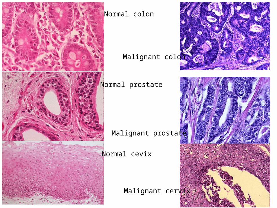
Normal colon
Malignant colon
Normal prostate
Malignant prostate
Normal cevix
Malignant cervix

Normal lung
Malignant lung
Normal breast
Malignant breast
Normal thyroid
Malignant thyroid
Those are easy cases!!
• Subtle changes in cell require further tests
• Immunohistochemsitry– We stain the proteins found in cells eg muco-
proteins, keratins, vimentins, osteobilins, neurofilaments etc
– Every protein has different characteristics and could be stained

The stain should be
• Permanent
• Able to differentiate acidic nucleus from basic cytoplasm
• Able to pick up loss of desmosomes when normal epithelial cell become malignant
• Able to detect changes in nucleus when cells become malignant

How to verify the stains?
• 100 Normal and 100 Epithelial Malignant human tissues obtained from the archives of Pathology laboratory.
• The ability to differentiate will be scored and statistically analysed

• This is possible innovation …but probably time consuming.
• Require several trials and errors
• Perseverance will be rewarded!
Related Documents