Identification of mutants in inbred Xenopus tropicalis Timothy C. Grammer a , Mustafa K. Khokha a,b , Maura A. Lane a , Kentson Lam a , Richard M. Harland a, * a Department of Molecular and Cell Biology and the Center for Integrative Genomics, University of California, 142 LSA, Berkeley, CA 94720-3204, USA b Department of Pediatrics, UCSF School of Medicine, 505 Parnassus Ave., San Francisco, CA 94143, USA Received 24 July 2004; received in revised form 8 October 2004; accepted 4 November 2004 Abstract Xenopus tropicalis offers the potential for genetic analysis in an amphibian. In order to take advantage of this potential, we have been inbreeding strains of frogs for future mutagenesis. While inbreeding a population of Nigerian frogs, we identified three mutations in the genetic background of this strain. These mutations are all recessive embryonic lethals. We show that multigenerational mutant analysis is feasible and demonstrate that mutations can be identified, propagated, and readily characterized using hybrid, dihybrid, and even trihybrid crosses. In addition, we are optimizing conditions to raise frogs rapidly and present our protocols for X. tropicalis husbandry. We find that males mature faster than females (currently 4 versus 6 months to sexual maturity). Here we document our progress in developing X. tropicalis as a genetic model organism and demonstrate the utility of the frog to study the genetics of early vertebrate development. q 2004 Published by Elsevier Ireland Ltd. Keywords: Xenopus; Tadpoles; Mutants; Husbandry; Genetics; grinch; curly; bubblehead 1. Introduction The advantages of Xenopus for studying early vertebrate development include year round fertility, large numbers of embryos per ovulation, external development, and long fertile lifespans (O10 years). Xenopus laevis is studied widely and has made many important contributions to the study of embryology. A major benefit of using Xenopus is the ease with which oocytes and embryos can be micro- injected and manipulated (Peng, 1991; Sive et al., 2000). Gain-of-function studies involving the overexpression of exogenous factors are facile in Xenopus (Grammer et al., 2000). Inhibitory molecules such as dominant-negative reagents have been successfully used in loss-of-function experiments in Xenopus. Antisense and morpholino oligo- nucleotide technologies have also proven to be highly effective in loss-of-function studies (Woolf et al., 1990; Heasman et al., 1994, 2000; Heasman, 2002). Another emerging frog model system, X. tropicalis, shares many of the features of X. laevis (Beck and Slack, 2001). Admittedly, microinjection of X. tropicalis embryos is more difficult than in X. laevis due to their smaller size and less tolerance of temperature regulation (Khokha et al., 2002). However, while X. laevis genetic mutants have been identified (Krotoski et al., 1985; Dudek et al., 1987), X. tropicalis appears to be more amenable to genetic analysis than X. laevis (Amaya et al., 1998; Hirsch et al., 2002). X. tropicalis has a shorter generation time and is diploid. X. tropicalis is smaller which reduces housing costs and space requirements for genetic screens. Because of its diploid genome and the availability of inbred lines, X. tropicalis genes may be easier to target than X. laevis with morpholino oligonucleotide loss-of-function studies (Khokha et al., 2002). Projects are currently underway to develop X. tropicalis as a genetically tractable model organism (Hirsch et al., 2002; Klein et al., 2002). In addition, multiple resources are being generated that will greatly facilitate genetic analysis. The genome of X. tropicalis is currently being sequenced and major efforts to generate X. tropicalis EST sequences, BAC libraries, and 0925-4773/$ - see front matter q 2004 Published by Elsevier Ireland Ltd. doi:10.1016/j.mod.2004.11.003 Mechanisms of Development 122 (2005) 263–272 www.elsevier.com/locate/modo * Corresponding author. Tel.: C1 510 643 6003; fax: C1 510 643 6334. E-mail address: [email protected] (R.M. Harland).

Welcome message from author
This document is posted to help you gain knowledge. Please leave a comment to let me know what you think about it! Share it to your friends and learn new things together.
Transcript
Identification of mutants in inbred Xenopus tropicalis
Timothy C. Grammera, Mustafa K. Khokhaa,b, Maura A. Lanea,Kentson Lama, Richard M. Harlanda,*
aDepartment of Molecular and Cell Biology and the Center for Integrative Genomics, University of California, 142 LSA, Berkeley, CA 94720-3204, USAbDepartment of Pediatrics, UCSF School of Medicine, 505 Parnassus Ave., San Francisco, CA 94143, USA
Received 24 July 2004; received in revised form 8 October 2004; accepted 4 November 2004
Abstract
Xenopus tropicalis offers the potential for genetic analysis in an amphibian. In order to take advantage of this potential, we have been
inbreeding strains of frogs for future mutagenesis. While inbreeding a population of Nigerian frogs, we identified three mutations in the
genetic background of this strain. These mutations are all recessive embryonic lethals. We show that multigenerational mutant analysis is
feasible and demonstrate that mutations can be identified, propagated, and readily characterized using hybrid, dihybrid, and even trihybrid
crosses. In addition, we are optimizing conditions to raise frogs rapidly and present our protocols for X. tropicalis husbandry. We find that
males mature faster than females (currently 4 versus 6 months to sexual maturity). Here we document our progress in developing X. tropicalis
as a genetic model organism and demonstrate the utility of the frog to study the genetics of early vertebrate development.
q 2004 Published by Elsevier Ireland Ltd.
Keywords: Xenopus; Tadpoles; Mutants; Husbandry; Genetics; grinch; curly; bubblehead
1. Introduction
The advantages of Xenopus for studying early vertebrate
development include year round fertility, large numbers of
embryos per ovulation, external development, and long
fertile lifespans (O10 years). Xenopus laevis is studied
widely and has made many important contributions to the
study of embryology. A major benefit of using Xenopus is
the ease with which oocytes and embryos can be micro-
injected and manipulated (Peng, 1991; Sive et al., 2000).
Gain-of-function studies involving the overexpression of
exogenous factors are facile in Xenopus (Grammer et al.,
2000). Inhibitory molecules such as dominant-negative
reagents have been successfully used in loss-of-function
experiments in Xenopus. Antisense and morpholino oligo-
nucleotide technologies have also proven to be highly
effective in loss-of-function studies (Woolf et al., 1990;
Heasman et al., 1994, 2000; Heasman, 2002).
0925-4773/$ - see front matter q 2004 Published by Elsevier Ireland Ltd.
doi:10.1016/j.mod.2004.11.003
* Corresponding author. Tel.: C1 510 643 6003; fax: C1 510 643 6334.
E-mail address: [email protected] (R.M. Harland).
Another emerging frog model system, X. tropicalis,
shares many of the features of X. laevis (Beck and Slack,
2001). Admittedly, microinjection of X. tropicalis embryos
is more difficult than in X. laevis due to their smaller size
and less tolerance of temperature regulation (Khokha et al.,
2002). However, while X. laevis genetic mutants have been
identified (Krotoski et al., 1985; Dudek et al., 1987),
X. tropicalis appears to be more amenable to genetic
analysis than X. laevis (Amaya et al., 1998; Hirsch et al.,
2002). X. tropicalis has a shorter generation time and is
diploid. X. tropicalis is smaller which reduces housing costs
and space requirements for genetic screens. Because of its
diploid genome and the availability of inbred lines,
X. tropicalis genes may be easier to target than X. laevis
with morpholino oligonucleotide loss-of-function studies
(Khokha et al., 2002). Projects are currently underway to
develop X. tropicalis as a genetically tractable model
organism (Hirsch et al., 2002; Klein et al., 2002).
In addition, multiple resources are being generated that
will greatly facilitate genetic analysis. The genome of
X. tropicalis is currently being sequenced and major efforts
to generate X. tropicalis EST sequences, BAC libraries, and
Mechanisms of Development 122 (2005) 263–272
www.elsevier.com/locate/modo
T.C. Grammer et al. / Mechanisms of Development 122 (2005) 263–272264
cDNAs are underway (Klein et al., 2002). Furthermore,
polymorphic X. tropicalis inbred lines are being generated
(Hirsch et al., 2002) that can be interbred for future mapping
efforts. Using these animals, a high resolution linkage map
is being generated (see website at http://tropmap.biology.
uh.edu). With these resources, there is great potential for
X. tropicalis to become an effective genetic model system.
The National Institutes of Health is supporting projects to
develop X. tropicalis genetic resources (see websites
at http://www.nih.gov/science/models/xenopus/ and http://
tropicalis.berkeley.edu/home/).
If X. tropicalis is to become a useful genetic model
system, then: (1) molecular and genetic resources need to be
assembled, (2) husbandry conditions must be established
which result in mature animals in a short period of time and
(3) mutants must be identified and propagated through
successive generations. As mentioned above, multiple
efforts are underway to establish the first criteria (Hirsch
et al., 2002; Klein et al., 2002). We present in this paper our
results to help establish the final two conditions.
In this paper, we present our husbandry results on sexual
maturation and sex ratios when raising numerous families
and generations of X. tropicalis. In addition, we have been
inbreeding strains of X. tropicalis. During these inbreeding
efforts, we have identified several genetic mutations in the
inbred Nigerian strain. We have identified single, double,
and triple mutant carrier frogs and show that the mutations
segregate independently. Here we present three X. tropicalis
mutations present in the genetic background of the Nigerian
strain: grinch, curly, and bubblehead. These mutations
clearly demonstrate that multigenerational mutant analysis
is feasible in X. tropicalis and that mutations can be
identified, propagated, and readily characterized using
hybrid, dihybrid, and even trihybrid crosses.
2. Results and discussion
2.1. Inbreeding and husbandry
We are inbreeding strains of X. tropicalis to generate
useful frog lines. Ten fifth generation (F5) inbred
Nigerian X. tropicalis frogs were obtained (kind gift of
Dr R. Grainger) and bred to generate F6–F9 generation frogs
(for more detail on inbred lines see http://tropicalis.berkeley.
edu/home/genetic_resources/Inbred-strains/inbred-strains.
html and Dr R. Grainger’s group at the University of Virginia
at http://faculty.virginia.edu/xtropicalis/). During this
inbreeding program, we have been optimizing our husbandry
protocols, since generation time and housing space dictate
the most severe limits to a genetic screen.
Natural matings were done since they have several
advantages over in vitro fertilizations to generate tadpoles
and frogs: male parents are not sacrificed and natural
matings routinely generate greater yields of embryos than in
vitro fertilizations. Natural matings are induced with hCG
injections and egg yields and fertilization rates do not
appear dependent on the time of year (data not shown).
Healthy, well-fed males can be mated every 2 weeks and
females every 1–2 months. We have generated over 2000
clutches of embryos, and raised approximately 4000
inbred frogs.
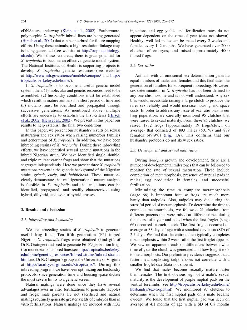
2.2. Sex ratios
Animals with chromosomal sex determination generate
equal numbers of males and females and this facilitates the
generation of families for subsequent inbreeding. However,
sex determination in X. tropicalis has not been defined to
any one chromosome and is not well understood. Any sex
bias would necessitate raising a large clutch to produce the
rarer sex reliably and would increase housing and space
costs. In order to address any issue of sex ratio bias in our
frog population, we carefully monitored 95 clutches that
were raised to sexual maturity. From these 95 clutches, we
raised 1782 frogs (approximately 19 frogs/clutch on
average) that consisted of 893 males (50.1%) and 889
females (49.9%) (Fig. 1A). This confirms that our
husbandry protocols do not skew sex ratios.
2.3. Development and sexual maturation
During Xenopus growth and development, there are a
number of developmental milestones that can be followed to
monitor the rate of sexual maturation. These include
completion of metamorphosis, presence of nuptial pads in
males, egg production in females, and successful
fertilization.
Minimizing the time to complete metamorphosis
(stage 66) is important because frogs are much more
hardy than tadpoles. Also, tadpoles may die during the
stressful period of metamorphosis. To determine the time to
complete metamorphosis, we followed 21 clutches from
different parents that were raised at different times during
the course of a year and noted when the first froglet (stage
66) occurred in each clutch. The first froglet occurred on
average at 33 days of age with a standard deviation (SD) of
2.5 days. We find that the entire clutch typically completes
metamorphosis within 2 weeks after the first froglet appears.
We saw no apparent trends or differences between what
time of year the clutch was generated and how long it took
to metamorphosis. Our preliminary evidence suggests that a
faster metamorphosing tadpole does not correlate with a
smaller froglet size (data not shown).
We find that males become sexually mature faster
than females. The first obvious sign of a male’s sexual
maturity is the development of purple nuptial pads on the
ventral forelimbs (see http://tropicalis.berkeley.edu/home/
husbandry/sex-trop.html). We monitored 97 clutches to
determine when the first nuptial pads on a male became
evident. We found that the first nuptial pad was seen on
average at 4.1 months of age with a SD of 0.7 months
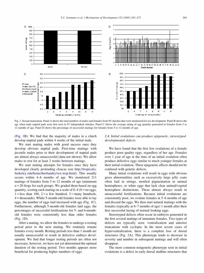
Fig. 1. Sexual maturation. Panel A shows the total numbers of males and females from 95 clutches that were monitored for sex development. Panel B shows the
age when male nuptial pads were first seen in 97 independent clutches. Panel C shows the average rating of egg quantity generated in females from 5 to
12 months of age. Panel D shows the percentage of successful matings for females from 5 to 12 months of age.
T.C. Grammer et al. / Mechanisms of Development 122 (2005) 263–272 265
(Fig. 1B). We find that the majority of males in a clutch
develop nuptial pads within 4 weeks of the initial male.
We start mating males with good success once they
develop obvious nuptial pads. First-time matings with
juvenile males prior to their development of nuptial pads
are almost always unsuccessful (data not shown). We allow
males to rest for at least 2 weeks between matings.
We start mating attempts for females once they have
developed clearly protruding cloacas (see http://tropicalis.
berkeley.edu/home/husbandry/sex-trop.html). This usually
occurs within 4–6 months of age. We monitored 211
matings of females from 5 to 12 months of age (minimum
nZ20 frogs for each group). We graded them based on egg
quantity, scoring each mating on a scale of 0–4 (0Zno eggs,
1Zless than 100, 2Za few hundred, 3Zmany hundreds,
4Zthousands). While 5-month-old females were able to lay
eggs, the number of eggs laid increased with age (Fig. 1C).
Furthermore, although 5-month-old females laid eggs, the
percentages of successful fertilizations for 5- and 6-month-
old females were consistently less than older females
(Fig. 1D).
After a mating, we allow the females to undergo a resting
period prior to the next mating. We routinely remate
females every month. Resting periods less than 1 month are
usually unsuccessful or result in defective embryo devel-
opment. We find that longer resting periods are often not
necessary, however, we have not yet determined the optimal
duration of the resting period. Two months appears more
beneficial for producing higher numbers of eggs.
2.4. Initial ovulations can produce epigenetic, stereotyped
developmental defects
We have found that the first few ovulations of a female
produce poor quality eggs, regardless of her age. Females
over 1 year of age at the time of an initial ovulation often
produce defective eggs similar to much younger females at
their initial ovulation. These epigenetic effects should not be
confused with genetic defects.
Many initial ovulations will result in eggs with obvious
gross abnormalities such as excessively large jelly coats
often laid in strings, mottled pigmentation in animal
hemispheres, or white eggs that lack clear animal/vegetal
hemisphere distinctions. These almost always result in
unsuccessful fertilizations. Because initial ovulations are
consistently poor, we ovulate females at 5–6 months of age
and discard the eggs. We then start natural matings with the
females (typically at 6–7 months of age) 1 month after their
first successful laying of normal looking eggs.
Stereotyped defects often occur in embryos generated in
the first several matings of immature females. Two types of
defects are typically seen: ventralization and anterior
truncations with cyclopia. In the most severe cases of
hyperventralization, there is a complete loss of dorsal
structures (Fig. 2A). This phenotype tends to decrease in
severity and number in subsequent matings and will often
disappear.
The more common nongenetic phenotype seen in initial
ovulations is a defect in early dorsal midline structures that
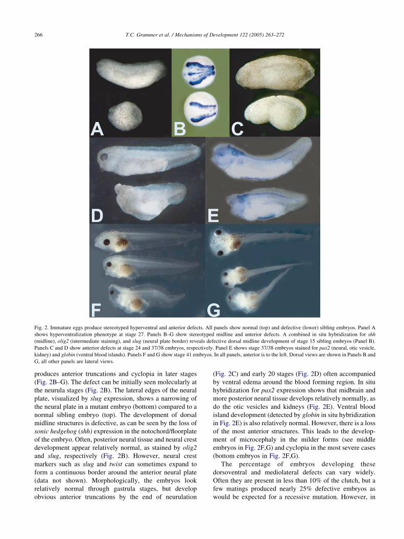
Fig. 2. Immature eggs produce stereotyped hyperventral and anterior defects. All panels show normal (top) and defective (lower) sibling embryos. Panel A
shows hyperventralization phenotype at stage 27. Panels B–G show stereotyped midline and anterior defects. A combined in situ hybridization for shh
(midline), olig2 (intermediate staining), and slug (neural plate border) reveals defective dorsal midline development of stage 15 sibling embryos (Panel B).
Panels C and D show anterior defects at stage 24 and 37/38 embryos, respectively. Panel E shows stage 37/38 embryos stained for pax2 (neural, otic vesicle,
kidney) and globin (ventral blood islands). Panels F and G show stage 41 embryos. In all panels, anterior is to the left. Dorsal views are shown in Panels B and
G, all other panels are lateral views.
T.C. Grammer et al. / Mechanisms of Development 122 (2005) 263–272266
produces anterior truncations and cyclopia in later stages
(Fig. 2B–G). The defect can be initially seen molecularly at
the neurula stages (Fig. 2B). The lateral edges of the neural
plate, visualized by slug expression, shows a narrowing of
the neural plate in a mutant embryo (bottom) compared to a
normal sibling embryo (top). The development of dorsal
midline structures is defective, as can be seen by the loss of
sonic hedgehog (shh) expression in the notochord/floorplate
of the embryo. Often, posterior neural tissue and neural crest
development appear relatively normal, as stained by olig2
and slug, respectively (Fig. 2B). However, neural crest
markers such as slug and twist can sometimes expand to
form a continuous border around the anterior neural plate
(data not shown). Morphologically, the embryos look
relatively normal through gastrula stages, but develop
obvious anterior truncations by the end of neurulation
(Fig. 2C) and early 20 stages (Fig. 2D) often accompanied
by ventral edema around the blood forming region. In situ
hybridization for pax2 expression shows that midbrain and
more posterior neural tissue develops relatively normally, as
do the otic vesicles and kidneys (Fig. 2E). Ventral blood
island development (detected by globin in situ hybridization
in Fig. 2E) is also relatively normal. However, there is a loss
of the most anterior structures. This leads to the develop-
ment of microcephaly in the milder forms (see middle
embryos in Fig. 2F,G) and cyclopia in the most severe cases
(bottom embryos in Fig. 2F,G).
The percentage of embryos developing these
dorsoventral and mediolateral defects can vary widely.
Often they are present in less than 10% of the clutch, but a
few matings produced nearly 25% defective embryos as
would be expected for a recessive mutation. However, in
T.C. Grammer et al. / Mechanisms of Development 122 (2005) 263–272 267
over 600 matings that have shown these defects, subsequent
matings show a continual decline in the numbers of embryos
developing these phenotypes. Therefore, these phenotypes
are epigenetic.
While these effects can alter the viability of early
embryos and complicate an evaluation of phenotypes in a
forward genetic screen, a number of these embryos will
be unaffected and survive. Therefore, this epigenetic
effect does not extend the generation time of X. tropicalis.
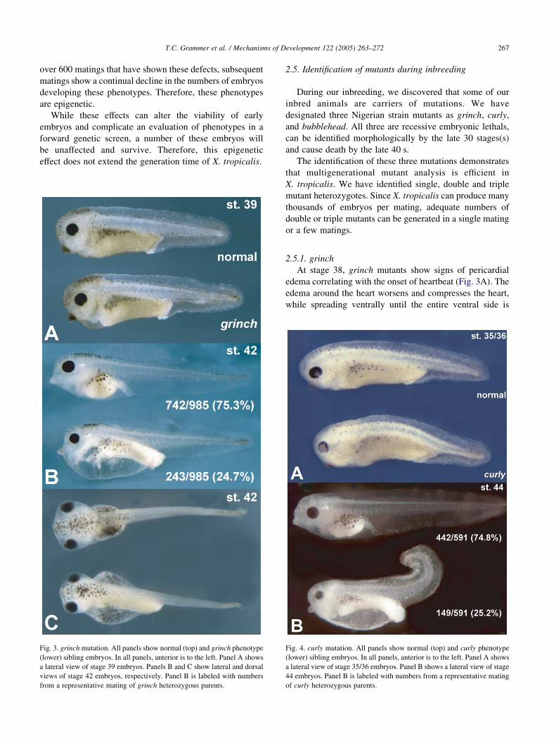
Fig. 3. grinch mutation. All panels show normal (top) and grinch phenotype
(lower) sibling embryos. In all panels, anterior is to the left. Panel A shows
a lateral view of stage 39 embryos. Panels B and C show lateral and dorsal
views of stage 42 embryos, respectively. Panel B is labeled with numbers
from a representative mating of grinch heterozygous parents.
2.5. Identification of mutants during inbreeding
During our inbreeding, we discovered that some of our
inbred animals are carriers of mutations. We have
designated three Nigerian strain mutants as grinch, curly,
and bubblehead. All three are recessive embryonic lethals,
can be identified morphologically by the late 30 stages(s)
and cause death by the late 40 s.
The identification of these three mutations demonstrates
that multigenerational mutant analysis is efficient in
X. tropicalis. We have identified single, double and triple
mutant heterozygotes. Since X. tropicalis can produce many
thousands of embryos per mating, adequate numbers of
double or triple mutants can be generated in a single mating
or a few matings.
2.5.1. grinch
At stage 38, grinch mutants show signs of pericardial
edema correlating with the onset of heartbeat (Fig. 3A). The
edema around the heart worsens and compresses the heart,
while spreading ventrally until the entire ventral side is
Fig. 4. curly mutation. All panels show normal (top) and curly phenotype
(lower) sibling embryos. In all panels, anterior is to the left. Panel A shows
a lateral view of stage 35/36 embryos. Panel B shows a lateral view of stage
44 embryos. Panel B is labeled with numbers from a representative mating
of curly heterozygous parents.
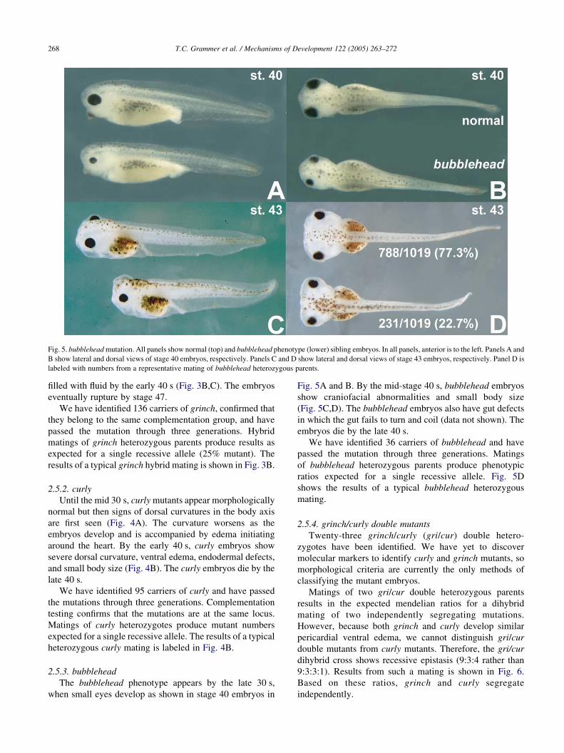
Fig. 5. bubblehead mutation. All panels show normal (top) and bubblehead phenotype (lower) sibling embryos. In all panels, anterior is to the left. Panels A and
B show lateral and dorsal views of stage 40 embryos, respectively. Panels C and D show lateral and dorsal views of stage 43 embryos, respectively. Panel D is
labeled with numbers from a representative mating of bubblehead heterozygous parents.
T.C. Grammer et al. / Mechanisms of Development 122 (2005) 263–272268
filled with fluid by the early 40 s (Fig. 3B,C). The embryos
eventually rupture by stage 47.
We have identified 136 carriers of grinch, confirmed that
they belong to the same complementation group, and have
passed the mutation through three generations. Hybrid
matings of grinch heterozygous parents produce results as
expected for a single recessive allele (25% mutant). The
results of a typical grinch hybrid mating is shown in Fig. 3B.
2.5.2. curly
Until the mid 30 s, curly mutants appear morphologically
normal but then signs of dorsal curvatures in the body axis
are first seen (Fig. 4A). The curvature worsens as the
embryos develop and is accompanied by edema initiating
around the heart. By the early 40 s, curly embryos show
severe dorsal curvature, ventral edema, endodermal defects,
and small body size (Fig. 4B). The curly embryos die by the
late 40 s.
We have identified 95 carriers of curly and have passed
the mutations through three generations. Complementation
testing confirms that the mutations are at the same locus.
Matings of curly heterozygotes produce mutant numbers
expected for a single recessive allele. The results of a typical
heterozygous curly mating is labeled in Fig. 4B.
2.5.3. bubblehead
The bubblehead phenotype appears by the late 30 s,
when small eyes develop as shown in stage 40 embryos in
Fig. 5A and B. By the mid-stage 40 s, bubblehead embryos
show craniofacial abnormalities and small body size
(Fig. 5C,D). The bubblehead embryos also have gut defects
in which the gut fails to turn and coil (data not shown). The
embryos die by the late 40 s.
We have identified 36 carriers of bubblehead and have
passed the mutation through three generations. Matings
of bubblehead heterozygous parents produce phenotypic
ratios expected for a single recessive allele. Fig. 5D
shows the results of a typical bubblehead heterozygous
mating.
2.5.4. grinch/curly double mutants
Twenty-three grinch/curly (gri/cur) double hetero-
zygotes have been identified. We have yet to discover
molecular markers to identify curly and grinch mutants, so
morphological criteria are currently the only methods of
classifying the mutant embryos.
Matings of two gri/cur double heterozygous parents
results in the expected mendelian ratios for a dihybrid
mating of two independently segregating mutations.
However, because both grinch and curly develop similar
pericardial ventral edema, we cannot distinguish gri/cur
double mutants from curly mutants. Therefore, the gri/cur
dihybrid cross shows recessive epistasis (9:3:4 rather than
9:3:3:1). Results from such a mating is shown in Fig. 6.
Based on these ratios, grinch and curly segregate
independently.
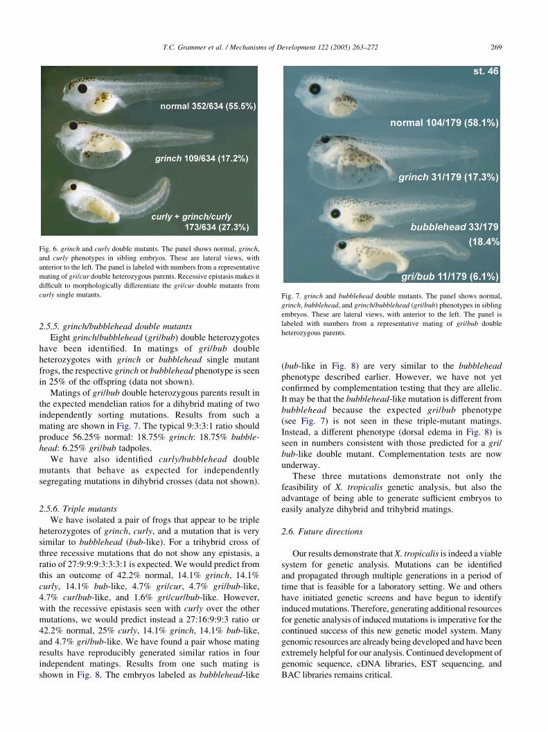
Fig. 6. grinch and curly double mutants. The panel shows normal, grinch,
and curly phenotypes in sibling embryos. These are lateral views, with
anterior to the left. The panel is labeled with numbers from a representative
mating of gri/cur double heterozygous parents. Recessive epistasis makes it
difficult to morphologically differentiate the gri/cur double mutants from
curly single mutants. Fig. 7. grinch and bubblehead double mutants. The panel shows normal,
grinch, bubblehead, and grinch/bubblehead (gri/bub) phenotypes in sibling
embryos. These are lateral views, with anterior to the left. The panel is
labeled with numbers from a representative mating of gri/bub double
heterozygous parents.
T.C. Grammer et al. / Mechanisms of Development 122 (2005) 263–272 269
2.5.5. grinch/bubblehead double mutants
Eight grinch/bubblehead (gri/bub) double heterozygotes
have been identified. In matings of gri/bub double
heterozygotes with grinch or bubblehead single mutant
frogs, the respective grinch or bubblehead phenotype is seen
in 25% of the offspring (data not shown).
Matings of gri/bub double heterozygous parents result in
the expected mendelian ratios for a dihybrid mating of two
independently sorting mutations. Results from such a
mating are shown in Fig. 7. The typical 9:3:3:1 ratio should
produce 56.25% normal: 18.75% grinch: 18.75% bubble-
head: 6.25% gri/bub tadpoles.
We have also identified curly/bubblehead double
mutants that behave as expected for independently
segregating mutations in dihybrid crosses (data not shown).
2.5.6. Triple mutants
We have isolated a pair of frogs that appear to be triple
heterozygotes of grinch, curly, and a mutation that is very
similar to bubblehead (bub-like). For a trihybrid cross of
three recessive mutations that do not show any epistasis, a
ratio of 27:9:9:9:3:3:3:1 is expected. We would predict from
this an outcome of 42.2% normal, 14.1% grinch, 14.1%
curly, 14.1% bub-like, 4.7% gri/cur, 4.7% gri/bub-like,
4.7% cur/bub-like, and 1.6% gri/cur/bub-like. However,
with the recessive epistasis seen with curly over the other
mutations, we would predict instead a 27:16:9:9:3 ratio or
42.2% normal, 25% curly, 14.1% grinch, 14.1% bub-like,
and 4.7% gri/bub-like. We have found a pair whose mating
results have reproducibly generated similar ratios in four
independent matings. Results from one such mating is
shown in Fig. 8. The embryos labeled as bubblehead-like
(bub-like in Fig. 8) are very similar to the bubblehead
phenotype described earlier. However, we have not yet
confirmed by complementation testing that they are allelic.
It may be that the bubblehead-like mutation is different from
bubblehead because the expected gri/bub phenotype
(see Fig. 7) is not seen in these triple-mutant matings.
Instead, a different phenotype (dorsal edema in Fig. 8) is
seen in numbers consistent with those predicted for a gri/
bub-like double mutant. Complementation tests are now
underway.
These three mutations demonstrate not only the
feasibility of X. tropicalis genetic analysis, but also the
advantage of being able to generate sufficient embryos to
easily analyze dihybrid and trihybrid matings.
2.6. Future directions
Our results demonstrate that X. tropicalis is indeed a viable
system for genetic analysis. Mutations can be identified
and propagated through multiple generations in a period of
time that is feasible for a laboratory setting. We and others
have initiated genetic screens and have begun to identify
induced mutations. Therefore, generating additional resources
for genetic analysis of induced mutations is imperative for the
continued success of this new genetic model system. Many
genomic resources are already being developed and have been
extremely helpful for our analysis. Continued development of
genomic sequence, cDNA libraries, EST sequencing, and
BAC libraries remains critical.
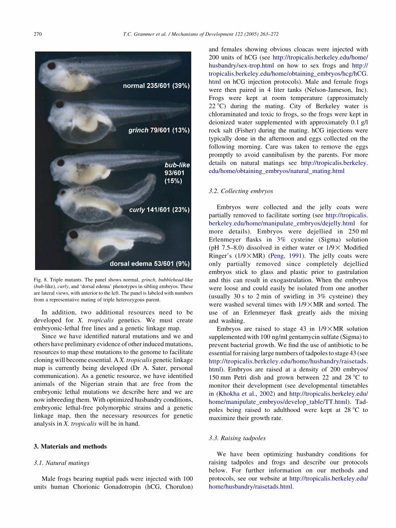
Fig. 8. Triple mutants. The panel shows normal, grinch, bubblehead-like
(bub-like), curly, and ‘dorsal edema’ phenotypes in sibling embryos. These
are lateral views, with anterior to the left. The panel is labeled with numbers
from a representative mating of triple heterozygous parent.
T.C. Grammer et al. / Mechanisms of Development 122 (2005) 263–272270
In addition, two additional resources need to be
developed for X. tropicalis genetics. We must create
embryonic-lethal free lines and a genetic linkage map.
Since we have identified natural mutations and we and
others have preliminary evidence of other induced mutations,
resources to map these mutations to the genome to facilitate
cloning will become essential. A X. tropicalis genetic linkage
map is currently being developed (Dr A. Sater, personal
communication). As a genetic resource, we have identified
animals of the Nigerian strain that are free from the
embryonic lethal mutations we describe here and we are
now inbreeding them. With optimized husbandry conditions,
embryonic lethal-free polymorphic strains and a genetic
linkage map, then the necessary resources for genetic
analysis in X. tropicalis will be in hand.
3. Materials and methods
3.1. Natural matings
Male frogs bearing nuptial pads were injected with 100
units human Chorionic Gonadotropin (hCG, Chorulon)
and females showing obvious cloacas were injected with
200 units of hCG (see http://tropicalis.berkeley.edu/home/
husbandry/sex-trop.html on how to sex frogs and http://
tropicalis.berkeley.edu/home/obtaining_embryos/hcg/hCG.
html on hCG injection protocols). Male and female frogs
were then paired in 4 liter tanks (Nelson-Jameson, Inc).
Frogs were kept at room temperature (approximately
22 8C) during the mating. City of Berkeley water is
chloraminated and toxic to frogs, so the frogs were kept in
deionized water supplemented with approximately 0.1 g/l
rock salt (Fisher) during the mating. hCG injections were
typically done in the afternoon and eggs collected on the
following morning. Care was taken to remove the eggs
promptly to avoid cannibalism by the parents. For more
details on natural matings see http://tropicalis.berkeley.
edu/home/obtaining_embryos/natural_mating.html
3.2. Collecting embryos
Embryos were collected and the jelly coats were
partially removed to facilitate sorting (see http://tropicalis.
berkeley.edu/home/manipulate_embryos/dejelly.html for
more details). Embryos were dejellied in 250 ml
Erlenmeyer flasks in 3% cysteine (Sigma) solution
(pH 7.5–8.0) dissolved in either water or 1/9! Modified
Ringer’s (1/9!MR) (Peng, 1991). The jelly coats were
only partially removed since completely dejellied
embryos stick to glass and plastic prior to gastrulation
and this can result in exogastrulation. When the embryos
were loose and could easily be isolated from one another
(usually 30 s to 2 min of swirling in 3% cysteine) they
were washed several times with 1/9!MR and sorted. The
use of an Erlenmeyer flask greatly aids the mixing
and washing.
Embryos are raised to stage 43 in 1/9!MR solution
supplemented with 100 ng/ml gentamycin sulfate (Sigma) to
prevent bacterial growth. We find the use of antibiotic to be
essential for raising large numbers of tadpoles to stage 43 (see
http://tropicalis.berkeley.edu/home/husbandry/raisetads.
html). Embryos are raised at a density of 200 embryos/
150 mm Petri dish and grown between 22 and 28 8C to
monitor their development (see developmental timetables
in (Khokha et al., 2002) and http://tropicalis.berkeley.edu/
home/manipulate_embryos/develop_table/TT.html). Tad-
poles being raised to adulthood were kept at 28 8C to
maximize their growth rate.
3.3. Raising tadpoles
We have been optimizing husbandry conditions for
raising tadpoles and frogs and describe our protocols
below. For further information on our methods and
protocols, see our website at http://tropicalis.berkeley.edu/
home/husbandry/raisetads.html.
T.C. Grammer et al. / Mechanisms of Development 122 (2005) 263–272 271
3.3.1. First 2 weeks of age
Tadpoles are raised in 1/9!MR with gentamycin in Petri
dishes for the first several days in our laboratory and kept in
28 8C incubators. They are regularly monitored for normal
development and to remove any debris. The dishes are
supplemented with fresh 1/9!MR with gentamycin once a
day. When the tadpoles become free swimming (st. 43), they
are introduced to our recirculating water system.
When introducing the tadpoles to the recirculating water
system, the tadpoles are transferred to 1/20!MR without
gentamycin and put into an empty, clean 2.75 liter tank
(Aquatic Habitats). Tadpoles from a single mating (referred
to as a clutch) are split into two or three tanks so that any
problem with one tank will not result in the loss of the entire
clutch. Tadpoles are initially raised at a density of 20–50
tadpoles per 2.75 liter tank. Higher numbers of tadpoles/
tank can be raised for the first 2 weeks, but this results in
slower growth and will result in severe overcrowding within
2 weeks.
The pH in the tanks is maintained between 7 and 7.5.
City of Berkeley water is often acidic. The incoming
water pH is adjusted by adding sodium bicarbonate to a
reservoir tank prior to its introduction to the recirculating
system. The water temperature is maintained between 26
and 27 8C.
For the first 2 weeks of life, tadpoles are fed a solution of
Sera Micron tadpole food (Pondside Herp Supply)
suspended in water from the recirculating system. We
feed once or twice a day. Feeding frequency should be
increased to 3 or 4 times a day (or more) if the water is
cleared quickly. This will depend on the density and size of
the tadpoles as well as the flow rate of the water through
the tank.
The water drip rate is initially set at 1 drop every
1–3 s. Faster flows will very often kill tadpoles, while
slower flow rates often result in complete stopping of
flow, probably due to periodic fluctuations in water
pressure. We first introduce the tadpoles in a minimal
amount of 1/20!MR supplemented with Sera Micron
solution and start the drip. The tank should fill up slowly
over a day, allowing for a gradual transition from 1/20!MR to recirculating water. Once the tank is full, the slow
drip provides a continuous exchange of water but without
a high current flow that can be detrimental to the young
tadpoles. As the tadpoles grow, the drip rate is gradually
increased to keep the water clean and prevent decay of
debris sitting in the tank. Increasing the drip incrementally
prevents shocking the tadpoles. By the time hindlimbs
appear (approximately 2–3 weeks of age) the drip rate is
about 2–3 drops per second.
After feeding the tadpoles, we routinely stop the inflow
drip for about an hour to facilitate feeding. This prevents
the food from being removed by the flushing action of
the water flow. Except for these feeding intervals, the
drips are continuously flowing. Over time, uneaten food
and debris will accumulate on the bottom of the tank.
We find that this is not detrimental to the tadpoles as long
as the drips are kept flowing to provide a continuous
exchange of water to maintain consistency in the water
quality in the tanks.
3.3.2. Care of older tadpoles through metamorphosis
Around 2 weeks of age, the largest 12–15 tadpoles in
each tank are kept and the rest removed or transferred to
more tanks. This lower density (3–5 tadpoles/l) results in
faster growth and metamorphosis (M.A. Lane, unpublished
results).
At 14 days of age through to metamorphosis, we feed the
tadpoles ‘Sifted Tadpole Diet’, in addition to Sera
Micron (see http://tropicalis.berkeley.edu/home/husbandry/
raisetads.html#Anchor-Sifted-6296). The supplemented
diet introduces a higher protein content and solid food for
metamorphosing tadpoles and young froglets. This
alleviates the need to remove froglets from the tank and
saves on housing space.
At 14 days of age, we feed each 2.75 liter (l) tank of
tadpoles about 1/2 cubic centimeter (cc) (1/8 teaspoon)
Sifted Tadpole Diet twice a day for 5 days in addition to the
Sera Micron solution. After 5 days of feeding, we increase
the amount to 1 cc (1/4 teaspoon)/2.75 l tank once or twice a
day. Food consumption is monitored and the amount of
feeding is adjusted as necessary to ensure that we are not
over- or underfeeding. By metamorphosis, we are generally
feeding 1 cc Sifted Tadpole Diet/2.75 l tank, two to three
times a day. When the tadpoles reach stage 62–63, we stop
feeding Sera Micron, and feed the Sifted Tadpole Diet alone
until stage 66 (complete metamorphosis).
3.3.3. Metamorphosis through adulthood
Once a tadpole has completely metamorphosed (stage
66), we refer to it as a froglet. When froglets begin to appear
in the tank, we begin to add HBH Frog and Tadpole Bites
(HBH, purchased from Pondside Herp Supply) to the Sifted
Tadpole Diet. HBH is high in fat and an ideal size and
consistency for froglets. We feed 1 cc of pellets per tank
once or twice a day for the first week. This amount is
gradually increased as the froglets grow. We generally
increase the amount of food when we find the first feeding is
consumed within 30 min.
When all tadpoles have metamorphosed, the flow rate in
the tank is turned up to a small steady stream to clear out
uneaten solid food. Approximately 2 week after metamor-
phosis, the froglets are transferred to a 9 liter tank (Aquatic
Habitats) to decrease their density. At lower densities, the
froglets grow faster and at a more even rate (M.A. Lane,
unpublished data).
Juvenile frogs are kept in 9 liter tanks through adulthood.
Juveniles are weaned from HBH onto larger, less expensive
food when they are 4 months old. We use Nasco
Postmetamorphic Frog Brittle (Nasco), which is a good
size for adult frogs. Other Nasco frog brittles tend to be
too large for X. tropicalis. We do not feed Nasco
T.C. Grammer et al. / Mechanisms of Development 122 (2005) 263–272272
Postmetamorphic Frog Brittle prior to 4 months of age
because it is too big for the froglets to eat and is not as
readily accepted by the froglets as is HBH. We wean
juvenile frogs without difficulty on to the new food by
mixing the Nasco with HBH for a few days, gradually
increasing the amount of Nasco. We feed about 2 cc (1/2
teaspoon) Nasco/10–12 frogs in a 9 liter tank. We feed a
second time if the first feeding is eaten within 30 min.
3.4. Adult frog care
Adult frogs are housed and fed the same as the juveniles.
As with the tadpole and juvenile frog tanks, we turn the
water off for 1 h after feeding to allow the frogs to eat.
From 4 to 5 months of age, healthy adult frogs require
very little care. We maintain both holding water and
recirculating water systems for adult frogs (http://tropicalis.
berkeley.edu/home/husbandry/husbandryWebDocs/index.
html). The most rapid growth is with frogs housed in a
recirculating water system where they are also less prone to
disease outbreaks. Adults are fed 1/5 cc Nasco per frog
every other day. The tanks are changed about every 2–3
weeks or as they become dirty.
3.5. Husbandry database
Husbandry and mating records are kept in a database
(Filemaker Pro) designed by M.A. Lane. A template of
the database is available in a downloadable format (http://
tropicalis.berkeley.edu/home/husbandry/database/index.
html).
Acknowledgements
We thank Kristin Trott who first noticed the grinch
mutation. We thank Rob Grainger for providing frogs. We
also thank the Grainger lab, Nick Hirsch, Lyle Zimmerman,
and Enrique Amaya for advice on husbandry. M.K.K. was
supported by the Pediatric Scientist Development Program
of the NICHD (K12-HD00850) and a K08-HD42550 award
from the NICHD/NIH. R.M.H. is supported by the NIH
(GM66684-01).
References
Amaya, E., Offield, M.F., Grainger, R.M., 1998. Frog genetics: Xenopus
tropicalis jumps into the future. Trends Genet. 14, 253–255.
Beck, C.W., Slack, J.M., 2001. An amphibian with ambition: a new role for
Xenopus in the 21st century. Genome. Biol. 2, 1029. Reviews.
Dudek, F.E., Ide, C.F., Tompkins, R., 1987. Unresponsive, a behavioral
mutant in Xenopus laevis: electrophysiological studies of the neuro-
muscular system. J. Neurobiol. 18, 237–243.
Grammer, T.C., Liu, K.J., Mariani, F.V., Harland, R.M., 2000. Use of large-
scale expression cloning screens in the Xenopus laevis tadpole to
identify gene function. Dev. Biol. 228, 197–210.
Heasman, J., 2002. Morpholino oligos: making sense of antisense?. Dev.
Biol. 243, 209–214.
Heasman, J., Ginsberg, D., Geiger, B., Goldstone, K., Pratt, T., Yoshida-
Noro, C., Wylie, C., 1994. A functional test for maternally inherited
cadherin in Xenopus shows its importance in cell adhesion at the
blastula stage. Development 120, 49–57.
Heasman, J., Kofron, M., Wylie, C., 2000. Beta-catenin signaling activity
dissected in the early Xenopus embryo: a novel antisense approach.
Dev. Biol. 222, 124–134.
Hirsch, N., Zimmerman, L.B., Grainger, R.M., 2002. Xenopus, the next
generation:X. tropicalis geneticsandgenomics.Dev. Dyn.225,422–433.
Khokha, M.K., Chung, C., Bustamante, E.L., Gaw, L.W., Trott, K.A.,
Yeh, J., et al., 2002. Techniques and probes for the study of Xenopus
tropicalis development. Dev. Dyn. 225, 499–510.
Klein, S.L., Strausberg, R.L., Wagner, L., Pontius, J., Clifton, S.W.,
Richardson, P., 2002. Genetic and genomic tools for Xenopus research:
The NIH Xenopus initiative. Dev. Dyn. 225, 384–391.
Krotoski, D.M., Reinschmidt, D.C., Tompkins, R., 1985. Developmental
mutants isolated from wild-caught Xenopus laevis by gynogenesis and
inbreeding. J. Exp. Zool. 233, 443–449.
Peng, H.B., 1991. Appendix A: solutions and protocols, in: Kay, B.K.,
Peng, H.B. (Eds.), Xenopus laevis: Practical Uses in Cell and Molecular
Biology, vol. 36, pp. 661–662.
Sive, H.L., Grainger, R.M., Harland, R.M., 2000. Early development of
Xenopus laevis: a laboratory manual, A Laboratory Manual. Cold
Spring Harbor Laboratory Press, New York.
Woolf, T.M., Jennings, C.G., Rebagliati, M., Melton, D.A., 1990.
The stability, toxicity and effectiveness of unmodified and phosphor-
othioate antisense oligodeoxynucleotides in Xenopus oocytes and
embryos. Nucleic Acids Res. 18, 1763–1769.
Related Documents