Identification of MicroRNAs in the Coral Stylophora pistillata Yi Jin Liew 1,2 , Manuel Aranda 1 , Adrian Carr 2 , Sebastian Baumgarten 1 , Didier Zoccola 3 , Sylvie Tambutte ´ 3 , Denis Allemand 3 , Gos Micklem 2 *, Christian R. Voolstra 1 * 1 Red Sea Research Center, King Abdullah University of Science and Technology (KAUST), Thuwal, Saudi Arabia, 2 Cambridge Systems Biology Centre & Department of Genetics, University of Cambridge, Cambridge, United Kingdom, 3 Centre Scientifique de Monaco, Monaco, Monaco Abstract Coral reefs are major contributors to marine biodiversity. However, they are in rapid decline due to global environmental changes such as rising sea surface temperatures, ocean acidification, and pollution. Genomic and transcriptomic analyses have broadened our understanding of coral biology, but a study of the microRNA (miRNA) repertoire of corals is missing. miRNAs constitute a class of small non-coding RNAs of ,22 nt in size that play crucial roles in development, metabolism, and stress response in plants and animals alike. In this study, we examined the coral Stylophora pistillata for the presence of miRNAs and the corresponding core protein machinery required for their processing and function. Based on small RNA sequencing, we present evidence for 31 bona fide microRNAs, 5 of which (miR-100, miR-2022, miR-2023, miR-2030, and miR- 2036) are conserved in other metazoans. Homologues of Argonaute, Piwi, Dicer, Drosha, Pasha, and HEN1 were identified in the transcriptome of S. pistillata based on strong sequence conservation with known RNAi proteins, with additional support derived from phylogenetic trees. Examination of putative miRNA gene targets indicates potential roles in development, metabolism, immunity, and biomineralisation for several of the microRNAs. Here, we present first evidence of a functional RNAi machinery and five conserved miRNAs in S. pistillata, implying that miRNAs play a role in organismal biology of scleractinian corals. Analysis of predicted miRNA target genes in S. pistillata suggests potential roles of miRNAs in symbiosis and coral calcification. Given the importance of miRNAs in regulating gene expression in other metazoans, further expression analyses of small non-coding RNAs in transcriptional studies of corals should be informative about miRNA- affected processes and pathways. Citation: Liew YJ, Aranda M, Carr A, Baumgarten S, Zoccola D, et al. (2014) Identification of MicroRNAs in the Coral Stylophora pistillata. PLoS ONE 9(3): e91101. doi:10.1371/journal.pone.0091101 Editor: Mo ´ nica Medina, Pennsylvania State University, United States of America Received November 6, 2013; Accepted February 6, 2014; Published March 21, 2014 Copyright: ß 2014 Liew et al. This is an open-access article distributed under the terms of the Creative Commons Attribution License, which permits unrestricted use, distribution, and reproduction in any medium, provided the original author and source are credited. Funding: This project was partially funded by bursaries from Cambridge Commonwealth Trust and Trinity Hall to YJL, and an Academic Excellence Alliance (AEA) Award (Award Number 1000000533) to CRV and GM. The funders had no role in study design, data collection and analysis, decision to publish, or preparation of the manuscript. Competing Interests: The authors have declared that no competing interests exist. * E-mail: [email protected] (GM); [email protected] (CRV) Introduction Scientific curiosity about corals has recently intensified, following observations of the deterioration of coral reefs at an unprecedented rate worldwide — for instance, in the Caribbean, Hughes [1] reported that coral cover has declined from over 50% in the 1970s to less than 5% in the 1990s; in the Indo-Pacific region, home to 75% of the world’s coral reefs, Bruno and Selig [2] estimated that coral cover declined ,1% annually in the past 20 years, and ,2% annually between 1997–2003. This trend is worrying, as coral reefs are important ecosystems, supporting more marine biodiversity per unit area than any other marine habitat [3]. There are many reasons behind the global decline of coral reefs, which include, but are not limited to, accelerated warming and acidification of oceans [4,5], overfishing [1], pollution [6,7], and disease [8]. In recent years, the increasing use of genomics has broadened our understanding of basic coral biology. The genome sequence of the coral Acropora digitifera [9] revealed a potential dependency of some coral species on their symbiont population for synthesis of an essential amino acid, and highlighted an unexpectedly diverse repertoire of immune-response genes [9]. Furthermore, micro- array and RNA sequencing studies on several coral species have shed light on their responses to environmental cues at the transcriptional level. Shifts in transcriptional landscapes have been noted, based on the composition of symbionts in the coral cell [10,11], or as a response to stressors such as increased temperatures [12–15]; long-term darkness [16]; elevated CO 2 levels [17,18], and ultraviolet radiation [19]. Despite the increasing accumulation of genomic data, some aspects of the molecular machinery potentially involved in these processes, such as microRNAs (miRNAs), have yet to be studied in corals. miRNAs are a class of small non-coding RNAs of ,22 nucleotides (nt) in length, which regulate gene expression through posttranscriptional degradation or translational repression via the RNA interference pathway (RNAi) [20–22]. Recent studies in plants and metazoans have discovered pivotal roles for miRNAs in regulating developmental timing [23–25]; cell cycle progression [26,27]; immune response [28,29]; metabolism [30]; response to stress [31–33]; and potentially biomineralisation [34–36]. miRNAs have been identified in more than 200 species that span major kingdoms of life: animals, plants, and protists (based on miRBase v20, June 2013) [37–40]. miRNAs have also been identified in the PLOS ONE | www.plosone.org 1 March 2014 | Volume 9 | Issue 3 | e91101

Welcome message from author
This document is posted to help you gain knowledge. Please leave a comment to let me know what you think about it! Share it to your friends and learn new things together.
Transcript
Identification of MicroRNAs in the Coral StylophorapistillataYi Jin Liew1,2, Manuel Aranda1, Adrian Carr2, Sebastian Baumgarten1, Didier Zoccola3, Sylvie Tambutte3,
Denis Allemand3, Gos Micklem2*, Christian R. Voolstra1*
1 Red Sea Research Center, King Abdullah University of Science and Technology (KAUST), Thuwal, Saudi Arabia, 2 Cambridge Systems Biology Centre & Department of
Genetics, University of Cambridge, Cambridge, United Kingdom, 3 Centre Scientifique de Monaco, Monaco, Monaco
Abstract
Coral reefs are major contributors to marine biodiversity. However, they are in rapid decline due to global environmentalchanges such as rising sea surface temperatures, ocean acidification, and pollution. Genomic and transcriptomic analyseshave broadened our understanding of coral biology, but a study of the microRNA (miRNA) repertoire of corals is missing.miRNAs constitute a class of small non-coding RNAs of ,22 nt in size that play crucial roles in development, metabolism,and stress response in plants and animals alike. In this study, we examined the coral Stylophora pistillata for the presence ofmiRNAs and the corresponding core protein machinery required for their processing and function. Based on small RNAsequencing, we present evidence for 31 bona fide microRNAs, 5 of which (miR-100, miR-2022, miR-2023, miR-2030, and miR-2036) are conserved in other metazoans. Homologues of Argonaute, Piwi, Dicer, Drosha, Pasha, and HEN1 were identified inthe transcriptome of S. pistillata based on strong sequence conservation with known RNAi proteins, with additional supportderived from phylogenetic trees. Examination of putative miRNA gene targets indicates potential roles in development,metabolism, immunity, and biomineralisation for several of the microRNAs. Here, we present first evidence of a functionalRNAi machinery and five conserved miRNAs in S. pistillata, implying that miRNAs play a role in organismal biology ofscleractinian corals. Analysis of predicted miRNA target genes in S. pistillata suggests potential roles of miRNAs in symbiosisand coral calcification. Given the importance of miRNAs in regulating gene expression in other metazoans, furtherexpression analyses of small non-coding RNAs in transcriptional studies of corals should be informative about miRNA-affected processes and pathways.
Citation: Liew YJ, Aranda M, Carr A, Baumgarten S, Zoccola D, et al. (2014) Identification of MicroRNAs in the Coral Stylophora pistillata. PLoS ONE 9(3): e91101.doi:10.1371/journal.pone.0091101
Editor: Monica Medina, Pennsylvania State University, United States of America
Received November 6, 2013; Accepted February 6, 2014; Published March 21, 2014
Copyright: � 2014 Liew et al. This is an open-access article distributed under the terms of the Creative Commons Attribution License, which permits unrestricteduse, distribution, and reproduction in any medium, provided the original author and source are credited.
Funding: This project was partially funded by bursaries from Cambridge Commonwealth Trust and Trinity Hall to YJL, and an Academic Excellence Alliance (AEA)Award (Award Number 1000000533) to CRV and GM. The funders had no role in study design, data collection and analysis, decision to publish, or preparation ofthe manuscript.
Competing Interests: The authors have declared that no competing interests exist.
* E-mail: [email protected] (GM); [email protected] (CRV)
Introduction
Scientific curiosity about corals has recently intensified,
following observations of the deterioration of coral reefs at an
unprecedented rate worldwide — for instance, in the Caribbean,
Hughes [1] reported that coral cover has declined from over 50%
in the 1970s to less than 5% in the 1990s; in the Indo-Pacific
region, home to 75% of the world’s coral reefs, Bruno and Selig
[2] estimated that coral cover declined ,1% annually in the past
20 years, and ,2% annually between 1997–2003. This trend is
worrying, as coral reefs are important ecosystems, supporting
more marine biodiversity per unit area than any other marine
habitat [3]. There are many reasons behind the global decline of
coral reefs, which include, but are not limited to, accelerated
warming and acidification of oceans [4,5], overfishing [1],
pollution [6,7], and disease [8].
In recent years, the increasing use of genomics has broadened
our understanding of basic coral biology. The genome sequence of
the coral Acropora digitifera [9] revealed a potential dependency of
some coral species on their symbiont population for synthesis of an
essential amino acid, and highlighted an unexpectedly diverse
repertoire of immune-response genes [9]. Furthermore, micro-
array and RNA sequencing studies on several coral species have
shed light on their responses to environmental cues at the
transcriptional level. Shifts in transcriptional landscapes have been
noted, based on the composition of symbionts in the coral cell
[10,11], or as a response to stressors such as increased
temperatures [12–15]; long-term darkness [16]; elevated CO2
levels [17,18], and ultraviolet radiation [19]. Despite the
increasing accumulation of genomic data, some aspects of the
molecular machinery potentially involved in these processes, such
as microRNAs (miRNAs), have yet to be studied in corals.
miRNAs are a class of small non-coding RNAs of ,22
nucleotides (nt) in length, which regulate gene expression through
posttranscriptional degradation or translational repression via the
RNA interference pathway (RNAi) [20–22]. Recent studies in
plants and metazoans have discovered pivotal roles for miRNAs in
regulating developmental timing [23–25]; cell cycle progression
[26,27]; immune response [28,29]; metabolism [30]; response to
stress [31–33]; and potentially biomineralisation [34–36]. miRNAs
have been identified in more than 200 species that span major
kingdoms of life: animals, plants, and protists (based on miRBase
v20, June 2013) [37–40]. miRNAs have also been identified in the
PLOS ONE | www.plosone.org 1 March 2014 | Volume 9 | Issue 3 | e91101
genome and transcriptome of the coral symbiont Symbiodinium
microadriaticum [41] as well as in the genomes of two other
cnidarians: Chapman et al. [42] reported 17 miRNAs for Hydra
magnipapillata, while Grimson et al. [43] reported 40 miRNAs in
the sea anemone Nematostella vectensis. The large evolutionary
distance from Hydra and Nematostella to corals (,500 million years
[9]) warranted a search for the presence of miRNAs and the
corresponding RNAi machinery in scleractinian corals. Here we
present a first assessment of the miRNA repertoire, the RNAi
machinery, and putative gene targets in the scleractinian coral S.
pistillata from the Red Sea.
Materials and Methods
Ethics statementCorals were kept in accordance with recommendations by the
Centre Scientifique de Monaco and appropriate guidelines for the
Care and Use of Laboratory Animals (Permit Number DCI/89/
32).
Growth conditions of S. pistillataS. pistillata was maintained in aquaria at the Centre Scientifique
de Monaco, Principality of Monaco, in controlled culture
conditions: semi-open circuit, Mediterranean seawater heated to
2560.5uC, salinity of 38.2 psu, illuminated with HQI-10000K;
BLV-Nepturion at a constant irradiance of 175 mmol photons
m22 s21 on a 12 h:12 h day:night light cycle. Corals were fed
three times a week with a mix of Artemia salina nauplii and A. salina
frozen adults and frozen krill.
mRNA sequencing and transcriptomeTotal RNA extraction was performed as described previously
[44]. Briefly, nubbins of coral (sampled at noon) were snap-frozen
in liquid nitrogen and ground into powder in a cryogrinder
(Freezer/Mill 6770, Spex Sample Prep) and then extracted with
TRIzol Reagent (Invitrogen, Carlsbad, CA) according to manu-
facturer’s instructions. Total RNA was quality-checked using a
Bioanalyzer 2100 (Agilent, Santa Clara, CA) and a Nanodrop
2000c (ThermoScientific, Wilmington, DE) prior to library
creation and sequencing by the KAUST Bioscience Core lab.
For mRNA sequencing, paired-end reads for Illumina sequencing
were generated from oligo-dT selected total RNA using the
Illumina TruSeq RNA Sample Prep Kit (Illumina, San Diego,
CA) according to manufacturer’s instructions. A total of
152,552,099 paired-end read pairs (read length: 101 bp, insert
size: 175 bp) were sequenced on the HiSeq 2000 platform
(Illumina, San Diego, CA).
For the transcriptome assembly, sequence adaptors were
trimmed from the raw sequences and low quality ends were cut
with trimmomatic [45]. The remaining read pairs were subjected
to digital normalization with Diginorm at k = 20 and C = 20 [46],
reducing the dataset to 51,023,864 read pairs. Further, in order to
remove contaminating sequence information from endosymbiotic
dinoflagellates, remaining read pairs were mapped to the
transcriptome of Symbiodinium microadriaticum [41] using Bowtie 2
[47]. This resulted in 38% of the remaining read pairs being
mapped to the S. microadriaticum transcriptome, and a significant
reduction in potential chimeric locus assemblies for the remaining
16,555,086 read pairs.
The transcriptome was assembled with Oases [48] using k-mer
values ranging from 29 to 69. To reduce redundancy within single
k-mer assemblies, only contigs with a minimum coverage of 7 were
reported. Based on contig lengths, number of distinct loci, and
number of transcripts, single k-mer assemblies from k = 45, 47, 49
were reassembled at k = 27, resulting in a final transcriptome
assembly of 43,493 unique loci/genes $250 bp (Supporting
Information S1). For gene annotation, the longest transcript per
loci was subjected to a BLASTX search (minimum e-value
threshold of 1025) against three protein databases: UniProtKB/
Swiss-Prot, UniProtKB/TrEMBL, and the non-redundant Gen-
Bank nr in a subsequent manner. Hits were selected preferentially
from Swiss-Prot as Swiss-Prot is manually curated, followed by
TrEMBL if no matches were found in Swiss-Prot, and lastly from
nr if neither Swiss-Prot nor TrEMBL yielded hits. Out of the
20,332 transcripts with an annotation, 15,177 (77.6%) were from
Swiss-Prot; 4,964 (24.4%) were from TrEMBL; and 193 (0.93%)
were from nr (Supporting Information S2). For transcripts with
annotations from Swiss-Prot or TrEMBL, a script was written to
assign GO (Gene Ontology) terms (and their parent GO terms)
from UniProt-GOA [49]. 14,558 (95.9%) of the Swiss-Prot hits
and 1,955 (39.4%) of the TrEMBL hits had at least 1 GO term
assigned to it (Supporting Information S2).
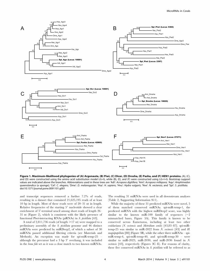
Identification of core RNAi proteinsIn order to identify homologues of the RNAi machinery in S.
pistillata, sequences from six families of proteins (Argonaute, Dicer,
Piwi, Drosha, Pasha, and HEN1) were drawn from five organisms
(H. sapiens, D. melanogaster, C. elegans, S. pombe, and A. thaliana). These
sequences were obtained from the UniRef100 database [50], and
clustered into groups with 90% sequence identity using CD-HIT
[51] to remove near-identical sequences. The clustered sequences
were used in a TBLASTN search against the S. pistillata
transcriptome to identify candidate RNAi-related transcripts.
Identified homologues (TBLASTN e-value,10210) of known
RNAi proteins were then searched for domains that are required
for the function of the protein using InterProScan [52–54]. The
domains that were determined to be essential for function were: a
Paz and Piwi domain for Argonaute and Piwi; a pair of RNase III
domains for Dicer and Drosha; a double-stranded RNA binding
domain for Pasha; and a methyltransferase (MTase) domain for
HEN1. Candidate homologues were not considered further in the
absence of any of these domains.
Additional support for the inferred function of candidate
homologues was obtained by carrying out a reciprocal BLASTP
search of these translated candidates against all proteins in the
Swiss-Prot database [55] (Supporting Information S3). The
candidate homologues were aligned against known RNAi proteins
on a per-family basis using Clustal Omega [56], and the
alignments were visualised using Jalview [57,58]. Key residues
were derived from literature [59–64]. In addition, for each of the
six protein families, phylogenies were constructed by aligning our
candidate homologues with selected sequences from Grimson et al.
[43] and Moran et al. [65] with MUSCLE [66]. Aligned regions of
low quality were removed with TrimAl, using the in-built
‘‘gappyout’’ parameter [67] (Supporting Information S4). Prot-
Test3 [68] was used to determine the best model for amino acid
substitution, and MEGA (version 6) [69] was used to construct
maximum-likelihood phylogenetic trees (support values were
computed from 1,000 bootstrap replicates).
Small RNA sequencing and processingThe small RNA (smRNA) fraction from S. pistillata was
selectively enriched from isolated total RNA (see above) using
the mirVana miRNA isolation kit (Ambion, Austin, TX) according
to manufacturer’s instructions. The small RNA fraction was
quality-checked using a Bioanalyzer 2100 (Agilent, Santa Clara,
CA) and a Nanodrop 2000c (ThermoScientific, Wilmington, DE).
The small RNA library was created using the Illumina Small RNA
MicroRNAs in Corals
PLOS ONE | www.plosone.org 2 March 2014 | Volume 9 | Issue 3 | e91101
Sample Prep Kit (Illumina, San Diego, CA) according to
manufacturer’s instructions, and sequenced on 1 lane on an
Illumina Genome Analyzer IIx (GA2x) machine. A total of 30.5
million small RNA reads of ,40 bp in length were produced. The
reads, along with associated Phred quality scores for each
sequenced base, were saved in a FASTQ file.
The raw FASTQ file was processed using several scripts to
remove low-quality reads resulting in a more compact FASTQ file
that contained high-quality reads for downstream analyses. First,
low quality 39 ends were trimmed from the reads. The 39 end of
the resulting reads had a Phred score .20, while the average
Phred score of the entire read was .20 as well. After trimming,
the overall error rate of the reads was calculated from the Phred
scores of individual bases. Reads were discarded if the error rate
exceeded 2%. Subsequently, the Illumina 59 and 39 adapter
sequences used in library generation were trimmed off using
Cutadapt v1.0 [70]. Last, in order to remove fragments of rRNA,
tRNA, and mRNA sequences, Velvet [71] was used to assemble
the short reads into contigs (at k = 25), which were then compared
to the GenBank nt database (nucleotide collection at NCBI). In
addition, we compared the assembled contigs to the S. pistillata
transcriptome assembly using BLASTN, in order to remove short
reads that matched known mRNA sequences.
miRNA prediction and filteringWe used miRDeep2 [72] to identify miRNAs. Briefly,
miRDeep2 mapped smRNAs to a preliminary draft genome of
S. pistillata using Bowtie, discarding reads that occurred more than
five times in the genome to avoid mapping to repetitive elements.
Potential pre-miRNA precursor sequences were identified based
on the pattern of the mappings, and subsequently folded using
RNAfold to ascertain whether they had the canonical hairpin
secondary structure [72]. Predicted pre-miRNAs that had a
miRDeep2 score of 10 or above were retained for further analyses
and inspected manually. A script was written to produce additional
information not found in the miRDeep2 output (i.e. length of 39
overhang, proportion of reads with consistent 59 end, number of
mismatched bases in stem) to further select a set of bona fide
miRNAs. Conserved miRNAs were identified using BLASTN
against all previously identified pre-miRNA sequences in miRBase
(ver 20) [37–40].
Functional analysis of miRNA targetsORFs were identified in the transcripts using TransDecoder
(part of the Trinity software pipeline [73]). Sequences downstream
of the longest ORF identified in the transcripts were treated as the
39 UTR of the transcript. 39 UTRs under 100 bp were filtered out
to remove transcripts associated with short UTR sequences. Out
of the 43,493 genes, 14,125 transcripts (32.4%) had a predicted 39
UTR longer than 100 bp.
For each miRNA, we ran PITA (Probability of Interaction by
Target Accessibility) [74] on the 39 UTRs at default settings to
produce a set of putative genes targeted by the miRNA. In the
absence of genomic data from other closely related organisms,
PITA achieves higher sensitivity and specificity than other target
prediction software (e.g. miRanda, TargetScan) as the latter
algorithms rely on a filter based on evolutionary conservation to
reduce the false positive rate. PITA works by calculating the
difference in Gibbs’ free energy (DDG) between the energy that is
required to unfold the secondary structure of the target site
(DGopen), and the energy of the mature miRNA binding its target
(DGduplex) [74]. Only miRNA targets with a DDG of #210 kcal -
mol21 were retained.
For GO enrichment of target genes, we used topGO (version
2.12.0), an R script that is available through Bioconductor 2.0.
topGO is a scoring algorithm that improves GO scoring by
eliminating local dependencies between related GO terms [75].
The threshold for significance was set at P,0.01, using otherwise
default topGO ‘‘weight01’’ settings, which produced GO terms
that were significantly enriched in the set of transcripts targeted by
each miRNA. The resulting P values were not corrected for
multiple testing, as non-independent tests are carried out on each
GO term by topGO [75].
Results
Identification of core RNAi proteinsThe miRNA machinery that processes and mediates the
function of miRNAs encompasses several key components that
appear to be conserved across the animal kingdom [76]. In order
to establish the presence of a functional miRNA machinery in S.
pistillata we conducted a BLAST-based search for key proteins
known to be essential for miRNA processing and function.
We identified seven candidate genes that are homologues to
known RNAi proteins: one Argonaute, two Piwi, one Dicer, one
Drosha, one Pasha, and one HEN1 in S. pistillata. We employed
several key metrics (i.e. matches to known RNAi families, presence
of protein domains crucial for catalytic activity, and a reciprocal
BLAST search against manually curated proteins in Swiss-Prot) to
identify candidate RNAi proteins (Supporting Information S3).
The per-family alignments of candidate homologues against
known sequences revealed a striking conservation of functionally
important amino acid residues located within the key protein
domains. Examples include the strong conservation of the DDX
triad in the Piwi domain of the Argonaute and Piwi homologues;
the aspartate and glutamate residues essential for Dicer activity;
and the pair of alanine/alanine and alanine/serine dipeptides
involved in the binding of dsRNA in Pasha (Supporting
Information S5, S6, S7, S8, S9, S10). Maximum-likelihood
phylogenetic trees that were constructed for all six protein families
(Figures 1A–1F) placed all of the candidate S. pistillata homologues
with those from other cnidarians. Judging from the presence of the
key RNAi proteins in S. pistillata in comparison to other organisms,
the RNAi machinery in S. pistillata is similar in composition to
those from sea anemone, worm, fruit fly, and humans (Table 1).
Besides the core RNAi proteins, we have also discovered
transcripts that are candidate homologues of HYL1 (one), GW182
(two), and RdRP (RNA-dependent RNA polymerase, eight) (data
not shown). HYL1 is thought to be a plant-specific partner to
Dicer [77], whereas GW182 helps Argonaute repress its targets
[78]. Both proteins have recently been discovered in four
cnidarians (Acropora digitifera, A. millepora, Hydra vulgaris, Nematostella
vectensis). However, although we could identify the PAM2 and P-
GL motif in one of our GW182 homologues, there were very few
GW repeats in this homologue (1 in S. pistillata, compared to 14 in
N. vectensis and 40 in humans) [65]. RdRPs, using small RNAs as
templates, amplify the silencing effect by directing the production
of secondary dsRNAs [79]. Functional RdRPs have been
discovered in plants [25,80] and C. elegans [81], but not in
mammals nor flies [79]. Four candidate homologues of RdRPs
have been found in N. vectensis [82], indicating that RdRPs might
be present in cnidarians.
Small RNA sequencing and miRNA repertoireSequencing produced 30,543,433 reads, of which 23,830,932
reads (78.0%) were kept after adapter trimming. The additional
step of removing short reads that matched known rRNA, tRNA,
MicroRNAs in Corals
PLOS ONE | www.plosone.org 3 March 2014 | Volume 9 | Issue 3 | e91101
and transcript sequences removed a further 7.2% of reads,
resulting in a dataset that contained 21,625,195 reads of at least
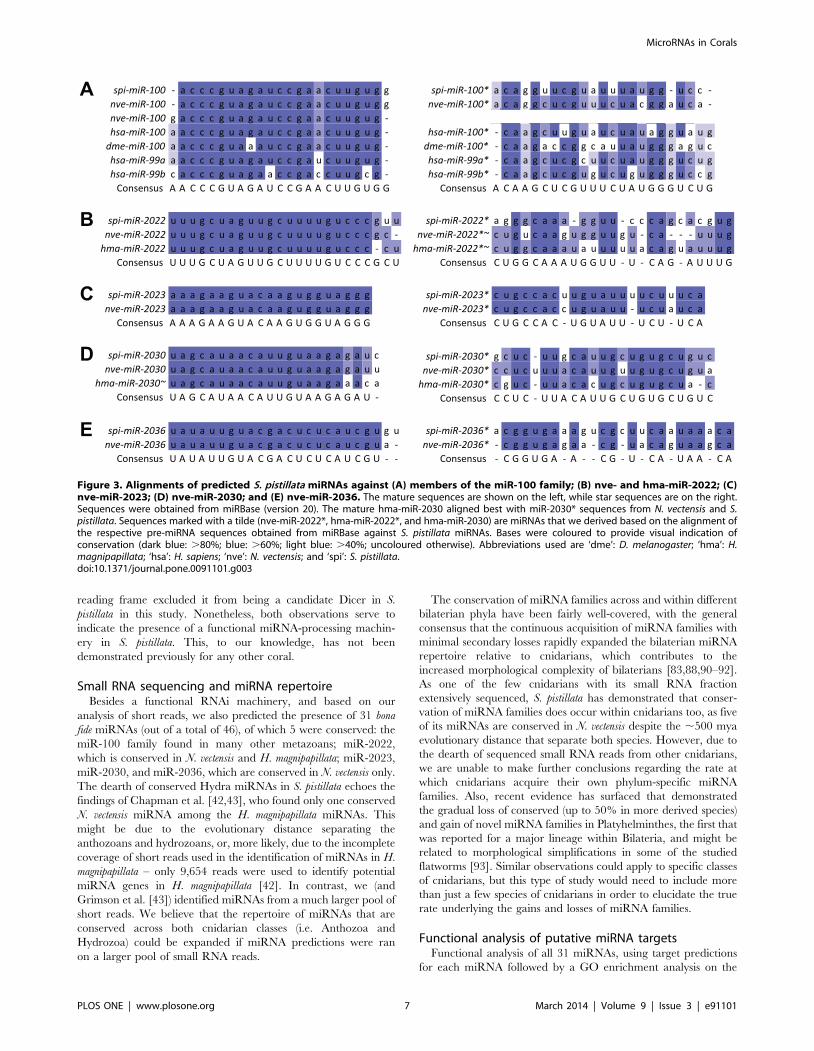
10 bp in length. Most of these reads were of 20–31 nt in length.
Relative frequencies of the starting 59 nucleotide showed a clear
enrichment of 59-terminal uracil among short reads of length 26–
31 nt (Figure 2), which is consistent with the likely presence of
functional Piwi-interacting RNAs (piRNAs) in S. pistillata [43].
A total of 2,811,736 reads (of length .17 nt) were mapped to a
preliminary assembly of the S. pistillata genome and 46 distinct
miRNAs were predicted by miRDeep2, of which a subset of 30
miRNAs passed additional filtering criteria (see Materials and
Methods). An exception was made for spi-miR-temp-25 –
although the precursor had a 3 bp 39 overhang, it was included
in the bona fide set as it was a close match to two known miRNAs.
The resulting 31 miRNAs were used in all downstream analyses
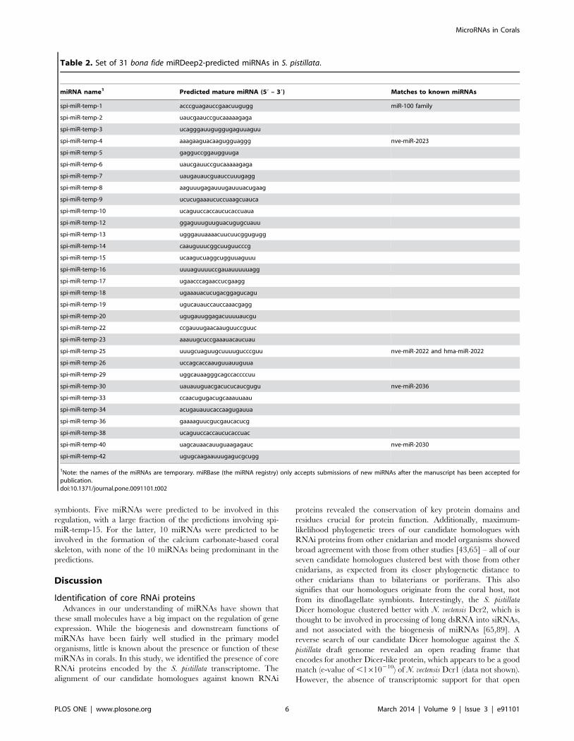
(Table 2, Supporting Information S11).
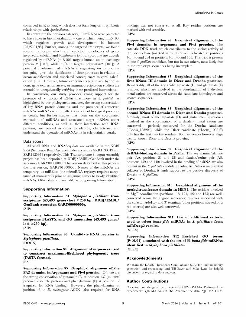
While the majority of these 31 predicted miRNAs were novel, 5
of them matched conserved miRNAs. spi-miR-temp-1, the
predicted miRNA with the highest miRDeep2 score, was highly
similar to the known miR-100 family of sequences (,2
mismatched bases, Figure 3A). This family is known to be
conserved across Eumetazoa, including at least two other
cnidarians (N. vectensis and Metridium senile) [43,83–87]. spi-miR-
temp-25 was similar to miR-2022 from N. vectensis [43] and H.
magnipapillata [88] (Figure 3B), while the other three miRNAs – spi-
miR-temp-4, spi-miR-temp-40, and spi-miR-temp-30 – were
similar to miR-2023, miR-2030, and miR-2036 found in N.
vectensis [43], respectively (Figures 3C–E). For reasons of clarity,
these five conserved miRNAs in S. pistillata will be referred to as
Figure 1. Maximum-likelihood phylogenies of (A) Argonaute, (B) Piwi, (C) Dicer, (D) Drosha, (E) Pasha, and (F) HEN1 proteins. (A), (C),and (D) were constructed using the amino acid substitution model LG+G, while (B), (E), and (F) were constructed using LG+I+G. Bootstrap supportvalues are indicated above the branches. Abbreviations used in these trees are ‘Adi’: Acropora digitifera; ‘Ami’: Acropora millepora; ‘Aqu’: Amphimedonqueenslandica (a sponge); ‘Cel’: C. elegans; ‘Dme’: D. melanogaster; ‘Hsa’: H. sapiens; ‘Hvu’: Hydra vulgaris; ‘Nve’: N. vectensis; and ‘Spi’: S. pistillata.doi:10.1371/journal.pone.0091101.g001
MicroRNAs in Corals
PLOS ONE | www.plosone.org 4 March 2014 | Volume 9 | Issue 3 | e91101
spi-miR-100, spi-miR-2022, spi-miR-2023, spi-miR-2030, and spi-
miR-2036, in accordance with miRNA naming conventions.
Although nve-miR-100 has been identified in two separate
studies (both of which utilised next-generation sequencing of short
reads to identify miRNAs in basal metazoans), Grimson et al. [43]
and Wheeler et al. [88] predicted mature miR-100 sequences
which are offset by a single nucleotide. From our read data, we
had .30,000 reads that exactly match the nve-miR-100 from
Grimson et al. [43], but none that matched the alternative version
from Wheeler et al. [88]. A negligible minority of reads (,10) did
start one nucleotide upstream, but unlike the Wheeler et al. [88]
version, this residue was an adenine, making this form identical to
the hsa-, dme- and xtr-miR-100s.
Functional analysis of putative miRNA targetsIn order to identify processes in S. pistillata that are potentially
regulated by miRNAs, we conducted a GO term enrichment
analysis. Briefly, for the set of 31 miRNAs, we searched for animal-
like target sites in the 39 UTRs of those 16,513 S. pistillata genes
that had available 39 UTR and GO annotation. This analysis was
performed for all miRNAs individually, and indicated that
miRNAs are likely to play roles in diverse processes (Table 3,
Supporting Information S12).
We categorised the resulting enriched GO terms under several
high-level groups based on known miRNA function – ‘‘immuni-
ty’’, ‘‘biomineralisation’’, ‘‘transcription’’, ‘‘cell cycle’’, ‘‘cytoskel-
eton’’, ‘‘metabolism’’, ‘‘transport/signalling’’, and ‘‘differentia-
tion/development’’. Other GO terms that did not fall under one of
these umbrella terms were categorised under ‘‘miscellaneous’’
(Supporting Information S12).
We paid particular attention to the ‘‘immunity’’ and ‘‘biomi-
neralisation’’ high-level groups, as we considered these terms to be
specifically relevant to the understanding of coral physiology
(Table 3). For the former, it is likely that immunity-related
transcripts are involved in the formation and retention of
symbiotic relationships between the coral host and its Symbiodinium
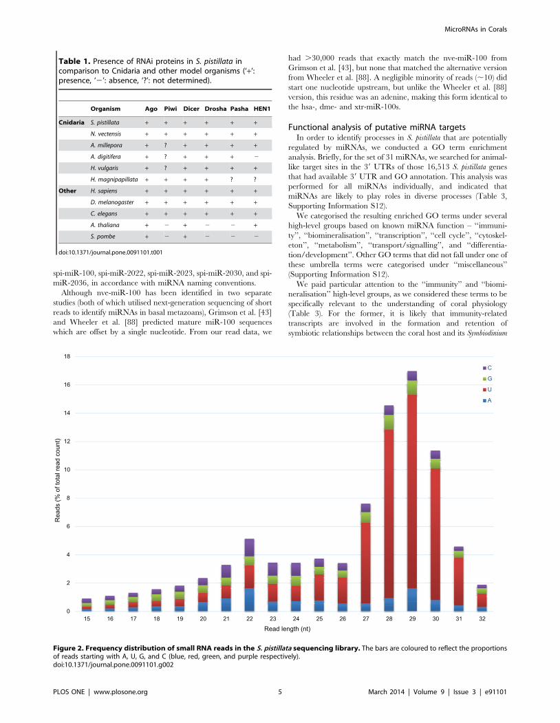
Table 1. Presence of RNAi proteins in S. pistillata incomparison to Cnidaria and other model organisms (‘+’:presence, ‘2’: absence, ‘?’: not determined).
Organism Ago Piwi Dicer Drosha Pasha HEN1
Cnidaria S. pistillata + + + + + +
N. vectensis + + + + + +
A. millepora + ? + + + +
A. digitifera + ? + + + 2
H. vulgaris + ? + + + +
H. magnipapillata + + + + ? ?
Other H. sapiens + + + + + +
D. melanogaster + + + + + +
C. elegans + + + + + +
A. thaliana + 2 + 2 2 +
S. pombe + 2 + 2 2 2
doi:10.1371/journal.pone.0091101.t001
Figure 2. Frequency distribution of small RNA reads in the S. pistillata sequencing library. The bars are coloured to reflect the proportionsof reads starting with A, U, G, and C (blue, red, green, and purple respectively).doi:10.1371/journal.pone.0091101.g002
MicroRNAs in Corals
PLOS ONE | www.plosone.org 5 March 2014 | Volume 9 | Issue 3 | e91101
symbionts. Five miRNAs were predicted to be involved in this
regulation, with a large fraction of the predictions involving spi-
miR-temp-15. For the latter, 10 miRNAs were predicted to be
involved in the formation of the calcium carbonate-based coral
skeleton, with none of the 10 miRNAs being predominant in the
predictions.
Discussion
Identification of core RNAi proteinsAdvances in our understanding of miRNAs have shown that
these small molecules have a big impact on the regulation of gene
expression. While the biogenesis and downstream functions of
miRNAs have been fairly well studied in the primary model
organisms, little is known about the presence or function of these
miRNAs in corals. In this study, we identified the presence of core
RNAi proteins encoded by the S. pistillata transcriptome. The
alignment of our candidate homologues against known RNAi
proteins revealed the conservation of key protein domains and
residues crucial for protein function. Additionally, maximum-
likelihood phylogenetic trees of our candidate homologues with
RNAi proteins from other cnidarian and model organisms showed
broad agreement with those from other studies [43,65] – all of our
seven candidate homologues clustered best with those from other
cnidarians, as expected from its closer phylogenetic distance to
other cnidarians than to bilaterians or poriferans. This also
signifies that our homologues originate from the coral host, not
from its dinoflagellate symbionts. Interestingly, the S. pistillata
Dicer homologue clustered better with N. vectensis Dcr2, which is
thought to be involved in processing of long dsRNA into siRNAs,
and not associated with the biogenesis of miRNAs [65,89]. A
reverse search of our candidate Dicer homologue against the S.
pistillata draft genome revealed an open reading frame that
encodes for another Dicer-like protein, which appears to be a good
match (e-value of ,1610210) of N. vectensis Dcr1 (data not shown).
However, the absence of transcriptomic support for that open
Table 2. Set of 31 bona fide miRDeep2-predicted miRNAs in S. pistillata.
miRNA name1 Predicted mature miRNA (59 – 39) Matches to known miRNAs
spi-miR-temp-1 acccguagauccgaacuugugg miR-100 family
spi-miR-temp-2 uaucgaauccgucaaaaagaga
spi-miR-temp-3 ucagggauuguggugaguuaguu
spi-miR-temp-4 aaagaaguacaagugguaggg nve-miR-2023
spi-miR-temp-5 gagguccggaugguuga
spi-miR-temp-6 uaucgauuccgucaaaaagaga
spi-miR-temp-7 uaugauaucguauccuuugagg
spi-miR-temp-8 aaguuugagauuugauuuacugaag
spi-miR-temp-9 ucucugaaaucuccuaagcuauca
spi-miR-temp-10 ucaguuccaccaucucaccuaua
spi-miR-temp-12 ggaguuuguuguacugugcuauu
spi-miR-temp-13 ugggauuaaaacuucuucggugugg
spi-miR-temp-14 caauguuucggcuuguucccg
spi-miR-temp-15 ucaagucuaggcugguuaguuu
spi-miR-temp-16 uuuaguuuuccgauauuuuuagg
spi-miR-temp-17 ugaacccagaaccucgaagg
spi-miR-temp-18 ugaaauacucugacggagucagu
spi-miR-temp-19 ugucauauccauccaaacgagg
spi-miR-temp-20 ugugauuggagacuuuuaucgu
spi-miR-temp-22 ccgauuugaacaauguuccguuc
spi-miR-temp-23 aaauugcuccgaaauacaucuau
spi-miR-temp-25 uuugcuaguugcuuuugucccguu nve-miR-2022 and hma-miR-2022
spi-miR-temp-26 uccagcaccaauguuauuguua
spi-miR-temp-29 uggcauaagggcagccaccccuu
spi-miR-temp-30 uauauuguacgacucucaucgugu nve-miR-2036
spi-miR-temp-33 ccaacugugacugcaaauuaau
spi-miR-temp-34 acugauauucaccaagugauua
spi-miR-temp-36 gaaaaguucgucgaucacucg
spi-miR-temp-38 ucaguuccaccaucucaccuac
spi-miR-temp-40 uagcauaacauuguaagagauc nve-miR-2030
spi-miR-temp-42 ugugcaagaauuugagucgcugg
1Note: the names of the miRNAs are temporary. miRBase (the miRNA registry) only accepts submissions of new miRNAs after the manuscript has been accepted forpublication.doi:10.1371/journal.pone.0091101.t002
MicroRNAs in Corals
PLOS ONE | www.plosone.org 6 March 2014 | Volume 9 | Issue 3 | e91101
reading frame excluded it from being a candidate Dicer in S.
pistillata in this study. Nonetheless, both observations serve to
indicate the presence of a functional miRNA-processing machin-
ery in S. pistillata. This, to our knowledge, has not been
demonstrated previously for any other coral.
Small RNA sequencing and miRNA repertoireBesides a functional RNAi machinery, and based on our
analysis of short reads, we also predicted the presence of 31 bona
fide miRNAs (out of a total of 46), of which 5 were conserved: the
miR-100 family found in many other metazoans; miR-2022,
which is conserved in N. vectensis and H. magnipapillata; miR-2023,
miR-2030, and miR-2036, which are conserved in N. vectensis only.
The dearth of conserved Hydra miRNAs in S. pistillata echoes the
findings of Chapman et al. [42,43], who found only one conserved
N. vectensis miRNA among the H. magnipapillata miRNAs. This
might be due to the evolutionary distance separating the
anthozoans and hydrozoans, or, more likely, due to the incomplete
coverage of short reads used in the identification of miRNAs in H.
magnipapillata – only 9,654 reads were used to identify potential
miRNA genes in H. magnipapillata [42]. In contrast, we (and
Grimson et al. [43]) identified miRNAs from a much larger pool of
short reads. We believe that the repertoire of miRNAs that are
conserved across both cnidarian classes (i.e. Anthozoa and
Hydrozoa) could be expanded if miRNA predictions were ran
on a larger pool of small RNA reads.
The conservation of miRNA families across and within different
bilaterian phyla have been fairly well-covered, with the general
consensus that the continuous acquisition of miRNA families with
minimal secondary losses rapidly expanded the bilaterian miRNA
repertoire relative to cnidarians, which contributes to the
increased morphological complexity of bilaterians [83,88,90–92].
As one of the few cnidarians with its small RNA fraction
extensively sequenced, S. pistillata has demonstrated that conser-
vation of miRNA families does occur within cnidarians too, as five
of its miRNAs are conserved in N. vectensis despite the ,500 mya
evolutionary distance that separate both species. However, due to
the dearth of sequenced small RNA reads from other cnidarians,
we are unable to make further conclusions regarding the rate at
which cnidarians acquire their own phylum-specific miRNA
families. Also, recent evidence has surfaced that demonstrated
the gradual loss of conserved (up to 50% in more derived species)
and gain of novel miRNA families in Platyhelminthes, the first that
was reported for a major lineage within Bilateria, and might be
related to morphological simplifications in some of the studied
flatworms [93]. Similar observations could apply to specific classes
of cnidarians, but this type of study would need to include more
than just a few species of cnidarians in order to elucidate the true
rate underlying the gains and losses of miRNA families.
Functional analysis of putative miRNA targetsFunctional analysis of all 31 miRNAs, using target predictions
for each miRNA followed by a GO enrichment analysis on the
Figure 3. Alignments of predicted S. pistillata miRNAs against (A) members of the miR-100 family; (B) nve- and hma-miR-2022; (C)nve-miR-2023; (D) nve-miR-2030; and (E) nve-miR-2036. The mature sequences are shown on the left, while star sequences are on the right.Sequences were obtained from miRBase (version 20). The mature hma-miR-2030 aligned best with miR-2030* sequences from N. vectensis and S.pistillata. Sequences marked with a tilde (nve-miR-2022*, hma-miR-2022*, and hma-miR-2030) are miRNAs that we derived based on the alignment ofthe respective pre-miRNA sequences obtained from miRBase against S. pistillata miRNAs. Bases were coloured to provide visual indication ofconservation (dark blue: .80%; blue: .60%; light blue: .40%; uncoloured otherwise). Abbreviations used are ‘dme’: D. melanogaster; ‘hma’: H.magnipapillata; ‘hsa’: H. sapiens; ‘nve’: N. vectensis; and ‘spi’: S. pistillata.doi:10.1371/journal.pone.0091101.g003
MicroRNAs in Corals
PLOS ONE | www.plosone.org 7 March 2014 | Volume 9 | Issue 3 | e91101

predicted target genes, revealed several putative processes and
pathways that are regulated by miRNAs in corals. For the miR-
100 homologue in S. pistillata, the GO terms ‘‘embryonic forelimb
morphogenesis’’ and ‘‘bone development’’ were enriched (P,0.01,
Supporting Information S12) in the predicted targets, which is
reminiscent of its reported function: in humans, miR-100 has been
shown to target genes involved in growth and development.
Examples include Plk1, a key mitotic checkpoint regulatory
protein [26]; RBSP3, involved in cell proliferation and myeloid
cell differentiation [27]; BMPR2, involved in osteogenesis [94];
and FRAP1/mTOR, which regulates cell growth [95]. It is
possible that miR-100 plays an analogous role in coral calcifica-
tion, making this miRNA a potentially important piece of the
puzzle in coral physiology, as well as a gene of interest when
investigating coral responses to ocean acidification. However, as
miRNA-mRNA target recognition depends critically on the
miRNA seed sequence (bases 2–7 of the mature RNA), it is
possible that the targets of bilaterian and cnidarian miR-100 will
differ due to the one nucleotide offset between the two miRNA
sequences. This 59 offset has also been observed for miR-2, miR-
10, miR-133, and miR-210 that are otherwise well-conserved
across two phylogenetically-related taxa, and presumably able to
regulate non-overlapping sets of target mRNAs [91]. Thus, further
experimentation is required to confirm the bona fide function of
cnidarian miR-100 in corals. Nonetheless, our spi-miR-100 adds
to the existing literature documenting the strong conservation of
miR-100 within metazoans.
Besides the only miRNA with documented function, we
identified miRNAs whose targets are involved in high-level
functions such as immunity, biomineralisation, regulation of cell
cycle, cellular motility, metabolism, signalling, and development,
analogous to functions that were previously ascribed to miRNAs in
other organisms [23–36]. We were interested in the first two high-
level groups, as immunity genes might regulate the relationship
with the symbiotic dinoflagellate Symbiodinium, and biomineralisa-
tion genes may control the rate of coral skeleton growth, two
processes that are arguably of importance to corals under
conditions of environmental change.
Out of the 5 miRNAs that were predicted to regulate coral
immunity genes, we speculate that spi-miR-temp-15 should
warrant further investigation due to the significant enrichment of
multiple immunity-related GO terms in the transcripts targeted by
this miRNA. Indeed, several of the predicted target genes of spi-
miR-temp-15 have homologues that are known to be regulated by
other miRNAs: Nod2 is repressed by miR-122 [96]; TLR2 is
regulated by miR-19 and miR-105 [97,98]; while caspase-8 is
targeted by miR-874 [99]. Interestingly, this miRNA is not
Table 3. Enriched immunity- and biomineralisation-related GO terms (default topGO settings, P,0.01) associated with predictedmiRNA target genes from S. pistillata.
miRNA GO ID Description P value
Immunity
spi-miR-2022 GO:0044003 modification by symbiont of host morphology or physiology 0.0015
spi-miR-2022 GO:0019048 virus-host interaction 0.0086
spi-miR-temp-15 GO:0044130 negative regulation of growth of symbiont in host 0.00634
spi-miR-temp-15 GO:0032735 positive regulation of interleukin2 production 0.00119
spi-miR-temp-15 GO:0034134 toll-like receptor 2 signaling pathway 0.00188
spi-miR-temp-15 GO:0034142 toll-like receptor 4 signaling pathway 0.00626
spi-miR-temp-15 GO:0038123 toll-like receptor TLR1:TLR2 signaling pathway 0.00314
spi-miR-temp-15 GO:0038124 toll-like receptor TLR6:TLR2 signaling pathway 0.00314
spi-miR-temp-16 GO:0070498 interleukin-mediated signaling pathway 0.00019
spi-miR-temp-16 GO:0042102 positive regulation of T cell proliferation 0.00366
spi-miR-temp-20 GO:0032088 negative regulation of NF-kappaB transcription factor activity 0.0038
spi-miR-temp-26 GO:0051703 intraspecies interaction between organisms 0.0086
Biomineralisation
spi-miR-100 GO:0060348 bone development 0.00475
spi-miR-temp-7 GO:0030574 collagen catabolic process 0.00733
spi-miR-temp-8 GO:0001958 endochondral ossification 0.0069
spi-miR-temp-12 GO:0003417 growth plate cartilage development 0.00891
spi-miR-temp-13 GO:0070588 calcium ion transmembrane transport 0.0046
spi-miR-temp-15 GO:0060346 bone trabecula formation 0.00629
spi-miR-temp-15 GO:0003417 growth plate cartilage development 0.00119
spi-miR-temp-15 GO:0043931 ossification involved in bone maturation 6.50E-05
spi-miR-temp-16 GO:0045672 positive regulation of osteoclast differentiation 0.00776
spi-miR-temp-18 GO:0030509 BMP signaling pathway 0.0084
spi-miR-temp-18 GO:0030501 positive regulation of bone mineralization 0.0031
spi-miR-temp-19 GO:0015701 bicarbonate transport 0.00172
spi-miR-temp-23 GO:0061036 positive regulation of cartilage development 0.00066
doi:10.1371/journal.pone.0091101.t003
MicroRNAs in Corals
PLOS ONE | www.plosone.org 8 March 2014 | Volume 9 | Issue 3 | e91101
conserved in N. vectensis, which does not form long-term symbiotic
relationships with Symbiodinium.
In contrast to the previous category, 10 miRNAs were predicted
to have roles in biomineralisation – one of which being miR-100,
which regulates growth and development in humans
[26,27,94,95]. Further, among the targeted transcripts, we found
several transcripts which are predicted homologues of genes
involved in calcium and bicarbonate ion transport that are directly
regulated by miRNAs (miR-506 targets human anion exchange
protein 2 [100], while miR-17 targets polycystin-2 [101]). A
potential involvement of miRNAs in regulating ion transport is
intriguing, given the significance of these processes in relation to
ocean acidification and associated consequences to coral calcifi-
cation [102]. However, future experiments (e.g in-situ hybridisa-
tions, gene expression assays, or immunoprecipitation studies) are
essential in unequivocally verifying these predicted interactions.
In conclusion, our study provides strong support for the
presence of a functional RNAi machinery in S. pistillata as
highlighted by our phylogenetic analyses, the strong conservation
of key RNAi protein domains, and the presence of conserved
miRNAs. miRNAs seem to affect a variety of biological processes
in corals, but further studies that focus on the coordinated
expression of miRNAs and associated target mRNAs under
different conditions, as well as their interaction with RNAi
proteins, are needed in order to identify, characterise, and
understand the operational miRNAome in scleractinian corals.
Data accessAll small RNA and RNASeq data are available in the NCBI
SRA (Sequence Read Archive) under accessions SRR1130519 and
SRR1125978 respectively. This Transcriptome Shotgun Assembly
project has been deposited at DDBJ/EMBL/GenBank under the
accession GARY00000000. The version described in this paper is
the first version, GARY01000000.. Names of the miRNAs are
temporary, as miRBase (the microRNA registry) requires accep-
tance of manuscripts prior to assigning names to newly identified
miRNAs. Other data are available as Supporting Information.
Supporting Information
Supporting Information S1 Stylophora pistillata tran-scriptome (43,493 genes/loci $250 bp, DDBJ/EMBL/GenBank accession GARY00000000).
(ZIP)
Supporting Information S2 Stylophora pistillata tran-scriptome BLASTX and GO annotation (43,493 genes/loci $250 bp).
(ZIP)
Supporting Information S3 Candidate RNAi proteins inStylophora pistillata.
(DOCX)
Supporting Information S4 Alignment of sequences usedto construct maximum-likelihood phylogenetic trees(FASTA format).
(FA)
Supporting Information S5 Graphical alignment of thePAZ domains in Argonaute and Piwi proteins. Of note are
the strong conservation of glutamate (E) at position 137 (mutants
produce insoluble protein) and phenylalanine (F) at position 72
(required for RNA binding). However, the phenylalanine at
position 48 in D. melanogaster AGO2 (also required for RNA
binding) was not conserved at all. Key residue positions are
marked with red asterisks.
(EPS)
Supporting Information S6 Graphical alignment of thePiwi domains in Argonaute and Piwi proteins. The
catalytic DDX triad, which contributes to the slicing activity of
the ribonuclease (marked in red asterisks), is located at positions
46, 140 and 284 or positions 46, 140 and 155. This triad is present
in one S. pistillata candidate, but not in two others, most likely due
to the transcript sequences being incomplete.
(EPS)
Supporting Information S7 Graphical alignment of thefirst RNase III domain in Dicer and Drosha proteins.Remarkably, all of the key acidic aspartate (D) and glutamate (E)
residues, which are involved in the coordination of a divalent
metal cation, are conserved across the candidate homologues and
known sequences.
(EPS)
Supporting Information S8 Graphical alignment of thesecond RNase III domain in Dicer and Drosha proteins.Similarly, most of the aspartate (D) and glutamate (E) residues
involved in the coordination of a divalent metal cation are
conserved - perfectly conserved for the Drosha candidate
(‘‘Locus_18820’’), while the Dicer candidate (‘‘Locus_10081’’)
only has the first two key residues. Both sequences however align
well to known Dicer and Drosha proteins.
(EPS)
Supporting Information S9 Graphical alignment of thedsRNA-binding domain in Pasha. The key alanine/alanine
pair (AA, positions 21 and 22) and alanine/serine pair (AS,
positions 139 and 140) involved in the binding of dsRNA are also
present in the S. pistillata candidate Pasha. As Pasha is an essential
cofactor of Drosha, it lends support to the positive discovery of
Drosha in S. pistillata.
(EPS)
Supporting Information S10 Graphical alignment of themethyltransferase domain in HEN1. The residues involved
in Mg2+ coordination (positions 118, 121, 122 and 123) are well-
conserved across the aligned sequences; residues associated with
the cofactor AdoHcy and 39 terminus (other positions marked by a
red asterisk) are also well conserved.
(EPS)
Supporting Information S11 List of additional criteriaused to select bona fide miRNAs in S. pistillata frommiRDeep2 results.
(XLSX)
Supporting Information S12 Enriched GO terms(P,0.01) associated with the set of 31 bona fide miRNAsidentified in Stylophora pistillata.
(XLSX)
Acknowledgments
We thank the KAUST Bioscience Core Lab and S. Ali for Illumina library
generation and sequencing, and Till Bayer and Mike Lyne for helpful
discussions in regard to data analyses.
Author Contributions
Conceived and designed the experiments: CRV GM MA. Performed the
experiments: YJL MA AC SB DZ. Analyzed the data: YJL MA CRV.
MicroRNAs in Corals
PLOS ONE | www.plosone.org 9 March 2014 | Volume 9 | Issue 3 | e91101
Contributed reagents/materials/analysis tools: DZ ST DA. Wrote the
paper: YJL MA CRV.
References
1. Hughes TP (1994) Catastrophes, phase shifts, and large-scale degradation of a
Caribbean coral reef. Science 265: 1547–1551.
2. Bruno JF, Selig ER (2007) Regional decline of coral cover in the Indo-Pacific:
timing, extent, and subregional comparisons. PLoS One 2: e711.
3. Knowlton N, Brainard RE, Fisher R, Moews M, Plaisance L, et al. (2010)
Coral reef biodiversity. Life in the World’s Oceans: Diversity Distribution and
Abundance: 65–74.
4. Hughes TP, Baird AH, Bellwood DR, Card M, Connolly SR, et al. (2003)
Climate change, human impacts, and the resilience of coral reefs. Science 301:
929–933.
5. Hoegh-Guldberg O, Mumby PJ, Hooten AJ, Steneck RS, Greenfield P, et al.
(2007) Coral reefs under rapid climate change and ocean acidification. Science
318: 1737–1742.
6. Bak R (1987) Effects of chronic oil pollution on a Caribbean coral reef. Marine
Pollution Bulletin 18: 534–539.
7. Pastorok RA, Bilyard GR (1985) Effects of sewage pollution on coral-reef
communities. Marine ecology progress series Oldendorf 21: 175–189.
8. Green EP, Bruckner AW (2000) The significance of coral disease epizootiology
for coral reef conservation. Biological Conservation 96: 347–361.
9. Shinzato C, Shoguchi E, Kawashima T, Hamada M, Hisata K, et al. (2011)
Using the Acropora digitifera genome to understand coral responses to
environmental change. Nature 476: 320–323.
10. DeSalvo MK, Sunagawa S, Fisher PL, Voolstra CR, Iglesias-Prieto R, et al.
(2010) Coral host transcriptomic states are correlated with Symbiodinium
genotypes. Mol Ecol 19: 1174–1186.
11. Voolstra CR, Schwarz JA, Schnetzer J, Sunagawa S, Desalvo MK, et al. (2009)
The host transcriptome remains unaltered during the establishment of coral-
algal symbioses. Mol Ecol 18: 1823–1833.
12. DeSalvo MK, Voolstra CR, Sunagawa S, Schwarz JA, Stillman JH, et al.
(2008) Differential gene expression during thermal stress and bleaching in the
Caribbean coral Montastraea faveolata. Mol Ecol 17: 3952–3971.
13. Polato NR, Voolstra CR, Schnetzer J, DeSalvo MK, Randall CJ, et al. (2010)
Location-specific responses to thermal stress in larvae of the reef-building coral
Montastraea faveolata. PLoS One 5: e11221.
14. Voolstra CR, Schnetzer J, Peshkin L, Randall CJ, Szmant AM, et al. (2009)
Effects of temperature on gene expression in embryos of the coral Montastraea
faveolata. BMC Genomics 10: 627.
15. Meyer E, Aglyamova GV, Matz MV (2011) Profiling gene expression responses
of coral larvae (Acropora millepora) to elevated temperature and settlement
inducers using a novel RNA-Seq procedure. Mol Ecol 20: 3599–3616.
16. DeSalvo M, Estrada A, Sunagawa S, Medina M (2012) Transcriptomic
responses to darkness stress point to common coral bleaching mechanisms.
Coral Reefs 31: 215–228.
17. Moya A, Huisman L, Ball EE, Hayward DC, Grasso LC, et al. (2012) Whole
transcriptome analysis of the coral Acropora millepora reveals complex
responses to CO(2)-driven acidification during the initiation of calcification.
Mol Ecol 21: 2440–2454.
18. Vidal-Dupiol J, Zoccola D, Tambutte E, Grunau C, Cosseau C, et al. (2013)
Genes related to ion-transport and energy production are upregulated in
response to CO2-driven pH decrease in corals: new insights from transcriptome
analysis. PLoS One 8: e58652.
19. Aranda M, Banaszak AT, Bayer T, Luyten JR, Medina M, et al. (2011)
Differential sensitivity of coral larvae to natural levels of ultraviolet radiation
during the onset of larval competence. Mol Ecol 20: 2955–2972.
20. Lagos-Quintana M, Rauhut R, Lendeckel W, Tuschl T (2001) Identification of
novel genes coding for small expressed RNAs. Science 294: 853–858.
21. Lau NC, Lim LP, Weinstein EG, Bartel DP (2001) An abundant class of tiny
RNAs with probable regulatory roles in Caenorhabditis elegans. Science 294:
858–862.
22. Lee RC, Ambros V (2001) An extensive class of small RNAs in Caenorhabditis
elegans. Science 294: 862–864.
23. Pasquinelli AE, Reinhart BJ, Slack F, Martindale MQ, Kuroda MI, et al.
(2000) Conservation of the sequence and temporal expression of let-7
heterochronic regulatory RNA. Nature 408: 86–89.
24. Axtell MJ, Bowman JL (2008) Evolution of plant microRNAs and their targets.
Trends Plant Sci 13: 343–349.
25. Chen X (2009) Small RNAs and their roles in plant development. Annu Rev
Cell Dev Biol 25: 21–44.
26. Li BH, Zhou JS, Ye F, Cheng XD, Zhou CY, et al. (2011) Reduced miR-100
expression in cervical cancer and precursors and its carcinogenic effect through
targeting PLK1 protein. Eur J Cancer 47: 2166–2174.
27. Zheng YS, Zhang H, Zhang XJ, Feng DD, Luo XQ, et al. (2012) MiR-100
regulates cell differentiation and survival by targeting RBSP3, a phosphatase-
like tumor suppressor in acute myeloid leukemia. Oncogene 31: 80–92.
28. Chen CZ, Schaffert S, Fragoso R, Loh C (2013) Regulation of immune
responses and tolerance: the microRNA perspective. Immunol Rev 253: 112–
128.
29. O’Connell RM, Rao DS, Chaudhuri AA, Baltimore D (2010) Physiological and
pathological roles for microRNAs in the immune system. Nat Rev Immunol 10:111–122.
30. Horie T, Ono K, Nishi H, Iwanaga Y, Nagao K, et al. (2009) MicroRNA-133
regulates the expression of GLUT4 by targeting KLF15 and is involved inmetabolic control in cardiac myocytes. Biochem Biophys Res Commun 389:
315–320.
31. Leung AK, Sharp PA (2010) MicroRNA functions in stress responses. Mol Cell
40: 205–215.
32. Sunkar R, Li YF, Jagadeeswaran G (2012) Functions of microRNAs in plant
stress responses. Trends Plant Sci 17: 196–203.
33. Babenko O, Golubov A, Ilnytskyy Y, Kovalchuk I, Metz GA (2012) Genomicand epigenomic responses to chronic stress involve miRNA-mediated
programming. PLoS One 7: e29441.
34. Cao H, Wang J, Li X, Florez S, Huang Z, et al. (2010) MicroRNAs play a
critical role in tooth development. J Dent Res 89: 779–784.
35. Jiao Y, Zheng Z, Du X, Wang Q, Huang R, et al. (2014) Identification andCharacterization of MicroRNAs in Pearl Oyster Pinctada martensii by Solexa
Deep Sequencing. Mar Biotechnol (NY) 16: 54–62.
36. van Wijnen AJ, van de Peppel J, van Leeuwen JP, Lian JB, Stein GS, et al.
(2013) MicroRNA functions in osteogenesis and dysfunctions in osteoporosis.Curr Osteoporos Rep 11: 72–82.
37. Griffiths-Jones S (2004) The microRNA Registry. Nucleic Acids Res 32: D109–
111.
38. Griffiths-Jones S, Grocock RJ, van Dongen S, Bateman A, Enright AJ (2006)
miRBase: microRNA sequences, targets and gene nomenclature. Nucleic AcidsRes 34: D140–144.
39. Griffiths-Jones S, Saini HK, van Dongen S, Enright AJ (2008) miRBase: tools
for microRNA genomics. Nucleic Acids Res 36: D154–158.
40. Kozomara A, Griffiths-Jones S (2011) miRBase: integrating microRNA
annotation and deep-sequencing data. Nucleic Acids Res 39: D152–157.
41. Baumgarten S, Bayer T, Aranda M, Liew YJ, Carr A, et al. (2013) Integrating
microRNA and mRNA expression profiling in Symbiodinium microadriati-cum, a dinoflagellate symbiont of reef-building corals. BMC Genomics 14: 704.
42. Chapman JA, Kirkness EF, Simakov O, Hampson SE, Mitros T, et al. (2010)
The dynamic genome of Hydra. Nature 464: 592–596.
43. Grimson A, Srivastava M, Fahey B, Woodcroft BJ, Chiang HR, et al. (2008)
Early origins and evolution of microRNAs and Piwi-interacting RNAs inanimals. Nature 455: 1193–1197.
44. Moya A, Tambutte S, Beranger G, Gaume B, Scimeca JC, et al. (2008)
Cloning and use of a coral 36B4 gene to study the differential expression ofcoral genes between light and dark conditions. Mar Biotechnol (NY) 10: 653–
663.
45. Lohse M, Bolger AM, Nagel A, Fernie AR, Lunn JE, et al. (2012) RobiNA: a
user-friendly, integrated software solution for RNA-Seq-based transcriptomics.Nucleic Acids Res 40: W622–627.
46. Brown CT, Howe A, Zhang Q, Pyrkosz AB, Brom TH (2012) A reference-freealgorithm for computational normalization of shotgun sequencing data. arXiv
preprint arXiv:12034802.
47. Langmead B, Salzberg SL (2012) Fast gapped-read alignment with Bowtie 2.Nat Methods 9: 357–359.
48. Schulz MH, Zerbino DR, Vingron M, Birney E (2012) Oases: robust de novoRNA-seq assembly across the dynamic range of expression levels. Bioinfor-
matics 28: 1086–1092.
49. Dimmer EC, Huntley RP, Alam-Faruque Y, Sawford T, O’Donovan C, et al.
(2012) The UniProt-GO Annotation database in 2011. Nucleic Acids Res 40:D565–570.
50. Suzek BE, Huang H, McGarvey P, Mazumder R, Wu CH (2007) UniRef:
comprehensive and non-redundant UniProt reference clusters. Bioinformatics23: 1282–1288.
51. Li W, Godzik A (2006) Cd-hit: a fast program for clustering and comparinglarge sets of protein or nucleotide sequences. Bioinformatics 22: 1658–1659.
52. Mulder N, Apweiler R (2007) InterPro and InterProScan: tools for protein
sequence classification and comparison. Methods Mol Biol 396: 59–70.
53. Quevillon E, Silventoinen V, Pillai S, Harte N, Mulder N, et al. (2005)
InterProScan: protein domains identifier. Nucleic Acids Res 33: W116–120.
54. Zdobnov EM, Apweiler R (2001) InterProScan–an integration platform for thesignature-recognition methods in InterPro. Bioinformatics 17: 847–848.
55. Magrane M, Consortium U (2011) UniProt Knowledgebase: a hub ofintegrated protein data. Database (Oxford) 2011: bar009.
56. Sievers F, Wilm A, Dineen D, Gibson TJ, Karplus K, et al. (2011) Fast, scalable
generation of high-quality protein multiple sequence alignments using ClustalOmega. Mol Syst Biol 7: 539.
57. Clamp M, Cuff J, Searle SM, Barton GJ (2004) The Jalview Java alignmenteditor. Bioinformatics 20: 426–427.
58. Waterhouse AM, Procter JB, Martin DM, Clamp M, Barton GJ (2009) Jalview
Version 2–a multiple sequence alignment editor and analysis workbench.
Bioinformatics 25: 1189–1191.
MicroRNAs in Corals
PLOS ONE | www.plosone.org 10 March 2014 | Volume 9 | Issue 3 | e91101
59. Huang Y, Ji L, Huang Q, Vassylyev DG, Chen X, et al. (2009) Structural
insights into mechanisms of the small RNA methyltransferase HEN1. Nature461: 823–827.
60. Lee YS, Nakahara K, Pham JW, Kim K, He Z, et al. (2004) Distinct roles for
Drosophila Dicer-1 and Dicer-2 in the siRNA/miRNA silencing pathways.Cell 117: 69–81.
61. Lingel A, Simon B, Izaurralde E, Sattler M (2003) Structure and nucleic-acidbinding of the Drosophila Argonaute 2 PAZ domain. Nature 426: 465–469.
62. Rivas FV, Tolia NH, Song JJ, Aragon JP, Liu J, et al. (2005) Purified
Argonaute2 and an siRNA form recombinant human RISC. Nat Struct MolBiol 12: 340–349.
63. Song JJ, Smith SK, Hannon GJ, Joshua-Tor L (2004) Crystal structure ofArgonaute and its implications for RISC slicer activity. Science 305: 1434–
1437.64. Yeom KH, Lee Y, Han J, Suh MR, Kim VN (2006) Characterization of
DGCR8/Pasha, the essential cofactor for Drosha in primary miRNA
processing. Nucleic Acids Res 34: 4622–4629.65. Moran Y, Praher D, Fredman D, Technau U (2013) The Evolution of
MicroRNA Pathway Protein Components in Cnidaria. Mol Biol Evol.66. Edgar RC (2004) MUSCLE: multiple sequence alignment with high accuracy
and high throughput. Nucleic Acids Res 32: 1792–1797.
67. Capella-Gutierrez S, Silla-Martinez JM, Gabaldon T (2009) trimAl: a tool forautomated alignment trimming in large-scale phylogenetic analyses. Bioinfor-
matics 25: 1972–1973.68. Darriba D, Taboada GL, Doallo R, Posada D (2011) ProtTest 3: fast selection
of best-fit models of protein evolution. Bioinformatics 27: 1164–1165.69. Tamura K, Stecher G, Peterson D, Filipski A, Kumar S (2013) MEGA6:
Molecular Evolutionary Genetics Analysis version 6.0. Mol Biol Evol 30: 2725–
2729.70. Martin M (2011) Cutadapt removes adapter sequences from high-throughput
sequencing reads. EMBnet journal 17: pp. 10–12.71. Zerbino DR, Birney E (2008) Velvet: algorithms for de novo short read
assembly using de Bruijn graphs. Genome Res 18: 821–829.
72. Friedlander MR, Mackowiak SD, Li N, Chen W, Rajewsky N (2012)miRDeep2 accurately identifies known and hundreds of novel microRNA
genes in seven animal clades. Nucleic Acids Res 40: 37–52.73. Grabherr MG, Haas BJ, Yassour M, Levin JZ, Thompson DA, et al. (2011)
Full-length transcriptome assembly from RNA-Seq data without a referencegenome. Nat Biotechnol 29: 644–652.
74. Kertesz M, Iovino N, Unnerstall U, Gaul U, Segal E (2007) The role of site
accessibility in microRNA target recognition. Nat Genet 39: 1278–1284.75. Alexa A, Rahnenfuhrer J, Lengauer T (2006) Improved scoring of functional
groups from gene expression data by decorrelating GO graph structure.Bioinformatics 22: 1600–1607.
76. Tarver JE, Donoghue PC, Peterson KJ (2012) Do miRNAs have a deep
evolutionary history? Bioessays 34: 857–866.77. Voinnet O (2009) Origin, biogenesis, and activity of plant microRNAs. Cell
136: 669–687.78. Huntzinger E, Izaurralde E (2011) Gene silencing by microRNAs: contribu-
tions of translational repression and mRNA decay. Nat Rev Genet 12: 99–110.79. Ghildiyal M, Zamore PD (2009) Small silencing RNAs: an expanding universe.
Nat Rev Genet 10: 94–108.
80. Song X, Wang D, Ma L, Chen Z, Li P, et al. (2012) Rice RNA-dependentRNA polymerase 6 acts in small RNA biogenesis and spikelet development.
Plant J 71: 378–389.81. Maniar JM, Fire AZ (2011) EGO-1, a C. elegans RdRP, modulates gene
expression via production of mRNA-templated short antisense RNAs. Curr
Biol 21: 449–459.82. Zong J, Yao X, Yin J, Zhang D, Ma H (2009) Evolution of the RNA-dependent
RNA polymerase (RdRP) genes: duplications and possible losses before andafter the divergence of major eukaryotic groups. Gene 447: 29–39.
83. Sempere LF, Cole CN, McPeek MA, Peterson KJ (2006) The phylogenetic
distribution of metazoan microRNAs: insights into evolutionary complexity
and constraint. J Exp Zool B Mol Dev Evol 306: 575–588.
84. Mourelatos Z, Dostie J, Paushkin S, Sharma A, Charroux B, et al. (2002)
miRNPs: a novel class of ribonucleoproteins containing numerous microRNAs.
Genes Dev 16: 720–728.
85. Sempere LF, Sokol NS, Dubrovsky EB, Berger EM, Ambros V (2003)
Temporal regulation of microRNA expression in Drosophila melanogaster
mediated by hormonal signals and broad-Complex gene activity. Dev Biol 259:
9–18.
86. Chen PY, Manninga H, Slanchev K, Chien M, Russo JJ, et al. (2005) The
developmental miRNA profiles of zebrafish as determined by small RNA
cloning. Genes Dev 19: 1288–1293.
87. Michalak P, Malone JH (2008) Testis-derived microRNA profiles of African
clawed frogs (Xenopus) and their sterile hybrids. Genomics 91: 158–164.
88. Wheeler BM, Heimberg AM, Moy VN, Sperling EA, Holstein TW, et al.
(2009) The deep evolution of metazoan microRNAs. Evol Dev 11: 50–68.
89. Mukherjee K, Campos H, Kolaczkowski B (2013) Evolution of animal and
plant dicers: early parallel duplications and recurrent adaptation of antiviral
RNA binding in plants. Mol Biol Evol 30: 627–641.
90. Campo-Paysaa F, Semon M, Cameron RA, Peterson KJ, Schubert M (2011)
microRNA complements in deuterostomes: origin and evolution of micro-
RNAs. Evol Dev 13: 15–27.
91. Peterson KJ, Dietrich MR, McPeek MA (2009) MicroRNAs and metazoan
macroevolution: insights into canalization, complexity, and the Cambrian
explosion. Bioessays 31: 736–747.
92. Prochnik SE, Rokhsar DS, Aboobaker AA (2007) Evidence for a microRNA
expansion in the bilaterian ancestor. Dev Genes Evol 217: 73–77.
93. Fromm B, Worren MM, Hahn C, Hovig E, Bachmann L (2013) Substantial
loss of conserved and gain of novel microRNA families in flatworms. Mol Biol
Evol 30: 2619–2628.
94. Zeng Y, Qu X, Li H, Huang S, Wang S, et al. (2012) MicroRNA-100 regulates
osteogenic differentiation of human adipose-derived mesenchymal stem cells by
targeting BMPR2. FEBS Lett 586: 2375–2381.
95. Nagaraja AK, Creighton CJ, Yu Z, Zhu H, Gunaratne PH, et al. (2010) A link
between mir-100 and FRAP1/mTOR in clear cell ovarian cancer. Mol
Endocrinol 24: 447–463.
96. Chen Y, Wang C, Liu Y, Tang L, Zheng M, et al. (2013) miR-122 targets
NOD2 to decrease intestinal epithelial cell injury in Crohn’s disease. Biochem
Biophys Res Commun 438: 133–139.
97. Philippe L, Alsaleh G, Suffert G, Meyer A, Georgel P, et al. (2012) TLR2
expression is regulated by microRNA miR-19 in rheumatoid fibroblast-like
synoviocytes. J Immunol 188: 454–461.
98. Benakanakere MR, Li Q, Eskan MA, Singh AV, Zhao J, et al. (2009)
Modulation of TLR2 protein expression by miR-105 in human oral
keratinocytes. J Biol Chem 284: 23107–23115.
99. Wang K, Liu F, Zhou LY, Ding SL, Long B, et al. (2013) miR-874 regulates
myocardial necrosis by targeting caspase-8. Cell Death Dis 4: e709.
100. Banales JM, Saez E, Uriz M, Sarvide S, Urribarri AD, et al. (2012) Up-
regulation of microRNA 506 leads to decreased Cl-/HCO3- anion exchanger
2 expression in biliary epithelium of patients with primary biliary cirrhosis.
Hepatology 56: 687–697.
101. Sun H, Li QW, Lv XY, Ai JZ, Yang QT, et al. (2010) MicroRNA-17 post-
transcriptionally regulates polycystic kidney disease-2 gene and promotes cell
proliferation. Mol Biol Rep 37: 2951–2958.
102. Erez J, Reynaud S, Silverman J, Schneider K, Allemand D (2011) Coral
Calcification Under Ocean Acidification and Global Change. In: Dubinsky Z,
Stambler N, editors. Coral Reefs: An Ecosystem in Transition: Springer
Netherlands. pp. 151–176.
MicroRNAs in Corals
PLOS ONE | www.plosone.org 11 March 2014 | Volume 9 | Issue 3 | e91101
Related Documents