Identification of Long stress-induced non-coding transcripts that have altered expression in cancer ☆ Jessica M. Silva a,b, ⁎, Damon S. Perez a , Jay R. Pritchett a , Meredith L. Halling a , Hui Tang c , David I. Smith a a Division of Experimental Pathology, Department of Laboratory Medicine and Pathology, Mayo Clinic and Foundation, 200 First Street, S.W., Rochester, MN 55905, USA b Department of Biochemistry and Molecular Biology, Mayo Clinic and Foundation, 200 First Street, S.W., Rochester, MN 55905, USA c Division of Biomedical statistics and informatics, Department of Health Sciences Research, Mayo Clinic and Foundation, 200 First Street, S.W., Rochester, MN 55905, USA abstract article info Article history: Received 4 December 2009 Accepted 24 February 2010 Available online 7 March 2010 Keywords: Long non-coding transcripts Non-coding RNA NNK(4-(methylnitrosamino)-1- (3-pyridyl)-1-butanone) Lung cancer Breast cancer Stress induction Tiling arrays Entire genome analysis It has recently become clear that the transcriptional output of the human genome is far more abundant than previously anticipated, with the vast majority of transcripts not coding for protein. Utilizing whole-genome tiling arrays, we analyzed the transcription across the entire genome in both normal human bronchial epithelial cells (NHBE) and NHBE cells exposed to the tobacco carcinogen NNK. Our efforts focused on the characterization of non-coding transcripts that were greater than 300 nucleotides in length and whose expression was increased in response to NNK. We identified 12 Long Stress-Induced Non-coding Transcripts that we term LSINCTs. Northern blot analysis revealed that these transcripts were larger than predicted from the tiling array data. Quantitative real-time RT-PCR performed across a panel of normal cell lines indicates that these transcripts are more abundantly expressed in rapidly growing tissues or in tissues that are more prone to cellular stress. These transcripts that have increased expression after exposure to NNK also had increased expression in a number of lung cancer cell lines and also in many breast cancer cell lines. Collectively, our results identified a new class of long stress responsive non-coding transcripts, LSINCTs, which have increased expression in response to DNA damage induced by NNK. LSINCTs interestingly also have increased expression in a number of cancer-derived cell lines, indicating that the expression is increased in both, correlating cellular stress and cancer. © 2010 Elsevier Inc. All rights reserved. Introduction The three gigabase pair human genome contains approximately 20,000 genes which themselves produce over one million proteins. However, less than 2% of the genome directly encodes proteins [1]. Recently new technologies, including whole-genome tiling arrays, have revealed that the majority of the genome is transcriptionally active and that the number of non-coding transcripts vastly exceeds the number of protein-coding transcripts [2–7]. The most abundantly expressed non-coding transcripts include the RNA components of the ribosome and the transfer RNAs, both of which are involved in translating coding transcripts into proteins. A second group of important non-coding transcripts are the microRNAs (miRNAs), which are much smaller (∼ 20-25 nucleotides) and perform many critical functions. The miRNAs function to regulate gene expression through binding to multiple transcripts that contain sequences homologous to the “core” sequence within each miRNA [8]. Several miRNAs (e.g. those derived from the miR-15a-16-1 and miR17-92 clusters) have also been correlated with carcinogenesis, acting as either tumor suppressor miRNAs or oncogenic miRNAs [9]. Other small non-coding RNAs (ncRNAs) include the piwiRNAs and the snoRNAs. piwiRNAs regulate gene expression through mRNA degradation and translational repression [10]. snoRNAs are involved in post-transcriptional hypermodification of rRNA. snoU5 has also been found to be a candidate tumor suppressor in prostate cancer [11,12]. There are also considerably larger non-coding transcripts that play important functional roles within the cell. Perhaps the best known long ncRNA transcript is the human XIST/TSIX transcript, which is involved in chromatin remodeling events associated with X-chromosome inactivation and dosage compensation in eukaryotes. Copies of this 8 kb transcript are responsible for coating the inactive X chromosome. Two adjacent long ncRNAs that have been identified are tncRNA (NEAT1) and MALAT1 (NEAT2). Both have been demonstrated to play Genomics 95 (2010) 355–362 ☆ "Sequence data from this article have been deposited with the DDBJ/EMBL/ GenBank Data Libraries under Accession Numbers: GU228573, GU228574, GU228575, GU228576, GU228577, GU228578, GU228579, GU228580, GU228581, GU228582, GU228583, GU228584, for LSINCT1 to LSINCT12 respectively." ⁎ Corresponding author. Division of Experimental Pathology, Department of Laboratory Medicine and Pathology, Mayo Clinic and Foundation, 200 First Street, S.W., Rochester, MN 55905. Fax: +1 507 266 5193. E-mail addresses: [email protected] (J.M. Silva), [email protected] (D.S. Perez), [email protected] (J.R. Pritchett), [email protected] (M.L. Halling), [email protected] (H. Tang), [email protected] (D.I. Smith). 0888-7543/$ – see front matter © 2010 Elsevier Inc. All rights reserved. doi:10.1016/j.ygeno.2010.02.009 Contents lists available at ScienceDirect Genomics journal homepage: www.elsevier.com/locate/ygeno

Welcome message from author
This document is posted to help you gain knowledge. Please leave a comment to let me know what you think about it! Share it to your friends and learn new things together.
Transcript
Genomics 95 (2010) 355–362
Contents lists available at ScienceDirect
Genomics
j ourna l homepage: www.e lsev ie r.com/ locate /ygeno
Identification of Long stress-induced non-coding transcripts that have alteredexpression in cancer☆
Jessica M. Silva a,b,⁎, Damon S. Perez a, Jay R. Pritchett a, Meredith L. Halling a, Hui Tang c, David I. Smith a
a Division of Experimental Pathology, Department of Laboratory Medicine and Pathology, Mayo Clinic and Foundation, 200 First Street, S.W., Rochester, MN 55905, USAb Department of Biochemistry and Molecular Biology, Mayo Clinic and Foundation, 200 First Street, S.W., Rochester, MN 55905, USAc Division of Biomedical statistics and informatics, Department of Health Sciences Research, Mayo Clinic and Foundation, 200 First Street, S.W., Rochester, MN 55905, USA
☆ "Sequence data from this article have been depGenBank Data Libraries under Accession Numbers: GU2GU228576, GU228577, GU228578, GU228579, GU22GU228583, GU228584, for LSINCT1 to LSINCT12 respect⁎ Corresponding author. Division of Experimental Patho
Medicine and Pathology,Mayo Clinic and Foundation, 20055905. Fax: +1 507 266 5193.
E-mail addresses: [email protected] (J.M. Silva)[email protected] (D.S. Perez), jrpri(J.R. Pritchett), [email protected] (M.L. Halling), [email protected] (D.I. Smith).
0888-7543/$ – see front matter © 2010 Elsevier Inc. Adoi:10.1016/j.ygeno.2010.02.009
a b s t r a c t
a r t i c l e i n f oArticle history:Received 4 December 2009Accepted 24 February 2010Available online 7 March 2010
Keywords:Long non-coding transcriptsNon-coding RNANNK(4-(methylnitrosamino)-1-(3-pyridyl)-1-butanone)Lung cancerBreast cancerStress inductionTiling arraysEntire genome analysis
It has recently become clear that the transcriptional output of the human genome is far more abundant thanpreviously anticipated, with the vast majority of transcripts not coding for protein. Utilizing whole-genometiling arrays, we analyzed the transcription across the entire genome in both normal human bronchialepithelial cells (NHBE) and NHBE cells exposed to the tobacco carcinogen NNK. Our efforts focused on thecharacterization of non-coding transcripts that were greater than 300 nucleotides in length and whoseexpression was increased in response to NNK. We identified 12 Long Stress-Induced Non-coding Transcriptsthat we term LSINCTs. Northern blot analysis revealed that these transcripts were larger than predicted fromthe tiling array data. Quantitative real-time RT-PCR performed across a panel of normal cell lines indicatesthat these transcripts are more abundantly expressed in rapidly growing tissues or in tissues that are moreprone to cellular stress. These transcripts that have increased expression after exposure to NNK also hadincreased expression in a number of lung cancer cell lines and also in many breast cancer cell lines.Collectively, our results identified a new class of long stress responsive non-coding transcripts, LSINCTs,which have increased expression in response to DNA damage induced by NNK. LSINCTs interestingly alsohave increased expression in a number of cancer-derived cell lines, indicating that the expression isincreased in both, correlating cellular stress and cancer.
osited with the DDBJ/EMBL/28573, GU228574, GU228575,8580, GU228581, GU228582,ively."logy, Department of LaboratoryFirst Street, S.W., Rochester,MN
,[email protected]@mayo.edu (H. Tang),
ll rights reserved.
© 2010 Elsevier Inc. All rights reserved.
Introduction
The three gigabase pair human genome contains approximately20,000 genes which themselves produce over one million proteins.However, less than 2% of the genome directly encodes proteins [1].Recently new technologies, including whole-genome tiling arrays,have revealed that the majority of the genome is transcriptionallyactive and that the number of non-coding transcripts vastly exceedsthe number of protein-coding transcripts [2–7].
The most abundantly expressed non-coding transcripts includethe RNA components of the ribosome and the transfer RNAs, both of
which are involved in translating coding transcripts into proteins. Asecond group of important non-coding transcripts are the microRNAs(miRNAs), which are much smaller (∼20-25 nucleotides) andperform many critical functions. The miRNAs function to regulategene expression through binding to multiple transcripts that containsequences homologous to the “core” sequence within eachmiRNA [8].Several miRNAs (e.g. those derived from the miR-15a-16-1 andmiR17-92 clusters) have also been correlated with carcinogenesis,acting as either tumor suppressor miRNAs or oncogenic miRNAs[9]. Other small non-coding RNAs (ncRNAs) include the piwiRNAs andthe snoRNAs. piwiRNAs regulate gene expression through mRNAdegradation and translational repression [10]. snoRNAs are involvedin post-transcriptional hypermodification of rRNA. snoU5 has alsobeen found to be a candidate tumor suppressor in prostate cancer[11,12].
There are also considerably larger non-coding transcripts that playimportant functional roles within the cell. Perhaps the best known longncRNA transcript is the human XIST/TSIX transcript, which is involvedin chromatin remodeling events associated with X-chromosomeinactivation and dosage compensation in eukaryotes. Copies of this8 kb transcript are responsible for coating the inactive X chromosome.Two adjacent long ncRNAs that have been identified are tncRNA(NEAT1) and MALAT1 (NEAT2). Both have been demonstrated to play
356 J.M. Silva et al. / Genomics 95 (2010) 355–362
roles in cancer development. tncRNA (trophoblast derived non-codingRNA) is a 4.5 kb transcript associated with the suppression of the MHCclass II antigen promoter in T cells thus, suppressing the display of fetalhistocompatibility antigens to allow fetal evasion of the maternalimmune system [13]. In addition, tncRNA has also been found to haveincreased expression in HELA cells. MALAT1 is an 8 kb ncRNA that isover-expressed in a variety of different cancers and is a marker formetastasis in early stage non-small cell lung cancer patients [14].Another long ncRNA HOTAIR is produced within one cluster of HOXgenes and is responsible for regulating another HOX gene cluster on adifferent chromosome [15]. Thus, the functions of the long ncRNAs arediverse and there is increasing evidence that many are importantcellular regulators.
ncRNAs may modulate several cellular stress responses [16]. Forinstance, Adapt33 ncRNA is a stress-responsive transcript that isinduced under conditions of a cytoprotective "adaptive response” andhas been shown to be a stress-inducible riboregulator [17]. Another,small ncRNA, DsrA, stimulates translation of the RpoS stress responsefactor in Escherichia coli by base-pairing with the 5' leader of the RpoSmRNA and opening a stem-loop that represses translation initiation[18]. Recently, Ben Amor et al. conducted a genome-wide bioinfor-matic analysis and identified 76 long non-protein-coding RNAs(npcRNA) involved in Arabidopsis differentiation and stress responses[19]. In humans, the expression of the ncRNAs PRINS (Psoriasissusceptibility-related RNA Gene Induced by Stress) was induced byultraviolet-B irradiation, viral infection (herpes simplex virus), andtranslational inhibition [20].
We were interested in identifying human non-coding transcriptsthat are both long and induced upon cellular stress. Our interest instress-responsive transcripts is because of the known associationbetween cellular stress and cancer.We performedwhole-genome tilingarray experiments using RNA prepared from Normal Human BronchialEpithelial cells (NHBE cells) exposed to NNK (4-(methylnitrosamino)-1-(3-pyridyl)-1-butanone), a potent tobacco carcinogen. NNK is one ofthe most carcinogenic tobacco-specific nitrosamines. Picomolar con-centrations of NNK can be detected in body fluids and tissues of cancerpatients and tobacco products [21,22]. NNK is an organ-selective lungcarcinogen in laboratory animals [23] and is formed from nicotineduring tobacco processing and smoking. NNK is metabolically activatedby the cytochrome P450 enzymes (predominately CYP2A6 andCYP2A13) to intermediates that methylate and pyridyloxobutylateDNA, resulting in DNA adduct formation, single strand breaks, andincreased levels of 8-oxodeoxyguanosine in the DNA [24]. The cellularstress NNK elicits has been shown in vivo and in vitro dysregulatingvarious genes affecting viability, cell movement, cell cycle, and cellproliferation [25–28]. Lung cancers induced by NNK are primarilyadenocarcinomas [22,29]. In a recentpaper, NNKhas been shown toalsopromote tumor growth and metastasis in mouse models of lung cancer[30]. It has also been shown to induce several other cancers includingnasal, oral cavity, liver, pancreas, and cervix[31,32]. Interestingly,NNK has been utilized to induce cancer in human breast epithelialcells [32] and has been shown to significantly increasemammary tumorincidence and/or multiplicity in human HRAS proto-oncogene trans-genic (Hras128) rats [33].
We hypothesize that long stress-induced non-coding transcriptsare encoded in the genome and that they are not only involved in theresponse to cellular stress, but also have altered expression in cancerderived cells. Our tiling array data has identified a unique group oflong stress-induced non-coding transcripts, which we term LSINCTs.These LSINCTs accumulate to higher levels in NNK-induced cells, andare more abundantly expressed in normal human tissues that aremore proliferative. In addition, LSINCTs have increased expression in anumber of lung cancer-derived cell lines and are also over-expressedin many breast cancers. These results support the concept that theexpression of these non-coding transcripts is correlated with cellularstress and carcinogenesis.
Materials and Methods
Cell Lines, Cell Culture and Chemical Treatment
All cell lines were grown in 5% CO2 at 37 °C incubator. HMEC,HCC1500, and HCC1569 cells were purchased from Lonza (Basel,Switzerland). Cryopreserved NHBE cells were purchased from CambrexBio Science (Walkerville, MD) and grown in defined BEBM medium(Cambrex) with supplements at manufacturer-suggested concentra-tions. All lung cancer cell lines were purchased from ATCC (Manassas,Virginia) and grown in ATCC RPMI-1640 medium containing 10% fetalbovine serum (FBS). T47D, BT474, HCC1500, HCC1569, and UACC893were grown in ATCC RPMI-1640 medium containing 10% fetal bovineserum (FBS). MCF7, MCF10A, MDA157, and MDA435 were grown inGibco DMEMwith L-glutamine and 10% FBS. The MDA468 cell line wascultured in L15 medium with L-glutamine and 10% FBS.
NNK-induced cells were prepared by treating NHBE and MCF7 cellswith 200 µM NNK (4-(methylnitrosamino)-1-(3-pyridyl)-1-butanone)(Toronto Research Chemicals) prepared in dimethyl sulfoxide for24 hours. We measured a strong induction of three DNA damagegenes; GADD45, CYP2A6, and CYP2A13 as an indication of DNA damageresponse to determine optimal concentration of NNK [32,34].
RNA and RNA isolation
Human normal total RNA was purchased from Applied BiosystemsInc. (Austin, TX) and served as a control when analyzing LSINCTsexpression across a panel of cancer specimens.
Total RNA was isolated from cells or tissues using the QiagenRNAeasy Plus Mini Kit (Valencia, CA) and Gentra Systems VersageneTotal RNA Cell Kit (Minneapolis, MN) according to the manufacturer'sprotocol.
Whole-genome tiling array design
The GeneChip® Human 35 bp Tiling Array 1.0R Set (Affymetrix)design is based on the human genome version 34 according to NCBIversioning system, as downloaded from the UCSCwebsite (http:/www.genome.ucsc.edu/) and repeat masked. Probes are roughly at a 35 bpresolution (center-to-center spacing of consecutive 25-mers), subjectonly to requirements of synthesis and probe quality. Arrays weresubdivided according to genome location into 14 separate microarrays.Each probe pair is formed by a perfectly-matched 25-mer (PM), iden-tical to the genome sequence at the selected position, and a mismatch(MM) probe that differs from PM at the central base. This tiling arraydesign contained 41 804 probe pairs representing 1 364 427 919 (91%)nucleotides of the repeat masked (http://www.repeatmasker.org/)human genome sequence that could be grouped into windows con-taining at least 5 probes.
Tiling array experimental procedure
Analysis was performed at the Microarray Core Facility at MayoClinic (Rochester, MN). Briefly, total RNA was isolated from NHBEcultured in T-150 flasks in triplicate using Gentra Systems VersageneTotal RNA Cell Kit (Minneapolis, MN), including DNase treatment. Tofocus on longer transcripts, total RNA was isolated using a columnwith low affinity for RNAs less than 80 nt. RNA quality was assessed byultraviolet spectrophotometry. Agilent traces were then obtained foreach sample.
The GeneChip® WT Double-Stranded DNA Terminal Labeling Kitand GeneChip® WT Amplified Double-Stranded cDNA Synthesis Kitfrom Affymetrix (Santa Clara, CA) was used. This protocol entails firstand second strand cDNA synthesis using random hexamers, RNAremoval and cDNA purification, a quality control cDNA step, a cDNAfragmentation step, TdT labeling, prehybridization of the chips, and
357J.M. Silva et al. / Genomics 95 (2010) 355–362
the final hybridization of the labeled cDNA onto the GeneChip® Human35 bp Tiling Array 1.0R Set. Labeled sampleswere re-hybridized to up toa total of four chips, as suggested by the protocol. Each tiling array chipwas completed in triplicate (i.e., three complete 14 chip sets werehybridized and scanned for both control NHBE and NHBE exposedto NNK).
Each array was scanned using the Affymetrix GeneChip® 300 G7scanner. The GeneChip® Operating Software (GCOS) automaticallygenerated four files required for data analysis, including the CEL file.The raw CEL files were used in our data analysis. Using IntegratedGenome Browser (IGB) from Affymetrix and the UCSC HumanGenome Browser, we were able to visualize the raw and normalizedhybridization signals from different data sets, together with annota-tion information from public and private data banks.
Northern Blot analysis
Probes were created as PCR products (300 bp) based on LSINCT-specific primers, designed using the Primer3 program (http://biotools.umassmed.edu/bioapps/primer3_www.cgi). Sequences are available inSupplemental data. PCR products were extracted from 2% low-meltingpoint agarose gels and purified using the QIAquick Gel Extraction Kit(Qiagen). Probes were then labeled with [±P32]-labeled deoxynucleo-tide triphosphates using theMegaprime DNA Labeling Systems kit fromAmersham Biosciences (Piscataway, NJ). Formaldehyde 1% agarose gelswere loaded with 5-20 µg total RNA, which was transferred ontoNitrocellulose Membrane (Bio-Rad) overnight and then fixed to themembrane by UV-crosslinking (Stratalinker, Stratagene). Membraneswere hybridized at 42° with labeled probes overnight using ULTRAhybUltrasensitive hybridization buffer (Ambion) in a rotating incubator.Membranes were washed in buffers and were exposed to CL-X PosureFilm (Thermo Scientific) for 1 to 3 days.
Quantitative Real-Time RT-PCR
Primers for LSINCTs were designed using Primer3 (Supp Table 7b).cDNA was synthesized using the Thermoscript RT-PCR System fromInvitrogen using 2 µg of total RNA and random primers. cDNAquantitation was then performed with specific primers using theSYBR greenmethod (ABI 7900HT Fast Real-time PCR system). Primerswere optimized for qPCR with β-actin as a control gene and then withthe transcript region of interest. When the optimal primer concen-tration produced a linear response to input cDNA concentration, RNAsamples were analyzed in triplicate for each tested transcript. Tonormalize the expression levels (Δ CT), the threshold cycle (CT) foreach transcript was subtracted from the CT of the more abundantly-expressed control gene (β-actin). Transcripts displaying consistentlyaltered expression levels consistently in each of the triplicate exper-iments were included for statistical analysis comparing normal versuscancer samples from breast and cells induced with NNK treatment.Stressed or cancer samples that displayed a 2-fold difference in expres-sion compared to appropriate controls underwent further analysis todetermine statistical significance.
Statistical analysis
Affymetrix provided two software programs for initial dataanalysis, Tiling Analysis Software (TAS) and Integrated GenomeBrowser (IGB). Tiling Analysis Software v1.1 User Guide (Affymetrix)was consulted. Briefly, the analyses provided within the TASapplication included analyzing feature intensity data stored in CELfiles to produce signal and p-values for each genomic position,computation of genomic intervals based on those computed signaland p-values, and computation of summary statistics and visualiza-tions for assessing the quality of the array data. The results of thisanalysis were imported into applications such as IGB or the UCSC
Human Genome Browser. With IGB, annotation variations were com-pared in different data sets. IGB combined in one viewer its ownexperimental or computational results, common reference information,and access to public and private data banks.
LSINCT Selection using Model-Based Analysis of Tiling Arrays (MAT)
The peak-finding algorithm, MAT, was used to analyze tiling arraydata. Raw CEL files were first normalized across all replicates usingquantile normalization [35], then the differential expression betweenNNK and NHBE cells was studied using MAT [36]. Using a stringentthreshold of FDR rate equal to zero, the process identified nearly 1000significantly differentially expressed genomic intervals. The length ofthese intervals ranges from 307 nt to 6680 nt (average 1017 nt).
University of California Santa Cruz Genome Bioinformatics Browser(UCSC)
UCSC Homo sapiens genome browser gateway March 2006 (NCBIBuild 36.1) was used to analyze the LSINCT regions. BED files createdwith IGBwere used to locate potential LSINCTs throughout the genome.Sequence coordinates from the BED files were imported into the UCSCbrowser where these regions underwent additional investigation inorder to confirm that these sequences are indeed non-coding. Thisanalysis included several of the UCSC browser fields. Transcript analysisexamined LSINCTs that contain noopen reading frames, nohomology toany RefSeq Genes, and no known mRNA homology.
Results
Whole genome tiling array of RNA from NHBE and NHBE-NNK inducedcells
A tiling array experiment was performed to obtain an unbiasedview of transcription changes across the entire genome in NHBE cellsexposed to NNK compared to unexposed NHBE cells. Initial experi-ments determined optimal NNK concentrations for induction of a DNAdamage response (data not shown). NHBE cells were exposed todifferent NNK concentrations for a 24 hour period and the expressionof three genes: GADD45, CYP2A6, and CYP2A13weremeasured. Theseare genes known to be induced in response to DNA damage [22]. Wefound that 200 µM NNK produced strong induction of these. It shouldbe noted that this inducing NNK concentration is extremely highrelative to the picomolar concentrations of NNK that are detected inthe blood of humans who smoke cigarettes however, in vitro studiesin bronchial epithelial cells confirm this concentration to be optimalfor metabolizing NNK [32,34].
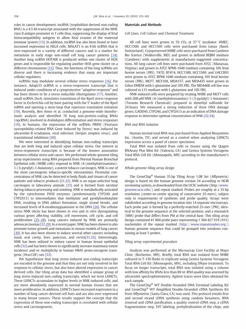
To identify NHBE NNK-induced Transcriptionally Active Regions(NNK- TARs), we utilized the Tiling Analysis Software (TAS) andIntegrated Genome Browser (IGB). We analyzed NHBE NNK-TARs ascompared with transcripts found from non-treated NHBE cells basedon a p-value of b0.01 using TAS. We analyzed the NNK-TARs by TAS/IGB and Model-Based Analysis of Tiling Arrays (MAT) to identifycandidate NNK-TARs for further investigation. A number of the NNK-TARs were identified with both methods, but there were many NNK-TARs that were only identified with one of the two methods. In total,over 231, 305 NNK-TARs were identified with their lengths rangingfrom 100 nt to greater than 3000 nt. This analysis was further char-acterized by identifying NNK-induced TARs (119 305) or NNK-decreased TARs (112 000). In this study we focused our efforts on theidentification of TARs which were induced by NNK. In order to identifythe longer non-coding transcripts and to intentionally exclude shortnon-coding RNAs including some pre-miRNAs; we excluded transcriptssmaller than 300 nts, thereby identifying 1 305 long NNK-induced TARs(Fig. 1).
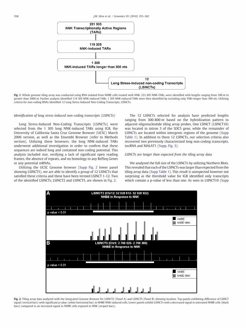
Fig. 1. Whole genome tiling array was conducted using RNA isolated from NHBE cells treated with NNK. 231,305 NNK-TARs, were identified with lengths ranging from 100 nt togreater than 3000 nt. Further analysis identified 119 305 NNK-induced TARs. 1 305 NNK-induced TARs were then identified by excluding only TARs longer than 300 nts. Utilizingcriteria for non-coding RNAs identified 12 Long Stress-Induced Non-Coding Transcripts, LSINCTs.
358 J.M. Silva et al. / Genomics 95 (2010) 355–362
Identification of long stress-induced non-coding transcripts (LSINCTs)
Long Stress-Induced Non-Coding Transcripts (LSINCTs) wereselected from the 1 305 long NNK-induced TARs using IGB, theUniversity of California Santa Cruz Genome Browser (UCSC) March2006 version, as well as the Ensembl Browser (refer to Methodssection). Utilizing these browsers, the long NNK-induced TARsunderwent additional investigation in order to confirm that thesesequences are indeed long and contained non-coding potential. Thisanalysis included size, verifying a lack of significant open readingframes, the absence of repeats, and no homology to any RefSeq Genesor any potential mRNAs.
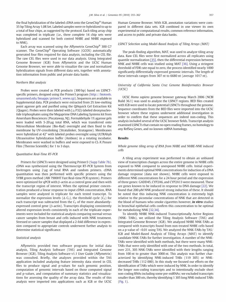
Utilizing the UCSC Genome browser (Supp Fig. 2 lower panelshowing LSINCT5), we are able to identify a group of 12 LSINCTs thatsatisfied these criteria and these have been termed LSINCT 1-12. Twoof the identified LSINCTs, LSINCT2 and LSINCT5, are shown in Fig. 2.
Fig. 2. Tiling array data analyzed with the Integrated Genome Browser for LSINCT2 (Panel Asignal (vertical bars) with significant p value (white horizontal bar) in NHBE NNK-induced cebars) compared to an increased signal in NHBE cells exposed to NNK (striped bars).
The 12 LSINCTs selected for analysis have predicted lengthsranging from 300-800 nt based on the hybridization pattern toadjacent oligonucleotide tiling array probes. One LSINCT (LSINCT10)was located in intron 3 of the SDC5 gene, while the remainder ofLSINCTs are located within intergenic regions of the genome (SuppTable 1). In addition to these 12 LSINCTs, our selection criteria alsorecovered two previously characterized long non-coding transcripts,tncRNA and MALAT1 (Supp. Fig. 3).
LSINCTs are longer than expected from the tiling array data
We analyzed the full size of the LSINCTs by utilizing Northern Blots.This revealed that eachof the LSINCTswas larger thanexpected fromthetiling array data (Supp Table 1). This result is unexpected however notsurprising as the threshold value for IGB identified only transcriptswhich contain a p-value of less than one. As seen in LSINCT10 (Supp
) and LSINCT5 (Panel B) showing location. Top panels exhibiting difference of LSINCTlls. Lower panels exhibit LSINCTs with a decreased signal in untreated NHBE cells (black
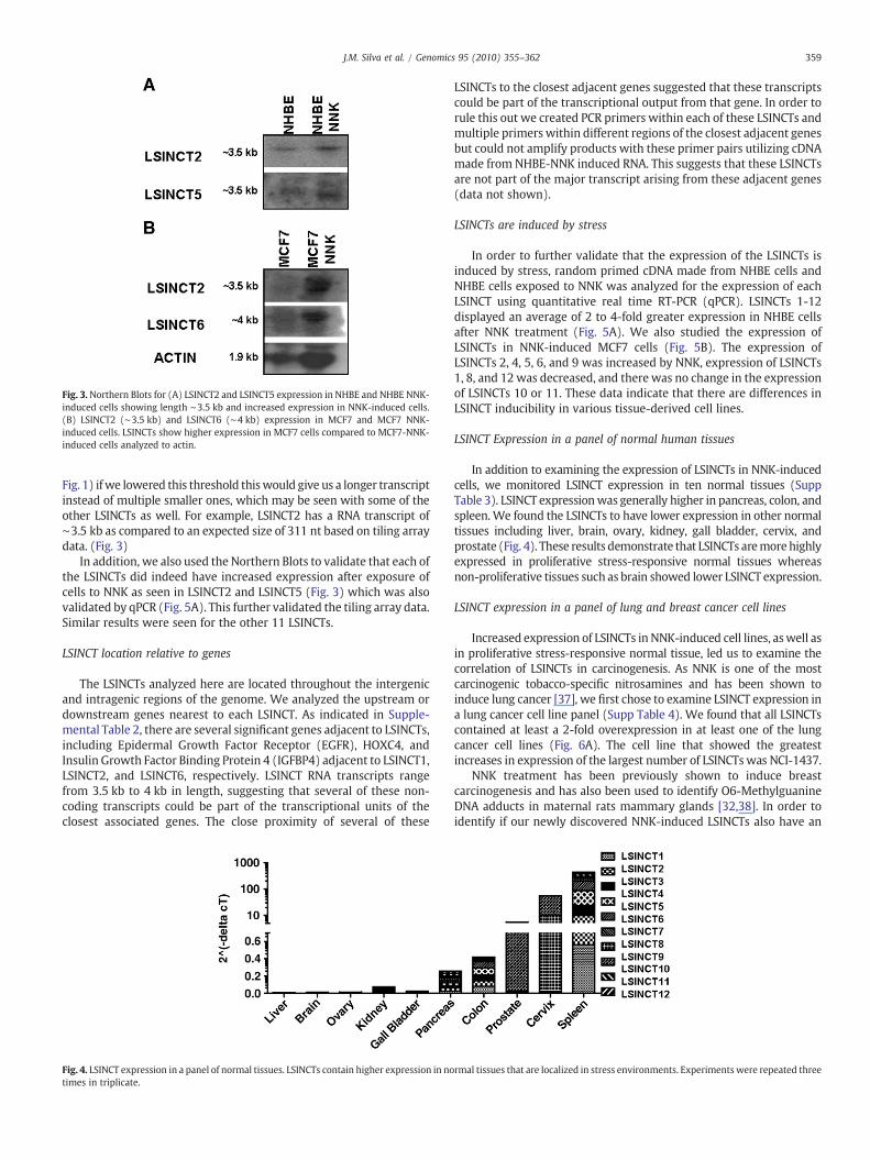
Fig. 3.Northern Blots for (A) LSINCT2 and LSINCT5 expression in NHBE and NHBE NNK-induced cells showing length ∼3.5 kb and increased expression in NNK-induced cells.(B) LSINCT2 (∼3.5 kb) and LSINCT6 (∼4 kb) expression in MCF7 and MCF7 NNK-induced cells. LSINCTs show higher expression in MCF7 cells compared to MCF7-NNK-induced cells analyzed to actin.
359J.M. Silva et al. / Genomics 95 (2010) 355–362
Fig. 1) ifwe lowered this threshold thiswould give us a longer transcriptinstead of multiple smaller ones, which may be seen with some of theother LSINCTs as well. For example, LSINCT2 has a RNA transcript of∼3.5 kb as compared to an expected size of 311 nt based on tiling arraydata. (Fig. 3)
In addition, we also used the Northern Blots to validate that each ofthe LSINCTs did indeed have increased expression after exposure ofcells to NNK as seen in LSINCT2 and LSINCT5 (Fig. 3) which was alsovalidated by qPCR (Fig. 5A). This further validated the tiling array data.Similar results were seen for the other 11 LSINCTs.
LSINCT location relative to genes
The LSINCTs analyzed here are located throughout the intergenicand intragenic regions of the genome. We analyzed the upstream ordownstream genes nearest to each LSINCT. As indicated in Supple-mental Table 2, there are several significant genes adjacent to LSINCTs,including Epidermal Growth Factor Receptor (EGFR), HOXC4, andInsulin Growth Factor Binding Protein 4 (IGFBP4) adjacent to LSINCT1,LSINCT2, and LSINCT6, respectively. LSINCT RNA transcripts rangefrom 3.5 kb to 4 kb in length, suggesting that several of these non-coding transcripts could be part of the transcriptional units of theclosest associated genes. The close proximity of several of these
Fig. 4. LSINCT expression in a panel of normal tissues. LSINCTs contain higher expression in notimes in triplicate.
LSINCTs to the closest adjacent genes suggested that these transcriptscould be part of the transcriptional output from that gene. In order torule this out we created PCR primers within each of these LSINCTs andmultiple primers within different regions of the closest adjacent genesbut could not amplify products with these primer pairs utilizing cDNAmade from NHBE-NNK induced RNA. This suggests that these LSINCTsare not part of the major transcript arising from these adjacent genes(data not shown).
LSINCTs are induced by stress
In order to further validate that the expression of the LSINCTs isinduced by stress, random primed cDNA made from NHBE cells andNHBE cells exposed to NNK was analyzed for the expression of eachLSINCT using quantitative real time RT-PCR (qPCR). LSINCTs 1-12displayed an average of 2 to 4-fold greater expression in NHBE cellsafter NNK treatment (Fig. 5A). We also studied the expression ofLSINCTs in NNK-induced MCF7 cells (Fig. 5B). The expression ofLSINCTs 2, 4, 5, 6, and 9 was increased by NNK, expression of LSINCTs1, 8, and 12 was decreased, and there was no change in the expressionof LSINCTs 10 or 11. These data indicate that there are differences inLSINCT inducibility in various tissue-derived cell lines.
LSINCT Expression in a panel of normal human tissues
In addition to examining the expression of LSINCTs in NNK-inducedcells, we monitored LSINCT expression in ten normal tissues (SuppTable 3). LSINCT expressionwas generally higher in pancreas, colon, andspleen. We found the LSINCTs to have lower expression in other normaltissues including liver, brain, ovary, kidney, gall bladder, cervix, andprostate (Fig. 4). These results demonstrate that LSINCTs aremore highlyexpressed in proliferative stress-responsive normal tissues whereasnon-proliferative tissues such as brain showed lower LSINCT expression.
LSINCT expression in a panel of lung and breast cancer cell lines
Increased expression of LSINCTs in NNK-induced cell lines, aswell asin proliferative stress-responsive normal tissue, led us to examine thecorrelation of LSINCTs in carcinogenesis. As NNK is one of the mostcarcinogenic tobacco-specific nitrosamines and has been shown toinduce lung cancer [37], we first chose to examine LSINCT expression ina lung cancer cell line panel (Supp Table 4). We found that all LSINCTscontained at least a 2-fold overexpression in at least one of the lungcancer cell lines (Fig. 6A). The cell line that showed the greatestincreases in expression of the largest number of LSINCTs was NCI-1437.
NNK treatment has been previously shown to induce breastcarcinogenesis and has also been used to identify O6-MethylguanineDNA adducts in maternal rats mammary glands [32,38]. In order toidentify if our newly discovered NNK-induced LSINCTs also have an
rmal tissues that are localized in stress environments. Experiments were repeated three
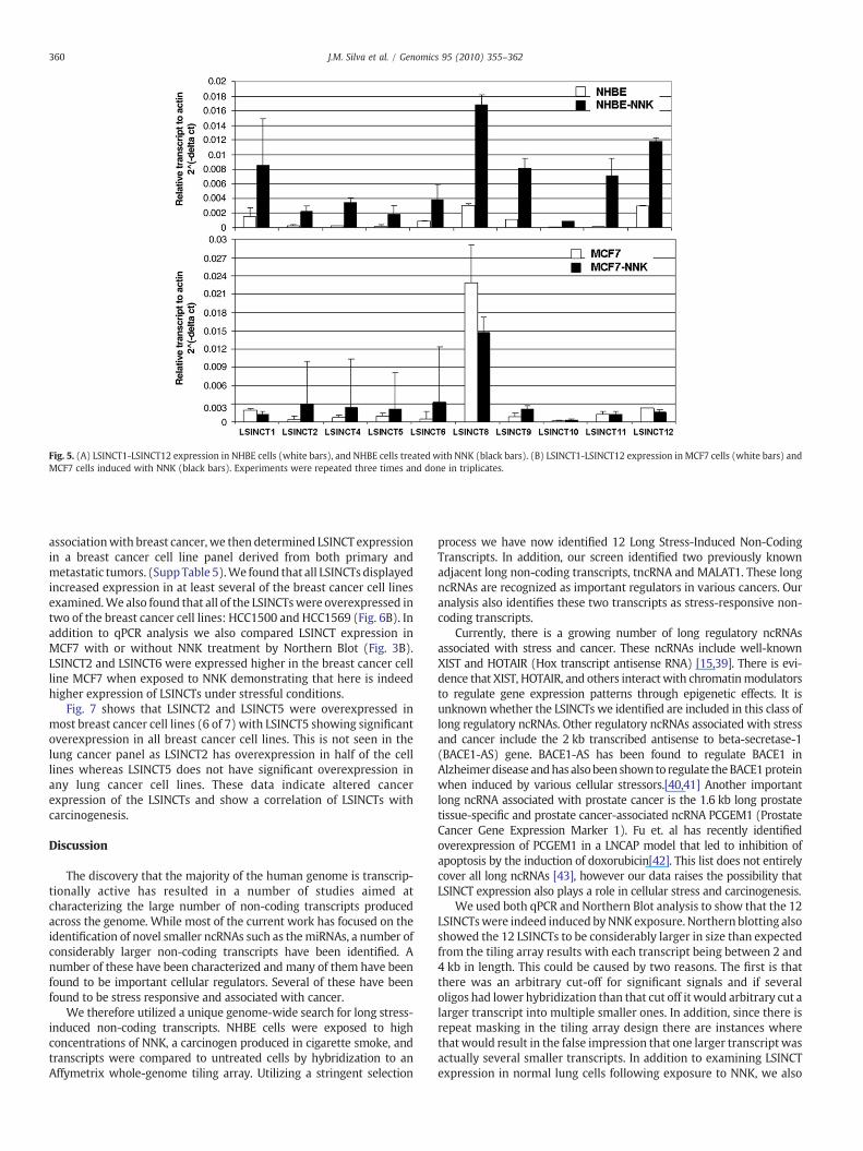
Fig. 5. (A) LSINCT1-LSINCT12 expression in NHBE cells (white bars), and NHBE cells treated with NNK (black bars). (B) LSINCT1-LSINCT12 expression in MCF7 cells (white bars) andMCF7 cells induced with NNK (black bars). Experiments were repeated three times and done in triplicates.
360 J.M. Silva et al. / Genomics 95 (2010) 355–362
associationwith breast cancer, we then determined LSINCT expressionin a breast cancer cell line panel derived from both primary andmetastatic tumors. (SuppTable 5).We found that all LSINCTsdisplayedincreased expression in at least several of the breast cancer cell linesexamined.We also found that all of the LSINCTswere overexpressed intwo of the breast cancer cell lines: HCC1500 and HCC1569 (Fig. 6B). Inaddition to qPCR analysis we also compared LSINCT expression inMCF7 with or without NNK treatment by Northern Blot (Fig. 3B).LSINCT2 and LSINCT6 were expressed higher in the breast cancer cellline MCF7 when exposed to NNK demonstrating that here is indeedhigher expression of LSINCTs under stressful conditions.
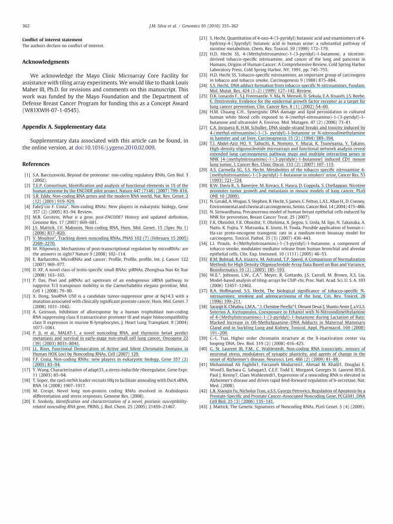
Fig. 7 shows that LSINCT2 and LSINCT5 were overexpressed inmost breast cancer cell lines (6 of 7) with LSINCT5 showing significantoverexpression in all breast cancer cell lines. This is not seen in thelung cancer panel as LSINCT2 has overexpression in half of the celllines whereas LSINCT5 does not have significant overexpression inany lung cancer cell lines. These data indicate altered cancerexpression of the LSINCTs and show a correlation of LSINCTs withcarcinogenesis.
Discussion
The discovery that the majority of the human genome is transcrip-tionally active has resulted in a number of studies aimed atcharacterizing the large number of non-coding transcripts producedacross the genome. While most of the current work has focused on theidentification of novel smaller ncRNAs such as themiRNAs, a number ofconsiderably larger non-coding transcripts have been identified. Anumber of these have been characterized and many of them have beenfound to be important cellular regulators. Several of these have beenfound to be stress responsive and associated with cancer.
We therefore utilized a unique genome-wide search for long stress-induced non-coding transcripts. NHBE cells were exposed to highconcentrations of NNK, a carcinogen produced in cigarette smoke, andtranscripts were compared to untreated cells by hybridization to anAffymetrix whole-genome tiling array. Utilizing a stringent selection
process we have now identified 12 Long Stress-Induced Non-CodingTranscripts. In addition, our screen identified two previously knownadjacent long non-coding transcripts, tncRNA and MALAT1. These longncRNAs are recognized as important regulators in various cancers. Ouranalysis also identifies these two transcripts as stress-responsive non-coding transcripts.
Currently, there is a growing number of long regulatory ncRNAsassociated with stress and cancer. These ncRNAs include well-knownXIST and HOTAIR (Hox transcript antisense RNA) [15,39]. There is evi-dence that XIST, HOTAIR, and others interact with chromatinmodulatorsto regulate gene expression patterns through epigenetic effects. It isunknownwhether the LSINCTs we identified are included in this class oflong regulatory ncRNAs. Other regulatory ncRNAs associated with stressand cancer include the 2 kb transcribed antisense to beta-secretase-1(BACE1-AS) gene. BACE1-AS has been found to regulate BACE1 inAlzheimerdisease andhas alsobeen shownto regulate theBACE1proteinwhen induced by various cellular stressors.[40,41] Another importantlong ncRNA associated with prostate cancer is the 1.6 kb long prostatetissue-specific and prostate cancer-associated ncRNA PCGEM1 (ProstateCancer Gene Expression Marker 1). Fu et. al has recently identifiedoverexpression of PCGEM1 in a LNCAP model that led to inhibition ofapoptosis by the induction of doxorubicin[42]. This list does not entirelycover all long ncRNAs [43], however our data raises the possibility thatLSINCT expression also plays a role in cellular stress and carcinogenesis.
We used both qPCR and Northern Blot analysis to show that the 12LSINCTswere indeed induced byNNKexposure. Northern blotting alsoshowed the 12 LSINCTs to be considerably larger in size than expectedfrom the tiling array results with each transcript being between 2 and4 kb in length. This could be caused by two reasons. The first is thatthere was an arbitrary cut-off for significant signals and if severaloligos had lower hybridization than that cut off it would arbitrary cut alarger transcript into multiple smaller ones. In addition, since there isrepeat masking in the tiling array design there are instances wherethat would result in the false impression that one larger transcript wasactually several smaller transcripts. In addition to examining LSINCTexpression in normal lung cells following exposure to NNK, we also
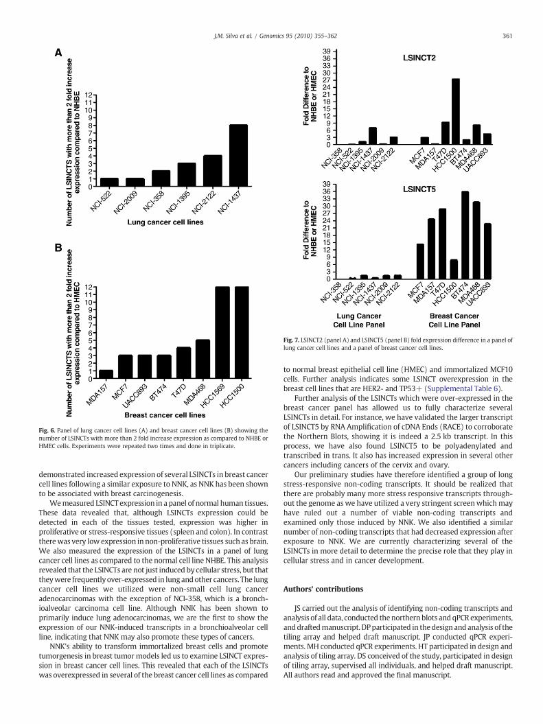
Fig. 7. LSINCT2 (panel A) and LSINCT5 (panel B) fold expression difference in a panel oflung cancer cell lines and a panel of breast cancer cell lines.
Fig. 6. Panel of lung cancer cell lines (A) and breast cancer cell lines (B) showing thenumber of LSINCTs with more than 2 fold increase expression as compared to NHBE orHMEC cells. Experiments were repeated two times and done in triplicate.
361J.M. Silva et al. / Genomics 95 (2010) 355–362
demonstrated increased expression of several LSINCTs in breast cancercell lines following a similar exposure to NNK, as NNK has been shownto be associated with breast carcinogenesis.
Wemeasured LSINCT expression in a panel of normal human tissues.These data revealed that, although LSINCTs expression could bedetected in each of the tissues tested, expression was higher inproliferative or stress-responsive tissues (spleen and colon). In contrasttherewas very low expression in non-proliferative tissues such as brain.We also measured the expression of the LSINCTs in a panel of lungcancer cell lines as compared to the normal cell line NHBE. This analysisrevealed that the LSINCTs are not just induced by cellular stress, but thattheywere frequentlyover-expressed in lungandother cancers. The lungcancer cell lines we utilized were non-small cell lung canceradenocarcinomas with the exception of NCI-358, which is a bronch-ioalveolar carcinoma cell line. Although NNK has been shown toprimarily induce lung adenocarcinomas, we are the first to show theexpression of our NNK-induced transcripts in a bronchioalveolar cellline, indicating that NNK may also promote these types of cancers.
NNK's ability to transform immortalized breast cells and promotetumorgenesis in breast tumormodels led us to examine LSINCT expres-sion in breast cancer cell lines. This revealed that each of the LSINCTswas overexpressed in several of the breast cancer cell lines as compared
to normal breast epithelial cell line (HMEC) and immortalized MCF10cells. Further analysis indicates some LSINCT overexpression in thebreast cell lines that are HER2- and TP53+ (Supplemental Table 6).
Further analysis of the LSINCTs which were over-expressed in thebreast cancer panel has allowed us to fully characterize severalLSINCTs in detail. For instance, we have validated the larger transcriptof LSINCT5 by RNA Amplification of cDNA Ends (RACE) to corroboratethe Northern Blots, showing it is indeed a 2.5 kb transcript. In thisprocess, we have also found LSINCT5 to be polyadenylated andtranscribed in trans. It also has increased expression in several othercancers including cancers of the cervix and ovary.
Our preliminary studies have therefore identified a group of longstress-responsive non-coding transcripts. It should be realized thatthere are probably many more stress responsive transcripts through-out the genome as we have utilized a very stringent screenwhichmayhave ruled out a number of viable non-coding transcripts andexamined only those induced by NNK. We also identified a similarnumber of non-coding transcripts that had decreased expression afterexposure to NNK. We are currently characterizing several of theLSINCTs in more detail to determine the precise role that they play incellular stress and in cancer development.
Authors' contributions
JS carried out the analysis of identifying non-coding transcripts andanalysis of all data, conducted thenorthernblots and qPCR experiments,anddraftedmanuscript. DPparticipated in thedesign and analysis of thetiling array and helped draft manuscript. JP conducted qPCR experi-ments. MH conducted qPCR experiments. HT participated in design andanalysis of tiling array. DS conceived of the study, participated in designof tiling array, supervised all individuals, and helped draft manuscript.All authors read and approved the final manuscript.
362 J.M. Silva et al. / Genomics 95 (2010) 355–362
Conflict of interest statementThe authors declare no conflict of interest.
Acknowledgments
We acknowledge the Mayo Clinic Microarray Core Facility forassistance with tiling array experiments. Wewould like to thank LouisMaher III, Ph.D. for revisions and comments on this manuscript. Thiswork was funded by the Mayo Foundation and the Department ofDefense Breast Cancer Program for funding this as a Concept Award(W81XWH-07-1-0545).
Appendix A. Supplementary data
Supplementary data associated with this article can be found, inthe online version, at doi:10.1016/j.ygeno.2010.02.009.
References
[1] S.A. Barciszewski, Beyond the proteome: non-coding regulatory RNAs, Gen Biol. 3(2002).
[2] T.E.P. Consortium, Identification and analysis of functional elements in 1% of thehuman genome by the ENCODE pilot project, Nature 447 (7146) (2007) 799–816.
[3] S.R. Eddy, Non-coding RNA genes and the modern RNA world, Nat. Rev. Genet. 2(12) (2001) 919–929.
[4] Fabrý´cio F. Costa*, Non-coding RNAs: New players in eukaryotic biology, Gene357 (2) (2005) 83–94. Review.
[5] M.B. Gerstein, What is a gene, post-ENCODE? History and updated definition,Genome Res. 17 (2007) 669–681.
[6] J.S. Mattick, I.V. Makunin, Non-coding RNA, Hum. Mol. Genet. 15 (Spec No 1)(2006) R17–R29.
[7] V. Moulton*, Tracking down noncoding RNAs, PNAS 102 (7) (February 15 2005)2269–2270.
[8] W. Filipowicz, Mechanisms of post-transcriptional regulation by microRNAs: arethe answers in sight? Nature 9 (2008) 102–114.
[9] E. Barbarotto, MicroRNAs and cancer: Profile, Profile, profile, Int. J. Cancer 122(2007) 969–977.
[10] D. XF, A novel class of testis-specific small RNAs: piRNAs, Zhonghua Nan Ke Xue(2008) 163–165.
[11] P. Das, Piwi and piRNAs act upstream of an endogenous siRNA pathway tosuppress Tc3 transposon mobility in the Caenorhabditis elegans germline, Mol.Cell 1 (2008) 79–90.
[12] X. Dong, SnoRNA U50 is a candidate tumor-suppressor gene at 6q14.3 with amutation associated with clinically significant prostate cancer, Hum. Mol. Genet. 7(2008) 1031–1042.
[13] A. Geirsson, Inhibition of alloresponse by a human trophoblast non-codingRNA suppressing class II transactivator promoter III and major histocompatibilityclass II expression in murine B-lymphocytes, J. Heart Lung Transplant. 9 (2004)1077–1081.
[14] P. Ji, et al., MALAT-1, a novel noncoding RNA, and thymosin beta4 predictmetastasis and survival in early-stage non-small cell lung cancer, Oncogene 22(39) (2003) 8031–8041.
[15] J.L. Rinn, Functional Demarcation of Active and Silent Chromatin Domains inHuman HOX Loci by Noncoding RNAs, Cell (2007) 129.
[16] F.F. Costa, Non-coding RNAs: new players in eukaryotic biology, Gene 357 (2)(2005) 83–94.
[17] Y. Wang, Characterization of adapt33, a stress-inducible riboregulator, Gene Expr.11 (2003) 85–94.
[18] T. Soper, the rpoSmrNA leader recruits Hfq to facilitate annealing with DsrA sRNA,RNA 14 (2008) 1907–1917.
[19] M. Crespi, Novel long non-protein coding RNAs involved in Arabidopsisdifferentiation and stress responses, Genome Res. (2008).
[20] E. Sonkoly, Identification and characterization of a novel, psoriasis susceptibility-related noncoding RNA gene, PRINS, J. Biol. Chem. 25 (2005) 21459–21467.
[21] S. Hecht, Quantitation of 4-oxo-4-(3-pyridyl) butanoic acid and enantiomers of 4-hydroxy-4-(3pyridyl) butanoic acid in human urine: a substantial pathway ofnicotine metabolism, Chem. Res. Toxicol. 59 (1999) 172–179.
[22] H.D. Hecht SS, 4-(Methylnitrosamino)-1-(3-pyridyl)-1-butanone, a nicotine-derived tobacco-specific nitrosamine, and cancer of the lung and pancreas inHumans, Origins of Human Cancer: A Comprehensive Review, Cold Spring HarborLaboratory Press, Cold Spring Harbor, NY, 1991, pp. 745–755.
[23] H.D. Hecht SS, Tobacco-specific nitrosamines, an important group of carcinogensin tobacco and tobacco smoke, Carcinogenesis 9 (1988) 875–884.
[24] S.S. Hecht, DNA adduct formation from tobacco-specific N-nitrosamines, Fundam.Mol. Mutat. Res. 424 (1–2) (1999) 127–142. Review.
[25] D.K. Lonardo F., S.J. Freemantle, Y. Ma, N.Memoli, D. Sekula, E.A. Knauth, J.S. Beebe,E. Dmitrovsky, Evidence for the epidermal growth factor receptor as a target forlung cancer prevention, Clin. Cancer Res. 8 (1) (2002) 54–60.
[26] H.M. Chuang C.H., Synergistic DNA damage and lipid peroxidation in culturedhuman white blood cells exposed to 4-(methyl-nitrosamino)-1-(3-pyridyl)-1-butanone and ultraviolet A, Environ. Mol. Mutagen. 47 (2) (2006) 73–81.
[27] C.A. Jorquera R, H.M. Schuller, DNA single-strand breaks and toxicity induced by4-(methyl-nitrosamino)-1-(3- pyridyl)-1-butanone or N-nitrosodimethylaminein hamster and rat liver, Carcinogenesis 15 (2) (1994) 389–394.
[28] T.I. Abdel-Aziz HO, Y. Tabuchi, K. Nomoto, Y. Murai, K. Tsuneyama, Y. Takano,High-density oligonucleotide microarrays and functional network analysis revealextended lung carcinogenesis pathway maps and multiple interacting genes inNNK [4-(methylnitrosamino)-1-(3-pyridyle)-1-butanone] induced CD1 mouselung tumor, J. Cancer Res. Clinic Oncol. 133 (2) (2007) 107–115.
[29] A.S. Carmella SG, S.S. Hecht, Metabolites of the tobacco specific nitrosamine 4-(methylnitrosamino)-1-(3-pyridyl)-1-butanone in smokers' urine, Cancer Res. 53(1993) 721–724.
[30] R.W. Davis R., S. Banerjee, M. Kovacs, E. Haura, D. Coppola, S. Chellappan, Nicotinepromotes tumor growth and metastasis in mouse models of lung cancer, PLoSONE 10 (2009).
[31] N.Gerald, A.Wogan, S. Stephen, B.Hecht, S. James, C. Felton, L.A.L. AllanH., D. Conney,Environmental and chemical carcinogenesis, Semin. Cancer Biol. 14 (2004) 473–486.
[32] N. Siriwardhana, Precancerous model of human breast epithelial cells induced byNNK for prevention, Breast Cancer Treat. 25 (2007).
[33] F.K. Ohnishit, F.K. Ohnishit, Y. Ohshima, X. Jiegou, S. Ueda, M. Iigo, N. Takasuka, A.Naito, K. Fujita, Y. Matsuoka, K. Izumi, H. Tsuda, Possible application of human c-Ha-ras proto-oncogene transgenic rats in a medium-term bioassay model forcarcinogens, Toxicol. Pathol. 35 (3) (2007) 436–443.
[34] L.I. Proulx, 4-(Methylnitrosamino)-1-(3-pyridyl)-1-butanone, a component oftobacco smoke, modulates mediator release from human bronchial and alveolarepithelial cells, Clin. Exp. Immunol. 10 (1111) (2005) 46–53.
[35] B.M. Bolstad, R.A. Irizarry, M. Astrand, T.P. Speed, A Comparison of NormalizationMethods for High Density Oligonucleotide Array Data Based on Bias and Variance,Bioinformatics 19 (2) (2003) 185–193.
[36] W.E.*. Johnson, L.W., C.A.*. Meyer, R. Gottardo, J.S. Carroll, M. Brown, X.S. Liu,Model-based analysis of tiling-arrays for ChIP-chi, Proc. Natl. Acad. Sci. U. S. A. 103(2006) 12457–12462.
[37] R.A. Hoffmannd, S.S. Hecht, The biological significance of tobacco-specific N-nitrosamines: smoking and adenocarcinoma of the lung, Crit. Rev. Toxicol. 26(1996) 199–211.
[38] Saranjit K. Chhabra, L.M.A., *,1, Christine Perella*,†, Dimant Desai ‡, ShantuAmin ‡, a.V.L.S.Soterios A. Kyrtopoulos, Coexposure to Ethanol with N-Nitrosodimethylamineof 4-(Methylnitrosamino)-1-(3-pyridyl)-1-butanone during Lactation of Rats:Marked Increase in O6-Methylguanine–DNA Adducts in Maternal MammaryGland and in Suckling Lung and Kidney, Toxicol. Appl. Pharmacol. 169 (2000)191–200.
[39] C.-L. Tsai, Higher order chromatin structure at the X-inactivation center vialooping DNA, Dev. Biol. 319 (2) (2008) 416–425.
[40] G. St. Laurent III, F.M., C. Wahlestedt, Non-coding RNA transcripts: sensors ofneuronal stress, modulators of synaptic plasticity, and agents of change in theonset of Alzheimer's disease, Neurosci. Lett. 466 (2) (2009) 81–88.
[41] Mohammad Ali Faghihi1, Farzaneh Modarresi1, Ahmad M. Khalil1, Douglas E.Wood3, Barbara G. Sahagan3, C.E.F. Todd E. Morgan4, Georges St. Laurent III5,6,Paul J. Kenny7, Claes Wahlestedt1, Expression of a noncoding RNA is elevated inAlzheimer's disease and drives rapid feed-forward regulation of b-secretase, Nat.Med. (2008).
[42] L.R. Xiaoqin Fu, Nicholas Tran, a.S.S. Gyorgy Petrovics, Regulation of Apoptosis by aProstate-Specific and Prostate Cancer-Associated Noncoding Gene, PCGEM1, DNACell Biol. 25 (3) (2006) 135–141.
[43] J. Mattick, The Genetic Signatures of Noncoding RNAs, PLoS Genet. 5 (4) (2009).
Related Documents