1348 / Molecular Plant-Microbe Interactions MPMI Vol. 19, No. 12, 2006, pp. 1348–1358. DOI: 10.1094/MPMI-19-1348. © 2006 The American Phytopathological Society Identification of Cell Wall–Associated Proteins from Phytophthora ramorum Harold J. G. Meijer, 1 Peter J. I. van de Vondervoort, 1 Qing Yuan Yin, 2 Chris G. de Koster, 2 Frans M. Klis, 2 Francine Govers, 1 and Piet W. J. de Groot 2 1 Laboratory of Phytopathology, Plant Sciences Group, Wageningen University, Binnenhaven 5, NL-6709 PD Wageningen, The Netherlands; 2 Swammerdam Institute for Life Sciences, University of Amsterdam, Nieuwe Achtergracht 166, NL-1018 WV Amsterdam, The Netherlands Submitted 31 May 2006. Accepted 24 July 2006. The oomycete genus Phytophthora comprises a large group of fungal-like plant pathogens. Two Phytophthora genomes recently have been sequenced; one of them is the genome of Phytophthora ramorum, the causal agent of sudden oak death. During plant infection, extracellular proteins, either soluble secreted proteins or proteins associated with the cell wall, play important roles in the interaction with host plants. Cell walls of P. ramorum contain 1 to 1.5% proteins, the remainder almost exclusively being accounted for by glucan polymers. Here, we present an inventory of cell- wall-associated proteins based on mass spectrometric se- quence analysis of tryptic peptides obtained by proteolytic digestion of sodium dodecyl sulfate-treated mycelial cell walls. In total, 17 proteins were identified, all of which are authentic secretory proteins. Functional classification based on homology searches revealed six putative mucins or mucin-like proteins, five putative glycoside hydrolases, two transglutaminases, one annexin-like protein, the elicitin protein RAM5, one protein of unknown function, and one Kazal-type protease inhibitor. We propose that the cell wall proteins thus identified are important for pathogenicity. Additional keywords: cell wall proteome. The fungal cell wall is an essential organelle that enables cells to resist internal turgor pressures which, in appressoria of plant pathogens, may reach values up to 8 MPa (Howard et al. 1991) The cell wall is also of vital importance for maintaining cell morphology, uptake of substances, interactions with plasma membrane proteins, signaling, and protection against adverse conditions in the cellular environment. The cell wall may function further as a barrier by preventing proteins from leaking into the medium and act as a scaffold for exposing cell wall proteins (CWPs) to the cell surface (De Groot et al. 2005). The large diversity of implicated biological roles of CWPs includes cell wall biosynthesis and adhesion. For patho- genic microorganisms, such as fungi and oomycetes, CWPs also can provide protection against host defense reactions (De Groot et al. 2004). Adhesion to host tissues is an important at- tribute in pathogenesis and, in many cases, indispensable for virulence (De Groot et al. 2005; Sundstrom 2002). Many fungi have bilayered cell walls. The inner, electron- transparent layer is primarily composed of polysaccharides such as 1,3-β-glucan, 1,6-β-glucan, 1,3-α-glucan, and chitin, and is surrounded by an electron-dense layer, mainly compris- ing covalently bound mannosylated proteins (Klis et al. 2002). In a few ascomycetes, the CWP composition has been studied in detail using tandem mass spectrometry (De Groot et al. 2004; Yin et al. 2005). In baker’s yeast (Saccharomyces cere- visiae) and the pleomorphic human pathogen Candida albi- cans, many of the CWPs are classified as carbohydrate- or gly- can-modifying enzymes. In addition, adhesins, heme-binding proteins involved in iron uptake, and superoxide dismutases in- volved in environmental protection are found in the cell wall (De Groot et al. 2005). Most of the identified CWPs in S. cere- visiae and C. albicans are covalently bound glycosylphos- phatidylinositol (GPI)-modified proteins. Typical features of GPI proteins are a canonical N-terminal signal peptide for se- cretion and a hydrophobic C-terminal region that functions as a signal peptide for the addition of a GPI membrane anchor (De Groot et al. 2005). GPI proteins in fungi have a unique char- acteristic depending on sequence features immediately upstream of the GPI-anchor attachment site. They may be cleaved from the plasma membrane and subsequently attached covalently to the cell wall glucan (De Groot et al. 2005). This covalent incor- poration of GPI CWPs in the cell wall of S. cerevisiae and other ascomycetes was shown to be achieved by direct attach- ment to 1,6-β-glucan, which interconnects CWPs to the 1,3-β- glucan network (Kapteyn et al. 1996; Kollár et al. 1997). Such covalently linked CWPs can be solubilized by glucanases but not by treatment with NaCl or hot sodium dodecyl sulfate (SDS) solutions. Other proteins in S. cerevisiae and C. albicans are bound to the cell wall polysaccharide network through a linkage that is sensitive to treatment with mild alkali (De Groot et al. 2004; Ecker et al. 2006; Kapteyn et al. 1999; Yin et al. 2005). Oomycetes are a unique group of fungal-like eukaryotes that includes notorious pathogens of plants and animals. The oomy- cete genus Phytophthora comprises over 70 species that all cause devastating plant diseases on a wide variety of commer- cially grown crops and in natural vegetations. Phytophthora ramorum is among the most destructive plant pathogens that have been discovered in the last decade. It originally was de- scribed as a pathogen on Rhododendron and Viburnum spp. (Werres et al. 2001) and subsequently found to be responsible for the sudden oak death syndrome, in particular, on the west coast of North America (Rizzo et al. 2002). Unlike most fungal cell walls, oomycete walls contain very little chitin. They pri- marily are composed of 1,3-β-glucan and some other β-linked glucose polymers; and, in Phytophthora spp., these β-glucans compose up to 90% of the dry weight (Bartnicki-Garcia and Wang 1983). Protein levels in the cell walls of fungi and oomy- Corresponding author: H. J. G. Meijer; Telephone: +31 317 483 403; Fax: +31 317 483 412; E-mail: Harold.Meijer@ wur.nl

Welcome message from author
This document is posted to help you gain knowledge. Please leave a comment to let me know what you think about it! Share it to your friends and learn new things together.
Transcript

1348 / Molecular Plant-Microbe Interactions
MPMI Vol. 19, No. 12, 2006, pp. 1348–1358. DOI: 10.1094 / MPMI -19-1348. © 2006 The American Phytopathological Society
Identification of Cell Wall–Associated Proteins from Phytophthora ramorum
Harold J. G. Meijer,1 Peter J. I. van de Vondervoort,1 Qing Yuan Yin,2 Chris G. de Koster,2 Frans M. Klis,2 Francine Govers,1 and Piet W. J. de Groot2 1Laboratory of Phytopathology, Plant Sciences Group, Wageningen University, Binnenhaven 5, NL-6709 PD Wageningen, The Netherlands; 2Swammerdam Institute for Life Sciences, University of Amsterdam, Nieuwe Achtergracht 166, NL-1018 WV Amsterdam, The Netherlands
Submitted 31 May 2006. Accepted 24 July 2006.
The oomycete genus Phytophthora comprises a large group of fungal-like plant pathogens. Two Phytophthora genomes recently have been sequenced; one of them is the genome of Phytophthora ramorum, the causal agent of sudden oak death. During plant infection, extracellular proteins, either soluble secreted proteins or proteins associated with the cell wall, play important roles in the interaction with host plants. Cell walls of P. ramorum contain 1 to 1.5% proteins, the remainder almost exclusively being accounted for by glucan polymers. Here, we present an inventory of cell-wall-associated proteins based on mass spectrometric se-quence analysis of tryptic peptides obtained by proteolytic digestion of sodium dodecyl sulfate-treated mycelial cell walls. In total, 17 proteins were identified, all of which are authentic secretory proteins. Functional classification based on homology searches revealed six putative mucins or mucin-like proteins, five putative glycoside hydrolases, two transglutaminases, one annexin-like protein, the elicitin protein RAM5, one protein of unknown function, and one Kazal-type protease inhibitor. We propose that the cell wall proteins thus identified are important for pathogenicity.
Additional keywords: cell wall proteome.
The fungal cell wall is an essential organelle that enables cells to resist internal turgor pressures which, in appressoria of plant pathogens, may reach values up to 8 MPa (Howard et al. 1991) The cell wall is also of vital importance for maintaining cell morphology, uptake of substances, interactions with plasma membrane proteins, signaling, and protection against adverse conditions in the cellular environment. The cell wall may function further as a barrier by preventing proteins from leaking into the medium and act as a scaffold for exposing cell wall proteins (CWPs) to the cell surface (De Groot et al. 2005). The large diversity of implicated biological roles of CWPs includes cell wall biosynthesis and adhesion. For patho-genic microorganisms, such as fungi and oomycetes, CWPs also can provide protection against host defense reactions (De Groot et al. 2004). Adhesion to host tissues is an important at-tribute in pathogenesis and, in many cases, indispensable for virulence (De Groot et al. 2005; Sundstrom 2002).
Many fungi have bilayered cell walls. The inner, electron-transparent layer is primarily composed of polysaccharides such as 1,3-β-glucan, 1,6-β-glucan, 1,3-α-glucan, and chitin,
and is surrounded by an electron-dense layer, mainly compris-ing covalently bound mannosylated proteins (Klis et al. 2002). In a few ascomycetes, the CWP composition has been studied in detail using tandem mass spectrometry (De Groot et al. 2004; Yin et al. 2005). In baker’s yeast (Saccharomyces cere-visiae) and the pleomorphic human pathogen Candida albi-cans, many of the CWPs are classified as carbohydrate- or gly-can-modifying enzymes. In addition, adhesins, heme-binding proteins involved in iron uptake, and superoxide dismutases in-volved in environmental protection are found in the cell wall (De Groot et al. 2005). Most of the identified CWPs in S. cere-visiae and C. albicans are covalently bound glycosylphos-phatidylinositol (GPI)-modified proteins. Typical features of GPI proteins are a canonical N-terminal signal peptide for se-cretion and a hydrophobic C-terminal region that functions as a signal peptide for the addition of a GPI membrane anchor (De Groot et al. 2005). GPI proteins in fungi have a unique char-acteristic depending on sequence features immediately upstream of the GPI-anchor attachment site. They may be cleaved from the plasma membrane and subsequently attached covalently to the cell wall glucan (De Groot et al. 2005). This covalent incor-poration of GPI CWPs in the cell wall of S. cerevisiae and other ascomycetes was shown to be achieved by direct attach-ment to 1,6-β-glucan, which interconnects CWPs to the 1,3-β-glucan network (Kapteyn et al. 1996; Kollár et al. 1997). Such covalently linked CWPs can be solubilized by glucanases but not by treatment with NaCl or hot sodium dodecyl sulfate (SDS) solutions. Other proteins in S. cerevisiae and C. albicans are bound to the cell wall polysaccharide network through a linkage that is sensitive to treatment with mild alkali (De Groot et al. 2004; Ecker et al. 2006; Kapteyn et al. 1999; Yin et al. 2005).
Oomycetes are a unique group of fungal-like eukaryotes that includes notorious pathogens of plants and animals. The oomy-cete genus Phytophthora comprises over 70 species that all cause devastating plant diseases on a wide variety of commer-cially grown crops and in natural vegetations. Phytophthora ramorum is among the most destructive plant pathogens that have been discovered in the last decade. It originally was de-scribed as a pathogen on Rhododendron and Viburnum spp. (Werres et al. 2001) and subsequently found to be responsible for the sudden oak death syndrome, in particular, on the west coast of North America (Rizzo et al. 2002). Unlike most fungal cell walls, oomycete walls contain very little chitin. They pri-marily are composed of 1,3-β-glucan and some other β-linked glucose polymers; and, in Phytophthora spp., these β-glucans compose up to 90% of the dry weight (Bartnicki-Garcia and Wang 1983). Protein levels in the cell walls of fungi and oomy-
Corresponding author: H. J. G. Meijer; Telephone: +31 317 483 403; Fax:+31 317 483 412; E-mail: Harold.Meijer@ wur.nl
Vol. 19, No. 12, 2006 / 1349
cetes are comparable and are reported to range from 3 to 11% depending on the species studied (Bartnicki-Garcia and Wang 1983).
Fungal and oomycete plant pathogens secrete a variety of extracellular effector molecules to promote the infection proc-ess (Birch et al. 2006; Dean et al. 2005; Westerink et al. 2004). Recognition of effector proteins can elicit plant defense re-sponses that ultimately result in a hypersensitivity response (HR) or programmed cell death. Several cell wall components and cell wall proteins are known to act as elicitors of plant de-fense responses (Cote et al. 1998; Hahn 1996; Rose et al. 2002). Therefore, understanding the pathogens’ cell wall com-position is expected to provide new insights into mechanisms underlying plant–pathogen interactions. The release of the complete genome sequence of two Phytophthora spp. provides the opportunity to search for genes encoding putative cell wall proteins (Tyler et al. 2006). A genome-wide inventory of se-creted proteins of P. sojae and P. ramorum already revealed putative secretomes of 1,464 and 1,188 proteins, respectively (Jiang 2006), of which a subset of approximately 100 proteins contain putative GPI anchor addition sequences. This suggests that, also in Phytophthora spp., GPI modification might be a way to tether proteins to the exterior of the cell (Jiang et al. 2006; R. H. Y. Jiang and F. Govers, unpublished).
In this study, we present a detailed analysis of the cell wall composition of P. ramorum. First, we investigated the chemi-cal composition of the mycelial cell walls and, second, we per-formed a detailed analysis of the cell wall proteome using liquid chromatography tandem mass spectrometry (LC/MS/ MS). By sequencing the peptides and exploiting the genome sequence, 17 CWPs could be identified, none of which contains a putative GPI anchor. They comprise proteins involved in polysaccharide modification, adhesion, and virulence. The iden-tification of these CWPs provides a first foray into Phytoph-thora cell wall proteomics.
RESULTS AND DISCUSSION
Chemical composition of the cell wall. In order to analyze the sugar composition of mycelial cell
walls of P. ramorum, polysaccharides in SDS-treated cell walls of strain Pr04 were hydrolyzed using sulfuric acid and ana-lyzed by high-performance liquid chromatography (HPLC). Glucose monomers appeared to represent 97 ± 4% of the cell wall mass, whereas mannose, galactose, and glucosamine were below detection levels. Chitin, which was determined sepa-rately, also was below the detection level, which is consistent with the lack of detectable levels of glucosamine by HPLC and with the very high percentage of glucose in P. ramorum cell walls. The amount of covalently bound proteins was quantified at 1.2 ± 0.2%. These results suggest that, in addition to a small amount of protein, the cell wall is composed almost exclu-sively of glucan. In comparison with earlier studies on Phy-tophthora cell walls, the glucan levels in P. ramorum seem to be relatively high. Bartnicki-Garcia and Wang (1983) reported analyses on the cell wall composition in P. cinnamomi, P. para-sitica, P. palmivora, P. heveae, and P. megasperma var. sojae. These analyses, based on enzymatic digestions and methyla-tion analysis, revealed 86 to 90% glucose, 3 to 11% protein, and minor amounts of lipids and polysaccharides, including mannose and glucosamine. Although this comparison suggests that glucan levels differ between species, it is more likely that the differences are due to the methodology that was used to isolate the cell walls or to quantify the components.
BLAST analysis using sequences from ascomycetous fungi as queries revealed that homologs of genes involved in 1,3-β-glucan synthesis (FKS genes), 1,3-β-glucan modification (GAS
and BGL), and 1,6-β-glucan synthesis (KRE6) are present in Phytophthora spp., whereas homologs of 1,3-α-glucan syn-thase-encoding genes (MOK1/AGS1 family) are absent. Inter-estingly, although chitin levels in the cell wall are below the detection levels, P. ramorum does contain clear homologs of chitin synthase and chitinase. Homologs of the CRH family, believed to mediate the covalent attachment of chitin to 1,3-β-glucan in yeast, are absent.
CWPs in P. ramorum. To identify putative CWPs of P. ramorum, isolated cell walls
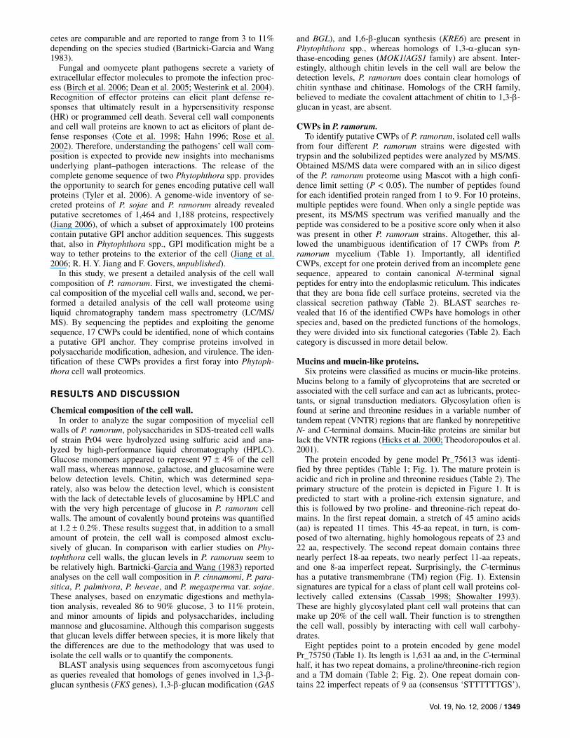
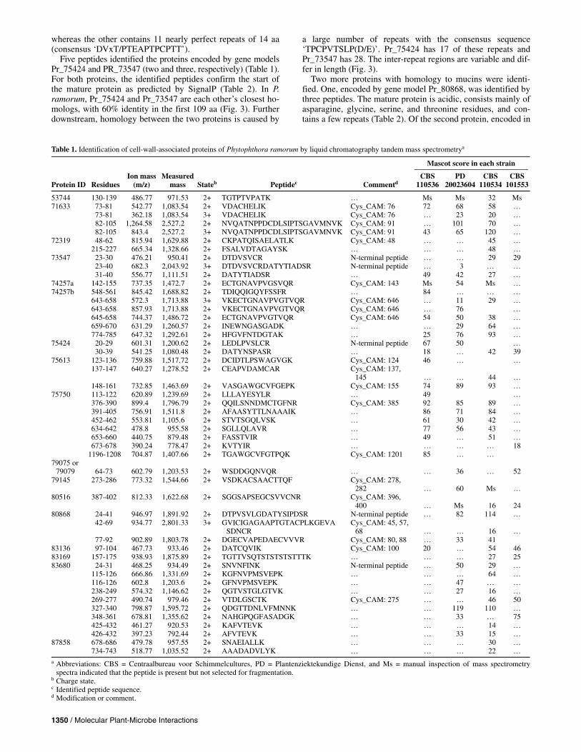
from four different P. ramorum strains were digested with trypsin and the solubilized peptides were analyzed by MS/MS. Obtained MS/MS data were compared with an in silico digest of the P. ramorum proteome using Mascot with a high confi-dence limit setting (P < 0.05). The number of peptides found for each identified protein ranged from 1 to 9. For 10 proteins, multiple peptides were found. When only a single peptide was present, its MS/MS spectrum was verified manually and the peptide was considered to be a positive score only when it also was present in other P. ramorum strains. Altogether, this al-lowed the unambiguous identification of 17 CWPs from P. ramorum mycelium (Table 1). Importantly, all identified CWPs, except for one protein derived from an incomplete gene sequence, appeared to contain canonical N-terminal signal peptides for entry into the endoplasmic reticulum. This indicates that they are bona fide cell surface proteins, secreted via the classical secretion pathway (Table 2). BLAST searches re-vealed that 16 of the identified CWPs have homologs in other species and, based on the predicted functions of the homologs, they were divided into six functional categories (Table 2). Each category is discussed in more detail below.
Mucins and mucin-like proteins. Six proteins were classified as mucins or mucin-like proteins.
Mucins belong to a family of glycoproteins that are secreted or associated with the cell surface and can act as lubricants, protec-tants, or signal transduction mediators. Glycosylation often is found at serine and threonine residues in a variable number of tandem repeat (VNTR) regions that are flanked by nonrepetitive N- and C-terminal domains. Mucin-like proteins are similar but lack the VNTR regions (Hicks et al. 2000; Theodoropoulos et al. 2001).
The protein encoded by gene model Pr_75613 was identi-fied by three peptides (Table 1; Fig. 1). The mature protein is acidic and rich in proline and threonine residues (Table 2). The primary structure of the protein is depicted in Figure 1. It is predicted to start with a proline-rich extensin signature, and this is followed by two proline- and threonine-rich repeat do-mains. In the first repeat domain, a stretch of 45 amino acids (aa) is repeated 11 times. This 45-aa repeat, in turn, is com-posed of two alternating, highly homologous repeats of 23 and 22 aa, respectively. The second repeat domain contains three nearly perfect 18-aa repeats, two nearly perfect 11-aa repeats, and one 8-aa imperfect repeat. Surprisingly, the C-terminus has a putative transmembrane (TM) region (Fig. 1). Extensin signatures are typical for a class of plant cell wall proteins col-lectively called extensins (Cassab 1998; Showalter 1993). These are highly glycosylated plant cell wall proteins that can make up 20% of the cell wall. Their function is to strengthen the cell wall, possibly by interacting with cell wall carbohy-drates.
Eight peptides point to a protein encoded by gene model Pr_75750 (Table 1). Its length is 1,631 aa and, in the C-terminal half, it has two repeat domains, a proline/threonine-rich region and a TM domain (Table 2; Fig. 2). One repeat domain con-tains 22 imperfect repeats of 9 aa (consensus ‘STTTTTTGS’),
1350 / Molecular Plant-Microbe Interactions
whereas the other contains 11 nearly perfect repeats of 14 aa (consensus ‘DVxT/PTEAPTPCPTT’).
Five peptides identified the proteins encoded by gene models Pr_75424 and PR_73547 (two and three, respectively) (Table 1). For both proteins, the identified peptides confirm the start of the mature protein as predicted by SignalP (Table 2). In P. ramorum, Pr_75424 and Pr_73547 are each other’s closest ho-mologs, with 60% identity in the first 109 aa (Fig. 3). Further downstream, homology between the two proteins is caused by
a large number of repeats with the consensus sequence ‘TPCPVTSLP(D/E)’. Pr_75424 has 17 of these repeats and Pr_73547 has 28. The inter-repeat regions are variable and dif-fer in length (Fig. 3).
Two more proteins with homology to mucins were identi-fied. One, encoded by gene model Pr_80868, was identified by three peptides. The mature protein is acidic, consists mainly of asparagine, glycine, serine, and threonine residues, and con-tains a few repeats (Table 2). Of the second protein, encoded in
Table 1. Identification of cell-wall-associated proteins of Phytophthora ramorum by liquid chromatography tandem mass spectrometrya
Mascot score in each strain
Protein ID
Residues
Ion mass (m/z)
Measured mass
Stateb
Peptidec
Commentd
CBS 110536
PD 20023604
CBS 110534
CBS 101553
53744 130-139 486.77 971.53 2+ TGTPTVPATK … Ms Ms 32 Ms 71633 73-81 542.77 1,083.54 2+ VDACHELIK Cys_CAM: 76 72 68 58 … 73-81 362.18 1,083.54 3+ VDACHELIK Cys_CAM: 76 … 23 20 … 82-105 1,264.58 2,527.2 2+ NVQATNPPDCDLSIPTSGAVMNVK Cys_CAM: 91 … 101 70 … 82-105 843.4 2,527.2 3+ NVQATNPPDCDLSIPTSGAVMNVK Cys_CAM: 91 43 65 120 … 72319 48-62 815.94 1,629.88 2+ CKPATQISAELATLK Cys_CAM: 48 … … 45 … 215-227 665.34 1,328.66 2+ FSALVDTAGAYSK … … … 48 … 73547 23-30 476.21 950.41 2+ DTDVSVCR N-terminal peptide … … 29 29 23-40 682.3 2,043.92 3+ DTDVSVCRDATYTIADSR N-terminal peptide … 3 … … 31-40 556.77 1,111.51 2+ DATYTIADSR … 49 42 27 … 74257a 142-155 737.35 1,472.7 2+ ECTGNAVPVGSVQR Cys_CAM: 143 Ms 54 Ms … 74257b 548-561 845.42 1,688.82 2+ TDIQQIGQYFSSFR … 84 … … … 643-658 572.3 1,713.88 3+ VKECTGNAVPVGTVQR Cys_CAM: 646 … 11 29 … 643-658 857.93 1,713.88 2+ VKECTGNAVPVGTVQR Cys_CAM: 646 … 76 … 645-658 744.37 1,486.72 2+ ECTGNAVPVGTVQR Cys_CAM: 646 54 50 38 … 659-670 631.29 1,260.57 2+ INEWNGASGADK … … 29 64 … 774-785 647.32 1,292.61 2+ HFGVFNTDGTAK … 25 76 93 … 75424 20-29 601.31 1,200.62 2+ LEDLPVSLCR N-terminal peptide 67 50 … 30-39 541.25 1,080.48 2+ DATYNSPASR … 18 … 42 39 75613 123-136 759.88 1,517.72 2+ DCIDTLPSWAGVGK Cys_CAM: 124 46 … … 137-147 640.27 1,278.52 2+ CEAPVDAMCAR Cys_CAM: 137,
145
…
…
44
… 148-161 732.85 1,463.69 2+ VASGAWGCVFGEPK Cys_CAM: 155 74 89 93 … 75750 113-122 620.89 1,239.69 2+ LLLAYESYLR … 49 … 376-390 899.4 1,796.79 2+ QQILSNNDMCTGFNR Cys_CAM: 385 92 85 89 … 391-405 756.91 1,511.8 2+ AFAASYTTLNAAAIK … 86 71 84 … 452-462 553.81 1,105.6 2+ STVTSGQLVSK … 61 30 42 … 634-642 478.8 955.58 2+ SGLLQLAVR … 77 56 43 … 653-660 440.75 879.48 2+ FASSTVIR … 49 … 51 … 673-678 390.24 778.47 2+ KVTYIR … … … … 18 1196-1208 704.87 1,407.66 2+ TGAWGCVFGTPQK Cys_CAM: 1201 85 … … 79075 or
79079
64-73
602.79
1,203.53
2+ WSDDGQNVQR
…
…
36
…
52
79145 273-286 773.32 1,544.66 2+ VSDKACSAACTTQF Cys_CAM: 278, 282
…
60
Ms
…
80516 387-402 812.33 1,622.68 2+ SGGSAPSEGCSVVCNR Cys_CAM: 396, 400
…
Ms
16
24
80868 24-41 946.97 1,891.92 2+ DTPVSVLGDATYSIPDSR N-terminal peptide … 82 114 … 42-69 934.77 2,801.33 3+ GVICIGAGAAPTGTACPLKGEVA
SDNCR Cys_CAM: 45, 57,
68
…
…
16
… 77-92 902.89 1,803.78 2+ DGECVAPEDAECVVVR Cys_CAM: 80, 88 … 33 41 83136 97-104 467.73 933.46 2+ DATCQVIK Cys_CAM: 100 20 … 54 46 83169 157-175 938.93 1,875.89 2+ TGTTVSQTSTSTSTSTTTK … … … 27 25 83680 24-31 468.25 934.49 2+ SNVNFINK N-terminal peptide … 50 29 … 115-126 666.86 1,331.69 2+ KGFNVPMSVEPK … … … 64 … 116-126 602.8 1,203.6 2+ GFNVPMSVEPK … … 47 … … 238-249 574.32 1,146.62 2+ QGTVSTGLGTVK … … 27 16 … 269-277 490.74 979.46 2+ VTDLGSCTK Cys_CAM: 275 … … 46 50 327-340 798.87 1,595.72 2+ QDGTTDNLVFMNNK … … 119 110 … 348-361 678.81 1,355.62 2+ NAHGPQGFASADGK … … 33 … 75 425-432 461.27 920.53 2+ KAFVTEVK … … … 14 … 426-432 397.23 792.44 2+ AFVTEVK … … 33 15 … 87858 678-686 479.78 957.55 2+ SNAEIALLK … … … 30 … 734-743 518.77 1,035.52 2+ AAADADVLYK … … … 22 … a Abbreviations: CBS = Centraalbureau voor Schimmelcultures, PD = Plantenziektekundige Dienst, and Ms = manual inspection of mass spectrometry
spectra indicated that the peptide is present but not selected for fragmentation. b Charge state. c Identified peptide sequence. d Modification or comment.
Vol. 19, No. 12, 2006 / 1351
gene model Pr_83136, only one peptide was identified; how-ever, this peptide was found in cell wall preparations of three P. ramorum strains. The protein is rich in alanine and glycine residues (Table 2). The mature protein has no obvious repeats and, therefore, is classified as a mucin-like protein.
Five of the mucin or mucin-like proteins identified in this study are most homologous to a mucin-like protein in the nematode Heterodera glycines (Table 2) and all six have ho-mology to the cyst germination-specific acidic repeat (Car) proteins in P. infestans (Gornhardt et al. 2000), based on a mo-tif spanning 67 to 69 aa (Fig. 4). All mucin or mucin-like pro-teins contain multiple repeats of the sequence “TPxP”. The lowest number (n = 6) was found in Pr_83136 and the highest (n = 45) in Pr_75613. These repeats are part of the repeats indicated in Figure 1 (box III) and Figure 3. This suggests that the mucin or mucin-like proteins have a common ancestor but have diverged over time.
In five of the six P. ramorum CWPs categorized as mucins, we found repeats that are relatively rich in proline, threonine, and serine (Table 2), and they resemble the VNTR-containing mucins described in other species (Hicks et al. 2000; Theodoropoulos et al. 2001). The VNTR-containing mucins are glycosylated through O-linked N-acetylgalactosamine to serine and threonine residues in their VNTR regions. As yet, there is no experimental evidence for O-glycosylation in Phytophthora spp. For the P. infestans mucin Car90, Gornhardt and asso-ciates (2000) failed to get clear-cut evidence for O-glycosyla-
tion with either a glycan-detection kit or by enzymatic degly-cosylation. In P. ramorum, the mannose level in cell walls is below threshold values, and the amount of glucose plus protein accounts for nearly the entire cell wall mass; therefore, one may assume that O-glycosylation is either sparse or even absent in Phytophthora spp. Consistent with this, obvious orthologs of genes that are involved in O- or N-mannosylation in asco-mycetous fungi such as the PMT1 family, MNT1 and MNT2, OCH1, and MNN4 (Bates et al. 2006; Hobson et al. 2004; Munro et al. 2005; Prill et al. 2005) are absent in P. ramorum. Alternatively, O-linked carbohydrate side chains may consist of glucose. Two of the identified P. ramorum mucins (Pr_75613 and Pr_75750) have putative TM domains close to their C-termini, which is rather surprising. Assuming that both gene models are correct, these TM domains are unlikely to serve as GPI-addition signals. Therefore, these proteins are expected to be integrated into the plasma membrane with the large N-termi-nal part of the protein facing the cell wall. Consistent with this, the presence of TM domains in cell wall proteins is unprece-dented in fungi. Immediately upstream of the TM domains, both proteins contain stretches of consecutive glutamine residues. Possibly, these regions may serve as acceptor sites for transglutaminases and may cause the proteins to be coval-ently coupled to cell wall proteins, explaining their presence in cell wall extracts. There are mucins in other species that have TM regions but there is no indication that these are associated with the cell wall (Gipson 2004; Hicks et al. 2000).
Table 2. Characteristics of identified Phytophthora ramorum cell wall proteinsa
Function, IDb
Size of MP (aa)
Peptide (aa)c
pI of MP
Amino acids with ≥10% frequency (%)
Repsd
Best BlastP match
GenBank no.
E value
Mucin 73547 646 22 3.44 T (27), P (18), S (11) Yes Mucin-like protein
(Heterodera glycines) AAC62109.1
3E-37
75424 324 19 3.49 T (22), P (21), A (10) Yes Mucin-like protein (H. glycines) AAC62109.1 2E-34 75613 804 18 3.72 P (24), T (20) Yes Mucin 2, intestinal/tracheal
(Methanospirillum hungatei JF-1) YP_502753.1
1E-59
75750 1,612 19 3.44 T (27), P (18), S (11) Yes Mucin-like protein (H. glycines) AAC62109.1 6E-25 80868 781 23 3.83 G (12), S (12), T (12),
D (11)
Yes Mucin-like protein (H. glycines)
AAC62109.1
7E-20
83136 275 19 5.28 G (19), A (11) … Mucin-like protein (H. glycines) AAC62109.1 2E-26 Glucanases 74257a 388 23 3.57 T (18), S (13), A (11) … Unnamed protein product
(Debaryomyces hansenii CBS767) CAG88512
3E-12
74257b 390 23 4.19 T (13), A (11) … Glycosyl hydrolase, family 17 (uncultured bacterium 443)
AAR37824.1
7E-12
72319 372 23 3.93 A (17), T (10) … Putative endo-1,3-beta-glucanase (P. infestans)
AAM18482.1
4E-120
79075/79079 359 19 7.73 G (11) Unknown protein (P. sojae) AAO24650.1 6E-23 83680 532 23 4.84 G (11) … Secretory protein OPEL
(P. parasitica) AAP85258.1
0
Transglutaminases 53744 518 26 4.6 T (12) … Transglutaminase elicitor M81C
(P. infestans) AAP70477.1
0
83169 749 26 4.35 T (17), S (15), A (11) … Transglutaminase elicitor M81C (P. infestans)
AAP70477.1
3E-163
Annexin-like proteine 87858 773+ ? 4.57 T (13), S (12), A (11),
E (11)
Yes Annexin A13 (Homo sapiens)
CAG46637.1
4E-18
Elicitin protein 71633 172 20 3.9 A (24), T (19) Yes Elicitin protein RAM5 (P. ramorum) ABB55988.1 3E-52
Kazal protein 79145 268 18 3.73 S (16), G (10) … Predicted: similar to agrin, partial
(Strongylocentrotus purpuratus) XP_782290.1
2E-39
Unknown 80516 571 19 4.48 T (14), S (13), A (11) … No significant hit … …
a Abbreviations: MP = mature protein, aa = amino acids, and pI = isoelectric point. b Predicted function and protein identification. c Signal peptide size. d Repeats were determined by rapid automatic detection and alignment of repeats. e This gene lacks the 5′ region.

1352 / Molecular Plant-Microbe Interactions
Glycoside hydrolases. Among the identified CWPs, there are five putative glyco-
side hydrolases. Glycoside hydrolases are ubiquitous enzymes that hydrolyze the glycoside bond between two or more carbo-hydrates or between a carbohydrate and a no carbohydrate moiety and may have transglycosidic activity (Henrissat and Bairoch 1996).
Six peptides were identified from a protein encoded by gene model Pr_74257. Two of the peptides (14 aa long) are nearly identical, with only 1 aa difference. They are located in two different regions of the predicted protein, which prompted us to analyze the current gene model in more detail. This revealed that, in fact, Pr_74257 represents two genes that are highly ho-mologous and encode putative 1,3-β-endoglucanase precursors (now designated as Pr_74257a and Pr_74257b) (Fig. 5). SignalP analysis validated that both genes encode a protein with a signal peptide of 23 aa. Directly adjacent to Pr_74257a and Pr_74257b are two additional 1,3-β-endoglucanase genes (Pr_74255 and Pr_74256) (Fig. 5). The homology of the proteins in this gene cluster is restricted to their N-terminal half, in a region directly
downstream of the signal peptides that specifies the catalytic domain and is shared by all copies. The closest S. cerevisiae homolog is Scw10p, which belongs to glycoside hydrolase (GH) family 17. Members of this family have been found in various fungi as well as plants (Henrissat 1991; Henrissat and Bairoch 1993, 1996). Scw10p has no consensus sequence for GPI modification and recently has been identified as a cova-lently bound CWP that is linked through an alkali-sensitive linkage (Yin et al. 2005).
Two peptides identified the protein encoded by Pr_72319, which also is annotated as an endo-1,3-β-glucanase of GH 17 (Table 1). The highest level of homology was found with its P. sojae ortholog Ps_141908 (82% similar, 75% identical) and with PiENDO1 of P. infestans (81% similar; 73% identical) (McLeod et al. 2003). Outside the genus Phytophthora, the closest homolog was a putative endo-1,3-β-glucanase (expressed sequence tag [EST] AAY53769) from the oomycete fish patho-gen Saprolegnia parasitica (Torto-Alalibo et al. 2005) (Table 2). The closest homolog in baker’s yeast is Bgl2p, which is associated with cell walls but, unlike the proteins identified in our study, is not resistant to extraction with hot SDS (Klebl and Tanner 1989).
Based on the current genome sequence assembly, one peptide could be assigned to two genes, Pr_79075 and Pr_79079, which are 100% identical (Table 1). Due to the absence of sequence information in the flanking untranscribed regions, we cannot de-termine if they are gene duplications or assembly artifacts. The encoded protein has a signal peptide of 19 aa and is relatively cysteine rich. The latter is a common feature of elicitor proteins (Jiang et al. 2006). The protein has two cellulose-binding do-
Fig. 2. Modular structure of the putative mucin cell wall protein encoded by Pr_75750. The putative mucin encoded by Pr_75750 is 1,631 amino acids (aa)long and has an N-terminal signal peptide, a proline/threonine-rich region, a putative transmembrane region, and two repeat domains, A and B. A has 22imperfect repeats of 9 aa and B has 11 nearly perfect repeats of 14 aa.
Fig. 1. Pr_75613 encodes a cell wall protein with mucin characteristics.The primary structure of Pr_75613 contains a predicted N-terminal signalpeptide (I), a proline-rich extensin signature (in II and III; underlined), tworepeat domains (III and IV), and a C-terminus with a transmembranedomain (bold) (V). Peptides identified by liquid chromatography tandemmass spectrometry are indicated by * and #.
Fig. 3. Pr_73547 and Pr_75424 encode two homologous cell wall proteins with mucin characteristics. Pairwise sequence alignment of the proteins encoded by Pr_73547 and Pr_75424. In the mucin proteins the signal peptides (bold and underlined), and the repeat sequences (shaded) are indicated. Identified peptides are indicated by * and #.

Vol. 19, No. 12, 2006 / 1353
mains and weak homology with endo-1,4-β-D-glucanases (cel-lobiohydrolases; GH 6; E value > 0.64) that degrade cellulose by splitting off cellobiose (glucose dimer) molecules. Several ho-mologous proteins are present in P. ramorum and P. sojae. One of the P. sojae homologs, encoded by EST 1-7c-MY, was selected as a putative elicitor based on its cysteine-rich features and tested for necrosis-inducing activity on Nicotiana bentha-miana using Potato virus X (PVX) vectors for in planta expres-sion. Expression of 1-7c-MY was found to affect the typical mo-saic lesions, suggesting that it interfered with viral replication or spreading (Qutob et al. 2002).
Nine tryptic peptides identified the protein encoded by Pr_83680 (Tables 1 and 2). BLAST analysis revealed homology with the secretory protein OPEL from P. parasitica (79% simi-lar; 71% identical) that was isolated as a putative polygalactu-ronase (R. F. Liou, personal communication). Homology to other proteins was restricted to two regions. The first 150 aa have homology with thaumatins, which are monomeric plant proteins with disulfide bonds and are composed of a signal peptide and a thaumatin domain. Some thaumatins have been shown to possess polymeric 1,3-β-glucanase activity (Barre et al. 2000; Grenier et al. 1999; Menu-Bouaouiche et al. 2003; Sakamoto et al. 2006). They do not belong to any classified glycoside hydrolase family (Henrissat 1991; Henrissat and Bairoch 1993, 1996). In Pr_83680, at least 5 of the 17 cysteine residues are conserved when compared with thaumatins. The C-terminal part has some homology with non-plant-derived proteins and its closest homolog in Saccharomyces cerevisiae is Tos1p (23% identity, 35% similarity). Tos1p is covalently bound to the yeast cell through a mild alkali-sensitive linkage and is not GPI modified (Yin et al. 2005). Interestingly, yeast mutants lacking Tos1p are highly resistant to the 1,3-β-glucan-degrading enzyme Quantazyme, underlining its importance in cell wall construction (Yin et al. 2005).
The identification of five CWPs representing various classes of glycoside hydrolases is in line with results obtained from S. cerevisiae and C. albicans, where various glycoside hydrolase representatives also were found to be located in the cell wall (De Groot et al. 2004, 2005; Yin et al. 2005). Glycoside hy-drolases have been proposed to be involved in cell wall expan-sion during growth, cell-cell fusion during mating, cell divi-sion, and biofilm formation (De Groot et al. 2005; Simmons 1994). Glycoside hydrolases in Phytophthora spp. could be involved in similar processes but also might be important for plant–pathogen interactions (McLeod et al. 2003).
Transglutaminases. Two CWPs were identified that have homology with trans-
glutaminases (Table 2). They belong to a family of enzymes that form covalent bonds between free amino groups (e.g., peptide-bound lysine residues and gamma-carboxamide groups of peptide-bound glutamines). They can form extensively cross-linked protein polymers.
The identified proteins are encoded by gene models Pr_53744 and Pr_83169 (Tables 1 and 2). Both genes reside on the same scaffold, approximately 20 kb apart, with additional transgluta-minase genes in the vicinity (not shown). A multigene transglu-taminase family has been identified previously in several Phy-tophthora spp. (Brunner et al. 2002; Fabritius and Judelson 2003) and, for one of them, transglutaminase activity was shown (GP42) (Brunner et al. 2002). Pr_53744 shows strong similarity with P. infestans M81C (76% identity; 86% similarity), a mem-ber of the transglutaminase elicitor-like family with structural similarity to the mating-induced glycoprotein M81 (Fabritius et al. 2002). Almost all M81-related proteins in P. infestans possess a Pep-13 peptide fragment. This peptide with the sequence ‘VWNQPVRGFKVYE’ was first identified in an abundant cell wall glycoprotein elicitor isolated from P. sojae (Nürnberger et al. 1994; Parker et al. 1991; Sacks et al. 1995) and is a so-called pathogen-associated molecular pattern (PAMP) that induces de-fense responses in plants. Within Pep-13, the same amino acids are indispensable for both transglutaminase activity and defense-eliciting activity. Pep-13 is fully conserved in the two cell-wall-derived transglutaminases in P. ramorum. Compared with Pr_53744, Pr_83169 has a 70-aa insertion following the first 150 aa, and a long extended C-terminal region that does not contain any typical features or known domains. Pr_83169 has homology with other transglutaminases in P. infestans and P. sojae.
The Phytophthora transglutaminases form a separate group and are structurally unrelated to other transglutaminases found in many eukaryotes, bacteria, and archaea (Brunner et al. 2002; Makarova et al. 1999). Many of the microbial transglu-taminases have protease activity, probably reflecting the ances-tral origin of the superfamily (Makarova et al. 1999). There-fore, the Phytophthora transglutaminases might have a role as either proteases or transglutaminases. The latter may result in cell wall strengthening, connecting the plasma membrane to the cell wall by cross-linking proteins, or in cell-cell adhesion.
Annexin-like protein. Two peptides identified the protein encoded in gene model
Pr_87858 (Table 1). This gene is located on a small scaffold which lacks the 5′ region of the gene. Consequently, the N-ter-minal region of the protein, specifying the start codon and the signal peptide for secretion, is missing. Unfortunately, its closest homolog in P. sojae (gene model Ps_145567) also is located on a small scaffold and seems to be incomplete as well (not shown). Assembly of the P. ramorum trace files (available at the National Center for Biotechnology Information [NCBI]) is compromised by the presence of an in-frame direct 486-bp repeat sequence, present in over 50 trace files (E value < e–107). The current model has 1.5 times this repeat (5′) and the encoded protein is rich in threonine and glutamate residues. In at least one trace file (gnl|ti|324361359), an in-frame start codon was detected that gave rise to a signal peptide of 19 aa (probability is 0.980). The C-terminus of Pr_87858 contains a putative annexin
Fig. 5. Two genes encoding cell-wall-localized glycoside hydrolases of Phytophthora ramorum are part of a glycoside hydrolase gene cluster on scaffold 7. The presented 11-kb region contains a guanylate cyclase gene (Pr_74254) and four glycoside hydrolases genes, of which Pr_74257a and Pr_74257b encode cell wall proteins. The 7-kb downstream region of Pr_74257a contains no putative exons or gene models.
Fig. 4. Cell-wall-localized mucin(-like) proteins contain conserved motifs.Multiple sequence alignment of the six identified mucin(-like) proteins, the cyst germination-specific acidic repeat (Car) proteins in Phytophthora infestans (AAC72308 and AAC72309), and the mucin-like protein fromHeterodera glycines (AAC62109). Amino acids that are identical in themajority of the proteins are shaded. Numbers indicate the positions of thelast amino acids of the presented sequences in the individual proteins.
1354 / Molecular Plant-Microbe Interactions
domain and has homology with ANXA13 of Homo sapiens (Ta-ble 2) and its homologs in Gallus gallus and Rattus norvegicus. The annexin domain contains the characteristic type 2 calcium binding motif GxGT-(38 residues)-E (Moss and Morgan 2004). Annexins originally were identified as calcium-dependent phos-pholipid-binding proteins and are diverse in gene and protein structure. Most of them have variable amino-terminal domains (Moss and Morgan 2004) and at least two of them (annexin A1 and A2) are localized at cell surfaces (Brownstein et al. 2004; Solito et al. 1994). The identification of this protein in SDS-treated cell walls implies that, in Phytophthora spp., annexins may have adapted to the requirements imposed by their hosts, resulting in cell wall localization.
Elicitin protein. One peptide specified gene model Pr_71633 (Table 1) that
previously was annotated as the elicitin RAM5 (Jiang et al. 2006). Elicitins are extracellular elicitor proteins that cause an HR in tobacco. Elicitins (ELIs) and elicitin-like proteins (ELLs) are characterized by an elicitin domain with six cysteine resi-dues which have a clade-specific spacing and form three disul-fide bonds involved in stabilizing the protein (Boissy et al. 1996; Fefeu et al. 1997; Jiang et al. 2006). ELIs and ELLs were shown to be unique to Phytophthora and Pythium spp. and belong to the most highly conserved and complex protein families in Phy-tophthora (Jiang et al. 2006). The canonical elicitins that form the ELI-1 clade consist solely of a signal peptide and an elicitin domain, and the mature ELI-1 proteins are soluble and have a globular structure. The majority of the ELIs and ELLs in the other clades have extended C-terminal domains with biased amino acid compositions and often are composed of repeats (Jiang et al. 2006). Their structure resembled the “lollipop on a stick” structure found in numerous surface and cell-wall-associ-ated proteins (Jentoft 1990). RAM5 is classified as an ELI-4 elicitin (Jiang et al. 2006). The proteins in this clade are charac-terized by an elicitin domain with conserved cysteine spacing (C-23-C-23-C-4-C-14-C-23-C) and a C-terminus that possesses large quantities of threonine, serine, and proline residues. RAM5 has a C-terminal domain of 74 aa, 27 of which are threonine or serine, and seven TTA repeats (Jiang et al. 2006).
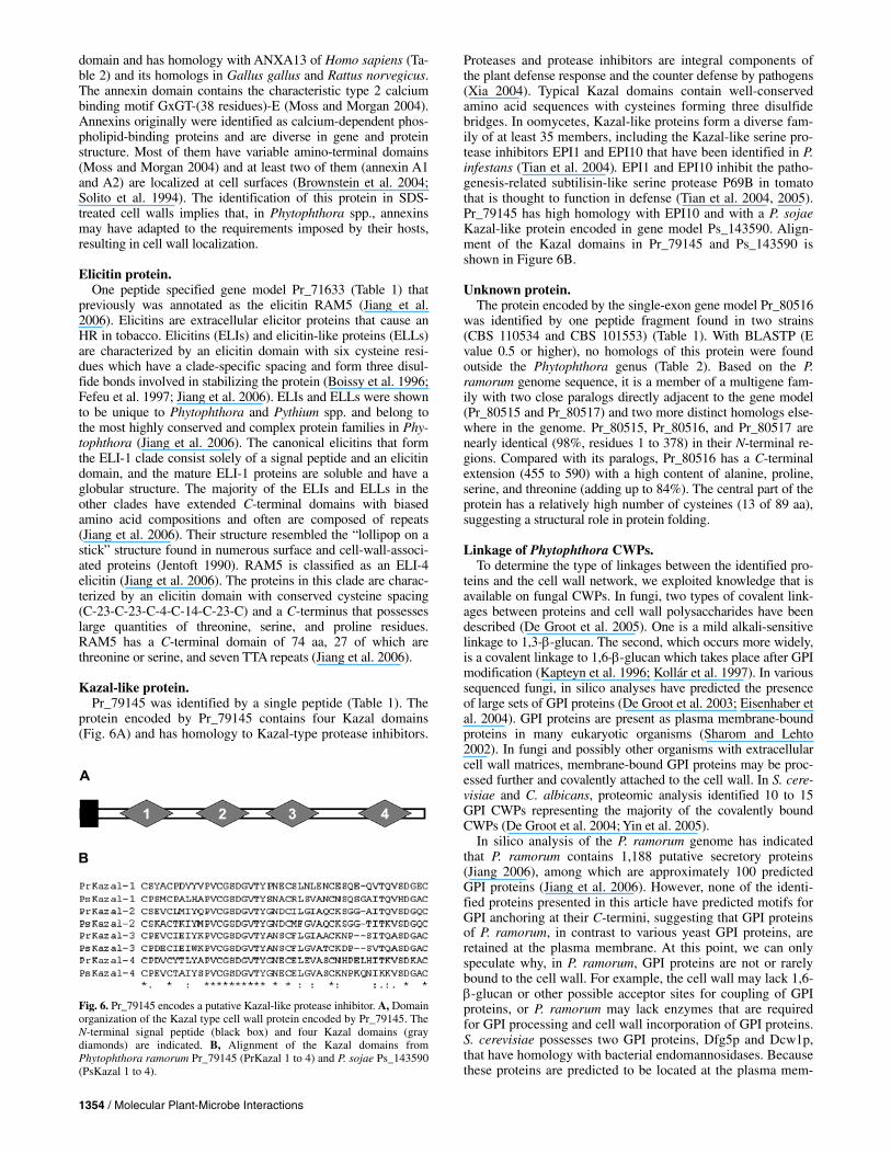
Kazal-like protein. Pr_79145 was identified by a single peptide (Table 1). The
protein encoded by Pr_79145 contains four Kazal domains (Fig. 6A) and has homology to Kazal-type protease inhibitors.
Proteases and protease inhibitors are integral components of the plant defense response and the counter defense by pathogens (Xia 2004). Typical Kazal domains contain well-conserved amino acid sequences with cysteines forming three disulfide bridges. In oomycetes, Kazal-like proteins form a diverse fam-ily of at least 35 members, including the Kazal-like serine pro-tease inhibitors EPI1 and EPI10 that have been identified in P. infestans (Tian et al. 2004). EPI1 and EPI10 inhibit the patho-genesis-related subtilisin-like serine protease P69B in tomato that is thought to function in defense (Tian et al. 2004, 2005). Pr_79145 has high homology with EPI10 and with a P. sojae Kazal-like protein encoded in gene model Ps_143590. Align-ment of the Kazal domains in Pr_79145 and Ps_143590 is shown in Figure 6B.
Unknown protein. The protein encoded by the single-exon gene model Pr_80516
was identified by one peptide fragment found in two strains (CBS 110534 and CBS 101553) (Table 1). With BLASTP (E value 0.5 or higher), no homologs of this protein were found outside the Phytophthora genus (Table 2). Based on the P. ramorum genome sequence, it is a member of a multigene fam-ily with two close paralogs directly adjacent to the gene model (Pr_80515 and Pr_80517) and two more distinct homologs else-where in the genome. Pr_80515, Pr_80516, and Pr_80517 are nearly identical (98%, residues 1 to 378) in their N-terminal re-gions. Compared with its paralogs, Pr_80516 has a C-terminal extension (455 to 590) with a high content of alanine, proline, serine, and threonine (adding up to 84%). The central part of the protein has a relatively high number of cysteines (13 of 89 aa), suggesting a structural role in protein folding.
Linkage of Phytophthora CWPs. To determine the type of linkages between the identified pro-
teins and the cell wall network, we exploited knowledge that is available on fungal CWPs. In fungi, two types of covalent link-ages between proteins and cell wall polysaccharides have been described (De Groot et al. 2005). One is a mild alkali-sensitive linkage to 1,3-β-glucan. The second, which occurs more widely, is a covalent linkage to 1,6-β-glucan which takes place after GPI modification (Kapteyn et al. 1996; Kollár et al. 1997). In various sequenced fungi, in silico analyses have predicted the presence of large sets of GPI proteins (De Groot et al. 2003; Eisenhaber et al. 2004). GPI proteins are present as plasma membrane-bound proteins in many eukaryotic organisms (Sharom and Lehto 2002). In fungi and possibly other organisms with extracellular cell wall matrices, membrane-bound GPI proteins may be proc-essed further and covalently attached to the cell wall. In S. cere-visiae and C. albicans, proteomic analysis identified 10 to 15 GPI CWPs representing the majority of the covalently bound CWPs (De Groot et al. 2004; Yin et al. 2005).
In silico analysis of the P. ramorum genome has indicated that P. ramorum contains 1,188 putative secretory proteins (Jiang 2006), among which are approximately 100 predicted GPI proteins (Jiang et al. 2006). However, none of the identi-fied proteins presented in this article have predicted motifs for GPI anchoring at their C-termini, suggesting that GPI proteins of P. ramorum, in contrast to various yeast GPI proteins, are retained at the plasma membrane. At this point, we can only speculate why, in P. ramorum, GPI proteins are not or rarely bound to the cell wall. For example, the cell wall may lack 1,6-β-glucan or other possible acceptor sites for coupling of GPI proteins, or P. ramorum may lack enzymes that are required for GPI processing and cell wall incorporation of GPI proteins. S. cerevisiae possesses two GPI proteins, Dfg5p and Dcw1p, that have homology with bacterial endomannosidases. Because these proteins are predicted to be located at the plasma mem-
Fig. 6. Pr_79145 encodes a putative Kazal-like protease inhibitor. A, Domainorganization of the Kazal type cell wall protein encoded by Pr_79145. The N-terminal signal peptide (black box) and four Kazal domains (graydiamonds) are indicated. B, Alignment of the Kazal domains fromPhytophthora ramorum Pr_79145 (PrKazal 1 to 4) and P. sojae Ps_143590 (PsKazal 1 to 4).

Vol. 19, No. 12, 2006 / 1355
brane, they might be involved in cleavage of GPI anchors from GPI proteins that are destined to be incorporated into the cell wall. This is supported by the observation that baker’s yeast cells partially deficient in Dfg5p and Dcw1p secrete increased amounts of Cwp1p, a normally abundant cell-wall-bound GPI protein that concentrates in the birth scar, into the medium (Kitagaki et al. 2002; Smits et al. 2006). P. ramorum has no obvious orthologs of Dfg5p and Dcw1p, indicating that this or-ganism may lack enzymes essential for the incorporation of GPI proteins in cell walls.
In this study, release of CWPs from the cell wall matrix of strain Pr04 was investigated by incubating walls with Quan-tazyme, a protease-free 1,3-β-glucanase, or with mild alkali (30 mM NaOH), followed by MS/MS. In both cases, no re-lease of CWPs was observed. The observation that these proto-cols for extracting fungal CWPs were ineffective for Phy-tophthora mycelium suggests that Phytophthora spp. contain a mechanism for retention of CWPs that is different from yeasts. Because 1,3-β-glucan is the major cell wall component in vari-ous Phytophthora spp. (Bartnicki-Garcia and Wang 1983), the inability of Quantazyme to extract detectable amounts of CWPs may suggest that proteins themselves cross-link into a water-insoluble SDS-resistant network.
Concluding remarks. In recent years, considerable progress has been made in the
understanding of the biology, pathology, and genetics of oo-mycetes in general and Phytophthora spp. in particular. This report describes the first cell wall proteomics approach based on the genome sequence of a Phytophthora sp. The procedure described here exemplifies the impact of novel, genome-based approaches toward control of Phytophthora infections. Analysis of CWPs is fast, easily applicable, and amenable to high-throughput procedures. Furthermore, it not only indicates whether genes are transcribed but also whether the transcripts are translated and the proteins incorporated into the cell wall. Although we cannot exclude the possibility that some CWPs may remain unidentified, the method is sensitive and repro-ducible, as shown by the identification of similar sets of proteins in different P. ramorum strains. The absence of GPI CWPs was illustrated by the inability to release CWPs with Quantazyme, and was confirmed by the absence of GPI anchor-addition se-quences in any of the identified CWPs.
This study is the first inventory of Phytophthora CWPs. In-vestigation of wall extracts of tissues grown under different conditions and from different developmental stages will result in the identification of additional, differentially regulated CWPs. Most of the identified CWPs could be categorized into functional groups with predicted biochemical or biological functions; however, these remain to be experimentally validated. Until then, classifications are based on sharing conserved do-mains with homologous proteins that are suggestive of their function. Interestingly, examination of expression of the P. sojae orthologs, based on the presence of ESTs, revealed that 14 of the 17 genes are expressed in infected plant tissue, suggesting a role in pathogenicity (data not shown). With the rapidly in-creasing amount of genome and proteome information at hand, the challenge will be to validate these functions and to eluci-date their biological role.
MATERIALS AND METHODS
Strains and media. The isolates used in this study are strain CBS 101553, isolated
from Rhododendron catawbiense in Germany; strain CBS 110536 (Pr04), isolated from Quercus kelloggii in Marin County, CA, U.S.A. (Ivors et al. 2004); strain CBS 110534
(Pr01), isolated from Quercus agrifolia in California, U.S.A. (Kroon et al. 2004), and PD 20023604, isolated from Viburnum bodnantense cv. Dawn in The Netherlands (Werres and Kaminski 2005). Isolates were obtained from the Centraalbu-reau voor Schimmelcultures (CBS; Fungal Biodiversity Centre, Utrecht, The Netherlands) and from the Plantenziektekundige Dienst (PD; Wageningen, The Netherlands). Isolates were grown in the dark on liquid Plich medium for 12 days at 18ºC. Plich medium was prepared as described previously (Van der Lee et al. 1997). The Plich medium was inoculated with myce-lium on rye sucrose agar plugs (Caten and Jinks 1968) from actively growing colony margins of cultures. Mycelium was har-vested and dried by pressing between filter paper.
Cell wall isolation. Mycelia were ground in a Braun Microdismembrator U (B.
Braun Biotech International GmbH, Melsungen, Germany), re-suspended, and washed in 10 mM KPO4 buffer, pH 7.5, in the presence of a complete protease inhibitor cocktail (1873580, Roche, Branchburg, NJ, U.S.A.; 1 tablet in 50 ml plus 30 μg of pepstatin). Ground cell material was resuspended in 10 mM Tris-HCl, pH 7.5 (plus protease inhibitors), and further disinte-grated with 0.25- to 0.50-mm (diameter) glass beads (Emergo BV, Landsmeer, The Netherlands) using a Bio-Savant Fast Prep 120 machine (Qbiogene, Carlsbad, CA, U.S.A.). Nonco-valently linked proteins and intracellular contaminants were removed by washing the isolated cell walls extensively with 1 M NaCl followed by extraction (twice) in 50 mM Tris-HCl, pH 7.8, 2% SDS, 100 mM Na-EDTA, and 40 mM β-mercap-toethanol for 5 min at 100ºC. The SDS-treated cell walls were washed three times with water and lyophilized.
Cell wall composition analysis. For determination of cell wall carbohydrates, the polysaccha-
rides in cell wall preparations were hydrolyzed using sulfuric acid (Dallies et al. 1998). Briefly, 4 mg of lyophilized cell walls were incubated with 100 μl of 72% (vol/vol) H2SO4 for 3 h at room temperature. Samples were then diluted with 575 μl of distilled H2O to obtain a 2 M H2SO4 solution and incubated for 4 h at 100ºC. Samples were analyzed by HPLC (LKB) on a REZEX organic acid analysis column (Phenomenex, Torrance, CA, U.S.A.) at 40ºC with 7.2 mM H2SO4 as eluent, using an RI 1530 refractive index detector (Jasco, Great Dunmow, U.K). The chromatograms were analyzed using AZUR chromatography software and compared with chromatograms of known amounts of mannose, glucose, galactose, and glucosamine. Protein and chitin contents in the cell wall were determined following the protocols described by Kapteyn and associates (2001).
Cell wall protein extraction. Two extraction methods that are known to solubilize cova-
lently bound CWPs in yeasts (De Groot et al. 2004; Kapteyn et al. 1999) were applied to P. ramorum. For enzymatic release of CWPs, SDS-treated cell walls were incubated overnight with Quantazyme (30 units/mg) (Qbiogene, Morgan Irvine, CA, U.S.A.), a recombinant endo-1,3-β-glucanase, followed by di-alysis against H2O and lyophilization of the solubilized mate-rial. For release of proteins that may be bound to the cell wall through a linkage that is sensitive to mild alkali, cell walls were incubated with 30 mM NaOH at 4ºC for 17 h (Mrsa et al. 1997). The reaction was stopped by adding neutralizing amounts of acetic acid, followed by dialysis and lyophilization of released protein.
Sample preparation for MS analysis. Cell walls and unfractionated protein extracts were reduced
with dithiothreitol and S-alkylated with iodoacetamide as de-
1356 / Molecular Plant-Microbe Interactions
scribed previously (Shevchenko et al. 1996; Yin et al. 2005). Samples were digested with sequencing grade trypsin (Roche Molecular Biochemicals) overnight at 37ºC with an estimated CWP/trypsin ratio of 50:1. Extracted peptides were desalted and concentrated on a μC18 ZipTip column (Millipore, Bedford, MA, U.S.A.).
MS analysis. Solubilized peptides were analyzed by nanoscale HPLC elec-
trospray ionization quadrupole time-of-flight LC/MS/MS using an Ultimate nano-LC system (Dionex, Sunnyvale, CA, U.S.A.) and a MicroMass Q-TOF mass spectrometer (Waters, Milford, MA, U.S.A.). Proteolytic digests derived from 40 μg of lyophi-lized cell walls were injected with a micro-autosampler (FA-MOS) on a precolumn (300-μm inner diameter by 5 mm). Thereafter, the peptides were loaded and separated on a PepMap column (75-μm inner diameter by 150 mm; Dionex). A linear gradient of 5 to 95% B, where mobile phase A was water + 0.1% formic acid and mobile phase B was acetonitrile + 0.1% formic acid, over 30 min was employed for separation at a flow rate of 300 nl/min. Online coupling to the Q-TOF was present using a coated fused-silica PicoTip (FS360-20-10CE; New Objective, Woburn, MA, U.S.A.). Masslynx software automatically selected peptides from the survey spectrum for collision-induced fragmentation. Fragmentation was achieved with argon as colli-sion gas at a pressure of 4 × 10⁻⁵ bar measured on the quadru-pole pressure gauge. MS/MS spectra were analyzed with Mass-lynx Pepseq software to generate peak lists. These data were used to search the P. ramorum proteome database with the Mas-cot search engine (version 2.0; Matrix Science website). The P. ramorum proteome is available at the website of the DOE Joint Genome Institute (JGI). To identify N-terminal peptides, signal peptidase cleavage sites were predicted using SignalP 3.0 server (Bendtsen et al. 2004) and sequences of matured proteins were added to the proteome file. The Mascot searching parameters consisted of allowing up to one missed cleavage, fixed carbami-domethyl modification, a peptide tolerance of 2.0 Da, and an MS/MS tolerance of 0.8 Da. Probability-based Mascot scores were used to evaluate protein identifications. Only matches with P < 0.05 for random occurrence were considered significant.
Database analysis. Genes encoding the identified proteins were analyzed with
the publicly accessible databases at the JGI of the United States Department of Energy or the Virginia Bioinformatics In-stitute microbial database (Tripathy et al. 2006). The databases contain a functional assignment based on automatic analysis. For additional functional assignment and homology studies, searches were initiated in publicly available datasets. Conserved domains were retrieved directly from the JGI database and also based on analyses using multiple databases including SMART, NCBI, and Interproscan. Signal peptides were detected via SignalP using default parameters (Bendtsen et al. 2004). Transmembrane regions identified by SMART or Interproscan were verified by using the PredCon II program (Arai et al. 2004). For GPI lipid anchor prediction, big-PI plant predictor (Eisenhaber et al. 2003) was used. Repeats in proteins were in-vestigated with rapid automatic detection and alignment of repeats, available at the European Bioinformatics Institute. Alignments were performed using the CLUSTAL program (version 1.83). Glycoside hydrolases are classified according to the carbohydrate-active enzymes database.
ACKNOWLEDGMENTS
The authors gratefully acknowledge the Department of Energy JGI for making the P. sojae and P. ramorum genome sequences publicly available.
This work was supported by grants to F. Govers by the Dutch Ministry of Agriculture, Nature and Food Quality (LNV427) and the Technology Foundation STW (WPB.5498) and to F. M. Klis from the European Union (FUNGWALL). The Q-TOF mass spectrometer has been funded largely by the Council for Medical Sciences of the Netherlands Organization for Scientific Research (NWO-ZonMW). The nano HPLC was funded by a grant from the Council for Chemical Sciences of The Netherlands Organi-zation for Scientific Research (NWO-CW). We thank M. Bekker and H. Dekker (UvA-SILS) for assistance with HPLC and mass spectrometric analysis, respectively.
LITERATURE CITED
Arai, M., Mitsuke, H., Ikeda, M., Xia, J. X., Kikuchi, T., Satake, M., and Shimizu, T. 2004. ConPred II: A consensus prediction method for ob-taining transmembrane topology models with high reliability. Nucleic Acids Res. 32:W390-393.
Barre, A., Peumans, W. J., Menu-Bouaouiche, L., Van Damme, E. J., May, G. D., Herrera, A. F., Van Leuven, F., and Rouge, P. 2000. Purification and structural analysis of an abundant thaumatin-like protein from ripe banana fruit. Planta 211:791-799.
Bartnicki-Garcia, S., and Wang, M. C. 1983. Biochemical aspects of morphogenesis in Phytophthora. Pages 121-138 in: Phytophthora. D. C. Erwin, S. Bartinicki-Garcia, and P. H. Tsao, eds. American Phytopa-thological Society Press, St. Paul, MN, U.S.A.
Bates, S., Hughes, H. B., Munro, C. A., Thomas, W. P., MacCallum, D. M., Bertram, G., Atrih, A., Ferguson, M. A., Brown, A. J., Odds, F. C., and Gow, N. A. 2006. Outer chain N-glycans are required for cell wall integrity and virulence of Candida albicans. J. Biol. Chem. 281:90-98.
Bendtsen, J. D., Nielsen, H., von Heijne, G., and Brunak, S. 2004. Im-proved prediction of signal peptides: SignalP 3.0. J. Mol. Biol. 340:783-795.
Birch, P. R., Rehmany, A. P., Pritchard, L., Kamoun, S., and Beynon, J. L. 2006. Trafficking arms: Oomycete effectors enter host plant cells. Trends Microbiol. 14:8-11.
Boissy, G., de La Fortelle, E., Kahn, R., Huet, J. C., Bricogne, G., Pernollet, J. C., and Brunie, S. 1996. Crystal structure of a fungal elicitor secreted by Phytophthora cryptogea, a member of a novel class of plant necrotic proteins. Structure 4:1429-1439.
Brownstein, C., Deora, A. B., Jacovina, A. T., Weintraub, R., Gertler, M., Khan, K. M., Falcone, D. J., and Hajjar, K. A. 2004. Annexin II medi-ates plasminogen-dependent matrix invasion by human monocytes: En-hanced expression by macrophages. Blood 103:317-324.
Brunner, F., Rosahl, S., Lee, J., Rudd, J. J., Geiler, C., Kauppinen, S., Rasmussen, G., Scheel, D., and Nürnberger, T. 2002. Pep-13, a plant defense-inducing pathogen-associated pattern from Phytophthora trans-glutaminases. EMBO (Eur. Mol. Biol. Organ.) J. 21:6681-6688.
Cassab, G. I. 1998. Plant cell wall proteins. Annu. Rev. Plant Physiol. Plant Mol. Biol. 49:281-309.
Caten, C. E., and Jinks, J. L. 1968. Spontaneous variability of single iso-lates of Phytophthora infestans. I. Cultural variation. Can. J. Bot. 46:329-348.
Cote, F., Ham, K. S., Hahn, M. G., and Bergmann, C. W. 1998. Oligosac-charide elicitors in host-pathogen interactions. Generation, perception, and signal transduction. Subcell. Biochem. 29:385-432.
Dallies, N., Francois, J., and Paquet, V. 1998. A new method for quantita-tive determination of polysaccharides in the yeast cell wall. Application to the cell wall defective mutants of Saccharomyces cerevisiae. Yeast 14:1297-1306.
Dean, R. A., Talbot, N. J., Ebbole, D. J., Farman, M. L., Mitchell, T. K., Orbach, M. J., Thon, M., Kulkarni, R., Xu, J. R., Pan, H., Read, N. D., Lee, Y. H., Carbone, I., Brown, D., Oh, Y. Y., Donofrio, N., Jeong, J. S., Soanes, D. M., Djonovic, S., Kolomiets, E., Rehmeyer, C., Li, W., Harding, M., Kim, S., Lebrun, M. H., Bohnert, H., Coughlan, S., Butler, J., Calvo, S., Ma, L. J., Nicol, R., Purcell, S., Nusbaum, C., Galagan, J. E., and Birren, B. W. 2005. The genome sequence of the rice blast fun-gus Magnaporthe grisea. Nature 434:980-986.
De Groot, P. W. J., De Boer, A. D., Cunningham, J., Dekker, H. L., de Jong, L., Hellingwerf, K. J., de Koster, C., and Klis, F. M. 2004. Proteo-mic analysis of Candida albicans cell walls reveals covalently bound carbohydrate-active enzymes and adhesins. Eukaryot. Cell 3:955-965.
De Groot, P. W. J., Hellingwerf, K. J., and Klis, F. M. 2003. Genome-wide identification of fungal GPI proteins. Yeast 20:781-796.
De Groot, P. W. J., Ram, A. F., and Klis, F. M. 2005. Features and func-tions of covalently linked proteins in fungal cell walls. Fungal Genet. Biol. 42:657-675.
Ecker, M., Deutzmann, R., Lehle, L., Mrsa, V., and Tanner, W. 2006. Pir proteins of Saccharomyces cerevisiae are attached to β-1,3-glucan by a new protein-carbohydrate linkage. J. Biol. Chem. 281:11523-11529.
Vol. 19, No. 12, 2006 / 1357
Eisenhaber, B., Schneider, G., Wildpaner, M., and Eisenhaber, F. 2004. A sensitive predictor for potential GPI lipid modification sites in fungal protein sequences and its application to genome-wide studies for Asper-gillus nidulans, Candida albicans, Neurospora crassa, Saccharomyces cerevisiae and Schizosaccharomyces pombe. J. Mol. Biol. 337:243-253.
Fabritius, A. L., Cvitanich, C., and Judelson, H. S. 2002. Stage-specific gene expression during sexual development in Phytophthora infestans. Mol. Microbiol. 45:1057-1066.
Fabritius, A. L., and Judelson, H. S. 2003. A mating-induced protein of Phytophthora infestans is a member of a family of elicitors with diver-gent structures and stage-specific patterns of expression. Mol. Plant-Microbe Interact. 16:926-935.
Fefeu, S., Bouaziz, S., Huet, J. C., Pernollet, J. C., and Guittet, E. 1997. Three-dimensional solution structure of beta cryptogein, a beta elicitin secreted by a phytopathogenic fungus Phytophthora cryptogea. Protein Sci. 6:2279-2284.
Gipson, I. K. 2004. Distribution of mucins at the ocular surface. Exp. Eye Res. 78:379-388.
Gornhardt, B., Rouhara, I., and Schmelzer, E. 2000. Cyst germination pro-teins of the potato pathogen Phytophthora infestans share homology with human mucins. Mol. Plant-Microbe Interact. 13:32-42.
Grenier, J., Potvin, C., Trudel, J., and Asselin, A. 1999. Some thaumatin-like proteins hydrolyse polymeric beta-1,3-glucans. Plant J. 19:473-480.
Hahn, M. G. 1996. Microbial elicitors and their receptors in plants. Annu. Rev. Phytopathol. 34:387-412.
Henrissat, B. 1991. A classification of glycosyl hydrolases based on amino acid sequence similarities. Biochem. J. 280:309-316.
Henrissat, B., and Bairoch, A. 1993. New families in the classification of glycosyl hydrolases based on amino acid sequence similarities. Bio-chem. J. 293 781-788.
Henrissat, B., and Bairoch, A. 1996. Updating the sequence-based classifi-cation of glycosyl hydrolases. Biochem. J. 316:695-696.
Hicks, S. J., Theodoropoulos, G., Carrington, S. D., and Corfield, A. P. 2000. The role of mucins in host-parasite interactions. Part I-protozoan parasites. Parasitol. Today 16:476-481.
Hobson, R. P., Munro, C. A., Bates, S., MacCallum, D. M., Cutler, J. E., Heinsbroek, S. E., Brown, G. D., Odds, F. C., and Gow, N. A. 2004. Loss of cell wall mannosylphosphate in Candida albicans does not in-fluence macrophage recognition. J. Biol. Chem. 279:39628-39635.
Howard, R. J., Ferrari, M. A., Roach, D. H., and Money, N. P. 1991. Pene-tration of hard substrates by a fungus employing enormous turgor pres-sures. Proc. Natl. Acad. Sci. U.S.A. 88:11281-11284.
Ivors, K. L., Hayden, K. J., Bonants, P. J., Rizzo, D. M., and Garbelotto, M. 2004. AFLP and phylogenetic analyses of North American and European populations of Phytophthora ramorum. Mycol. Res. 108:378-392.
Jentoft, N. 1990. Why are proteins O-glycosylated? Trends Biochem. Sci. 15:291-294.
Jiang, R. H. Y. 2006. Footprints of evolution: The dynamics of effector genes in the Phytophthora genome. Ph.D. thesis, Wageningen Univer-sity, The Netherlands.
Jiang, R. H. Y., Tyler, B. M., Whisson, S. C., Hardham, A. R., and Govers, F. 2006. Ancient origin of elicitin gene clusters in Phytophthora ge-nomes. Mol. Biol. Evol. 23:338-351.
Kapteyn, J. C., Montijn, R. C., Vink, E., de la Cruz, J., Llobell, A., Douwes, J. E., Shimoi, H., Lipke, P. N., and Klis, F. M. 1996. Retention of Saccharomyces cerevisiae cell wall proteins through a phosphodiester-linked beta-1,3-/beta-1,6-glucan heteropolymer. Glycobiology 6:337-345.
Kapteyn, J. C., ter Riet, B., Vink, E., Blad, S., De Nobel, H., Van Den Ende, H., and Klis, F. M. 2001. Low external pH induces HOG1-de-pendent changes in the organization of the Saccharomyces cerevisiae cell wall. Mol. Microbiol. 39:469-479.
Kapteyn, J. C., Van Egmond, P., Sievi, E., Van Den Ende, H., Makarow, M., and Klis, F. M. 1999. The contribution of the O-glycosylated pro-tein Pir2p/Hsp150 to the construction of the yeast cell wall in wild-type cells and beta 1,6-glucan-deficient mutants. Mol. Microbiol. 31:1835-1844.
Kitagaki, H., Wu, H., Shimoi, H., and Ito, K. 2002. Two homologous genes, DCW1 (YKL046c) and DFG5, are essential for cell growth and encode glycosylphosphatidylinositol (GPI)-anchored membrane pro-teins required for cell wall biogenesis in Saccharomyces cerevisiae. Mol. Microbiol. 46:1011-1022.
Klebl, F., and Tanner, W. 1989. Molecular cloning of a cell wall exo-beta-1,3-glucanase from Saccharomyces cerevisiae. J. Bacteriol. 171:6259-6264.
Klis, F. M., Mol, P., Hellingwerf, K. J., and Brul, S. 2002. Dynamics of cell wall structure in Saccharomyces cerevisiae. FEMS (Fed. Eur. Mi-crobiol. Soc.) Microbiol. Rev. 26:239-256.
Kollár, R., Reinhold, B. B., Petráková, E., Yeh, H. J., Ashwell, G., Drgonova, J., Kapteyn, J. C., Klis, F. M., and Cabib, E. 1997. Architec-ture of the yeast cell wall. β(1→6)-glucan interconnects mannoprotein, β (1→)3-glucan, and chitin. J. Biol. Chem. 272:17762-17775.
Kroon, L. P. N. M., Verstappen, E. C. P., Cox, L. F. F., Flier, W. G., and Bonants, P. 2004. A rapid diagnostic test to distinguish between Ameri-can and European populations of Phytophthora ramorum. Phytopathol-ogy 94:613-620.
Makarova, K. S., Aravind, L., and Koonin, E. V. 1999. A superfamily of archaeal, bacterial, and eukaryotic proteins homologous to animal transglutaminases. Protein Sci. 8:1714-1719.
McLeod, A., Smart, C. D., and Fry, W. E. 2003. Characterization of 1,3-beta-glucanase and 1,3;1,4-beta-glucanase genes from Phytophthora in-festans. Fungal Genet. Biol. 38:250-263.
Menu-Bouaouiche, L., Vriet, C., Peumans, W. J., Barre, A., Van Damme, E. J., and Rouge, P. 2003. A molecular basis for the endo-beta 1,3-glu-canase activity of the thaumatin-like proteins from edible fruits. Bio-chimie 85:123-131.
Moss, S. E., and Morgan, R. O. 2004. The annexins. Genome Biol. 5:219. Mrsa, V., Seidl, T., Gentzsch, M., and Tanner, W. 1997. Specific labeling
of cell wall proteins by biotinylation. Identification of four covalently linked O-mannosylated proteins of Saccharomyces cerevisiae. Yeast 13:1145-1154.
Munro, C. A., Bates, S., Buurman, E. T., Hughes, H. B., Maccallum, D. M., Bertram, G., Atrih, A., Ferguson, M. A., Bain, J. M., Brand, A., Hamilton, S., Westwater, C., Thomson, L. M., Brown, A. J., Odds, F. C., and Gow, N. A. 2005. Mnt1p and Mnt2p of Candida albicans are partially redundant alpha-1,2-mannosyltransferases that participate in O-linked mannosylation and are required for adhesion and virulence. J. Biol. Chem. 280:1051-1060.
Nürnberger, T., Nennstiel, D., Jabs, T., Sacks, W. R., Hahlbrock, K., and Scheel, D. 1994. High affinity binding of a fungal oligopeptide elicitor to parsley plasma membranes triggers multiple defense responses. Cell 78:449-460.
Parker, J. E., Schulte, W., Hahlbrock, K., and Scheel, D. 1991. An extra-cellular glycoprotein from Phytophthora megasperma f. sp. glycinea elicits phytoalexin synthesis in cultured parsley cells and protoplasts. Mol. Plant-Microbe Interact. 4:19-27.
Prill, S. K., Klinkert, B., Timpel, C., Gale, C. A., Schroppel, K., and Ernst, J. F. 2005. PMT family of Candida albicans: Five protein mannosyl-transferase isoforms affect growth, morphogenesis and antifungal resis-tance. Mol. Microbiol. 55:546-560.
Qutob, D., Kamoun, S., and Gijzen, M. 2002. Expression of a Phy-tophthora sojae necrosis-inducing protein occurs during transition from biotrophy to necrotrophy. Plant J. 32:361-373.
Rizzo, D. M., Garbelotto, M., Davidson, J. M., Slaughter, G. W., and Koike, S. T. 2002. Phytophthora ramorum as the cause of extensive mortality of Quercus spp. and Lithocarpus densiflorus in California. Plant Dis. 86:205-214.
Rose, J. K., Ham, K. S., Darvill, A. G., and Albersheim, P. 2002. Molecu-lar cloning and characterization of glucanase inhibitor proteins: Coevo-lution of a counterdefense mechanism by plant pathogens. Plant Cell 14:1329-1345.
Sacks, W., Nürnberger, T., Hahlbrock, K., and Scheel, D. 1995. Molecular characterization of nucleotide sequences encoding the extracellular gly-coprotein elicitor from Phytophthora megasperma. Mol. Gen. Genet. 246:45-55.
Sakamoto, Y., Watanabe, H., Nagai, M., Nakade, K., Takahashi, M., and Sato, T. 2006. Lentinula edodes tlg1 encodes a thaumatin-like protein that is involved in lentinan degradation and fruiting body senescence. Plant Physiol. 141:793-801.
Sharom, F. J., and Lehto, M. T. 2002. Glycosylphosphatidylinositol-an-chored proteins: Structure, function, and cleavage by phosphatidylinosi-tol-specific phospholipase C. Biochem. Cell. Biol. 80:535-549.
Shevchenko, A., Wilm, M., Vorm, O., and Mann, M. 1996. Mass spectro-metric sequencing of proteins silver-stained polyacrylamide gels. Anal. Chem. 68:850-858.
Showalter, A. M. 1993. Structure and function of plant cell wall proteins. Plant Cell 5:9-23.
Simmons, C. R. 1994. The physiology and molecular biology of plant 1,3-beta-D-glucanases and 1,3;1,4-beta-D-glucanases. Crit. Rev. Plant Sci. 13:325-387.
Smits, G. J., Schenkman, L. R., Brul, S., Pringle, J. R., and Klis, F. M. 2006. Role of cell cycle-regulated expression in the localized incorpora-tion of cell wall proteins in yeast. Mol. Biol. Cell. 17:3267-3280.
Solito, E., Nuti, S., and Parente, L. 1994. Dexamethasone-induced translo-cation of lipocortin (annexin) 1 to the cell membrane of U-937 cells. Br. J. Pharmacol. 112:347-348.
Sundstrom, P. 2002. Adhesion in Candida spp. Cell Microbiol. 4:461-469. Theodoropoulos, G., Hicks, S. J., Corfield, A. P., Miller, B. G., and
1358 / Molecular Plant-Microbe Interactions
Carrington, S. D. 2001. The role of mucins in host-parasite interactions: Part II—helminth parasites. Trends Parasitol. 17:130-135.
Tian, M., Benedetti, B., and Kamoun, S. 2005. A second Kaza1-like prote-ase inhibitor from Phytophthora infestans inhibits and interacts with the apoplastic pathogenesis-related protease P69B of tomato. Plant Physiol. 138:1785-1793.
Tian, M., Huitema, E., Da Cunha, L., Torto-Alalibo, T., and Kamoun, S. 2004. A Kazal-like extracellular serine protease inhibitor from Phytoph-thora infestans targets the tomato pathogenesis-related protease P69B. J. Biol. Chem. 279:26370-26377.
Torto-Alalibo, T., Tian, M., Gajendran, K., Waugh, M. E., van West, P., and Kamoun, S. 2005. Expressed sequence tags from the oomycete fish pathogen Saprolegnia parasitica reveal putative virulence factors. BMC Microbiol. 5:46.
Tripathy, S., Pandey, V. N., Fang, B., Salas, F., and Tyler, B. M. 2006. VMD: A community annotation database for oomycetes and microbial genomes. Nucleic Acids Res. 34:D379-D381.
Tyler, B. M., Tripathy, S., Zhang, X., Dehal, P., Jiang, R. H. Y., Aerts, A., Arredondo, F., Baxter, L., Bensasson, D., Beynon, J. L., Damasceno, C. M. B., Dickerman, A., Dorrance, A. E., Dou, D., Dubchak, I., Garbelotto, M., Gijzen, M., Gordon, S., Govers, F., Grunwald, N. J., Huang, W., Ivors, K., Jones, R. W., Kamoun, S., Krampis, K., Lamour, K., Lee, M.-K., McDonald, W. H., Medina, M., Meijer, H. J. G., Nordberg, E., Maclean, D. J., Ospina-Giraldo, M. D., Morris, P. F., Phuntumart, V., Putnam, N., Rash, S., Rose, J. K. C., Sakihama, Y., Salamov, A., Savidor, A., Scheuring, C., Smith, B., Sobral, B. W. S., Terry, A., Torto-Alalibo, T., Win, J., Xu, Z., Zhang, H., Grigoriev, I., Rokhsar, D., and Boore, J. 2006. Phytophthora genome sequences uncover evolutionary origins and mechanisms of pathogenesis. Science. 313:1261-1266.
Van der Lee, T., De Witte, I., Drenth, A., Alfonso, C., and Govers, F. 1997. AFLP linkage map of the oomycete Phytophthora infestans. Fungal Genet. Biol. 21:278-291.
Werres, S., and Kaminski, K. 2005. Characterisation of European and North American Phytophthora ramorum isolates due to their morphol-
ogy and mating behaviour in vitro with heterothallic Phytophthora spe-cies. Mycol. Res. 109:860-871.
Werres, S., Marwitz, R., Man in ‘t Veld, W. A., de Cock, A. W. A. M., Bonants, P. J. M., De Weerdt, M., Themann, K., Ilievea, E., and Baayen, R. P. 2001. Phytophthora ramorum sp. nov., a new pathogen on Rhodo-dendron and Viburnum. Mycol. Res. 105:1155-1165.
Westerink, N., Joosten, M. H. A. J., and de Wit, P. J. G. M. 2004. Fungal (A)virulence factors at the crossroads of disease susceptibility and re-sistance. Pages 93-127 in: Fungal Disease Resistance in Plants. Bio-chemistry, Molecular Biology and Genetic Engineering. Z. Punja, ed. Haworth Press, Binghamton, NY, U.S.A.
Xia, Y. 2004. Proteases in pathogenesis and plant defence. Cell Microbiol. 6:905-913.
Yin, Q. Y., de Groot, P. W. J., Dekker, H. L., de Jong, L., Klis, F. M., and de Koster, C. G. 2005. Comprehensive proteomic analysis of Saccharo-myces cerevisiae cell walls: Identification of proteins covalently attached via glycosylphosphatidylinositol remnants or mild alkali-sensitive link-ages. J. Biol. Chem. 280:20894-20901.
AUTHOR-RECOMMENDED INTERNET RESOURCES
Carbohydrate-active enzymes (CAZY) database: afmb.cnrs-mrs.fr/CAZY DOE Joint Genome Institute (JGI) P. ramorum genome database:
genome.jgi-psf.org/ramorum1/ramorum1.home.html DOE JGI P. sojae genome database:
genome.jgi-psf.org/sojae1/sojae1.home.html DOE JGI T. pseudonana genome database:
genome.jgi-psf.org/thaps1/thaps1.home.html European Bioinformatics Institute (EBI): www.ebi.ac.uk/Radar/ Matrix Science Mascot search website: www.matrixscience.com United States Department of Energy JGI: www.jgi.doe.gov The Virginia Bioinformatics Institute (VBI) microbial database:
phytophthora.vbi.vt.edu
Related Documents