ORIGINAL ARTICLE Reproductive genetics Identification of a duplication within the GDF9 gene and novel candidate genes for primary ovarian insufficiency (POI) by a customized high-resolution array comparative genomic hybridization platform A. Norling 1,2,3, * , A.L. Hirschberg 2 , K.A. Rodriguez-Wallberg 4 , E. Iwarsson 1,3 , A. Wedell 3,5,6 , and M. Barbaro 1,3,6 1 Department of Molecular Medicine and Surgery, Karolinska Institutet, Karolinska University Hospital, Stockholm 171 76, Sweden 2 Department of Women’s and Children’s Health, Karolinska Institutet, Karolinska University Hospital, Stockholm 171 76, Sweden 3 Centre for Molecular Medicine, Karolinska Institutet, Stockholm, Sweden 4 Department of Clinical Science, Intervention and Technology, Section for Obstetrics and Gynaecology and Fertility Unit, Karolinska University Hospital, Stockholm, Sweden 5 Department of Molecular Medicine and Surgery, Science for Life Laboratory, Karolinska Institutet, Stockholm, Sweden 6 Centre for Inherited Metabolic Diseases (CMMS), Karolinska University Hospital, Stockholm 171 76, Sweden *Correspondence address. Tel: +46-8-517-714-50; Fax: +46-8-517-736-20; E-mail: [email protected] Submitted on December 11, 2013; resubmitted on May 18, 2014; accepted on May 21, 2014 study question: Can high-resolution array comparative genomic hybridization (CGH) analysis of DNA samples from women with primary ovarian insufficiency (POI) improve the diagnosis of the condition and identify novel candidate genes for POI? summary answer: A mutation affecting the regulatory region of growth differentiation factor 9 (GDF9) was identified for the first time together with several novel candidate genes for POI. what is known already: Most patients with POI do not receive a molecular diagnosis despite a significant genetic component in the pathogenesis. study design, size, duration: We performed a case– control study. Twenty-six patients were analyzed by array CGH for identi- fication of copy number variants. Novel changes were investigated in 95 controls and in a separate population of 28 additional patients with POI. The experimental procedures were performed during a 1-year period. participants/materials, setting, methods: DNA samples from 26 patients with POI were analyzed by a customized 1M array-CGH platform with whole genome coverage and probe enrichment targeting 78 genes in sex development. By PCR amplification and sequencing, the breakpoint of an identified partial GDF9 gene duplication was characterized. A multiplex ligation-dependent probe amplification (MLPA) probe set for specific identification of deletions/duplications affecting GDF9 was developed. An MLPA probe set for the identification of additional cases or controls carrying novel candidate regions identified by array-CGH was developed. Sequencing of three candidate genes was performed. main results and the role of chance: Eleven unique copy number changes were identified in a total of 11 patients, including a tandem duplication of 475 bp, containing part of the GDF9 gene promoter region. The duplicated region contains three NOBOX-binding elements and an E-box, important for GDF9 gene regulation. This aberration is likely causative of POI. Fifty-four patients were investigated for copy number changes within GDF9, but no additional cases were found. Ten aberrations constituting novel candidate regions were detected, including a second DNAH6 deletion in a patient with POI. Other identified candidate genes were TSPYL6, SMARCC1, CSPG5 and ZFR2. & The Author 2014. Published by Oxford University Press on behalf of the European Society of Human Reproduction and Embryology. This is an Open Access article distributed under the terms of the Creative Commons Attribution Non-Commercial License (http://creativecommons.org/licenses/by-nc/3.0/), which permits non-commercial re-use, distribution, and reproduction in any medium, provided the original work is properly cited. For commercial re-use, please contact [email protected] Human Reproduction, Vol.29, No.8 pp. 1818 –1827, 2014 Advanced Access publication on June 17, 2014 doi:10.1093/humrep/deu149

Welcome message from author
This document is posted to help you gain knowledge. Please leave a comment to let me know what you think about it! Share it to your friends and learn new things together.
Transcript

ORIGINAL ARTICLE Reproductive genetics
Identification of a duplication within theGDF9 gene and novel candidate genesfor primary ovarian insufficiency (POI)by a customized high-resolution arraycomparative genomic hybridizationplatformA. Norling1,2,3,*, A.L. Hirschberg2, K.A. Rodriguez-Wallberg4,E. Iwarsson1,3, A. Wedell3,5,6, and M. Barbaro1,3,6
1Department of Molecular Medicine and Surgery, Karolinska Institutet, Karolinska University Hospital, Stockholm 171 76, Sweden 2Departmentof Women’s and Children’s Health, Karolinska Institutet, Karolinska University Hospital, Stockholm 171 76, Sweden 3Centre for MolecularMedicine, Karolinska Institutet, Stockholm, Sweden 4Department of Clinical Science, Intervention and Technology, Section for Obstetrics andGynaecology and Fertility Unit, Karolinska University Hospital, Stockholm, Sweden 5Department of Molecular Medicine and Surgery, Science forLife Laboratory, Karolinska Institutet, Stockholm, Sweden 6Centre for Inherited Metabolic Diseases (CMMS), Karolinska University Hospital,Stockholm 171 76, Sweden
*Correspondence address. Tel: +46-8-517-714-50; Fax: +46-8-517-736-20; E-mail: [email protected]
Submitted on December 11, 2013; resubmitted on May 18, 2014; accepted on May 21, 2014
study question: Can high-resolution array comparative genomic hybridization (CGH) analysis of DNA samples from women withprimary ovarian insufficiency (POI) improve the diagnosis of the condition and identify novel candidate genes for POI?
summary answer: A mutation affecting the regulatory region of growth differentiation factor 9 (GDF9) was identified for the first timetogether with several novel candidate genes for POI.
what is known already: Most patients with POI do not receive a molecular diagnosis despite a significant genetic component in thepathogenesis.
study design, size, duration: We performed a case–control study. Twenty-six patients were analyzed by array CGH for identi-fication of copy number variants. Novel changes were investigated in 95 controls and in a separate population of 28 additional patients with POI.The experimental procedures were performed during a 1-year period.
participants/materials, setting, methods: DNA samples from 26 patients with POI were analyzed by a customized 1Marray-CGH platform with whole genome coverage and probe enrichment targeting 78 genes in sex development. By PCR amplification andsequencing, the breakpoint of an identified partial GDF9 gene duplication was characterized. A multiplex ligation-dependent probe amplification(MLPA) probe set for specific identification of deletions/duplications affecting GDF9 was developed. An MLPA probe set for the identification ofadditional cases or controls carrying novel candidate regions identified by array-CGH was developed. Sequencing of three candidate genes wasperformed.
main results and the role of chance: Eleven unique copy number changes were identified in a total of 11 patients, including atandem duplication of 475 bp, containing part of the GDF9 gene promoter region. The duplicated region contains three NOBOX-binding elementsand an E-box, important for GDF9 gene regulation. This aberration is likely causative of POI. Fifty-four patients were investigated for copy numberchanges within GDF9, but no additional cases were found. Ten aberrations constituting novel candidate regions were detected, including a secondDNAH6 deletion in a patient with POI. Other identified candidate genes were TSPYL6, SMARCC1, CSPG5 and ZFR2.
& The Author 2014. Published by Oxford University Press on behalf of the European Society of Human Reproduction and Embryology.This is an Open Access article distributed under the terms of the Creative Commons Attribution Non-Commercial License (http://creativecommons.org/licenses/by-nc/3.0/), which permitsnon-commercial re-use, distribution, and reproduction in any medium, provided the original work is properly cited. For commercial re-use, please contact [email protected]
Human Reproduction, Vol.29, No.8 pp. 1818–1827, 2014
Advanced Access publication on June 17, 2014 doi:10.1093/humrep/deu149

limitations, reasons for caution: This is a descriptive study and no functional experiments were performed.
wider implications of the findings: The study illustrates the importance of analyzing small copy number changes in addition tosequence alterations in the genetic investigation of patients with POI. Also, promoter regions should be included in the investigation.
study funding/competing interest(s): The study was supported by grants from the Swedish Research council (projectno 12198 to A.W. and project no 20324 to A.L.H.), Stockholm County Council (E.I., A.W. and K.R.W.), Foundation FrimurareBarnhuset (A.N., A.W. and M.B.), Karolinska Institutet (A.N., A.L.H., E.I., A.W. and M.B.), Novo Nordic Foundation (A.W.) andSvenska Lakaresallskapet (M.B.). The funding sources had no involvement in the design or analysis of the study. The authors have nocompeting interests to declare.
trial registration number: Not applicable.
Key words: array-CGH / GDF9 / DNAH6 / TSPYL6 / ZFR2
IntroductionA clinical diagnosis of primary ovarian insufficiency (POI) is defined by thepresence of primary or secondary amenorrhea (PA or SA) and hypergo-nadotropic hypogonadism, occurring before the age of 40 (Nelson,2009; De Vos et al., 2010). This condition has previously also beentermed premature ovarian failure in patients with SA and gonadal dysgen-esis in PA.
POI pathogenesis can be divided into follicle dysfunction andfollicle depletion. Follicle depletion can be caused by a small initialgerm cell count or a rapid germ cell loss. External factors, such assurgery, irradiation and infection as well as autoimmune and meta-bolic disorders also lead to POI. Most cases however remainidiopathic, and there is a significant genetic component (Simpson andRajkovic, 1999; Persani et al., 2010). The most common are chromo-somal aberrations including Turner’s syndrome (Baronchelli et al.,2012). Premutation of the FMR1 gene is responsible for 2–5% of spor-adic cases with POI presenting with SA (Wittenberger et al., 2007;De Vos et al., 2010).
Other known genetic causes are mutations in genes for oocyte-secreted factors, GDF9 (growth differentiation factor 9) and BMP15(Dixit et al., 2006; Kovanci et al., 2007), for oocyte nuclear transcriptionfactors NOBOX and FIGLA (Qin et al., 2007; Zhao et al., 2008) and for thehormone receptors FSHR and NR5A1 (Aittomaki et al., 1995; Janse et al.,2012). However, the majority (90%) of patients with POI do not receive amolecular diagnosis (Nelson, 2009) and it is likely that many causativegenes still remain unknown.
By genome-wide detection of small genomic rearrangements, it ispossible to identify novel genes involved in ovarian development,both on the X chromosome and on autosomes. The aim of this studywas to identify novel candidate genes for ovarian development andfunction by investigating submicroscopic genetic imbalances in patientswith POI. A 1 M array-CGH (comparative genomic hybridization)platform with 2.2 kb probe spacing for whole genome coverage andenrichment targeting 78 genes involved in gonadal developmentwas used. At present, all studies reporting copy number variants(CNVs) in patients with POI by array-CGH have used lower resolutionplatforms (Aboura et al., 2009; Ledig et al., 2010; Liao et al., 2011).Studies using whole genome SNP array (McGuire et al., 2011) andtargeted analysis of the X chromosome using array-based techniqueshave also been reported (Dudding et al., 2010; Quilter et al., 2010;Knauff et al., 2011).
Materials and Methods
PatientsFifty-four patients with POI (23 with PA, 31 with SA) referred to the ClinicalGenetic Laboratory of Karolinska University Hospital Stockholm, Sweden,were included in the study. Samples from 26 patients (17 with PA and 9with SA) were available for array-CGH experiments.
The diagnosis of POI was set by PA or SA in a girl with female external geni-talia as well as internal Mullerian structures (uterus) and hypergonadotropichypogonadism (FSH . 30 IU/l), determined twice. In the group with SA theoverall median age at diagnosis was 22 years (12–37) (Table I). The ninepatients with SA included in the array-CGH experiments were diagnosedat a median age of 16 (13–22) years.
One patient with PA has several affected sisters with POI who did not wantto be included in the study. Among patients with SA, four have mothers withPOI, one a sister with POI and one an affected paternal aunt, who is includedin the study.
None of the patientshad undergone previousovarian surgery, chemother-apy or radiotherapy or presented with syndromic forms of POI. Five patientspresented with hypothyroidism with anti-thyroid autoantibodies, after POIdiagnosis in four with SA, and at 11 years of age in one patient with PA.None had autoantibodies against adrenal cortex or 21-hydroxylase protein.
Nine patients with POI underwent laparoscopy with diagnostic ovarian bi-opsies. In four patients with PA, streak gonads with no visible follicles werefound. Three patients with SA also had streak gonads with no visible follicles.One patient with SA at 23 had multiple primordial follicles and one patientwith SA at 15 had a few visible primordial and primary follicles.
........................................................................................
Table I Clinical overview of investigated patients.
Clinical data PA(n 5 23)
SA(n 5 31)
Median age at diagnosis, years (range) 16 (13–18) 22 (12–37)
Median age at menarche, years (range) – 13 (10–15)
Median FSH level at diagnosis, IU/l(range)a
89 (39–150) 80 (34–155)
Swedish Caucasian, n (%) 17 (74) 26 (84)
Known heredity for POI, n (%) 1 (4) 6 (19)
Spontaneous conception beforediagnosis, n (%)
– 11 (35)
PA, primary amenorrhea; SA, secondary amenorrhea. aReference value: ,30 IU/l.
Partial GDF9 duplication in ovarian insufficiency 1819

Ethical approvalAll participants gave informed consent and the regional Ethics Committee atKarolinska Institutet, Sweden, approved the study with registration number2007/263-31/2 and 2011/276-32.
Genetic investigationA 46, XX karyotype in peripheral blood was confirmed in all but in Patient24 in whom a balanced Robertsonian translocation identified (45, XX, der(13; 14)) was not considered causative.
Sex chromosome mosaicism was excluded using fluorescent in situ hybrid-ization and DNA probes from chromosomes X and Y on peripheral bloodsmears, and when available, on touch preparations from gonadal tissue.Genetic investigations included sequencing of the NR5A1, BMP15, FSHR,FIGLA, NOBOX and GDF9 genes. FMR1 mutations were also excluded inpatients with SA.
ControlsDNA samples from 95 healthy women were collected. All women wereabove the age of 40 and had given birth to at least one child. The exclusioncriteria were previous egg donation, in vitro fertilization, infertility treatmentor menopause before the age of 40.
DNA extractionDNA was extracted from peripheral blood lymphocytes. Some sampleswere further purified using the QiAmp DNA minikit (QIAGEN, Sweden)to achieve acceptable quality values for array-CGH analysis.
Array-CGHTwenty-six unrelated patients with POI were analyzed using a customized1 M oligomarker array-CGH platform developed at Oxford Gene Technol-ogy (OGT, UK). In addition to whole genome coverage, the platform isenriched with probes targeting 78 genes implicated in sex development (Sup-plementary data, Table SI). The theoretical average probe resolution is2.2 kb.
Preparation of labeled DNA and subsequent hybridization were per-formed according to the ‘Agilent oligonucleotide array-based CGH forgenomic DNA analysis’ protocol (v6.2) and as previously described(Norling et al., 2013). A commercial DNA sample with pooled humangenomic DNA from 10 female controls (Promega, Sweden) was used as ref-erence DNA.
Data was analyzed using the Cytosure interpret software v3.4.3 (OGT).Circular binary segmentation analysis to detect copy number changes wasperformed using the following parameters: minimum probe count: 5, thresh-old for gains: 0.35, threshold for losses: 0.65, chromosome average method:median segment.
By comparing data with the online database of genomic variation (DGV)version 10 released Nov 2010 (Iafrate et al., 2004), common CNVs foundin healthy control samples were excluded from further investigation. Thisexclusion was done with caution, as phenotypic effects of genetic imbalancesaffecting sexual development can be dependent on chromosomal sex. Thisinformation is not always available for controls included in the DGV. Aberra-tions only partially overlapping with rare reported CNVs were not excluded.Small intronic variations and aberrations not affecting genes were excludedafter verification that they were not located just upstream or downstreama known gene causing disorders of sex development as a positional effectshould then be considered.
Multiplex ligation-dependent probeamplificationMultiplex ligation-dependent probe amplification (MLPA) (Schouten et al.,2002) probes were designed according to the recommendations by Sternet al. (2004) and combined in different probe sets (Supplementary data,Table SII). The reference probes and the pilot probe used have beendescribed earlier (Barbaro et al., 2008).
MLPA reactions were performed according to the EK1 reagent kit(MRC-Holland, the Netherlands) recommendations using 200 ng of DNAand the in-house designed probe set, as previously described (Barbaroet al., 2008). The reference DNA sample used for array-CGH was used asa control in all runs, along with at least another control DNA.
Confirmation of array-CGH findingsCopy number variations left after exclusion criteria were confirmed by MLPAusing two probes peraberration. When available, parental samples wereana-lyzed for control of inheritance. Trace data were exported and analyzed in anExcel 2007 spreadsheet. Each sample’s peak heights were normalized to theaverage peak height of the reference probes and subsequently normalized tothe average peak height of the control samples. The analysis was accepted ifthe reference probe ratios were between 0.8 and 1.2. Threshold values fordeletions and duplications were set at 0.75 and 1.25, respectively.
MLPA for GDF9A specific synthetic probe set for MLPA analysis of the GDF9 gene wasdesigned. Two probes were placed in each exon, two upstream GDF9 andone probe downstream (Fig. 1E). The probe set was validated for consistencyby analysis of 10 healthy controls, with a standard deviation ,0.1 for eachprobe. Trace datawere analyzed using the GeneMarker v1.90 (Soft Genetics,USA) software using internal control probe normalization, quantification bypeak height and ratio threshold values for deletion and duplication were 0.75and 1.3, respectively.
Patient cohort and control screeningA specific synthetic probe set for MLPA analysis of array findings wasdesigned, using one probe per each novel candidate copy number variationidentified by array-CGH experiments. In addition, one probe within thekeratin-associated protein (KRTAP)2-1 and the KRTAP2-2 gene, respectively,were included. The probe set was validated for consistency. Trace data wereanalyzed with GeneMarker v1.90.
Sequencing of TSPYL6, KRTAP2-3 andKRTAP2-4The single exon genes TSPYL6, KRTAP2-3 and KRTAP2-4, including part of theUTRs, were amplified by PCR using DyNAzyme EXT polymerase. Primerswere designed using Primer3 software (Koressaar and Remm, 2007; Unter-gasser et al., 2012) (Supplementary data, Table SIII).
PCR products were cleaned and sequenced using the ABI BigDye Termin-ator v3.1 kit (Applied Biosystems, Sweden). Electropherograms were ana-lyzed against the reference sequences: NM_001003937.2 for TSPYL6,NM_001165252.1 for KRTAP2-3 and NM_033184.3 for KRTAP2-4, usingthe SeqScape v2.5 program (Applied Biosystems).
Characterization of the GDF9 duplicationPrimers for the amplification and sequencing of the duplication junction weredesigned using Primer 3 software (Rozen and Skaletsky, 2000) (Supplemen-tary data, Table SIII) with the hypothesis of a tandem duplication.
1820 Norling et al.
Partial GDF9 duplication in ovarian insufficiency 1821

In silico analysisFor all identified candidate genes the following databases were searched forinformation. Relevant data are presented in the discussion section.
NCBI (http://www.ncbi.nlm.nih.gov/) including PubMed, UCSC (http://genome.ucsc.edu/) (Dreszer et al., 2012), GeneCards (http://www.genecards.org/), The Human Protein Atlas (http://www.proteinatlas.org/)(Uhlen et al., 2010), Gene expression profiles during sex determination byDr Serge Nef (http://nef.unige.ch/microarrays.php) (Nef et al., 2005), DE-CIPHER (Database of Chromosomal Imbalance and Phenotype in HumansUsing Ensembl Resources) (http://decipher.sanger.ac.uk/), LifeMap Data-base of Embryonic development, Stem Cell Research and RegenerativeMedicine (http://discovery.lifemapsc.com) (Edgar et al., 2013).
Polyphen2 was used to evaluate amino acid substitutions (http://genetics.bwh.harvard.edu/pph2).
Results
Array-CGHA total of 1720 aberrations were detected by array-CGH analysis of 26samples with an average of 66 changes per patient (ranging from 29 to142). After exclusion criteria, 13 copy number changes remained, 6 inthe PA group and 7 in the SA (Table II). Two pairs of patients carriedthe same aberration and one patient carried 3 changes, leaving 11unique copy number changes identified in a total of 11 patients. One ab-erration affected the GDF9 gene, already associated with POI, the other10 changes unraveled novel candidate regions. All aberrations were con-firmed by MLPA. When possible, inheritance pattern was investigated.
The entire patient cohort of 54 patients with POI and 95 healthy con-trols were screened by MLPA for additional cases with copy numberchanges within the novel candidate regions. One patient and two con-trols were heterozygous for KRTAP2-3 and KRTAP2-4 deletion. Onecontrol was hemizygous for the KRTAP2-2 gene. No other patient orcontrol was found to carry any other identified aberration.
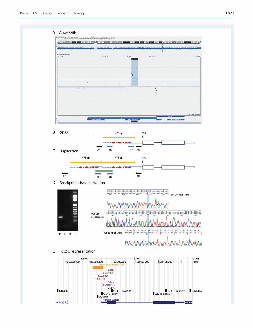
GDF9 duplication investigation and GDF9MLPA analysisA duplication between 475 and 1729 bp within the GDF9 gene was iden-tified by array-CGH in Patient 20. We amplified the duplication junction(Fig. 1D) and by sequencing, we confirmed a tandem head-to-tail dupli-cation of 475 bp containing the first part of the 5′ UTR and a short up-stream DNA sequence (Chr5.hg19:g.132,201,170_132,201,644dup).
Analysis of the entire patient cohort by an MLPA probe set for theidentification of deletions/duplications within or encompassing GDF9did not identify any additional case.
SequencingThe KRTAP2-3 and KRTAP2-4 genes were sequenced in the two hemizy-gous patients. No mutations were identified in KRTAP2-4, while the vari-ation c.218G.A (p.Cys73Tyr), that is reported in dbSNP asrs113397060, was identified. KRTAP2-3 sequencing was extended toparental samples and the entire patient–control cohorts. One morepatient and one control were homozygous (A/A) for this substitution.
Sequencing of the TSPYL6 gene in the entire patient cohort revealed nomutations.
DiscussionOut of a group of 26 patients with POI, we have identified 11 uniquenovel aberrations in 11 patients, using a customized high-resolutionarray-CGH platform. Our detection rate is comparable to Ledig et al.(2010), and higher than studies using platforms with lower resolution(Aboura et al., 2009; Liao et al., 2011; McGuire et al., 2011).
One change affects GDF9 and the other 10 involve new regions. Ma-ternally inherited changes, even though less likely pathogenic if assuminga haploinsufficiency mechanism, have not been excluded by default asautosomal recessive inheritance, and polygenic or environmentalfactor affecting phenotype penetrance cannot be excluded.
GDF9 on 5q31.1A duplication affecting GDF9 was detected in Patient 20 with SA at 15 yearsof age (Fig. 1). This oocyte-secreted factor, expressed from the primary fol-licle stage, is necessary for normal folliculogenesis and fertility. Mutationsaffecting the open reading frame of the GDF9 gene have been associatedwith POI at a low frequency (Dixit et al., 2005; Laissue et al., 2006; Zhaoet al., 2007). No such mutations were identified in our patient cohort.
Within the duplicated region there are three NOBOX-binding ele-ments (NBEs) and an E-box sequence. In mouse NBEs regulate Gdf9 ex-pression (Choi and Rajkovic, 2006), and the E-box sequence is a targetfor transcription factors necessary for the ovarian specific expressionof Gdf9 (Yan et al., 2006). Both the NBEs and the E-box are conservedbetween mice and humans, as well as several other species. Thus, theyare likely important also for the regulation of human GDF9 expression(Choi and Rajkovic, 2006; Yan et al., 2006). It is likely that the duplicationcauses altered GDF9 expression in the developing ovary, leading to POI.This represents the first mutation identified affecting the regulatoryregion of GDF9.
An enrichment of CNVs has been identified in promoter regions, sug-gesting that promoter sequences can be mildly unstable. This is a
Figure 1 GDF9 duplication. (A) Representation of an array-CGH result from the Cytosure software. The duplicated segment is indicated by blue back-ground with a positive baseline offset. Blue dispersed dots represent oligomarkers. Blue arrows on bottom indicate gene location and purple smaller arrowsrepresent exons. Black horizontal line on bottom represents location of duplication. Chromosome ideogram shown on top with the current segment mag-nification indicated by blue background color. (B) Schematic representation of the GDF9 gene. The ATG indicates the initiation of translation. Red dotsrepresent NBE. Violet dot represents the E-box. Yellowarrow indicates the duplicated segment. Black and blue arrows represent PCR primers for wild-typeallele (wt) and for duplication breakpoint amplification, respectively. (C) Schematic representation of the duplication. The green line represents the PCRproduct containing the duplication junction. (D) Breakpoint characterization. Gel image of amplified PCR breakpoint fragment. P, patient; C, control; B,blank; L, ladder. Electropherograms with sequence of the breakpoint (in the middle), partially aligned with the wt allele (upper and lower electrophero-grams). (E) Representation from the UCSC genome browser, GRCh37/hg19 assembly. Horizontal yellow arrow line indicates the duplicated segment.Locations of NBE (TAATTA, TAATTG) and E-box (CAGCTG) are shown as vertical red and purple lines, respectively. Vertical black boxes representMLPA probes.
1822 Norling et al.

suggested evolutionary mechanism for changes in gene regulation(Conrad et al., 2010).
Candidate genesTSPY-Like 6, acylphosphatase 2, muscle type 1 and C2ORF73 on2p16.2The deletion, affecting TSPY-Like 6 (TSPYL6) and acylphosphatase 2,muscle type 1 (ACYP2), in Patient 1 was not inherited from themother. The TSPYL6 gene function is unknown, but homozygous
mutations of TSPYL1 (TSPY-Like 1) have been found to cause dysgenesisof the testis and sudden infant death syndrome (MIM 608800). Weconsidered TSPYL6 an interesting candidate gene for POI andsequenced the entire patient cohort, but no inactivating mutationswere identified.
ACYP2 encodes a muscle type form of acylphosphatase 2 and we con-sider it a less likely candidate for POI. The deletion occurs just upstreamof C2ORF73, an uncharacterized gene expressed in both ovaries andtestis. A positional effect of the deletion on this gene expressioncannot be excluded as a potential cause of POI.
.............................................................................................................................................................................................
Table II Results from 26 patients, copy numbers shown after application of exclusion criteria.
Pat.no.
Age atamen.
Chr.band
Start End Size(kb)
Probecount
Del/dup
Gene(s) Inheritance Other information
1 PA 2p16.2 54296546 54398100 102 39 Del ACYP2, TSPYL6 Not maternal –
2 PA – – – – – – – – Ovarian biopsy
3 PA 15q26.2 92580470 92619918 39 17 Del MCTP2 Maternal –
4 PA 3p21.31 47588558 47723275 135 54 Dup SMARCC1,CSPG5
Maternal Mother breast cancer 41years
5 PA 15q26.2 92580470 92619918 39 17 Del MCTP2 Paternal –
6 PA – – – – – – – – –
7 PA – – – – – – – – –
8 PA – – – – – – – – –
9 PA 12p13.33 2666671 2718905 52 15 Dup CACNA1C Maternal –
10 PA – – – – – – – – –
11 PA – – – – – – – – –
12 PA 15q25.2 81917995 81997930 8 25 Dup SH3GL3 Maternal –
13 PA – – – – – – – – –
14 PA – – – – – – – – –
15 PA – – – – – – – – –
16 PA – – – – – – – – –
17 PA – – – – – – – – –
18 16 years – – – – – – – – –
19 14 years 19p13.3 3688186 3816117 128 51 Del RAX2, TJP3,ZFR2, MRPL54,APBA3, MATK
Maternal Hypothyroidism ovarianbiopsy
Xp22.33 1529221 1572694 43 17 Dup P2RY8, ASMTL Not maternal –17q21.2 36465290 36473792 9 5 Del KRTAP2-3,
KRTAP2-4Not maternal –
20 15 years 5q31.1 132228266 132228740 0.5 7 Dup GDF9 NA –
21 22 years 2p11.2 84620455 84791661 171 57 Del DNAH6 NA Paternal aunt POI 34
22 20 years 17q21.2 36465290 36473792 9 5 Del KRTAP2-3,KRTAP2-4a
Motherhemizygous
Hypothyroidismconsanguinity motherwith bilateral breastcancer
23 17 years 2q34 210703481 210739467 36 14 Del KANSL1L Maternal –
24 21 years – – – – – – – – 45,XX,der(13;14)Ovarian biopsy
25 15 years – – – – – – – – –
26 13 years – – – – – – – – Hypothyroidism withanti-TPO ovarian biopsy
All coordinates are given using the NCBI36/hg18 build. Pat no., patient number; Age at amen., Age at amenorrhea; Chr. Band., chromosomal band; Del., deletion; Dup., duplication; PA,primary amenorrhea; NA, no available sample.aHomozygous deletion.
Partial GDF9 duplication in ovarian insufficiency 1823

Multiple C2 domains, transmembrane 2 on 15q26.2In two unrelated patients with PA (Patients 3 and 5), a 39 kb deletion wasdetected, removing a large part of intron 1 in one of the several isoformsof the multiple C2 domains, transmembrane 2 (MCTP2) gene(ENST00000543482). MCTP2 is a transmembrane protein that bindscalcium in the absence of phospholipids. The deletion could affect spli-cing or regulation or have no effect at all. It is difficult to find an immediateconnection between MCTP2 and POI with the available data.
SWI/SNF-related matrix-associated, actin-dependent regulator ofchromatin subfamily c member 1 and chondroitin sulfate proteoglycan5 on 3p21.31A 135 kb duplication on chromosome 3p21.31 was found in Patient 4with PA, spanning the SWI/SNF-related matrix-associated, actin-dependent regulator of chromatin subfamily c member 1 (SMARCC1)gene and at least the first two exons of the CSPG5 gene. SMARCC1 isa member of the SWI/SNF family of proteins that regulate transcriptionby chromatin remodeling. It is highlyexpressed in both follicle and ovarianstroma cells. Chondroitin sulfate proteoglycan 5 (CSPG5) is a proteogly-can that may function as a differentiation factor. In mice, there is a sex dif-ferential expression profile of Cspg5 in Sf1+ somatic cells in developinggonads at embryonic day 12.5 (E12.5) and E13.5 (Nef et al., 2005) indi-cating a potential role in gonad differentiation. We consider bothSMARCC1 and CSPG5 possible candidate genes for POI.
Calcium channel, voltage-dependent l type alpha 1C subunit on12p13.33Patient 9, with PA, has a maternally inherited duplication involving thecalcium channel, voltage-dependent l type alpha 1C subunit (CACNA1C)gene, likely not affecting expression as loss of function mutations causesautosomal dominant cardiac conduction abnormalities (Brugada syn-drome, MIM 611875; Timothy syndrome, MIM 601005). We do notconsider CACNA1C a candidate gene for POI.
SH3-domain GRB2-like 3 on 15q25.2Patient 12, presenting with PA, carries a maternally inherited duplicationof almost the entire sequence of the first intron of the SH3GL3 gene.SH3-domain GRB2-like 3 (SH3GL3) expression is abundant in testis(Ringstad et al., 1997), where it is believed important for spermatogen-esis (Li et al., 2009). Moderate expression in follicle cells is reported in theHuman Protein Atlas. SH3GL3 interacts with the similar protein SH3GL2(SH3-domain GRB2-like-2) (Franceschiniet al., 2013) that in mice Sh3Gl2has a sex differential expression profile at E12.5 and E13.5 (Nef et al.,2005). An association between SH3GL3 and POI should be furtherinvestigated.
Dynein axonemal heavy chain 6 on 2p11.2In Patient 21 we have identified a 171 kb deletion that removes atleast 52 of the 77 exons of the dynein axonemal heavy chain 6(DNAH6) gene encoding a heavy axonemal dynein chain. Dyneins aremicrotubule-associated motor protein complexes, important for ciliarand flagellar motility. There are heavy, light and intermediate chains.The mouse intermediate chain Dnaic2 (dynein, axonemal intermediatechain 2) has been found on the surface of oocytes in secondary andantral stages, but not in primordial or primary follicles suggesting a rolefor dyneins in ovarian development (Yang and Wu, 2008).
Interestingly, in a previous array-CGH study a smaller but partiallyoverlapping deletion of DNAH6 was detected in a patient with secondaryamenorrhea (Fig. 2) (Ledig et al., 2010) strengthening the possible rolefor DNAH6 in POI. Another study has also identified the relatedDNAH5 as a candidate gene for POI (Aboura et al., 2009). Mutationsin DNAH5 are otherwise known to cause primary ciliary dyskinesia(PCD, MIM244400). We believe that DNAH6 is a very interesting candi-date gene for POI.
KRTAP2-3 and KRTAP2-4 on 17q21.2A 9 kb deletion encompassing the KRTAP2-3 and KRTAP2-4 genes wasidentified in two unrelated patients with SA and by MLPA screeningone additional hemizygous patient was found.
We hypothesized an autosomal recessive causative mechanism, andby sequencing identified a potentially damaging amino acid substitutionin the KRTAP2-3 gene in the hemizygous patients and in homozygousform in one more patient with POI. However, also one control wasfound to be homozygous for the substitution, contradicting KRTAP2-3mutations as causative for POI. It is possible that the identifiedKRTAP2-3 and KRTAP2-4 deletion is a normal variant in the Swedish popu-lation as also two controls are hemizygous.
Purinergic receptor P2Y G-protein coupled 8 and acetylserotoninO-methyltransferase-like on Xp22.33In addition to a deletion of KRTAP2-3 and KRTAP2-4, Patient 22 alsocarries a duplication in the pseudoautosomal region 1 on the X chromo-some. The duplication includes the last exon of purinergic receptor P2YG-protein coupled 8 (P2RY8) and the first exon of the ASMTL gene.P2RY8 is a G-protein coupled receptor expressed in lymphocytes. Acet-ylserotonin O-methyltransferase-like (ASMTL) is a generally expressedenzyme, with high levels in ovarian follicle cells. We consider bothP2RY8 and ASTML less likely candidate genes for POI.
Del 19p13.3A 128 kb deletion on chromosome 19 was also identified in Patient 22affecting six genes: TJP3, APBA3, MRPL54, RAX2, MATK and zinc fingerRNA-binding protein 2 (ZFR2).
Knock-out mouse models for TJP3 (Xu et al., 2008), APBA3 (Hara et al.,2011) and MATK (Lee et al., 2006) have been described without particu-lar phenotypes and with normal fertility excluding these genes as candi-dates for POI.
MRPL54 encodes for a mitochondrial ribosome subunit necessary forthe synthesis of mitochondrial proteins (Koc et al., 2001). Defects of mito-chondrial protein synthesis lead to a more wide and severe phenotype thanisolated POI. RAX2 heterozygous missense mutations cause age-relatedmacular degeneration (MIM 610362) and cone-rod dystrophy type 11(MIM 610381). We exclude MRPL54 and RAX2 as candidate genes for POI.
ZFR2 has no known function, but is expressed in many adult cell types,including ovarian follicle and stroma cells, as well as in female gameto-cytes and germ cells (Edgar et al., 2013). Ovarian biopsies from thepatient show bilateral complete lack of oocytes and follicles and we con-sider ZFR2 an interesting candidate gene for POI.
KAT8 regulatory NSL complex subunit 1-like on 2q34In Patient 23, with SA at 17, we detected a 36 kb deletion removing thefirst coding exon of the KAT8 regulatory NSL complex subunit 1-like(KANSL1L) gene on chromosome 2q34. KANSL1L, together with
1824 Norling et al.

KANSL1 on chromosome 17, are the human homologs of the DrosophilaWaharan gene (Lone et al., 2010). KANSL1 is a regulator of KAT8 influ-encing gene expression through histone H4 lysine 16 acetylation(Mendjan et al., 2006). Mutations in this gene cause 17q21.31 microdele-tion syndrome (MIM 612452). No specific information on KANSL1Lfunction is reported, but as an altered chromatin acetylation could pos-sibly affect developing germ cells and follicle formation, we considerKANSL1L a potential candidate gene for POI.
ConclusionUsing array-CGH, we have identified the first mutation affecting the regu-latory region of GDF9 in a patientwith SA, most likelypathogenic. Import-antly, the small, 475 bp duplication would not have been detectedwithout the customized probe enrichment, making clear that platformresolution and probe targeting are crucial. We have also identified asecond DNAH6 deletion in a patient with SA, corroborating DNAH6 asa candidate gene for POI. In addition, we describe several novel candi-date genes such as TSPYL6, SMARCC1, CSPG5 and ZFR2.
We consider array-CGH a powerful tool to identify candidate genesfor POI and we recommend that patients without a molecular diagnosisshould undergo analysis for copy number alterations. Soon next gener-ation sequencing will be available and able to provide reliable data oncopy numbers, once this occurs whole genome sequencing should beconsidered for diagnostics of POI patients, as most known causativegenes can either harbor small pathogenic sequence changes or bepresent in abnormal copy numbers.
Supplementary dataSupplementary data areavailable athttp://humrep.oxfordjournals.org/.
AcknowledgementsWe thank Johan Svensson (Department of Child and Adolescent Medi-cine, Skane University Hospital, Malmo, Sweden) for contributing withpatient samples and clinical characterization. We thank Oxford GeneTechnology, and Duarte Molha, Lee Eastoe, John Shovelton and Brian
Figure 2 DNAH6 deletion. (A) Representation of an array-CGH result from the Cytosure software. The duplicated segment is indicated by blue back-ground with a negative baseline offset. Blue dispersed dots represent oligomarkers. Blue arrows on bottom indicate gene location and purple vertical linesrepresent exons. The DNAH6 gene is shown with previous name DNHL1. The black horizontal line on bottom represents the location of deletion. Chromo-some ideogram shown on top with the current segment magnification indicated by blue background color. (B) Representation from the UCSC genomebrowser, GRCh37/hg19 assembly. The green line represents the deletion detected in Patient 21. The orange line shows the deletion reported byLedig et al. Vertical black lines represent MLPA probes.
Partial GDF9 duplication in ovarian insufficiency 1825

Woodhouse in particular, for their input and assistance in designing ourcustomized 1 M array-CGH platform, as well as their support through-out experimentation and computerized analysis.
Authors’ rolesAs first author A.N. performed the experimental work, results interpret-ation and writing of the first draft of the manuscript. A.L.H., K.R.W. andE.I. performed the clinical characterization and sample acquisition. E.I.also performed sample analysis. A.W. and M.B. were integral in resultsinterpretation. A.L.H., E.I., A.W. and M.B. planned the study and experi-ments. M.B. performed literature evaluation and manuscript revision. Allauthors have contributed significantly to critical revision of article for im-portant intellectual content and preparation of the final manuscript sub-mitted for publication.
FundingThe study was supported by grants from the Swedish Research council(project no 12198 to A.W., and project no 20324 to A.L.H.), StockholmCounty Council (E.I., A.W., K.R.W.), Foundation Frimurare Barnhuset(A.N., A.W., M.B.), Karolinska Institutet (A.N., A.L.H., E.I., A.W.,M.B.), Novo Nordic Foundation (A.W.) and Svenska Lakaresallskapet(M.B.). The funding sources had no involvement in the design or analysisof the study. Funding to pay the Open Access publication charges for thisarticle was provided by the Swedish Research Council.
Conflict of interestNone declared.
ReferencesAboura A, Dupas C, Tachdjian G, Portnoi MF, Bourcigaux N, Dewailly D,
Frydman R, Fauser B, Ronci-Chaix N, Donadille B et al. Array comparativegenomic hybridization profiling analysis reveals deoxyribonucleic acid copynumber variations associated with premature ovarian failure. J ClinEndocrinol Metab 2009;94:4540–4546.
Aittomaki K, Lucena JL, Pakarinen P, Sistonen P, Tapanainen J, Gromoll J,Kaskikari R, Sankila EM, Lehvaslaiho H, Engel AR et al. Mutation in thefollicle-stimulating hormone receptor gene causes hereditaryhypergonadotropic ovarian failure. Cell 1995;82:959–968.
Barbaro M, Cicognani A, Balsamo A, Lofgren A, Baldazzi L, Wedell A,Oscarson M. Gene dosage imbalances in patients with 46,XY gonadalDSD detected by an in-house-designed synthetic probe set for multiplexligation-dependent probe amplification analysis. Clin Genet 2008;73:453–464.
Baronchelli S, Villa N, Redaelli S, Lissoni S, Saccheri F, Panzeri E, Conconi D,Bentivegna A, Crosti F, Sala E et al. Investigating the role of X chromosomebreakpoints in premature ovarian failure. Mol Cytogenet 2012;5:32.
Choi Y, Rajkovic A. Characterization of NOBOX DNA binding specificityand its regulation of Gdf9 and Pou5f1 promoters. J Biol Chem 2006;281:35747–35756.
Conrad DF, Pinto D, Redon R, Feuk L, Gokcumen O, Zhang Y, Aerts J,Andrews TD, Barnes C, Campbell P et al. Origins and functional impactof copy number variation in the human genome. Nature 2010;464:704–712.
De Vos M, Devroey P, Fauser BC. Primary ovarian insufficiency. Lancet 2010;376:911–921.
Dixit H, Rao LK, Padmalatha V, Kanakavalli M, Deenadayal M, Gupta N,Chakravarty B, Singh L. Mutational screening of the coding region ofgrowth differentiation factor 9 gene in Indian women with ovarianfailure. Menopause 2005;12:749–754.
Dixit H, Rao LK, Padmalatha VV, Kanakavalli M, Deenadayal M, Gupta N,Chakrabarty B, Singh L. Missense mutations in the BMP15 gene areassociated with ovarian failure. Hum Genet 2006;119:408–415.
Dreszer TR, Karolchik D, Zweig AS, Hinrichs AS, Raney BJ, Kuhn RM,Meyer LR, Wong M, Sloan CA, Rosenbloom KR et al. The UCSCGenome Browser database: extensions and updates 2011. Nucleic AcidsRes 2012;40:D918–D923.
Dudding TE, Lawrence O, Winship I, Froyen G, Vandewalle J, Scott R,Shelling AN. Array comparative genomic hybridization for the detectionof submicroscopic copy number variations of the X chromosome inwomen with premature ovarian failure. Hum Reprod 2010;25:3159–3160. Author reply 3160–3151.
Edgar R, Mazor Y, Rinon A, Blumenthal J, Golan Y, Buzhor E, Livnat I,Ben-Ari S, Lieder I, Shitrit A et al. LifeMap Discovery: the embryonicdevelopment, stem cells, and regenerative medicine research portal.PLoS One 2013;8:e66629.
Franceschini A, Szklarczyk D, Frankild S, Kuhn M, Simonovic M, Roth A, Lin J,Minguez P, Bork P, von Mering C et al. STRING v9.1: protein-proteininteraction networks, with increased coverage and integration. NucleicAcids Res 2013;41:D808–D815.
Hara T, Mimura K, Abe T, Shioi G, Seiki M, Sakamoto T. Deletion of theMint3/Apba3 gene in mice abrogates macrophage functions andincreases resistance to lipopolysaccharide-induced septic shock. J BiolChem 2011;286:32542–32551.
Iafrate AJ, Feuk L, Rivera MN, Listewnik ML, Donahoe PK, Qi Y, Scherer SW,Lee C. Detection of large-scale variation in the human genome. Nat Genet2004;36:949–951.
Janse F, de With LM, Duran KJ, Kloosterman WP, Goverde AJ, Lambalk CB,Laven JS, Fauser BC, Giltay JC. Limited contribution of NR5A1 (SF-1)mutations in women with primary ovarian insufficiency (POI). Fertil Steril2012;97:141–146, e142.
Knauff EA, Blauw HM, Pearson PL, Kok K, Wijmenga C, Veldink JH, van denBerg LH, Bouchard P, Fauser BC, Franke L. Copy number variants on the Xchromosome in women with primary ovarian insufficiency. Fertil Steril2011;95:1584–1588, e1581.
Koc EC, Burkhart W, Blackburn K, Moyer MB, Schlatzer DM, Moseley A,Spremulli LL. The large subunit of the mammalian mitochondrialribosome. Analysis of the complement of ribosomal proteins present.J Biol Chem 2001;276:43958–43969.
Koressaar T, Remm M. Enhancements and modifications of primer designprogram Primer3. Bioinformatics 2007;23:1289–1291.
Kovanci E, Rohozinski J, Simpson JL, Heard MJ, Bishop CE, Carson SA.Growth differentiating factor-9 mutations may be associated withpremature ovarian failure. Fertil Steril 2007;87:143–146.
Laissue P, Christin-Maitre S, Touraine P, Kuttenn F, Ritvos O, Aittomaki K,Bourcigaux N, Jacquesson L, Bouchard P, Frydman R.et al. Mutationsand sequence variants in GDF9 and BMP15 in patients with prematureovarian failure. Eur J Endocrinol 2006;154:739–744.
Ledig S, Ropke A, Wieacker P. Copy number variants in premature ovarianfailure and ovarian dysgenesis. Sex Dev 2010;4:225–232.
Lee BC, Avraham S, Imamoto A, Avraham HK. Identification of thenonreceptor tyrosine kinase MATK/CHK as an essential regulator ofimmune cells using Matk/CHK-deficient mice. Blood 2006;108:904–907.
Li S, Qiao Y, Di Q, Le X, Zhang L, Zhang X, Zhang C, Cheng J, Zong S,Koide SS et al. Interaction of SH3P13 and DYDC1 protein: a germ cellcomponent that regulates acrosome biogenesis during spermiogenesis.Eur J Cell Biol 2009;88:509–520.
1826 Norling et al.

Liao C, Fu F, Yang X, Sun YM, Li DZ. Analysis of Chinese women with primaryovarian insufficiency by high resolution array-comparative genomichybridization. Chin Med J (Engl) 2011;124:1739–1742.
Lone M, Kungl T, Koper A, Bottenberg W, Kammerer R, Klein M,Sweeney ST, Auburn RP, O’Kane CJ, Prokop A. The nuclear proteinWaharan is required for endosomal-lysosomal trafficking in Drosophila.J Cell Sci 2010;123:2369–2374.
McGuire MM, Bowden W, Engel NJ, Ahn HW, Kovanci E, Rajkovic A.Genomic analysis using high-resolution single-nucleotide polymorphismarrays reveals novel microdeletions associated with premature ovarianfailure. Fertil Steril 2011;95:1595–1600.
Mendjan S, Taipale M, Kind J, Holz H, Gebhardt P, Schelder M, Vermeulen M,Buscaino A, Duncan K, Mueller J et al. Nuclear pore components areinvolved in the transcriptional regulation of dosage compensation inDrosophila. Mol Cell 2006;21:811–823.
Nef S, Schaad O, Stallings NR, Cederroth CR, Pitetti JL, Schaer G, Malki S,Dubois-Dauphin M, Boizet-Bonhoure B, Descombes P et al. Geneexpression during sex determination reveals a robust female geneticprogram at the onset of ovarian development. Dev Biol 2005;287:361–377.
Nelson LM. Clinical practice. Primary ovarian insufficiency. N Engl J Med 2009;360:606–614.
Norling A, Hirschberg AL, Iwarsson E, Persson B, Wedell A, Barbaro M.Novel candidate genes for 46,XY gonadal dysgenesis identified by acustomized 1 M array-CGH platform. Eur J Med Genet 2013;12:661–668.
Persani L, Rossetti R, Cacciatore C. Genes involved in human prematureovarian failure. J Mol Endocrinol 2010;45:257–279.
Qin Y, Choi Y, Zhao H, Simpson JL, Chen ZJ, Rajkovic A. NOBOXhomeobox mutation causes premature ovarian failure. Am J Hum Genet2007;81:576–581.
Quilter CR, Karcanias AC, Bagga MR, Duncan S, Murray A, Conway GS,Sargent CA, Affara NA. Analysis of X chromosome genomic DNAsequence copy number variation associated with premature ovarianfailure (POF). Hum Reprod 2010;25:2139–2150.
Ringstad N, Nemoto Y, De Camilli P. The SH3p4/Sh3p8/SH3p13 proteinfamily: binding partners for synaptojanin and dynamin via a Grb2-like Srchomology 3 domain. Proc Natl Acad Sci USA 1997;94:8569–8574.
Rozen S, Skaletsky H. Primer3 on the WWW for general users and forbiologist programmers. Methods Mol Biol 2000;132:365–386.
Schouten JP, McElgunn CJ, Waaijer R, Zwijnenburg D, Diepvens F, Pals G.Relative quantification of 40 nucleic acid sequences by multiplexligation-dependent probe amplification. Nucleic Acids Res 2002;30:e57.
Simpson JL, Rajkovic A. Ovarian differentiation and gonadal failure. Am J MedGenet 1999;89:186–200.
Stern RF, Roberts RG, Mann K, Yau SC, Berg J, Ogilvie CM. Multiplexligation-dependent probe amplification using a completely syntheticprobe set. Biotechniques 2004;37:399–405.
Uhlen M, Oksvold P, Fagerberg L, Lundberg E, Jonasson K, Forsberg M,Zwahlen M, Kampf C, Wester K, Hober S.et al. Towards aknowledge-based Human Protein Atlas. Nat Biotechnol 2010;28:1248–1250.
Untergasser A, Cutcutache I, Koressaar T, Ye J, Faircloth BC, Remm M,Rozen SG. Primer3—new capabilities and interfaces. Nucleic Acids Res2012;40:e115.
Wittenberger MD, Hagerman RJ, Sherman SL, McConkie-Rosell A, Welt CK,Rebar RW, Corrigan EC, Simpson JL, Nelson LM. The FMR1 premutationand reproduction. Fertil Steril 2007;87:456–465.
Xu J, Kausalya PJ, Phua DC, Ali SM, Hossain Z, Hunziker W. Earlyembryonic lethality of mice lacking ZO-2, but Not ZO-3, revealscritical and nonredundant roles for individual zonula occludensproteins in mammalian development. Mol Cell Biol 2008;28:1669–1678.
Yan C, Elvin JA, Lin YN, Hadsell LA, Wang J, DeMayo FJ, Matzuk MM.Regulation of growth differentiation factor 9 expression in oocytes invivo: a key role of the E-box. Biol Reprod 2006;74:999–1006.
Yang Z, Wu J. Mouse dynein axonemal intermediate chain 2: cloning andexpression. DNA Cell Biol 2008;27:479–488.
Zhao H, Qin Y, Kovanci E, Simpson JL, Chen ZJ, Rajkovic A. Analysesof GDF9mutation in 100 Chinese women with premature ovarian failure. Fertil Steril2007;88:1474–1476.
Zhao H, Chen ZJ, Qin Y, Shi Y, Wang S, Choi Y, Simpson JL, Rajkovic A.Transcription factor FIGLA is mutated in patients with prematureovarian failure. Am J Hum Genet 2008;82:1342–1348.
Partial GDF9 duplication in ovarian insufficiency 1827
Related Documents