Vol:.(1234567890) Apoptosis (2017) 22:1310–1318 DOI 10.1007/s10495-017-1405-z 1 3 SHORT COMMUNICATION Identification and validation of uterine stimulant methylergometrine as a potential inhibitor of caspase-1 activation Guillermo García-Laínez 1 · Mónica Sancho 1 · Vanessa García-Bayarri 1 · Mar Orzáez 1 Published online: 28 July 2017 © The Author(s) 2017. This article is an open access publication pro-inflammatory stimuli. Our results suggest that MEM has the potential to reposition in the treatment of inflamma- tory diseases with the advantages of established safety and clinical data. Keywords Caspase-1 inhibitor · Ergometrine · Inflammasome inhibitor · Inflammation · Methylergometrine Introduction Inflammation is a protective response of the body to ensure removal of detrimental stimuli and repair damaged tis- sue [1]. Cells of the innate immune system, such as mac- rophages and dendritic cells, recognize the conserved microbial structures called pathogen-associated molecular patterns (PAMPs) and danger-associated molecular pat- terns (DAMPs) [2]. At least four major pattern recognition receptor (PRR) families operate cooperatively to detect invading microbes or endogenous danger signals: Toll-like receptors, RIG-I-like receptors, C-type lectin receptors, and nucleotide-binding oligomerization domain leucine-rich repeat containing receptors (NLRs) [3, 4]. After microbial recognition, PRRs induce the activation of different sign- aling pathways that result in the synthesis of pro-inflam- matory cytokines such as interleukin (IL)-1β (IL-1β) and IL-18 [5]. IL-1β and IL-18 are synthesized as precursor molecules, which can be cleaved into their mature and active forms by caspase-1 protease. This activation occurs when the cell receives a second signal (PAMP or DAMP) to activate NLRs and promotes the assembly of a large multiprotein complex called the inflammasome. Inflam- masomes are a group of multimeric protein complexes formed by the NLR sensor protein, the adaptor protein Abstract Inflammasomes are intracellular multiprotein complexes of the innate immune system. Upon an inflam- matory insult, such as infection or intracellular damage, a nucleotide-binding oligomerization domain-like receptor (NLR) sensor protein and the adaptor protein ASC (apopto- sis-associated speck-like protein containing a caspase acti- vation and recruitment domain) are assembled to activate protease procaspase-1. This protease processes pro-IL-1β and pro-IL-18 cytokines, which are released to induce the inflammatory response. De-regulation of inflammasome contributes to the progression of several diseases, such as Alzheimer’s disease, diabetes, cancer, inflammatory and autoimmune disorders. We herein describe the identifica- tion of methylergometrine (MEM), a drug currently used as a smooth muscle constrictor during postpartum hem- orrhage, as an inhibitor of the inflammasome complex in ASC-mediated procaspase-1 activation screening. MEM inhibits the activation of the nucleotide-binding oligomeri- zation domain-like receptor protein 1 (NLRP1) and nucleo- tide-binding oligomerization domain-like receptor protein 3 (NLRP3) inflammasomes in cellular models upon different Guillermo García-Laínez and Mónica Sancho equally contributed to the work. Electronic supplementary material The online version of this article (doi:10.1007/s10495-017-1405-z) contains supplementary material, which is available to authorized users. * Mónica Sancho [email protected] * Mar Orzáez [email protected] 1 Laboratory of Peptide and Protein Chemistry, Centro de Investigación Príncipe Felipe, C/ Eduardo Primo Yúfera, 3, 46012 Valencia, Spain

Welcome message from author
This document is posted to help you gain knowledge. Please leave a comment to let me know what you think about it! Share it to your friends and learn new things together.
Transcript
Vol:.(1234567890)
Apoptosis (2017) 22:1310–1318DOI 10.1007/s10495-017-1405-z
1 3
SHORT COMMUNICATION
Identification and validation of uterine stimulant methylergometrine as a potential inhibitor of caspase-1 activation
Guillermo García-Laínez1 · Mónica Sancho1 · Vanessa García-Bayarri1 · Mar Orzáez1
Published online: 28 July 2017 © The Author(s) 2017. This article is an open access publication
pro-inflammatory stimuli. Our results suggest that MEM has the potential to reposition in the treatment of inflamma-tory diseases with the advantages of established safety and clinical data.
Keywords Caspase-1 inhibitor · Ergometrine · Inflammasome inhibitor · Inflammation · Methylergometrine
Introduction
Inflammation is a protective response of the body to ensure removal of detrimental stimuli and repair damaged tis-sue [1]. Cells of the innate immune system, such as mac-rophages and dendritic cells, recognize the conserved microbial structures called pathogen-associated molecular patterns (PAMPs) and danger-associated molecular pat-terns (DAMPs) [2]. At least four major pattern recognition receptor (PRR) families operate cooperatively to detect invading microbes or endogenous danger signals: Toll-like receptors, RIG-I-like receptors, C-type lectin receptors, and nucleotide-binding oligomerization domain leucine-rich repeat containing receptors (NLRs) [3, 4]. After microbial recognition, PRRs induce the activation of different sign-aling pathways that result in the synthesis of pro-inflam-matory cytokines such as interleukin (IL)-1β (IL-1β) and IL-18 [5]. IL-1β and IL-18 are synthesized as precursor molecules, which can be cleaved into their mature and active forms by caspase-1 protease. This activation occurs when the cell receives a second signal (PAMP or DAMP) to activate NLRs and promotes the assembly of a large multiprotein complex called the inflammasome. Inflam-masomes are a group of multimeric protein complexes formed by the NLR sensor protein, the adaptor protein
Abstract Inflammasomes are intracellular multiprotein complexes of the innate immune system. Upon an inflam-matory insult, such as infection or intracellular damage, a nucleotide-binding oligomerization domain-like receptor (NLR) sensor protein and the adaptor protein ASC (apopto-sis-associated speck-like protein containing a caspase acti-vation and recruitment domain) are assembled to activate protease procaspase-1. This protease processes pro-IL-1β and pro-IL-18 cytokines, which are released to induce the inflammatory response. De-regulation of inflammasome contributes to the progression of several diseases, such as Alzheimer’s disease, diabetes, cancer, inflammatory and autoimmune disorders. We herein describe the identifica-tion of methylergometrine (MEM), a drug currently used as a smooth muscle constrictor during postpartum hem-orrhage, as an inhibitor of the inflammasome complex in ASC-mediated procaspase-1 activation screening. MEM inhibits the activation of the nucleotide-binding oligomeri-zation domain-like receptor protein 1 (NLRP1) and nucleo-tide-binding oligomerization domain-like receptor protein 3 (NLRP3) inflammasomes in cellular models upon different
Guillermo García-Laínez and Mónica Sancho equally contributed to the work.
Electronic supplementary material The online version of this article (doi:10.1007/s10495-017-1405-z) contains supplementary material, which is available to authorized users.
* Mónica Sancho [email protected]
* Mar Orzáez [email protected]
1 Laboratory of Peptide and Protein Chemistry, Centro de Investigación Príncipe Felipe, C/ Eduardo Primo Yúfera, 3, 46012 Valencia, Spain
1311Apoptosis (2017) 22:1310–1318
1 3
ASC (the apoptosis-associated speck-like protein contain-ing a caspase activation and recruitment domain (CARD)) and protease procaspase-1 (PC1). ASC interacts through the pyrin domain with sensors, and through the CARD with PC1. The formation of this complex leads to the prox-imity-induced activation of PC1 [6]. Once activated, cas-pase-1 hydrolyses the inactive forms of pro-inflammatory cytokines IL-1β and IL-18 (pro-IL-1β and pro-IL-18) by triggering their secretion and initiating the inflammation process. The most reported consequence of caspase-1 acti-vation is the rapid secretion of cytokines, but caspase-1 activation in macrophages also leads to caspase-1-depend-ent cell death called pyroptosis [7–9].
Abnormal inflammasome activation has been implicated in the pathophysiology of Alzheimer’s disease [10, 11], diabetes [12] and cancer [13, 14], and also leads to many inflammatory and autoimmune diseases, including gout, silicosis, rheumatoid arthritis and genetically-inherited periodic fever syndromes [15–18].
The identification of small molecule inhibitors that tar-get the inflammasome is an important step in developing effective therapeutics to treat inflammation. Recently devel-oped treatments for inflammasome-related diseases target processes downstream of PC1 activation. These include Anakinra, a recombinant IL-1 receptor antagonist [19, 20], Canakinumab, a neutralizing IL-1β antibody [21, 22], and Rilonacept, a soluble decoy IL-1 receptor [23]. Several relevant strategies have also identified new drugs that tar-get different intervention points in the cascade of inflam-masome activation, such as MCC950 [24], Glyburide [25], β-hydroxybutyrate [26] and VX-765 [27].
In this study, we address the identification of new modu-lators of the inflammasome by targeting ASC-mediated PC1 activation and employing a drug repurposing strategy. The major advantage of drug repurposing is that it saves the time and cost for drug development, and permits the rapid inclusion of the molecule in clinical trials [28, 29]. We describe the identification of methylergometrine (MEM), a synthetic analog of ergonovine prescribed for treating post-partum hemorrhage, as an inhibitor of ASC-mediated PC1 activation and demonstrate the inflammasome inhibition activity of this drug in cellular models of inflammation.
Results
Identification of modulators of the ASC-mediated activation of PC1
Identification of inflammasome inhibitors was accom-plished by targeting the specific interaction of PC1 with the adaptor molecule ASC. The Prestwick Chemical Library® composed of 880 FDA-approved drugs, selected for their
chemical and pharmacological diversity and safety in humans, was evaluated in a caspase-1 activity fluorescence assay. Recombinant PC1 and ASC were combined to obtain a functional complex. Once reconstituted, self-cleavage produces PC1 activation. Active caspase-1 hydrolyzes sub-strate Ac-WEDH-AFC by releasing the fluorogenic AFC molecule (Fig. 1a). Compounds were pre-incubated with recombinant ASC to facilitate the selection of the mol-ecules that affect the ASC/PC1 interaction interface. A 60% inhibition threshold was applied, and those compounds that showed intrinsic fluorescence or color were ruled out. Three compounds were initially selected as inhibitors of ASC-mediated PC1 activation: ursolic acid, MEM and sul-fasalazine. A secondary screening was performed to con-firm positive hits and to rule out direct inhibitors of active caspase-1 (Fig. 1a). Ursolic acid and MEM were selected as inhibitors of the ASC-mediated activation of caspase-1 (Fig. 1b).
In order to confirm ursolic acid and MEM as positive hits, the dose–response effect on the ASC-dependent PC1 activation assay was analyzed. In the presence of ursolic acid and MEM, activity was inhibited in a dose–response manner with an IC50 value (compound concentration that provides 50% inhibition of activity) within the low micro-molar range (15.5 and 16.3 µM, respectively) (Fig. 2a, b). Both compounds were then evaluated in a cellular environ-ment for toxicity. For this purpose, we employed THP-1 cells, a human monocyte-derived cell line frequently used as a mammalian cellular model for inflammasome activa-tion studies. Ursolic acid was ruled out because of the high toxicity observed in THP-1 cells (Fig. 2c). However, MEM showed a non toxic profile at the range of concentrations from 1 to 50 µM (Fig. 2d).
Anti-inflammatory capacity of MEM as an inhibitor of the ASC-mediated activation of PC1
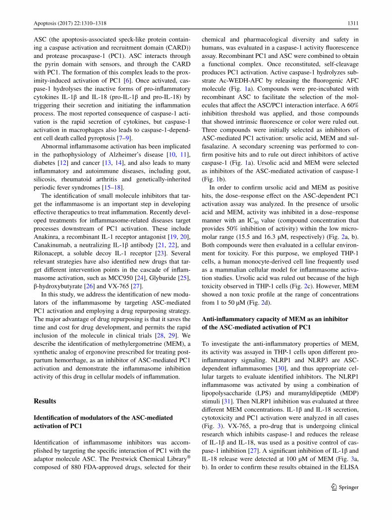
To investigate the anti-inflammatory properties of MEM, its activity was assayed in THP-1 cells upon different pro-inflammatory signaling. NLRP1 and NLRP3 are ASC-dependent inflammasomes [30], and thus appropriate cel-lular targets to evaluate identified inhibitors. The NLRP1 inflammasome was activated by using a combination of lipopolysaccharide (LPS) and muramyldipeptide (MDP) stimuli [31]. Then NLRP1 inhibition was evaluated at three different MEM concentrations. IL-1β and IL-18 secretion, cytotoxicity and PC1 activation were analyzed in all cases (Fig. 3). VX-765, a pro-drug that is undergoing clinical research which inhibits caspase-1 and reduces the release of IL-1β and IL-18, was used as a positive control of cas-pase-1 inhibition [27]. A significant inhibition of IL-1β and IL-18 release were detected at 100 µM of MEM (Fig. 3a, b). In order to confirm these results obtained in the ELISA

1312 Apoptosis (2017) 22:1310–1318
1 3
assays, immunoblotting of cellular supernatants was also performed. A significant inhibition of caspase-1 and IL-1β cleavage was observed at the highest MEM dose (Fig. 3c and Supp Data 1). The observed drop in proIL-1β in the MEM-treated samples could be a consequence of IL-1β cleavage inhibition. The active form of this cytokine acts as a positive autocrine signal to induce a feedback loop of proIL-1β expression [32].
Inflammasome activation also produces pyroptosis and, consequently, the release of lactate dehydrogenase (LDH) to media. However, VX-765, the inhibitor of caspase-1, does not affect pyroptosis in THP-1 cells. Interestingly, MEM treatment reduces the release of LDH. Thus MEM is able to partially inhibit pyroptosis produced by inflamma-some activation (Fig. 3d).
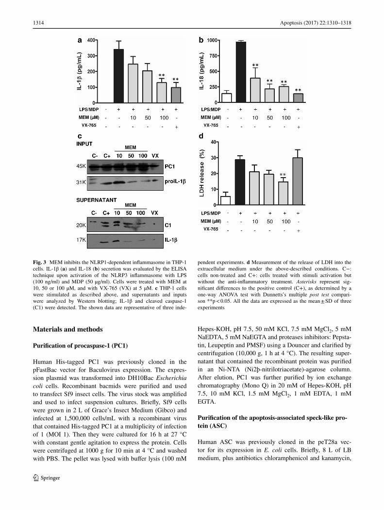
In order to activate the NLRP3 inflammasome, a com-bination of LPS and adenosine triphosphate (ATP) stimuli was used [31]. Inhibition of IL-1β and IL-18 secretion by MEM gave similar results as in the case of NLRP1 (Fig. 4a, b). Immunoblotting experiments showed that proIL-1β
decreased at the highest MEM concentration [32], as well as diminished caspase-1 and IL-1β processing (Fig. 4c and Supp data 1). The analysis of cellular pyroptosis by the release of LDH to media showed similar results to the NLRP1 inflammasome activation study. A lower cytotoxic-ity was observed in the MEM-treated cells than in the VX-765-treated cells (Fig. 4d), which indicates that MEM pro-tects cells from pyroptotic damage.
Discussion
MEM (or methergine) is a synthetic analog of ergonovine, a psychedelic alkaloid found in ergot. It is a smooth muscle constrictor that functions via the antagonism of the dopa-mine D1 receptor which mostly acts on the uterus [33, 34]. To this end, it is widely used in obstetrics to prevent or control excessive bleeding following childbirth and abor-tion. This study shows that MEM also exerts an inhibitory effect on PC1 activation, a key protein in the inflammation
Fig. 1 Identification of the modulators of the ASC-mediated acti-vation of PC1. a Scheme of the 96-well plate assays performed to identify inhibitors. Primary screening: ASC-dependent PC1 activa-tion was monitored in vitro by the release of AFC from fluorogenic PC1 substrate Ac-WEHD-AFC in the presence of 880 FDA-approved compounds (20 µM) from the Prestwick Chemical Library. Second-ary screening: caspase-1 activation was monitored in vitro by the release of AFC from fluorogenic PC1 substrate Ac-WEHD-AFC in
the presence of the positive hits (20 µM) obtained in the previous assay. b Graphical representation of the behavior of the three selected compounds in the primary (black bars) and secondary screening (white bars) at 20 µM in DMSO. A Student’s t-test statistical analysis (***p < 0.01) was performed to compare the caspase-1 inhibition for each compound in the presence or absence of ASC. Data represent the mean ± SD of three experiments

1313Apoptosis (2017) 22:1310–1318
1 3
process, in vitro and cell-based assays with an IC50 within the micromolar range. Similar results were obtained in both the NLRP1 and NLRP3 inflammasome activation models.
It is noteworthy that MEM is also able to significantly reduce the release of LDH caused by inflammasome acti-vation in THP-1 cells. Compound VX-765 is the most effective inhibitor of caspase-1 as it acts on the catalytic site of the enzyme [27], but does not decrease pyropto-sis in THP-1 cells. MEM is able to inhibit pyroptosis and enhance cell recovery. This result supports the hypothesis that targeting ASC-mediated PC1 activation is an advan-tage for cell protection over active caspase-1 inhibition. In line with our data, recent research has given rise to the development of a single domain antibody fragment that specifically recognizes the CARD domain of human ASC [35]. This antibody impairs ASC (CARD) interactions, inhibits inflammasome activation and, more importantly,
protects cells from inflammatory cell death. Our results reinforce the idea that the perturbation of ASC interac-tion interfaces could be a potential target for the devel-opment of broad-spectrum anti-inflammatory agents with improved cell recovery capabilities.
MEM is the first pass metabolite of the methysergide maleate drug [36]. The anti-inflammatory activity of methysergide maleate has been reported in systemic burn edema, asthma and endotoxemia animal models, but the mechanism of action has not been thoroughly explored [37–39]. In these models, the contribution of the inflam-masome has been recently reported [40, 41]. Although further research will be required to clarify the in vivo anti-inflammatory activity of MEM, our results suggest the contribution of inflammasome inhibition to the anti-inflammatory effects observed in asthma and endotox-emia models.
Fig. 2 The activity and toxicity profiles of ursolic acid and MEM. Dose–response activity of ursolic acid (a) and MEM (b) in vitro. The inhibitory effect of both compounds on ASC-dependent PC1 activation was followed by incubating PC1 and ASC with different concentrations of each compound: ursolic acid (2.5, 5, 10, 20, 50 and 100 µM) and MEM (2.5, 5, 10, 20, 50 and 100 µM). Data are reported as the percentage of inhibition compared with the untreated sample. The concentration that provided 50% inhibition of the com-plex (IC50) was calculated. Data represent the mean ± SD of three
experiments. Toxicity was evaluated in THP-1 cells. The cell viability measured by the MTS assay was monitored after 24 h of treatment with the indicated concentrations of both compounds, ursolic acid (c) and MEM (d). Data are reported as the percentage of live cells compared with the untreated sample. Data represent the mean ± SD of three experiments. Asterisks represent significant differences, as determined by a one-way ANOVA test with Dunnetts’s multiple post test comparison **p < 0.05, ns means non significant
1314 Apoptosis (2017) 22:1310–1318
1 3
Materials and methods
Purification of procaspase-1 (PC1)
Human His-tagged PC1 was previously cloned in the pFastBac vector for Baculovirus expression. The expres-sion plasmid was transformed into DH10Bac Escherichia coli cells. Recombinant bacmids were purified and used to transfect Sf9 insect cells. The virus stock was amplified and used to infect suspension cultures. Briefly, Sf9 cells were grown in 2 L of Grace’s Insect Medium (Gibco) and infected at 1,500,000 cells/mL with a recombinant virus that contained His-tagged PC1 at a multiplicity of infection of 1 (MOI 1). Then they were cultured for 16 h at 27 °C with constant gentle agitation to express the protein. Cells were centrifuged at 1000 g for 10 min at 4 °C and washed with PBS. The pellet was lysed with buffer lysis (100 mM
Hepes-KOH, pH 7.5, 50 mM KCl, 7.5 mM MgCl2, 5 mM NaEDTA, 5 mM NaEGTA and proteases inhibitors: Pepsta-tin, Leupeptin and PMSF) using a Douncer and clarified by centrifugation (10,000 g, 1 h at 4 °C). The resulting super-natant that contained the recombinant protein was purified in an Ni-NTA (Ni2þ-nitrilotriacetate)-agarose column. After elution, PC1 was further purified by ion exchange chromatography (Mono Q) in 20 mM of Hepes-KOH, pH 7.5, 10 mM KCl, 1.5 mM MgCl2, 1 mM EDTA, 1 mM EGTA.
Purification of the apoptosis-associated speck-like pro-tein (ASC)
Human ASC was previously cloned in the peT28a vec-tor for its expression in E. coli cells. Briefly, 8 L of LB medium, plus antibiotics chloramphenicol and kanamycin,
Fig. 3 MEM inhibits the NLRP1-dependent inflammasome in THP-1 cells. IL-1β (a) and IL-18 (b) secretion was evaluated by the ELISA technique upon activation of the NLRP3 inflammasome with LPS (100 ng/ml) and MDP (50 µg/ml). Cells were treated with MEM at 10, 50 or 100 µM, and with VX-765 (VX) at 5 µM. c THP-1 cells were stimulated as described above, and supernatants and inputs were analyzed by Western blotting; IL-1β and cleaved caspase-1 (C1) were detected. The shown data are representative of three inde-
pendent experiments. d Measurement of the release of LDH into the extracellular medium under the above-described conditions. C−: cells non-treated and C+: cells treated with stimuli activation but without the anti-inflammatory treatment. Asterisks represent sig-nificant differences to the positive control (C+), as determined by a one-way ANOVA test with Dunnetts’s multiple post test compari-son **p < 0.05. All the data are expressed as the mean ± SD of three experiments
1315Apoptosis (2017) 22:1310–1318
1 3
were inoculated with an overnight culture of freshly trans-formed His tagged-ASC-pet28a-BL21 (DE3) pLys E. coli cells (Invitrogen), and grown at 37 °C until OD 0.7. The ASC expression was induced with 0.7 mM IPTG at 28 °C for 4 h. Cells were harvested (15 min, 6000 rpm at 4 °C), lysed in 300 mL of Buffer A (50 mM NaH2PO4, 300 mM NaCl, pH 8.0) and sonicated. After centrifugation at 11,000 rpm for 1 h at 4 °C, ASC was present in the inclu-sion bodies (pellet). Pellets were then resuspended in 100 mL of denaturing Buffer B (50 mM NaH2PO4, 300 mM NaCl, 8 M urea, pH 8.0) and resonicated. Under these conditions, the His tagged-ASC protein was solubilized and purified using TALON resin (Qiagen). Re-folding was performed in the TALON resin at a gradient of decreas-ing urea concentrations and the protein was finally eluted in 15 mL of Buffer C (50 mM NaH2PO4, 300 mM NaCl, 500 mM imidazol, pH 8.0). Desalting was performed in a
PD-10 column and the protein was finally concentrated in a 10K Amicon concentrator unit.
Primary screening: the ASC-mediated PC1 reconstitu-tion in vitro assay
Recombinant PC1 (75 nM) was pre-incubated with the indicated concentrations of compounds for 20 min at RT in Assay Buffer (20 mM Hepes pH 7.5, 10 mM KCl, 1.5 mM MgCl2, 1 mM EDTA, 1 mM EGTA, 1 mM DTT). Then 300 nM of ASC were added and incubated for another 10-min period. Caspase-1 activation was measured using fluorogenic substrate Ac-WEHD-AFC (80 µM; PeptaNova) by continuously monitoring the release of the AFC prod-uct in a Wallac Victor 1420 workstation. The Prestwick Chemical Library® is composed of 880 FDA-approved drugs dissolved in DMSO. The compounds were initially
Fig. 4 MEM inhibits the NLRP3-dependent inflammasome in THP-1 cells. IL-1β (a) and IL-18 (b) secretion was evaluated by the ELISA technique upon activation of the NLRP3 inflammasome with LPS (100 ng/ml) and ATP (2.5 mM). Cells were treated with MEM at 10, 50 or 100 µM, and with VX-765 (VX) at 5 µM. c THP-1 cells were stimulated as described above, and supernatants and inputs were analyzed by Western blotting; IL-1β and cleaved caspase-1 (C1) were detected. The shown data are representative of three inde-
pendent experiments. d Measurement of the release of LDH into the extracellular medium under the above-described conditions. C−: cells non-treated and C+: cells treated with stimuli activation but without the anti-inflammatory treatment. Asterisks represent sig-nificant differences to the positive control (C+), as determined by a one-way ANOVA test with Dunnetts’s multiple post test compari-son **p < 0.05. All the data are expressed as the mean ± SD of three experiments

1316 Apoptosis (2017) 22:1310–1318
1 3
tested at 20 µM with 1% of DMSO. Data are reported as the percentage of inhibition compared with the absence of the compound.
Secondary screening: the caspase-1 in vitro activation assay
Recombinant caspase-1 (75 nM) was incubated with the indicated concentrations of compounds for 20 min at RT in Assay Buffer. Then fluorogenic substrate Ac-WEHD-AFC (80 µM) was added and activity was monitored in a Wallac Victor 1420 workstation. Data are reported as the percentage of inhibition compared with the absence of the inhibitor.
Toxicity assay: the MTS (3-(4,5-dimethylthia-zol-2-yl)-5-(3-carboxymethoxyphenyl)-2-(4-sul-fophenyl)-2H-tetrazolium, inner salt) assay
THP-1 is a cell line that is derived from human acute mono-cytic leukemia purchased from ATCC. THP-1 cells were cultured in RPMI supplemented with 10% FBS (Sigma) at 37 °C and 5% CO2.
Cell proliferation was measured with the CellTiter 96® Aqueous Non-Radioactive Cell Proliferation Assay kit (Promega) according to the manufacturer’s instructions. THP-1 cells were plated in 96-well plates at a cellular den-sity of 10,000 cells/well. Cells were treated with the indi-cated compounds at five concentrations for 24 h. Plates were read at 490 nm by a Wallac Victor 1420 workstation.
The inflammasome activation cellular assays
The molecules were evaluated in the THP-1 cells stimu-lated with LPS and MDP, or with LPS plus ATP, to stimu-late ASC-dependent inflammasomes NLRP1 and NLRP3, respectively. Briefly, 1 × 10⁶ cells were seeded in 6-well plates in 1 mL RPMI media that contained 1% FBS. Cells were either mock-treated or primed with compounds at the indicated concentrations for 30 min, followed by treatment with 100 ng/mL LPS (Sigma) and 50 μg/mL MDP (Sigma) or 2.5 mM ATP (Sigma) for 6 h at 37 °C. Supernatants were harvested and clarified by centrifugation at 1500 rpm at RT and a cytokine analysis was performed.
Both IL-1β secretion and the release of IL-18 were mon-itored by an ELISA assay (BD OptEIA™ Human IL-1β ELISA Kit and Human IL-18 Module Set eBioscience) following the manufacturer’s instructions. Cell viability was analyzed in parallel by evaluating the release of LDH according to a commercial kit (CytoTox-ONE™ Homoge-neous Membrane Integrity Assay; Promega). The release of LDH was calculated using the formula: release of LDH (%) = 100 × (Abs490 treated − Abs490 untreated cells)/
Abs490 untreated cells lysed with Triton 9% (maximum release of LDH).
Immunoblotting
The supernatants of the treated cells were precipitated by the chloroform–methanol method, as described by De Nardo and Latz [42]. Pellets were obtained by lysing cells in 25 mM of Tris-HCl, pH 7.4, 1 mM EDTA, 1 mM EGTA and 1% SDS, plus protease and phosphatase inhibitors. The protein concentration was determined by the BCA pro-tein assay. Samples were separated in a 14% SDS-PAGE gel, transferred to a nitrocellulose membrane and blocked with 5% skimmed milk for 1 h. Then the membrane was incubated overnight with primary antibodies: α-casp1 (1:1000; Cell Signaling #2225) and α-IL-1β (1:1000; Acris #R1130P) at 4 °C. Membranes were washed and probed with the appropriate secondary antibody conjugated with peroxidase for enhanced chemiluminescence detection (Amersham Pharmacia Biotech).
Statistical analysis
Data were analyzed with the GraphPad software and statis-tical significance was assessed by a Student’s t-test or a one-way analysis of variance (ANOVA) test with Dunnetts’s multiple post test comparison (**p < 0.05; ***p < 0.01).
Acknowledgements This paper is dedicated to the memory of Enrique Pérez-Payá. We thank all the members of our laboratory for discussion. This work was supported by grants from the Spanish Ministry of Economy and Competitiveness (Grant BFU2016-79487 and SAF2014-52614-R), and the Generalitat Valenciana (Prometeo 2014/061 and GV/2016/197). García-Laínez was funded by a predoc-toral fellowship from JAE-pre CSIC from the Spanish government (JaePre021).
Compliance with ethical standards
Conflict of interest The authors declare that they have no conflict of interest.
Open Access This article is distributed under the terms of the Creative Commons Attribution 4.0 International License (http://creativecommons.org/licenses/by/4.0/), which permits unrestricted use, distribution, and reproduction in any medium, provided you give appropriate credit to the original author(s) and the source, provide a link to the Creative Commons license, and indicate if changes were made.
References
1. Medzhitov R (2008) Origin and physiological roles of inflamma-tion. Nature 454(7203):428–435. doi:10.1038/nature07201

1317Apoptosis (2017) 22:1310–1318
1 3
2. Akira S, Uematsu S, Takeuchi O (2006) Pathogen recogni-tion and innate immunity. Cell 124(4):783–801. doi:10.1016/j.cell.2006.02.015
3. Bauernfeind F, Ablasser A, Bartok E, Kim S, Schmid-Burgk J, Cavlar T, Hornung V (2010) Inflammasomes: current under-standing and open questions. Cell Mol Life Sci 68(5):765–783. doi:10.1007/s00018-010-0567-4
4. Ting JP, Lovering RC, Alnemri ES, Bertin J, Boss JM, Davis BK, Flavell RA, Girardin SE, Godzik A, Harton JA, Hoffman HM, Hugot JP, Inohara N, Mackenzie A, Maltais LJ, Nunez G, Ogura Y, Otten LA, Philpott D, Reed JC, Reith W, Schreiber S, Steimle V, Ward PA (2008) The NLR gene family: a stand-ard nomenclature. Immunity 28(3):285–287. doi:10.1016/j.immuni.2008.02.005
5. Takeuchi O, Akira S (2010) Pattern recognition recep-tors and inflammation. Cell 140(6):805–820. doi:10.1016/j.cell.2010.01.022
6. Lu A, Magupalli VG, Ruan J, Yin Q, Atianand MK, Vos MR, Schroder GF, Fitzgerald KA, Wu H, Egelman EH (2014) Unified polymerization mechanism for the assembly of ASC-depend-ent inflammasomes. Cell 156(6):1193–1206. doi:10.1016/j.cell.2014.02.008
7. Bergsbaken T, Fink SL, Cookson BT (2009) Pyroptosis: host cell death and inflammation. Nat Rev Microbiol 7(2):99–109. doi:10.1038/nrmicro2070
8. Miao EA, Leaf IA, Treuting PM, Mao DP, Dors M, Sarkar A, Warren SE, Wewers MD, Aderem A (2010) Caspase-1-induced pyroptosis is an innate immune effector mechanism against intra-cellular bacteria. Nat Immunol 11(12):1136–1142. doi:10.1038/ni.1960
9. Sollberger G, Strittmatter GE, Garstkiewicz M, Sand J, Beer HD (2014) Caspase-1: the inflammasome and beyond. Innate immun 20(2):115–125. doi:10.1177/1753425913484374
10. Halle A, Hornung V, Petzold GC, Stewart CR, Monks BG, Reinheckel T, Fitzgerald KA, Latz E, Moore KJ, Golenbock DT (2008) The NALP3 inflammasome is involved in the innate immune response to amyloid-beta. Nat Immunol 9(8):857–865. doi:10.1038/ni.1636
11. Olsen I, Singhrao SK (2016) Inflammasome involvement in Alz-heimer’s disease. J Alzheimer’s Dis 54(1):45–53. doi:10.3233/JAD-160197
12. Masters SL, Dunne A, Subramanian SL, Hull RL, Tannahill GM, Sharp FA, Becker C, Franchi L, Yoshihara E, Chen Z, Mullooly N, Mielke LA, Harris J, Coll RC, Mills KH, Mok KH, News-holme P, Nunez G, Yodoi J, Kahn SE, Lavelle EC, O’Neill LA (2010) Activation of the NLRP3 inflammasome by islet amyloid polypeptide provides a mechanism for enhanced IL-1beta in type 2 diabetes. Nat Immunol 11(10):897–904. doi:10.1038/ni.1935
13. Ghiringhelli F, Apetoh L, Tesniere A, Aymeric L, Ma Y, Ortiz C, Vermaelen K, Panaretakis T, Mignot G, Ullrich E, Perfettini JL, Schlemmer F, Tasdemir E, Uhl M, Genin P, Civas A, Ryffel B, Kanellopoulos J, Tschopp J, Andre F, Lidereau R, McLaugh-lin NM, Haynes NM, Smyth MJ, Kroemer G, Zitvogel L (2009) Activation of the NLRP3 inflammasome in dendritic cells induces IL-1beta-dependent adaptive immunity against tumors. Nat Med 15(10):1170–1178. doi:10.1038/nm.2028
14. Guo B, Fu S, Zhang J, Liu B, Li Z (2016) Targeting inflammas-ome/IL-1 pathways for cancer immunotherapy. Scientific reports 6:36107. doi:10.1038/srep36107
15. Heymann MC, Rosen-Wolff A (2013) Contribution of the inflam-masomes to autoinflammatory diseases and recent mouse models as research tools. Clin Immunol 147(3):175–184. doi:10.1016/j.clim.2013.01.006
16. Cassel SL, Eisenbarth SC, Iyer SS, Sadler JJ, Colegio OR, Tephly LA, Carter AB, Rothman PB, Flavell RA, Sutterwala FS (2008) The Nalp3 inflammasome is essential for the development
of silicosis. Proc Natl Acad Sci USA 105(26):9035–9040. doi:10.1073/pnas.0803933105
17. Masters SL (2013) Specific inflammasomes in complex diseases. Clin Immunol 147(3):223–228. doi:10.1016/j.clim.2012.12.006
18. Moll M, Kuemmerle-Deschner JB (2013) Inflammasome and cytokine blocking strategies in autoinflammatory disorders. Clin Immunol 147(3):242–275. doi:10.1016/j.clim.2013.04.008
19. Granowitz EV, Porat R, Mier JW, Pribble JP, Stiles DM, Bloe-dow DC, Catalano MA, Wolff SM, Dinarello CA (1992) Phar-macokinetics, safety and immunomodulatory effects of human recombinant interleukin-1 receptor antagonist in healthy humans. Cytokine 4(5):353–360
20. Wakabayashi G, Gelfand JA, Burke JF, Thompson RC, Din-arello CA (1991) A specific receptor antagonist for interleukin 1 prevents Escherichia coli-induced shock in rabbits. FASEB J 5(3):338–343
21. Dinarello CA, van der Meer JW (2013) Treating inflammation by blocking interleukin-1 in humans. Semin Immunol 25(6):469–484. doi:10.1016/j.smim.2013.10.008
22. Rondeau JM, Ramage P, Zurini M, Gram H (2015) The molec-ular mode of action and species specificity of canakinumab, a human monoclonal antibody neutralizing IL-1beta. MAbs 7(6):1151–1160. doi:10.1080/19420862.2015.1081323
23. Hoffman Hal M. YUaKP (2008) Rilonacept. Nat Rev Drug Dis-cov 7:385–386. doi:10.1038/nrd2579
24. Coll RC, Robertson AA, Chae JJ, Higgins SC, Munoz-Planillo R, Inserra MC, Vetter I, Dungan LS, Monks BG, Stutz A, Cro-ker DE, Butler MS, Haneklaus M, Sutton CE, Nunez G, Latz E, Kastner DL, Mills KH, Masters SL, Schroder K, Cooper MA, O’Neill LA (2015) A small-molecule inhibitor of the NLRP3 inflammasome for the treatment of inflammatory diseases. Nat Med 21(3):248–255. doi:10.1038/nm.3806
25. Lamkanfi M, Mueller JL, Vitari AC, Misaghi S, Fedorova A, Deshayes K, Lee WP, Hoffman HM, Dixit VM (2009) Glybur-ide inhibits the Cryopyrin/Nalp3 inflammasome. J Cell Biol 187(1):61–70. doi:10.1083/jcb.200903124
26. Youm YH, Nguyen KY, Grant RW, Goldberg EL, Bodogai M, Kim D, D’Agostino D, Planavsky N, Lupfer C, Kanneganti TD, Kang S, Horvath TL, Fahmy TM, Crawford PA, Biragyn A, Alnemri E, Dixit VD (2015) The ketone metabolite beta-hydroxybutyrate blocks NLRP3 inflammasome-mediated inflam-matory disease. Nat Med 21(3):263–269. doi:10.1038/nm.3804
27. Stack JH, Beaumont K, Larsen PD, Straley KS, Henkel GW, Randle JC, Hoffman HM (2005) IL-converting enzyme/cas-pase-1 inhibitor VX-765 blocks the hypersensitive response to an inflammatory stimulus in monocytes from familial cold autoin-flammatory syndrome patients. J Immunol 175(4):2630–2634
28. Padhy BM, Gupta YK (2011) Drug repositioning: re-investigat-ing existing drugs for new therapeutic indications. J Postgrad Med 57(2):153–160 doi:10.4103/0022-3859.81870
29. Sekhon B (2013) Repositioning drugs and biologics:Retargeting old/existing drugs for potential new therapeutic applications. J Pharm Educ Res 4:1
30. Franchi L, Eigenbrod T, Munoz-Planillo R, Nunez G (2009) The inflammasome: a caspase-1-activation platform that regu-lates immune responses and disease pathogenesis. Nat Immunol 10(3):241–247. doi:10.1038/ni.1703
31. Gregory SM, Davis BK, West JA, Taxman DJ, Matsuzawa S, Reed JC, Ting JP, Damania B (2011) Discovery of a viral NLR homolog that inhibits the inflammasome. Science 331(6015):330–334. doi:10.1126/science.1199478
32. Toda Y, Tsukada J, Misago M, Kominato Y, Auron PE, Tanaka Y (2002) Autocrine induction of the human pro-IL-1beta gene pro-moter by IL-1beta in monocytes. J Immunol 168(4):1984–1991
33. Berde B (1978) Pharmacology of ergot alkaloids in clinical use. Med J Aust 2(3 Suppl):3–13
1318 Apoptosis (2017) 22:1310–1318
1 3
34. Pertz HH, Milhahn H, Eich E (1999) Cycloalkanecarboxylic esters derived from lysergol, dihydrolysergol-I, and elymoclavine as partial agonists and antagonists at rat 5-HT2A receptors: phar-macological evidence that the indolo[4,3-fg]quinoline system of the ergolines is responsible for high 5-HT2A receptor affinity. J Med Chem 42(4):659–668. doi:10.1021/jm981092u
35. Schmidt FI, Lu A, Chen JW, Ruan J, Tang C, Wu H, Ploegh HL (2016) A single domain antibody fragment that recognizes the adaptor ASC defines the role of ASC domains in inflam-masome assembly. J Exp Med 213(5):771–790. doi:10.1084/jem.20151790
36. Bredberg U, Eyjolfsdottir GS, Paalzow L, Tfelt-Hansen P, Tfelt-Hansen V (1986) Pharmacokinetics of methysergide and its metabolite methylergometrine in man. Eur J Clin Pharmacol 30(1):75–77
37. Hernekamp JF, Hu S, Schmidt K, Walther A, Lehnhardt M, Kre-mer T (2013) Methysergide attenuates systemic burn edema in rats. Microvasc Res 89:115–121. doi:10.1016/j.mvr.2013.03.002
38. Lima C, Souza VM, Soares AL, Macedo MS, Tavares-de-Lima W, Vargaftig BB (2007) Interference of methysergide,
a specific 5-hydroxytryptamine receptor antagonist, with airway chronic allergic inflammation and remodelling in a murine model of asthma. Clinical Exp Allergy 37(5):723–734. doi:10.1111/j.1365-2222.2007.02700.x
39. Walther A, Yilmaz N, Schmidt W, Bach A, Gebhard MM, Mar-tin E, Schmidt H (2001) Methysergide attenuates leukocyte-inde-pendent plasma extravasation during endotoxemia. J Crit Care 16(3):121–126. doi:10.1053/jcrc.2001.28975
40. Besnard AG, Guillou N, Tschopp J, Erard F, Couil-lin I, Iwakura Y, Quesniaux V, Ryffel B, Togbe D (2011) NLRP3 inflammasome is required in murine asthma in the absence of aluminum adjuvant. Allergy 66(8):1047–1057. doi:10.1111/j.1398-9995.2011.02586.x
41. Kim SR, Kim DI, Kim SH, Lee H, Lee KS, Cho SH, Lee YC (2014) NLRP3 inflammasome activation by mitochondrial ROS in bronchial epithelial cells is required for allergic inflammation. Cell Death Dis 5:e1498. doi:10.1038/cddis.2014.460
42. De Nardo CM, Latz E (2013) The inflammasome. Biology MiM. Springer, New York
Related Documents