Identification and preliminary functional analysis of alternative splicing of Siah1 in Xenopus laevis Luan Wen a,b , Jiantao Liu a,c , Yonglong Chen a , Donghai Wu a, * a Key Laboratory of Regenerative Biology, Guangzhou Institute of Biomedicine and Health, Chinese Academy of Sciences, 190 Kaiyuan Road, Guangzhou Science Park, 510530 Guangzhou, China b Graduate University of Chinese Academy of Sciences, 100049 Beijing, China c Laboratory of Veterinary Pharmacology, College of Veterinary Medicine South China Agricultural University, Guangzhou, China article info Article history: Received 11 April 2010 Available online 22 April 2010 Keywords: Xenopus xSiah1a xSiah1b abstract Siah proteins are vertebrate homologs of the Drosophila ‘seven in absentia’ gene. In this study, we char- acterized two splicing forms, Siah1a and Siah1b, of the Xenopus seven in absentia homolog 1 gene (Siah1). Overexpression of xSiah1a led to severe suppression of embryo cleavage, while that of xSiah1b was not effective even at a high dose. Competition analysis demonstrated that co-expression of xSiah1a and 1b generated the same phenotype as overexpression of xSiah1a alone, suggesting that xSiah1b does not interfere with the function of xSiah1a. Since xSiah1b has an additional 31 amino acids in the N-terminus compared to xSiah1a, progressive truncation of xSiah1b from the N-terminus showed that inability of xSiah1b to affect embryo cleavage was associated with the length of the N-terminal extension of extra amino acids. The possible implication of this finding is discussed. Ó 2010 Elsevier Inc. All rights reserved. 1. Introduction The seven in absentia homolog (Siah) proteins are homologs of the Drosophila seven in absentia (sina) gene. This gene codes for a protein downstream from the Sevenless tyrosine kinase receptor that degrades the transcriptional repressor Tramtrack and is re- quired for R7 cell fate specification during Drosophila eye develop- ment [1–3]. Humans have two unlinked Siah genes, SIAH1 and SIAH2 [4], but mice have two Siah1 (Siah1a and Siah1b) and single Siah2 genes [5]. The Siah1 protein contains an N-terminal RING fin- ger domain, required for interaction with E2 ubiquitin conjugating enzymes [6], as well as a coiled coil domain to form homo- and heterodimers [7]. As an essential component of E3 ubiquitin ligase complexes, Siah1 is responsible for the ubiquitin proteosome deg- radation that terminates cellular signaling and also for turnover of many key proteins, such as DCC [8], b-catenin [9], Kid [10], c-Myb [11], OBF-1 [12,13], Numb [14], Synaptophysin [15], TIEG-1 [16], Synphilin-1 [17], CtIP [18], T-STAR [19], Polycystin [20], Af4 [21], PHD1 and PHD3 [22], HIPK2 [23], FIH [24,25], PLCepsilon [26] and TRB3 [27]. Siah1a knockout mice exhibit severe growth retardation and male sterility due to a blockade in spermatogenesis [28]. Several splicing variants of SIAH1 appear to play important regulatory roles; for example, SIAH1S, an alternative splicing form of SIAH1, acts as a dominant negative inhibitor of SIAH1 in the regulation of b-catenin activity [29]. SIAH1L, another splicing variant of SIAH1, is induced in response to p53 and plays a key role in the regulation of b-catenin activity [9,30,31]. In Xenopus laevis, overexpression of xSiah2 led to small eyes [32] due to degradation of PHD2 [33], indicating its critical role in retinal development. However, the function of Siah1 in Xenopus embryogenesis still remains largely unknown. In the present study, we report for the first time the identification of two splicing forms of Siah1 in X. laevis and their distinct functional roles. 2. Materials and methods 2.1. Isolation of xSiah1a and xSiah1b in X. laevis Two Xenopus Siah1protein sequences were found based on a Blastp search using the human Siah1 coding sequence; one was from X. laevis (NP_001085438), the other was from the Xenopus tropicalis (NP_001015836). The predicted open reading frame (ORF) of the X. laevis gene was longer than that of X. tropicalis. Com- parison of the genomic organization of the two ORFs using Blat re- vealed that the X. laevis Siah1 has an alternative exon in the UTR (Fig. 2A). The full X. laevis Siah1a sequence (GU377277) was cloned by RT-PCR using the forward primer 5 0 -TCCGGTGTCTTTCTATG- GAAGCGGT-3 0 and the reverse primer 5 0 -CATGTACACACCCAG- CTGGGCATCTTTTGTA-3 0 . The correct clones were verified by sequencing. 0006-291X/$ - see front matter Ó 2010 Elsevier Inc. All rights reserved. doi:10.1016/j.bbrc.2010.04.107 * Corresponding author. E-mail address: [email protected] (D. Wu). Biochemical and Biophysical Research Communications 396 (2010) 419–424 Contents lists available at ScienceDirect Biochemical and Biophysical Research Communications journal homepage: www.elsevier.com/locate/ybbrc

Welcome message from author
This document is posted to help you gain knowledge. Please leave a comment to let me know what you think about it! Share it to your friends and learn new things together.
Transcript
Biochemical and Biophysical Research Communications 396 (2010) 419–424
Contents lists available at ScienceDirect
Biochemical and Biophysical Research Communications
journal homepage: www.elsevier .com/locate /ybbrc
Identification and preliminary functional analysis of alternative splicing of Siah1in Xenopus laevis
Luan Wen a,b, Jiantao Liu a,c, Yonglong Chen a, Donghai Wu a,*
a Key Laboratory of Regenerative Biology, Guangzhou Institute of Biomedicine and Health, Chinese Academy of Sciences, 190 Kaiyuan Road,Guangzhou Science Park, 510530 Guangzhou, Chinab Graduate University of Chinese Academy of Sciences, 100049 Beijing, Chinac Laboratory of Veterinary Pharmacology, College of Veterinary Medicine South China Agricultural University, Guangzhou, China
a r t i c l e i n f o
Article history:Received 11 April 2010Available online 22 April 2010
Keywords:XenopusxSiah1axSiah1b
0006-291X/$ - see front matter � 2010 Elsevier Inc. Adoi:10.1016/j.bbrc.2010.04.107
* Corresponding author.E-mail address: [email protected] (D. Wu).
a b s t r a c t
Siah proteins are vertebrate homologs of the Drosophila ‘seven in absentia’ gene. In this study, we char-acterized two splicing forms, Siah1a and Siah1b, of the Xenopus seven in absentia homolog 1 gene (Siah1).Overexpression of xSiah1a led to severe suppression of embryo cleavage, while that of xSiah1b was noteffective even at a high dose. Competition analysis demonstrated that co-expression of xSiah1a and 1bgenerated the same phenotype as overexpression of xSiah1a alone, suggesting that xSiah1b does notinterfere with the function of xSiah1a. Since xSiah1b has an additional 31 amino acids in the N-terminuscompared to xSiah1a, progressive truncation of xSiah1b from the N-terminus showed that inability ofxSiah1b to affect embryo cleavage was associated with the length of the N-terminal extension of extraamino acids. The possible implication of this finding is discussed.
� 2010 Elsevier Inc. All rights reserved.
1. Introduction
The seven in absentia homolog (Siah) proteins are homologs ofthe Drosophila seven in absentia (sina) gene. This gene codes for aprotein downstream from the Sevenless tyrosine kinase receptorthat degrades the transcriptional repressor Tramtrack and is re-quired for R7 cell fate specification during Drosophila eye develop-ment [1–3]. Humans have two unlinked Siah genes, SIAH1 andSIAH2 [4], but mice have two Siah1 (Siah1a and Siah1b) and singleSiah2 genes [5]. The Siah1 protein contains an N-terminal RING fin-ger domain, required for interaction with E2 ubiquitin conjugatingenzymes [6], as well as a coiled coil domain to form homo- andheterodimers [7]. As an essential component of E3 ubiquitin ligasecomplexes, Siah1 is responsible for the ubiquitin proteosome deg-radation that terminates cellular signaling and also for turnover ofmany key proteins, such as DCC [8], b-catenin [9], Kid [10], c-Myb[11], OBF-1 [12,13], Numb [14], Synaptophysin [15], TIEG-1 [16],Synphilin-1 [17], CtIP [18], T-STAR [19], Polycystin [20], Af4 [21],PHD1 and PHD3 [22], HIPK2 [23], FIH [24,25], PLCepsilon [26]and TRB3 [27].
Siah1a knockout mice exhibit severe growth retardation andmale sterility due to a blockade in spermatogenesis [28]. Severalsplicing variants of SIAH1 appear to play important regulatoryroles; for example, SIAH1S, an alternative splicing form of SIAH1,
ll rights reserved.
acts as a dominant negative inhibitor of SIAH1 in the regulationof b-catenin activity [29]. SIAH1L, another splicing variant ofSIAH1, is induced in response to p53 and plays a key role in theregulation of b-catenin activity [9,30,31].
In Xenopus laevis, overexpression of xSiah2 led to small eyes[32] due to degradation of PHD2 [33], indicating its critical rolein retinal development. However, the function of Siah1 in Xenopusembryogenesis still remains largely unknown. In the present study,we report for the first time the identification of two splicing formsof Siah1 in X. laevis and their distinct functional roles.
2. Materials and methods
2.1. Isolation of xSiah1a and xSiah1b in X. laevis
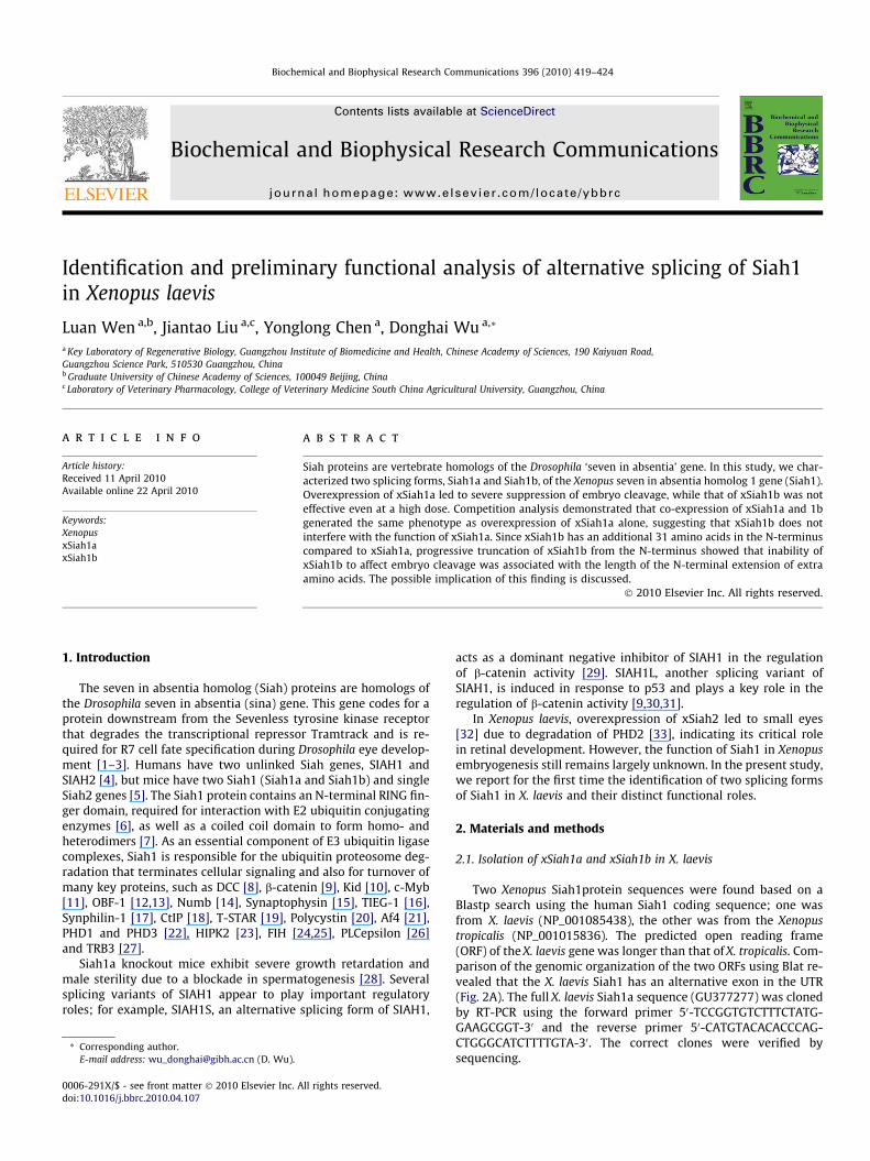
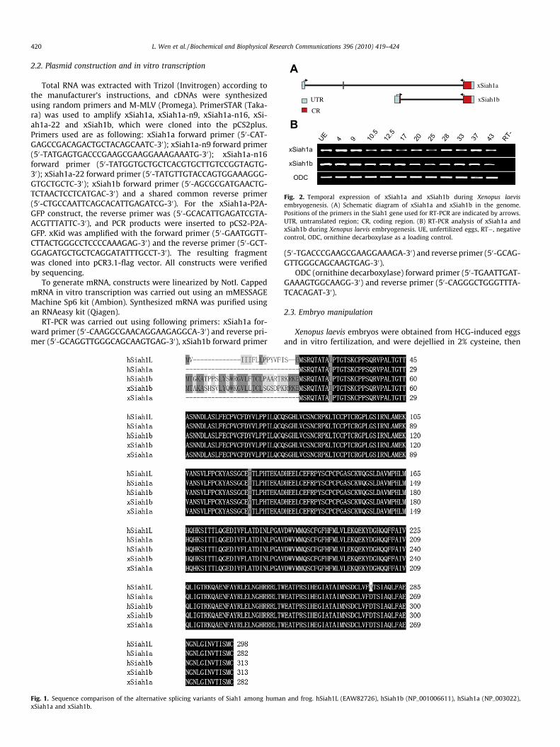
Two Xenopus Siah1protein sequences were found based on aBlastp search using the human Siah1 coding sequence; one wasfrom X. laevis (NP_001085438), the other was from the Xenopustropicalis (NP_001015836). The predicted open reading frame(ORF) of the X. laevis gene was longer than that of X. tropicalis. Com-parison of the genomic organization of the two ORFs using Blat re-vealed that the X. laevis Siah1 has an alternative exon in the UTR(Fig. 2A). The full X. laevis Siah1a sequence (GU377277) was clonedby RT-PCR using the forward primer 50-TCCGGTGTCTTTCTATG-GAAGCGGT-30 and the reverse primer 50-CATGTACACACCCAG-CTGGGCATCTTTTGTA-30. The correct clones were verified bysequencing.
UTR
CR
xSiah1a
xSiah1b
A
RT-
4337332825201712.5
10.5
94UE
xSiah1a
xSiah1b
ODC
B
Fig. 2. Temporal expression of xSiah1a and xSiah1b during Xenopus laevisembryogenesis. (A) Schematic diagram of xSiah1a and xSiah1b in the genome.Positions of the primers in the Siah1 gene used for RT-PCR are indicated by arrows.UTR, untranslated region; CR, coding region. (B) RT-PCR analysis of xSiah1a andxSiah1b during Xenopus laevis embryogenesis. UE, unfertilized eggs, RT�, negativecontrol, ODC, ornithine decarboxylase as a loading control.
420 L. Wen et al. / Biochemical and Biophysical Research Communications 396 (2010) 419–424
2.2. Plasmid construction and in vitro transcription
Total RNA was extracted with Trizol (Invitrogen) according tothe manufacturer’s instructions, and cDNAs were synthesizedusing random primers and M-MLV (Promega). PrimerSTAR (Taka-ra) was used to amplify xSiah1a, xSiah1a-n9, xSiah1a-n16, xSi-ah1a-22 and xSiah1b, which were cloned into the pCS2plus.Primers used are as following: xSiah1a forward primer (50-CAT-GAGCCGACAGACTGCTACAGCAATC-30); xSiah1a-n9 forward primer(50-TATGAGTGACCCGAAGCGAAGGAAAGAAATG-30); xSiah1a-n16forward primer (50-TATGGTGCTGCTCACGTGCTTGTCCGGTAGTG-30); xSiah1a-22 forward primer (50-TATGTTGTACCAGTGGAAAGGG-GTGCTGCTC-30); xSiah1b forward primer (50-AGCGCGATGAACTG-TCTAACTCCTCATGAC-30) and a shared common reverse primer(50-CTGCCAATTCAGCACATTGAGATCG-30). For the xSiah1a-P2A-GFP construct, the reverse primer was (50-GCACATTGAGATCGTA-ACGTTTATTC-30), and PCR products were inserted to pCS2-P2A-GFP. xKid was amplified with the forward primer (50-GAATGGTT-CTTACTGGGCCTCCCCAAAGAG-30) and the reverse primer (50-GCT-GGAGATGCTGCTCAGGATATTTGCCT-30). The resulting fragmentwas cloned into pCR3.1-flag vector. All constructs were verifiedby sequencing.
To generate mRNA, constructs were linearized by NotI. CappedmRNA in vitro transcription was carried out using an mMESSAGEMachine Sp6 kit (Ambion). Synthesized mRNA was purified usingan RNAeasy kit (Qiagen).
RT-PCR was carried out using following primers: xSiah1a for-ward primer (50-CAAGGCGAACAGGAAGAGGCA-30) and reverse pri-mer (50-GCAGGTTGGGCAGCAAGTGAG-30), xSiah1b forward primer
Fig. 1. Sequence comparison of the alternative splicing variants of Siah1 among humaxSiah1a and xSiah1b.
(50-TGACCCGAAGCGAAGGAAAGA-30) and reverse primer (50-GCAG-GTTGGGCAGCAAGTGAG-30).
ODC (ornithine decarboxylase) forward primer (50-TGAATTGAT-GAAAGTGGCAAGG-30) and reverse primer (50-CAGGGCTGGGTTTA-TCACAGAT-30).
2.3. Embryo manipulation
Xenopus laevis embryos were obtained from HCG-induced eggsand in vitro fertilization, and were dejellied in 2% cysteine, then
n and frog. hSiah1L (EAW82726), hSiah1b (NP_001006611), hSiah1a (NP_003022),
L. Wen et al. / Biochemical and Biophysical Research Communications 396 (2010) 419–424 421
cultured in 0.1 MBS. In vitro transcribed mRNA and MG132 (Beyo-time) were injected into the embryos at the 1 or 2 cell stage. Em-bryos were fixed in MEMFA (0.1 M MOPS, pH 7.4, 2 mM EDTAand 3.7% formaldehyde) for 1 h, washed in ethanol three timesand stored at �20 �C in ethanol. For b-galactosidase staining, em-bryos were fixed in MEMFA for 20 min at room temperature, andthen washed in PBS twice before staining in a buffer containing1 mg/ml X-gal, 20 mM K3Fe(CN)6, 20 mM K4Fe(CN)6 and 2 mMMgCl2. For DAPI whole mount staining, MEMFA fixed embryos
xSiah1a P2A GFP
C
38/3840/40
A
xSiah1a
82/82 78/78
B
xSiah1bxSiah1a
E
Fig. 3. xSiah1a suppresses Xenopus embryo cleavage through a proteosome dependentcleavage in a dose-dependent manner. (B) Two hundred picograms xSiah1a mRNA injectconstruct of xSiah1a-2A-GFP. (D) Expression of XSiah1a was suppressed by MG132, a pxSiah1a-2A-GFP mRNA. (E) Overexpression of xSiah1a formed multinuclear cells. Redinterpretation of the references to color in this figure legend, the reader is referred to th
were washed with PBS five times, stained with DAPI for 30 s, andthen washed in PBS five times.
2.4. Cell transfection and Western blotting analysis
293T cells were cultured in Dulbecco’s modified Eagle’s med-ium (DMEM) supplemented with 10% FBS. Cells were transfectedwith Lipofectamine 2000 (Invitrogen) according to the manufac-turer’s instructions. Cells were homogenized with RIPA lysis buffer
xSiah1a-2A-GFP
MG132
+
+-
+
40/42
47/50
D
54/54 41/41
degradation pathway. (A) Overexpression of xSiah1a suppresses Xenopus embryoion compared to 2 ng xSiah1b mRNA injection. (C) Schematic representation of theroteosome inhibitor. MG132 was injected to embryos after injection of 500 pg of
arrow shows an enlarged nucleus; yellow arrow indicates a normal nucleus. (Fore web version of this article.)
422 L. Wen et al. / Biochemical and Biophysical Research Communications 396 (2010) 419–424
(Beyotime), and protease inhibitor cocktail (Sigma–Aldrich) and100 mM PMSF were added according to the manufacturer’s recom-mendation. Proteins were separated by 10% SDS–PAGE and trans-ferred to nitrocellulose membrane (Millipore). Anti-flag (Sigma–Aldrich) and anti-GAPDH (Abcam) antibody were diluted as recom-mended by the suppliers. Detection was by HRP-labeled secondaryantibodies and ECL.
3. Results and discussion
3.1. Isolation of xSiah1a and xSiah1b
Searching the Xenopus protein database using the human SIAH1protein sequence found one X. tropicalis Siah1 (xtSiah1) and one X.laevis Siah1 (xSiah1). Searching the nucleotide sequences in the X.tropicalis genome using Blat tools revealed large differences in the50 untranslated region. The xSiah1 has an upstream initiation co-don and encoded an additional 31 amino acids compared to humanSiah1s (Fig. 1). We then obtained the short xSiah1 through predic-tion based on bioinformatics using xSiah1 sequences, was and des-ignated this as xSiah1a (GU377277), while the long form wasdesignated xSiah1b.
Searching the X. tropicalis genome for xSiah1a and xSiah1b usingthe Blat program revealed that xSiah1a and xSiah1b shared mostsequences except the N-terminus. XSiah1b consists of two exons
xSiah1a xSiah1a-n9 xSiah1a-n
57/57 53/61 60/6
C
52/52 55/55
81/81 73/73
A
Control
xSiah1b
xSi
D
Flag
GAPDH
Flag-xKidxSiah1axSiah1b
++ +
+- -
--+
pCS2 - -+
Fig. 4. Analysis of xSiah1b function. (A) Co-injection of xSiah1a and xSiah1b. Two narespectively. (B) Diagram of the constructs of xSiah1b with different additional N-termexpression to detect xSiah1a and xSiah1a activity in 293T cells. Equivalent amounts of
and encodes a 313 amino acid protein, while xSiha1a consists ofthree exons and encodes a 282 amino acid protein (Fig. 2A). The se-quences are conserved between humans and Xenopus (Fig. 1).These splicing variants have also been found in humans, but theirfunctional significance is not known.
Expression analysis during different stages of embryogenesisrevealed that mRNAs for xSiah1a and xSiah1b are both maternaland can be detected at all developmental stages analyzed(Fig. 2B). XSiah1a and xSiah1b are abundantly present before thegastrulation stages (Fig. 2B), indicating their potential roles inXenopus embryogenesis.
3.2. Xenopus cleavage was exclusively suppressed by xSiah1a
Siah1 is involved in the cell cycle, apoptosis and proliferation[10,34,35]. Stable overexpression of SIAH1 in a human lung cancerMCF7 cell line showed cell growth inhibition due to perturbedmitosis through degradation of Kid [10,35], a protein needed forchromosome alignment in metaphase and that should be degradedin anaphase to allow for chromosome separation [36]. Gene abla-tion of Siah1a in mice demonstrated that Siah1a is also requiredfor traversing metaphase during meiosis I of spermatogenesis [28].
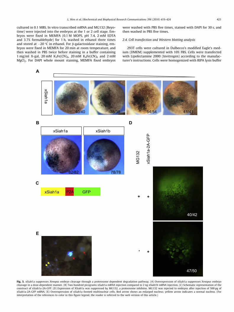
In our experiments, overexpression of xSiah1a through mRNAinjection was found to suppress cleavage of Xenopus embryos ina dose-dependent manner (Fig. 3A) and eventually led to severe
16 xSiah1a-n22 xSiah1b
48/4845/450
ah1a
BxSiah1bxSiah1a-n22xSiah1a-n16xSiah1a-n9xSiah1a
nograms of xSiah1b mRNA was co-injected with 100 and 200 pg xSiah1a mRNA,inal truncations. (C) Injection of 50 pg of mRNA. (D) Western blot analysis of xKidxSiah1a, xSiah1b and pCS2 (250 ng), were co-transfected with Flag-xKid (1 lg).
L. Wen et al. / Biochemical and Biophysical Research Communications 396 (2010) 419–424 423
apoptosis (data not shown). However, xSiah1b was unable to sup-press Xenopus embryo cleavage, even at a high dose (Fig. 2B). Over-expression of human SIAH1 in cell lines showed cell growthinhibition and more than 50% cells formed multinucleated giantcells [35]. DAPI staining showed that the enlarged cells suppressedby xSiah1a overexpression had giant nuclei (Fig. 3E). Overexpres-sion of xSiah1a has been suggested to disturb mitosis during Xeno-pus early cleavage. To better trace the mRNA distribution inXenopus embryos, we used 2A peptide [37] to link xSiah1a withGFP (Fig. 3D) so that GFP positive cells would express xSiah1a.Co-injection of MG132, a well-known inhibitor of the proteosomewith xSiah1a-P2A-GFP diminished the phenotype of cell cycle sup-pression (Fig. 3C), indicating that suppression of embryo cleavageby xSiah1a is proteosome dependent. In contrast, injection of2 ng xSiah1b mRNA into embryos had no significant effect, evenat a later tadpole stage (data not shown). This result suggests thatxSiah1b is inactivated in Xenopus.
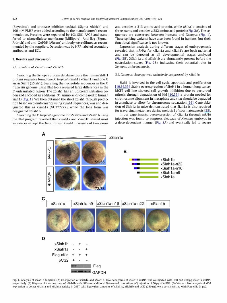
Siah1 can form homo- or heterodimers with other Siah1pro-teins through its C-terminal domain [7]. To determine whether xSi-ah1b can affect xSiah1a activity by formation of a heterodimerwith xSiah1a, a low dose of xSiah1a was co-injected with a highdose of xSiah1b. The effect was the same as injection of xSiah1aalone (Fig. 4A), thus eliminating the possibility that xSiah1b servesas a dominant negative form of xSiah1a in Xenopus.
3.3. N-terminal 31 amino acids are responsible for xSiah1b activity inthe Xenopus embryo
The underlying mechanism for inactivation of xSiah1b duringXenopus embryonic development was further explored by examin-ing the functional role of the additional N-terminal sequences. Todetermine which amino acids at the N-terminal of xSiah1b are re-quired for suppression of xSiah1 activity, we made a series of con-structs of Siah1b with different lengths of the N-terminus (Fig. 4B).Overexpression by mRNA injection demonstrated that xSiah1bactivity was correlated with the length of the N-terminus(Fig. 4C). On the other hand, xSiah1a and xSiah1b had the same ef-fect with respect to degradation of substrate xKid when transfec-ted into 293T cells (Fig. 4D), indicating that other protein(s) inthe Xenopus embryo is (are) likely to bind to xSiah1b through the31 N-terminal amino acids, resulting in inhibition of xSiah1b.Clearly additional efforts are required to determine the detailedmechanism.
In summary, this study reports, for the first time, the presence ofalternative splicing variants of Siah1—xSiah1a and 1b—in earlyembryogenesis of X. laevis. xSiah1b seems to be inactivated in Xeno-pus. xSiah1 activity also is apparently regulated at the posttran-scriptional level. Transient overexpression of SIAH1L, which hasan additional 15 amino acids in the N-terminal than does SIAH1(Fig. 1), in a 293 cell line resulted in cell growth arrest [31]. Injectionof SIAH1L mRNA at 1.5 ng per egg caused hypodorsalization ofXenopus embryos due to downregulation of b-catenin signalingbut had no effect on early cleavage during Xenopus embryogenesis[9]. Overexpression of xSiah2, which is conserved with xSiah1a inits C-terminal, also did not show suppression of embryo cleavageat 2 ng per embryo (data not shown) but resulted in the develop-ment of small eyes in the tadpole stage [32,33]. A recent report sug-gests that suppression of Siah1 activity can reduce tumor growthand prolong survival of the mice with neoangiogenesis [38]. Ourpresent results may prove useful in providing more insight intothe regulation of xSiah1 activity in a Xenopus model system.
Acknowledgments
This work was supported in part by Funds from the Key Projectof Knowledge Innovation Program of the Chinese Academy of Sci-
ences (KSCX2-YW-R-083), the National Science Foundation of Chi-na (30811120429, 3097063 and 90813033), the National 973program of China (2006CB910202) and the National Basic ResearchProgram of China (2009CB941202 and 2010CB9455000).
References
[1] A.H. Tang, T.P. Neufeld, E. Kwan, G.M. Rubin, PHYL acts to down-regulateTTK88, a transcriptional repressor of neuronal cell fates, by a SINA-dependentmechanism, Cell 90 (1997) 459–467.
[2] S.C. Li, Z. Songyang, S.J. Vincent, C. Zwahlen, S. Wiley, L. Cantley, L.E. Kay, J.Forman-Kay, T. Pawson, High-affinity binding of the Drosophila Numbphosphotyrosine-binding domain to peptides containing a Gly-Pro-(p)Tyrmotif, Proc. Natl. Acad. Sci. USA 94 (1997) 7204–7209.
[3] R.W. Carthew, G.M. Rubin, Seven in absentia, a gene required for specificationof R7 cell fate in the Drosophila eye, Cell 63 (1990) 561–577.
[4] G. Hu, Y.L. Chung, T. Glover, V. Valentine, A.T. Look, E.R. Fearon,Characterization of human homologs of the Drosophila seven in absentia(sina) gene, Genomics 46 (1997) 103–111.
[5] N.G. Della, P.V. Senior, D.D. Bowtell, Isolation and characterisation of murinehomologues of the Drosophila seven in absentia gene (sina), Development 117(1993) 1333–1343.
[6] K.L. Lorick, J.P. Jensen, S. Fang, A.M. Ong, S. Hatakeyama, A.M. Weissman, RINGfingers mediate ubiquitin-conjugating enzyme (E2)-dependent ubiquitination,Proc. Natl. Acad. Sci. USA 96 (1999) 11364–11369.
[7] G. Hu, E.R. Fearon, Siah-1 N-terminal RING domain is required for proteolysisfunction, and C-terminal sequences regulate oligomerization and binding totarget proteins, Mol. Cell. Biol. 19 (1999) 724–732.
[8] G. Hu, S. Zhang, M. Vidal, J.L. Baer, T. Xu, E.R. Fearon, Mammalian homologs ofseven in absentia regulate DCC via the ubiquitin-proteasome pathway, GenesDev. 11 (1997) 2701–2714.
[9] J. Liu, J. Stevens, C.A. Rote, H.J. Yost, Y. Hu, K.L. Neufeld, R.L. White, N.Matsunami, Siah-1 mediates a novel beta-catenin degradation pathway linkingp53 to the adenomatous polyposis coli protein, Mol. Cell 7 (2001) 927–936.
[10] A. Germani, H. Bruzzoni-Giovanelli, A. Fellous, S. Gisselbrecht, N. Varin-Blank,F. Calvo, SIAH-1 interacts with alpha-tubulin and degrades the kinesin Kid bythe proteasome pathway during mitosis, Oncogene 19 (2000) 5997–6006.
[11] J. Tanikawa, E. Ichikawa-Iwata, C. Kanei-Ishii, A. Nakai, S. Matsuzawa, J.C. Reed,S. Ishii, P53 suppresses the c-Myb-induced activation of heat shocktranscription factor 3, J. Biol. Chem. 275 (2000) 15578–15585.
[12] R. Tiedt, B.A. Bartholdy, G. Matthias, J.W. Newell, P. Matthias, The RING fingerprotein Siah-1 regulates the level of the transcriptional coactivator OBF-1,EMBO J. 20 (2001) 4143–4152.
[13] J. Boehm, Y. He, A. Greiner, L. Staudt, T. Wirth, Regulation of BOB.1/OBF.1stability by SIAH, EMBO J. 20 (2001) 4153–4162.
[14] L. Susini, B.J. Passer, N. Amzallag-Elbaz, T. Juven-Gershon, S. Prieur, N. Privat,M. Tuynder, M.C. Gendron, A. Israel, R. Amson, M. Oren, A. Telerman, Siah-1binds and regulates the function of Numb, Proc. Natl. Acad. Sci. USA 98 (2001)15067–15072.
[15] T.C. Wheeler, L.S. Chin, Y. Li, F.L. Roudabush, L. Li, Regulation of synaptophysindegradation by mammalian homologues of seven in absentia, J. Biol. Chem.277 (2002) 10273–10282.
[16] S.A. Johnsen, M. Subramaniam, D.G. Monroe, R. Janknecht, T.C. Spelsberg,Modulation of transforming growth factor beta (TGFbeta)/Smadtranscriptional responses through targeted degradation of TGFbeta-inducibleearly gene-1 by human seven in absentia homologue, J. Biol. Chem. 277 (2002)30754–30759.
[17] Y. Nagano, H. Yamashita, T. Takahashi, S. Kishida, T. Nakamura, E. Iseki, N.Hattori, Y. Mizuno, A. Kikuchi, M. Matsumoto, Siah-1 facilitates ubiquitinationand degradation of synphilin-1, J. Biol. Chem. 278 (2003) 51504–51514.
[18] A. Germani, A. Prabel, S. Mourah, M.P. Podgorniak, A. Di Carlo, R. Ehrlich, S.Gisselbrecht, N. Varin-Blank, F. Calvo, H. Bruzzoni-Giovanelli, SIAH-1 interactswith CtIP and promotes its degradation by the proteasome pathway, Oncogene22 (2003) 8845–8851.
[19] J.P. Venables, C. Dalgliesh, M.P. Paronetto, L. Skitt, J.K. Thornton, P.T. Saunders,C. Sette, K.T. Jones, D.J. Elliott, SIAH1 targets the alternative splicing factor T-STAR for degradation by the proteasome, Hum. Mol. Genet. 13 (2004) 1525–1534.
[20] H. Kim, W. Jeong, K. Ahn, C. Ahn, S. Kang, Siah-1 interacts with the intracellularregion of polycystin-1 and affects its stability via the ubiquitin-proteasomepathway, J. Am. Soc. Nephrol. 15 (2004) 2042–2049.
[21] P.L. Oliver, E. Bitoun, J. Clark, E.L. Jones, K.E. Davies, Mediation of Af4 proteinfunction in the cerebellum by Siah proteins, Proc. Natl. Acad. Sci. USA 101(2004) 14901–14906.
[22] K. Nakayama, I.J. Frew, M. Hagensen, M. Skals, H. Habelhah, A. Bhoumik, T.Kadoya, H. Erdjument-Bromage, P. Tempst, P.B. Frappell, D.D. Bowtell, Z. Ronai,Siah2 regulates stability of prolyl-hydroxylases, controls HIF1alphaabundance, and modulates physiological responses to hypoxia, Cell 117(2004) 941–952.
[23] M. Winter, D. Sombroek, I. Dauth, J. Moehlenbrink, K. Scheuermann, J. Crone,T.G. Hofmann, Control of HIPK2 stability by ubiquitin ligase Siah-1 andcheckpoint kinases ATM and ATR, Nat. Cell Biol. 10 (2008) 812–824.
[24] H. Fukuba, H. Yamashita, Y. Nagano, H.G. Jin, M. Hiji, T. Ohtsuki, T. Takahashi, T.Kohriyama, M. Matsumoto, Siah-1 facilitates ubiquitination and degradation
424 L. Wen et al. / Biochemical and Biophysical Research Communications 396 (2010) 419–424
of factor inhibiting HIF-1alpha (FIH), Biochem. Biophys. Res. Commun. 353(2007) 324–329.
[25] H. Fukuba, T. Takahashi, H.G. Jin, T. Kohriyama, M. Matsumoto, Abundance ofasparaginyl-hydroxylase FIH is regulated by Siah-1 under normoxicconditions, Neurosci. Lett. 433 (2008) 209–214.
[26] S. Yun, A. Moller, S.K. Chae, W.P. Hong, Y.J. Bae, D.D. Bowtell, S.H. Ryu, P.G. Suh,Siah proteins induce the epidermal growth factor-dependent degradation ofphospholipase Cepsilon, J. Biol. Chem. 283 (2008) 1034–1042.
[27] Y. Zhou, L. Li, Q. Liu, G. Xing, X. Kuai, J. Sun, X. Yin, J. Wang, L. Zhang, F. He, E3ubiquitin ligase SIAH1 mediates ubiquitination and degradation of TRB3, Cell.Signal. 20 (2008) 942–948.
[28] R.A. Dickins, I.J. Frew, C.M. House, M.K. O’Bryan, A.J. Holloway, I. Haviv, N.Traficante, D.M. de Kretser, D.D. Bowtell, The ubiquitin ligase componentSiah1a is required for completion of meiosis I in male mice, Mol. Cell. Biol. 22(2002) 2294–2303.
[29] Y. Mei, C. Xie, W. Xie, Z. Wu, M. Wu, Siah-1S, a novel splice variant of Siah-1(seven in absentia homolog), counteracts Siah-1-mediated downregulation ofbeta-catenin, Oncogene 26 (2007) 6319–6331.
[30] A. Iwai, H. Marusawa, S. Matsuzawa, T. Fukushima, M. Hijikata, J.C. Reed, K.Shimotohno, T. Chiba, Siah-1L, a novel transcript variant belonging to thehuman Siah family of proteins, regulates beta-catenin activity in a p53-dependent manner, Oncogene 23 (2004) 7593–7600.
[31] S. Matsuzawa, S. Takayama, B.A. Froesch, J.M. Zapata, J.C. Reed, P53-induciblehuman homologue of Drosophila seven in absentia (Siah) inhibits cell growth:suppression by BAG-1, EMBO J. 17 (1998) 2736–2747.
[32] S. Bogdan, S. Senkel, F. Esser, G.U. Ryffel, E. Pogge, E. Pogge v Strandmann,Misexpression of Xsiah-2 induces a small eye phenotype in Xenopus, Mech.Dev. 103 (2001) 61–69.
[33] S. Imaoka, T. Muraguchi, T. Kinoshita, Isolation of Xenopus HIF-prolyl 4-hydroxylase and rescue of a small-eye phenotype caused by Siah2 over-expression, Biochem. Biophys. Res. Commun. 355 (2007) 419–425.
[34] J.P. Roperch, F. Lethrone, S. Prieur, L. Piouffre, D. Israeli, M. Tuynder, M.Nemani, P. Pasturaud, M.C. Gendron, J. Dausset, M. Oren, R.B. Amson, A.Telerman, SIAH-1 promotes apoptosis and tumor suppression through anetwork involving the regulation of protein folding, unfolding, and trafficking:identification of common effectors with p53 and p21(Waf1), Proc. Natl. Acad.Sci. USA 96 (1999) 8070–8073.
[35] H. Bruzzoni-Giovanelli, A. Faille, G. Linares-Cruz, M. Nemani, F. Le Deist, A.Germani, D. Chassoux, G. Millot, J.P. Roperch, R. Amson, A. Telerman, F. Calvo,SIAH-1 inhibits cell growth by altering the mitotic process, Oncogene 18(1999) 7101–7109.
[36] H. Funabiki, A.W. Murray, The Xenopus chromokinesin Xkid is essential formetaphase chromosome alignment and must be degraded to allow anaphasechromosome movement, Cell 102 (2000) 411–424.
[37] A.L. Szymczak, C.J. Workman, Y. Wang, K.M. Vignali, S. Dilioglou, E.F. Vanin,D.A. Vignali, Correction of multi-gene deficiency in vivo using a single ‘self-cleaving’ 2A peptide-based retroviral vector, Nat. Biotechnol. 22 (2004) 589–594.
[38] A. Moller, C.M. House, C.S. Wong, D.B. Scanlon, M.C. Liu, Z. Ronai, D.D. Bowtell,Inhibition of Siah ubiquitin ligase function, Oncogene 28 (2009) 289–296.
Related Documents


