Identification and nomenclature of the genus Penicillium C.M. Visagie 1 , J. Houbraken 1* , J.C. Frisvad 2* , S.-B. Hong 3 , C.H.W. Klaassen 4 , G. Perrone 5 , K.A. Seifert 6 , J. Varga 7 , T. Yaguchi 8 , and R.A. Samson 1 1 CBS-KNAW Fungal Biodiversity Centre, Uppsalalaan 8, NL-3584 CT Utrecht, The Netherlands; 2 Department of Systems Biology, Building 221, Technical University of Denmark, DK-2800 Kgs. Lyngby, Denmark; 3 Korean Agricultural Culture Collection, National Academy of Agricultural Science, RDA, Suwon, Korea; 4 Medical Microbiology & Infectious Diseases, C70 Canisius Wilhelmina Hospital, 532 SZ Nijmegen, The Netherlands; 5 Institute of Sciences of Food Production, National Research Council, Via Amendola 122/O, 70126 Bari, Italy; 6 Biodiversity (Mycology), Agriculture and Agri-Food Canada, Ottawa, ON K1A0C6, Canada; 7 Department of Microbiology, Faculty of Science and Informatics, University of Szeged, H-6726 Szeged, Köz ep fasor 52, Hungary; 8 Medical Mycology Research Center, Chiba University, 1-8-1 Inohana, Chuo-ku, Chiba 260-8673, Japan *Correspondence: J. Houbraken, [email protected]; J.C. Frisvad, [email protected] Abstract: Penicillium is a diverse genus occurring worldwide and its species play important roles as decomposers of organic materials and cause destructive rots in the food industry where they produce a wide range of mycotoxins. Other species are considered enzyme factories or are common indoor air allergens. Although DNA sequences are essential for robust identification of Penicillium species, there is currently no comprehensive, verified reference database for the genus. To coincide with the move to one fungus one name in the International Code of Nomenclature for algae, fungi and plants, the generic concept of Penicillium was re-defined to accommodate species from other genera, such as Chromocleista, Eladia, Eupenicillium, Torulomyces and Thysanophora, which together comprise a large monophyletic clade. As a result of this, and the many new species described in recent years, it was necessary to update the list of accepted species in Penicillium. The genus currently contains 354 accepted species, including new combinations for Aspergillus crystallinus, A. malodoratus and A. paradoxus, which belong to Penicillium section Paradoxa. To add to the taxonomic value of the list, we also provide information on each accepted species MycoBank number, living ex-type strains and provide GenBank accession numbers to ITS, β-tubulin, calmodulin and RPB2 sequences, thereby supplying a verified set of sequences for each species of the genus. In addition to the nomenclatural list, we recommend a standard working method for species descriptions and identifications to be adopted by laboratories working on this genus. Key words: Aspergillaceae, Fungal identification, phylogeny, media, nomenclature. Taxonomic novelties: New combinations: Penicillium crystallinum (Kwon-Chung & Fennell) Samson, Houbraken, Visagie & Frisvad, Penicillium malodoratum (Kwon-Chung & Fennell) Samson, Houbraken, Visagie & Frisvad, Penicillium paradoxum (Fennell & Raper) Samson, Houbraken, Visagie & Frisvad. Published online 22 September 2014; http://dx.doi.org/10.1016/j.simyco.2014.09.001. Hard copy: June 2014. INTRODUCTION Penicillium is well known and one of the most common fungi occurring in a diverse range of habitats, from soil to vegetation to air, indoor environments and various food products. It has a worldwide distribution and a large economic impact on human life. Its main function in nature is the decomposition of organic materials, where species cause devastating rots as pre- and postharvest pathogens on food crops (Frisvad & Samson 2004, Pitt & Hocking 2009, Samson et al. 2010), as well as producing a diverse range of mycotoxins (Frisvad et al. 2004). Some species also have positive impacts, with the food industry exploiting some species for the production of speciality cheeses, such as Camembert or Roquefort (Thom 1906, Nelson 1970, Karahadian et al. 1985, Giraud et al. 2010) and fermented sausages (L opez- Díaz et al. 2001, Ludemann et al. 2010). Their degradative ability has resulted in species being screened for the production of novel enzymes (Raper & Thom 1949, Li et al. 2007, Adsul et al. 2007, Terrasan et al. 2010). Its biggest impact and claim to fame is the production of penicillin, which revolutionised medical ap- proaches to treating bacterial diseases (Fleming 1929, Chain et al. 1940, Abraham et al. 1941, Thom 1945). Many other extrolites have since been discovered that are used for a wide range of applications (Frisvad et al. 2004). Pitt (1979) considered it axiomatic that Penicillium or one of its products has affected every modern human. It is now more than 200 years since Link (1809) introduced the generic name Penicillium, meaning ‘brush’, and described the three species P. candidum, P. glaucum and the generic type P. expansum. Since then, more than 1000 names were intro- duced in the genus. Many of these names are not recognisable today because descriptions were incomplete by modern criteria. Some names were published invalidly, or are now considered synonyms of other species. Thom (1930) revised all species described until 1930 and accepted 300 species. In later studies, Raper & Thom (1949) accepted 137 species, Pitt (1979) accepted 150 species, and Ramírez (1982) accepted 252 spe- cies (numbers include species described in Eupenicillium). At that time, a morphological species concept was used for Peni- cillium classification and identification, with DNA sequencing starting to be used during the 1990's. DNA sequencing created the threat that old names previously considered of uncertain application, because their ex-type cultures were no longer morphologically representative, could replace more commonly used but younger names. As such, the List of “Names in Current Use” (NCU) for the family Trichocomaceae (Pitt & Samson 1993) accepted 223 species and disregarded all other names as if not published. This list was updated by Pitt et al. (2000) who Peer review under responsibility of CBS-KNAW Fungal Biodiversity Centre. Copyright © 2014, CBS-KNAW Fungal Biodiversity Centre. Production and hosting by ELSEVIER B.V. This is an open access article under the CC BY-NC-ND license (http://creativecommons.org/ licenses/by-nc-nd/3.0/). available online at www.studiesinmycology.org STUDIES IN MYCOLOGY 78: 343 – 371. Studies in Mycology 343

Welcome message from author
This document is posted to help you gain knowledge. Please leave a comment to let me know what you think about it! Share it to your friends and learn new things together.
Transcript

available online at www.studiesinmycology.org STUDIES IN MYCOLOGY 78: 343–371.St
udie
s in
Myc
olog
y
Identification and nomenclature of the genus PenicilliumC.M. Visagie1, J. Houbraken1*, J.C. Frisvad2*, S.-B. Hong3, C.H.W. Klaassen4, G. Perrone5, K.A. Seifert6, J. Varga7, T. Yaguchi8, andR.A. Samson1
1CBS-KNAW Fungal Biodiversity Centre, Uppsalalaan 8, NL-3584 CT Utrecht, The Netherlands; 2Department of Systems Biology, Building 221, Technical University ofDenmark, DK-2800 Kgs. Lyngby, Denmark; 3Korean Agricultural Culture Collection, National Academy of Agricultural Science, RDA, Suwon, Korea; 4MedicalMicrobiology & Infectious Diseases, C70 Canisius Wilhelmina Hospital, 532 SZ Nijmegen, The Netherlands; 5Institute of Sciences of Food Production, NationalResearch Council, Via Amendola 122/O, 70126 Bari, Italy; 6Biodiversity (Mycology), Agriculture and Agri-Food Canada, Ottawa, ON K1A0C6, Canada; 7Departmentof Microbiology, Faculty of Science and Informatics, University of Szeged, H-6726 Szeged, Köz�ep fasor 52, Hungary; 8Medical Mycology Research Center, ChibaUniversity, 1-8-1 Inohana, Chuo-ku, Chiba 260-8673, Japan
*Correspondence: J. Houbraken, [email protected]; J.C. Frisvad, [email protected]
Abstract: Penicillium is a diverse genus occurring worldwide and its species play important roles as decomposers of organic materials and cause destructive rots in thefood industry where they produce a wide range of mycotoxins. Other species are considered enzyme factories or are common indoor air allergens. Although DNAsequences are essential for robust identification of Penicillium species, there is currently no comprehensive, verified reference database for the genus. To coincide withthe move to one fungus one name in the International Code of Nomenclature for algae, fungi and plants, the generic concept of Penicillium was re-defined toaccommodate species from other genera, such as Chromocleista, Eladia, Eupenicillium, Torulomyces and Thysanophora, which together comprise a large monophyleticclade. As a result of this, and the many new species described in recent years, it was necessary to update the list of accepted species in Penicillium. The genus currentlycontains 354 accepted species, including new combinations for Aspergillus crystallinus, A. malodoratus and A. paradoxus, which belong to Penicillium section Paradoxa.To add to the taxonomic value of the list, we also provide information on each accepted species MycoBank number, living ex-type strains and provide GenBankaccession numbers to ITS, β-tubulin, calmodulin and RPB2 sequences, thereby supplying a verified set of sequences for each species of the genus. In addition to thenomenclatural list, we recommend a standard working method for species descriptions and identifications to be adopted by laboratories working on this genus.
Key words: Aspergillaceae, Fungal identification, phylogeny, media, nomenclature.Taxonomic novelties: New combinations: Penicillium crystallinum (Kwon-Chung & Fennell) Samson, Houbraken, Visagie & Frisvad, Penicillium malodoratum(Kwon-Chung & Fennell) Samson, Houbraken, Visagie & Frisvad, Penicillium paradoxum (Fennell & Raper) Samson, Houbraken, Visagie & Frisvad.
Published online 22 September 2014; http://dx.doi.org/10.1016/j.simyco.2014.09.001. Hard copy: June 2014.
INTRODUCTION
Penicillium is well known and one of the most common fungioccurring in a diverse range of habitats, from soil to vegetation toair, indoor environments and various food products. It has aworldwide distribution and a large economic impact on humanlife. Its main function in nature is the decomposition of organicmaterials, where species cause devastating rots as pre- andpostharvest pathogens on food crops (Frisvad & Samson 2004,Pitt & Hocking 2009, Samson et al. 2010), as well as producing adiverse range of mycotoxins (Frisvad et al. 2004). Some speciesalso have positive impacts, with the food industry exploiting somespecies for the production of speciality cheeses, such asCamembert or Roquefort (Thom 1906, Nelson 1970, Karahadianet al. 1985, Giraud et al. 2010) and fermented sausages (L�opez-Díaz et al. 2001, Ludemann et al. 2010). Their degradative abilityhas resulted in species being screened for the production ofnovel enzymes (Raper & Thom 1949, Li et al. 2007, Adsul et al.2007, Terrasan et al. 2010). Its biggest impact and claim to fameis the production of penicillin, which revolutionised medical ap-proaches to treating bacterial diseases (Fleming 1929, Chainet al. 1940, Abraham et al. 1941, Thom 1945). Many otherextrolites have since been discovered that are used for a widerange of applications (Frisvad et al. 2004). Pitt (1979) considered
Peer review under responsibility of CBS-KNAW Fungal Biodiversity Centre.Copyright © 2014, CBS-KNAW Fungal Biodiversity Centre. Production and hosting by ELSEVIER B.Vlicenses/by-nc-nd/3.0/).
it axiomatic that Penicillium or one of its products has affectedevery modern human.
It is now more than 200 years since Link (1809) introducedthe generic name Penicillium, meaning ‘brush’, and describedthe three species P. candidum, P. glaucum and the generic typeP. expansum. Since then, more than 1000 names were intro-duced in the genus. Many of these names are not recognisabletoday because descriptions were incomplete by modern criteria.Some names were published invalidly, or are now consideredsynonyms of other species. Thom (1930) revised all speciesdescribed until 1930 and accepted 300 species. In later studies,Raper & Thom (1949) accepted 137 species, Pitt (1979)accepted 150 species, and Ramírez (1982) accepted 252 spe-cies (numbers include species described in Eupenicillium). Atthat time, a morphological species concept was used for Peni-cillium classification and identification, with DNA sequencingstarting to be used during the 1990's. DNA sequencing createdthe threat that old names previously considered of uncertainapplication, because their ex-type cultures were no longermorphologically representative, could replace more commonlyused but younger names. As such, the List of “Names in CurrentUse” (NCU) for the family Trichocomaceae (Pitt & Samson 1993)accepted 223 species and disregarded all other names as if notpublished. This list was updated by Pitt et al. (2000) who
. This is an open access article under the CC BY-NC-ND license (http://creativecommons.org/
343
VISAGIE ET AL.
accepted 225 species. Species names not accepted on theselists were not to be disregarded permanently, as stated by Pittet al. (2000), because they were not formally rejected underthe nomenclatural code and could still be reintroduced in arevised taxonomy. In fact, this became common practice asmany old species were shown to be distinct and were reintro-duced (Peterson et al. 2005, Serra et al. 2008, Houbraken et al.2011a,b, Houbraken et al. 2012a, Visagie et al. 2013).
The abandonment of article 59 in the new International Codeof Nomenclature for algae, fungi and plants (ICN) (McNeill et al.2012) resulted in single name nomenclature for fungi. In antici-pation of this change, Houbraken & Samson (2011) redefined thegenera in the family Trichocomaceae based on a four genephylogeny. They segregated the Trichocomaceae into threefamilies, namely the Aspergillaceae (Aspergillus, Hamigera,Leiothecium, Monascus, Penicilliopsis, Penicillium, Phialomyces,Sclerocleista, Warcupiella, Xeromyces), Thermoascaceae(Byssochlamys/Paecilomyces, Thermoascus) and the Trichoco-maceae (Rasamsonia, Sagenomella, Talaromyces, Thermo-myces, Trichocoma). Penicillium subgenus Biverticillium andTalaromyces were shown to form a monophyletic clade distinctfrom the other subgenera of Penicillium, with these namesrecombined as necessary into Talaromyces (Samson et al.2011). The remaining Penicillium species formed a mono-phyletic clade together with species classified in Eupenicillium,Eladia, Hemicarpenteles, Torulomyces, Thysanophora andChromocleista. These generic names were synonymised withPenicillium, while their species were given Penicillium names(Houbraken & Samson 2011). The remaining three Aspergillusspecies, A. paradoxus (≡ Hemicarpenteles paradoxus),A. malodoratus and A. crystallinus, phylogenetically belonging inPenicillium, are transferred to Penicillium below in the Taxonomysection. To accommodate the morphological variation, thegeneric diagnosis of Penicillium was amended in Houbraken &Samson (2011). Most importantly, in comparison with the pre-vailing generic concept (Raper & Thom 1949, Pitt 1979), it nowexcludes the acerose phialides and usually symmetricallybranched conidiophores of species now included in Talaromyces,and was expanded to include the conidiophores with solitaryphialides of species in section Torulomyces, and the darklypigmented stipes that formerly characterised the genus Thysa-nophora, which show secondary growth by means of the prolif-eration of an apical penicillus. For infrageneric classification, thegenus was divided into two subgenera, Aspergilloides andPenicillium, and 25 sections.
Thom (1954) attempted to explicitly define species conceptsused for Penicillium. He was the pioneer of standardised workingtechniques and emphasised that Penicillium taxonomy demands aconsistent, logical approach. He demonstrated these tendencieshimself by taking into account infraspecies variation when delin-eating species. To minimise infraspecies variation, the importanceof standardised working techniques were again emphasised byPitt (1979), Samson & Pitt (1985), Okuda (1994) and Okuda et al.(2000). Although this was relatively effective when dealing withfreshly isolated or wild-type strains, comparing strains using onlymorphology requires experience and nuance, because of thedegeneration of characters in old reference material and the largenumber of species in the genus. New techniques incorporated intotaxonomic studies resulted in the physiological species concept(Ciegler & Pitt 1970, Pitt 1973, Frisvad 1981, Frisvad & Filtenborg1983, Cruickshank & Pitt 1987a,b, El-Banna et al. 1987, Frisvad &Filtenborg 1989, Paterson et al. 1989), phylogenetic species
344
concept, including Genealogical Concordance PhylogeneticSpecies Recognition (GCPSR) (LoBuglio et al. 1993, Berbee et al.1995, Boysen et al. 1996, Geiser et al. 1998, Skouboe et al. 1999,O'Donnell et al. 1998, Peterson 2000a, Taylor et al. 2000) andeventually led to the combined approach using morphological,extrolite and genetic data in a polyphasic species concept(Christensen et al. 2000, Frisvad & Samson 2004). In the moderntaxonomy, however, sequence data and GCPSR carries moreweight than morphology or extrolite data.
Even though Penicillium species are very common and thetaxonomic structure of the genus is well defined, species iden-tification is still problematic. Problems include an out-datedaccepted species list and a lack of a verified, completesequence database. The aim of this paper is to address theseissues. In honour of Charles Thom and Kenneth B. Raper, thefathers of Penicillium taxonomy, and all the other mycologistswho have contributed in the last 200 years towards our currentunderstanding of the genus, we offer this contribution towards apractical, stable, consistent, logical approach to Penicillium tax-onomy, identification and nomenclature.
RECOMMENDED METHODS FORIDENTIFICATION AND CHARACTERISATION OFPENICILLIUM
Morphological species recognition
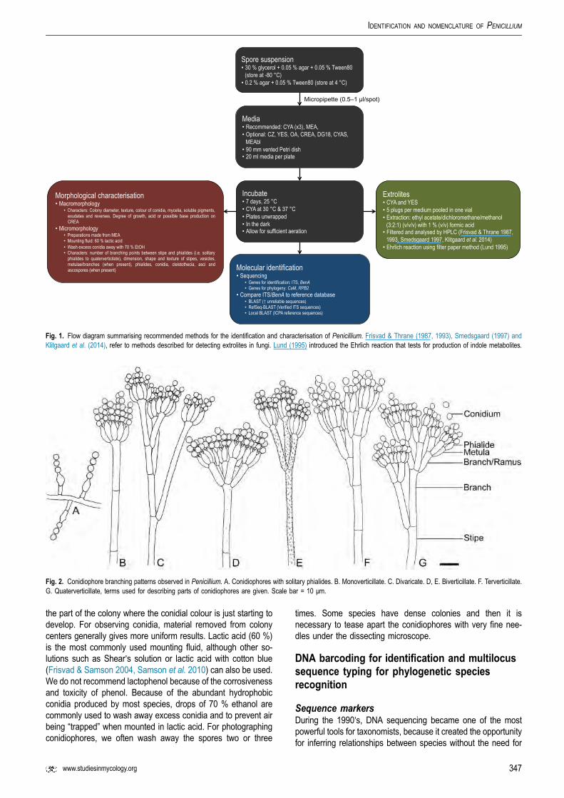
Morphology in the past has been central to the taxonomy ofPenicillium and along with multigene phylogenetics and extroliteprofiling comprises the polyphasic species concept adopted forPenicillium. Morphology is the physical architecture throughwhich an organism functions in and adapts to its environment,but some aspects may vary or be induced by specific cues in theimmediate environment. As a result, strains characterised in onelaboratory might look different when grown in another because ofsubtle differences in nutrients, temperature, lighting or humidity.This sometimes makes comparisons between different studiesvery difficult. These effects can be minimised using strictlystandardised working techniques for medium preparation, inoc-ulation technique and incubation conditions (Samson & Pitt 1985,Okuda 1994, Okuda et al. 2000). We recommend the followingstandardised methods for laboratories identifying and describingPenicillium species (summarised in Fig. 1).
MacromorphologyColony characters and diameters on specific media are impor-tant features for species identification. Czapek Yeast Autolysateagar (CYA) and Malt Extract agar (MEA, Oxoid) is recommendedas standard media for Penicillium. Even though malt extract fromOxoid is recommended, many laboratories prefer Difco. It shouldbe noted that two different MEA formulations are widely used inmodern taxonomic studies. Blakeslee's MEA was historicallywidely used, but lately CBS switched to a different formulation(given in Table 1). Both are suitable for characterisation, butstudies should state which MEA (Oxoid or Difco) and formulationwas used. The following alternative media can be used forobserving additional taxonomic characters: Czapek's agar (CZ),Yeast Extract Sucrose agar (YES), Oatmeal agar (OA), CreatineSucrose agar (CREA), Dichloran 18 % Glycerol agar (DG18),Blakeslee's MEA and CYA with 5 % NaCl (CYAS). CZ was used

Table 1. Media and stock solutions used for morphologicalcharacterisation.
Czapek stock solution (100 ml) (Pitt 1979)
NaNO3 NaNO3 30 g
KCl 5 g
MgSO4$7H2OMgSO4$7H2O 5 g
FeSO4$7H2OFeSO4$7H2O 0.1 g
dH2OdH2O 100 ml
*Store at 4–10 °C
Trace elements stock solution (100 ml)
CuSO4$5H2OCuSO4$5H2O 0.5 g
ZnSO4$7H2OZnSO4$7H2O 0.1 g
dH2OdH2O 100 ml
*Store at 4–10 °C
Blakeslee's Malt extract agar (MEAbl, Blakeslee 1915)
Malt extract (Oxoid) 20 g
Peptone (Oxoid) 1 g
Glucose 20 g
Trace elements 1 ml
Agar 20 g
dH2O 1000 ml
*Mix well and autoclave at 121 °C for 15 min. pH 5.3 ± 0.2.
Creatine sucrose agar (CREA, Frisvad 1981)
Sucrose 30 g
Creatine$1H2O 3 g
K3PO4$7H2O 1.6 g
MgSO4$7H2O 0.5 g
KCl 0.5 g
FeSO4$7H2O 0.01 g
Trace elements stock solution 1 ml
Bromocresol purple 0.05 g
Agar 20 g
dH2O 1000 ml
*Mix well and autoclave at 121 °C for 15 min. pH 8.0 ± 0.2.
Czapek's agar (CZ, Raper & Thom 1949)
Czapek concentrate 10 ml
Sucrose 30 g
Trace elements stock solution 1 ml
Agar 20 g
dH2O 1000 ml
*Mix well and autoclave at 121 °C for 15 min.
Czapek Yeast Autolysate agar (CYA, Pitt 1979)
Czapek concentrate 10 ml
Sucrose 30 g
Yeast extract (Difco) 5 g
K2HPO4 1 g
Trace elements stock solution 1 ml
Agar 20 g
dH2O 1000 ml
*Mix well and autoclave at 121 °C for 15 min. pH 6.2 ± 0.2.
Table 1. (Continued).
Czapek Yeast Autolysate agar with 5 % NaCl (CYAS)
Czapek concentrate 10 ml
Sucrose 30 g
Yeast extract (Difco) 5 g
K2HPO4K2HPO4 1 g
Trace elements stock solution 1 ml
NaCl 50 g
Agar 20 g
dH2OdH2O 1000 ml
*Mix well and autoclave at 121 °C for 15 min. pH 6.2 ± 0.2.
Dichloran 18 % Glycerol agar (DG18, Hocking & Pitt 1980)
Dichloran-Glycerol-agar-base (Oxoid) 31.5 g
Glycerol (anhydrous) 220 g
Trace elements stock solution 1 ml
Chloramphenicol 0.05 g
Agar 20 g
dH2O 1000 ml
*Mix well and autoclave at 121 °C for 15 min. After autoclaving, add 0.05chlortetracycline. pH 5.6 ± 0.2.
Malt Extract agar (MEA, Samson et al. 2010)
Malt extract (Oxoid CM0059) 50 g
Trace elements stock solution 1 ml
dH2O 1000 ml
*Mix well and autoclave at 115 °C for 10 min. pH 5.4 ± 0.2.
Oatmeal agar (OA, Samson et al. 2010)
Oatmeal / flakes 30 g
Trace elements stock solution 1 ml
Agar 20 g
dH2O 1000 ml
*First autoclave flakes (121 °C for 15 min) in 1000 ml dH2O. Squeeze mixturethrough cheese cloth and use flow through, topping up to 1000 ml with dH2Owith 20 g agar. Autoclave at 121 °C for 15 min. pH 6.5 ± 0.2.
Yeast extract sucrose agar (YES, Frisvad 1981)
Yeast extract (Difco) 20 g
Sucrose 150 g
MgSO4$7H2O 0.5 g
Trace elements stock solution 1 ml
Agar 20 g
dH2O 885 ml
*Mix well and autoclave at 121 °C for 15 min. pH 6.5 ± 0.2.
IDENTIFICATION AND NOMENCLATURE OF PENICILLIUM
www.studiesinmycology.org 345

VISAGIE ET AL.
in the taxonomic treatments of Raper & Thom (1949) andRamírez (1982) and is chemically well defined. However, it is notwidely used for Penicillium studies. YES is the recommendedmedium for extrolite profiling of species. Sexual reproductionmost commonly occurs when strains are grown on OA and thusoften provide valuable taxonomic information. OA should beprepared using organic uncooked flakes and not the unsuitablequick cook oats (“3 minute oats”) or prefabricated OA formula-tions available. Acid production is observed by the colour reac-tion in CREA (from purple to yellow) and is often useful fordistinguishing between closely related species. In some speciesacid production is followed by base production and is most oftenobserved from the colony reverse. DG18 and CYAS often pro-vide useful information with regards to growth rates on low wateractivity media. For consistent conidial colours, the addition ofzinc-sulphate and copper-sulphate as trace elements (1 gZnSO4.7H2O and 0.5 g CuSO4.5H2O in 100 ml distilled water) isof utmost importance because these metals vary widely in watersources in different locations and are critical for pigment pro-duction. For inorganic chemicals, analytical grade should beused. Experience has shown that the brand of agar used in-fluences colony appearance. As such, it is important to test theagar for consistent character development and note the brand inspecies descriptions. Even though we do not recommend abrand here as standard, after extensive comparisons CBS optedto use So-BI-Gel agar (Bie & Berntsen, BBB 100030) for mediumpreparations. Medium formulations are given in Table 1.
Media are prepared in 90 mm Petri-dishes with a volume of20 ml (Okuda et al. 2000). Glass Petri dishes was traditionallyconsidered best for character observation. However, today it isnot feasible using them, with polystyrene Petri dishes recom-mended. The Petri dishes should preferably be vented whichallows for a bit of air exchange. Inoculations are made from sporesuspensions in a semi-solid agar solution containing 0.2 % agarand 0.05 % Tween80 (Pitt 1979). In some laboratories, sporesuspensions are made in a 30 % glycerol, 0.05 % agar and0.05 % Tween80 solution. Variation between these two inocula-tion suspension solutions has not been shown. We recommendusing a micropipette for inoculating the spore suspension in three-point fashion (0.5–1 μl per spot). They should not be wrappedwith Parafilm, which by restricting air exchange often inhibitsgrowth and sporulation (Okuda et al. 2000). When using walk-inincubators, laboratory rules often require plates to be placed inplastic containers or plastic bags. In this case, the boxes or bagsshould allow for enough aeration and care should be taken not tohave too many plates in one box or in one incubator. For standardbench-top incubators, plates do not need to be incubated in boxesor bags, unless there is a strong air-current in the incubator. Allmedia are incubated at the standard temperature of 25 °C for 7 d,with additional CYA plates at 30 and 37 °C that are useful todistinguish between species. It is crucial that temperatures arecarefully checked as small differences have a large impact oncolony growth. No effect on colonies has been shown for incu-bating Petri dishes upside down or vice versa.
After 7 d, colony diameters are measured at the widest part ofthe colony. Important characters used for describing Penicilliuminclude colony texture, degree of sporulation, the colour ofconidia, the abundance, texture and colour of mycelia, thepresence and colours of soluble pigments and exudates, colonyreverse colours, and degree of growth and acid production (insome species acid production followed by base production) onCREA. The use of a standard colour chart is recommended for
346
descriptions. Although many are available, the most commonlyused colour chart in Penicillium is the Methuen Handbook ofColor (Kornerup & Wanscher 1967), which although long out ofprint is available in many libraries. It is worth noting that colournames used by Raper & Thom (1949) were based on Ridgeway(1912) and are also still in use. When colour charts are notavailable, it is recommended to publish full color photoplates toaccompany descriptions of new species. Some species producespecialised structures such as cleistothecia or sclerotia, mostlyafter longer incubation times, especially on OA. We thusrecommend OA plates be incubated for prolonged periods tohave a chance of observing these structures. For definitions andexplanations of terms used in descriptions of Penicillium col-onies, readers are referred to Frisvad & Samson (2004).
MicromorphologyConidiophore and cleistothecium (when produced) characters ofPenicillium are of great taxonomic importance. Conidiophorebranching patterns (Fig. 2) were traditionally used in the classifi-cation of Penicillium (Thom 1930, Raper & Thom 1949, Pitt 1979).Although these branching patterns do not correspond perfectlywith the sections currently accepted for Penicillium, characterisingthem accurately is still considered important. The conidiophoresrange from being simple (solitary phialides, Fig. 2A) to verycomplex patterns with multiple levels of branching resulting inoverall symmetrical or asymmetrical patterns. Monoverticillateconidiophores (Fig. 2B) have a terminal whorl of phialides and insome species, the terminal cell of the conidiophore is slightlyswollen or vesiculate; such species could be confused withdiminutive Aspergillus conidiophores, but they have septa in thestipes unlike species of the latter genus. Divaricate conidiophores,previously also referred to as irregular (Pitt 1979, Fig. 2C), are bestdescribed as having a simple to complex branching pattern withnumerous subterminal branches formed, but where conidiophoreparts are divergent. Biverticillate conidiophores (Fig. 2D, E) have awhorl of three or more metulae between the end of the stipe andthe phialides; the metulae may be of unequal or equal length, varyin their degree of divergence, are usually more or less cylindricalbut can also be clavate or slightly vesiculate. Terverticillate co-nidiophores (Fig. 2F) have another level of branching between thestipe and themetulae, often just a continuation of the stipe axis andone side branch, sometimes a true whorl of three or morebranches. Quaterverticillate conidiophores (Fig. 2G) are producedby only a few species, and have one extra level of branchingbeyond the terverticillate pattern. Terverticillate and quaterverti-cillate conidiophores tend to be conspicuously asymmetrical. Incolonies of many species, especially as cultures begin to degen-erate, there may be more than one branching pattern or inter-mediate forms, and it can be challenging to decide which pattern istypical or most developed.
Other important microscopic characters include the walltexture/ornamentation of stipes and conidia, as well as di-mensions, ornamentation and sometimes colours of all elementsof the conidiophore (Fig. 1). In our experience, wall textures arevery sensitive to minute differences in media composition andaeration. For best observation of conidial ornamentation, differ-ential interference contrast (= Nomarski) is recommended ifpossible; ornamentation is sometimes most conspicuouslyvisible in air pockets in the preparation.
For microscopic observations, slides are prepared from7–10 d old MEA colonies. Remove material from the zoneswhere adjacent colonies are closest or growing together, from
Spore suspension • 30 % glycerol + 0.05 % agar + 0.05 % Tween80
(store at -80 °C)• 0.2 % agar + 0.05 % Tween80 (store at 4 °C)
Media • Recommended: CYA (x3), MEA, • Optional: CZ, YES, OA, CREA, DG18, CYAS,
MEAbl• 90 mm vented Petri dish • 20 ml media per plate
Micropipette (0.5–1 μl/spot)
Incubate • 7 days, 25 °C • CYA at 30 °C & 37 °C • Plates unwrapped • In the dark • Allow for sufficient aeration
Morphological characterisation• Macromorphology
• Characters: Colony diameter, texture, colour of conidia, mycelia, soluble pigments, exudates and reverses. Degree of growth, acid or possible base production on CREA
• Micromorphology • Preparations made from MEA• Mounting fluid: 60 % lactic acid • Wash excess conidia away with 70 % EtOH• Characters: number of branching points between stipe and phialides (i.e. solitary
phialides to quaterverticilate), dimension, shape and texture of stipes, vesicles, metulae/branches (when present), phialides, conidia, cleistothecia, asci and ascospores (when present) Molecular identification
• Sequencing • Genes for identification: ITS, BenA• Genes for phylogeny: CaM, RPB2
• Compare ITS/BenA to reference database • BLAST ( unreliable sequences) • RefSeq-BLAST (Verified ITS sequences) • Local BLAST (ICPA reference sequences)
Extrolites • CYA and YES • 5 plugs per medium pooled in one vial • Extraction: ethyl acetate/dichloromethane/methanol
(3:2:1) (v/v/v) with 1 % (v/v) formic acid • Filtered and analysed by HPLC (Frisvad & Thrane 1987,
1993, Smedsgaard 1997, Klitgaard et al. 2014) • Ehrlich reaction using filter paper method (Lund 1995)
Fig. 1. Flow diagram summarising recommended methods for the identification and characterisation of Penicillium. Frisvad & Thrane (1987, 1993), Smedsgaard (1997) andKlitgaard et al. (2014), refer to methods described for detecting extrolites in fungi. Lund (1995) introduced the Ehrlich reaction that tests for production of indole metabolites.
Fig. 2. Conidiophore branching patterns observed in Penicillium. A. Conidiophores with solitary phialides. B. Monoverticillate. C. Divaricate. D, E. Biverticillate. F. Terverticillate.G. Quaterverticillate, terms used for describing parts of conidiophores are given. Scale bar = 10 μm.
IDENTIFICATION AND NOMENCLATURE OF PENICILLIUM
the part of the colony where the conidial colour is just starting todevelop. For observing conidia, material removed from colonycenters generally gives more uniform results. Lactic acid (60 %)is the most commonly used mounting fluid, although other so-lutions such as Shear's solution or lactic acid with cotton blue(Frisvad & Samson 2004, Samson et al. 2010) can also be used.We do not recommend lactophenol because of the corrosivenessand toxicity of phenol. Because of the abundant hydrophobicconidia produced by most species, drops of 70 % ethanol arecommonly used to wash away excess conidia and to prevent airbeing “trapped” when mounted in lactic acid. For photographingconidiophores, we often wash away the spores two or three
www.studiesinmycology.org
times. Some species have dense colonies and then it isnecessary to tease apart the conidiophores with very fine nee-dles under the dissecting microscope.
DNA barcoding for identification and multilocussequence typing for phylogenetic speciesrecognition
Sequence markersDuring the 1990's, DNA sequencing became one of the mostpowerful tools for taxonomists, because it created the opportunityfor inferring relationships between species without the need for
347

VISAGIE ET AL.
standardising culturing regimes and eliminated problems relatedto deteriorated cultures. It also created the opportunity forsequence based identifications. DNA barcoding was launched tomake species identification of any eukaryotic organism possiblefor anybody, by using a standardised short DNA sequence and acurated reference database linked to authoritatively identifiedvouchers (Blaxter 2003, Tautz et al. 2003, Hebert et al. 2003,Blaxter et al. 2005, DeSalle et al. 2005, Ratnasingham &Hebert 2007, Seifert et al. 2007, Min & Hickey 2007a,b,Schoch et al. 2012). Only recently however, was the internaltranscribed spacer rDNA area (ITS) accepted as the officialbarcode for fungi (Schoch et al. 2012). ITS is the most widelysequenced marker for fungi, and universal primers are available(Schoch et al. 2012). In Penicillium, it works well for placingstrains into a species complex or one of the 25 sections, andsometimes provides a species identification (Visagie et al.2014a). Unfortunately, for Penicillium and many other generaof ascomycetes, the ITS is not variable enough for distinguishingall closely related species (Skouboe et al. 1999, Seifert et al.2007, Schoch et al. 2012). The open source sequence re-pository GenBank contains a large proportion of incorrectlyidentified sequences, making identifications of Penicillium usingBLAST very tricky for inexperienced workers. This particularproblem is addressed in a number of publications (K~oljalg et al.2005, Santamaria et al. 2012, K~oljalg et al. 2013, Schoch et al.2014). For Penicillium, the International Commission of Penicil-lium and Aspergillus (ICPA), in conjunction with the publication ofan updated accepted species list presented below, decided toinclude GenBank accession numbers to reference barcode se-quences for each species when available.
Because of the limitations associated with ITS as a speciesmarker in Penicillium, a secondary barcode or identificationmarker is often needed for identifying isolates to species level.The requirements for a secondary identification marker are clear.It should be easy to amplify, distinguish among closely relatedspecies and most importantly, the reference data set should becomplete, meaning that there should be representative se-quences for all species. It would be an added bonus if thismarker is useful for phylogenetic studies, as it will by defaultbecome the gene most widely sequenced in future. Based onthese criteria, we propose the use of β-tubulin (BenA) as the bestoption for a secondary identification marker for Penicillium. BenAdoes, however, have problems associated with it. Although notinfluencing BLAST identifications, alignments across a diversegenus like Penicillium is difficult and often contains a largeproportion ambiguously aligned sites, which can make phylog-enies difficult. Also, there is evidence for the amplification ofBenA paralogous genes in Aspergillus (Peterson 2008, Hubka &Kolarik 2012) and Talaromyces (Peterson & Jurjevi�c 2013) and itcan thus be assumed that the same might be happening in somePenicillium species, although this has not been shown. Otherpossible secondary marker options include calmodulin (CaM) orthe RNA polymerase II second largest subunit (RPB2) genes.Both these genes have similar discriminatory power as BenA.RPB2 has the added advantage of lacking introns in the ampli-con, allowing robust and easy alignments when used for phy-logenies, but it is sometimes difficult to amplify and the databaseis incomplete. Similarly, we lack a complete CaM database.Thus, for routine identifications BenA is currently recommended,while for the description of new species, we suggest the use ofITS, BenA, CaM and RPB2 among the markers for multilocus
348
sequence typing and GCPSR. We consider it good practise toinclude at least ITS and BenA sequences when describing newspecies, to allow others to more easily recognise the newspecies.
BenA can successfully be used for accurately identifyingPenicillium species. However, as is the case for genes otherthan BenA, care should be taken in specific groups or situa-tions. Infraspecies variation in BenA occurs in some Penicilliumspecies as is observed in phylogenies published for Penicillium(Frisvad & Samson 2004, Barreto et al. 2011, Peterson et al.2011, Houbraken et al. 2011b,c, Rivera & Seifert 2011,Rivera et al. 2012, Houbraken et al. 2012a, Visagie et al.2013, 2014a, 2014b). This variation must be considered foridentification purposes and especially when consideringwhether a strain might represent a new species. This meansthat in addition to the reference sequences of ex-type culturessanctioned by ICPA, additional reference sequences arenecessary to document sequence variation that differ from theex-type. ICPA is currently working on populating such a vali-dated database to capture infraspecies variation, but for thetime being critical phylogenetic revisions of different sectionsshould be referred to for reliable data. Alternatively, combiningITS, BenA, CaM and RPB2 from a suspected new species withsequences of the same markers from related species will aid indeciding whether a species is new or not, using GCPSR asexplained in detail by Taylor et al. (2000). This is in factcommon practise in most studies describing and characterisingPenicillium species.
β-tubulin as a secondary marker in practiseAs discussed above, BenA works well for species identificationsin Penicillium. Penicillium chrysogenum and P. allii-sativi is oneexample where identification based on BenA should be madewith care (Houbraken et al. 2012a). This is a consequence of thevariation observed among strains of P. chrysogenum, which re-sults in P. allii-sativi forming a clade within the P. chrysogenumclade in BenA species trees. Even though CaM distinguishesP. chrysogenum from P. allii-sativi, it does not distinguishP. chrysogenum and P. rubens. As a result, molecular identifi-cations should be made with great care in section Chrysogena,with the revision by Houbraken et al. (2012a) used as guide.Penicillium kongii was recently introduced as a close relative ofP. brevicompactum (Wang & Wang 2013). Even though thisspecies formed a coherent clade distinct from theP. brevicompactum ex-type strain, on examining additionalstrains previously identified as P. brevicompactum, the P. kongiiclade is resolved within the P. brevicompactum clade. This cladethus requires more work to resolve the species from the com-plex. A similar situation is observed for P. desertorum and thenewly described P. glycyrrhizacola (Chen et al. 2013), wheremore strains and additional genes included in the phylogeny willhelp to resolve species. Another species that cannot be identifiedusing molecular data is P. camemberti, P. caseifulvum andP. commune, which shares identical BenA and other gene se-quences (Giraud et al. 2010). Although these could be consid-ered taxonomic synonyms, their different applications andimportance in the cheese industry suggest that it is of more valueto keep them as separate species. Morphological identification isnecessary in this case, with P. camemberti having white conidia,P. caseifulvum having an orange reverse on YES andP. commune producing green conidia (Frisvad & Samson 2004).

Table 2. Primers used for amplification and sequencing.
Locus Primer name Direction Primer sequence (5′-3′) Reference
Internal Transcribed Spacer (ITS) ITS1 Forward TCC GTA GGT GAA CCT GCG G White et al. 1990
ITS4 Reverse TCC TCC GCT TAT TGA TAT GC White et al. 1990
V9G Forward TTA CGT CCC TGC CCT TTG TA de Hoog & Gerrits van den Ende 1998
LS266 Reverse GCA TTC CCA AAC AAC TCG ACT C Masclaux et al. 1995
β-tubulin (BenA) Bt2a Forward GGT AAC CAA ATC GGT GCT GCT TTC Glass & Donaldson 1995
Bt2b Reverse ACC CTC AGT GTA GTG ACC CTT GGC Glass & Donaldson 1995
Calmodulin (CaM) CMD5 Forward CCG AGT ACA AGG ARG CCT TC Hong et al. 2006
CMD6 Reverse CCG ATR GAG GTC ATR ACG TGG Hong et al. 2006
CF1 Forward GCC GAC TCT TTG ACY GAR GAR Peterson et al. 2005
CF4 Reverse TTT YTG CAT CAT RAG YTG GAC Peterson et al. 2005
RNA polymerase II secondlargest subunit (RPB2)
5F Forward GAY GAY MGW GAT CAY TTY GG Liu et al. 1999
7CR Reverse CCC ATR GCT TGY TTR CCC AT Liu et al. 1999
5Feur Forward GAY GAY CGK GAY CAY TTC GG Houbraken et al. 2012b
7CReur Reverse CCC ATR GCY TGY TTR CCC AT Houbraken et al. 2012b
IDENTIFICATION AND NOMENCLATURE OF PENICILLIUM
Amplification and identificationPrimers used for amplification of the ITS, BenA, CaM and RPB2genes are included in Table 2 and amplification profiles are givenin Table 3. A standard thermal cycle with an annealing temper-ature of 55 °C is generally used. Sometimes amplification suc-cess is low for CaM, especially in sections Canescentia andRamosa. In this case, dropping the annealing temperature to52 °C gives good results. The RPB2 amplification is morecomplicated. We recommend using a touch-up PCR(50–52–55 °C) with primer pair 5Feur and 7CReur for bestamplification. When amplification is problematic, the alternativetouch-up PCR (48–50–52 °C) profile can be used and/or thealternative primer pair 5F and 7CR.
After sequences are obtained, there is a number of ways touse them to identify the strain. The most widely used method isthe BLAST search on NCBI. Using this method, one is able tosearch all sequences in the database, but as noted above, thereare many unidentified and misidentified sequences in the data-base. For ITS the RefSeq data set, accessible from the NCBIhomepage (http://www.ncbi.nlm.nih.gov/refseq/), is now availableand can be used to query ITS sequences against a verified ITSdatabase (Schoch et al. 2014). Unfortunately, the RefSeq data-base does not cover alternative genes. For this, we suggestsetting up Local BLAST files and using the verified BenA data-base provided here.
Table 3. Thermal cycle programs used for amplification.
Gene Profile type Initialdenaturing
Cycles Denat
General ITS, BenA, CaM standard 94 °C, 5 min 35 94 °C, 4
General alternative standard 94 °C, 5 min 35 94 °C, 4
RPB2 touch-up 94 °C, 5 min 5 94 °C, 4
5 94 °C, 4
30 94 °C, 4
RPB2 alt. touch-up 94 °C, 5 min 5 94 °C, 4
5 94 °C, 4
30 94 °C, 4
www.studiesinmycology.org
Extrolite data
Extrolites are produced by the mycelium and sporulating struc-tures of Penicillium species, and exudates, diffusible pigments,and reverse colours are also mixtures of secondary metabolites.Studies of extrolite profiles were very useful for unravelling somemorphological species concepts into biologically meaningfulsegregate species before DNA sequencing provided similarpossibilities. As an example, the P. aurantiogriseum complex,critical contaminants of grain, was divided first into species usingextrolite profiles by Frisvad & Filtenborg (1983, 1989), delim-itations that were subsequently supported by BenA sequencing(Seifert & Louis-Seize 2000).
Identification of unknown strains using extrolites is possiblefor well-equipped chemical laboratories. The best way of usingextrolites as identification aids is to extract and then separatethem by HPLC and then partially or fully identify as many of thesecondary metabolites as possible, generally using massspectroscopy based technology (Frisvad et al. 2008). The mediaused for identification, especially CYA and YES agars, areoptimal for production of most major diagnostic extrolites inPenicillium after incubation for 7 d at 25 °C in darkness. Agarplugs are extracted with a mixture of dichloromethane, ethyl-acetate and methanol. The metabolites extracted can then beanalysed using advanced separation and detection techniques,
uring Annealing Elongation Finalelongation
Rest period
5 s 55 °C, 45 s 72 °C, 60 s 72 °C, 7 min 10 °C, ∞
5 s 52 °C, 45 s 72 °C, 60 s 72 °C, 7 min 10 °C, ∞
5 s 50 °C, 45 s 72 °C, 60 s
5 s 52 °C, 45 s 72 °C, 60 s
5 s 55 °C, 45 s 72 °C, 60 s 72 °C, 7 min 10 °C, ∞
5 s 48 °C, 45 s 72 °C, 60 s
5 s 50 °C, 45 s 72 °C, 60 s
5 s 52 °C, 45 s 72 °C, 60 s 72 °C, 7 min 10 °C, ∞
349

VISAGIE ET AL.
for example ultra high performance liquid chromatography withdiode array detection and high resolution mass spectrometricdetection (UHPLC-DAD-HRMS) (Kildgaard et al. 2014, Klitgaardet al. 2014). However, simpler HPLC-diode array detectionmethods can also be used (Frisvad & Thrane 1987, 1993).
Laboratories with less sophisticated equipment can performvaluable confirmatory tests. Although Thin Layer Chromatography(TLC) is no longer considered state of the art for chemicalresearch, it is still a useful technique for detecting coloured oruncoloured extrolites that can be used to confirm the identificationof a Penicillium strain. For example, P. brevicompactum consis-tently produces large amounts of the colourless mycophenolicacid, and this metabolite will make a green colour reaction withferric chloride (Clutterbuck & Raistrick 1933).
Sometimes only a few extrolites are needed to confirm theidentity of a Penicillium isolate. If an isolate produces griseo-fulvin and roquefortine C, it can only be P. coprophilum,P. griseofulvum or P. sclerotigenum and if the isolate also pro-duces cyclopiazonic acid, it can only be P. griseofulvum (Frisvad& Samson 2004).
Identification based purely on secondary metabolites is notyet possible for all species of Penicillium, but future databaseswill be better developed and an optimal battery of media forsecondary metabolite production may allow this method to beused for identification of all species in the future.
Misidentifications in PenicilliumA few examples demonstrate why Penicillium identification isdifficult or how misleading results might be obtained in practiseusing morphology or sequencing. For example, a fungus pro-ducing penicillones A and B and chloctanspirones A, B andterrestrols was identified as P. terrestre (patent strain CCTCC M204077, strain unavailable) (Liu et al. 2005). The nameP. terrestre was used by Raper & Thom (1949), but has sincebeen considered a synonym of P. crustosum. However, addi-tional secondary metabolites produced by the isolate, such assorbicillin and trichodimerol, are never seen in P. crustosum(Frisvad et al. 2004), suggesting the strain was P. chrysogenumor P. rubens. Liu et al. (2005) did not describe how their isolatewas identified. Another strain of P. chrysogenum or P. rubens,SD-118 (HQ652873), was misidentified as a P. commune (Shanget al. 2012, Zhao et al. 2012), based on “100 % sequenceidentity” with P. commune (FJ499541). The production ofchrysogine, sorbicillins and meleagrin indicated that the strain,SD-118, was in fact P. chrysogenum. We propose that the use ofthe reference sequence databases provided here, will minimisethese type of misidentifications.
Matrix-assisted laser desorption/ionization time-of-flight mass spectrometry fingerprinting(MALDI-TOF MS)
Another detection technique receiving a lot of attention, especiallyfrom the medical field, is matrix-assisted laser desorption/ioniza-tion time-of-flight mass spectrometry fingerprinting (MALDI-TOFMS). It was successfully applied to the identification of bacteria(Hettick et al. 2006, Siegrist et al. 2007) and yeast species (Amiri-Eliasi & Fenselau 2001, Kolecka et al. 2013). Only a limitednumber of studies included or focused on Penicillium (Welhamet al. 2000, Hettick et al. 2008, Del Chierico et al. 2012,Chalupov�a et al. 2014) and the closely related Aspergillus (Bille
350
et al. 2011, De Carolis et al. 2011, Iriart et al. 2012, Verwer et al.2013). Although these studies promisingly report that MALDI-TOF MS distinguish between species, a large degree of varia-tion is observed within a species, even between duplicates of thesame strain (Hettick et al. 2008). This means that data from a highnumber of strains will have to be included in the database to makerobust identifications feasible. Difficulties with identifications arealso reported in Aspergillus, where not all strains could be iden-tified with 100 % accuracy (Iriart et al. 2012, Verwer et al. 2013).Thus we can conclude that although this technique shows prom-ise, a lot of work remains to make routine identifications feasible.
TAXONOMY
Phylogenetic analyses revealed that Hemicarpenteles paradoxusshould not be classified in Aspergillus, and together with its closestrelatives A. crystallinus and A. malodoratus, should be transferredtoPenicillium (Peterson 2000a, 2008, Houbraken&Samson 2011).Houbraken & Samson (2011) included them in Penicillium sect.Paradoxa together with P. atramentosum. The other Hemi-carpenteles species were shown to belong in Aspergillus sect.Clavati (H. acanthosporus ≡ A. acanthosporus ≡ Neocarpentelesacanthosporus (Udagawa&Takada) Udagawa &Uchiy.) (Peterson2000b, Tamura et al. 2000, Varga et al. 2007, Peterson 2008), whileH. ornatus and H. thaxteri are classified in Sclerocleista (Pitt et al.2000, Houbraken & Samson 2011).
Aspergillus paradoxus was described by Fennell & Raper(1955) for strains producing small clavate vesicles suggestiveof those observed in aberrant strains of A. clavatus. Later, Raper& Fennell (1965) placed A. paradoxus in the A. ornatus speciesgroup, based on the resemblance of its sclerotia to youngcleistothecia of A. ornatus and A. citrisporus. Rai et al. (1964,1967) also isolated A. paradoxus from soil in India, and foundthat conditions favouring sclerotia production also inhibited theformation of conidial heads and the yellow pigment in the me-dium. Sarbhoy & Elphick (1968) observed that a strain ofA. paradoxus (CBS 793.68 = IMI 117502), isolated from dogdung in the United Kingdom, produced mature unilocular cleis-tothecia with tough peridial walls, embedding smooth-walledlenticular ascospores. They introduced the name Hemi-carpenteles for the sexual morph, with H. paradoxus as type.Houbraken & Samson (2011) noted that the sexual morph mostclosely resembles Eupenicillium, both producing sclerotoidcleistothecia that ripen from the centre outwards (Sarbhoy &Elphick 1968, Pitt 1979, Stolk & Samson 1983). In our freshisolates, some strains produce sclerotia abundantly while othershave only a few sclerotia. Only two fresh isolates producedascospores after incubation at 25 °C for three weeks on OA.Aspergillus paradoxus, A. crystallinus and A. malodoratus (Figs3–6) have a striking similarity with members of section Olsoniiand Coronata (Frisvad & Samson 2004, Samson et al. 2004), inparticular the long, wide, smooth-walled conidiophores termi-nating in densely branched penicilli with metulae and phialides.Particularly P. olsonii bears a morphological resemblance withA. paradoxus and A. crystallinus.
Extrolite data also suggest that A. paradoxus, A. crystallinusand A. malodoratus belong in Penicillium (summarised inTable 4). Pseurotins are found in both Penicillium and Aspergillusspecies (Frisvad et al. 2004). Sorbicillins were previously onlyconfirmed in P. rubens and P. chrysogenum, and not so far in

Fig. 3. Penicillium paradoxum. A. Colonies: top row left to right, obverse CYA, YES, DG18 and MEA; bottom row left to right, reverse CYA, reverse YES, reverse DG18 andCREA. B. Young sclerotia. C. Phototropic conidiophores after two weaks growth. D–H. Conidiophores. I. Conidia. Scale bars: D– I = 10 μm.
IDENTIFICATION AND NOMENCLATURE OF PENICILLIUM
www.studiesinmycology.org 351

Fig. 4. Penicillium paradoxum sexual reproduction. A–C. Cleistothecia. D, E. Asci. F. Ascospores. Scale bars: A, B = 500 μm; C–F = 10 μm.
VISAGIE ET AL.
Aspergillus, although Basaran & Demirbas (2010) reportedsorbicillin production in A. parasiticus; we have never observedsorbicillins in any A. parasiticus strain. Penicillium atramento-sum, also classified in section Paradoxa, produces andrastin A,atpenins, meleagrin and oxaline, roquefortine C & D and rugu-lovasine A & B (Frisvad et al. 2004, and reported here). AmongPenicillium and Aspergillus species, the atpenins have only beenfound in P. atramentosum (Omura et al. 1988, Kawada et al.2009), but the other listed extrolites are common in species ofPenicillium sections Paradoxa and Chrysogena. Rugulovasinsare an exception and have only been found in P. commune(Frisvad et al. 2004). Aspergillus paradoxus produces brefeldin,an extrolite also reported in P. brefeldianum (Hutchinson et al.1983), but never in Aspergillus apart from a report forA. clavatus (Wang et al. 2002) that could not be confirmed (Vargaet al. 2007). The latter strain was probably actually A. ingratus,first regarded as belonging to Aspergillus section Clavati(Yaguchi et al. 1993) but now considered a synonym ofA. paradoxus. Xanthocillins are produced by A. paradoxusstrains, and also by P. chrysogenum and P. egyptiacum.Meleagrin is commonly produced in Penicillium sections Para-doxa and Chrysogena, and species of several other sections(Frisvad et al. 2004). Only the closely related extrolite, neoxaline,has been reported in A. japonicus (Hirano et al. 1979). Asper-gillus crystallinus produces chrysogine, typical of P. rubens andP. chrysogenum (Frisvad et al. 2004, Houbraken et al. 2012a),and a compound also reported in A. nomius (Varga et al. 2011).Chrysophanic acid, produced in A. crystallinus, was found inTalaromyces islandicus (Howard & Raistrick 1950), and pachy-basin was found in P. vulpinum (Frisvad et al. 2004). Aspergillusmalodoratus produces andrastin A, meleagrin and oxaline.
352
Andrastin A is produced by several Penicillium species includingP. rubens and P. chrysogenum (Houbraken et al. 2011a, 2012a),but has not been found in Aspergillus. On balance, chemotax-onomic evidence points to the placement of A. paradoxus,A. crystallinus and A. malodoratus in Penicillium.
From the phenotypic, molecular and extrolite data we canconclude that the three species formerly placed in Aspergillusbelong to Penicillium. Consequently we propose the followingnew combinations:
Penicillium paradoxum (Fennell & Raper) Samson,Houbraken, Visagie & Frisvad, comb. nov. MycoBankMB547045. Figs 3, 4.Basionym: Aspergillus paradoxus Fennell & Raper, Mycologia47: 69. 1955. MycoBank MB292853
≡ Hemicarpenteles paradoxus Sarbhoy & Elphick, Trans. Brit. Mycol.Soc. 51: 156. 1968. MycoBank MB265229= Aspergillus ingratus Yaguchi, Someya & Udagawa, Trans. Mycol.Soc. Japan 34: 305. 1993. MycoBank MB361187[Non Trichocoma paradoxa Jungh., Praemissa in floram cryptogamicamJavae insulae: 9. 1838. MycoBank MB161024]
Typus: New Zealand, Wellington, dung of opossum, 1948, iso-lated by J.H. Warcup, Neotype IMI 061446, culture ex-type CBS527.65 = NRRL 2162 = ATCC 16918 = IMI 061446
Penicillium paradoxum (and P. malodoratum) deviate fromother Penicillium species by being phototropic (Raper & Fennell1965) and occasionally having a well-developed vesicle thatmakes the conidophore appears like an aspergillum (illustrated byYaguchi et al. 1993 in the protologue of Aspergillus ingratus).

Fig. 5. Penicillium crystallinum. A. Colonies: top row left to right, obverse CYA, YES, DG18 and MEA; bottom row left to right, reverse CYA, reverse YES, reverse DG18 andCREA. B. Colony texture on MEA. C–G. Conidiophores. H. Conidia. Scale bars: C–H = 10 μm.
IDENTIFICATION AND NOMENCLATURE OF PENICILLIUM
www.studiesinmycology.org 353
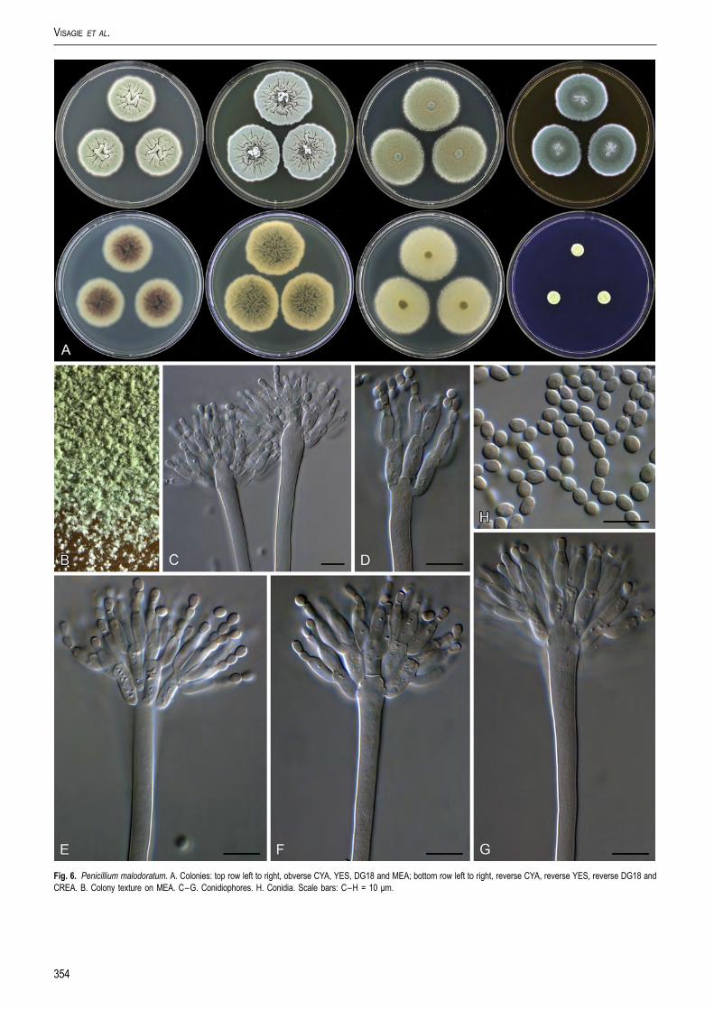
Fig. 6. Penicillium malodoratum. A. Colonies: top row left to right, obverse CYA, YES, DG18 and MEA; bottom row left to right, reverse CYA, reverse YES, reverse DG18 andCREA. B. Colony texture on MEA. C–G. Conidiophores. H. Conidia. Scale bars: C–H = 10 μm.
VISAGIE ET AL.
354
Table 4. Origin and extrolites of the strains examined in this study.
Species Strain Origin Extrolites
A. crystallinus CBS 479.65T = IBT 21947 Forest soil, Tilaran, Costa Rica chrysophanic acid, chrysogine, meleagrin, pachybasin
A. malodoratus CBS 490.65T = IBT 21948 Forest soil, Barranca, Costa Rica andrastin A, oxaline, meleagrin
A. paradoxus CBS 123898 = IBT 29765 = DTO 76F5 Dung of dog, the Netherlands “astyl”, brefeldin A, “SLOF”
CBS 123898 = IBT 29768 = DTO 76F6 Dung of dog, the Netherlands “astyl”, brefeldin A, pseurotin, “SLOF”, sorbicillins
CBS 527.65T = IBT 19547 Dung of opossum, New Zealand “astyl”, brefeldin A, pseurotin, “SLOF”, sorbicillins
CBS 643.95 = IBT 17513 = PF 1116 Soil, USA, California (ex-type of A. ingratus) “asyl”, brefeldin A, pseurotin, “SLOF”, sorbicillins
CBS 793.68 = IBT 19572 Dung of dog, UK “astyl”, brefeldin A, “PAS”, pseurotin, “SLOF”, sorbicillins
CBS 793.91 = IBT 12297 Dung of dog, Denmark brefeldin A, “PAS”, pseurotin, “SLOF”, sorbicillins
IBT 29766 = DTO 76F7 Dung of dog, the Netherlands brefeldin A, pseurotin, “SLOF”, sorbicillins
IMI 173765 = IBT 19364 Jet fuel, New Zealand “astyl”, brefeldin A, pseurotin, sorbicillins
IMI 320026 = IBT 19365 Air, UK brefeldin A, pseurotin, sorbicillins
IDENTIFICATION AND NOMENCLATURE OF PENICILLIUM
However, stipes were described as small, thin-walled and evenseptate, and the illustration by Raper & Fennell (1965) indicates aconidial head not unlike P. crystallinum and P. malodoratum. Thelatter authors also noted that A. crystallinus and A. malodoratusoccasionally produced triseriate “sterigmata” and “conidialstructures, particularly when small, tend to resemble those of thegenus Penicillium”. Most isolates of P. paradoxum are apparentlyfrom dung. Penicillium paradoxum, P. malodoratus andP. crystallinum produce strong penetrating odours, similar to thoseknown from coprophilous species in Penicillium series Clav-iformia, apparently an adaptation to the dung habitat.
Penicillium crystallinum (Kwon-Chung & Fennell)Samson, Houbraken, Visagie & Frisvad, comb. nov.MycoBank MB809315. Fig. 5.Basionym: Aspergillus crystallinus Kwon-Chung & Fennell, TheGenus Aspergillus: 471. 1965. MycoBank MB326624
Typus: Costa Rica, Province of Guanacaste, Tilaran, forest soil,1962, isolated by K.J. Kwon & D.I. Fennell, Neotype IMI 139270,culture ex-type CBS 479.65 = NRRL 5082 = ATCC 16833 = IMI139270
Penicillium crystallinum is only known from its ex-type strain(CBS 479.65 = NRRL 5082 = ATCC 16833 = IMI 139270),isolated from forest soil in Costa Rica. The strain shows typicalsporulation, but the soft compacted masses of thin-walled cellssuggesting sclerotia described by Raper & Fennell (1965) werenot observed. The yellow needle crystals of pachybasinobserved in the type strain are no longer conspicuously present,but the extrolite profile shows that the strain still produces thiscompound. The strain also produces the pungent odour noted inP. paradoxum and P. malodoratum.
Penicillium malodoratum (Kwon-Chung & Fennell)Samson, Houbraken, Visagie & Frisvad, comb. nov.MycoBank MB809316. Fig. 6.Basionym: Aspergillus malodoratus Kwon-Chung & Fennell, Thegenus Aspergillus: 468. 1965. MycoBank MB326644
Typus: Costa Rica, Province of Puntarenas, Barranca, forestsoil, 1962, isolated by K.J. Kwon & D.I. Fennell, Neotype IMI
www.studiesinmycology.org
172289, culture ex-type CBS 490.65 = NRRL 5083 = IMI172289 = ATCC 16834
Penicillium malodoratum is only known from its ex-type strain(CBS 490.65 = NRRL 5083 = IMI 172289 = ATCC 16834),isolated from forest soil in Costa Rica. Colonies containnumerous long, phototropic conidiophores. Raper & Fennell(1965) observed abundant soft sclerotium-like bodies withthick-walled cells, but we could not detect them. As the namesuggests, this fungus produces an unpleasant odour on all mediatested. Although A. malodoratus and A. crystallinus were isolatedfrom forest soil in Costa Rica and have similar morphologicalcharacters, the taxa can be distinguished by the colony diameter,the length of the conidiophores and the size and shape of theconidia. Conidia of A. crystallinus are globose, 4–7 μm, echi-nulate, while those of A. malodoratus are subglobose to ellip-soidal, 3–3.5 × 3.5–4 μm and mainly smooth.
PROPOSED LIST OF ACCEPTED SPECIES INTHE GENUS PENICILLIUM
The following list includes species names that are accepted inthe genus Penicillium on 8 August 2014. This list is updated fromprevious lists published by Pitt & Samson (1993) and Pitt et al.(2000). This revision was considered necessary followingchanges made to the ICN and the move to single namenomenclature (McNeill et al. 2012), the large number of speciesdescribed since the 2000 list and new taxonomic informationprovided by molecular data.
The most dramatic changes in Penicillium are the incorpo-ration of Eupenicillium and several other genera as synonyms(Houbraken & Samson 2011), and the transfer of species of theformer Penicillium subgenus Biverticillium species to Talar-omyces (Samson et al. 2011). Aspergillus paradoxus,A. crystallinus and A. malodoratus were shown to belong toPenicillium and were transferred above. Several speciesdescribed as Penicillium belongs to other genera and not toPenicillium (Houbraken & Samson 2011). They are listed belowthe accepted species list.
Even though the updating of the accepted species list beganfor nomenclatural purposes, we aim to make this list more
355

VISAGIE ET AL.
functional by including additional information linked to the spe-cies names. The list thus includes MycoBank numbers wherecomplete nomenclatural data can be obtained, collectionnumbers of ex-type strains for future taxonomists requiringauthenticated reference material, the species current sectionalclassification, and GenBank accession numbers for ITS barc-odes and, where available, alternative molecular identificationmarkers for BenA, CaM and RPB2.
Despite the considerable amount of time and effort spent oncompleting this list, there is the possibility of errors or oversights.As such we solicit and gratefully accept any comments onmissing names, errors or new data that has become available,especially when publishing a new species. We would alsoappreciate suggestions for making the list more useful. Theactive version of the list is currently hosted on the ICPA website(http://www.aspergilluspenicillium.org) where it will be updated asnew information comes to light. The website contains a portal forcomments, for the convenience of our users. The list publishedhere contains only accepted species; the online version will infuture also include data for synonyms. Similar lists are availablefor Aspergillus (Samson et al. 2014) and Talaromyces (Yilmazet al. 2014).
Penicillium Link, Mag. Ges. Naturf. Freunde Berlin 3: 16.1809. MycoBank MB9257.
356
= Coremium Link, Mag. Ges. Naturf. Freunde Berlin 3: 19. 1809, fideRaper & Thom 1949, Seifert & Samson 1985. [MB7782]. anamorphicsynonym.= Floccaria Grev., Scott. crypt. fl.: pl. 301. 1827, fide Seifert & Samson1985. [MB8260]. anamorphic synonym.?= Hormodendrum Bonord., Handbuch allg. Mykol.: 76. 1851 fide deHoog & Hermanides-Nijhof 1977, but see Hughes 1958. [MB8558].anamorphic synonym.?= Walzia Sorokin, Trudy Obshch. Ispyt. Prir. Imp. Kharkov: 47. 1871,fide Constantin 1888. [MB10429]. anamorphic synonym.= Pritzeliella Henn., Hedwigia Beibl.42: 88. 1903, fide Clements & Shear1931. [MB9529]. anamorphic synonym.= Eupenicillium F. Ludw., Lehrbuch der Niederen Kryptogamen: 263.1892, fide Houbraken & Samson 2011. [MB1933]. teleomorphicsynonym.= Citromyces Wehmer, Ber. Deutsch. Bot. Ges. 11: 333. 1893, fideThom 1930. [MB7672]. anamorphic synonym.= Aspergillopsis Sopp, Skr. Vidensk.-Selsk. Christiana Math.-Nat. Kl. 11:204. 1912, non Aspergillopsis Speg. 1910, fide Pitt 1979. [MB22043].anamorphic synonym.= Carpenteles Langeron, Crypt. Fr. Exs.: 344. 1922, fide Stolk & Scott1967. [MB826]. teleomorphic synonym (= Eupenicillium).= Torulomyces Delitsch, Systematik der Schimmelpilze: 91. 1943, fideStolk & Samson 1983 and Houbraken & Samson 2011. [MB10252].anamorphic synonym.= Eladia Smith, Trans. Brit. Mycol. Soc. 44: 47. 1961, fide Samson1981, Houbraken & Samson 2011. [MB8134]. anamorphic synonym.= Thysanophora Kendrick, Can. J. Bot. 39: 820. 1961, fide Houbraken &Samson 2011. [MB10230]. anamorphic synonym.= Hemicarpenteles Sarbhoy & Elphick, Trans. Brit. Mycol. Soc. 51: 156.1968, fide Houbraken & Samson 2011. [MB2279]. teleomorphicsynonym.= Penicillium Link ex Gray sensu Pitt, The Genus Penicillium: 154. 1979(nom. inval., art 13e). anamorphic synonym.= Chromocleista Yaguchi & Udagawa, Trans. Mycol. Soc. Japan 34:101. 1993, fide Houbraken & Samson 2011. [MB25855]. teleomorphicsynonym.
Penicillium abidjanum Stolk, Antonie van Leeuwenhoek 34: 49.1968 ≡ Eupenicillium abidjanum Stolk, Antonie van Leeuwenhoek 34: 49.1968. [MB335705]. — Herb.: CBS 246.67. Ex-type: CBS 246.67 = ATCC18385 = FRR 1156 = IMI 136244. Section Lanata-Divaricata. ITS barcode:GU981582. (Alternative markers: BenA = GU981650; RPB2 = JN121469;CaM = KF296383).
Penicillium adametzii K.M. Zalessky, Bull. Int. Acad. Polon. Sci., S�er. B., Sci.Nat., 1927: 507. 1927. [MB119777]. — Herb.: IMI 39751. Ex-type: CBS209.28 = ATCC 10407 = IMI 039751 = MUCL 29106 = NRRL 737. SectionSclerotiora. ITS barcode: JN714929. (Alternative markers:BenA = JN625957; RPB2 = JN121455; CaM = KC773796).
Penicillium adametzioides S. Abe ex G. Sm., Trans. Brit. Mycol. Soc. 46: 335.1963 ≡ Penicillium adametzioides S. Abe, J. Gen. Appl. Microbiol. 2: 68. 1956(nom. inval., Art. 36). [MB302372]. — Herb.: CBS 313.59. Ex-type: CBS313.59 = ATCC 18306 = FAT1302 = IFO 6055 = IMI 068227 = NRRL3405 = QM 7312. Section Sclerotiora. ITS barcode: JN686433. (Alternativemarkers: BenA = JN799642; RPB2 = JN406578; CaM = JN686387).
Penicillium albocoremium (Frisvad) Frisvad, Int. Mod. Tax. Meth. Pen. Asp.Clas.: 275. 2000 ≡ Penicillium hirsutum var. albocoremium Frisvad, Myco-logia 81: 856. 1990. [MB459817]. — Herb.: IMI 285511. Ex-type: CBS472.84 = FRR 2931 = IBT 10682 = IBT 21502 = IMI 285511. Section Fas-ciculata. ITS barcode: AJ004819. (Alternative markers: BenA = AY674326;RPB2 = n.a.; CaM = n.a.).
Penicillium alexiae Visagie Houbraken & Samson, Persoonia 31: 59. 2013.[MB803785]. — Herb.: CBS H-21142. Ex-type: CBS 134558. Section Scle-rotiora. ITS barcode: KC790400. (Alternative markers: BenA = KC773778;RPB2 = n.a.; CaM = KC773803).
Penicillium alfredii Visagie, Seifert & Samson, Stud. Mycol. 78: 116. 2014.[MB809180]. — Herb.: CBS H-21800. Ex-type: CBS 138224 = DTO 269A4.Section unclassified. ITS barcode: KJ775684. (Alternative markers:BenA = KJ775177; RPB2 = KJ834520; CaM = KJ775411).
Penicillium allii Vincent & Pitt, Mycologia 81: 300. 1989 ≡ Penicillium hirsutumvar. allii (Vincent & Pitt) Frisvad, Mycologia 81: 855. 1989. [MB125498]. —Herb.: MU Vincent 114. Ex-type: CBS 131.89 = IMI 321505 = NRRL13630 = ATCC 64636 = IMI 321506. Section Fasciculata. ITS barcode:AJ005484. (Alternative markers: BenA = AY674331; RPB2 = n.a.;CaM = n.a.).
Penicillium allii-sativi Frisvad, Houbraken & Samson, Persoonia 29: 89. 2012.[MB801873]. — Herb.: CBS H-21058. Ex-type: CBS 132074 = DTO149A8 = IBT 26507 = LJC 206. Section Chrysogena. ITS barcode:JX997021. (Alternative markers: BenA = JX996891; RPB2 = JX996627;CaM = JX996232).
Penicillium alutaceum D.B. Scott, Mycopathol. Mycol. Appl. 36: 17.1968 ≡ Eupenicillium alutaceum D.B. Scott, Mycopathol. Mycol. Appl. 36: 17.1968. [MB335708]. — Herb.: CBS 317.67. Ex-type: CBS 317.67 = ATCC18542 = FRR 1158 = IFO 31728 = IMI 136243. Section Exilicaulis. ITSbarcode: AF033454. (Alternative markers: BenA = KJ834430;RPB2 = JN121489; CaM = n.a.).
Penicillium amaliae Visagie, Houbraken & K. Jacobs, Persoonia 31: 52. 2013.[MB803784]. — Herb.: CBS H-21141. Ex-type: CBS 134209 = CV1875 = DTO 183F3 = DAOM 241034. Section Sclerotiora. ITS barcode:JX091443. (Alternative markers: BenA = JX091563; RPB2 = n.a.;CaM = JX141557).
Penicillium anatolicum Stolk, Antonie van Leenwenhoek 34: 46.1968 ≡ Eupenicillium anatolicum Stolk, Antonie van Leenwenhoek 34: 46.1968. [MB335710]. — Herb.: CBS 479.66. Ex-type: CBS 479.66 = IBT30764. Section Citrina. ITS barcode: AF033425. (Alternative markers:BenA = JN606849; RPB2 = JN606593; CaM = JN606571).
Penicillium angulare S.W. Peterson, E.M. Bayer & Wicklow, Mycologia 96:1289. 2004. [MB487891]. — Herb.: BPI 842268. Ex-type: CBS 130293 = IBT27051 = NRRL 28157. Section Sclerotiora. ITS barcode: AF125937.(Alternative markers: BenA = KC773779; RPB2 = JN406554;CaM = KC773804).
Penicillium angustiporcatum Takada & Udagawa, Trans. Mycol. Soc. Japan24: 143. 1983 ≡ Eupenicillium angustiporcatum Takada & Udagawa, Trans.Mycol. Soc. Japan 24: 143. 1983. [MB108322]. — Herb.: NHL 6481. Ex-type:CBS 202.84. Section Gracilenta. ITS barcode: KC411690. (Alternativemarkers: BenA = KJ834431; RPB2 = JN406617; CaM = n.a.).
Penicillium antarcticum A.D. Hocking & C.F. McRae, Polar Biol. 21: 103. 1999.[MB482749]. — Herb.: DAR 72813. Ex-type: CBS 100492 = FRR 4989.Section Canescentia. ITS barcode: KJ834503. (Alternative markers:BenA = KJ834432; RPB2 = JN406653; CaM = n.a.).
Penicillium araracuaraense Houbraken, et al., Int. J. Syst. Evol. Microbiol. 61:1469. 2011. [MB518025]. — Herb.: HUA 170334. Ex-type: CBS113149 = IBT 23247. Section Lanata-Divaricata. ITS barcode: GU981597.(Alternative markers: BenA = GU981642; RPB2 = KF296414;CaM = KF296373).
Penicillium ardesiacum Novobr., Novosti Sist. Nizsh. Rast. 11: 228. 1974.[MB319257]. — Herb.: IMI 174719. Ex-type: CBS 497.73 = ATCC24719 = FRR 1479 = IFO 30540 = IMI 174719 = VKMF-1749. Section

IDENTIFICATION AND NOMENCLATURE OF PENICILLIUM
Aspergilloides. ITS barcode: KM189565. (Alternative markers:BenA = KM088805; RPB2 = KM089577; CaM = KM089190).
Penicillium argentinense Houbraken, Frisvad & Samson, Stud. Mycol. 70: 78.2011. [MB563185]. — Herb.: CBS H-20461. Ex-type: CBS 130371 = IBT30761. Section Citrina. ITS barcode: JN831361. (Alternative markers:BenA = JN606815; RPB2 = n.a.; CaM = JN606549).
Penicillium arianeae Visagie, Houbraken & Samson, Persoonia 31: 59. 2013.[MB803786]. — Herb.: CBS H-21143. Ex-type: CBS 134559. Section Scle-rotiora. ITS barcode: KC773833. (Alternative markers: BenA = KC773784;RPB2 = n.a.; CaM = KC773811).
Penicillium armarii Houbraken, et al. Stud. Mycol. 78: 410. 2014. [MB809955].— Herb.: CBS H-21870. Ex-type: CBS 138171 = DTO 235-F1. SectionAspergilloides. ITS barcode: KM189758. (Alternative markers:BenA = KM089007; RPB2 = KM089781; CaM = KM089394).
Penicillium astrolabium R. Serra & S.W. Peterson, Mycologia 99: 80. 2007.[MB504766]. — Herb.: BPI 872160. Ex-type: CBS 122427 = NRRL35611 = MUM 06.161. Section Brevicompacta. ITS barcode: DQ645804.(Alternative markers: BenA = DQ645793; RPB2 = JN406634;CaM = DQ645808).
Penicillium athertonense Houbraken, Stud. Mycol. 78: 412. 2014. [MB809956].— Herb.: CBS H-21874. Ex-type: CBS 138161 = DTO 030-C2. SectionAspergilloides. ITS barcode: KM189462. (Alternative markers:BenA = KM088690; RPB2 = KM089462; CaM = KM089075).
Penicillium atramentosum Thom, U.S.D.A. Bur. Animal Industr. Bull. 118: 65.1910. [MB237291]. — Herb.: IMI 39752. Ex-type: CBS 291.48 = ATCC10104 = FRR 795 = IBT 6616 = IFO 8137 = IMI 039752 = IMI039752ii = LSHBP 1 = MUCL 29071 = MUCL 29126 = NRRL 795 = QM 7483.Section Paradoxa. ITS barcode: AF033483. (Alternative markers:BenA = AY674402; RPB2 = JN406584; CaM = FJ530964).
Penicillium atrofulvum Houbraken, Frisvad & Samson, Stud. Mycol. 70: 80.2011. [MB563183]. — Herb.: CBS H-20650. Ex-type: CBS 109.66 = DTO31B2 = FRR 799 = IBT 30032 = IBT 29667. Section Citrina. ITS barcode:JN617663. (Alternative markers: BenA = JN606677; RPB2 = JN606620;CaM = JN606387).
Penicillium atrosanguineum B.X. Dong, Cesk�a Mycol. 27: 174. 1973.[MB319260]. — Herb.: CBS H-15524. Ex-type: CBS 380.75 = FRR1726 = IMI 197488. Section Exilicaulis. ITS barcode: JN617706. (Alternativemarkers: BenA = KJ834435; RPB2 = JN406557; CaM = n.a.).
Penicillium atrovenetum G. Sm., Trans. Brit. Mycol. Soc. 39: 112. 1956.[MB302377]. — Herb.: IMI 061837. Ex-type: CBS 241.56 = ATCC13352 = FRR 2571 = IFO 8138 = = IMI 061837 = LSHBSm683 = QM 6963.Section Canescentia. ITS barcode: AF033492. (Alternative markers:BenA = JX140944; RPB2 = JN121467; CaM = KJ867004).
Penicillium aurantiacobrunneum Houbraken, Frisvad & Samson, Stud. Mycol.70: 80. 2011. [MB563206]. — Herb.: CBS H-20662. Ex-type: CBS126228 = IBT 18753. Section Citrina. ITS barcode: JN617670. (Alternativemarkers: BenA = JN606702; RPB2 = n.a.; CaM = JN606522).
Penicillium aurantiogriseum Dierckx, Ann. Soc. Sci. Bruxelles 25: 88. 1901.[MB247956]. — Herb.: IMI 195050. Ex-type: CBS 249.89 = ATCC48920 = FRR 971 = IBT 14016 = IMI 195050 = MUCL 29090 = NRRL 971.Section Fasciculata. ITS barcode: AF033476. (Alternative markers:BenA = AY674296; RPB2 = JN406573; CaM = n.a.).
Penicillium aurantioviolaceum Biourge, Cellule 33: 282. 1923. [MB257885]. —Herb.: CBS H-21954. Ex-type: CBS 137777 = NRRL 762 = ATCC 14974.Section Aspergilloides. ITS barcode: KM189756. (Alternative markers:BenA = KM089005; RPB2 = KM089779; CaM = KM089392).
Penicillium austroafricanum Houbraken & Visagie, Stud. Mycol. 78: 412. 2014.[MB809957]. — Herb.: CBS H-21864. Ex-type: CBS 137773 = DTO 133-G5.Section Aspergilloides. ITS barcode: KM189610. (Alternative markers:BenA = KM088854; RPB2 = KM089628; CaM = KM089241).
Penicillium bialowiezense K.M. Zalessky, Bull. Int. Acad. Polon. Sci., S�er. B.,Sci. Nat. 1927: 450. 1927. [MB258429]. — Herb.: unknown. Ex-type: CBS227.28 = IBT 23044 = IMI 092237 = LSHBP 71 = NRRL 865. Section Bre-vicompacta. ITS barcode: EU587315. (Alternative markers:BenA = AY674439; RPB2 = JN406604; CaM = AY484828).
Penicillium biforme Thom, U.S.D.A. Bur. Animal Industr. Bull. 118: 54. 1910.[MB240764]. — Herb.: unnknown. Ex-type: CBS 297.48 = ATCC 10416 = FRR885 = IFO 7722 = IMI 039820 = LSHB P72 = MUCL 29165 = NRRL 885 = QM7492. Section Fasciculata. ITS barcode: KC411731. (Alternative markers:BenA = FJ930944; RPB2 = n.a.; CaM = n.a.).
Penicillium bilaiae Chalab., Bot. Mater. Otd. Sporov. Rast. 6: 165. 1950.[MB302379]. — Herb.: IMI 113677. Ex-type: CBS 221.66 = ATCC22348 = ATCC 48731 = CCRC 31675 = FRR 3391 = IJFM 5025 = IMI113677 = MUCL 31187 = VKMF-854. Section Sclerotiora. ITS barcode:
www.studiesinmycology.org
JN714937. (Alternative markers: BenA = JN625966; RPB2 = JN406610;CaM = JN626009).
Penicillium boreae S.W. Peterson & Sigler, Mycol. Res. 106: 1112. 2002.[MB483980]. — Herb.: BPI 841395. Ex-type: CBS 111717 = NRRL31002 = UAMH 3896. Section Stolkia. ITS barcode: AF481122. (Alternativemarkers: BenA = JN617715; RPB2 = n.a.; CaM = AF481138).
Penicillium bovifimosum (Tuthill & Frisvad) Houbraken & Samson, Stud. Mycol.70: 47. 2011 ≡ Eupenicillium bovifimosum Tuthill & Frisvad, Mycologia 94:241. 2002. [MB561957]. — Herb.: WY RMF 82071. Ex-type: CBS102825 = RMF 9598. Section Turbata. ITS barcode: AF263347. (Alternativemarkers: BenA = KJ834436; RPB2 = JN406649; CaM = FJ530989).
Penicillium brasilianum Bat., Anais Soc. Biol. Pernambuco 15: 162. 1957.[MB302381]. — Herb.: URM IMUR 56. Ex-type: CBS 253.55 = ATCC12072 = FRR 3466 = QM 6947. Section Lanata-Divaricata. ITS barcode:GU981577. (Alternative markers: BenA = GU981629; RPB2 = KF296420;CaM = AB667857).
Penicillium brefeldianum B.O. Dodge, Mycologia 25: 92. 1933 ≡ Carpentelesbrefeldianum (B.O. Dodge) Shear, Mycologia 26: 107. 1934 ≡ Eupenicilliumbrefeldianum (B.O. Dodge) Stolk & D.B. Scott, Persoonia 4: 400.1967 ≡ Penicillium dodgei Pitt, Genus Penicillium: 117. 1980. [MB258851]. —Herb.: IMI 216896. Ex-type: CBS 235.81 = NRRL 710 = FRR 710 = IFO31731 = IMI 216896 = LCP 89.2573 = LCP 89.2578 = MUCL 38762 = QM1872 = Thom 5296. Section Lanata-Divaricata. ITS barcode: AF033435.(Alternative markers: BenA = GU981623; RPB2 = KF296421;CaM = EU021683).
Penicillium brevicompactum Dierckx, Ann. Soc. Sci. Bruxelles 25: 88. 1901.[MB149773]. — Herb.: IMI 40225. Ex-type: CBS 257.29 = ATCC10418 = ATCC 9056 = DSM3825 = FRR 862 = IBT 23045 = IMI040225 = LSHBP 75 = MUCL 28647 = MUCL 28813 = MUCL 28935 = MUCL30240 = MUCL 30241 = MUCL 30256 = MUCL 30257 = NRRL 2011 = NRRL862 = NRRL 864 = QM 7496. Section Brevicompacta. ITS barcode:AY484912. (Alternative markers: BenA = AY674437; RPB2 = JN406594;CaM = AY484813).
Penicillium brevistipitatum L. Wang & W.Y. Zhuang, Mycotaxon 93: 234. 2005.[MB356064]. — Herb.: HMAS 130354-1-4. Ex-type: AS 3.6887. SectionPenicillium. ITS barcode: DQ221696. (Alternative markers:BenA = DQ221695; RPB2 = JN406528; CaM = n.a.).
Penicillium brocae S.W. Peterson et al., Mycologia 95: 143. 2003. [MB373658].— Herb.: BPI 841763. Ex-type: CBS 116113 = IBT 26293 = NRRL 31472.Section Sclerotiora. ITS barcode: AF484398. (Alternative markers:BenA = KC773787; RPB2 = JN406639; CaM = KC773814).
Penicillium brunneoconidiatum Visagie, Houbraken & K. Jacobs, Stud. Mycol.78: 415. 2014. [MB809958]. — Herb.: CBS H-21873. Ex-type: CBS137732 = DTO 182-E4 = CV 949 = DAOM 241359. Section Aspergilloides.ITS barcode: KM189666. (Alternative markers: BenA = KM088911;RPB2 = KM089685; CaM = KM089298).
Penicillium buchwaldii Frisvad & Samson, FEMS Microbiol. Lett. 339: 86. 2013.[MB800966]. — Herb.: IMI 304286. Ex-type: CBS 117181 = IBT 6005 = IMI304286. Section Brevicompacta. ITS barcode: JX313164. (Alternativemarkers: BenA = JX313182; RPB2 = JN406637; CaM = JX313148).
Penicillium burgense Quintan., Av. Aliment. Majora Anim. 30: 176. 1990.[MB130241]. — Herb.: S�aez 1538. Ex-type: CBS 325.89. Section Exilicaulis.ITS barcode: KC411736. (Alternative markers: BenA = KJ834437;RPB2 = JN406572; CaM = n.a.).
Penicillium bussumense Houbraken, Stud. Mycol. 78: 415. 2014. [MB809959].— Herb.: CBS H-21869. Ex-type: CBS 138160 = DTO 018-B2. SectionAspergilloides. ITS barcode: KM189458. (Alternative markers:BenA = KM088685; RPB2 = KM089457; CaM = KM089070).
Penicillium cainii K.G. Rivera, Malloch & Seifert, Stud. Mycol. 70: 147. 2011.[MB563159]. — Herb.: DAOM 239914. Ex-type: CCFC 239914. SectionSclerotiora. ITS barcode: JN686435. (Alternative markers:BenA = JN686366; RPB2 = n.a.; CaM = JN686389).
Penicillium cairnsense Houbraken, Frisvad & Samson, Stud. Mycol. 70: 83.2011. [MB563184]. — Herb.: CBS H-20686. Ex-type: CBS 124325 = IBT29042. Section Citrina. ITS barcode: JN617669. (Alternative markers:BenA = JN606692; RPB2 = n.a.; CaM = JN606512).
Penicillium camemberti Thom, U.S.D.A. Bur. Animal Industr. Bull. 82: 33. 1906.[MB175171]. — Herb.: IMI 27831. Ex-type: CBS 299.48 = ATCC1105 = ATCC 4845 = FRR 878 = IBT 21508 = IMI 027831 = IMI092200 = LCP 66.584 = LSHBP 11 = MUCL 29790 = NCTC 582 = NRRL877 = NRRL 878. Section Fasciculata. ITS barcode: AB479314. (Alternativemarkers: BenA = FJ930956; RPB2 = JN121484; CaM = n.a.).
Penicillium canariense S.W. Peterson & Sigler, Mycol. Res. 106: 1113. 2002.[MB483981]. — Herb.: BPI 841396. Ex-type: CBS 111720 = NRRL
357

VISAGIE ET AL.
31003 = IJFM 536 = UAMH 6403. Section Stolkia. ITS barcode: AF481121.(Alternative markers: BenA = JN617714; RPB2 = n.a.; CaM = AF481137).
Penicillium canescens Sopp, Skr. Vidensk.-Selsk. Christiana Math.-Nat. Kl. 11:181. 1912. [MB153765]. — Herb.: IMI 28260. Ex-type: CBS 300.48 = ATCC10419 = DSM1215 = FRR 910 = IMI 028260 = MUCL 29169 = NCTC6607 = NRRL 910 = QM 7550 = VKMF-1148. Section Canescentia. ITSbarcode: AF033493. (Alternative markers: BenA = JX140946;RPB2 = JN121485; CaM = KJ867009).
Penicillium canis S.W. Peterson, J. Clin. Microbiol. (in press). [MB807056]. —Herb.: BPI 892763. Ex-type: NRRL 62798. Section Canescentia. ITS bar-code: KJ511291. (Alternative markers: BenA = KF900167;RPB2 = KF900196; CaM = KF900177).
Penicillium caperatum Udagawa & Y. Horie, Trans. Mycol. Soc. Japan 14: 371.1973 ≡ Eupenicillium caperatum Udagawa and Y. Horie, Trans. Mycol. Soc.Japan 14: 371. 1973. [MB319262]. — Herb.: unknown. Ex-type: CBS443.75 = ATCC 28046 = DSM2209 = NHL 6465. Section Lanata-Divaricata.ITS barcode: KC411761. (Alternative markers: BenA = GU981660;RPB2 = KF296422; CaM = KF296392).
Penicillium capsulatum Raper & Fennell, Mycologia 40: 528. 1948.[MB289079]. — Herb.: IMI 40576. Ex-type: CBS 301.48 = ATCC10420 = DSM2210 = FRR 2056 = IJFM 5120 = IMI 040576 = NRRL2056 = QM 4869 = VKMF-445. Section Ramigena. ITS barcode:AF033429. (Alternative markers: BenA = FJ004390; RPB2 = JN406582;CaM = n.a.).
Penicillium carneum (Frisvad) Frisvad, Microbiology, UK, 142: 546.1996 ≡ Penicillium roqueforti var. carneum Frisvad, Mycologia 81: 858. 1990.[MB415652]. — Herb.: IMI 293204. Ex-type: CBS 112297 = IBT 6884 = IBT18419 = IMI 293204. Section Roquefortorum. ITS barcode: HQ442338.(Alternative markers: BenA = AY674386; RPB2 = JN406642;CaM = HQ442322).
Penicillium cartierense Houbraken, Stud. Mycol. 78: 415. 2014. [MB809960].— Herb.: CBS H-21861. Ex-type: CBS 137956 = DTO 092-H9. SectionAspergilloides. ITS barcode: KM189564. (Alternative markers:BenA = KM088804; RPB2 = KM089576; CaM = KM089189).
Penicillium caseifulvum Lund, Filt. & Frisvad, J. Food Mycol. 1: 97. 1998.[MB446013]. — Herb.: C 24999. Ex-type: CBS 101134 = IBT 18282 = IBT21510. Section Fasciculata. ITS barcode: KJ834504. (Alternative markers:BenA = AY674372; RPB2 = n.a.; CaM = n.a.).
Penicillium catenatum D.B. Scott, Mycopathol. Mycol. Appl. 36: 24.1968 ≡ Eupenicillium catenatum D.B. Scott, Mycopathol. Mycol. Appl. 36: 24.1968. [MB335719]. — Herb.: CBS 352.67. Ex-type: CBS 352.67 = ATCC18543 = CSIR 1097 = IFO 31774 = IMI 136241. Section Exilicaulis. ITSbarcode: KC411754. (Alternative markers: BenA = KJ834438;RPB2 = JN121504; CaM = n.a.).
Penicillium cavernicola Frisvad & Samson, Stud. Mycol. 49: 31. 2004.[MB370976]. — Herb.: CBS H-13441. Ex-type: CBS 100540 = IBT 14499.Section Fasciculata. ITS barcode: KJ834505. (Alternative markers:BenA = KJ834439; RPB2 = n.a.; CaM = n.a.).
Penicillium chalybeum Pitt & A.D. Hocking, Mycotaxon 22: 204. 1985.[MB105608]. — Herb.: FRR 2660. Ex-type: CBS 254.87 = FRR 2660.Section Exilicaulis. ITS barcode: KC411713. (Alternative markers:BenA = KJ834440; RPB2 = JN406596; CaM = n.a.).
Penicillium charlesii G. Sm., Trans. Brit. Mycol. Soc. 18: 90. 1933. [MB260433].— Herb.: unknown. Ex-type: CBS 304.48 = ATCC 8730 = CBS342.51 = CECT 2277 = FRR 778 = IMI 040232 = LSHBBB127 = LSHBP146 = NRRL 1887 = NRRL 778 = QM 6338 = QM 6838. Section Charlesia.ITS barcode: AF033400. (Alternative markers: BenA = JX091508;RPB2 = JN121486; CaM = AY741727).
Penicillium chermesinum Biourge, Cellule 33: 284. 1923. [MB260472]. —Herb.: IMI 191730. Ex-type: CBS 231.81 = NRRL 2048 = FRR 2048 = IFO31745 = IMI 191730. Section Cinnamopurpurea. ITS barcode: AY742693.(Alternative markers: BenA = KJ834441; RPB2 = JN406581;CaM = AY741728).
Penicillium christenseniae Houbraken, Frisvad & Samson, Stud. Mycol. 70: 85.2011. [MB563187]. — Herb.: CBS H-20656. Ex-type: CBS 126236 = IBT23355. Section Citrina. ITS barcode: JN617674. (Alternative markers:BenA = JN606680; RPB2 = JN606624; CaM = JN606373).
Penicillium chrysogenum Thom, U.S.D.A. Bur. Animal Industr. Bull. 118: 58.1910. [MB165757]. — Herb.: IMI 24314. Ex-type: CBS 306.48 = ATCC10106 = ATHUM2889 = CCRC 30564 = FRR 807 = IBT 5233 = IMI024314 = IMI 092208 = LSHBAd 3 = LSHBP 19 = MUCL 29079 = MUCL29145 = NCTC 589 = NRRL 807 = NRRL 810 = QM 7500. Section Chrys-ogena. ITS barcode: AF033465. (Alternative markers: BenA = AY495981;RPB2 = JN121487; CaM = JX996273).
358
Penicillium chrzaszczii K.M. Zalessky, Bull. Int. Acad. Polon. Sci., S�er. B., Sci.Nat. 1927: 464. 1927. [MB260609]. — Herb.: unknown. Ex-type: CBS217.28 = FRR 903 = MUCL 29167 = NRRL 1741 = NRRL 903. SectionCitrina. ITS barcode: GU944603. (Alternative markers: BenA = JN606758;RPB2 = JN606628; CaM = JN606423).
Penicillium cinerascens Biourge, Cellule 33: 308. 1923. [MB260785]. — Herb.:IMI 92234. Ex-type: NRRL 748 = ATCC 48693 = BIOURGE 90 = FRR748 = IMI 92234 = QM 7555 = Thom 4733.34. Section Exilicaulis. ITSbarcode: AF033455. (Alternative markers: BenA = JX141041; RPB2 = n.a.;CaM = n.a.).
Penicillium cinereoatrum Chalab., Bot. Mater. Otd. Sporov. Rast. 6: 167. 1950.[MB302385]. — Herb.: CBS H-7469. Ex-type: CBS 222.66 = ATCC22350 = FRR 3390 = IJFM 5024 = IMI 113676 = VKMF-856. Section Exili-caulis. ITS barcode: KC411700. (Alternative markers: BenA = KJ834442;RPB2 = JN406608; CaM = KJ867002).
Penicillium cinnamopurpureum Udagawa, J. Agric. Food Sci., Tokyo 5: 1.1959. [MB302386]. — Herb.: unknown. Ex-type: CBS 429.65 = CBS847.68 = NRRL 162 = ATCC 18489 = CSIR 936 = FAT 362 = IAM 7016 = IFO6032 = NHL 6359 = QM 7888. Section Cinnamopurpurea. ITS barcode:EF626950. (Alternative markers: BenA = EF626948; RPB2 = JN406533;CaM = EF626949).
Penicillium citreonigrum Dierckx, Ann. Soc. Sci. Bruxelles 25: 86. 1901.[MB165197]. — Herb.: IMI 92209i. Ex-type: CBS 258.29 = ATCC48736 = FRR 761 = IMI 092209 = LSHBP 20 = LSHBP 98 = MUCL28648 = MUCL 29062 = MUCL 29116 = NRRL 761. Section Exilicaulis. ITSbarcode: AF033456. (Alternative markers: BenA = EF198621;RPB2 = JN121474; CaM = EF198628).
Penicillium citrinum Thom, U.S.D.A. Bur. Animal Industr. Bull. 118: 61. 1910.[MB165293]. — Herb.: IMI 92196ii. Ex-type: CBS 139.45 = ATCC1109 = ATCC 36382 = CECT 2269 = FRR 1841 = IMI 091961 = IMI092196 = LSHBAd 95 = LSHBP 25 = LSHBP 6 = MUCL 29781 = NRRL1841 = NRRL 1842. Section Citrina. ITS barcode: AF033422. (Alternativemarkers: BenA = GU944545; RPB2 = JF417416; CaM = GU944638).
Penicillium clavigerum Demelius, Verh. Zool.-Bot. Ges. Wien 72: 74. 1923.[MB261069]. — Herb.: IMI 39807. Ex-type: CBS 310.48 = ATCC 10427 = CBS255.94 = FRR 1003 = IMI 039807 = IMI 039807ii = MUCL 15623 = NRRL1003 = QM 1918. Section Penicillium. ITS barcode: DQ339555. (Alternativemarkers: BenA = AY674427; RPB2 = n.a.; CaM = n.a.).
Penicillium clavistipitatum Visagie, Houbraken & K. Jacobs, Stud. Mycol. 78:419. 2014. [MB809961]. — Herb.: CBS H-21882. Ex-type: CBS138650 = DTO 182-E5 = CV 336 = KAS 4112 = DAOM 241092. SectionAspergilloides. ITS barcode: KM189667. (Alternative markers:BenA = KM088912; RPB2 = KM089686; CaM = KM089299).
Penicillium cluniae Quintan., Av. Aliment. Mejora Anim. 30: 174. 1990.[MB130240]. — Herb.: unknown. Ex-type: CBS 326.89. Section Lanata-Divaricata. ITS barcode: KF296406. (Alternative markers: BenA = KF296471;RPB2 = KF296424; CaM = KF296402).
Penicillium coeruleum Sopp apud Biourge, Cellule 33: 102. 1923. [MB446014].— Herb.: unknown. Ex-type: CBS 141.45 = NCTC 6595. Section Lanata-Divaricata. ITS barcode: GU981606. (Alternative markers:BenA = GU981655; RPB2 = KF296425; CaM = KF296393).
Penicillium coffeae S.W. Peterson et al., Mycologia 97: 662. 2005. [MB340281].— Herb.: BPI 863480. Ex-type: CBS 119387 = IBT 27866 = NRRL 35363.Section Charlesia. ITS barcode: AY742702. (Alternative markers:BenA = KJ834443; RPB2 = JN121436; CaM = AY741747).
Penicillium commune Thom, U.S.D.A. Bur. Animal Industr. Bull. 118: 56. 1910.[MB164241]. — Herb.: IMI 39812. Ex-type: CBS 311.48 = ATCC10428 = ATCC 1111 = CCRC 31554 = DSM2211 = IBT 6200 = IFO5763 = IMI 039812ii = IMI 039812iii = NRRL 890 = QM 1269 = VKMF-3233.Section Fasciculata. ITS barcode: AY213672. (Alternative markers:BenA = AY674366; RPB2 = n.a.; CaM = n.a.).
Penicillium concentricum Samson, Stolk & Hadlock, Stud. Mycol. 11: 17. 1976.[MB319263]. — Herb.: CBS 477.75. Ex-type: CBS 477.75 = IBT 14571 = IBT6577. Section Penicillium. ITS barcode: KC411763. (Alternative markers:BenA = AY674413; RPB2 = n.a.; CaM = n.a.).
Penicillium confertum (Frisvad, Filt. & Wicklow) Frisvad, Mycologia 81: 852.1990 ≡ Penicillium glandicola var. confertum Frisvad, Filt. & Wicklow, Can. J.Bot. 65: 769. 1987. [MB126404]. — Herb.: IMI 296930. Ex-type: CBS171.87 = IBT 21515 = IBT 3098 = IBT 5672 = IMI 296930 = NRRL13488 = NRRL A-26904. Section Chrysogena. ITS barcode: JX997081.(Alternative markers: BenA = AY674373; RPB2 = JX996708;CaM = JX996963).
Penicillium contaminatum Houbraken, Stud. Mycol. 78: 419. 2014.[MB809962]. — Herb.: CBS H-21866. Ex-type: CBS 345.52 = DTO 091-

IDENTIFICATION AND NOMENCLATURE OF PENICILLIUM
A3 = IMI 049057. Section Aspergilloides. ITS barcode: KM189554.(Alternative markers: BenA = KM088793; RPB2 = KM089565;CaM = KM089178).
Penicillium coprobium Frisvad, Mycologia 81: 853. 1990. [MB126405]. —Herb.: IMI 293209. Ex-type: CBS 561.90 = ATCC 58615 = IBT 21516 = IBT4583 = IBT 6932. Section Penicillium. ITS barcode: DQ339559. (Alternativemarkers: BenA = AY674425; RPB2 = n.a.; CaM = n.a.).
Penicillium coprophilum (Berk. & M.A. Curtis) Seifert & Samson, Adv. Peni-cillium Aspergillus Syst.: 145. 1985 ≡ Coremium coprophilum Berk. & M.A.Curtis, J. Linn. Soc., Bot., 10: 363. 1868. [MB114760]. — Herb.: K CubaWright 666. Ex-type: CBS 110760 = IBT 5551 = IBT 3064 = NRRL 13627.Section Penicillium. ITS barcode: AF033469. (Alternative markers:BenA = AY674421; RPB2 = JN406645; CaM = n.a.).
Penicillium copticola Houbraken, Frisvad & Samson, Stud. Mycol. 70: 88. 2011.[MB563205]. — Herb.: CBS H-20643. Ex-type: CBS 127355 = IBT 30771.Section Citrina. ITS barcode: JN617685. (Alternative markers:BenA = JN606805; RPB2 = JN606599; CaM = JN606553).
Penicillium coralligerum Nicot & Pionnat, Bull. Soc. Mycol. France 78: 245.1963 [1962]. [MB335721]. — Herb.: IMI 99159. Ex-type: CBS123.65 = ATCC 16968 = FRR 3465 = IFO 9578 = IHEM 4511 = IMI099159 = LCP 58.1674 = NRRL 3465. Section Canescentia. ITS barcode:JN617667. (Alternative markers: BenA = KJ834444; RPB2 = JN406632;CaM = KJ866994).
Penicillium corylophilum Dierckx, Ann. Soc. Sci. Bruxelles 25: 86. 1901.[MB178294]. — Herb.: IMI 39754. Ex-type: CBS312.48 = TCC9784 = ATHUM2890 = CECT 2270 = FRR 802 = IMI039754 = MUCL 28671 = MUCL 29073 = MUCL 29131 = NRRL 802 = QM7510. Section Exilicaulis. ITS barcode: AF033450. (Alternative markers:BenA = JX141042; RPB2 = n.a.; CaM = n.a.).
Penicillium cosmopolitanum Houbraken, Frisvad & Samson, Stud. Mycol. 70:91. 2011. [MB563188]. — Herb.: CBS H-20665. Ex-type: CBS 126995 = IBT30681. Section Citrina. ITS barcode: JN617691. (Alternative markers:BenA = JN606733; RPB2 = n.a.; CaM = JN606472).
Penicillium cremeogriseum Chalab., Bot. Mater. Otd. Sporov. Rast. 6: 168.1950. [MB302390]. — Herb.: CBS 223.60. Ex-type: CBS 223.66 = ATCC18320 = ATCC 18323 = FRR 1734 = IJFM 5011 = IMI 197492 = NRRL3389 = VKMF-1034. Section Lanata-Divaricata. ITS barcode: GU981586.(Alternative markers: BenA = GU981624; RPB2 = KF296426;CaM = KF296403).
Penicillium crocicola W. Yamam., Sci. Rep. Hyogo Univ. Agric. 2: 28. 1956.[MB302391]. — Herb.: CBS H-7528. Ex-type: CBS 745.70 = NRRL6175 = ATCC 18313 = QM 7778. Section Aspergilloides. ITS barcode:KM189581. (Alternative markers: BenA = KJ834445; RPB2 = JN406535;CaM = KM089210).
Penicillium crustosum Thom, The Penicillia: 399. 1930. [MB262401]. — Herb.:IMI 91917. Ex-type: CBS 115503 = ATCC 52044 = FRR 1669 = IBT5528 = IBT 6175 = IMI 091917 = NCTC 4002. Section Fasciculata. ITSbarcode: AF033472. (Alternative markers: BenA = AY674353; RPB2 = n.a.;CaM = DQ911132).
Penicillium cryptum Goch., Mycotaxon 26: 349. 1986 ≡ Eupenicillium cryptumGoch., Mycotaxon 26: 349. 1986. [MB103648]. — Herb.: NY 769. Ex-type:CBS 271.89 = ATCC 60138 = IMI 296794 = NRRL 13460. Section Tor-ulomyces. ITS barcode: KF303647. (Alternative markers: BenA = KF303608;RPB2 = JN121478; CaM = KF303628).
Penicillium crystallinum (Kwon-Chung & Fennell) Samson et al., (publishedhere) ≡ Aspergillus crystallinus Kwon-Chung & Fennell, The Genus Asper-gillus: 471 1965. [MB809315]. — Herb.: IMI 139270. Ex-type: CBS479.65 = NRRL 5082 = ATCC 16833 = IMI 139270. Section Paradoxa. ITSbarcode: AF033486. (Alternative markers: BenA = EF669682;RPB2 = EF669669; CaM = FJ530973).
Penicillium cyaneum (Bainier & Sartory) Biourge, Cellule 33: 102.1923 ≡ Citromyces cyaneus Bainier & Sartory, Bull. Soc. Mycol. France 29:157. 1913. [MB251712]. — Herb.: IMI 39744. Ex-type: CBS 315.48 = ATCC10432 = FRR 775 = IFO 5337 = IMI 039744 = NRRL 775 = QM 7516. SectionRamigena. ITS barcode: AF033427. (Alternative markers: BenA = JX091552;RPB2 = JN406575; CaM = n.a.).
Penicillium cyclopium Westling, Ark. Bot. 11: 90. 1911. [MB156739]. — Herb.:unknown. Ex-type: CBS 144.45 = ATCC 8731 = ATHUM2888 = CECT2264 = DSM1250 = IBT 5130 = IMI 089372 = LSHBP 123 = MUCL15613 = NRRL 1888 = QM 6839 = VKMF-265. Section Fasciculata. ITSbarcode: JN097811. (Alternative markers: BenA = AY674310;RPB2 = JN985388; CaM = n.a.).
Penicillium daejeonium S.H. Yu & H.K. Sang, J. Microbiol. 51: 537. 2013.[MB561572]. — Herb.: KACC 46609. Ex-type: KACC 46609. Section
www.studiesinmycology.org
Sclerotiora. ITS barcode: JX436489. (Alternative markers:BenA = JX436493; RPB2 = n.a.; CaM = JX436491).
Penicillium daleae K.M. Zalessky, Bull. Int. Acad. Polon. Sci., S�er. B., Sci. Nat.1927: 495. 1927. [MB262773]. — Herb.: IMI 89338. Ex-type: CBS211.28 = ATCC 10435 = DSM 2449 = FRR 2025 = IFO 6087 = IFO9072 = IMI 034910 = MUCL 29234 = NRRL 2025. Section Lanata-Divaricata.ITS barcode: GU981583. (Alternative markers: BenA = GU981649;RPB2 = KF296427; CaM = KF296385).
Penicillium decaturense S.W. Peterson, E.M. Bayer & Wicklow, Mycologia 96:1290. 2004. [MB487890]. — Herb.: BPI 842267. Ex-type: CBS117509 = NRRL 28152 = IBT 27117. Section Citrina. ITS barcode:GU944604. (Alternative markers: BenA = JN606685; RPB2 = JN606621;CaM = JN606413).
Penicillium decumbens Thom, U.S.D.A. Bur. Animal Industr. Bull. 118: 71.1910. [MB156582]. — Herb.: IMI 190875. Ex-type: CBS 230.81 = FRR741 = IMI 190875 = MUCL 29107 = NRRL 741. Section Exilicaulis. ITSbarcode: AY157490. (Alternative markers: BenA = KJ834446;RPB2 = JN406601; CaM = n.a.).
Penicillium desertorum Frisvad, Houbraken & Samson, Persoonia 29: 90.2012. [MB801874]. — Herb.: CBS H-21056. Ex-type: CBS 131543 = IBT16321 = DTO 148I6. Section Chrysogena. ITS barcode: JX997010.(Alternative markers: BenA = JX996818; RPB2 = JX996682;CaM = JX996937).
Penicillium dierckxii Biourge, Cellule 33: 313. 1923. [MB263175]. — Herb.: IMI92216. Ex-type: CBS 185.81 = IMI 092216 = LSHBP 32 = MUCL28665 = NRRL 755. Section Ramigena. ITS barcode: EF634444. (Alternativemarkers: BenA = EF634442; RPB2 = EF634445; CaM = EF634443).
Penicillium digitatum (Pers.: Fr.) Sacc., Fung. Ital.: tab. 894. 1881 ≡ Aspergillusdigitatus Pers., Disp. meth. Fung.: 41. 1794 ≡ Monilia digitata Pers., Syn.Meth. Fung.: 693. 1801: Fr., Syst. Mycol. 3: 411. 1832 ≡ Mucor digitata(Pers.) M�erat, Nouvelle flore des environs de Paris 1: 14. 1821. [MB169502].— Herb.: Lectotype = icon in Saccardo, Fung. Ital.: tab. 894. Jul 1881. Ex-type: CBS 112082 = IBT 13068. Section Digitata. ITS barcode: KJ834506.(Alternative markers: BenA = KJ834447; RPB2 = JN121426; CaM = n.a.).
Penicillium dimorphosporum H.J. Swart, Trans. Brit. Mycol. Soc. 55: 310.1970. [MB120334]. — Herb.: CBS 456.70. Ex-type: CBS 456.70 = ATCC22783 = ATCC 52501 = FRR 1120 = IMI 149680. Section Exilicaulis. ITSbarcode: AF081804. (Alternative markers: BenA = KJ834448;RPB2 = JN121517; CaM = n.a.).
Penicillium dipodomyicola (Frisvad, Filt. & Wicklow) Frisvad, Int. Mod. Meth.Pen. Asp. Clas.: 275. 2000. [MB459818]. — Herb.: IMI 296935. Ex-type:CBS 173.87 = IBT 21521 = IMI 296935 = ATCC 64187. Section Penicil-lium. ITS barcode: AY371616. (Alternative markers: BenA = AY674409;RPB2 = n.a.; CaM = n.a.).
Penicillium dipodomyis (Frisvad, Filt. & Wicklow) Banke, Firsvad & S. Rosend,Int. Mod. Meth. Pen. Asp. Clas. 270. 2000. [MB274307]. — Herb.: IMI296926. Ex-type: IBT 5333 = CBS 110412 = NRRL 13485 = NRRL A-26836 = IMI 296926. Section Chrysogena. ITS barcode: AY371615. (Alter-native markers: BenA = AY495991; RPB2 = JF909932; CaM = JX996950).
Penicillium discolor Frisvad & Samson, Antonie van Leeuwenhoek, 72: 120.1997. [MB442902]. — Herb.: IMI 285513. Ex-type: CBS 474.84 = IBT21523 = IBT 5738 = IBT 14440 = IMI 285513 = FRR 2933. Section Fas-ciculata. ITS barcode: AJ004816. (Alternative markers: BenA = AY674348;RPB2 = n.a.; CaM = n.a.).
Penicillium donkii Stolk, Persoonia 7: 333. 1973. [MB319267]. — Herb.: CBS188.72. Ex-type: CBS 188.72 = NRRL 5562 = ATCC 48439 = CCRC31694 = FRR 1738 = IFO 31746 = IMI 197489 = MUCL 31188. SectionStolkia. ITS barcode: AF033445. (Alternative markers: BenA = JN617718;RPB2 = n.a.; CaM = AF481136).
Penicillium dravuni Janso, Mycologia 97: 445. 2005. [MB501442]. — Herb.:BPI 844248. Ex-type: F01V25. Section Exilicaulis. ITS barcode: AY494856.(Alternative markers: BenA = n.a.; RPB2 = n.a.; CaM = n.a.).
Penicillium dunedinense Visagie, Seifert & Samson, Stud. Mycol. 78: 121.2014. [MB809183]. — Herb.: CBS H-21803. Ex-type: CBS 138218 = DTO244G1. Section Canescentia. ITS barcode: KJ775678. (Alternative markers:BenA = KJ775171; RPB2 = n.a.; CaM = KJ775405).
Penicillium echinulatum Raper & Thom ex Fassat., Acta Univ. Carol., Biol. 1974:326. 1977. [MB319269]. — Herb.: PRM 778523. Ex-type: CBS 317.48 = IBT6294 = IMI 040028 = ATCC 10434 = NRRL 1151 = FRR 1151 = IFO7760 = MUCL 15615 = QM 7519. Section Fasciculata. ITS barcode: AF033473.(Alternative markers: BenA = AY674341; RPB2 = n.a.; CaM = DQ911133).
Penicillium egyptiacum J.F.H. Beyma, Zentralbl. Bakteriol. Parasitenk., Abt. 288: 137. 1933 ≡ Eupenicillium egyptiacum (J.F.H. Beyma) Stolk & D.B. Scott,Persoonia 4: 401. 1967. [MB263790]. — Herb.: IMI 040580. Ex-type: CBS
359

VISAGIE ET AL.
244.32 = ATCC 10441 = CSIR 707 = FRR 2090 = IBT 14684 = IFO6094 = IFO 8141 = IFO 8847 = IMI 040580 = NRRL 2090 = QM 1875. SectionChrysogena. ITS barcode: AF033467. (Alternative markers:BenA = AY674374; RPB2 = JN406598; CaM = JX996969).
Penicillium ehrlichii Kleb., Ber. Deutsch. Bot. Ges. 48: 374.1930 ≡ Eupenicillium ehrlichii (Kleb.) Stolk & D.B. Scott, Persoonia 4: 400.1967 ≡ Penicillium klebahnii Pitt, Genus Penicillium: 122. 1980. [MB319270].— Herb.: IMI 039737. Ex-type: CBS 324.48 = ATCC 10442 = IMI039737 = IMI 039737ii = NRRL 708 = QM 1874 = VKMF-273. SectionLanata-Divaricata. ITS barcode: AF033432. (Alternative markers:BenA = KF296464; RPB2 = KF296428; CaM = KF296395).
Penicillium elleniae Houbraken et al., Int. J. Syst. Evol. Microbiol. 61: 1470.2011. [MB518028]. — Herb.: HUA 170339. Ex-type: CBS 118135 = IBT23229. Section Lanata-Divaricata. ITS barcode: GU981612. (Alternativemarkers: BenA = GU981663; RPB2 = KF296429; CaM = KF296389).
Penicillium ellipsoideosporum L. Wang & W.Y. Kong, Mycosystema 19: 463.2000. [MB467721]. — Herb.: HMAS 71768. Ex-type: CBS 112493 = AS3.5688. Section Cinnamopurpurea. ITS barcode: JX012224. (Alternativemarkers: BenA = JQ965104; RPB2 = JN121427; CaM = AY678559).
Penicillium erubescens D.B. Scott, Mycopathol. Mycol. Appl. 36: 14.1968 ≡ Eupenicillium erubescens D.B. Scott, Mycopathol. Mycol. Appl. 36:14. 1968. [MB335726]. — Herb.: CBS 318.67. Ex-type: CBS 318.67 = ATCC18544 = CSIR 1040 = FRR 814 = IFO 31734 = IMI 136204 = NRRL 6223.Section Exilicaulis. ITS barcode: AF033464. (Alternative markers:BenA = HQ646566; RPB2 = JN121490; CaM = EU427281).
Penicillium estinogenum A. Komatsu & S. Abe ex G. Sm., Trans. Brit. Mycol.Soc. 46: 335. 1963 ≡ Penicillium estinogenum A. Komatsu & S. Abe, J. Gen.Appl. Microbiol., Tokyo 2: 132. 1956 (nom. inval., Art. 36). [MB302397]. —Herb.: IMI 68241. Ex-type: CBS 329.59 = ATCC 18310 = CCRC31557 = FAT1196 = FRR 3428 = IFO 6230 = IMI 068241 = QM8149 = VKMF-274. Section Gracilenta. ITS barcode: n.a. (Alternativemarkers: BenA = n.a.; RPB2 = n.a.; CaM = n.a.).
Penicillium euglaucum J.F.H. Beyma, Antonie van Leeuewenhoek 6: 269.1940 ≡ Eupenicillium euglaucum (J.F.H. Beyma) Stolk & Samson, Stud.Mycol. 23: 90. 1983. [MB289081]. — Herb.: CBS 323.71. Ex-type: CBS323.71 = IBT 30767. Section Citrina. ITS barcode: JN617699. (Alternativemarkers: BenA = JN606856; RPB2 = JN606594; CaM = JN606564).
Penicillium expansum Link, Mag. Ges. Naturf. Freunde Berlin 3: 16. 1809.[MB159382]. — Herb.: CBS H-7082. Ex-type: CBS 325.48 = ATCC7861 = ATHUM2891 = CCRC 30566 = FRR 976 = IBT 3486 = IBT 5101 = IMI039761 = IMI 039761ii = MUCL 29192 = NRRL 976 = VKMF-275. SectionPenicillium. ITS barcode: AY373912. (Alternative markers:BenA = AY674400; RPB2 = JF417427; CaM = n.a.).
Penicillium fagi C. Ramírez & A.T. Martínez, Mycopathologia 63: 57. 1978.[MB283595]. — Herb.: IJFM 3049. Ex-type: CBS 689.77 = CCMF-696 = IJFM 3049 = IMI 253806 = VKMF-2178. Section Exilicaulis. ITS bar-code: AF481124. (Alternative markers: BenA = KJ834449;RPB2 = JN406540; CaM = n.a.).
Penicillium fellutanum Biourge, Cellule 33: 262. 1923. [MB264748]. — Herb.:IMI 39734. Ex-type: CBS 229.81 = CBS 326.48 = ATCC 10443 = FRR746 = IFO 5761 = IMI 039734 = IMI 039734iii = NRRL 746 = QM 7554.Section Charlesia. ITS barcode: AF033399. (Alternative markers:BenA = KJ834450; RPB2 = JN121460; CaM = AY741753).
Penicillium fennelliae Stolk, Antonie van Leeuwenhoek 35: 261. 1969.[MB335728]. — Herb.: CBS 711.68. Ex-type: CBS 711.68 = ATCC22050 = ATCC 52492 = FRR 521 = IHEM 4389 = IMI 151747 = MUCL 31322.Section Brevicompacta. ITS barcode: JX313169. (Alternative markers:BenA = JX313185; RPB2 = JN406536; CaM = JX313151).
Penicillium flavidostipitatum C. Ramírez & C.C. Gonz�alez, Mycopathologia 88:3. 1984. [MB106338]. — Herb.: CBS 202.87. Ex-type: CBS 202.87 = IJFM7824. Section Exilicaulis. ITS barcode: KC411691. (Alternative markers:BenA = KJ834451; RPB2 = n.a.; CaM = n.a.).
Penicillium flavigenum Frisvad & Samson, Mycol. Res. 101: 620. 1997.[MB437441]. — Herb.: CBS 419.89. Ex-type: CBS 419.89 = BT21526 = IBT3091 = IBT V1035 = IMI 293207. Section Chrysogena. ITS barcode:JX997105. (Alternative markers: BenA = AY495993; RPB2 = JN406551;CaM = JX996281).
Penicillium flavisclerotiatum Visagie, Houbraken & K. Jacobs, Stud. Mycol. 78:419. 2014. [MB809963]. — Herb.: CBS H-21879. Ex-type: CBS137750 = DTO 180-I8 = CV 100 = DAOM 241157. Section Aspergilloides. ITSbarcode: KM189644. (Alternative markers: BenA = KM088888;RPB2 = KM089662; CaM = KM089275).
Penicillium formosanum H.M. Hsieh, H.J. Su & Tzean, Trans. Mycol. Soc.Republ. China 2: 159. 1987. [MB126488]. — Herb.: PPEH 10001. Ex-type:
360
CBS 211.92 = IBT 19748 = IBT 21527. Section Penicillium. ITS barcode:KC411696. (Alternative markers: BenA = AY674426; RPB2 = JN406615;CaM = n.a.).
Penicillium fractum Udagawa, Trans. Mycol. Soc. Japan 9: 51.1968 ≡ Eupenicillium fractum Udagawa, Trans. Mycol. Soc. Japan 9: 51.1968. [MB335729]. — Herb.: CBS H-7086. Ex-type: CBS 124.68 = ATCC18567 = FRR 3448 = IMI 136701 = NHL 6104 = NRRL 3448. Section Fracta.ITS barcode: KC411674. (Alternative markers: BenA = KJ834452;RPB2 = JN121441; CaM = n.a.).
Penicillium freii Frisvad & Samson, Stud. Mycol. 49: 28. 2004. [MB369274]. —Herb.: IMI 285513. Ex-type: CBS 476.84 = IBT 5137 = IMI 285513. SectionFasciculata. ITS barcode: JN942696. (Alternative markers:BenA = AY674290; RPB2 = JN985430; CaM = n.a.).
Penicillium frequentans Westling, Ark. Bot. 11: 133. 1911. [MB152118]. —Herb.: CBS 105.11. Ex-type: CBS 105.11. Section Aspergilloides. ITS bar-code: KM189525. (Alternative markers: BenA = KM088762;RPB2 = KM089534; CaM = KM089147).
Penicillium fuscum (Sopp) Biourge, Cellule 33: 103. 1923 ≡ Citromyces fuscusSopp, Skr. Vidensk.-Selsk. Christiana Math.-Nat. Kl. 11: 120.1912 ≡ Eupenicillium pinetorum Stolk, Antonie van Leeuwenhoek 34: 37.1968. [MB289082]. — Herb.: WIS WSF 15-C. Ex-type: CBS 295.62 = ATCC14770 = CCRC 31517 = DSM2438 = IFO 7743 = IMI 094209 = MUCL31196 = NRRL 3008 = WSF15c. Section Aspergilloides. ITS barcode:AF033411. (Alternative markers: BenA = GQ367513; RPB2 = JN121483;CaM = GQ367539).
Penicillium fusisporum L. Wang, PloS ONE 9: e101454-P2. 2014. [MB806119].— Herb.: HMAS 244961. Ex-type: CBS 137463 = NRRL 62805 = AS3.15338. Section Aspergilloides. ITS barcode: KF769424. (Alternativemarkers: BenA = KF769400; RPB2 = n.a.; CaM = KF769413).
Penicillium galliacum C. Ramírez, A.T. Martínez & Berer., Mycopathologia 72:30. 1980. [MB113021]. — Herb.: CBS H-7464. Ex-type: CBS 167.81 = ATCC42232 = IJFM 5597. Section Citrina. ITS barcode: JN617690. (Alternativemarkers: BenA = JN606837; RPB2 = JN606609; CaM = JN606548).
Penicillium georgiense S.W. Peterson & B.W. Horn, Mycologia 101: 79. 2009.[MB509290]. — Herb.: BPI 877332. Ex-type: CBS 132826 = NRRL 35509.Section Charlesii. ITS barcode: EF422852. (Alternative markers:BenA = EF506223; RPB2 = n.a.; CaM = EF506239).
Penicillium glabrum (Wehmer) Westling, Ark. Bot. 11: 131. 1911 ≡ Citromycesglaber Wehmer, Beitr. Einh. Pilze 1: 24. 1893. [MB120545]. — Herb.: IMI91944. Ex-type: CBS 125543 = IBT 22658 = IMI 91944. Section Aspergil-loides. ITS barcode: GU981567. (Alternative markers: BenA = GU981619;RPB2 = JF417447; CaM = GQ367545).
Penicillium gladioli L. McCulloch & Thom, Science 67: 217.1928 ≡ Eupenicillium crustaceum F. Ludw., Lehrb. Nied. Krypt.: 263. 1892.[MB266048]. — Herb.: IMI 34911. Ex-type: CBS 332.48 = ATCC10448 = FRR 939 = IBT 14772 = IMI 034911 = IMI 034911ii = LCP89.202 = MUCL 29174 = NRRL 939 = = QM 1955 = VKMF-2088. SectionPenicillium. ITS barcode: AF033480. (Alternative markers:BenA = AY674287; RPB2 = JN406567; CaM = n.a.).
Penicillium glandicola (Oudem.) Seifert & Samson, Adv. Penicillium AspergillusSyst.: 147. 1985 ≡ Coremiun glandicola Oudem., Ned. Kruidk. Arch. 2: 918.1903. [MB114761]. — Herb.: Netherlands, Valkenburg, Jul 1901, Rick inherb. Oudemans (L). Ex-type: CBS 498.75 = IBT 21529 = IMI 154241.Section Penicillium. ITS barcode: AB479308. (Alternative markers:BenA = AY674415; RPB2 = n.a.; CaM = n.a.).
Penicillium glaucoroseum Demelius, Verh. Zool.-Bot. Ges. Wien 72: 72. 1923(1922). [MB158423]. — Herb.: unknown. Ex-type: NRRL 908 (authentic acc.Raper & Thom 1949). Section Lanata-Divaricata. ITS barcode: KF296407.(Alternative markers: BenA = KF296469; RPB2 = KF296430;CaM = KF296400).
Penicillium glycyrrhizacola A.J. Chen, B.D. Sun & W.W. Gao, PloS ONE 8:e78285-P3. 2013. [MB804682]. — Herb.: HMAS 244707. Ex-type:CGMCC3.15271 (G4432). Section Chrysogena. ITS barcode: n.a. (Alterna-tive markers: BenA = KF021538; CaM = n.a.; RPB2 = KF021554).
Penicillium godlewskii K.M. Zalessky, Bull. Int. Acad. Polon. Sci., S�er. B., Sci.Nat. 1927: 466. 1927. [MB266206]. — Herb.: unknown. Ex-type: CBS215.28 = ATCC 10449 = ATCC 48714 = FRR 2111 = IFO 7724 = IMI040591 = MUCL 29243 = NRRL 2111 = QM 7566 = VKMF-1826. SectionCitrina. ITS barcode: JN617692. (Alternative markers: BenA = JN606768;RPB2 = JN606626; CaM = JN606443).
Penicillium goetzii J. Rogers et al., Persoonia 29: 92. 2012. [MB801876]. —Herb.: CBS H-21061. Ex-type: CBS 285.73 = DTO 88G6 = IBT 30199.Section Chrysogena. ITS barcode: JX997091. (Alternative markers:BenA = JX996847; RPB2 = JX996716; CaM = JX996971).

IDENTIFICATION AND NOMENCLATURE OF PENICILLIUM
Penicillium gorlenkoanum Baghd., Novosti Sist. Nizsh. Rast. 5: 97. 1968.[MB335731]. — Herb.: CBS H-7490. Ex-type: CBS 408.69 = FRR 511 = IMI140339 = VKMF-1079. Section Citrina. ITS barcode: GU944581. (Alternativemarkers: BenA = GU944520; RPB2 = JN606601; CaM = GU944608).
Penicillium gracilentum Udagawa & Y. Horie, Trans. Mycol. Soc. Japan 14: 373.1973 ≡ Eupenicillium gracilentum Udagawa & Y. Horie, Trans. Mycol. Soc.Japan 14: 373. 1973. [MB319272]. — Herb.: NHL 6452. Ex-type: CBS599.73 = ATCC 28047 = ATCC 48258 = FRR 1557 = IMI 216900 = NHL6452. Section Gracilenta. ITS barcode: KC411768. (Alternative markers:BenA = KJ834453; RPB2 = JN121537; CaM = n.a.).
Penicillium grancanariae C. Ramírez, A.T. Martínez & Ferrer, Mycopathologia66: 79. 1978. [MB319273]. — Herb.: IJFM 3745. Ex-type: CBS 687.77 = IJFM3745 = IMI 253783. Section Aspergilloides. ITS barcode: KM189529. (Alter-native markers: BenA = KM088766; RPB2 = KM089538; CaM = KM089151).
Penicillium grevilleicola Houbraken & Quaedvlieg, Stud. Mycol. 78: 423. 2014.[MB809964]. — Herb.: CBS H-21871. Ex-type: CBS 137775 = DTO 174-E6.Section Aspergilloides. ITS barcode: KM189630. (Alternative markers:BenA = KM088874; RPB2 = KM089648; CaM = KM089261).
Penicillium griseofulvum Dierckx, Ann. Soc. Sci. Bruxelles 25: 88. 1901.[MB120566]. — Herb.: IMI 75832. Ex-type: CBS 185.27 = ATCC11885 = ATHUM2893 = CECT 2605 = DSM896 = IBT 6740 = IFO 7640 = IFO7641 = IMI 075832 = IMI 075832ii = LCP 79.3245 = LSHBP 68 = MUCL28643 = NRRL 2152 = NRRL 2300 = QM 6902 = VKMF-286. SectionPenicillium. ITS barcode: AF033468. (Alternative markers:BenA = AY674432; RPB2 = JN121449; CaM = n.a.).
Penicillium griseolum G. Sm., Trans. Br. Mycol. Soc. 40: 485. 1957.[MB302401]. — Herb.: unknown. Ex-type: CBS 277.58 = ATCC18239 = FRR 2671 = IFO 8175 = IMI 071626 = LSH BB323 = NRRL2671 = QM 7523. Section unclassified. ITS barcode: EF422848. (Alternativemarkers: BenA = EF506213; CaM = EF506232; RPB2 = JN121480).
Penicillium griseopurpureum G. Sm., Trans. Brit. Mycol. Soc. 48: 275. 1965.[MB335732]. — Herb.: IMI 96157. Ex-type: CBS 406.65 = ATCC22353 = FRR 3429 = IFO 9147 = IMI 096157. Section Lanata-Divaricata. ITSbarcode: KF296408. (Alternative markers: BenA = KF296467;RPB2 = KF296431; CaM = KF296384).
Penicillium guanacastense K.G. Rivera, Urb & Seifert, Mycotaxon 119: 324.2011. [MB563044]. — Herb.: DAOM 239912. Ex-type: CCFC 239912.Section Sclerotiora. ITS barcode: JN626098. (Alternative markers:BenA = JN625967; RPB2 = n.a.; CaM = JN626010).
Penicillium guttulosum J.C. Gilman & E.V. Abbott, Iowa St. Coll. J. Sci. 1: 298.1927. [MB266689]. — Herb.: unknown. Ex-type: ATCC 48734 = NRRL907 = FRR 907 = Thom 4894.16. Section Exilicaulis. ITS barcode:HQ646592. (Alternative markers: BenA = HQ646576; RPB2 = n.a.;CaM = HQ646587).
Penicillium halotolerans Frisvad, Houbraken & Samson, Persoonia 29: 92.2012. [MB801875]. — Herb.: CBS H-21060. Ex-type: CBS 131537 = DTO148H9 = IBT 4315. Section Chrysogena. ITS barcode: JX997005. (Alterna-tive markers: BenA = JX996816; RPB2 = JX996680; CaM = JX996935).
Penicillium hennebertii Houbraken & Samson, Stud. Mycol. 70: 47.2011 ≡ Thysanophora canadensis Stolk & Hennebert, Persoonia 5: 189.1968. [MB561964]. — Herb.: CBS H-7854. Ex-type: CBS 334.68 = ATCC18741 = IMI 137644 = MUCL 21216 = VKMF-2999. Section Thrysanophora.ITS barcode: KJ834507. (Alternative markers: BenA = KJ834454;RPB2 = JN121493; CaM = n.a.).
Penicillium herquei Bainier & Sartory, Bull. Soc. Mycol. France 28: 121. 1912.[MB536431]. — Herb.: IMI 28809. Ex-type: CBS 336.48 = NRRL1040 = ATCC 10118 = BIOURGE 452 = FRR 1040 = IFO 31747 = IMI28809 = MUCL 29213 = NCTC 1721 = QM 1926 = Thom 4640.447. SectionSclerotiora. ITS barcode: JN626101. (Alternative markers:BenA = JN625970; RPB2 = JN121494; CaM = JN626013).
Penicillium heteromorphum H.Z. Kong & Z.T. Qi, Mycosystema 1: 107. 1988.[MB135444]. — Herb.: CBS 226.89. Ex-type: CBS 226.89. Section Exili-caulis. ITS barcode: KC411702. (Alternative markers: BenA = KJ834455;RPB2 = JN406605; CaM = n.a.).
Penicillium hetheringtonii Houbraken, Frisvad & Samson, Fungal Divers. 44:125. 2010. [MB518292]. — Herb.: CBS 122392. Ex-type: CBS 122392 = IBT29057. Section Citrina. ITS barcode: GU944558. (Alternative markers:BenA = GU944538; RPB2 = JN606606; CaM = GU944642).
Penicillium hirayamae Udagawa, J. Agric. Soc. Tokyo 5: 6. 1959 ≡ Eupenicilliumhirayamae D.B. Scott & Stolk, Antonie van Leeuwenhoek 33: 305. 1967.[MB302402]. — Herb.: IMI 78255. Ex-type: CBS 229.60 = ATCC18312 = IFO 6435 = IMI 078255 = IMI 078255ii = NHL 6046 = NRRL143 = QM 7885. Section Sclerotiora. ITS barcode: JN626095. (Alternativemarkers: BenA = JN625955; RPB2 = JN121459; CaM = JN626003).
www.studiesinmycology.org
Penicillium hirsutum Dierckx, Ann. Soc. Sci. Bruxelles 25: 89. 1901.[MB152720]. — Herb.: IMI 40213. Ex-type: CBS 135.41 = ATCC10429 = FRR 2032 = IBT 21531 = IFO 6092 = IMI 040213 = MUCL15622 = NRRL 2032. Section Fasciculata. ITS barcode: AY373918. (Alter-native markers: BenA = AF003243; RPB2 = JN406629; CaM = n.a.).
Penicillium hispanicum C. Ramírez, A.T. Martínez & Ferrer, Mycopathologia 66:77. 1978. [MB319274]. — Herb.: IJFM 3223. Ex-type: CBS 691.77 = ATCC38667 = DSM2416 = IJFM 3223 = IMI 253785 = VKMF-2179. SectionRamigena. ITS barcode: JX841247. (Alternative markers: BenA = KJ834456;RPB2 = JN406539; CaM = n.a.).
Penicillium hoeksii Houbraken, Stud. Mycol. 78: 423. 2014. [MB809965]. —Herb.: CBS H-21860. Ex-type: CBS 137776 = DTO 192-H4. SectionAspergilloides. ITS barcode: KM189707. (Alternative markers:BenA = KM088954; RPB2 = KM089728; CaM = KM089341).
Penicillium hordei Stolk, Antonie van Leeuwenhoek 35: 270. 1969.[MB335734]. — Herb.: CBS 701.68. Ex-type: CBS 701.68 = ATCC22053 = CECT 2290 = FRR 815 = IBT 17804 = IBT 6980 = IMI151748 = MUCL 39559. Section Fasciculata. ITS barcode: n.a. (Alternativemarkers: BenA = AY674347; RPB2 = n.a.; CaM = n.a.).
Penicillium idahoense Paden, Mycopathol. Mycol. Appl. 43: 259.1971 ≡ Eupenicillium idahoense Paden, Mycopathol. Mycol. appl. 43: 259.1971. [MB319275]. — Herb.: UVIC JWP 66-32. Ex-type: CBS341.68 = NRRL 5274 = ATCC 22055 = FRR 881 = IMI 148393. SectionCinnamopurpurea. ITS barcode: KC411747. (Alternative markers:BenA = EF626953; RPB2 = JN121499; CaM = EF626954).
Penicillium incoloratum L.Q. Huang & Z.T. Qi, Acta Mycol. Sin. 13: 264. 1994.[MB363421]. — Herb.: HMAS 65949. Ex-type: CBS 101753 = AS 3.4672.Section Cinnamopurpurea. ITS barcode: KJ834508. (Alternative markers:BenA = KJ834457; RPB2 = JN406651; CaM = KJ866984).
Penicillium indicum D.K. Sandhu & R.S. Sandhu, Can. J. Bot. 41: 1273. 1963.[MB335735]. — Herb.: CBS 7476. Ex-type: CBS 115.63 = NRRL3387 = ATCC 18324 = FRR 3387 = IFO 31744 = IMI 166620. SectionCharlesia. ITS barcode: AY742699. (Alternative markers: BenA = EU427263;RPB2 = JN406640; CaM = AY741744).
Penicillium infra-aurantiacum Visagie, Houbraken & K. Jacobs, Stud. Mycol.78: 426. 2014. [MB809966]. — Herb.: CBS H-21880. Ex-type: CBS137747 = DTO 183-C3 = CV 1518 = DAOM 241145. Section Aspergilloides.ITS barcode: KM189684. (Alternative markers: BenA = KM088930;RPB2 = KM089704; CaM = KM089317).
Penicillium infrapurpureum Visagie, Seifert & Samson, Stud. Mycol. 78: 116.2014. [MB809181]. — Herb.: CBS H-21801. Ex-type: CBS 138219 = DTO235F6. Section Cinnamopurpurea. ITS barcode: KJ775679. (Alternativemarkers: BenA = KJ775172; RPB2 = n.a.; CaM = KJ775406).
Penicillium inusitatum D.B. Scott, Mycopathol. Mycol. Appl. 36: 20.1968 ≡ Eupenicillium inusitatum D.B. Scott, Mycopathol. Mycol. Appl. 36: 20.1968. [MB335736]. — Herb.: CBS 351.67. Ex-type: CBS 351.67 = ATCC18622 = CSIR 1096 = FRR 1163 = IMI 136214 = NRRL 5810. Section Fracta.ITS barcode: AF033431. (Alternative markers: BenA = KJ834458;RPB2 = JN121503; CaM = n.a.).
Penicillium isariiforme Stolk & J.A. Mey., Trans. Brit. Mycol. Soc. 40: 187. 1957.[MB302403]. — Herb.: IMI 60371. Ex-type: CBS 247.56 = ATCC18425 = CCRC 31699 = IFO 6393 = IHEM 4376 = IMI060371 = LSHBBB308 = MUCL 31191 = MUCL 31323 = NRRL 2638 = QM1897. Section Ochrosalmonea. ITS barcode: AF454077. (Alternativemarkers: BenA = KJ834459; RPB2 = JN121470; CaM = n.a.).
Penicillium italicum Wehmer, Hedwigia 33: 211. 1894. [MB162660]. — Herb.:CBS 339.48. Ex-type: CBS 339.48 = ATCC 10454 = DSM2754 = FRR983 = IBT 23029 = IMI 039760 = MUCL 15608 = NRRL 983 = QM 7572.Section Penicillium. ITS barcode: KJ834509. (Alternative markers:BenA = AY674398; RPB2 = JN121496; CaM = n.a.).
Penicillium jacksonii K.G. Rivera, Houbraken & Seifert, Stud. Mycol. 70: 151.2011. [MB563160]. — Herb.: DAOM 239937. Ex-type: CCFC 239937.Section Sclerotiora. ITS barcode: JN686437. (Alternative markers:BenA = JN686368; RPB2 = n.a.; CaM = JN686391).
Penicillium jamesonlandense Frisvad & Overy, Int. J. Syst. Evol. Microbiol. 56:1435. 2006. [MB521421]. — Herb.: DAOM 234087. Ex-type: CBS102888 = DAOM 234087 = IBT 21984 = IBT 24411. Section Ramosa. ITSbarcode: DQ267912. (Alternative markers: BenA = DQ309448; RPB2 = n.a.;CaM = KJ866985).
Penicillium janczewskii K.M. Zalessky, Bull. Int. Acad. Polon. Sci., S�er. B., Sci.Nat. 1927: 488. 1927. [MB120703]. — Herb.: IMI 191499. Ex-type: CBS221.28 = FRR 919 = IMI 191499 = NRRL 919. Section Canescentia. ITSbarcode: AY157487. (Alternative markers: BenA = KJ834460;RPB2 = JN406612; CaM = KJ867001).
361
VISAGIE ET AL.
Penicillium janthinellum Biourge, Cellule 33: 258. 1923. [MB119134]. — Herb.:IMI 40238. Ex-type: CBS 340.48 = ATCC 10455 = IMI 040238 = NRRL2016 = QM 6865. Section Lanata-Divaricata. ITS barcode: GU981585.(Alternative markers: BenA = GU981625; RPB2 = JN121497;CaM = KF296401).
Penicillium javanicum J.F.H. Beyma, Verh. Kon. Ned. Akad. Wetensch., Afd.Natuurk. 26: 17. 1929 ≡ Carpenteles javanicum (J.F.H. Beyma) Shear,Mycologia 26: 107. 1934 ≡ Eupenicillium javanicum (J.F.H. Beyma) Stolk &D.B. Scott, Persoonia 4: 398. 1967 ≡ Penicillium indonesiae Pitt, GenusPenicillium: 114. 1980. [MB268394]. — Herb.: CBS H-7088. Ex-type: CBS341.48 = ATCC 9099 = CSIR 831 = FRR 707 = IFO 31735 = IMI039733 = MUCL 29099 = NRRL 707 = QM 1876. Section Lanata-Divaricata.ITS barcode: GU981613. (Alternative markers: BenA = GU981657;RPB2 = JN121498; CaM = KF296387).
Penicillium jensenii K.M. Zalessky, Bull. Int. Acad. Polon. Sci., S�er. B., Sci. Nat.1927: 494. 1927. [MB120708]. — Herb.: IMI 39768. Ex-type: CBS327.59 = ATCC 18317 = FRR 909 = IFO 5764 = IMI 039768 = LCP89.1389 = NRRL 909 = QM 7587. Section Canescentia. ITS barcode:AY443470. (Alternative markers: BenA = JX140954; RPB2 = JN406614;CaM = AY443490).
Penicillium jiangxiense H.Z. Kong & Z.Q. Liang, Mycosystema 22: 4. 2003.[MB489161]. — Herb.: HMAS 82540. Ex-type: AS 3.6521. Section Cinna-mopurpurea. ITS barcode: KJ890411. (Alternative markers:BenA = KJ890409; RPB2 = n.a.; CaM = KJ890407).
Penicillium johnkrugii K.G. Rivera, Houbraken & Seifert, Stud. Mycol. 70: 151.2011. [MB563161]. — Herb.: DAOM 239943. Ex-type: CCFC 239943.Section Sclerotiora. ITS barcode: JN686447. (Alternative markers:BenA = JN686378; RPB2 = n.a.; CaM = JN686401).
Penicillium jugoslavicum C. Ramírez & Munt.-Cvetk., Mycopathologia 88: 65.1984. [MB124173]. — Herb.: CBS 192.87. Ex-type: CBS 192.87 = IJFM7785 = IMI 314508. Section Sclerotiora. ITS barcode: KC773836. (Alternativemarkers: BenA = KC773789; RPB2 = JN406618; CaM = KC773815).
Penicillium kananaskense Seifert, Frisvad & McLean, Can. J. Bot. 72: 20. 1994.[MB362160]. — Herb.: MU-F-39531. Ex-type: CBS 530.93 = ATCC90282 = DAOM 216105 = IBT 11775 = IMI 356791. Section Aspergilloides.ITS barcode: KM189780. (Alternative markers: BenA = KM089030;RPB2 = KM089804; CaM = KM089417).
Penicillium katangense Stolk, Antonie van Leeuwenhoek 34: 42.1968 ≡ Eupenicillium katangense Stolk, Antonie van Leenwenhoek 34: 42.1968. [MB120725]. — Herb.: CBS 247.67. Ex-type: CBS 247.67 = ATCC18388 = IMI 136206 = NRRL 5182. Section Exilicaulis. ITS barcode:AF033458. (Alternative markers: BenA = n.a.; RPB2 = n.a.; CaM = n.a.).
Penicillium kewense G. Sm., Trans. Brit. Mycol. Soc. 44: 42. 1961. [MB335740].— Herb.: CBS H-7077. Ex-type: CBS 344.61 = ATCC 18240 = FRR3441 = IFO 8113 = IMI 086561 = LSHBBB400 = MUCL 2685 = NRRL3332 = QM 7958. Section Chrysogena. ITS barcode: AF033466. (Alternativemarkers: BenA = JX996849; RPB2 = JF417428; CaM = JX996973).
Penicillium kiamaense Houbraken & Pitt, Stud. Mycol. 78: 426. 2014.[MB809967]. — Herb.: CBS H-21857. Ex-type: CBS 137947 = FRR6087 = DTO 056-I6. Section Aspergilloides. ITS barcode: KM189506.(Alternative markers: BenA = KM088743; RPB2 = KM089515;CaM = KM089128).
Penicillium kojigenum G. Sm., Trans. Brit. Mycol. Soc. 44: 43. 1961.[MB335741]. — Herb.: LSHTM BB394. Ex-type: CBS 345.61 = ATCC18227 = CCRC 31515 = FRR 3442 = IFO 9581 = IMI086562 = LSHBBB394 = MUCL 2457 = NRRL 3442 = QM 7957. SectionRamosa. ITS barcode: AF033489. (Alternative markers: BenA = KJ834463;RPB2 = JN406564; CaM = KJ867011).
Penicillium kongii L. Wang, Mycologia 105: 1549. 2013. [MB803185]. — Herb.:HMAS 244382. Ex-type: AS 3.15329. Section Brevicompacta. ITS barcode:KC427191. (Alternative markers: BenA = KC427171; RPB2 = n.a.;CaM = KC427151).
Penicillium laeve (K. Ando & Manoch) Houbraken & Samson, Stud. Mycol. 70:47. 2011 ≡ Torulomyces laevis K. Ando & Manoch, Mycoscience 39: 317.1998. [MB561960]. — Herb.: TNS-F-238517. Ex-type: CBS 136665 = KY12727 = NBRC 109724. Section Exilicaulis. ITS barcode: KF667369.(Alternative markers: BenA = KF667365; RPB2 = KF667371;CaM = KF667367).
Penicillium lagena (Delitsch) Stolk & Samson, Stud. Mycol. 23: 100.1983 ≡ Torulomyces lagena Delitsch, Systematik der Schimmelpilze, Neu-damm: 9. 1943. [MB109162]. — Herb.: CBS 185.65. Ex-type: CBS185.65 = MUCL 8221 = JCM10149 = OAC10034. Section Torulomyces. ITSbarcode: KF303665. (Alternative markers: BenA = KF303619;RPB2 = JN121450; CaM = KF303634).
362
Penicillium lanosocoeruleum Thom, Penicillia: 322. 1930. [MB268949]. —Herb.: unknown. Ex-type: CBS 215.30 = CBS 334.48 = ATCC 10459 = IFO7761 = IMI 039818 = NRRL 888 = QM 6755 = VKMF-3089. SectionChrysogena. ITS barcode: KC411740. (Alternative markers:BenA = JX996843; RPB2 = JX996723; CaM = JX996967).
Penicillium lanosum Westling, Ark. Bot. 11: 97. 1911. [MB178497]. — Herb.:IMI 40224. Ex-type: CBS 106.11 = ATCC 10458 = FRR 2009 = IFO5851 = IFO 6099 = IMI 040224 = LSHBP 86 = MUCL 29232 = NRRL2009 = QM 7591. Section Ramosa. ITS barcode: DQ304540. (Alternativemarkers: BenA = DQ285627; RPB2 = n.a.; CaM = FJ530974).
Penicillium lapidosum Raper & Fennell, Mycologia 40: 524.1948 ≡ Eupenicillium lapidosum D.B. Scott & Stolk, Antonie van Leeu-wenhoek 33: 298. 1967. [MB289094]. — Herb.: IMI 39743. Ex-type: CBS343.48 = ATCC 10462 = CCT4477 = IFO 6100 = IMI 039743 = NRRL718 = QM 1928. Section Exilicaulis. ITS barcode: AF033409. (Alternativemarkers: BenA = KJ834465; RPB2 = JN121500; CaM = FJ530984).
Penicillium lassenii Paden, Mycopathol. Mycol. Appl. 43: 266.1971 ≡ Eupenicillium lassenii Paden, Mycopathol. Mycol. Appl. 43: 266.1971. [MB319281]. — Herb.: UVIC JWP 69-26. Ex-type: CBS277.70 = NRRL 5272 = ATCC 22054 = FRR 858 = IMI 148395. SectionTorulomyces. ITS barcode: KF303648. (Alternative markers:BenA = KF303607; RPB2 = JN121481; CaM = KF303629).
Penicillium lenticrescens Visagie, Seifert & Samson, Stud. Mycol. 78: 123.2014. [MB809184]. — Herb.: CBS H-21804. Ex-type: CBS 138215 = DTO129A8. Section Ramosa. ITS barcode: KJ775675. (Alternative markers:BenA = KJ775168; RPB2 = n.a.; CaM = KJ775404).
Penicillium levitum Raper & Fennell, Mycologia 40: 511. 1948 ≡ Carpenteleslevitum (Raper & Fennell) C.R. Benj., Mycologia 47: 685.1955 ≡ Eupenicillium levitum (Raper & Fennell) Stolk & D.B. Scott, Persoonia4: 402. 1967 ≡ Eupenicillium javanicum var. levitum (Raper & Fennell) Stolk &Samson, Stud. Mycol. 23: 134. 1983. [MB289096]. — Herb.: IMI 039735. Ex-type: CBS 345.48 = ATCC 10464 = IFO 6101 = IFO 8849 = IMI039735 = NRRL 705 = QM 1877. Section Lanata-Divaricata. ITS barcode:GU981607. (Alternative markers: BenA = GU981654; RPB2 = KF296432;CaM = KF296394).
Penicillium lilacinoechinulatum S. Abe ex G. Sm., Trans. Brit. Mycol. Soc.46: 335. 1963 ≡ Penicillium lilacinoechinulatum S. Abe, J. Gen. Appl.Microbiol., Tokyo 2: 54. 1956 (nom. inval., Art. 36). [MB120793]. — Herb.:unknown. Ex-type: CBS 454.93 = ATCC 18309 = FAT 84 = FRR3451 = IFO 6231 = IMI 068211 = QM 7289. Section Sclerotiora. ITSbarcode: AY157489. (Alternative markers: BenA = KC773790; RPB2 = n.a.;CaM = KC773816).
Penicillium limosum S. Ueda, Mycoscience 36: 451. 1995 ≡ Eupenicilliumlimosum S. Ueda, Mycoscience 36: 451. 1995. [MB415136]. — Herb.: CBMNEI-5220. Ex-type: CBS 339.97 = NEI5220. Section Lanata-Divaricata. ITSbarcode: GU981568. (Alternative markers: BenA = GU981621;RPB2 = KF296433; CaM = KF296398).
Penicillium lineolatum Udagawa & Y. Horie, Mycotaxon 5: 493.1977 ≡ Eupenicillium lineolatum Udagawa & Y. Horie, Mycotaxon 5: 493.1977 ≡ Eupenicillium javanicum var. lineolatum (Udagawa & Y. Horie) Stolk &Samson, Stud. Mycol. 23: 134. 1983. [MB319283]. — Herb.: NHL 2776. Ex-type: CBS 188.77 = NHL 2776. Section Lanata-Divaricata. ITS barcode:GU981579. (Alternative markers: BenA = GU981620; RPB2 = KF296434;CaM = KF296397).
Penicillium lividumWestling, Ark. Bot. 11: 134. 1911. [MB178817]. — Herb.: IMI39736. Ex-type: CBS 347.48 = ATCC 10102 = CCRC31286 = DSM1180 = IFO 6102 = IMI 039736 = NRRL 754 = QM1930 = VKMF-303. Section Aspergilloides. ITS barcode: KM189582. (Alter-native markers: BenA = KM088825; RPB2 = KM089598; CaM = KM089211).
Penicillium longicatenatum Visagie, et al., Stud. Mycol. 78: 429. 2014.[MB809968]. — Herb.: CBS H-21875. Ex-type: CBS 137735 = DTO 180-D9 = CV 2847 = DAOM 241119. Section Aspergilloides. ITS barcode:KM189636. (Alternative markers: BenA = KM088880; RPB2 = KM089654;CaM = KM089267).
Penicillium longisporum (W.B. Kend.) Houbraken & Samson, Stud. Mycol. 70:47. 2011 ≡ Thysanophora longispora W.B. Kend., Can. J. Bot. 39: 826. 1961.[MB561966]. — Herb.: unknown. Ex-type: CBS 354.62 = DAOM63073 = MUCL 4168. Section Thysanophora. ITS barcode: n.a. (Alternativemarkers: BenA = KJ834467; RPB2 = n.a.; CaM = n.a.).
Penicillium ludwigii Udagawa, Trans. Mycol. Soc. Japan 10: 2.1969 ≡ Eupenicillium ludwigii Udagawa, Trans. Mycol. Soc. Japan 10: 2.1969. [MB335744]. — Herb.: NHL 6118. Ex-type: CBS 417.68 = FRR 559.Section Lanata-Divaricata. ITS barcode: KF296409. (Alternative markers:BenA = KF296468; RPB2 = KF296435; CaM = KF296404).

IDENTIFICATION AND NOMENCLATURE OF PENICILLIUM
Penicillium maclennaniae H.Y. Yip, Trans. Brit. Mycol. Soc. 77: 202. 1981.[MB112523]. — Herb.: DAR 35238. Ex-type: CBS 198.81 = DAR35238.Section Exilicaulis. ITS barcode: KC411689. (Alternative markers:BenA = KJ834468; RPB2 = n.a.; CaM = n.a.).
Penicillium macrosclerotiorum L. Wang, X.M. Zhang & W.Y. Zhuang, Mycol.Res. 111: 1244. 2007. [MB492622]. — Herb.: HMAS 133177-1-4. Ex-type:CBS 116871 = AS 3.6581. Section Gracilenta. ITS barcode: KJ834511.(Alternative markers: BenA = KJ834469; RPB2 = JN121432;CaM = DQ911123).
Penicillium madriti G. Sm., Trans. Brit. Mycol. Soc. 44: 44. 1961. [MB335747].— Herb.: IMI 86563. Ex-type: CBS 347.61 = ATCC 18233 = CCRC31672 = FRR 3452 = IFO 9148 = IMI 086563 = LSHBBB389 = MUCL2456 = MUCL 31193 = NRRL 3452 = QM 7959. Section Turbata. ITS bar-code: AF033482. (Alternative markers: BenA = KJ834470;RPB2 = JN406561; CaM = EU644076).
PenicilliummagnielliptisporumVisagie, Seifert & Samson, Stud. Mycol. 78: 127.2014. [MB809186]. — Herb.: CBS H-21806. Ex-type: CBS 138225 = DTO128H8. Section Paradoxa. ITS barcode: KJ775686. (Alternative markers:BenA = KJ775179; RPB2 = n.a.; CaM = KJ775413).
Penicillium malacaense C. Ramírez & A.T. Martínez, Mycopathologia 72: 186.1980. [MB113025]. — Herb.: IJFM 7093. Ex-type: CBS 160.81 = NRRL35754 = ATCC 42241 = IJFM 7093 = IMI 253801 = VKMF-2197. SectionCinnamopurpurea. ITS barcode: EU427300. (Alternative markers:BenA = EU427268; RPB2 = JN406626; CaM = KJ866997).
Penicillium malachiteum (Yaguchi & Udagawa) Houbraken & Samson, Stud.Mycol. 70: 47. 2011 ≡ Chromocleista malachitea Yaguchi & Udagawa, Trans.Mycol. Soc. Japan 34: 102. 1993. [MB561971]. — Herb.: CBS 647.95. Ex-type: CBS 647.95 = IBT 17515. Section Sclerotiora. ITS barcode:KC773838. (Alternative markers: BenA = KC773794; RPB2 = n.a.;CaM = KC773820).
Penicillium mallochii K.G. Rivera, Urb & Seifert, Mycotaxon 119: 322. 2012.[MB563043]. — Herb.: DAOM 239917. Ex-type: CCFC 239917. SectionSclerotiora. ITS barcode: JN626104. (Alternative markers:BenA = JN625973; RPB2 = n.a.; CaM = JN626016).
Penicillium malmesburiense Visagie, Houbraken & K. Jacobs, Stud. Mycol. 78:429. 2014. [MB809969]. — Herb.: CBS H-21872. Ex-type: CBS137744 = DTO 182-H5 = CV 1180 = DAOM 241144. Section Aspergilloides.ITS barcode: KM189676. (Alternative markers: BenA = KM088921;RPB2 = KM089695; CaM = KM089308).
Penicillium malodoratum (Kwon-Chung & Fennell) Samson et al., (publishedhere) ≡ Aspergillus malodoratus Kwon-Chung & Fennell, Gen. Aspergillus:468. 1965. [MB809316]. — Herb.: IMI 172289. Ex-type: CBS 490.65 = NRRL5083 = IMI 172289 = ATCC 16834. Section Paradoxa. ITS barcode:AF033485. (Alternative markers: BenA = EF669681; RPB2 = EF669672;CaM = FJ530972).
Penicillium manginii Duch�e & R. Heim, Trav. Cryptog.: 450. 1931. [MB270490].— Herb.: CBS 253.31. Ex-type: CBS 253.31 = NRRL 2134. Section Citrina.ITS barcode: GU944599. (Alternative markers: BenA = JN606651;RPB2 = JN606618; CaM = JN606381).
Penicillium mariae-crucis Quintan., Av. Aliment. Majora Anim. 23: 334. 1982.[MB114171]. — Herb.: CBS 270.83. Ex-type: CBS 271.83 = IMI 256075.Section Lanata-Divaricata. ITS barcode: GU981593. (Alternative markers:BenA = GU981630; RPB2 = KF296439; CaM = KF296374).
Penicillium marinum Frisvad & Samson, Stud. Mycol. 49: 20. 2004.[MB370974]. — Herb.: CBS 109550. Ex-type: CBS 109550 = IBT 14360.Section Penicillium. ITS barcode: KJ834512. (Alternative markers:BenA = AY674392; RPB2 = n.a.; CaM = n.a.).
Penicillium maximae Visagie, Houbraken & Samson, Persoonia 31: 52. 2013.[MB803783]. — Herb.: CBS H-21144. Ex-type: CBS 134565 = NRRL 2060.Section Sclerotiora. ITS barcode: EU427298. (Alternative markers:BenA = KC773795; RPB2 = n.a.; CaM = KC773821).
Penicillium melanoconidium (Frisvad) Frisvad & Samson, Stud. Mycol. 49: 28.2004 ≡ Penicillium aurantiogriseum var. melanoconidium Frisvad, Mycologia81: 849. 1989. [MB368219]. — Herb.: IMI 321503. Ex-type: CBS 115506 = IBT3444 = IMI 321503. Section Fasciculata. ITS barcode: AJ005483. (Alternativemarkers: BenA = AY674304; RPB2 = n.a.; CaM = n.a.).
Penicillium melinii Thom, Penicillia: 273. 1930. [MB270876]. — Herb.: IMI40216. Ex-type: CBS 218.30 = ATCC 10469 = FRR 2041 = IFO 7675 = IMI040216 = MUCL 29235 = NRRL 2041 = QM 7599. Section Exilicaulis. ITSbarcode: AF033449. (Alternative markers: BenA = KJ834471;RPB2 = JN406613; CaM = n.a.).
Penicillium meloforme Udagawa & Y. Horie, Trans. Mycol. Soc. Japan 14: 376.1973 ≡ Eupenicillium meloforme Udagawa & Y. Horie, Trans. Mycol. Soc.Japan 14: 376. 1973 ≡ Eupenicillium javanicum var. meloforme (Udagawa &
www.studiesinmycology.org
Y. Horie) Stolk & Samson, Stud. Mycol. 23: 136. 1983. [MB120882]. — Herb.:NHL 6468. Ex-type: CBS 445.74 = ATCC 28049 = IMI 216903 = NHL 6468.Section Lanata-Divaricata. ITS barcode: KC411762. (Alternative markers:BenA = GU981656; RPB2 = KF296440; CaM = KF296396).
Penicillium menonorum S.W. Peterson, IMA Fungus 2: 122. 2011. [MB519297].— Herb.: BPI 881018. Ex-type: NRRL 50410. Section Exilicaulis. ITS bar-code: HQ646591. (Alternative markers: BenA = HQ646573; RPB2 = n.a.;CaM = HQ646584).
Penicillium meridianum D.B. Scott, Mycopathol. Mycol. Appl. 36: 12.1968 ≡ Eupenicillium meridianum D.B. Scott, Mycopathol. Mycol. Appl. 36:12. 1968. [MB335750]. — Herb.: CBS 314.67. Ex-type: CBS 314.67 = ATCC18545 = CSIR 1052 = IMI 136209. Section Exilicaulis. ITS barcode:AF033451. (Alternative markers: BenA = KJ834472; RPB2 = JN406576;CaM = n.a.).
Penicillium mexicanum Visagie, Seifert & Samson, Stud. Mycol. 78: 125. 2014.[MB809185]. — Herb.: CBS H-21805. Ex-type: CBS 138227 = DTO 270F1.Section Paradoxa. ITS barcode: KJ775685. (Alternative markers:BenA = KJ775178; RPB2 = n.a.; CaM = KJ775412).
Penicillium miczynskii K.M. Zalessky, Bull. Int. Acad. Polon. Sci., S�er. B., Sci.Nat. 1927: 482. 1927. [MB271171]. — Herb.: IMI 40030. Ex-type: CBS220.28 = ATCC 10470 = DSM2437 = FRR 1077 = IFO 7730 = IMI040030 = MUCL 29228 = NRRL 1077 = QM 1957. Section Citrina. ITSbarcode: GU944600. (Alternative markers: BenA = JN606706;RPB2 = JN606623; CaM = JN606526).
Penicillium mononematosum (Frisvad, Filt. & Wicklow) Frisvad, Mycologia 81:857. 1990 ≡ Penicillium glandicola var. mononematosum Frisvad, Filt. &Wicklow, Can. J. Bot. 65: 767. 1987. [MB126406]. — Herb.: IMI 296925. Ex-type: CBS 172.87 = IBT 3072 = IBT 5518 = IBT 21535 = IMI 296925 = NRRL13482 = NRRL A-26709. Section Chrysogena. ITS barcode: JX997082.(Alternative markers: BenA = AY495997; RPB2 = JX996709;CaM = JX996964).
Penicillium montanense M. Chr. & Backus, Mycologia 54: 574. 1962.[MB335752]. — Herb.: WIS Cryptogamic Herb. No. GW1-6. Ex-type: CBS310.63 = ATCC 14941 = FRR 3407 = IFO 7740 = IHEM 4375 = IMI099468 = MUCL 31326 = NRRL 3407. Section Aspergilloides. ITS barcode:KM189551. (Alternative markers: BenA = KM088789; RPB2 = KM089561;CaM = KM089174).
Penicillium multicolor Grig.-Man. & Porad., Arch. des Sciences Biol. Leningrad19: 120. 1915. [MB119288]. — Herb.: unknown. Ex-type: CBS501.73 = ATCC 24723 = IMI 174716 = VKM F-1745. Section Charlesia. ITSbarcode: KC790402. (Alternative markers: BenA = JN799645;RPB2 = EU427262; CaM = JN799646).
Penicillium nalgiovense Laxa, Zentralbl. Bakteriol. Parasitenk., Abt. 2 86: 160.1932. [MB114239]. — Herb.: CBS 352.48. Ex-type: CBS 352.48 = ATCC10472 = IBT 21536 = IMI 039804 = MUCL 31194 = NRRL 911. SectionChrysogena. ITS barcode: AY371617. (Alternative markers:BenA = AY495999; RPB2 = JX996719; CaM = JX996974).
Penicillium namyslowskii K.M. Zalessky, Bull. Int. Aead. Polonc. Sci., Cl. Sci.Math., S�er. B, Sci. Nat. 1927: 479. 1927 ≡ Geosmithia namyslowskii (K.M.Zalessky) Pitt, Can. J. Bot. 57: 2024. 1979. [MB272006]. — Herb.: CBS353.48. Ex-type: CBS 353.48 = ATCC 11127 = IMI 040033 = MUCL29226 = NRRL 1070. Section Exilicaulis. ITS barcode: AF033463. (Alter-native markers: BenA = JX141067; RPB2 = JF417430; CaM = n.a.).
Penicillium neocrassum R. Serra & S.W. Peterson, Mycologia 99: 81. 2007.[MB504767]. — Herb.: BPI 872161. Ex-type: CBS 122428 = NRRL35639 = MUM 06.160. Section Brevicompacta. ITS barcode: DQ645805.(Alternative markers: BenA = DQ645794; RPB2 = JN406633;CaM = DQ645809).
Penicillium neoechinulatum (Frisvad, Filt. & Wicklow) Frisvad & Samson, Stud.Mycol. 49: 28. 2004 ≡ Penicillium aurantiogriseum var. neoechinulatumFrisvad, Filt. & Wicklow, Can. J. Bot. 65: 767. 1987. [MB368218]. — Herb.:IMI 296937. Ex-type: CBS 169.87 = CBS 101135 = IBT 3493 = IBT21537 = IMI 296937 = NRRL 13486. Section Fasciculata. ITS barcode:JN942722. (Alternative markers: BenA = AF003237; RPB2 = JN985406;CaM = n.a.).
Penicillium neomiczynskii A.L.J. Cole et al., Stud. Mycol. 70: 105. 2011.[MB563192]. — Herb.: CBS H-20661. Ex-type: CBS 126231 = IBT 23560.Section Citrina. ITS barcode: JN617671. (Alternative markers:BenA = JN606705; RPB2 = n.a.; CaM = JN606523).
Penicillium nepalense Takada & Udagawa, Trans. Mycol. Soc. Japan 24: 146.1983 ≡ Eupenicillium nepalense Takada & Udagawa, Trans. Mycol. Soc.Japan 24: 146. 1983. [MB108327]. — Herb.: NHL 6482. Ex-type: CBS203.84 = NHL 6482. Section Exilicaulis. ITS barcode: KC411692. (Alternativemarkers: BenA = KJ834474; RPB2 = JN121453; CaM = n.a.).
363

VISAGIE ET AL.
Penicillium nodulum H.Z. Kong & Z.T. Qi, Mycosystema 1: 108. 1988.[MB135445]. — Herb.: CBS 227.89. Ex-type: CBS 227.89. Section Cinna-mopurpurea. ITS barcode: KC411703. (Alternative markers:BenA = KJ834475; RPB2 = JN406603; CaM = KJ867003).
Penicillium nordicum Dragoni & Cantoni ex C. Ramírez, Adv. PenicilliumAspergillus Syst.: 139. 1986 ≡ Penicillium nordicum Dragoni & Cantoni, Ind.Aliment. 155: 283. 1979 (nom. inval., Art. 36). [MB114762]. — Herb.: ATCC44219. Ex-type: ATCC 44219 = IBT 13307. Section Fasciculata. ITS barcode:KJ834513. (Alternative markers: BenA = KJ834476; RPB2 = n.a.;CaM = n.a.).
Penicillium nothofagi Houbraken, Frisvad & Samson, Stud. Mycol. 70: 105.2011. [MB563189]. — Herb.: CBS H-20655. Ex-type: CBS 130383 = IBT23018 = DTO 76C2. Section Citrina. ITS barcode: JN617712. (Alternativemarkers: BenA = JN606732; RPB2 = n.a.; CaM = JN606507).
Penicillium novae-zeelandiae J.F.H. Beyma, Antonie van Leeuwenhoek 6: 275.1940. [MB522253]. — Herb.: IMI 40584ii. Ex-type: CBS 137.41 = ATCC10473 = IFO 31748 = IMI 040584ii = NRRL 2128 = QM 1934 = VKMF-2886.Section Canescentia. ITS barcode: JN617688. (Alternative markers:BenA = KJ834477; RPB2 = JN406628; CaM = KJ866996).
Penicillium ochrochloron Biourge, Cellule 33: 269. 1923. [MB272701]. —Herb.: IMI 39806. Ex-type: CBS 357.48 = ATCC 10540 = IMI 039806 = NRRL926 = QM 7604. Section Lanata-Divaricata. ITS barcode: GU981604.(Alternative markers: BenA = GU981672; RPB2 = KF296445;CaM = KF296378).
Penicillium ochrosalmoneum Udagawa, J. Agric. Sci. Tokyo Nogyo Daig. 5: 10.1959 ≡ Eupenicillium ochrosalmoneum D.B. Scott & Stolk, Antonie vanLeeuwenhoek 33: 302. 1967. [MB302409]. — Herb.: NHL 6048. Ex-type:CBS 489.66 = ATCC 18338 = CSIR 145 = IMI 116248ii = NRRL 35499.Section Ochrosalmonea. ITS barcode: EF626961. (Alternative markers:BenA = EF506212; RPB2 = JN121524; CaM = EF506237).
Penicillium odoratum M. Chr. & Backus, Mycologia 53: 459. 1961. [MB335755].— Herb.: WSF 2000. Ex-type: CBS 294.62 = CBS 296.62 = ATCC14769 = DSM2419 = IFO 7741 = IMI 094208ii = NRRL 3007 = WSF2000.Section Aspergilloides. ITS barcode: KC411730. (Alternative markers:BenA = KJ834478; RPB2 = JN406583; CaM = KM089363).
Penicillium olsonii Bainier & Sartory, Ann. Mycol. 10: 398. 1912. [MB121021].— Herb.: IMI 192502. Ex-type: CBS 232.60 = IBT 23473 = IMI192502 = NRRL 13058 = NRRL 13716. Section Brevicompacta. ITS barcode:EU587341. (Alternative markers: BenA = AY674445; RPB2 = JN121464;CaM = DQ658165).
Penicillium onobense C. Ramírez & A.T. Martínez, Mycopathologia 74: 44.1981. [MB112525]. — Herb.: CBS 174.81. Ex-type: CBS 174.81 = ATCC42225 = IJFM 3026 = VKMF-2183. Section Lanata-Divaricata. ITS barcode:GU981575. (Alternative markers: BenA = GU981627; RPB2 = KF296447;CaM = KF296371).
Penicillium ornatum Udagawa, Trans. Mycol. Soc. Japan 9: 49.1968 ≡ Eupenicillium ornatum Udagawa, Trans. Mycol. Soc. Japan 9: 49.1968. [MB335756]. — Herb.: NHL 6101. Ex-type: CBS 190.68 = ATCC18608 = IFO 31739 = IMI 137977 = NHL 6101 = NRRL 3471. SectionRamigena. ITS barcode: KC411687. (Alternative markers: BenA = KJ834479;RPB2 = JN121451; CaM = n.a.).
Penicillium osmophilum Stolk & Veenb.-Rijks, Antonie van Leenwenhoek 40: 1.1974 ≡ Eupenicillium osmophilum Stolk & Veenb.-Rijks, Antonie van Leeu-wenhoek 40: 1. 1974. [MB319288]. — Herb.: CBS 462.72. Ex-type: CBS462.72 = CBS 439.73 = IBT 14678 = NRRL 5922. Section Fasciculata. ITSbarcode: EU427295. (Alternative markers: BenA = AY674376;RPB2 = JN121518; CaM = n.a.).
Penicillium ovatum (K. Ando & Nawawi) Houbraken & Samson, Stud. Mycol.70: 48. 2011 ≡ Torulomyces ovatus K. Ando & Nawawi, Mycoscience 39: 317.1998. [MB561961]. — Herb.: TNS-F-238518. Ex-type: CBS 136664 = KY12726. Section Exilicaulis. ITS barcode: KF667370. (Alternative markers:BenA = KF667366; RPB2 = KF667372; CaM = KF667368).
Penicillium oxalicum Currie & Thom, J. Biol. Chem. 22: 289. 1915. [MB121033].— Herb.: IMI 192332. Ex-type: CBS 219.30 = ATCC 1126 = FRR 787 = IMI192332 = MUCL 29047 = NRRL 787 = QM 7606. Section Lanata-Divaricata.ITS barcode: AF033438. (Alternative markers: BenA = KF296462;RPB2 = JN121456; CaM = KF296367).
Penicillium palitans Westling, Ark Bot. 11: 83. 1911. [MB203604]. — Herb.:CBS H-7531. Ex-type: CBS 107.11 = ATCC 10477 = IBT 23034 = IMI040215 = NRRL 2033 = VKMF-3088. Section Fasciculata. ITS barcode:KJ834514. (Alternative markers: BenA = KJ834480; RPB2 = n.a.;CaM = n.a.).
Penicillium palmense C. Ramírez & A.T. Martínez, Mycopathologia 66: 80.1978. [MB319289]. — Herb.: IJFM 3840. Ex-type: CBS 336.79 = ATCC
364
38669 = IJFM 3840. Section Aspergilloides. ITS barcode: KJ834515.(Alternative markers: BenA = GQ367508; RPB2 = JN406566;CaM = GQ367534).
Penicillium pancosmium Houbraken, Frisvad & Samson, Stud. Mycol. 70: 108.2011. [MB563191]. — Herb.: CBS H-20651. Ex-type: CBS 276.75 = DAOM147467 = IBT 29991. Section Citrina. ITS barcode: JN617660. (Alternativemarkers: BenA = JN606790; RPB2 = n.a.; CaM = JN606446).
Penicillium paneum Frisvad, Microbiology 142: 546. 1996. [MB415570]. —Herb.: C 25000. Ex-type: CBS 101032 = IBT 21541 = IBT 12407. SectionRoquefortorum. ITS barcode: HQ442346. (Alternative markers:BenA = AY674387; RPB2 = n.a.; CaM = HQ442331).
Penicillium paradoxum (Fennell & Raper) Samson et al., (publishedhere) ≡ Aspergillus paradoxus Fennell & Raper, Mycologia 47: 69.1955 ≡ Hemicarpenteles paradoxus Sarbhoy & Elphick, Trans. Brit. Mycol.Soc. 51: 156. 1968. [MB547045]. — Herb.: IMI 061446. Ex-type: CBS527.65 = NRRL 2162 = ATCC 16918 = IMI 061446. Section Paradoxa. ITSbarcode: EF669707. (Alternative markers: BenA = EF669683;RPB2 = EF669670; CaM = EF669692).
Penicillium paraherquei S. Abe ex G. Sm., Trans. Brit. Mycol. Soc. 46: 335.1963 ≡ Penicillium paraherquei S. Abe, J. Gen. Appl. Microbiol., Tokyo 2:131. 1956 (nom. inval., Art. 36). [MB302412]. — Herb.: IMI 68220. Ex-type:CBS 338.59 = ATCC 22354 = ATCC 46903 = FAT964 = FRR 3454 = IFO6234 = IMI 068220 = NRRL 3454. Section Lanata-Divaricata. ITS barcode:AF178511. (Alternative markers: BenA = KF296465; RPB2 = KF296449;CaM = KF296372).
Penicillium parvulum S.W. Peterson & B.W. Horn, Mycologia 101: 75. 2009.[MB509289]. — Herb.: BPI 877331. Ex-type: CBS 132825 = NRRL 35504.Section Cinnamopurpurea. ITS barcode: EF422845. (Alternative markers:BenA = EF506218; RPB2 = n.a.; CaM = EF506225).
Penicillium parvum Raper & Fennell, Mycologia 40: 508. 1948 ≡ Carpentelesparvum (Raper & Fennell) Udagawa, Trans. Mycol. Soc. Japan 6: 79.1965 ≡ Eupenicillium parvum (Raper & Fennell) Stolk & D.B. Scott, Persoonia4: 402. 1967. [MB289101]. — Herb.: CBS H-7105. Ex-type: CBS359.48 = ATCC 10479 = IFO 7732 = IMI 040587 = NRRL 2095 = QM 1878.Section Exilicaulis. ITS barcode: AF033460. (Alternative markers:BenA = HQ646568; RPB2 = JN406559; CaM = KF900173).
Penicillium pasqualense Houbraken, Frisvad & Samson, Stud. Mycol. 70: 108.2011. [MB563190]. — Herb.: CBS H-20663. Ex-type: CBS 126330 = IBT14235. Section Citrina. ITS barcode: JN617676. (Alternative markers:BenA = JN606673; RPB2 = JN606617; CaM = JN606394).
Penicillium patens Pitt & A.D. Hocking, Mycotaxon 22: 205. 1985. [MB105611].— Herb.: FRR 2661. Ex-type: CBS 260.87 = FRR 2662. Section Aspergil-loides. ITS barcode: KC411718. (Alternative markers: BenA = KJ834481;RPB2 = JN406593; CaM = n.a.).
Penicillium paxilli Bainier, Bull. Soc. Mycol. France 23: 95. 1907. [MB203838].— Herb.: IMI 40226. Ex-type: CBS 360.48 = ATCC 10480 = FRR 2008 = IMI040226 = NRRL 2008 = QM 725. Section Citrina. ITS barcode: GU944577.(Alternative markers: BenA = JN606844; RPB2 = JN606610;CaM = JN606566).
Penicillium penarojense Houbraken et al., Int. J. Syst. Evol. Microbiol. 61: 1471.2011. [MB518024]. — Herb.: HUA 170335. Ex-type: CBS 113178 = IBT23262. Section Lanata-Divaricata. ITS barcode: GU981570. (Alternativemarkers: BenA = GU981646; RPB2 = KF296450; CaM = KF296381).
Penicillium persicinum L. Wang et al., Antonie van Leeuwenhoek 86: 177.2004. [MB500259]. — Herb.: HMAS 80638-1-4. Ex-type: CBS 111235 = AS3.5891 = IBT 24565. Section Chrysogena. ITS barcode: JX997072. (Alter-native markers: BenA = AY495982; RPB2 = JN406644; CaM = JX996954).
Penicillium philippinense Udagawa & Y. Horie, J. Jap. Bot. 47: 341. 1972.[MB319291]. — Herb.: CBS H-7099. Ex-type: CBS 623.72 = FRR1532 = NHL 6130. Section Exilicaulis. ITS barcode: KC411770. (Alternativemarkers: BenA = KJ834482; RPB2 = JN406543; CaM = n.a.).
Penicillium phoeniceum J.F.H. Beyma, Zentralbl. Bakteriol. Parasitenk., Abt. 288: 136. 1933 ≡ Eupenicillium cinnamopurpureum D.B. Scott & Stolk, Antonievan Leeuwenhoek 33: 308. 1967. [MB274284]. — Herb.: IMI 40585. Ex-type:CBS 249.32 = ATCC 10481 = IJFM 5122 = IMI 040585 = NRRL 2070 = QM7608 = VKMF-321. Section Charlesia. ITS barcode: KC411711. (Alternativemarkers: BenA = KJ834483; RPB2 = JN406597; CaM = AY741729).
Penicillium pimiteouiense S.W. Peterson, Mycologia 91: 271. 1999.[MB460126]. — Herb.: BPI 806262. Ex-type: CBS 102479 = NRRL 25542.Section Exilicaulis. ITS barcode: AF037431. (Alternative markers:BenA = HQ646569; RPB2 = JN406650; CaM = HQ646580).
Penicillium piscarium Westling, Ark. Bot. 11: 86. 1911. [MB211321]. — Herb.:IMI 40032. Ex-type: CBS 362.48 = ATCC 10482 = FRR 1075 = IFO8111 = IMI 040032 = NRRL 1075 = VKMF-1823. Section Lanata-Divaricata.
IDENTIFICATION AND NOMENCLATURE OF PENICILLIUM
ITS barcode: GU981600. (Alternative markers: BenA = GU981668;RPB2 = KF296451; CaM = KF296379).
Penicillium polonicum K.M. Zalessky, Bull. Int. Acad. Polon. Sci., S�er. B., Sci.Nat. 1927: 445. 1927 ≡ Penicillium aurantiogriseum var. polonicum (K.M.Zalessky) Frisvad & Filt., Mycologia 81: 850. 1990. [MB274889]. — Herb.:CBS 222.28. Ex-type: CBS 222.28 = IBT 12821 = IMI 291194 = MUCL29204 = NRRL 995. Section Fasciculata. ITS barcode: AF033475. (Alter-native markers: BenA = AY674305; RPB2 = JN406609; CaM = n.a.).
Penicillium porphyreum Houbraken & Samson, Stud. Mycol. 70: 48.2011 ≡ Monocillium humicola G.L. Barron, Can. J. Bot. 39: 1575.1961 ≡Monocillium humicola var. brunneum M. Chr. & Backus, Mycologia 56:498. 1964 ≡ Torulomyces brunneus (M. Chr. & Backus) K. Ando, Mycoscience39: 314. 1998. [MB561959]. — Herb.: CBS 382.64 = WSF 9-c. Ex-type: CBS382.64 = KY 12723. Section Torulomyces. ITS barcode: KF303666. (Alter-native markers: BenA = KF303621; RPB2 = KF303677; CaM = KF303636).
Penicillium psychrosexualis Houbraken & Samson, IMA Fungus 1: 174. 2010.[MB519086]. — Herb.: CBS H-20501. Ex-type: CBS 128137 = IBT 29551.Section Roquefortorum. ITS barcode: HQ442345. (Alternative markers:BenA = HQ442356; RPB2 = n.a.; CaM = HQ442330).
Penicillium pullum S.W. Peterson & Sigler, Mycol. Res. 106: 1115. 2002.[MB483982]. — Herb.: BPI 841398. Ex-type: CBS 331.48 = ATCC10447 = NRRL 721 = FRR 721 = IFO 6097 = IMI 39747 = QM 1925 = Thom5179.4. Section Stolkia. ITS barcode: AF033443. (Alternative markers:BenA = JN617719; RPB2 = n.a.; CaM = AF481134).
Penicillium pulvillorum Turfitt, Trans. Brit. Mycol. Soc. 23: 186. 1939.[MB275682]. — Herb.: CBS 280.39. Ex-type: CBS 280.39 = IFO7763 = NRRL 2026. Section Lanata-Divaricata. ITS barcode: AF178517.(Alternative markers: BenA = GU981670; RPB2 = KF296452;CaM = KF296377).
Penicillium pulvis Houbraken, Visagie, Samson & Seifert, Stud. Mycol. 78: 429.2014. [MB809970]. — Herb.: CBS H-21878. Ex-type: CBS 138432 = DTO180-B7. Section Aspergilloides. ITS barcode: KM189632. (Alternativemarkers: BenA = KM088876; RPB2 = KM089650; CaM = KM089263).
Penicillium purpurescens (Sopp) Raper & Thom, A manual of the Penicillia:177. 1949 ≡ Citromyces purpurascens Sopp, Skr. Vidensk.-Selsk. Christiana,Math.-Naturvidensk. Kl. 11: 117. 1912. [MB335761]. — Herb.: IMI 39745. Ex-type: CBS 366.48 = NRRL 720 = FRR 720 = ATCC 10485 = IMI 39745.Section Aspergilloides. ITS barcode: KM189561. (Alternative markers:BenA = KM088801; RPB2 = KM089573; CaM = KM089186).
Penicillium quebecense Houbraken, Frisvad & Samson, Stud. Mycol. 70: 111.2011. [MB563202]. — Herb.: CBS H-20666. Ex-type: CBS 101623 = IBT29050. Section Citrina. ITS barcode: JN617661. (Alternative markers:BenA = JN606700; RPB2 = JN606622; CaM = JN606509).
Penicillium quercetorum Baghd., Novosti Sist. Nizsh. Rast. 5: 110. 1968.[MB335762]. — Herb.: CBS H-7527. Ex-type: CBS 417.69 = NRRL3758 = ATCC 48727 = CCRC 31668 = FRR 516 = IFO 31749 = IMI140342 = MUCL 31203 = VKMF-1074. Section Aspergilloides. ITS barcode:KM189556. (Alternative markers: BenA = KM088795; RPB2 = KM089567;CaM = KM089180).
Penicillium raciborskii K.M. Zalessky, Bull. Int. Acad. Polon. Sci., S�er. B., Sci.Nat. 1927: 454. 1927. [MB276002]. — Herb.: IMI 40568. Ex-type: CBS224.28 = ATCC 10488 = DSM2422 = FRR 2150 = IFO 7676 = IMI040568 = LSHBP 92 = MUCL 29246 = NRRL 2150. Section Exilicaulis. ITSbarcode: AF033447. (Alternative markers: BenA = JX141069;RPB2 = JN406607; CaM = n.a.).
Penicillium radicicola Overy & Frisvad, Syst. Appl. Microbiol. 26: 633. 2003.[MB488233]. — Herb.: C 60161. Ex-type: CBS 112430 = IBT 10696. SectionFasciculata. ITS barcode: KJ834516. (Alternative markers:BenA = AY674357; RPB2 = n.a.; CaM = n.a.).
Penicillium raistrickii G. Sm., Trans. Brit. Mycol. Soc. 18: 90. 1933.[MB276069]. — Herb.: IMI 40221. Ex-type: CBS 261.33 = ATCC10490 = FRR 1044 = IFO 6104 = IMI 040221 = LSHBB100 = NRRL1044 = NRRL 2039 = QM 1936 = VKMF-337. Section Ramosa. ITS barcode:AY373927. (Alternative markers: BenA = KJ834485; RPB2 = JN406592;CaM = KJ867006).
Penicillium ramusculum Bat. & H. Maia, Anais Soc. Biol. Pernambuco 13: 27.1955. [MB302419]. — Herb.: unknown. Ex-type: CBS 251.56 = ATCC12292 = FRR 3459 = IMI 063546 = IMUR478 = LSHBBB324 = NRRL3459 = QM 7057. Section Ramigena. ITS barcode: EF433765. (Alternativemarkers: BenA = EU427269; RPB2 = JN121472; CaM = EU427278).
Penicillium ranomafanaense Houbraken & Hagen, Stud. Mycol. 78: 433. 2014.[MB809971]. — Herb.: CBS H-21862. Ex-type: CBS 137953 = DTO 085-A5.Section Aspergilloides. ITS barcode: KM189541. (Alternative markers:BenA = KM088779; RPB2 = KM089551; CaM = KM089164).
www.studiesinmycology.org
Penicillium raperi G. Sm., Trans. Brit. Mycol. Soc. 40: 486. 1957. [MB302421].— Herb.: IMI 71625. Ex-type: CBS 281.58 = ATCC 22355 = IFO 8179 = IMI071625 = LSHBBB338 = NRRL 2674 = QM 7527. Section Lanata-Divaricata.ITS barcode: AF033433. (Alternative markers: BenA = GU981622;RPB2 = KF296453; CaM = KF296399).
Penicillium raphiae Houbraken, Frisvad & Samson, Stud. Mycol. 70: 114. 2011.[MB563203]. — Herb.: CBS H-20660. Ex-type: CBS 126234 = IBT 22407.Section Citrina. ITS barcode: JN617673. (Alternative markers:BenA = JN606657; RPB2 = JN606619; CaM = JN606409).
Penicillium restrictum J.C. Gilman & E.V. Abbott, Iowa St. Coll. J. Sci. 1: 297.1927. [MB276289]. — Herb.: IMI 40228. Ex-type: CBS 367.48 = ATCC11257 = FRR 1748 = IMI 040228 = NRRL 1748 = QM 1962. Section Exili-caulis. ITS barcode: AF033457. (Alternative markers: BenA = KJ834486;RPB2 = JN121506; CaM = n.a.).
Penicillium restingae J.P. Andrade et al., Persoonia 32: 293. 2014. [MB807051].— Herb.: CMR H-12. Ex-type: URM 7075. Section Sclerotiora. ITS barcode:KF803355. (Alternative markers: BenA = KF803349; RPB2 = n.a.;CaM = KF803352).
Penicillium reticulisporum Udagawa, Trans. Mycol. Soc. Japan 9: 52.1968 ≡ Eupenicillium reticulisporum Udagawa, Trans. Mycol. Soc. Japan 9:52. 1968. [MB335763]. — Herb.: NHL 6105. Ex-type: CBS 122.68 = ATCC18566 = IFO 9024 = IMI 136700 = NHL 6105 = NRRL 3447. Section Lanata-Divaricata. ITS barcode: AF033437. (Alternative markers:BenA = GU981665; RPB2 = KF296454; CaM = KF296391).
Penicillium ribium Frisvad & Overy, Int. J. Syst. Evol. Microbiol. 56: 1436. 2006.[MB501061]. — Herb.: DAOM 234091. Ex-type: CBS 127809 = DAOM234091 = IBT 16537 = IBT 24431. Section Ramosa. ITS barcode:DQ267916. (Alternative markers: BenA = DQ285625; RPB2 = JN406631;CaM = KJ866995).
Penicillium rolfsii Thom, Penicillia: 489. 1930. [MB276674]. — Herb.: IMI40029. Ex-type: CBS 368.48 = ATCC 10491 = FRR 1078 = IFO 7735 = IMI040029 = MUCL 29229 = NRRL 1078 = QM 1961. Section Lanata-Divaricata.ITS barcode: JN617705. (Alternative markers: BenA = GU981667;RPB2 = KF296455; CaM = KF296375).
Penicillium roqueforti Thom, U.S.D.A. Bur. Animal Industr. Bull. 82: 35. 1906.[MB213525]. — Herb.: IMI 24313. Ex-type: CBS 221.30 = ATCC10110 = ATCC 1129 = CECT 2905 = IBT 6754 = IFO 5459 = IMI024313 = LSHBP 93 = NCTC 588 = NRRL 849 = QM 1937. SectionRoquefortorum. ITS barcode: EU427296. (Alternative markers:BenA = AF000303; RPB2 = JN406611; CaM = HQ442332).
Penicillium roseomaculatum Biourge, Cellule 33: 301. 1923. [MB276785]. —Herb.: unknown. Ex-type: CBS 137962 = IMI 189696 = NRRL 728 = FRR728. Section Aspergilloides. ITS barcode: KM189755. (Alternative markers:BenA = KM089004; RPB2 = KM089778; CaM = KM089391).
Penicillium roseopurpureum Dierckx, Ann. Soc. Sci. Bruxelles 25: 86. 1901.[MB213447]. — Herb.: IMI 40573. Ex-type: CBS 226.29 = ATCC10492 = ATHUM2895 = FRR 2064 = IMI 040573 = MUCL 28654 = MUCL29237 = NRRL 2064 = NRRL 2064A. Section Citrina. ITS barcode:GU944605. (Alternative markers: BenA = JN606838; RPB2 = JN606613;CaM = JN606556).
Penicillium roseoviride Stapp & Bortels, Zentralbl. Bakteriol. Parasitenk., Abt. 293: 51. 1935. [MB492646]. — Herb.: unknown. Ex-type: CBS 267.35 = ATCC10412 = IFO 6089 = IMI 039740ii = NRRL 760 = QM 7485. SectionAspergilloides. ITS barcode: KM189549. (Alternative markers:BenA = KM088787; RPB2 = KM089559; CaM = KM089172).
Penicillium rubefaciens Quintan., Mycopathologia 80: 73. 1982. [MB109998].— Herb.: CBS 145.83. Ex-type: CBS 145.83 = CECT 2752. Section Exili-caulis. ITS barcode: KC411677. (Alternative markers: BenA = KJ834487;RPB2 = JN406627; CaM = n.a.).
Penicillium rubens Biourge, Cellule 33: 265. 1923. [MB276884]. — Herb.: CBSH-20595. Ex-type: CBS 129667 = NRRL 792 = IBT 30129 = ATCC 9783.Section Chrysogena. ITS barcode: JX997057. (Alternative markers:BenA = JF909949; RPB2 = JX996658; CaM = JX996263).
Penicillium rubidurum Udagawa & Y. Horie, Trans. Mycol. Soc. Japan 14: 381.1973 ≡ Eupenicillium rubidurum Udagawa & Y. Horie, Trans. Mycol. Soc.Japan 14: 381. 1973. [MB319295]. — Herb.: NHL 6460. Ex-type: CBS609.73 = NRRL 6033 = ATCC 28051 = ATCC 48238 = FRR 1558 = IMI228551 = NHL 6460. Section Exilicaulis. ITS barcode: AF033462. (Alterna-tive markers: BenA = HQ646574; RPB2 = JN406545; CaM = HQ646585).
Penicillium rudallense Houbraken, Visagie & Pitt, Stud. Mycol. 78: 433. 2014.[MB809972]. — Herb.: CBS H-21867. Ex-type: CBS 138162 = FRR6085 = DTO 056-I4. Section Aspergilloides. ITS barcode: KM088741.(Alternative markers: BenA = KM089126; RPB2 = KM089513;CaM = KM189504).
365

VISAGIE ET AL.
Penicillium sacculum E. Dale, Ann. Mycol. 24: 137. 1926 ≡ Eladia saccula (E.Dale) G. Sm., Trans. Brit. Mycol. Soc. 44: 47. 1961. [MB277209]. — Herb.:CBS 231.61. Ex-type: CBS 231.61 = ATCC 18350 = IFO 8114 = IFO9454 = IMI 051498 = LSHBBB298 = UC4505. Section Eladia. ITS barcode:KC411707. (Alternative markers: BenA = KJ834488; RPB2 = JN121462;CaM = n.a.).
Penicillium sajarovii Quintan., Av. Aliment. Majora Anim. 22: 539. 1981.[MB114172]. — Herb.: CBS 277.83. Ex-type: CBS 277.83 = CECT2751 = IMI 259992. Section Ramosa. ITS barcode: KC411724. (Alternativemarkers: BenA = KJ834489; RPB2 = JN406588; CaM = KJ867007).
Penicillium sanguifluum (Sopp) Biourge, Cellule 33: 105. 1923 ≡ Citromycessanguifluus Sopp, Skr. Vidensk.-Selsk. Christiana Math.-Nat. Kl. 11: 115.1912. [MB356682]. — Herb.: CBS H-20645. Ex-type: CBS 127032 = IBT29041. Section Citrina. ITS barcode: JN617681. (Alternative markers:BenA = JN606819; RPB2 = n.a.; CaM = JN606555).
Penicillium saturniforme (L. Wang & W.Y. Zhuang) Houbraken & Samson,Stud. Mycol. 70: 48. 2011 ≡ Eupenicillium saturniforme L. Wang & W.Y.Zhuang Mycopathologia 167: 300. 2009. [MB561958]. — Herb.: AS 3.6886.Ex-type: CBS 122276 = AS 3.6886. Section Aspergilloides. ITS barcode:EU644081. (Alternative markers: BenA = EU644080; RPB2 = JN121439;CaM = EU644062).
Penicillium scabrosum Frisvad, Samson & Stolk, Persoonia 14: 177. 1990.[MB136735]. — Herb.: IMI 285533. Ex-type: CBS 683.89 = FRR 2950 = IBT3736 = IMI 285533 = DAOM 214786. Section Ramosa. ITS barcode:DQ267906. (Alternative markers: BenA = DQ285610; RPB2 = JN406541;CaM = FJ530987).
Penicillium sclerotigenum W. Yamam., Sci. Rep. Hyogo Univ Agric. 1: 69.1955. [MB302424]. — Herb.: IMI 68616. Ex-type: CBS 101033 = CBS343.59 = ATCC 18488 = IBT 14346 = IFO 6167 = IMI 068616 = NRRL3461 = QM 7779. Section Penicillium. ITS barcode: AF033470. (Alternativemarkers: BenA = AY674393; RPB2 = JN406652; CaM = n.a.).
Penicillium sclerotiorum J.F.H. Beyma, Zentralbl. Bakteriol. Parasitenk., Abt. 296: 418. 1937. [MB277708]. — Herb.: IMI 40569. Ex-type: CBS287.36 = ATCC 10494 = IFO 6105 = IMI 040569 = NRRL 2074 = QM1938 = VKMF-353. Section Sclerotiora. ITS barcode: JN626132. (Alternativemarkers: BenA = JN626001; RPB2 = JN406585; CaM = JN626044).
Penicillium senticosum D.B. Scott, Mycopathol. Mycol. Appl. 36: 5.1968 ≡ Eupenicillium senticosum D.B. Scott, Mycopathol. Mycol. Appl. 36: 5.1968. [MB335764]. — Herb.: CBS 316.67. Ex-type: CBS 316.67 = ATCC18623 = CSIR 1042 = IMI 136211 = IMI 216905. Section Eladia. ITS barcode:KC411733. (Alternative markers: BenA = KJ834490; RPB2 = n.a.;CaM = n.a.).
Penicillium shearii Stolk & D.B. Scott, Persoonia 4: 396. 1967 ≡ Eupenicilliumshearii Stolk & D.B. Scott, Persoonia 4: 396. 1967. [MB335765]. — Herb.:CBS 290.48. Ex-type: CBS 290.48 = ATCC 10410 = IFO 6088 = IMI039739 = IMI 039739iv = NRRL 715 = QM 1870. Section Citrina. ITS bar-code: GU944606. (Alternative markers: BenA = JN606840;RPB2 = JN121482; CaM = EU644068).
Penicillium shennangjianum H.Z. Kong & Z.T. Qi, Mycosystema 1: 110. 1988.[MB119509]. — Herb.: CBS 228.89. Ex-type: CBS 228.89. Section Cinna-mopurpurea. ITS barcode: KC411705. (Alternative markers:BenA = KJ834491; RPB2 = JN121458; CaM = AY678561).
Penicillium simile Davolos et al., Int. J. Syst. Evol. Microbiol. 62: 457. 2012.[MB509645]. — Herb.: ATCC MYA-4591. Ex-type: CBS 129191 = ATCCMYA-4591. Section Ramosa. ITS barcode: FJ376592. (Alternative markers:BenA = FJ376595; RPB2 = n.a.; CaM = GQ979710).
Penicillium simplicissimum (Oudem.) Thom, Penicillia: 335. 1930 ≡ Spicariasimplicissima Oudem., Ned. Kruidk. Arch. 2: 763. 1902. [MB278201]. —Herb.: CUP Jensen. 1912: No. 5921 (CUP). Ex-type: CBS 372.48 = ATCC10495 = FRR 902 = IFO 5762 = IMI 039816 = QM 1939. Section Lanata-Divaricata. ITS barcode: GU981588. (Alternative markers:BenA = GU981632; RPB2 = JN121507; CaM = KF296368).
Penicillium sinaicum Udagawa & S. Ueda, Mycotaxon 14: 266.1982 ≡ Eupenicillium sinaicum Udagawa & S. Ueda, Mycotaxon 14: 266.1982. [MB110862]. — Herb.: NHL 2894. Ex-type: CBS 279.82 = NHL 2894.Section Chrysogena. ITS barcode: JX997090. (Alternative markers:BenA = JX996846; RPB2 = JN406587; CaM = JX996970).
Penicillium singorense Visagie, Seifert & Samson, Stud. Mycol. 78: 119. 2014.[MB809182]. — Herb.: CBS H-21802. Ex-type: CBS 138214 = DTO 133C6.Section Lanata-Divaricata. ITS barcode: KJ775674. (Alternative markers:BenA = KJ775167; RPB2 = n.a.; CaM = KJ775403).
Penicillium sizovae Baghd., Novosti Sist. Nizsh. Rast. 1968: 103. 1968.[MB335767]. — Herb.: CBS 413.69. Ex-type: CBS 413.69 = FRR 518 = IMI
366
140344 = VKMF-1073. Section Citrina. ITS barcode: GU944588. (Alternativemarkers: BenA = GU944535; RPB2 = JN606603; CaM = GU944618).
Penicillium skrjabinii Schmotina & Golovleva, Mikol. Fitopatol. 8: 530. 1974.[MB319296]. — Herb.: IMI 196528. Ex-type: CBS 439.75 = NRRL13055 = FRR 1945 = IMI 196528 = VKMF-1940. Section Lanata-Divaricata.ITS barcode: GU981576. (Alternative markers: BenA = GU981626;RPB2 = EU427252; CaM = KF296370).
Penicillium smithii Quintan., Av. Aliment. Majora Anim. 23: 340. 1982.[MB114173]. — Herb.: CBS 276.83. Ex-type: CBS 276.83 = CECT2744 = IMI 259693. Section Exilicaulis. ITS barcode: KC411723. (Alternativemarkers: BenA = KJ834492; RPB2 = JN406589; CaM = n.a.).
Penicillium solitum Westling, Ark. Bot. 11: 65. 1911. [MB206172]. — Herb.:CBS 424.89. Ex-type: CBS 424.89 = ATCC 9923 = CBS 288.36 = FRR937 = IBT 3948 = IFO 7765 = IMI 039810 = IMI 092225 = LSHBP 52 = MUCL28668 = MUCL 29173 = NRRL 937. Section Fasciculata. ITS barcode:AY373932. (Alternative markers: BenA = AY674354; RPB2 = n.a.;CaM = n.a.).
Penicillium soppii K.M. Zalessky, Bull. Int. Acad. Polon. Sci., S�er. B., Sci. Nat.1927: 476. 1927. [MB121424]. — Herb.: IMI 40217. Ex-type: CBS226.28 = ATCC 10496 = FRR 2023 = IFO 7766 = IMI 040217 = MUCL29233 = NRRL 2023 = QM 1964 = IBT 18220. Section Ramosa. ITS barcode:AF033488. (Alternative markers: BenA = DQ285616; RPB2 = JN406606;CaM = n.a.).
Penicillium spathulatum Frisvad & Samson, FEMS Microbiol. Lett. 339: 88.2013. [MB492650]. — Herb.: CBS 117192. Ex-type: CBS 117192 = IBT22220. Section Brevicompacta. ITS barcode: JX313165. (Alternativemarkers: BenA = JX313183; RPB2 = JN406636; CaM = JX313149).
Penicillium spinulosum Thom, U.S.D.A. Bur. Animal Industr. Bull. 118: 76.1910. [MB215401]. — Herb.: IMI 24316i. Ex-type: CBS 374.48 = ATCC10498 = FRR 1750 = IMI 024316 = LSHBAd 29 = MUCL 13910 = MUCL13911 = NCTC 591 = NRRL 1750 = QM 7654. Section Aspergilloides. ITSbarcode: AF033410. (Alternative markers: BenA = KJ834493;RPB2 = JN406558; CaM = GQ367524).
Penicillium steckii K.M. Zalessky, Bull. Int. Acad. Polon. Sci., S�er. B., Sci. Nat.1927: 469. 1927. [MB278769]. — Herb.: IMI 40583. Ex-type: CBS260.55 = ATCC 10499 = CECT 2268 = DSM1252 = IMI 040583 = NRRL2140 = QM 6413. Section Citrina. ITS barcode: GU944597. (Alternativemarkers: BenA = GU944522; RPB2 = JN606602; CaM = GU944611).
Penicillium sterculiniicola Houbraken, Stud. Mycol. 78: 436. 2014.[MB809973]. — Herb.: CBS H-21877. Ex-type: CBS 122426 = DTO 031-A4.Section Aspergilloides. ITS barcode: KM189464. (Alternative markers:BenA = KM088693; RPB2 = KM089465; CaM = KM089078).
Penicillium stolkiae D.B. Scott, Mycopathol. Mycol. Appl. 36: 8.1968 ≡ Eupenicillium stolkiae D.B. Scott, Mycopathol. Mycol. Appl. 36: 8.1968. [MB335768]. — Herb.: CBS 315.67. Ex-type: CBS 315.67 = ATCC18546 = CSIR 1041 = FRR 534 = IMI 136210 = NRRL 5816. Section Stolkia.ITS barcode: AF033444. (Alternative markers: BenA = JN617717;RPB2 = JN121488; CaM = AF481135).
Penicillium striatisporum Stolk, Antonie van Leeuwenhoek 35: 268. 1969.[MB335769]. — Herb.: CBS 705.68. Ex-type: CBS 705.68 = ATCC22052 = CCRC 31679 = FRR 827 = IMI 151749 = MUCL 31202. SectionExilicaulis. ITS barcode: AF038938. (Alternative markers: BenA = JX141156;RPB2 = JN406538; CaM = n.a.).
Penicillium subarcticum S.W. Peterson & Sigler, Mycol. Res. 106: 1116. 2002.[MB483983]. — Herb.: BPI 841397. Ex-type: CBS 111719 = NRRL31108 = UAMH 3897. Section Stolkia. ITS barcode: AF481120. (Alternativemarkers: BenA = JN617716; RPB2 = n.a.; CaM = AF481141).
Penicillium sublateritium Biourge, Cellule 33: 315. 1923. [MB279265]. —Herb.: IMI 40594. Ex-type: CBS 267.29 = ATCC 10502 = FRR 2071 = IFO6107 = IMI 040594 = LSHBP 55 = MUCL 28655 = NRRL 2071 = QM7652 = NBRC 6107. Section Ramigena. ITS barcode: EU427288. (Alterna-tive markers: BenA = JX091557; RPB2 = JN406590; CaM = AB566102).
Penicillium sublectaticum Houbraken, et al., Stud. Mycol. 78: 436. 2014.[MB809974]. — Herb.: CBS H-21955. Ex-type: CBS 138217 = DTO 244-G2.Section Aspergilloides. ITS barcode: KM189761. (Alternative markers:BenA = KM089010; RPB2 = KM089784; CaM = KM089397).
Penicillium subrubescens Houbraken et al., Antonie van Leeuwenhoek 103:1354. 2013. [MB801306]. — Herb.: CBS H-21029. Ex-type: CBS132785 = DTO 188D6 = FBCC 1632 = IBT 31985. Section Lanata-Divaricata.ITS barcode: KC346350. (Alternative markers: BenA = KC346327;RPB2 = KC346306; CaM = KC346330).
Penicillium subspinulosum Houbraken, Stud. Mycol. 78: 436. 2014.[MB809975]. — Herb.: CBS H-21856. Ex-type: CBS 137946 = DTO 041-F2.

IDENTIFICATION AND NOMENCLATURE OF PENICILLIUM
Section Aspergilloides. ITS barcode: KM189483. (Alternative markers:BenA = KM088719; RPB2 = KM089491; CaM = KM089104).
Penicillium sucrivorum Visagie & K. Jacobs, Mycologia 106: 546. 2014.[MB804723]. — Herb.: CBS H-21331. Ex-type: CBS 135116 = DAOM241042 = DTO 183E5. Section Citrina. ITS barcode: JX140872. (Alternativemarkers: BenA = JX141015; RPB2 = n.a.; CaM = JX141506).
Penicillium sumatraense Szilvinyi [as ‘sumatrense’], Archiv. Hydrobiol. 14,Suppl. 6: 535. 1936. [MB319297]. — Herb.: unknown. Ex-type: CBS281.36 = NRRL 779 = FRR 779. Section Citrina. ITS barcode: GU944578.(Alternative markers: BenA = JN606639; RPB2 = n.a.; CaM = JN606368).
Penicillium svalbardense Frisvad, Sonjak & Gundae-Cim, Antonie van Leeu-wenhoek 92: 48. 2007. [MB529943]. — Herb.: EX-F 1307. Ex-type: CBS122416 = IBT 23856. Section Lanata-Divaricata. ITS barcode: GU981603.(Alternative markers: BenA = KC346325; RPB2 = KF296457;CaM = KC346338).
Penicillium swiecickii K.M. Zalessky, Bull. Int. Acad. Polon. Sci., S�er. B., Sci.Nat. 1927: 474. 1927. [MB279618]. — Herb.: unknown. Ex-type: CBS119391 = FRR 918 = IBT 27865 = IMI 191500 = NRRL 918. Section Ramosa.ITS barcode: AF033490. (Alternative markers: BenA = KJ834494;RPB2 = n.a.; CaM = KJ866993).
Penicillium tardochrysogenum Frisvad, Houbraken & Samson, Persoonia 29:93. 2012. [MB801877]. — Herb.: CBS H-21057. Ex-type: CBS 132200 = IBT30075 = DTO 149B9. Section Chrysogena. ITS barcode: JX997027. (Alter-native markers: BenA = JX996898; RPB2 = JX996634; CaM = JX996239).
Penicillium taxi R. Schneid., Zentralbl. Bakteriol. Parasitenk., Abt. 2 110: 43.1956 ≡ Thysanophora taxi (R. Schneid.) Stolk & Hennebert, Persoonia 5:193. 1968. [MB282799]. — Herb.: unknown. Ex-type: CBS 206.57 = ATCC18484 = BBA 7480 = MUCL 11402 = QM 8153. Section Thysanophora. ITSbarcode: KJ834517. (Alternative markers: BenA = KJ834495;RPB2 = JN121454; CaM = n.a.).
Penicillium terrenum D.B. Scott, Mycopathol. Mycol. Appl. 36: 1.1968 ≡ Eupenicillium terrenum D.B. Scott, Mycopathol. Mycol. Appl. 36: 1.1968. [MB335771]. — Herb.: CBS 313.67. Ex-type: CBS 313.67 = ATCC18547 = CSIR 1022 = IMI 136208. Section Exilicaulis. ITS barcode:AM992111. (Alternative markers: BenA = KJ834496; RPB2 = JN406577;CaM = n.a.).
Penicillium terrigenum Seifert et al., Stud. Mycol. 70: 125. 2011. [MB563204].— Herb.: CBS H-20667. Ex-type: CBS 127354 = IBT 30769. Section Citrina.ITS barcode: JN617684. (Alternative markers: BenA = JN606810;RPB2 = JN606600; CaM = JN606583).
Penicillium thiersii S.W. Peterson, E.M. Bayer & Wicklow, Mycologia 96: 1283.2004. [MB487738]. — Herb.: BPI 842269. Ex-type: CBS 117503 = IBT27050 = NRRL 28162. Section Aspergilloides. ITS barcode: AF125936.(Alternative markers: BenA = KJ834497; RPB2 = JN121434;CaM = AY741726).
Penicillium thomii Maire, Bull. Soc. Hist. Nat. Afrique N. 8: 189.1917 ≡ Citromyces thomii (Maire) Sacc., Syll. Fung. 25: 683.1931 ≡ Penicillium lividum var. thomii (Maire) Stolk & Samson, Adv. Peni-cillium Aspergillus Syst.: 170. 1986. [MB202819]. — Herb.: IMI 189694. Ex-type: CBS 225.81 = IMI 189694 = NRRL 2077. Section Aspergilloides. ITSbarcode: KM189560. (Alternative markers: BenA = KM088799;RPB2 = KM089571; CaM = KM089184).
Penicillium thymicola Frisvad & Samson, Stud. Mycol. 49: 29. 2004.[MB370969]. — Herb.: CBS 111225. Ex-type: CBS 111225 = IBT 5891.Section Fasciculata. ITS barcode: KJ834518. (Alternative markers:BenA = AY674321; RPB2 = n.a.; CaM = FJ530990).
Penicillium tricolor Frisvad et al., Can. J. Bot. 72: 937. 1994. [MB541710]. —Herb.: DAOM 216240. Ex-type: CBS 635.93 = IBT 12493 = DAOM 216240.Section Fasciculata. ITS barcode: JN942704. (Alternative markers:BenA = AY674313; RPB2 = JN985422; CaM = n.a.).
Penicillium tropicoides Houbraken, Frisvad & Samson, Fungal Divers. 44: 127.2010. [MB518293]. — Herb.: CBS 122410. Ex-type: CBS 122410 = IBT29043. Section Citrina. ITS barcode: GU944584. (Alternative markers:BenA = GU944531; RPB2 = JN606608; CaM = GU944624).
Penicillium tropicum Houbraken, Frisvad & Samson, Fungal Divers. 44: 129.2010 ≡ Eupenicillium tropicum Tuthill & Frisvad, Mycol. Progress 3: 14. 2004.[MB518294]. — Herb.: SC42-1. Ex-type: CBS 112584 = IBT 24580. SectionCitrina. ITS barcode: GU944582. (Alternative markers: BenA = GU944532;RPB2 = JN606607; CaM = GU944625).
Penicillium trzebinskii K.M. Zalessky, Bull. Int. Acad. Polon. Sci., S�er. B., Sci.Nat. 1927: 498. 1927. [MB280795]. — Herb.: unknown. Ex-type: CBS382.48 = ATCC 10507 = FRR 731 = IFO 6110 = IMI 039749 = MUCL29102 = NRRL 731 = QM 7678. Section Aspergilloides. ITS barcode:
www.studiesinmycology.org
KM189784. (Alternative markers: BenA = KM089034; RPB2 = KM089808;CaM = KM089421).
Penicillium tsitsikammaense Houbraken, Stud. Mycol. 78: 440. 2014.[MB809976]. — Herb.: CBS H-21881. Ex-type: CBS 328.71 = DTO 006-I3 = CSIR 1092. Section Aspergilloides. ITS barcode: KM189451. (Alterna-tive markers: BenA = KM088675; RPB2 = KM089447; CaM = KM089060).
Penicillium tularense Paden, Mycopathol. Mycol. Appl. 43: 264.1971 ≡ Eupenicillium tularense Paden, Mycopathol. Mycol. Appl. 43: 264.1971. [MB319298]. — Herb.: UVIC JWP 68-31. Ex-type: CBS430.69 = ATCC 22056 = FRR 899 = IFO 31740 = IMI 148394 = NRRL5273 = AS 3.14006. Section Brevicompacta. ITS barcode: AF033487.(Alternative markers: BenA = KC427175; RPB2 = JN121516;CaM = JX313135).
Penicillium tulipae Overy & Frisvad, Syst. Appl. Microbiol. 26: 634. 2003.[MB488954]. — Herb.: C 60162. Ex-type: CBS 109555 = CBS 187.88 = IBT3458. Section Fasciculata. ITS barcode: KJ834519. (Alternative markers:BenA = AY674344; RPB2 = n.a.; CaM = n.a.).
Penicillium turbatum Westling, Ark. Bot. 11: 128. 1911. [MB202895]. — Herb.:IMI 39738. Ex-type: CBS 383.48 = CBS 237.60 = ATCC9782 = DSM2426 = FRR 757 = IFO 7767 = IMI 039738 =MUCL 29115 = NRRL757 = NRRL 758 = QM 1941. Section Turbata. ITS barcode: AF034454.(Alternative markers: BenA = KJ834499; RPB2 = JN406556; CaM = n.a.).
Penicillium turcosoconidiatum Visagie, Houbraken & K. Jacobs, Stud. Mycol.78: 440. 2014. [MB809977]. — Herb.: CBS H-21876. Ex-type: CBS138557 = DTO 181-A3 = CV 110 = DAOM 241130. Section Aspergilloides.ITS barcode: KM189645. (Alternative markers: BenA = KM088889;RPB2 = KM089663; CaM = KM089276).
Penicillium ubiquetum Houbraken, Frisvad & Samson, Stud. Mycol. 70: 127.2011. [MB563201]. — Herb.: CBS H-20659. Ex-type: CBS 126437 = IBT22226. Section Citrina. ITS barcode: JN617680. (Alternative markers:BenA = JN606800; RPB2 = n.a.; CaM = JN606460).
Penicillium ulaiense H.M. Hsieh, H.J. Su & Tzean, Trans. Mycol. Soc. Rep.China 2: 161. 1987. [MB126489]. — Herb.: PPEH 29001.87. Ex-type: CBS210.92 = CBS 261.94 = CCRC 32655 = IBT 18387 = IBT 23037. SectionPenicillium. ITS barcode: KC411695. (Alternative markers:BenA = AY674408; RPB2 = n.a.; CaM = n.a.).
Penicillium vagum Houbraken, et al., Stud. Mycol. 78: 443. 2014. [MB809978].— Herb.: CBS H-21926. Ex-type: CBS 137728 = DTO 180-G3 = CV25 = DAOM 241357. Section Aspergilloides. ITS barcode: KM189642.(Alternative markers: BenA = KM088886; RPB2 = KM089660;CaM = KM089273).
Penicillium valentinum C. Ramírez and A.T. Martínez, Mycopathologia 72: 183.1980. [MB113027]. — Herb.: unknown. Ex-type: CBS 172.81 = ATCC42227 = IJFM 5071. Section Aspergilloides. ITS barcode: KM189550.(Alternative markers: BenA = KM088788; RPB2 = KM089560;CaM = KM089173).
Penicillium vancouverense Houbraken, Frisvad & Samson, Stud. Mycol. 70:131. 2011. [MB563207]. — Herb.: CBS H-20646. Ex-type: CBS126323 = IBT 20700. Section Citrina. ITS barcode: JN617675. (Alternativemarkers: BenA = JN606663; RPB2 = n.a.; CaM = JN606399).
Penicillium vanderhammenii Houbraken et al., Int. J. Syst. Evol. Microbiol. 61:1473. 2011. [MB518027]. — Herb.: HUA 170337. Ex-type: CBS126216 = IBT 23203. Section Lanata-Divaricata. ITS barcode: GU981574.(Alternative markers: BenA = GU981647; RPB2 = KF296458;CaM = KF296382).
Penicillium vanluykii Frisvad, Houbraken & Samson, Persoonia 29: 97. 2012.[MB801878]. — Herb.: CBS H-21059. Ex-type: CBS 131539 = DTO148I2 = IBT 14505. Section Chrysogena. ITS barcode: JX997007. (Alter-native markers: BenA = JX996879; RPB2 = JX996615; CaM = JX996220).
Penicillium vanoranjei Visagie, Houbraken & Samson, Persoonia 31: 46. 2013.[MB803782]. — Herb.: CBS H-21145. Ex-type: CBS134406. Section Scle-rotiora. ITS barcode: KC695696. (Alternative markers: BenA = KC695686;RPB2 = n.a.; CaM = KC695691).
Penicillium vasconiae C. Ramírez & A.T. Martínez, Mycopathologia 72: 189.1980. [MB113028]. — Herb.: CBS 339.79. Ex-type: CBS 339.79 = ATCC42224 = IJFM 3008. Section Lanata-Divaricata. ITS barcode: GU981599.(Alternative markers: BenA = GU981653; RPB2 = KF296459;CaM = KF296386).
Penicillium velutinum J.F.H. Beyma, Zentralbl. Bakteriol. Parasitenk., Abt. 2 91:353. 1935. [MB283175]. — Herb.: IMI 40571. Ex-type: CBS 250.32 = ATCC10510 = CECT 2318 = IJFM 5108 = IMI 040571 = NRRL 2069 = QM7686 = VKMF-379. Section Exilicaulis. ITS barcode: AF033448. (Alternativemarkers: BenA = JX141170; RPB2 = n.a.; CaM = n.a.).
367

VISAGIE ET AL.
Penicillium venetum (Frisvad) Frisvad, Adv. Penicillium Aspergillus Syst.: 275.2000 ≡ Penicillium hirsutum var. venetum Frisvad, Mycologia 81: 856. 1990.[MB459816]. — Herb.: IMI 321520. Ex-type: IBT 10661 = IMI 321520.Section Fasciculata. ITS barcode: AJ005485. (Alternative markers:BenA = AY674335; RPB2 = n.a.; CaM = n.a.).
Penicillium verhagenii Houbraken, Stud. Mycol. 78: 443. 2014. [MB809979]. —Herb.: CBS H-21865. Ex-type: CBS 137959 = DTO 193-A1. SectionAspergilloides. ITS barcode: KM189708. (Alternative markers:BenA = KM088955; RPB2 = KM089729; CaM = KM089342).
Penicillium verrucosum Dierckx, Ann. Soc. Sci. Bruxelles 25: 88. 1901.[MB212252]. — Herb.: IMI 200310. Ex-type: CBS 603.74 = ATCC48957 = ATHUM2897 = CECT 2906 = FRR 965 = IBT 12809 = IBT4733 = IMI 200310 = IMI 200310ii = MUCL 28674 = MUCL29089 = MUCL 29186 = NRRL 965. Section Fasciculata. ITS barcode:AY373938. (Alternative markers: BenA = AY674323; RPB2 = JN121539;CaM = DQ911138).
Penicillium vinaceum J.C. Gilman & E.V. Abbott, Iowa St. Coll. J. Sci. 1: 299.1927. [MB281754]. — Herb.: IMI 29189. Ex-type: CBS 389.48 = ATCC10514 = FRR 739 = IMI 029189 = NRRL 739 = QM 6746. Section Exilicaulis.ITS barcode: AF033461. (Alternative markers: BenA = HQ646575;RPB2 = JN406555; CaM = HQ646586).
Penicillium virgatum Nirenberg & Kwasna, Mycol. Res. 109: 977. 2005.[MB341488]. — Herb.: BBA 65745. Ex-type: CBS 114838 = BBA 65745.Section Ramosa. ITS barcode: AJ748692. (Alternative markers:BenA = KJ834500; RPB2 = JN406641; CaM = KJ866992).
Penicillium viridicatum Westling, Ark. Bot. 11: 88. 1911 ≡ Penicillium aur-antiogriseum var. viridicatum (Westling) Frisvad & Filt., Mycologia 81: 850.1990. [MB163349]. — Herb.: IMI 39758ii. Ex-type: CBS 390.48 = ATCC10515 = IBT 23041 = IFO 7736 = IMI 039758 = IMI 039758ii = NRRL963 = QM 7683. Section Fasciculata. ITS barcode: AY373939. (Alternativemarkers: BenA = AY674295; RPB2 = JN121511; CaM = n.a.).
Penicillium viticola Nonaka & Masuma, Mycoscience 52: 339. 2011.[MB516048]. — Herb.: TNS-F38702. Ex-type: JCM 17636 = FKI-4410.Section Sclerotiora. ITS barcode: AB606414. (Alternative markers:BenA = AB540174; RPB2 = n.a.; CaM = n.a.).
Penicillium vulpinum (Cooke & Massee) Seifert & Samson, Adv. PenicilliumAspergillus Syst.: 144. 1985 ≡ Coremium vulpinum Cooke & Massee, Gre-villea 16: 81. 1888. [MB114763]. — Herb.: “on dung”, s. coll., in herb. Cooke(K). Ex-type: CBS 126.23 = ATCC 10426 = IMI 040237 = NRRL2031 = VKMF-257. Section Penicillium. ITS barcode: AF506012. (Alternativemarkers: BenA = KJ834501; RPB2 = n.a.; CaM = n.a.).
Penicillium waksmanii K.M. Zalessky, Bull. Int. Acad. Polon. Sci., S�er. B., Sci.Nat.: 468. 1927. [MB121677]. — Herb.: IMI 39746i. Ex-type: CBS230.28 = ATCC 10516 = FRR 777 = IFO 7737 = IMI 039746 = IMI039746i = MUCL 29120 = NRRL 777 = QM 7681. Section Citrina. ITSbarcode: GU944602. (Alternative markers: BenA = JN606779;RPB2 = JN606627; CaM = JN606431).
Penicillium wellingtonense A.L.J. Cole et al., Stud. Mycol. 70: 133. 2011.[MB563208]. — Herb.: CBS H-20657. Ex-type: CBS 130375 = IBT23557 = DTO 76C6. Section Citrina. ITS barcode: JN617713. (Alternativemarkers: BenA = JN606670; RPB2 = JN606616; CaM = JN606395).
Penicillium westlingii K.M. Zalessky, Bull. Int. Acad. Polon. Sci., S�er. B., Sci.Nat. 1927: 473. 1927. [MB282076]. — Herb.: IMI 92272. Ex-type: CBS231.28 = IMI 092272. Section Citrina. ITS barcode: GU944601. (Alternativemarkers: BenA = JN606718; RPB2 = JN606625; CaM = JN606500).
Penicillium wotroi Houbraken et al., Int. J. Syst. Evol. Microbiol. 61: 1474. 2011.[MB518026]. — Herb.: HUA 170336. Ex-type: CBS 118171 = IBT 23253.Section Lanata-Divaricata. ITS barcode: GU981591. (Alternative markers:BenA = GU981637; RPB2 = KF296460; CaM = KF296369).
Penicillium yarmokense Baghd., Novosti Sist. Nizsh. Rast. 5: 99. 1968.[MB335774]. — Herb.: CBS H-7536. Ex-type: CBS 410.69 = FRR 520 = IMI140346 = VKMF-1076. Section Canescentia. ITS barcode: KC411757.(Alternative markers: BenA = KJ834502; RPB2 = JN406553;CaM = KJ867013).
Penicillium yezoense Hanzawa ex Houbraken, Stud. Mycol. 78: 443. 2014.[MB809980]. — Herb.: CBS H-21863. Ex-type: CBS 350.59 = ATCC18333 = FRR 3395 = IFO 5362 = IMI 068615. Section Aspergilloides. ITSbarcode: KM189553. (Alternative markers: BenA = KM088792;RPB2 = KM089564; CaM = KM089177).
Penicillium zonatum Hodges & J.J. Perry, Mycologia 65: 697.1973 ≡ Eupenicillium zonatum Hodges & J.J. Perry, Mycologia 65: 697. 1973.[MB319303]. — Herb.: BPI FSL 525. Ex-type: CBS 992.72 = ATCC 24353.Section Lanata-Divaricata. ITS barcode: GU981581. (Alternative markers:BenA = GU981651; RPB2 = KF296461; CaM = KF296380).
368
Penicillium zhuangii L. Wang, PloS ONE 9: e101454-P4. 2014. [MB805945]. —Herb.: HMAS 244922. Ex-type: CBS 137464 = NRRL 62806 = AS 3.15341.Section Aspergilloides. ITS barcode: KF769435. (Alternative markers:BenA = KF769411; RPB2 = n.a.; CaM = KF769422).
Doubtful species
Penicillium asymmetricum (Subram. & Sudha) Houbraken & Samson, Stud.Mycol. 70: 47. 2011 ≡ Thysanophora asymmetrica Subram. & Sudha, Kavaka12: 88. 1985. [MB561963]. — Herb.: unknown. Ex-type: No culture available.Note: No material is available for this species. As such, we cannot confirm thetaxonomic position of the species and consider it as doubtful.
Penicillium coniferophilum Houbraken & Samson, Stud. Mycol. 70: 47.2011 ≡ Thysanophora striatispora Barron & Cooke, Mycopathol. Mycol. Appl.40: 353. 1970. [MB561968]. — Herb.: unknown. Ex-type: No culture avail-able. Note: No material is available for this species. As such, we cannotconfirm the taxonomic position of the species and consider it as doubtful.
Penicillium glaucoalbidum (Desm.) Houbraken & Samson, Stud. Mycol. 70: 47.2011 ≡ Sclerotium glaucoalbidum Desm., Ann. Sci. Nat., Bot. 16: 329.1851 ≡ Thysanophora glaucoalbida (Desm.) M. Morelet, Ann. Soc. Sci. Nat.Arch�eol. Toulon Var 20: 104. 1968. [MB561965]. — Herb.: unknown. Ex-type:No culture available. Note: No material is available for this species. As such,we cannot confirm the taxonomic position of the species and consider it asdoubtful.
Penicillium melanostipe Houbraken & Samson, Stud. Mycol. 70: 47.2011 ≡ Thysanophora verrucosa Mercado, Gen�e & Guarro, Mycotaxon 67:419. 1998. [MB561970]. — Herb.: HAC (M) 9165. Ex-type: No cultureavailable. Note: No material is available for this species. As such, we cannotconfirm the taxonomic position of the species and consider it as doubtful.
Penicillium parviverrucosum (K. Ando & Pitt) Houbraken & Samson, Stud. Mycol.70: 48. 2011 ≡ Torulomyces parviverrucosus K. Ando & Pitt, Mycoscience 39:317. 1998. — Herb.: TNS-F-238516. Ex-type: KY 12720. Note: Material forthis species is not available for study. As such, we cannot confirm thetaxonomic position of the species and consider it as doubtful.
Penicillium taiwanense (Matsush.) Houbraken & Samson, Stud. Mycol. 70: 48.2011 ≡ Phialomyces taiwanensis Matsush., Matsushima Mycol. Mem. 4: 12.1985 ≡ Thysanophora taiwanensis (Matsush.) Mercado, Gen�e & Guarro,Mycotaxon 67: 421. 1998. [MB561969]. — Herb.: unknown. Ex-type: Noculture available. Section Thysanophora. Note: No material is available forthis species. As such, we cannot confirm the taxonomic position of thespecies and consider it as doubtful.
Excluded species
Penicillium arenicola Chalabuda, Bot. Mater. Otd. Sporov. Rast. 6: 162. 1950.[MB302375]. — Herb.: IMI 117658. Ex-type: CBS 220.66 = ATCC18321 = ATCC 18330 = DSM 2435 = FRR 3392 = IMI 117658 = NRRL3392 = VKM F-1035. ITS barcode: GU092964. (Alternative markers:BenA = n.a.; CaM = n.a.; RPB2 = JN121457). Note: Molecular data suggestthat this species is related to Phialomyces (Houbraken & Samson 2011).
Penicillium canadense G. Sm., Trans. Br. Mycol. Soc. 39: 113. 1956 = Penicilliumarenicola Chalab., Bot. Mater. Otd. Sporov. Rast. 6: 162. 1950. [MB302384].— Herb.: unknown. Ex-type: CBS 245.56 = ATCC 18424 = FRR 2553 = IMI061834 = LSHB BB300 = NRRL 2553 = QM 6970 = WB 4155. ITS barcode:GU092963. (Alternative markers: BenA = GU092758; CaM = n.a.;RPB2 = n.a). Note: Molecular data suggest that this species is a synonym ofP. arenicola and is related to Phialomyces (Houbraken & Samson 2011).
Penicillium kabunicum Baghdadi, Novosti Sist. Nizs. Rast.: 98. 1968. [MB335738].— Herb.: CBS 575.90. Ex-type: CBS 575.90 = CBS 409.69 = FRR 513 = IMI140341 = VKM F-1072. ITS barcode: n.a.. (Alternative markers: BenA = n.a.;CaM = n.a.; RPB2 = n.a.). Note: Molecular data suggest that this species doesnot belong to Penicillium (Houbraken, unpubl. data).
Penicillium moldavicum Milko & Beliakova, Novosti Sist. Nizs. Rast. 1967: 255.1967. [MB335751]. — Herb.: unknown. Ex-type: CBS 574.90 = ATCC18355 = CBS 627.67 = FRR 665 = IMI 129966 = VKM F-922. ITS barcode:

IDENTIFICATION AND NOMENCLATURE OF PENICILLIUM
n.a.. (Alternative markers: BenA = n.a.; CaM = n.a.; RPB2 = n.a.). Note:Molecular data suggest that this species does not belong to Penicillium(Houbraken, unpubl. data).
Penicillium nodositatum Valla, Pl. Soil 114: 146. 1989. [MB126535]. — Herb.:CBS 330.90. Ex-type: CBS 333.90. ITS barcode: KC790403. (Alternativemarkers: BenA = KC790399; CaM = n.a.; RPB2 = n.a.). Note: Molecular datasuggest that this species is closely related to P. kabunicum and does notbelong in Penicillium (Visagie et al. 2013).
Penicillium resedanum McLennan & Ducker, Aust. J. Bot. 2: 360. 1954.[MB302422]. — Herb.: IMI 062877. Ex-type: CBS 181.71 = ATCC22356 = FRR 578 = IMI 062877 = NRRL 578. ITS barcode: AF033398.(Alternative markers: BenA = n.a.; CaM = n.a.; RPB2 = n.a.). Note: Pitt (1979)noted that this species form acerose phialides with weak growth on G25N,suggesting a relationship with Talaromyces, which is confirmed by ITS data(Houbraken & Samson 2011).
ACKNOWLEDGEMENTS
This research was supported by grants from the Alfred P. Sloan FoundationProgram on the Microbiology of the Built Environment.
REFERENCES
Abraham E, Chain E, Fletcher C, et al. (1941). Further observations on penicillin.The Lancet 238: 177–188.
Adsul MG, Bastawde KB, Varma AJ, et al. (2007). Strain improvement ofPenicillium janthinellum NCIM 1171 for increased cellulase production.Bioresource Technology 98: 1467–1473.
Amiri-Eliasi B, Fenselau C (2001). Characterization of protein biomarkers des-orbed by MALDI from whole fungal cells. Analytical Chemistry 73:5228–5231.
Barreto MC, Houbraken J, Samson RA, et al. (2011). Taxonomic studies of thePenicillium glabrum complex and the description of a new speciesP. subericola. Fungal Diversity 49: 23–33.
Basaran P, Demirbas RM (2010). Spectroscopic detection of pharmaceuticalcompounds from an aflatoxigenic strain of Aspergillus parasiticus. Microbi-ological Research 165: 516–522.
Berbee M, Yoshimura A, Sugiyama J, et al. (1995). Is Penicillium Monophyletic?An evaluation of phylogeny in the family Trichocomaceae from 18S, 5.8Sand ITS ribosomal DNA sequence data. Mycologia 87: 210–222.
Bille E, Dauphin B, Leto J, et al. (2011). MALDI-TOF MS Andromas strategy for theroutine identification of bacteria, mycobacteria, yeasts, Aspergillus spp. andpositive blood cultures. Clinical Microbiology and Infection 18: 1117–1125.
Blakeslee A (1915). Lindner's roll tube method of separation cultures. Phyto-pathology 5: 68–69.
Blaxter M (2003). Counting angels with DNA. Nature 421: 1–3.Blaxter M, Mann J, Chapman T, et al. (2005). Defining operational taxonomic
units with DNA barcodes data. Philosophical Transactions of the RoyalSociety B 360: 1935–1943.
Boysen M, Skouboe P, Frisvad JC, Rossen L (1996). Reclassification of thePenicillium roqueforti group into three species on the basis of moleculargenetic and biochemical profiles. Microbiology 142: 541–549.
Chain E, Florey H, Gardner A, et al. (1940). Penicillin as a chemotherapeuticagent. The Lancet 236: 226–228.
Chalupov�a J, Raus M, Sedl�a�rov�a M, et al. (2014). Identification of fungal mi-croorganisms by MALDI-TOF mass spectrometry. Biotechnology Advances32: 230–241.
Chen AJ, Tang D, Zhou YQ, et al. (2013). Identification of Ochratoxin A pro-ducing fungi associated with fresh and dry liquorice. PLoS One 8: e78285.
Christensen M, Frisvad JC, Tuthill DE (2000). Penicillium species diversity in soiland some taxonomic and ecological notes. In: Integration of modern taxo-nomic methods for Penicillium and Aspergillus classification (Samson RA,Pitt JI, eds). Harwood Academic Publishers, Amsterdam: 309–321.
Ciegler A, Pitt JI (1970). Survey of the genus Penicillium for tremorgenic toxinproduction. Mycopathologia et Mycologia applicata 42: 119–124.
Clutterbuck PW, Raistrick H (1933). Studies in the biochemistry of microor-ganisms XXXI. The molecular constitution of the metabolic products ofPenicillium brevicompactum Dierckx and related species. II. Mycophenolicacid. Biochemical Journal 27: 654–667.
www.studiesinmycology.org
Cruickshank RH, Pitt JI (1987a). Identification of species in Penicillium subgenusPenicillium by enzyme electrophoresis. Mycologia, 614–620.
Cruickshank RH, Pitt JI (1987b). The zymogram technique – isoenzyme pat-terns as an aid in Penicillium classification. Microbiological Sciences 4:14–17.
De Carolis E, Posteraro B, Lass-Flörl C, et al. (2011). Species identification ofAspergillus, Fusarium and Mucorales with direct surface analysis by matrix-assisted laser desorption ionization time-of-flight mass spectrometry. ClinicalMicrobiology and Infection 18: 475–484.
Del Chierico F, Masotti A, Onori M, et al. (2012). MALDI-TOF MS proteomicphenotyping of filamentous and other fungi from clinical origin. Journal ofProteomics 75: 3314–3330.
DeSalle R, Egan MG, Siddal M (2005). The unholy trinity: taxonomy, speciesdelimitation and DNA barcoding. Philosophical Transactions of the RoyalSociety B 360: 1905–1916.
El-Banna AA, Pitt JI, Leistner L (1987). Production of mycotoxins by Penicilliumspecies. Systematic and Applied Microbiology 10: 42–46.
Fennell DI, Raper KB (1955). New species and varieties of Aspergillus.Mycologia 47: 68–89.
Fleming A (1929). On the antibacterial action of cultures of a Penicillium, withspecial reference to their use in the isolation of B. influenzae. British Journalof Experimental Pathology 10: 226–236.
Frisvad JC (1981). Physiological criteria and mycotoxin production as aids inidentification of common asymmetric Penicillia. Applied and EnvironmentalMicrobiology 41: 568–579.
Frisvad JC, Andersen B, Thrane U (2008). The use of secondary metaboliteprofiling in fungal taxonomy. Mycological Research 112: 231–240.
Frisvad JC, Filtenborg O (1983). Classification of terverticillate Penicillia basedon profiles of mycotoxins and other secondary metabolites. Applied andEnvironmental Microbiology 46: 1301–1310.
Frisvad JC, Filtenborg O (1989). Terverticillate Penicillia: chemotaxonomy andmycotoxin production. Mycologia 81: 837–861.
Frisvad JC, Samson RA (2004). Polyphasic taxonomy of Penicillium subgenusPenicillium. A guide to identification of food and air-borne terverticillatePenicillia and their mycotoxins. Studies in Mycology 49: 1–174.
Frisvad JC, Smedsgaard J, Larsen TO, et al. (2004). Mycotoxins, drugs andother extrolites produced by species in Penicillium subgenus Penicillium.Studies in Mycology 49: 201–241.
Frisvad JC, Thrane U (1987). Standardized High Performance Liquid Chro-matography of 182 mycotoxins and other fungal metabolites based onalkylphenone indices and UV VIS spectra (diode array detection). Journal ofChromatography 404: 195–214.
Frisvad JC, Thrane U (1993). Liquid column chromatography of mycotoxins.In: Chromatography of mycotoxins: techniques and applications (Betina V,ed), Journal of Chromatography Library, 54. Elsevier, Amsterdam:253–372.
Geiser DM, Pitt JI, Taylor JW (1998). Cryptic speciation and recombination in theaflatoxin-producing fungus Aspergillus flavus. Proceedings of the NationalAcademy of Sciences of the United States of America 95: 388–393.
Giraud F, Giraud T, Aguileta G, et al. (2010). Microsatellite loci to recognize speciesfor the cheese starter and contaminating strains associated with cheesemanufacturing. International Journal of Food Microbiology 137: 204–213.
Glass NL, Donaldson GC (1995). Development of premier sets designed for usewith the PCR to amplify conserved genes from filamentous Ascomycetes.Applied and Environmental Microbiology 61: 1323–1330.
Hebert PDN, Ratnasingham S, deWaard JR (2003). Barcoding animal life: cy-tochrome c oxidase subunit 1 divergences among closely related species.Proceedings of the Royal Society London B (Suppl.) 270: S96–S99.
Hettick JM, Kashon ML, Slaven JE, et al. (2006). Discrimination of intactmycobacteria at the strain level: A combined MALDI-TOF MS and biosta-tistical analysis. Proteomics 6: 6416–6425.
Hettick JM, Green BJ, Buskirk AD, et al. (2008). Discrimination of Penicilliumisolates by matrix-assisted laser desorption/ionization time-of-flight massspectrometry fingerprinting. Rapid Communications in Mass Spectrometry22: 2555–2560.
Hirano A, Iwai Y, Masuma R, et al. (1979). Neoxaline, a new alkaloid producedby Aspergillus japonicus. Production, isolation and properties. The Journal ofAntibiotics 32: 781–785.
Hocking AD, Pitt JI (1980). Dichloran-glycerol medium for enumeration ofxerophilic fungi from low-moisture foods. Applied and EnvironmentalMicrobiology 39: 488–492.
Hoog GS de, Gerrits van den Ende AH (1998). Molecular diagnostics of clinicalstrains of filamentous Basidiomycetes. Mycoses 41: 183–189.
369
VISAGIE ET AL.
Hong S-B, Cho H-S, Shin H-D, et al. (2006). Novel Neosartorya species isolatedfrom soil in Korea. International Journal of Systematic and EvolutionaryMicrobiology 56: 477–486.
Houbraken J, Frisvad JC, Samson RA (2011a). Fleming's penicillin producingstrain is not Penicillium chrysogenum but P. rubens. IMA Fungus 2: 87–95.
Houbraken J, Frisvad JC, Samson RA (2011b). Taxonomy of Penicillium sectionCitrina. Studies in Mycology 70: 53–138.
Houbraken J, Frisvad JC, Seifert KA, et al. (2012a). New penicillin-producingPenicillium species and an overview of section Chrysogena. Persoonia29: 78–100.
Houbraken J, L�opez-Quintero CA, Frisvad JC, et al. (2011c). Penicillium ara-racuarense sp. nov., Penicillium elleniae sp. nov., Penicillium penarojensesp. nov., Penicillium vanderhammenii sp. nov. and Penicillium wotroi sp.nov., isolated from leaf litter. International Journal of Systematic andEvolutionary Microbiology 61: 1462–1475.
Houbraken J, Samson RA (2011). Phylogeny of Penicillium and the segregationof Trichocomaceae into three families. Studies in Mycology 70: 1–51.
Houbraken J, Spierenburg H, Frisvad JC (2012b). Rasamsonia, a new genuscomprising thermotolerant and thermophilic Talaromyces and Geosmithiaspecies. Antonie van Leeuwenhoek 101: 403–421.
Howard BH, Raistrick H (1950). Studies in the biochemistry of micro-organisms.81. The colouring matters of Penicillium islandicum Sopp. Part 2. Chrys-ophanic acid, 4:5-dihydroxy-2-methylanthraquinone. Biochemical Journal 46:49–53.
Hubka V, Kolarik M (2012). β-tubulin paralogue tubC is frequently misidentifiedas the benA gene in Aspergillus section Nigri taxonomy: primer specificitytesting and taxonomic consequences. Persoonia 29: 1–10.
Hutchinson CR, Li S-W, McInnes AG, et al. (1983). Comparative biochemistry offatty acid and macrolide antibiotic (brefeldin-A) formation in Penicilliumbrefeldianum. Tetrahedron 39: 3507–3513.
Iriart X, Lavergne RA, Fillaux J, et al. (2012). Routine identification of medicalfungi by the new Vitek MS Matrix-Assisted Laser Desorption Ionization-Timeof Flight System with a new time-effective strategy. Journal of ClinicalMicrobiology 50: 2107–2110.
Karahadian C, Josephson DB, Lindsay RC (1985). Volatile compoundsfrom Penicillium sp. contributing musty-earthy notes to Brie andCamembert cheese flavors. Journal of Agricultural and Food Chemistry33: 339–343.
Kawada M, Momose I, Someno T, et al. (2009). New atpenins, NBRI23477 Aand B, inhibit the growth of human prostrate cancer cells. Journal of Anti-biotics 62: 243–246.
Kildgaard S, Mansson M, Dosen I, et al. (2014). Accurate dereplication ofbioactive secondary metabolites from marine-derived fungi by UHPLC-DAD-QTOFMS and MS/HRMS library. Marine Drugs 12: 3681–3705.
Klitgaard A, Iversen A, Andersen MR, et al. (2014). Aggressive dereplication usingUHPLC-DAD-QTOF – screening extracts for up to 3000 fungal secondarymetabolites. Analytical and Bioanalytical Chemistry 406: 1933–1943.
Kolecka A, Khayhan K, Groenewald M, et al. (2013). Identification of medicallyrelevant species of arthroconidial yeasts by use of matrix-assisted laserdesorption ionization-time of flight mass spectrometry. Journal of ClinicalMicrobiology 51: 2491–2500.
Kornerup A, Wanscher JH (1967). Methuen handbook of colour. Methuen & CoLtd, Copenhagen.
K~oljalg U, Larsson K-H, Abarenkov K, et al. (2005). UNITE: a database providingweb-based methods for the molecular identification of ectomycorrhizal fungi.New Phytologist 166: 1063–1068.
K~oljalg U, Nilsson RH, Abarenkov K, et al. (2013). Towards a unified paradigmfor sequence-based identification of fungi. Molecular Ecology 22:5271–5277.
Li Y, Cui F, Liu Z, et al. (2007). Improvement of xylanase production by Peni-cillium oxalicum ZH-30 using response surface methodology. Enzyme andMicrobial Technology 40: 1381–1388.
Link HF (1809). Observationes in Ordines plantarum naturales. Dissertatio 1ma.Magazin der Gesellschaft Naturforschenden Freunde Berlin 3: 3–42.
Liu YJ, Whelen S, Hall BD (1999). Phylogenetic relationships among ascomy-cetes: evidence from an RNA polymerse II subunit. Molecular Biology andEvolution 16: 1799–1808.
Liu W, Gu Q, Zhu W, et al. (2005). Penicillones A and B, two novel polyketideswith tricyclo [5.3.1.03,8] undecane skeleton, from amarine-derived fungusPenicillium terrestre. Tehtrahedron Letters 46: 4993–4996.
L�opez-Díaz T-M, Santos J-A, García-L�opez M-L, et al. (2001). Surface myco-flora of a Spanish fermented meat sausage and toxigenicity of Penicilliumisolates. International Journal of Food Microbiology 68: 69–74.
370
LoBuglio KF, Pitt JI, Taylor JW (1993). Phylogenetic analysis of two ribosomalDNA regions indicates multiple independent losses of a sexual Talaromycesstate among asexual Penicillium species in subgenus Biverticillium. Myco-logica 85: 592–604.
Ludemann V, Greco M, Rodríguez MP, et al. (2010). Conidial production byPenicillium nalgiovense for use as starter cultures in dry fermented sau-sages by solid state fermentation. LWT – Food Science and Technology 43:315–318.
Lund F (1995). Differentiating Penicillium species by detection of indole me-tabolites using a filter paper method. Letters in Applied Microbiology 20:228–231.
Masclaux F, Gu�eho E, Hoog GS de, et al. (1995). Phylogenetic relationships ofhuman-pathogenic Cladosporium (Xylohypha) species inferred from partialLS rRNA sequences. Medical Mycology 33: 327–338.
McNeill J, Barrie FF, Buck WR, et al. (eds) (2012). International code ofnomenclature for algae, fungi, and plants (Melbourne Code). Koeltz Sci-entific Books, Königstein. [Regnum vegetabile no. 154.]
Min XJ, Hickey DA (2007a). Assessing the effect of varying sequence length onDNA barcoding of fungi. Molecular Notes 7: 365–373.
Min XJ, Hickey DA (2007b). DNA barcodes provide a quick preview of mito-chondrial genome composition. PLoS One 3: e325.
Nelson JH (1970). Production of blue cheese flavor via submerged fermentationby Penicillium roqueforti. Journal of Agricultural and Food Chemistry 18:567–569.
O'Donnell K, Cigelnik E, Nirenberg HI (1998). Molecular systematics and phy-logeography of the Gibberella fujikuroi species complex. Mycologia 90: 465.
Okuda T (1994). Variation in colony characteristics of Penicillium strainsresulting from minor variations in culture conditions. Mycologia 86: 259–262.
Okuda T, Klich MA, Seifert KA, et al. (2000). Media and incubation effect onmorphological characteristics of Penicillium and Aspergillus. In: Integration ofmodern taxonomic methods for Penicillium and Aspergillus classification(Samson RA, Pitt JI, eds). Harwood Academic Publishers, Amsterdam:83–99.
Omura S, Tomoda H, Kimura K, et al. (1988). Atpenins, new antifungal antibi-otics produced by Penicillium sp. — production, isolation, physicochemicaland biological properties. Journal of Antibiotics 41: 1769–1774.
Paterson RRM, Bridge PD, Crosswaite MJ, et al. (1989). A reappraisal of theterverticillate Penicillia using biochemical, physiological and morphologicalfeatures iii. An evaluation of pectinase and amylase isoenzymes for speciescharacterization. Journal of General Microbiology Journal of GeneralMicrobiology 135: 2979–2991.
Peterson SW (2000a). Phylogenetic analysis of Penicillium species based onITS and LSU-rDNA nucleotide sequences. In: Integration of modern taxo-nomic methods for Penicillium and Aspergillus classification (Samson RA,Pitt JI, eds). Harwood Academic Publishers, Amsterdam: 163–178.
Peterson SW (2000b). Phylogenetic relationships in Aspergillus based on rDNAsequence analysis. In: Integration of modern taxonomic methods for Peni-cillium and Aspergillus classification (Samson RA, Pitt JI, eds). HarwoodAcademic Publishers, Amsterdam: 323–355.
Peterson SW (2008). Phylogenetic analysis of Aspergillus species using DNAsequences from four loci. Mycologia 100: 205–226.
Peterson SW, Jurjevi�c Z (2013). Talaromyces columbinus sp. nov., and genea-logical concordance analysis in Talaromyces Clade 2a. PLoS One 8: e78084.
Peterson SW, Orchard SS, Menon S (2011). Penicillium menonorum, a newspecies related to P. pimiteouiense. IMA Fungus 2: 121–125.
Peterson SW, Vega F, Posada F, et al. (2005). Penicillium coffeae, a newendophytic species isolated from a coffee plant and its phylogenetic rela-tionship to P. fellutanum, P. thiersii and P. brocae based on parsimonyanalysis of multilocus DNA sequences. Mycologia 97: 659–666.
Pitt JI (1973). An appraisal of identification methods for Penicillium speciesNovel taxonomic criteria based on temperature and water relations. Myco-logia 65: 1135–1157.
Pitt JI (1979). The genus Penicillium and its teleomorphic states Eupenicilliumand Talaromyces. Academic Press Inc, London.
Pitt JI, Hocking AD (2009). Fungi and Food Spoilage. Springer, New York.Pitt JI, Samson RA (1993). Names in current use in the family Trichocomaceae.
In: Names in current use in the family Trichocomaceae, Cladoniaceae,Pinaceae, and Lemnaceae (Greuter W, ed). Koeltz Scientific Books,Konigstein: 13–57.
Pitt JI, Samson RA, Frisvad JC (2000). List of accepted species and their syno-nyms in the family Trichocomaceae. In: Integration of modern taxonomicmethods for Penicillium and Aspergillus classification (Samson RA, Pitt JI,eds). Harwood Academic Publishers, Amsterdam: 9–79.

IDENTIFICATION AND NOMENCLATURE OF PENICILLIUM
Rai JN, Tewari JP, Mukerji KG (1964). A new Aspergillus from Indian soils:A. striatus spec. nov. Canadian Journal of Botany 42: 1521–1524.
Rai JN, Tewari JP, Sinha AK (1967). Effect of environmental conditions onsclerotia and cleistothecia production in Aspergillus. Mycopathologia etmycologia applicata 31: 209–224.
Ramírez C (1982). Manual and atlas of the Penicillia. Elsevier Biomedical Press,Amsterdam.
Raper KB, Fennell DI (1965). The genus Aspergillus. The Williams & WilkinsCompany, Baltimore.
Raper KB, Thom C (1949). A manual of the penicillia. The Williams & WilkinsCompany, Baltimore.
Ratnasingham S, Hebert PDN (2007). Bold: The Barcode of Life Data System.Molecular Ecology Notes 7: 355–364.
Ridgeway R (1912). Color standards and color nomenclature. Washington DC.Rivera KG, Díaz J, Chavarría-Díaz F, et al. (2012). Penicillium mallochii and
P. guanacastense, two new species isolated from Costa Rican caterpillars.Mycotaxon 119: 315–328.
Rivera KG, Seifert KA (2011). A taxonomic and phylogenetic revision of thePenicillium sclerotiorum complex. Studies in Mycology 70: 139–158.
Samson RA, Houbraken J, Thrane U, et al. (2010). Food and indoor fungi. CBSKNAW Biodiversity Center, Utrecht.
Samson RA, Pitt JI (1985). General recommendations. In: Advances in Peni-cillium and Aspergillus systematics (Samson RA, Pitt JI, eds). PlenumPress, London: 455–460.
Samson RA, Seifert KA, Kuijpers A, et al. (2004). Phylogenetic analysis ofPenicillium subgenus Penicillium using partial B-tubulin sequences. Studiesin Mycology 49: 175–200.
Samson RA, Visagie CM, Houbraken J, et al. (2014). Phylogeny, identificationand nomenclature of the genus Aspergillus. Studies in Mycology 78:141–173.
Samson RA, Yilmaz N, Houbraken J, et al. (2011). Phylogeny and nomenclatureof the genus Talaromyces and taxa accommodated in Penicillium subgenusBiverticillium. Studies in Mycology 70: 159–183.
Santamaria M, Fosso B, Consiglio A, et al. (2012). Reference databases fortaxonomic assignment in metagenomics. Briefings in Bioinformatics 13:682–695.
Sarbhoy AK, Elphick JJ (1968). Hemicarpenteles paradoxus gen. & sp.nov.: theperfect state of Aspergillus paradoxus. Transactions of the British Myco-logical Society 51: 155–157.
Schoch CL, Robbertse B, Robert V, et al. (2014). Finding needles in haystacks:linking scientific names, reference specimens and molecular data for fungi.Database 2014. http://dx.doi.org/10.1093/database/bau061.
Schoch CL, Seifert KA, Huhndorf S, et al. (2012). Nuclear ribosomal internaltranscribed spacer (ITS) region as a universal DNA barcode marker forfungi. Proceedings of the National Academy of Sciences of the UnitedStates of America 109: 6241–6246.
Seifert KA, Louis-Seize G (2000). Phylogeny and species concepts in thePenicillium aurantiogriseum complex as inferred from partial β-tubulin geneDNA sequences. In: Integration of modern taxonomic methods for Penicil-lium and Aspergillus classification (Samson RA, Pitt JI, eds). HarwoodAcademic Publishers, Amsterdam: 189–198.
Seifert KA, Samson RA, deWaard JR, et al. (2007). Prospects for fungusidentification using CO1 DNA barcodes, with Penicillium as a test case.Proceedings of the National Academy of Sciences of the United States ofAmerica 104: 3901–3906.
Serra R, Peterson SW, CTCOR, et al. (2008). Multilocus sequence identificationof Penicillium species in cork bark during plank preparation for the manu-facture of stoppers. Research in Microbiology 159: 178–186.
Shang Z, Li X, Meng L, et al. (2012). Chemical profile of the secondary me-tabolites produced by a deep-sea sediment derived fungus Penicilliumcommune. Chinese Journal of Oceanology and Limnology 30: 305–314.
Siegrist TJ, Anderson PD, Huen WH, et al. (2007). Discrimination and char-acterization of environmental strains of Escherichia coli by matrix-assistedlaser desorption/ionization time-of-flight mass spectrometry (MALDI-TOF-MS). Journal of Microbiological Methods 68: 554–562.
www.studiesinmycology.org
Skouboe P, Frisvad JC, Taylor JW, et al. (1999). Phylogenetic analysis ofnucleotide sequences from the ITS region of terverticillate Penicillium spe-cies. Mycological Research 103: 873–881.
Smedsgaard J (1997). Micro-scale extraction procedure for standardizedscreening of fungal metabolite production in cultures. Journal of Chroma-tography A 760: 264–270.
Stolk AC, Samson RA (1983). The Ascomycete genus Eupenicillium and relatedPenicillium anamorphs. Studies in Mycology 23: 1–149.
Tamura M, Kawahara K, Sugiyama J (2000). Molecular phylogeny of Aspergillusand associated teleomorphs in the Trichocomaceae (Eurotiales). In: Inte-gration of modern taxonomic methods for Penicillium and Aspergillus clas-sification (Samson RA, Pitt JI, eds). Harwood Academic Publishers,Amsterdam: 357–372.
Tautz D, Arctander P, Minelli A, et al. (2003). A plea for DNA taxonomy. Trendsin Ecology and Evolution 18: 70–74.
Taylor JW, Jacobson DJ, Kroken S, et al. (2000). Phylogenetic speciesrecognition and species concepts in fungi. Fungal Genetics and Biology 31:21–32.
Terrasan CRF, Temer B, Duarte MCT, et al. (2010). Production of xylanolyticenzymes by Penicillium janczewskii. Bioresource Technology 101:4139–4143.
Thom C (1906). Fungi in cheese ripening: Camembert and Roquefort. U.S.Department of Agriculture, Bureau of Animal Industry – Bulletin 82: 1–39.
Thom C (1930). The Penicillia. The Williams & Wilkins Company, Baltimore.Thom C (1945). Mycology present penicillin. Mycologia 37: 460–475.Thom C (1954). The evolution of species concepts in Aspergillus and Penicil-
lium. Annals of the New York Academy of Sciences 60: 24–34.Varga J, Due M, Frisvad JC, et al. (2007). Taxonomic revision of Aspergillus
section Clavati based on molecular, morphological and physiological data.Studies in Mycology 59: 89–106.
Varga J, Frisvad JC, Samson RA (2011). Two new aflatoxin producing species,and an overview of Aspergillus section Flavi. Studies in Mycology 69: 57–80.
Verwer PEB, Leeuwen WB, Girard V, et al. (2013). Discrimination of Aspergilluslentulus from Aspergillus fumigatus by Raman spectroscopy and MALDI-TOF MS. European Journal of Clinical Microbiology & Infectious Diseases33: 245–251.
Visagie CM, Hirooka Y, Tanney JB, et al. (2014a). Aspergillus, Penicillium andTalaromyces isolated from in house dust samples collected around theworld. Studies in Mycology 78: 63–139.
Visagie CM, Houbraken J, Rodriques C, et al. (2013). Five new Penicilliumspecies in section Sclerotiora: a tribute to the Dutch Royal family. Persoonia31: 42–62.
Visagie CM, Seifert KA, Houbraken J, et al. (2014b). Diversity of Penicilliumsection Citrina within the fynbos biome of South Africa, including a newspecies from a Protea repens infructescence. Mycologia 106: 537–552.
Wang B, Wang L (2013). Penicillium kongii, a new terverticillate species isolatedfrom plant leaves in China. Mycologia 105: 1547–1554.
Wang J, Huang Y, Fang M, et al. (2002). Brefeldin A, a cytotoxin produced byPaecilomyces sp. and Aspergillus clavatus isolated from Taxus mairei andTorreya grandis. FEMS Immunology & Medical Microbiology 34: 51–57.
White TJ, Bruns T, Lee S, et al. (1990). Amplification and direct sequencing offungal ribosomal RNA genes for phylogenetics. In: PCR protocols: a guide tomethods and applications (Innis MA, Gelfand DH, Shinsky TJ, White TJ,eds). Academic Press Inc, New York: 315–322.
Welham KJ, Domin MA, Johnson K, et al. (2000). Characterization of fungalspores by laser desorption/ionization time-of-flight mass spectrometry. RapidCommunications in Mass Spectrometry 14: 307–310.
Yaguchi T, Someya A, Miyadoh S, et al. (1993). Aspergillus ingratus, a newspecies in Aspergillus section Clavati. Transactions of the Mycological So-ciety of Japan 34: 305–310.
Yilmaz N, Visagie CM, Houbraken J, et al. (2014). Polyphasic taxonomy of thegenus Talaromyces. Studies in Mycology 78: 175–341.
Zhao Y, Chen H, Shang Z, et al. (2012). SD-118 – Xanthocillin X (1). A novelmarine agent extracted from Penicillium commune, induces autophagythrough the inhibition of the MEK/ERK pathway.Marine Drugs 10: 1345–1359.
371
Related Documents