Indian Journal Of Natural Sciences www.tnsroindia.org. © IJONS Vol.5 / Issue 26/ October 2014 International Bimonthly ISSN: 0976 – 0997 1757 Identification and Molecular Characterization of Shigella flexneri ST-02 from Urinary Tract Infected Patient by 16S ribosomal RNA Gene Partial Sequence Analysis. Sridhar Ramachandran 1 *, Aarthi N 2 , Hemamalini V 3 , Pandia Vadivu P 4 , Mohan Kumar B 5 and Thiyagarajan S 6 1 Department of Biological Science, Gojan College of Teacher Education, Chennai, TamilNadu, India &Ph.D Scholar, School of Education, TamilNadu Open University, Saidapet, Chennai-600 01,India. 2 Department of Zoology, School of Life Science, Bharathiar University, Coimbatore-641046, Tamilnadu, India. 3 Department of Plant Biology and Plant Biotechnology, Quaid-e-Milleth Government Arts College for Women (Autonomous), Chennai, TamilNadu, India. 4 School of Education, TamilNadu Open University, Saidapet, Chennai-600 015, TamilNadu, India. 5 Ph.D (UGC-JRF) Scholar, School of Education, Pondicherry University, Puducherry-605 014, India. 6 Department of Microbiology, Faculty of Applied Medical Sciences, Northern Border University, Arar, Kingdom of Saudi Arabia. Received: 17 Aug 2014 Revised: 25 Sep 2014 Accepted: 30 Sep 2014 *Address for correspondence Mr.Sridhar Ramachandran No.2, Ragavendra Second Street, “Kasthuri Nivas” Kavangarai, Puzhal (Po.),Chennai- 600 066, TamilNadu, India. Mobile: +91-98849 84504 / +91-80154 59902 E.mail : [email protected]. This is an Open Access Journal / article distributed under the terms of the Creative Commons Attribution License (CC BY-NC-ND 3.0) which permits unrestricted use, distribution, and reproduction in any medium, provided the original work is properly cited. All rights reserved. Urinary Tract Infection (UTI) is mainly due to the entry of microorganisms and start developing to multiply in the urinary bladder. Cystitis and Urethritis are the two most common UTI among infected patients and mostly affect the bladder and urethra. The UTIs contribute significantly to the cost of providing health care in economically developed countries and it may be symptomatic or asymptomatic. Several studies such as multi-drug resistant strain, Extended Spectrum Beta Lactamase producing strain, recurrent urinary tract infection, symptomatic Shigella sonnei,UTI in pregnancy, polymicrobial septicemia ABSTRACT RESEARCH ARTICLE

Welcome message from author
This document is posted to help you gain knowledge. Please leave a comment to let me know what you think about it! Share it to your friends and learn new things together.
Transcript

Indian Journal Of Natural Sciences www.tnsroindia.org. © IJONS
Vol.5 / Issue 26/ October 2014 International Bimonthly ISSN: 0976 – 0997
1757
Identification and Molecular Characterization of Shigella flexneri ST-02 from Urinary Tract Infected Patient by 16S ribosomal RNA Gene Partial Sequence Analysis. Sridhar Ramachandran1*, Aarthi N2, Hemamalini V3, Pandia Vadivu P4, Mohan Kumar B5 and Thiyagarajan S6
1Department of Biological Science, Gojan College of Teacher Education, Chennai, TamilNadu, India &Ph.D Scholar, School of Education, TamilNadu Open University, Saidapet, Chennai-600 01,India. 2Department of Zoology, School of Life Science, Bharathiar University, Coimbatore-641046, Tamilnadu, India. 3Department of Plant Biology and Plant Biotechnology, Quaid-e-Milleth Government Arts College for Women (Autonomous), Chennai, TamilNadu, India. 4School of Education, TamilNadu Open University, Saidapet, Chennai-600 015, TamilNadu, India. 5Ph.D (UGC-JRF) Scholar, School of Education, Pondicherry University, Puducherry-605 014, India. 6Department of Microbiology, Faculty of Applied Medical Sciences, Northern Border University, Arar, Kingdom of Saudi Arabia.
Received: 17 Aug 2014 Revised: 25 Sep 2014 Accepted: 30 Sep 2014
*Address for correspondence Mr.Sridhar Ramachandran No.2, Ragavendra Second Street, “Kasthuri Nivas” Kavangarai, Puzhal (Po.),Chennai- 600 066, TamilNadu, India. Mobile: +91-98849 84504 / +91-80154 59902 E.mail : [email protected].
This is an Open Access Journal / article distributed under the terms of the Creative Commons Attribution License (CC BY-NC-ND 3.0) which permits unrestricted use, distribution, and reproduction in any medium, provided the original work is properly cited. All rights reserved. Urinary Tract Infection (UTI) is mainly due to the entry of microorganisms and start developing to multiply in the urinary bladder. Cystitis and Urethritis are the two most common UTI among infected patients and mostly affect the bladder and urethra. The UTIs contribute significantly to the cost of providing health care in economically developed countries and it may be symptomatic or asymptomatic. Several studies such as multi-drug resistant strain, Extended Spectrum Beta Lactamase producing strain, recurrent urinary tract infection, symptomatic Shigella sonnei,UTI in pregnancy, polymicrobial septicemia
ABSTRACT
RESEARCH ARTICLE

Indian Journal Of Natural Sciences www.tnsroindia.org. © IJONS
Vol.5 / Issue 26/ October 2014 International Bimonthly ISSN: 0976 – 0997
1758
etc. had been conducted with reference to Shigella flexneri. However, a study on Uropathogenic and their molecular typing and characterisation of Shigella flexneri using 16s r RNA gene sequencing is scanty. In the present study, the investigators were isolated Shigella species from the UTI patient and identified using standard microbiological procedures. The isolated strain was further confirmed by 16S rRNA gene sequencing. The sequenced strain has been submitted to GENBANK, USA and received gene accession number (JX444058). The Electropherogram report has been generated by Quality Control of Applied Biosystem, Hyderabad and reported that the Shigella flexneri ST-02 consist of 939 base pairs. Finally the sequenced strain was subjected to bioinformatic tools such as BLAST and phylogenetic tree for explorative and comparative studies. To the best of our knowledge, this constitutes the first report in which Shigella flexneri clinical strain had been isolated for UTI patient and was characterized by molecular typing using 16s ribosomal RNA gene partial sequencing analysis. Keywords: 16s rRNA, Bioinformatics, BLAST, Electropherogram, Molecular typing, Shigella flexneri. INTRODUCTION
Urinary tract infections (UTI) contribute significantly to the cost of providing health care in economically developed countries. Urinary tract infections may be lower or upper. As early as in eighteenth century Louis Pasteur recognized urine as good culture medium for bacteria. Bacteriological examination of the urine is considered to be the major aid to the diagnosis of infection. Urine culture may constitute 25–45% of the work of the average clinical laboratory. Cultural techniques are employed not only to detect but also enumerate bacterial in the urine and their susceptibility pattern to antimicrobial agents [1].Shigella flexneri is species of Gram-negative bacteria and belonging to the family Enterobacteriaceae. This species is mainly associated with diarrhea in humans. But in the modern era, Shigella species is responsible for ESBL production [2], recurrent UTI [3], symptomatic UTI [4], polymicrobial septicemia [5], and third generation cephalosporin resistance [6]. In view of increasing reports on uropathogenic infection around the world, the purpose of the present study was to identify and molecular typing the pathogen from the patients with UTI. This work was to intends to observer the differences among the species and to carry out the sequence using 16s rRNA sequencing technique for the identification of pathogen and phylogenetic analysis of the same using bioinformatics tools.
MATERIALS AND METHODS
Clinical isolates The UTI isolate was identified by standard microbiological procedures [7]. The isolated strain was further confirmed by molecular typing. Reagents required for molecular studies Sucrose (1 M) 34.23g of sucrose solution of 1 M was prepared by dissolving 34.2g in 80 ml distilled water and the volume was made up to 100 ml. Lysozyme stock 10 mg of lysozyme was dissolved in 1.0 ml of RNAse free water (Promega, USA).
Sridhar Ramachandran et al.

Indian Journal Of Natural Sciences www.tnsroindia.org. © IJONS
Vol.5 / Issue 26/ October 2014 International Bimonthly ISSN: 0976 – 0997
1759
Tris-HCl (50 mM) Tris, 12.1 g was dissolved in 80 ml of distilled water and the pH was adjusted to 8.0 with 4.1 ml of con. HCl. The volume was made up to 100 ml with distilled water. EDTA (0.25 M) EDTA, 9.3 g was dissolved in 80 ml of distilled water and the final volume was made up to 100 ml. Proteinase K 20 mg of proteinase K was dissolved in 1.0 ml of RNAse free water. N-Lauroyl sarcosine (10%) 10 g of N-Lauroyl sarcosine was dissolved in 80 ml of distilled water and the final volume was made up to 100 ml. RNase RNase was purchased from Genei, Banglore. Phenol Phenol was mixed with 10 mM of Tris-HCl (pH 8.0) and 1.0 mM EDTA. Chloroform and Isoamyl alcohol Chloroform 24 ml and Isoamyl alcohol 1.0 ml was mixed. TE Buffer Tris 1.576 g was dissolved in 50 mL of glass distilled water. Then it was added with 2.0 mL of 0.5 M EDTA and the volume was made up to 100 mL with glass distilled water. Loading dye (6x) Sucrose, 40 g and bromophenol blue, 25 mg were dissolved in 50 ml of glass distilled water. Tris (1.0 M), 0.6 ml and EDTA (0.5M) 120 µl were added and the volume was made up to 100 ml with glass distilled water. Ammonium acetate Ammonium acetate solution of 7.5 M was prepared with distilled water. Ethanol Seventy ml of ethanol was dissolved in 30 ml of distilled water. Isolation of genomic DNA The culture of Shigella sp.was separately grown in 50 ml of LB medium for 16 h. Three ml of the culture was centrifuged at 7,000 rpm for 15 min at 10C. The pellet was suspended in 3.0 ml of Tris-EDTA buffer and this was followed by the addition of 600 µl of 1 M sucrose solution and 600 µl of a 10 mg/ml solution of lysozyme and the mixture was incubated for 1 h. The suspension was centrifuged at 14,000 rpm for 10 min and the supernatant was discarded. The pellet was resuspended in 300 µl of 50 mM solution of Tris-HCl, 100 µl of a 0.25 M solution of EDTA, and 10 µl of a 20 mg/ml solution of proteinase K and incubated for 30 min at 37°C. This was followed by the addition of 100 µl of 10% solution of N-Lauroylsarcosine and incubated for 20 min at 37°C to ensure the complete lysis of the cells. After that 500 µl of sterile glass distilled water was added along with 10 µl of RNAse and incubated for 30 min at 37°C. After incubation equal volumes of phenol was added and gently shake for 2 h followed by centrifugation at 14,000 rpm for 30 sec. Then the aqueous top layer was separated and the bottom layer was discarded. Chloroform
Sridhar Ramachandran et al.

Indian Journal Of Natural Sciences www.tnsroindia.org. © IJONS
Vol.5 / Issue 26/ October 2014 International Bimonthly ISSN: 0976 – 0997
1760
was added with the aqueous top layer and shake the tube by hand for about 30 sec and 520 µl of TE buffer was added to bring in to the original volume. Then the 260 µl of 7.5 M ammonium acetate was added to enhance the DNA precipitation. After that 1.6 ml of ice cold 95% ethanol was added to precipitate the DNA. It was centrifuged at 14,000 rpm for 5 min at 4°C and the supernatant was discarded. The DNA was washed with 1.0 ml of 70% ethanol followed by centrifugation at 2,800 rpm for 5 min. Then the pellet was collected, air dried and resuspended in 100 µl of RNAse free water [8]. Spectrophotometric Estimation of DNA The DNA was quantified by measuring its absorbance at 260 nm in a spectrophotometer. The absorption of 1.0 OD corresponds to 50 µg/ml of double stranded DNA and 40 µg/ml of single stranded DNA[9]. The purity of DNA was determined at 260 and 280 nm. DNA solution that had a value of 1.8 obtained from the data recorded at 260 nm/280 nm is considered as pure. DNA samples, which gave the ratio of 1.8 and above were used. Agarose gel preparation To 20 mL of Tris-EDTA buffer (TE), 0.2 g of agarose was added. This was kept in a boiling water bath until the agarose melted. Then the temperature was brought to 65oC and 1.0 µl of ethidium bromide was added. This was poured onto the platform to form a gel at room temperature. Agarose electrophoresis Electrophoresis was performed in a horizontal sub-marine apparatus. Agarose (1%) was melted with TE buffer and 1µl of ethidium bromide at the concentration of 4 mg/ml. TE buffer was used as the tank buffer and electrophoresis was carried out for 30 min at constant voltage. The gel was visualized under UV transilluminator and photographed. PCR primer design 16s rRNA and universal primer In the present study 16s rRNA PCR primers were designed to amplify approximately 1,300 bp of a consensus 16s rRNA gene: forward primer 63f (5`-CAG GCC TAA CAC ATG CAA GTC-3`) and reverse primer 1387r (5`-GGG CGG WGT GTA CAA GGC-3`). The consensus 16S rRNA gene was amplified by the procedure prescribed by Julian et al., (1998) [10]. Polymerase chain reaction Polymerase chain reaction was performed in a T3-Thermocycler (Bio Metra Apparatus, USA) to produce multi copies of a specified DNA. The quantity of PCR reaction mixture for 16s rRNA is summarized in the Table-I. The reaction mixture was mixed gently and centrifuged at 1000 rpm for 1 min and incubated for reaction in thermocycler. The overall Thermocycler reactions are mentioned in Table-II and the quantity of PCR reaction mixture for gene specific serine protease is tabulated in Table-III. The reaction mixture was mixed gently and centrifuged at 1000 rpm for 1 min and incubated for reaction in Thermocycler. Agarose gel electrophoresis (PCR product) Electrophoresis was performed in a horizontal sub-marine apparatus (Medox, India). Agarose (2%) was melted with TE buffer and 2 µL of ethidium bromide at the concentration of 4 mg/mL was added. TE buffer was used as the tank buffer and electrophoresis was carried out for 30 minutes at a constant voltage. The gel was visualized under UV transilluminator and photographed.
Sridhar Ramachandran et al.
Indian Journal Of Natural Sciences www.tnsroindia.org. © IJONS
Vol.5 / Issue 26/ October 2014 International Bimonthly ISSN: 0976 – 0997
1761
Purification of PCR product The amplified PCR product carrying 16s rRNA gene was purified using Quiagen kit method and analyzed by sequencing. DNA sequence The purified PCR product was partially sequenced using Applied Biosystem Instrument (ABI) Prism 310 Genetic, Hyderabad, India. 16s rRNA Sequence analysis Sequence alignments provide a powerful way to compare novel sequences with previously characterized genes. Both functional and evolutionary information can be inferred from well designed queries and alignments. BLAST- Basic Local Alignment Search Tool (http://www.ncbi.nlm.nih.gov/blast/) provides a method for rapid searching of nucleotide and protein databases. Since the BLAST algorithm detects local as well as global alignments, regions of similarity embedded in otherwise unrelated nucleotides and proteins can be detected. Both types of similarity may provide important clues to the function of uncharacterized nucleotides and proteins. Phylogenetic tree analysis Phylogenetic tree analysis is the technique of methodically demonstrating an evolutionary relationship between species. This method is useful to determine whether group of genes are related through a process of divergent evolution from a common ancestor or the result of convergent evolution. In the present study, CLUSTALW was employed to analysis the evolutionary phylogeny.
RESULTS AND DISCUSSION
The data on DNA extraction and PCR amplification of uropathogenic bacteria is shown in the Figure-1. The isolated product was subjected to 16s rRNA gene sequencing. A total of 939 base pairs were obtained and the nucleotides sequence is mentioned in the Figure-2. Further the sequenced partial gene was submitted to GenBank, USA and got gene accession number JX444058. Ishrat J. Azmi et al. (2014) [11] had investigated the prevalence and mechanisms of fluoroquinolone resistance in Shigella species in Bangladesh from diarrhoeal disease. The researchers performed antibiotic susceptibility testing, plasmid profile analysis, determination of resistant factor, DNA sequence analysis in comparison of strain isolated from China. However, the present investigating group had reported the same methodology for Escherichia coli clinical strain for the exploration of molecular identification of uropathogenic bacteria from UTI patients [12]. Electropherogram, a report generated by gene sequencer by tracing of specific nucleotide compound in different colors which are eluted from the column. The evidence of Electropherogram of the 16s rRNA gene sequencing of clinical strain Shigella flexneri ST-02 is illustrated in the Figure-3. In another study the researchers had studied the restriction endonuclease cleavage patterns of rRNA genes of 72 isolates of Shigella flexneri [13]. In order to confirm the identity and phylogenetic relationship, the sequenced strain was compared with other strains of the same species to study the evolutionary phylogeny and illustrated in Figure-IV. The sequence of E. coli clinical strain was subjected with the similar species available in GENBANK for the comparative analysis sequence using Basic Local Alignment Search Tool (BLAST) technique. Furthermore, the phylogenetic relationship between the query and the subject were subjected to phylogenetic tree software tool [CLUSTALW].The report of phylogram is illustrated in the Figure-4.
Sridhar Ramachandran et al.

Indian Journal Of Natural Sciences www.tnsroindia.org. © IJONS
Vol.5 / Issue 26/ October 2014 International Bimonthly ISSN: 0976 – 0997
1762
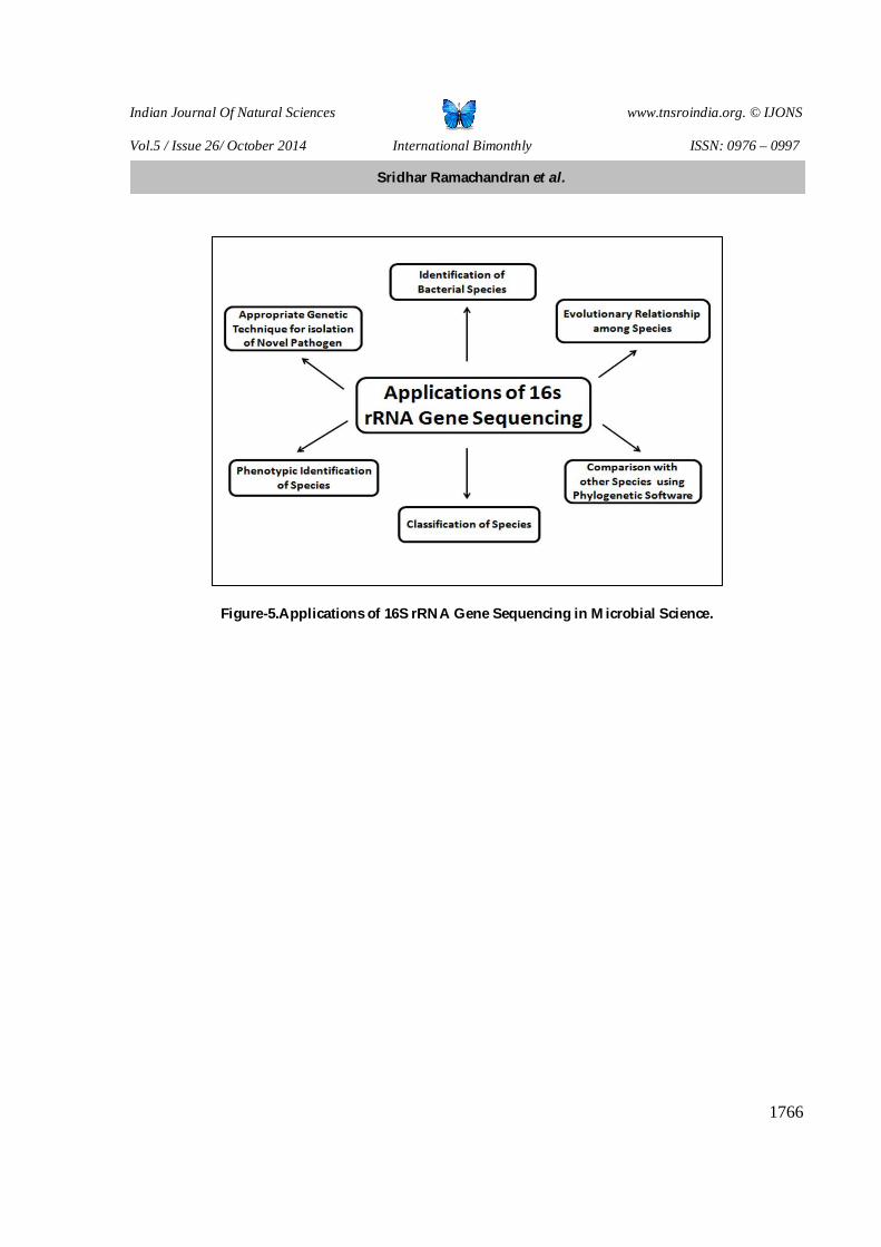
Clarridge JE (2004) [14] reviewed the impact 16S rRNA Gene Sequence Analysis for Identification of Bacteria on Clinical Microbiology and Infectious Diseases. The poor inaccuracy results in phenotypic identification can be replaced by genotypic identification in the routine clinical microbiological laboratories. In order to depict the significance of 16s rRNA gene in Clinical microbiological laboratories, the researchers pointed out the application of 16s rRNA gene sequencing in microbiology in the Figure-5.
CONCLUSION
The patients with renal aliments are vulnerable to malfunctioning of vital components of urinary tract if they are superimposed with infections by uropathogens. Several studies had portrayed the complications caused by UTI pathogens. Thus the attempts of the treatment of these categories of UTIs shall be challenged. Therefore early diagnosis UTI with special attention to the detection of Uropathogens becomes a vital step in effective treatment regime.
REFERENCES 1. Sridhar Ramachandran, Aarthi N, Hemamalini V and Thiyagarajan S. Extended Spectrum Beta Lactamase. 2014,
First Ed., Kancheepurum, Tamilnadu, India, Excellent Publisher. [ISBN- 978-81-928101-6-5]. 2. Sattar A, Abbasi S A, Usman J, Fagir F, Kaleem F and Hanif F. Extended Spectrum β-Lactamase production in
Shigella flexneri. J Coll Physician Surg Pak 2010; 20(11): 768-769. 3. Jafar Naqvi S A, Ahmed S I and Yagin H. Recurrent urinary tract infection due to Shigella flexneri-Case report. J
Pak Med Assoc. 1976; 26(8): 169-170. 4. Baka S, Spathi A, Tsouma I and Kouskouni E. symptomatic Shigella sonnei urinary tract infection in pregnancy.
Clin Exp Obstet Gynecol. 2013; 40(1): 116-117. 5. Hussain Qadri S M and Salim H Khalil. Polymicrobial septicemia due to Shigella flexneri and Pseudomonas
aeruginosa: First Report. J Natl Med Assoc 1987; 79(12): 1289-1292. 6. Bhattacharya D, Purushottaman SA, Bhattacharjee H, Thamizhmani R, Sudharama SD, Manimunda SP, et al.
Rapid Emergence of Third-Generation Cephalosporin Resistance in Shigella Sp. Isolated in Andaman and Nicobar Islands, India. Microbial Drug Resistance 2011;17(2): 329-332.
7. Koneman E w, Allen S D, Janda W M, Schreckenberger P C and Win W C, editiors. The Enterobacteriaceae. In: color Atlas and textbook of diagnostics microbiology. 5th ed. JB Lippinocott co: Philadelphia. 2006; 211-302.
8. Lewington, J., S.D. Greenaway and B.J. Splillane. 1987. Rapid cloning and hybridization analysis. Letters in Applied Microbiology Arihar, K., Cassens R.G. and Luchansky, J.B. 1993: 51–53.
9. Maniatis T, Fritsch EF, and Sambrook J. Molecular cloning - A laboratory manual. Cold Spring Harbor Laboratory Press 1982, Plainview, New York, 545.
10. Julian R M, Takuichi S., Andrew J W, et al. Design and evaluation of useful bacterium-specific PCR primers that amplify genes coding for bacterial 16s-rRNA. Appl Environ. Microbiol 1998. 64, 795-799.
11. Azmi IJ, Khajanchi BK, Akte r F , Hasan TN, Shahnaij M, Mahmuda Akter et al. Fluoroquinolone Resistance Mechanisms of Shigella flexneri Isolated in Bangladesh. PLoS ONE 2014; 9(7): 1-7.
12. Sridhar R, Thiyagarajan S and Tamilvendan M. Extended Spectrum β -Lactamase Producing Klebsiella pneumoniae and Escherichia coli isolated From UTI Patients: Prevalence, Antibiogram Science, ESBL Analysis and Molecular Typing. Biomedicine 2010; 30(4): 495-504.
13. Shah M. Faruque, Khaleda Haider, M. Monzur Rahman, A.R.M. Abdul Alim, Q.Shafiahmad, M.Johnalbert et al. differentiation of Shigella flexneri strains by rRNA gene restriction pattern. J Clin Microbiol 1992; 30(11): 2996-2999.
14. Clarridge JE. Impact 16S rRNA Gene Sequence Analysis for Identification of Bacteria on Clinical Microbiology and Infectious Diseases. Clin Microbiol Rev 2004; 17(4): 840-862.
Sridhar Ramachandran et al.

Indian Journal Of Natural Sciences www.tnsroindia.org. © IJONS
Vol.5 / Issue 26/ October 2014 International Bimonthly ISSN: 0976 – 0997
1763
Table-1. PCR reaction mixture for 16S rRNA.
Table-2. Thermocycler reaction.
Table-3. PCR reaction mixture for gene specific serine protease.
S. No.
Required PCR reaction mixture for 16s rRNA
Quantity
1 Forward primer 2.0 μl 2 Reverse primer 2.0 μl 3 Template DNA 4.0 μl 4 PCR master mixture 25 μl 5 RNase free water 17 μl 6 Total volume 50 μl
Figure-1.Genomic DNA (I-A) and PCR amplification (1-B) of 16s rRNA gene of Shigella flexneri ST-02.
S. No. Required PCR reaction mixture for 16s rRNA
Quantity
1 Forward primer 2.0 μl 2 Reverse primer 2.0 μl 3 Template DNA 4.0 μl 4 PCR master mixture 25 μl 5 RNase free water 17 μl 6 Total volume 50 μl
S. No. Temperature Time interval Reaction 1 95°C 3.0 min Initial denaturation 2 95°C 1 min Denaturation 3 55°C 1 min Annealing 4 72°C 1.5 min Cycle extension 5 72°C 10 min Final extension
I-A
I-B
Sridhar Ramachandran et al.

Indian Journal Of Natural Sciences www.tnsroindia.org. © IJONS
Vol.5 / Issue 26/ October 2014 International Bimonthly ISSN: 0976 – 0997
1764
1 gcccccggtg gaactaaacg gacgcacttt ttattgaggt ccgcttgctc tcgcgaggtc 61 gcttctcttt gtatgcgcca ttgtagcacg tgtgtagccc tggtcgtaag ggccatgatg 121 acttgacgtc atccccacct tcctccagtt tatcactggc agtctccttt gagttcccgg 181 ccggaccgct ggcaacaaag gataagggtt gcgctcgttg cgggacttaa cccaacattt 241 cacaacacga gctgacgaca gccatgcagc acctgtctca cggttcccga aggcacattc 301 tcatctctga aaacttccgt ggatgtcaag accaggtaag gttcttcgcg ttgcatcgaa 361 ttaaaccaca tgctccaccg cttgtgcggg cccccgtcaa ttcatttgag ttttaacctt 421 gcggccgtac tccccaggcg gtcgacttaa cgcgttagct ccggaagcca cgcctcaagg 481 gcacaacctc caagtcgaca tcgtttacgg cgtggactac cagggtatct aatcctgttt 541 gctccccacg ctttcgcacc tgagcgtcag tcttcgtcca gggggccgcc ttcgccaccg 601 gtattcctcc agatctctac gcatttcacc gctacacctg gaattctacc cccctctacg 661 agactcaagc ttgccagtat cagatgcagt tcccaggttg agcccggggg atttcacatc 721 tgacttaaca aaccgcctgc gtgcgcttta cgcccagtaa ttccgattaa cgcttgcacc 781 ctccgtatta ccgcggctgc tggcacggag taagccggtg cttcttctgc gggtaacgtc 841 aatgagcaaa ggtattaact ttactccctt cctcccccgc tgatagtaac tttacaaac 901 cgaatgcctt cttcataaca cgcggcaatg ggctgcatg //
Figure-2. Molecular Sequencing Report on 16s rRNA Gene (939 base pairs).
BASE COUNT 192 a 297 c 216 g 234 t ORIGIN Gene Accession Number (GENBANK, USA)-JX444058
Sridhar Ramachandran et al.

Indian Journal Of Natural Sciences www.tnsroindia.org. © IJONS
Vol.5 / Issue 26/ October 2014 International Bimonthly ISSN: 0976 – 0997
1765
Figure- 3.Electropherogram of the 16S rRNA Gene Sequencing of Clinical Strain Shigella flexneri ST-02.
Figure- 4.Phylogenetic analysis of Shigella flexneri ST-02 using clustalw.
Sridhar Ramachandran et al.
Indian Journal Of Natural Sciences www.tnsroindia.org. © IJONS
Vol.5 / Issue 26/ October 2014 International Bimonthly ISSN: 0976 – 0997
1766
Figure-5.Applications of 16S rRNA Gene Sequencing in Microbial Science.
Sridhar Ramachandran et al.
Related Documents