University of New Orleans ScholarWorks@UNO University of New Orleans eses and Dissertations Dissertations and eses 8-4-2011 Identification and characterization of enzymes involved in the biosynthesis of different phycobiliproteins in cyanobacteria Avijit Biswas University of New Orleans, [email protected] Follow this and additional works at: hp://scholarworks.uno.edu/td is Dissertation-Restricted is brought to you for free and open access by the Dissertations and eses at ScholarWorks@UNO. It has been accepted for inclusion in University of New Orleans eses and Dissertations by an authorized administrator of ScholarWorks@UNO. e author is solely responsible for ensuring compliance with copyright. For more information, please contact [email protected]. Recommended Citation Biswas, Avijit, "Identification and characterization of enzymes involved in the biosynthesis of different phycobiliproteins in cyanobacteria" (2011). University of New Orleans eses and Dissertations. Paper 446.

Welcome message from author
This document is posted to help you gain knowledge. Please leave a comment to let me know what you think about it! Share it to your friends and learn new things together.
Transcript
University of New OrleansScholarWorks@UNO
University of New Orleans Theses and Dissertations Dissertations and Theses
8-4-2011
Identification and characterization of enzymesinvolved in the biosynthesis of differentphycobiliproteins in cyanobacteriaAvijit BiswasUniversity of New Orleans, [email protected]
Follow this and additional works at: http://scholarworks.uno.edu/td
This Dissertation-Restricted is brought to you for free and open access by the Dissertations and Theses at ScholarWorks@UNO. It has been acceptedfor inclusion in University of New Orleans Theses and Dissertations by an authorized administrator of ScholarWorks@UNO. The author is solelyresponsible for ensuring compliance with copyright. For more information, please contact [email protected].
Recommended CitationBiswas, Avijit, "Identification and characterization of enzymes involved in the biosynthesis of different phycobiliproteins incyanobacteria" (2011). University of New Orleans Theses and Dissertations. Paper 446.
Identification and characterization of enzymes involved in
biosynthesis of different phycobiliproteins in cyanobacteria
A Thesis
Submitted to the Graduate Faculty of the
University of New Orleans
in partial fulfillment of the
requirements for the degree of
Doctor of Philosophy
In
Chemistry
(Biochemistry)
By
Avijit Biswas
B.S. Pharmacy, Utkal University, India, 2001
M.S. Biology, Rutgers, The State University of New Jersey, 2006

ii
ACKNOWLEDGMENT
First, I want to thank to my PhD advisor Dr. Wendy Schluchter, she is an excellent mentor with
great personality, professionalism, and knowledge. It’s always been a great pleasure working
with her and in her lab. She makes me feel the importance of research, gave me every possible
chance to learn, make myself better to excel in my career. Her door was always open to discuss
about, she knows when to get tough on me and when to hand over helpful advices. Not only she
is my advisor also a good friend, she gave chances to entertain myself when I get overwhelmed
with lab work. Thank you very much Dr. Schluchter for accepting me to you lab, giving me
every opportunity to work and helping me every way in building my career. It always being a
great pleasure working in your lab, I feel like my second home. Last but not the least Thanks for
arranging a place to stay during hurricane and for the Hornet’s tickets!!!!! Yeah…..
Second, I want to thank my mom and dad, for their support and encouragement. Although they
have no idea about my research work, still they gave constant motivation in my path of success.
Dad, thank you for all you did for me and you are the driving force that made me come to this
level of career. Mom, I can’t express in words for your help you did to me for my entire life that
make me a perfect person to face the real world. I love you mom and dad.
Third, I like to thank my uncle (Subhasis) and aunt (Esther). Thank you very much, for all you
guys did for me, helping in every way starting for giving scientific suggestions, helping me with
understanding what is the important of research and how to handle the situations. Esther thanks
a lot for providing me with opportunity to volunteer in your lab, which helped a lot in getting
hands on Molecular Biology/ Biochemistry.

iii
Now I should spend some time thanking the people who have worked with and helped me in the
lab in putting this thesis work together. First, I should thank Yasmin, she had been working with
me for 3 yrs and provided a lots of help starting from doing my experiments doing some parts of
my thesis. She never told no whenever I asked her to do something for me. I really appreciate her
help. Yasmin I wish you good luck with you graduate school at Baylor University. Above all
she is a great friend, thanks for checking my mails and baking cakes in my Birthday. Second, I
should thank Tierna, she is a great co-worker and friend. She always provided me company,
helped with my lab works and provided me moral supports. Thank you very much, Tierna for
getting lunch for me all the times. I wish you good luck in your admission to Medical School.
Third, I should thank Christina and Corry for helping me out with my clones and other
experiments. Thank you, Christina and Corry. Last but not the least I should thank all the
undergraduate and high school students who had worked in my project during my years in Dr.
Schluchter’s lab.
I should thank to my committee members Dr. Zengchang Liu, Dr. Steve Rick and Dr. Edward
Stevens, especially Dr. Liu since I got lots of helpful comments about my research. Thanks a lot
for providing us with different reagents and chemical, which helps us a lot.
Thanks to Dr. Rick and Dr. Stevens for their helpful inputs in putting my thesis together.
Now I should thank to all our collaborators who provided us with different plasmids construct
and research ideas which helped me in working with new projects and getting papers published
in peer reviewed journals. First, Thank you very much to Dr. Bryant, you gave us a lot of help
with your research ideas putting my paper together. Thanks to Rick, you helped me a lots with
all the cloned you made for us along with research helps. Thanks, to Dr. Frankenberg for
providing us with the pebS/ ho1 construct it helped us a lot. Thanks to Dr. Kehoe’s lab

iv
(Aminesh and Adrian) for making the clones and mutants, which provided me a lot of help in
putting this thesis together.
I should also express my thanks to one of the most important person in life in my wife Sudeshna
Das (Tina), she has always been with me, in my good and bad days. She always listened to my
complains, achievements and successes I had in my research. “I love you, Tina”
Lastly I should thank to numerous other people; undergraduate, high school students and high
school teacher who are involved in this project.
v
Table of Contents
List of figures .................................................................................................................... iv
List of Tables .................................................................................................................... vi
Table for abbreviations .................................................................................................. vii
Abstract ........................................................................................................................... viii
1.0 Introduction ..................................................................................................................1
1.1 Cyanobacteria: Background and History ...........................................................1
1.2 Phycobilisome: Structure and Function .............................................................2
1.3 Structure elucidation of phycobiliproteins .........................................................7
1.4 Application of fluorescent proteins ..................................................................11
1.4.1 Application of Cyanobacterial phycobiliproteins as fluorescent tags.....11
1.4.2 Application of Cyanobacterial phycobiliproteins as commercial
Commodities ...........................................................................................15
1.5 Application of green fluorescent protein .........................................................17
1.6 Bilin: Types and Biosynthetic pathway ...........................................................21
1.7 Bilin addition to phycobiliproteins ..................................................................28
1.7.1 E/F type lyase ..........................................................................................28
1.7.2 SU type lyase ..........................................................................................30
1.7.3 T type lyase .............................................................................................31
1.7.4 Autoctalytic lyase....................................................................................32
1.8 Other posttranslational modifications to phycobiliproteins .............................32
1.9 Chromatic acclimation .....................................................................................33
1.10 Purpose of work .............................................................................................39
2.0 Materials and Methods .............................................................................................41
2.1 Construction of expression vectors ..................................................................41
2.1.1 cpcS-I and cpcU expression construct ....................................................44
2.1.2 cpcT expression construct .......................................................................44
2.1.3 pcyA/ho1 expression constructs ..............................................................44
2.1.4 cpcBA and cpcB expression constructs ...................................................45
2.1.5 apcE expression construct.......................................................................46
2.1.6 cpcEF expression construct ....................................................................46
2.1.7 cpeA expression construct .......................................................................46
2.1.8 cpeZ and cpeY expression construct .......................................................47
2.1.9 cpeB expression construct .......................................................................47
2.1.10 cpeS expression construct .....................................................................47
2.1.11 pebS/ho1 expression constructs ............................................................48
2.1.12 cpeA and cpeB site directed mutant constructs .....................................48
2.2 In vivo heterologous expression and purification of recombinant proteins .....60
2.3 Fluorescence emission and absorbance spectra ...............................................62
2.4 Protein and bilin analysis .................................................................................62
2.5 Calculating fluorescence quantum yield ..........................................................64
2.6 Tryptic digestion of Phycoerythrin ..................................................................65
2.7 Growth condition for Fremyella diplosiphon ..................................................66
2.9 Separation of phycobilisome ............................................................................66
2.9 Isolation of Phycoerythrin................................................................................66
vi
2.10 Isolation of PEI and PEII from Synechococcus sp. RS 9916 .........................67
3.0 Results ........................................................................................................................70
3.1Biosynthesis of cyanobacterial phycobiliproteins in E. coli: chromophorylation
efficiency and specificity of all bilin lyases from Synechococcus sp. strain
PCC 7002 ..........................................................................................................70
3.1.1 Examination of Synechococcus sp. strain PCC 7002 PcyA activity in
E. coli ..............................................................................................................70
3.1.2 Development and use of a multi-plasmid system for expression of
holo-AP ............................................................................................................73
3.1.3 Chromophorylation Requirements for HT-ApcD ...................................78
3.1.4 Chromophorylation requirements for HT-ApcF .....................................82
3.1.5 Chromophorylation requirements of ApcE .............................................84
3.1.6 Creation of partially chromophorylated PBPs in E. coli.........................86
3.2 Creation of unique phycobiliproteins using PEB in E. coli for potential
biotechnological applications...........................................................................91
3.2.1 Creation of unique phycobiliproteins in E. coli ......................................91
3.2.2 Comaparison of CpcEF vs CpcSU ligation specificity for PEB on
phycobilirprotein subunits in E. coli ............................................................94
3.3 Characterization of CpeY, CpeZ and CpeS bilin lyases involved in
phycoerythrin biosynthesis in Fremyella diplosiphon strain UTEX 481 .....98
3.3.1 Characterization of bilin lyase activity of CpeY and CpeZ with
CpeA ......................................................................................................98
3.3.2 Analysis of which cysteine residues on -PE are chromophorylated by
the CpeY/CpeZ lyase ............................................................................104
3.3.3 Does CpeS also chromophorylate CpeA ...............................................106
3.3.4 Comparison of PEB ligation activity of CpeY/CpeY and CpeS
bilin lyase with CpeB ............................................................................111
3.3.5 Analysis of specific Cys residues(s) of CpeB chromophorylated by CpeS
in E. coli ................................................................................................113
3.4 Mutant in cpeY and cpeZ genes are defective in phycoerythrin biosynthesis in
Fremyella diplosiphon sp. Strain UTEX 481 ................................................119
3.4.1 Characterization of F. diplosiphon cpeY mutants .................................120
3.4.2 PCC 6803 CpcEF lyase activity on PCB ligation on CpeA .................123
3.4.3 Biochemical characterization of PE from the cpeZ mutant ..................125
3.5 mpeZ gene is involved in Type IV chromatic adaptation in marine
Synechococcus cyanobacteria ............................................................. 127 3.5.1 MpeZ is a novel phycoerythrin II:phycoerythrin lyase-isomerase
involved in Type IV chromatic acclimation .................................................128
3.5.2 Specificity of cysteine residues on MpeA ............................................131
3.5.3 Analyses of lyase activity on CpeA from RS 9916 ..............................133
4.0 Discussions ................................................................................................................137
4.1 Chromophorylation efficiency and specificity of all bilin lyases from
Synechococcus sp. Strain PCC 7002 ............................................................137
vii
4.2 Creation of unique phycobiliproteins using PEB in E. coli for potential
biotechnological applications.......................................................................144
4.3 Characterization of CpeY, CpeZ and CpeS bilin lyases involved in
phycoerythrin biosynthesis in Fremyella diplosiphon strain UTEX 481 ...146
4.4 Mutant in cpeY and cpeZ genes are defective in phycoerythrin biosynthesis in
Fremyella diplosiphon sp. UTEX 481 ......................................................150
4.5 The mpeZ gene is involved in Type IV chromatic adaptation in marine
Synechococcus sp. RS 9916 ...........................................................................152
5.0 Appendix ...................................................................................................................155
5.1 Analysis of lyase activity of CpcS type lyase fom
Thermosynechococcus elongatus on phycocyanin subunit ............................157
5.2 Analysis of lyase activity of CpcS type lyase fom
Thermosynechococcus elongatus on allophycocyanin subunit ......................160
5.3 TE CpcS activity on AP -like subunit ApcD ...............................................162
5.4 TE CpcS activity on AP -like subunit ApcF ................................................164
Reference ........................................................................................................................179
Vita ..................................................................................................................................197
viii
List of Figures
1. Phycobilisome structure .....................................................................................5
2. Phycobilisome structure in marine cyanobacteria .............................................6
3. Ribbon structure of phycobiliproteins ...............................................................8
4. Location within the GFP crystal structure .......................................................20
5. Structures of bound bilins ................................................................................22
6. Biosynthesis of PEB and PCB .........................................................................26
7. Biosynthesis of PVB and PUB ........................................................................27
8. The color phenotypes of F. diplosiphon filaments grown on agar plates ........36
9. Whole absorbance spectrum along with PBSs of F. diplosiphon grown in
different light conditions ..................................................................................37
10. Proposed models of PBS structure for the different Synechococcus pigment
types and subtypes ...........................................................................................38
11. Plasmid map of PCC 7002 cpcUS in pCOLA Duet .........................................51
12. Vector map of PCC 7002 cpcT in pCOLA Duet vector ..................................52
13. Plasmid map representing PCC7002 pcyA/ PCC 6803ho1 cloned in
pACYC Duet vector ........................................................................................53
14. Vector map of 6803 cpcBA in pCDF Duet ......................................................54
15. Vector map of HT-CpeA from F. diplosiphon cloned in pET Duet vector .....55
16. Vector map of Fd cpeY/cpeZ cloned in pCOLA Duet .....................................56
17. Vector map of Fd cpeB cloned in pET Duet ....................................................57
18. Vector map of Fd cpeS cloned in pCOLA Duet ..............................................58
19. Vector map of pebS/ho1 cloned in pACYC Duet ...........................................59
20. Photographs of E. coli pellets after growth with the plasmids listed on the
legend above or below each pellet ...................................................................71
21. Analyses of holo-HT-CpcA purified from E. coli cells ...................................72
22. Analyses of major AP subunits ApcA and ApcB synthesized in E. coli .........76
23. Analyses of HT-ApcAB purified from E. coli produced in the presence of
CpcEF or CpcT ...............................................................................................77
24. Amino acid sequence alignment of Synechococcus sp. PCC 7002 ApcE with
sequences of ApcA, ApcB, ApcD, ApcF sequences .......................................80
25. Analyses of AP-B α-subunits (ApcD) synthesized in E. coli ..........................81
26. Analyses of AP β18
-subunit (ApcF) synthesized in E. coli ..............................83
27. Analyses of GST-ApcE purified from E. coli cells .........................................85
28. Analysis of HT CpcB purified from E. coli chromophorylated by
CpcS-I/CpcU at Cys-82 ................................................................................88
29. Analysis of HT CpcB purified from E. coli chromophorylated by CpcT at
Cys-153 ............................................................................................................89
30. Analyses of Synechocystis sp. PCC 6803 HT-CpcA purified from
E. coli cells .......................................................................................................93
31. Analyses of Synechocystis sp. PCC 6803 HT-CpcB/CpcA purified from
E. coli cells .......................................................................................................96
32. Amino acid sequence alignment between CpeY from Fremyella diplosiphon
and a fusion of CpcE with CpcF from Synechococcus sp. PCC 6803 ............ 101
ix
33. Picture of the E. coli cell pellets from cells containing HT-CpeS, pPebS and
with either pCpeYZ or CpeS ................................................................................ 102
34. Analyses of HT-CpeA produced with CpeY and CpeZ in E. coli .................103
35. Analyses of the specific cysteine residue on HT-CpeA required for PEB
addition by CpeYZ .........................................................................................105
36. Analyses of HT-CpeA produced with CpeS in E. coli ..................................108
37. Analyses of the specific cysteine residue on HT-CpeA for PEB addition by
CpeS ...............................................................................................................110
38. Tryptic digest of partial holo HT-CpeA.........................................................115
39. Analyses of the HT-CpeB (β-PE) produced in the presence of various
lyases in E. coli ..............................................................................................112
40. Analyses of the specific cysteine residue on HT-CpeB required for PEB
addition by CpeS ...........................................................................................115
41. Analyses of HT-CpeA-PCB produced in the presence of pPcyA and
pCpeYZ ..........................................................................................................116
42. Whole cell spectra from wild type and mutant cells ......................................119
43. Analysis of Phycoerythrin purified from wild type and the cpeY mutant
cells ................................................................................................................122
44. PCB ligation on CpeA from PCC 7601 catalyzed by CpcEF from
PCC 6803 .......................................................................................................124
45. Analysis of Phycoerythrin purified from wild type and the cpeZ mutant
cells ................................................................................................................126
46. Analyses of MpeZ lyase for PEB addition to Phycoerythrin α subunit (PEII)
in E. coli .........................................................................................................130
47. Site directed mutant analysis of specific cysteine residue for PEB addition
to α-subunit PEII by MpeZ ............................................................................132
48. Analyzing TECpcS lyase activity on RS 9916 CpeA chromophorylation ....135
49. Analyzing Fd CpeS lyase activity on RS 9916 CpeA chromophorylation ....136
50. Amino acid sequence alignment of Synechococcus sp. PCC 7002 ApcE .....143
51. Structure of Tlr 1699/ CpcS-III ( Ter13) from Thermosynechococcus
elongatus BP-1 (PDB ID:3BDR) ..................................................................156
52. Comparison of chromophorylation between CpcSU and TECpcS on β-PC
........................................................................................................................159
53. Analysis of holo HT-ApcAB purified from E. coli cells chromophorylated
by TECpcS .....................................................................................................161
54. Analysis of holo HT-ApcBD purified from E. coli cells chromophorylated
by TECpcS .....................................................................................................163
55. Analysis of holo HT-ApcF purified from E. coli cells chromophorylated
by TECpcS .....................................................................................................165
x
List of Tables
1. List of potential lyases/ isomerases....................................................................10
2. Comparison of physical data between various flourophores and fluorescent
proteins ...............................................................................................................14
3. Commercially used products from Cyanobacterial Phycobiliproteins ..............16
4. Summary of plasmids used for experimental studies described in this thesis ...42
5. Primer sequences used in this thesis ..................................................................49
6. Properties of recombinant holo-PBPs for PC and AP subunits .........................90
7. Properties of recombinant holo-PBPs with non-cognate lyases ........................97
8. Comparison of spectral properties for various PE subunits produced with
bilin lyases ......................................................................................................117
9. Comparising Fluorescence intensities for various recombinant holo α-PE .....118
10. Comparison of fluorescence emission for recombinant PEII subunits ..........133
11. Spectral properties of holo PC and AP subunits chromophorylated with
multiple bilins aided by TE CpcS ..................................................................167
12. Lists of clones made which are not discussed in the results ..........................168
13. Coexpression attempted with negative result ................................................170
13. Oligonucleotides of the clones used in the appendix .....................................173
xi
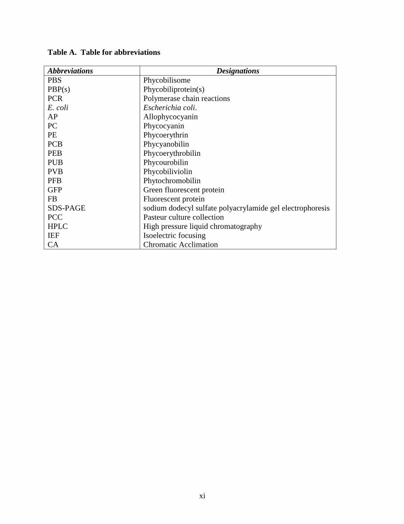
Table A. Table for abbreviations
Abbreviations Designations
PBS Phycobilisome
PBP(s) Phycobiliprotein(s)
PCR Polymerase chain reactions
E. coli Escherichia coli.
AP Allophycocyanin
PC Phycocyanin
PE Phycoerythrin
PCB Phycyanobilin
PEB Phycoerythrobilin
PUB Phycourobilin
PVB Phycobiliviolin
PFB Phytochromobilin
GFP Green fluorescent protein
FB Fluorescent protein
SDS-PAGE sodium dodecyl sulfate polyacrylamide gel electrophoresis
PCC Pasteur culture collection
HPLC High pressure liquid chromatography
IEF Isoelectric focusing
CA Chromatic Acclimation
xii
Abstract
A multi-plasmid, co-expression system was used to recreate the biosynthetic pathway for
phycobiliproteins from the cyanobacterium Synechococcus sp. PCC 7002 in E. coli. This system
efficiently produced chromophorylated allophycocyanin (ApcA/ApcB), -phycocyanin, and -
phycocyanin. This system was used to demonstrate that CpcS-I and CpcU proteins are both
required attaching PCB to allophycocyanin subunits ApcD (AP-B
) and ApcF (18
). The N-
terminal, AP-like domain of ApcE (LCM99
) was produced in soluble form and shown to have
intrinsic bilin lyase activity. In addition, this system was used to chromophorylated CpcA from
Synechococystis sp. PCC 6803 with a non-cognate bilin; PEB with the aid of CpcEF type bilin
lyase. However, the CpcSU type lyase displays much higher specificity for PCB (the native bilin
in these species) than PEB.
Next, using a heterologous, co-expression system in E. coli, the PEB ligation activity of
putative lyase subunits CpeY, CpeZ, and CpeS was tested on the CpeA and CpeB subunits from
F. diplosiphon. CpeY/CpeZ was found to ligate PEB on CpeA, although CpeY alone had only
60% chromophorylation activity compared to CpeYZ together. Studies with site-directed
variants of CpeA (C82S and C139S), revealed that CpeY/CpeZ attached PEB at Cys-82 on HT-
CpeA. The CpeS bilin lyase ligated PEB at both Cys-82 and Cys-139 of CpeA, but the yield of
attached PEB at Cys 82 was much lower than observed with CpeY or CpeY/CpeZ. However,
CpeS efficiently attached PEB to Cys-82 of CpeB. Purified PE from cpeY deletion mutants in F.
diplosiphon was found to have PCB added on α-PE instead of PEB, which was likely performed
by CpcEF in vivo. However, a cpeZ knock-out mutant is affected in chromophorylation of both
and subunits of PE with a red-shifted absorbance compared to wild type PE probably due to
missing PEB on PE subunits.
xiii
Next a new type of bilin lyase isomerase for PEII ( subunit) named MpeZ from
Synechococcus sp. RS 9916, was analyzed using the E. coli heterologous coexpression system.
MpeZ acted as bilin lyase/isomerase chromophorylating α-PEII (MpeA) with PUB on Cys 83.
Keywords: Phycobiliprotein, Allophycocyanin, Phycocyanin, Phycoerythrin, Phycocyanobilin,
Phycoerythrobilin, Phycourobilin.
1
1.0. INTRODUCTION
1.1 Cyanobacteria: Background and History
Cyanobacteria are fascinating photosynthetic, gram-negative prokaryotic organisms with
immense biological importance. They are known to be the world’s oldest oxygen-evolving
organisms, and are found in fossils dating back more than 3.5 billion years old (Schopf 1983;
Bengston 1994). These oxygenic photosynthetic organisms created a wonderful oxygen-rich
atmosphere that we can breathe in today (Bengston 1994). Through endosymbiosis,
cyanobacteria also contributed to the origin of plants and other oxygen-evolving organisms such
as red, green, and cryptophyte algae (Bengston 1994; Sidler 1994). In addition, they can fix N2
and survive in extreme environments (down to -60oC). This makes them ideal model systems for
studying fundamental processes such as nitrogen fixation and photosynthesis. In addition,
cyanobacteria produce an array of bioactive compounds, some of which could become novel
antimicrobial agents, anti-cancer drugs, UV protectants etc. (Gerwick, Mrozek et al. 1989; Eggen
and Georg 2002; Mohammed and Vermaas 2004). The amazing versatility of cyanobacteria has
attracted huge scientific interest in recent years especially in the field of engineering
cyanobacterial for the production of renewable fuels or biofuels (Zhou and Li 2010). The
genome sequences of 35 different species of cyanobacteria have been completed and are
available in searchable databases. Having these genome sequences allows one to identify
potential enzymes involved in phycobiliprotein biosynthesis (Yoshikawa, Adachi et al. 2000;
Lluisma, Karmacharya et al. 2001; Pomati, Burns et al. 2004) based on similarity to previously
characterized enzymes.
2
1.2. Phycobilisome: Structure and Function:
The light reactions in all photosynthetic organisms like cyanobacteria, and red-algae,
begin with the absorption of photons by protein (antenna) complexes called phycobilisome (PBS)
(Glazer 1989; Bryant, Stirewalt et al. 1991; Grant and Conti 1996) (Fig. 1). PBS were first
purified from the cyanobacterial species; Anacystis nidulans (Gantt and Lipschultz 1972; Evans
and Allen 1973) and are composed of brilliantly colored proteins known as phycobiliproteins
(PBPs). PBS are present on the cytoplasmic surface of the thylakoid membrane and transfer
energy to the membrane photosystem II complexes (Fig. 1) (Glazer 1985; Grossman, Bhaya et al.
2001) and Fig. 2. The PBP are highly water-soluble. The chomophores (eg. Chlorophyll) of all
other photosynthetic complexes are extractable by organic solvents, but those of PBPs are not
because they are covalently attached to polypeptides (Glazer 1988). The brilliant fluorescent
colors of PBPs did not go unnoticed by early investigators. Sorby in 1877 (Sorby 1877) made a
comment “It would be difficult to find another series of coloring matters of greater beauty or
with such remarkable and instructive chemical and physical peculiarities”
PBP can make up 40-50% of the total proteins in cyanobacteria (Glazer 1989). Each PBP
is mainly composed of two different polypeptides known as α and β in a 1:1 molar ratio. The α
subunit has a molecular weight between 10-19 kDa and β subunit has between 14-21 kDa
(Bennett and Bogorad 1971; Glazer and Cohen-Bazire 1971; P. and Killilea 1971; Gantt and
Zuber 1974; Gysi and Zuber 1974). Each α and β subunit has at least one but up to three
covalently attached bilin chromophore(s)s, which contributes to each PBP’s unique spectroscopic
properties allowing absorption of light energy in the visible range between 450-655 nm,
chromophores. The linker polypeptides are colorless with exception of the γ –subunit linker.
Found in marine Synechococcus sp. (and red-algae) this γ-subunit has a covalently attached bilin
3
(PUB) (Glazer and Hixson 1977; Klotz and Glazer 1985; Ong 1988). The phycobiliproteins are
isolated from cyanobacteria as trimeric (αβ) 3 or as hexameric (αβ) 6 complexes or are isolated as
dimeric (αβ) 2 or monomeric (αβ) forms. Certain phycoerythrins (PEs) are isolated as assemblies
with the composition (αβ) 6 where the subunit has a molecular weight of 30,000 Da. Specific
linker proteins (LCM) mediate the association of one hexameric disc to another and modulate the
spectroscopic properties of the phycobiliproteins (Zhao, Ping et al. 2005), promoting
unidirectional energy flow to photosynthetic reaction centers (Glazer, 1985). The linkers are
usually non chromophorylated except in some marine cyanobacterial species (WH 8020, WH
8102, RS9916 etc.) they have one chromophore attached (Six, Thomas et al. 2005).
Phycobiliprotein trimers are disc-shaped with a thickness of ~ 30A and a diameter of ~ 120A
and two trimers associated into hexamric assemblies in a face to face manner. Spectroscopic data
and electron microscopic analyses indicate that these complexes share common structural features
with those within phycobilisomes (Glazer 1989). There are four different classes of
phycobiliproteins; phycocyanin (PC, λ max=615-640nm), allophycocyanin (AP, λ max= 650-655),
phycoerythrin (PE, λ max= 495-575 nm) and phycoerythrocyanin (PEC, λ max= 575 nm) (Ong and
Glazer 1991).
A single PBS is generally composed of a central core (containing AP) and 6 to 8 radiating
rods (containing PC) (Fig. 1). Some cyanobacterial species contain PE on the end of the rods
adjacent to PC for more efficient light capture (Glazer and Hixson 1977) . In this thesis two
phycoerythrin- containing cyanobacteria were studied; fresh-water-grown cyanobacteria F.
diplosiphon UTEX 481 (also known as Tolypothrix sp. PCC 7601 and marine cyanobacterial
species Synechococcus sp. RS 9916. F. diplosiphon contains PE and PC. The PE contained in
the rods of F. diplosiphon have red colored bilins, designated as phycoerythrobilin (PEB)
4
(described in detail later) having an absorbance maximum at 560 nm and fluorescence emission
maximum at 572 nm (Fairchild and Glazer 1994) .
Marine Synechococcus strains contain two types of PE designated as PEI and PEII, which
have different protein compositions and different chromophores (between five to six
chromophores) (Ong and Glazer 1991) (See Fig 2). PE(II) and PE(I) exist in a weight ratio of 2-
4:1, respectively. The energy absorbed by PE(II) gets transferred to PE(I). Their PE subunits
contain a different group of bilin isomer the yellowish-orange colored; phycourobilin (PUB)
(described later) as well as PEB. Because PUB absorbs light efficiently at 495 nm the
phycobilisome in these species are more efficient in capturing blue light (BL), the main
wavelength of light penetrating deep in the ocean (Ong, Glazer et al. 1984).
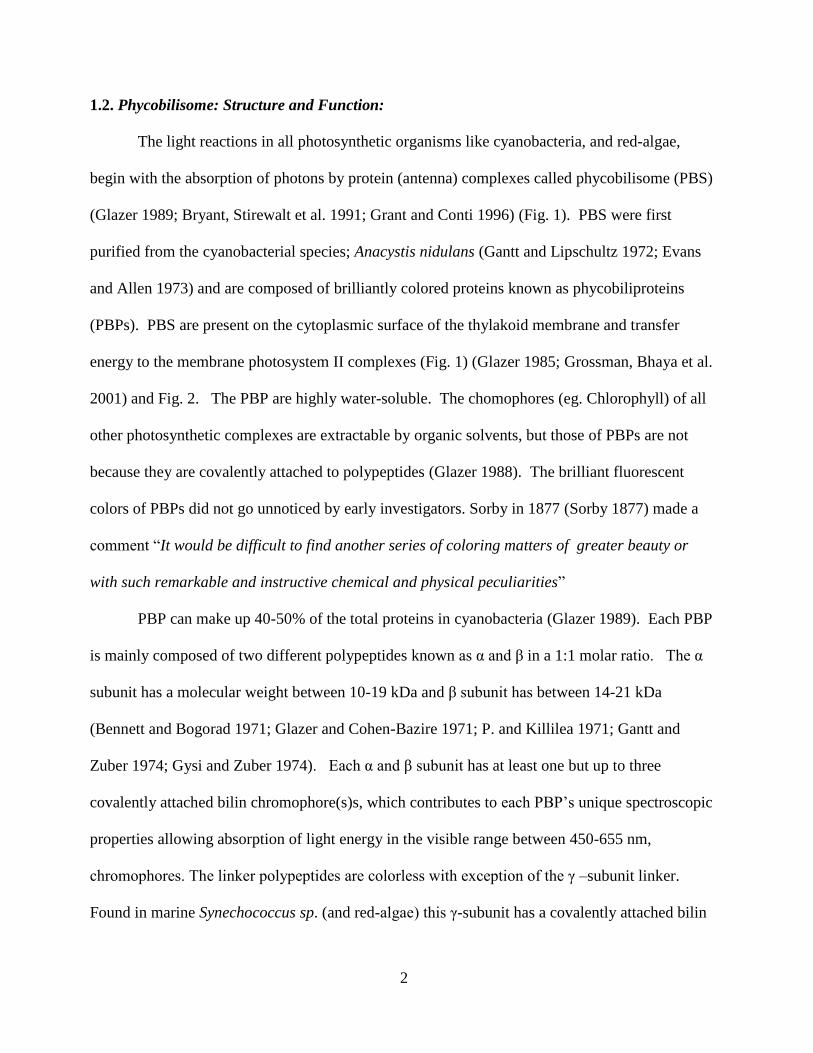
5
Fig. 1. Phycobilisome structure in cyanobacteria: The two photosystem (PS) shown
separately; PSI and PS II. On the outer membrane of PSII contain the donut shaped phybilisome.
Each phycobilisome consist of central code formed of Allophycocyanin (AP) and the radiating
rods formed of Phycocyanin (PC) and in marine cyanobacteria the rods also contain the
phycoerythrin (PE). The light energy shown in red arrow are first absorbed by PE, transferred to
PC then to AP, finally reached the chloroplast of PSII.This figure is a modified version as drawn
by N. Tandeau de Marsac (Tandeau de Marsac 1994)
6
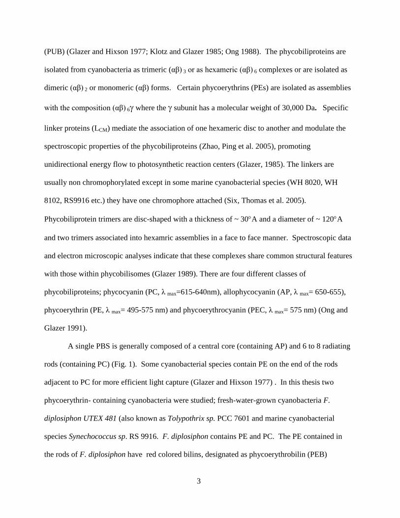
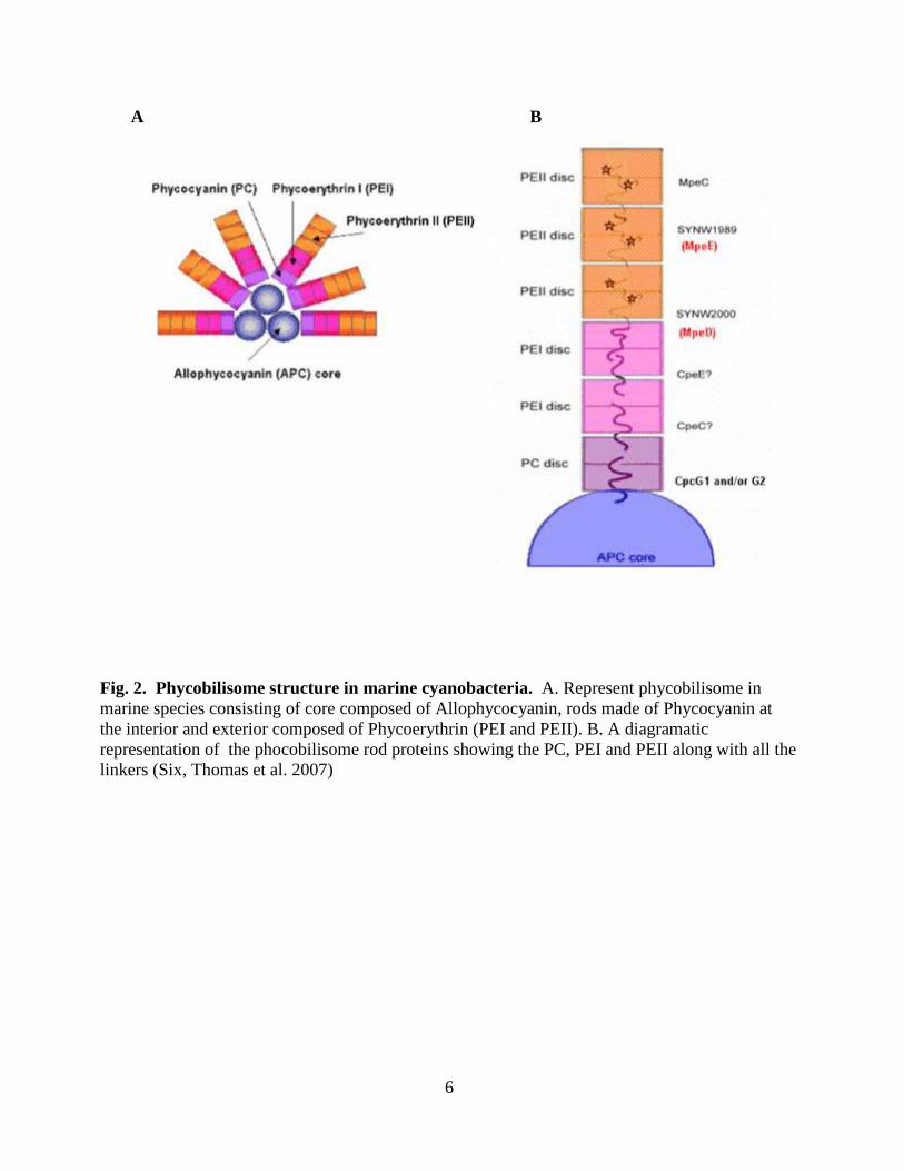
A B
Fig. 2. Phycobilisome structure in marine cyanobacteria. A. Represent phycobilisome in
marine species consisting of core composed of Allophycocyanin, rods made of Phycocyanin at
the interior and exterior composed of Phycoerythrin (PEI and PEII). B. A diagramatic
representation of the phocobilisome rod proteins showing the PC, PEI and PEII along with all the
linkers (Six, Thomas et al. 2007)
7
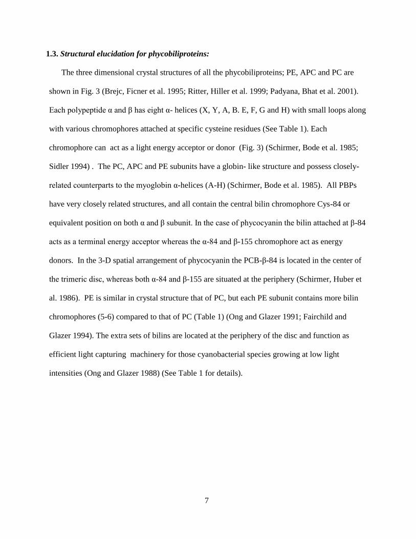
1.3. Structural elucidation for phycobiliproteins:
The three dimensional crystal structures of all the phycobiliproteins; PE, APC and PC are
shown in Fig. 3 (Brejc, Ficner et al. 1995; Ritter, Hiller et al. 1999; Padyana, Bhat et al. 2001).
Each polypeptide α and β has eight α- helices (X, Y, A, B. E, F, G and H) with small loops along
with various chromophores attached at specific cysteine residues (See Table 1). Each
chromophore can act as a light energy acceptor or donor (Fig. 3) (Schirmer, Bode et al. 1985;
Sidler 1994) . The PC, APC and PE subunits have a globin- like structure and possess closely-
related counterparts to the myoglobin α-helices (A-H) (Schirmer, Bode et al. 1985). All PBPs
have very closely related structures, and all contain the central bilin chromophore Cys-84 or
equivalent position on both α and β subunit. In the case of phycocyanin the bilin attached at β-84
acts as a terminal energy acceptor whereas the α-84 and β-155 chromophore act as energy
donors. In the 3-D spatial arrangement of phycocyanin the PCB-β-84 is located in the center of
the trimeric disc, whereas both α-84 and β-155 are situated at the periphery (Schirmer, Huber et
al. 1986). PE is similar in crystal structure that of PC, but each PE subunit contains more bilin
chromophores (5-6) compared to that of PC (Table 1) (Ong and Glazer 1991; Fairchild and
Glazer 1994). The extra sets of bilins are located at the periphery of the disc and function as
efficient light capturing machinery for those cyanobacterial species growing at low light
intensities (Ong and Glazer 1988) (See Table 1 for details).
8
1
2
3
PEB

9
Fig. 3. Represents ribbon structure of Phycobiliproteins (a) the α-subunit and (b) the β-
subunit. 1a and 1b represents PE subunits, 2a and 2b are AP subunits. 3a and 3b represent PC
subunits. The chromphores denoted by the amino acid residue numbers (Brejc, Ficner et al. 1995;
Ritter, Hiller et al. 1999; Padyana, Bhat et al. 2001).
10
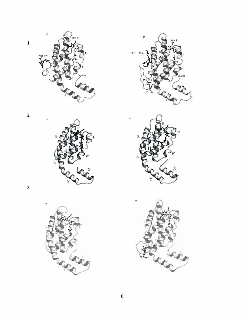
Table 1. Lists of the potential lyase/ isomerase that are involved in attaching different bilin
in Synechococcus sp. PCC 7002, F. diplosiphon PCC 7601, and Synechococcus sp. RS 9916. It
also includes the one have been published (in bold) and the rest of them are proposed as being
involved. Organism PBP
name
Gene name Attachment sites-
Bilina
Candidate Lyaseb
Synechococcus sp. PCC 7002 Syn. PCC 7002 PC cpcA Cys-84-PCB CpcE/CpcF (Fairchild, Zhao et al. 1992; Zhou, Gasparich et
al. 1992)
Syn. PCC 7002 PC cpcB Cys-82-PCB
Cys-153-PCB CpcS-I/CpcU (Saunée, Williams et al. 2008; Shen, Schluchter
et al. 2008)
CpcT (Shen, Saunee et al. 2006; Biswas, Vasquez et al. 2010)
Syn. PCC 7002 AP apcA Cys-81-PCB CpcS-I/CpcU (Shen, Saunee et al. 2006; Biswas, Vasquez et
al. 2010)
Syn. PCC 7002 AP apcB Cys-81-PCB CpcS-I/CpcU (Shen, Saunee et al. 2006; Biswas, Vasquez et
al. 2010)
Syn. PCC 7002 18 apcF Cys-81-PCB CpcS-I/CpcU (Biswas, Vasquez et al. 2010)
Syn. PCC 7002 AP-B apcD Cys-81-PCB CpcS-I/CpcU (Biswas, Vasquez et al. 2010)
Syn. PCC 7002 LCM99 apcE Cys-186 Autocatalytic (Zhao et al., 2005; Biswas, Vasquez et al. 2010)
Fremyella diplosiphon UTEX 481 Fd PCC 7601 PE cpeA Cys-82-PEB
Cys-139-PEB CpeY/CpeZ (Biswas et al. Manuscript in preparation)
? Fd PCC 7601 AP cpeB Cys-48,59-PEB
Cys-80-PEB
Cys-165-PEB
?
CpeS (Zhao, Su et al. 2007); Biswas et al. Manuscript in
preparation)
?CpeT
Synechococcus sp. RS 9916 (GL) Syn. RS9916 RPC rpcA Cys-84-PUB RpcG (Blot, Wu et al. 2009)
Syn. RS9916 RPC rpcB Cys-82-PCB Cys-155-PEB
?CpcS ?RpcT
Syn. RS9916 PE-I cpeA Cys-83-PEB
Cys-140-PEB CpeS (Zhao, Su et al. 2007 or CpeY/CpeZ
? Syn. RS9916 PE-I cpeB Cys-50, 61-PEB
Cys82-PEB
Cys159-PEB
?
CpeS
CpeT Syn. RS9916 PE-II mpeA Cys-75-PUB
Cys-83-PEB
Cys-140-PEB
?
Described in this thesis
? Syn. RS9916 PE-II mpeB Cys-50,61-PUB
Cys-82-PEB
Cys-159-PEB
?
CpeU or CpeS
CpeT Syn. RS9916 LR mpeC Cys-49-PUB ?
Synechococcus sp. RS 9916 (BL) Syn. RS9916 RPC rpcA Cys-84-PUB RpcG (Blot, Wu et al. 2009)
Syn. RS9916 RPC rpcB Cys-82-PCB
Cys-155-PEB
?CpcS
?RpcT
Syn. RS9916 PE-I cpeA Cys-83-PUB Cys-140-PEB
CpeS (Zhao, Su et al. 2007) or CpeY/CpeZ
?
Syn. RS9916 PE-I cpeB Cys-50, 61-PEB
Cys82-PEB
Cys159-PEB
?
CpeS or CpeU
CpeT
Syn. RS9916 PE-II mpeA Cys-75-PUB Cys-83-PUB
Cys-140-PUB
CpeS or CpeU
Described in this thesis
?
Syn. RS9916 PE-II mpeB Cys-50,61-PUB Cys-82-PEB
Cys-159-PEB
? CpeU or CpeS
CpeT
Syn. RS9916 LR mpeC Cys-49-PUB ? aTerminal acceptor bilin in rod proteins is underlined (Ong and Glazer, 1991). bIn bold, confirmed by experiment and citation is in parentheses; in italics, suggested by analogous position and other experiments from other
systems, but not yet confirmed; ?- candidate less clear from experimental data on paralagous proteins
GL represents grown in Green light , BL represents grown in Blue Light
11
1.4. Application of fluorescent proteins (FB):
The major fluorescent protein complex in cyanobacteria is known as PBS (Described
earlier). The presence of covalently attached tetrapyrrole pigments (or chromophores) on
phycobiliproteins makes them highly fluorescent. There are several unique features compared
to other flourophores like flourescein, tyrosine, tryptophan etc. and Green Flourescence
proteins (GFP) and its derivatives which make cyanobacterial phycobiliproteins ideal
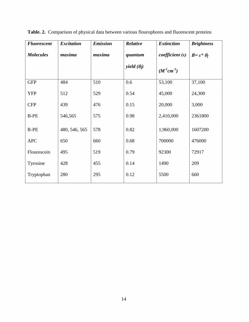
candidates for various biological applications; they have high quantum yields (~ 0.65-0.98)
(See Table 2), wide range of absorbance spectra (490-650 nm), they are stable at a wide range
of biological pH (4.5-8.0), the fluorescence property of phycobiliproteins are free of
interference from biological molecules, large Strokes shift provide greater signal to noise ratio
compared to other small flourophores, and they are stable to photobleaching. Three major
phycobiliproteins; Allophycocyanin (AP), R-phycoerythrin (R-PE), and B-phycoerythrin (B-
PE) currently serve as fluorescent tags with several biological applications in flow cytometry,
histochemistry, fluorescence activated cell sorting, and detection of reactive oxygen species
etc.
1.4.1. Application of Cyanobacterial phycobiliproteins as fluorescent tags:
Cyanobacterial proteins require three components to be fluorescent, for the use as a
fluorescent tags; namely phycobiliprotein subunits, the lyases which attach the chromophore and
the chromophore (bilin) itself. The apo-protein chains of phycobiliprotein subunits contain amino
and carboxyl groups that can form bonds to other molecules (Glazer and Stryer 1984; Glazer
1994; Sun, Wang et al. 2003). Oi et al. (Oi, Glazer et al. 1982) conjugated phycobiliproteins to
immunoglobulins, protein A and avidin to develop fluorescent probes. These conjugates have
been widely used in histochemistry, fluorescence microscopy, flow cytometry, fluorescence-
12
activated cell sorting and fluorescence immunoassays (Glazer and Stryer 1984; Glazer 1994; Sun,
Wang et al. 2003). Phycobiliproteins can exist as hexamers (α6β6) and trimers (α3β3) or
monomers (αβ). Hexamers tend to have higher molar extinction coefficients (Edwards, Hauer et
al. 1997; Thoren, Connell et al. 2006) and greater quantum yields compared to monomers (Glazer
and Stryer 1984; Glazer 1994; Sun, Wang et al. 2003), whereas denatured forms of
phycobiliproteins have lower molar extinction coefficient values and almost no fluorescence
(Fukui, Saito et al. 2004; Kupka, Jhang et al. 2009).
Back in the 1980s, phycoerythrin (PE) became one of the most widely used PBP in
different biological applications mainly, as a fluorescent tag. Glazer et al. isolated R-PE as α6β6
hexamers (Oi, Glazer et al. 1982; Glazer and Stryer 1984) with a fluorescence quantum yield of
81-90% (Oi, Glazer et al. 1982) (See Table 2). Phycoerythrin-immonoglobin, phycoerythrin-
protein A, and phycoerythrin-avidin conjugates were made, (Oi, Glazer et al. 1982), and these
bind specifically to beads containing covalently attached target molecules which renders them
highly fluorescent. Femptomole (10-15
mole) quantities of phycoerythrin conjugates can be
detected because of high extinction coefficient (εM= 2.4 X 106 cm
-1 M
-1 for 2.4 X 10
5 daltons)
and high fluorescence quantum yield (Q= 0.8) of the PBP moiety. These conjugates are used for
fluorescence-activated cell sorting and analyses, fluorescence microscopy, and fluorescence
immunoassays. In 1983, Glazer and Stryer (Glazer and Stryer 1983 ) developed fluorescent
tandem phycobiliprotein conjugates with a very large Stokes shift by covalently attaching PE to
AP. The efficiency of energy transfer from PE to AP in this disulphide-linked conjugate was
90%. One of its distinctive features is the wide separation between the intense absorption
maximum of phycoerythrin at 545 nm and the fluorescence emission maximum of
allophycocyanin at 660 nm. This tandem conjugate was found to have more advantages than
13
APC or PE alone in fluorescence-activated cell sorting and analysis, fluorescence microscopy,
and fluorescence immunoassays due to the large Stokes shift.
Oi et al. also isolated AP and PC, but it was a mixture of hexamers, trimers, and
monomers with lower quantum yields (68 % for AP and 50% for PC) (Oi, Glazer et al. 1982). PC
trimers can be stabilized by chemical cross-linking of polypeptide chains (Fukui, Saito et al.
2004; Sun, Wang et al. 2006). These stabilized PC trimers have similar spectral properties as
native PC can be used in fluorescent probes different from other PBPs. Also complete
phycobilisomes from Arthospira platensis composed of PC and AP have been chemically
stabilized, combined to streptavidin and used as a fluorescent probes in flow-cytometry (Telford,
Moss et al. 2001).
Another important use of PBP is in Flourescence immunoassay technique (FIT), a
process used for the identification of various proteins or enzymes in diseased cells.
Phycobiliproteins from different cyanobacteria and red-algae act as a valuable source for
flourscent tag in this immunoassay technique. The phycobiliproteins isolated from various
cyanobacterial and algal species possess certain chrateristics which make them ideal probes for
the use in FIT: red shifted excitation and emission spectra causing less interference with
biomolecules, a large Strokes shift, so that interferences from Rayleigh and Raman scatter and
other fluorescing components is less significant, stability toward naturally occurring biological
substances to be quenched, high solubility in an aqueous environment decreasing nonspecific
binding effect, and high fluorescence quantum yield independent of pH (O'Donnel and Suffin
1979; Soini and Hemmila 1979).
14
Table. 2. Comparison of physical data between various flourophores and fluorescent proteins
Fluorescent
Molecules
Excitation
maxima
Emission
maxima
Relative
quantum
yield (f)
Extinction
coefficient (ε)
(M-1
cm-1
)
Brightness
B= ε* f
GFP 484 510 0.6 53,100 37,100
YFP 512 529 0.54 45,000 24,300
CFP 439 476 0.15 20,000 3,000
B-PE 546,565 575 0.98 2,410,000 2361800
R-PE 480, 546, 565 578 0.82 1,960,000 1607200
APC 650 660 0.68 700000 476000
Flourescein 495 519 0.79 92300 72917
Tyrosine 428 455 0.14 1490 209
Tryptophan 280 295 0.12 5500 660
15
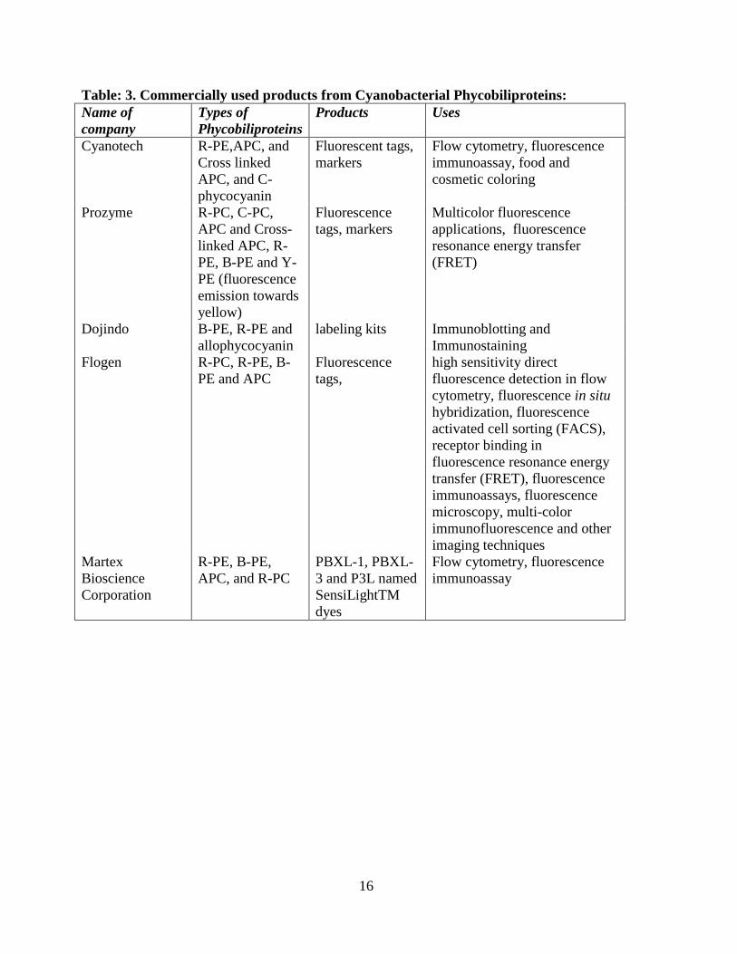
1.4.2. Application of Cyanobacterial phycobiliproteins as commercial commodities:
In addition to their use as fluorescent tags, PBP also have other uses. It might to helpful to
review the medical and biotechnological research industries involved in using phycobiliproteins
as biological tools. Phycocyanin from cyanobacteria has several pharmaceutical applications such
as to stimulate the immune defense system and possess antioxidant, anti-inflammatory, anti-viral,
anti-cancer, and cholesterol-lowering effects (Jensen, Ginsberg et al. 2001).
Several Biotechnology companies sell Cyanobacterial phycobiliprotein products (as
summarized in Table 3):
16
Table: 3. Commercially used products from Cyanobacterial Phycobiliproteins:
Name of
company
Types of
Phycobiliproteins
Products Uses
Cyanotech R-PE,APC, and
Cross linked
APC, and C-
phycocyanin
Fluorescent tags,
markers
Flow cytometry, fluorescence
immunoassay, food and
cosmetic coloring
Prozyme R-PC, C-PC,
APC and Cross-
linked APC, R-
PE, B-PE and Y-
PE (fluorescence
emission towards
yellow)
Fluorescence
tags, markers
Multicolor fluorescence
applications, fluorescence
resonance energy transfer
(FRET)
Dojindo
B-PE, R-PE and
allophycocyanin
labeling kits Immunoblotting and
Immunostaining
Flogen R-PC, R-PE, B-
PE and APC
Fluorescence
tags,
high sensitivity direct
fluorescence detection in flow
cytometry, fluorescence in situ
hybridization, fluorescence
activated cell sorting (FACS),
receptor binding in
fluorescence resonance energy
transfer (FRET), fluorescence
immunoassays, fluorescence
microscopy, multi-color
immunofluorescence and other
imaging techniques
Martex
Bioscience
Corporation
R-PE, B-PE,
APC, and R-PC
PBXL-1, PBXL-
3 and P3L named
SensiLightTM
dyes
Flow cytometry, fluorescence
immunoassay
17
1.5. Application of Green fluorescent proteins (GFP):
Although cyanobacterial fluorescent proteins have varied usage, their utility as FB have been
over-shadowed by the ground-breaking discovery of Green fluorescent protein (GFP) which
earned the Noble prize in chemistry in 2008 (Shimomura, Chalfie et al. 2008) .
GFP was discovered by Shimomura et al. (Shimomura, Johnson et al. 1962) as a companion
protein to aequorin, the famous bioluminescent protein from jellyfish Aequorea victoria. It is a
protein composed of 238 amino acid (Prasher, McCann et al. 1985) residue with a molecular
weight of 29.6 kDa exhibiting green fluorescence when exposed to blue light (Prendergast and
Mann 1978; Tsein 1998). In a footnote to Shimomura’s account of aequorin purification, they
noted “A protein giving solutions that look slightly greenish in sunlight though only yellowish
under tungsten lights, and exhibiting a very bright, greenish fluorescence in the ultraviolet of
Mineralite, has also been isolated from squeezates”(Shimomura, Johnson et al. 1962).
GFP was first crystallized in 1974 (Morrise, O et al. 1974) but it took 22 years to solve the
X-ray crystal structure (Ormo, Cubitt et al. 1996). It consists of 11 β-barrel strands, threaded by a
α-helix running up the axis of cylinder. Residues 65-67 (Ser-Tyr-Gly) in the GFP sequence
spontaneously form a fluorescent chromophore p-hydroxylbenzylideneimidazolinone
(Shimomura 1979; Cody, Prasher et al. 1993), which is attached to the α-helix and which provides
its fluorescent properties (Ormo, Cubitt et al. 1996). The crystal structure gave researchers
insight as to how the amino acid residues in the GFP molecule interact with each other towards to
contribute to its physical properties.
The chromophore formation occurs via a stepwise chemical reaction; first, GFP folds in a
nearly native conformation, and then the imidazolinone is formed by nucleophilic attack
(cyclilization) of the amide of Gly-67 on the carbonyl of residue Ser 65, followed by dehydration.
18
Finally, the presence of molecular oxygen dehydrogenates the - bond of the Tyr 66 residue
creating a conjugated bond with its aromatic group and with imidazolinone (Heim, Prasher et al.
1994; Cubitt AB, Onno et al. 1995). At this stage the mature form of GFP has absorbance and
fluorescent properties. So, unlike most cyanobacterial fluorescent proteins, GFP fluorescence is
autocatalytic.
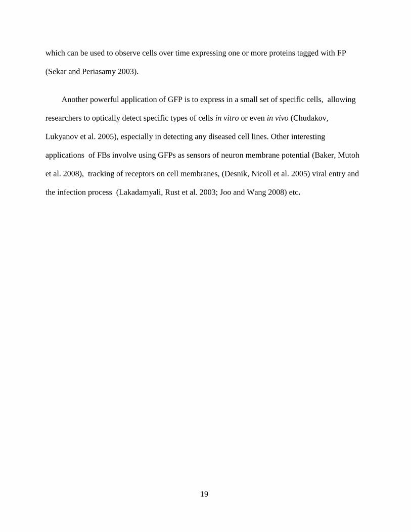
For the purpose of biotechnology applications scientists have mutated certain amino acid
residues (replacing the bulky residues with the smaller ones) (Cubitt AB and Biol 1997; Patterson,
Knobel et al. 1997; Ward 1997), which leads to the production of more soluble GFP (See Fig. 4).
Currently, GFP is one the most widely used fluorescence protein with a wide array of
biotechnology applications (Tsein 1998).
One of the major uses of GFP, involves fusing the gene for GFP in frame with a protein of
interest in any cell to create a fluorescent fusion protein. In an ideal situation, if the fused protein
maintains its original function and localization, it will now fluoresce. GFP localization has been
accomplished in all major cellular organelles such as the mitochrondria (Perozzo, Ward et al.
1988; Murray and Kirschner 1989; DeGiorgi, Brini et al. 1996), the nucleus (Perozzo, Ward et al.
1988; Lim, Kimata et al. 1995; Hanakam, Albrecht et al. 1996), and the endoplasmic reticulum
(Miyawaki, Llopis et al. 1997; Presley, Cole et al. 1997; Subramanian and Meyer 1997) etc.
The discovery of GFP and its derivatives (mutated versions) has revolutionized the use of
flouresence microscopy techniques in different biological disciplines (Ormo, Cubitt et al. 1996).
Compared to most small fluorescent molecules such as fluorescein isothiocyanate (FITC), which
is strongly phototoxic, GFP is usually not harmful when illuminated in live cells (Tsein 1998).
This triggered the development of highly automated live cell fluorescence microscopy systems,
19
which can be used to observe cells over time expressing one or more proteins tagged with FP
(Sekar and Periasamy 2003).
Another powerful application of GFP is to express in a small set of specific cells, allowing
researchers to optically detect specific types of cells in vitro or even in vivo (Chudakov,
Lukyanov et al. 2005), especially in detecting any diseased cell lines. Other interesting
applications of FBs involve using GFPs as sensors of neuron membrane potential (Baker, Mutoh
et al. 2008), tracking of receptors on cell membranes, (Desnik, Nicoll et al. 2005) viral entry and
the infection process (Lakadamyali, Rust et al. 2003; Joo and Wang 2008) etc.
20
Fig. 4. Location within the GFP crystal structure (Ormo, Cubitt et al. 1996) of the most
important sites that improve folding at 37C. The amino acids shown in space-filling
representation are the wild-type residues that are replaced by mutation.
21
1.6. Bilin: Types and Biosynthetic pathway:
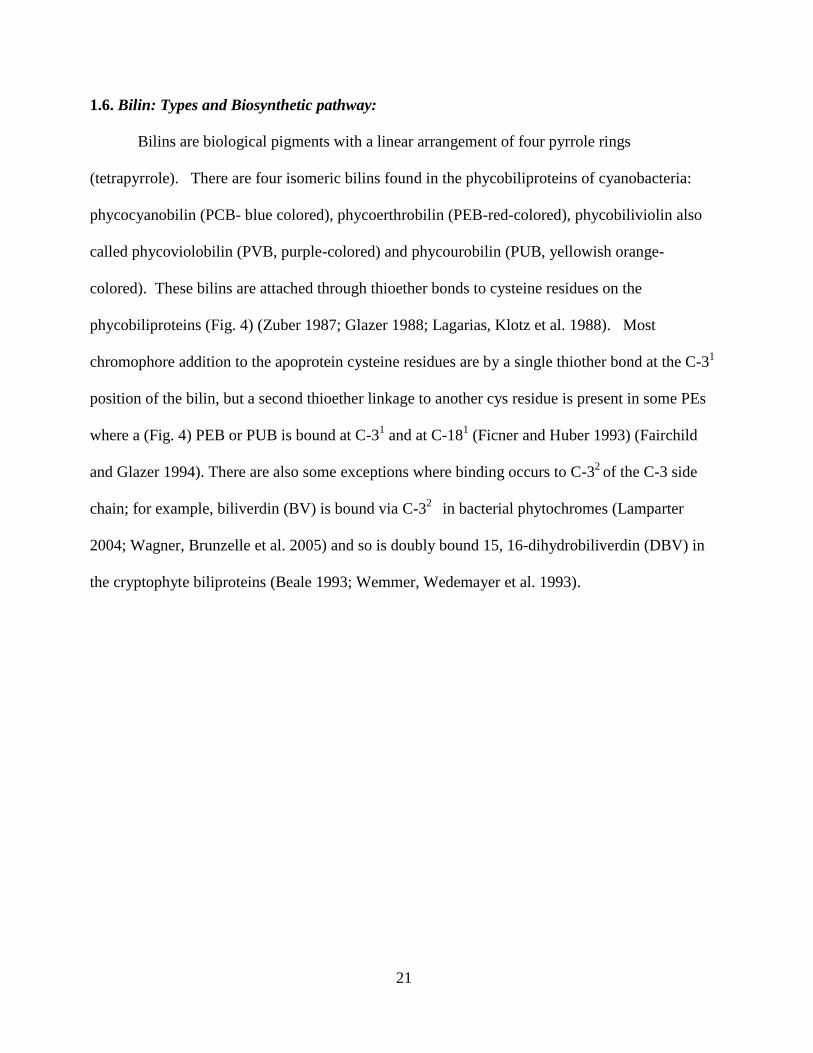
Bilins are biological pigments with a linear arrangement of four pyrrole rings
(tetrapyrrole). There are four isomeric bilins found in the phycobiliproteins of cyanobacteria:
phycocyanobilin (PCB- blue colored), phycoerthrobilin (PEB-red-colored), phycobiliviolin also
called phycoviolobilin (PVB, purple-colored) and phycourobilin (PUB, yellowish orange-
colored). These bilins are attached through thioether bonds to cysteine residues on the
phycobiliproteins (Fig. 4) (Zuber 1987; Glazer 1988; Lagarias, Klotz et al. 1988). Most
chromophore addition to the apoprotein cysteine residues are by a single thiother bond at the C-31
position of the bilin, but a second thioether linkage to another cys residue is present in some PEs
where a (Fig. 4) PEB or PUB is bound at C-31 and at C-18
1 (Ficner and Huber 1993) (Fairchild
and Glazer 1994). There are also some exceptions where binding occurs to C-32
of the C-3 side
chain; for example, biliverdin (BV) is bound via C-32
in bacterial phytochromes (Lamparter
2004; Wagner, Brunzelle et al. 2005) and so is doubly bound 15, 16-dihydrobiliverdin (DBV) in
the cryptophyte biliproteins (Beale 1993; Wemmer, Wedemayer et al. 1993).
22
Fig. 5. Structures of bound bilins. (Top row) Type 1 chromophores with a single bond between
C-2 and C-3; bottom row: type 2 chromophores with a Δ2,3-double bond. There is always a 31-
linkage present, for some chromophores an optional second linkage (181) is indicated . The figure
is modified from (Storf, Parbel et al. 2001) .
23
The phycobiliproteins in Synechocystis sp. PCC 6803 and Synechococcus sp. PCC 7002
have only PCB attached, whereas the PBP of F. diplosiphon have PCB and PEB (Fairchild and
Glazer 1994). The PBPs of Synechococcus sp. WH8020, WH8102, or RS 9916 have PCB, PEB,
and PUB attached (Lagarias, Klotz et al. 1988; Wilbanks and Glazer 1993; Six, Thomas et al.
2007). Among the different types of phycobiliproteins, AP exclusively contains PCB (Glazer
1985). PC mostly contains PCB (Glazer 1985) with exceptions such as in certain marine
cyanobacterial species like Synechococcus sp. RS 9916 (known as R-PC instead of just PC)
where it carries PCB, PEB, and PUB (Blot, Wu et al. 2009). PE may also contain PEB and/or
PUB (Alberte, Wood et al. 1984; Kahn, Mazel et al. 1997; Six, Thomas et al. 2005).
The biosynthetic pathways for all bilins start with heme, also called protoheme (Beale
1999). The heme molecule undergoes an oxidative cleavage by the enzyme heme oxygenase
(encoded by ho1 gene) (Cornejo, Willows et al. 1998) to form the common precursor molecule
for all bilins known as biliverdin IXα (BV) (See Fig. 6). The molecular mechanism of heme
degradation may proceed via three independent steps involving attack by molecular oxygen,
followed by elimination of carbon monoxide and formation of iron-biliverdin (Brown and Troxler
1982). The overall heme degradation pathway occurs via two intermediates: α-hydroxyheme and
verdoheme, however, the redox stoichiometry for the overall HO1 reaction remains unclear
(Sakamoto, Sugishima et al. 2002). BV then undergoes further reduction by highly specific
ferredoxin- dependent bilin reductases (FDBRs) (Frankenberg, Mukougawa et al. 2001). These
enzymes lack organic or metal cofactors (Frankenberg and Lagarias 2003; Dammeyer, Bagby et
al. 2008) and are comprised of several members, each targeting specific double bonds in the
tetrapyrrole (Ponkratov, Friedrich et al. 2004) with each electron coming from the FeS protein,
ferredoxin. Phycocyanobilin: ferredoxin oxidoreductase (PcyA), which belongs to the family of
24
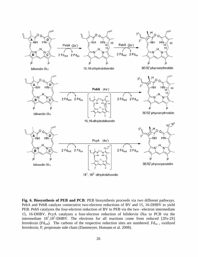
FDBRs, catalyzes the four-electron reduction of BV to PCB (See Fig 6); (Dammeyer, Homann et
al. 2008). FDBRs are found exclusively in oxygenic phytosynthetic organisms. This enzyme
family can be distinguished from the NADPH-dependent biliverdin reducatses BVR (Kapitulnik
and Maines 2009) and BvdR (Schluchter and Glazer 1997) by their ferredoxin-dependency and
their double bond reduction regiospecificity (Frankenberg, Mukougawa et al. 2001; Frankenberg
and Lagarias 2003). The latter property is responsible for the large diversity of their bilin
products which absorb light throughout the visible and near- IR spectral regions (Tu, Gunn et al.
2004). PcyA mediates two, two-electron reductions at both vinyl groups of BV (Fig. 6) (Storf,
Parbel et al. 2001; Frankenberg and Lagarias 2003; Dammeyer and Frankenberg-Dinkel 2006).
In this reaction it converts BV to PCB through a visible (greenish-colored) semi-reduced
intermediate 181, 18
2- dihydrobiliverdin (DHBV) (Frankenberg and Lagarias 2003). The DHBV
undergoes further two-electron reduction forming PCB, which is evident from its native blue
color and absorbance maximum at 665 nm (Glazer 1988) (See Fig. 6).
To form PEB there are two consecutive two-electron reduction steps catalyzed by two
enzymes; PebA and PebB, belonging to the FDBRs family of radical enzymes (Dammeyer and
Frankenberg-Dinkel 2006; Dammeyer, Michaelsen et al. 2007). BV reduction is first catalyzed
by 15, 16- DHBV: ferredoxin oxidoreductase (PebA) and yields 15, 16- DHBV by reducing the
C-15 methine bridge of BV. The 15, 16 DHBV undergoes further reduction by PEB: ferredoxin
oxidoreductase (PebB) on the A-ring 2, 3 31,3
2- diene system to form PEB. PebA lacks the metal
ion cofactors, and the reaction most likely proceeds via radical intermediates. Interestingly it was
observed that DHBV bound to PebA can be re-oxidized to BV by molecular oxygen (Dammeyer
and Frankenberg-Dinkel 2006). Reactive oxygen species (ROS) like peroxyradicals are known to
reoxidize albumin bound bilirubin (BR) to BV (Stocker, Glazer et al. 1987) Recently a new
25
enzyme called phycoerythrobilin synthase (PebS) was discovered in the sequencing of a genome
of a myovirus that infects a type of cyanobacteria called Procholorococcus (cyanophage PSSM-2)
(Dammeyer, Bagby et al. 2008; Dammeyer, Homann et al. 2008). PebS was shown to catalyze a
four-electron reduction of BV to PEB.
The four bilins found in cyanobacteria fall into two groups based upon their structure,
reactivities, and abundance. PCB and PEB are the most abundant in PBP and can be cleaved
from PBP producing a Δ 3, 3 ethylidene group (see Fig. 6). The biosynthetic pathways for these
two bilins have been characterized and were previously described.
The second group of bilins includes PUB and PVB. These bilins cannot be cleaved
directly from PBP and contain a vinyl group at C3, so they should be added or produced via a
different mechanism. The pathway for PVB is known; it is produced by a bilin lyase/ isomerase
composed of PecE and PecF. This enzyme attaches PCB to the α subunit of PEC and then
performs Δ4 Δ2 isomerization to form PVB (see Fig. 7) (Jung, Chan et al. 1995; Zhao, Deng et
al. 2000; Storf, Parbel et al. 2001; Tooley and Glazer 2002; Zhao, Wu et al. 2002).
Recently the biosynthetic pathway of PUB of R-PC-V was elucidated. It is produced by a
bilin lyase/isomerase composed of RpcG (Blot, Wu et al. 2009), where this enzyme attaches PEB
to the α subunit of R-PC-V and then performs Δ4 Δ2 isomerization to form PUB. Part of this
thesis project will focus on characterizing a new type of bilin lyase/isomerase specific for PEII
subunits.
26
Fig. 6. Biosynthesis of PEB and PCB: PEB biosynthesis proceeds via two different pathways.
PebA and PebB catalyze consecutive two-electron reductions of BV and 15, 16-DHBV to yield
PEB. PebS catalyzes the four-electron reduction of BV to PEB via the two –electron intermediate
15, 16-DHBV. PcyA catalyzes a four-electron reduction of bilidervin IX to PCB via the
intermediate 181,18
2-DHBV. The electrons for all reactions come from reduced [2Fe-2S]
ferredoxin (Fdred). The carbons of the respective reduction sites are numbered. Fdox , oxidized
ferredoxin; P, propionate side chain (Dammeyer, Homann et al. 2008).
27
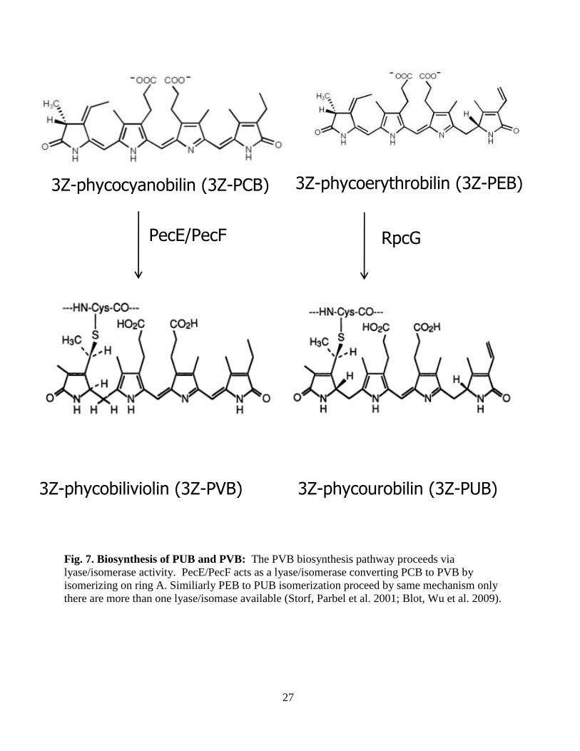
Fig. 7. Biosynthesis of PUB and PVB: The PVB biosynthesis pathway proceeds via
lyase/isomerase activity. PecE/PecF acts as a lyase/isomerase converting PCB to PVB by
isomerizing on ring A. Similiarly PEB to PUB isomerization proceed by same mechanism only
there are more than one lyase/isomase available (Storf, Parbel et al. 2001; Blot, Wu et al. 2009).
3Z-phycocyanobilin (3Z-PCB) 3Z-phycoerythrobilin (3Z-PEB)
3Z-phycobiliviolin (3Z-PVB) 3Z-phycourobilin (3Z-PUB)
PecE/PecF RpcG
28
1.7. Bilin addition to phycobiliproteins:
When any one of the four known cyanobacterial bilins get attached to the PBP in the
correct manner associating with the PBP amino acids residues so that it is held in stretched
conformation, the PBP becomes highly fluorescent (Scheer and Zhao 2008). The bilin ligation is
aided by a category of enzymes designated as bilin lyases. Various published data show that
different cyanobacterial bilin lyase enzymes are involved in bilin addition, isomerization and/ or
detachment of bilin chromophores to the cysteine residues of PBP (Arciero, Bryant et al. 1988;
Fairchild, Zhao et al. 1992; Zhou, Gasparich et al. 1992; Fairchild and Glazer 1994; Dolganov
and Grossman 1999; Zhao, Deng et al. 2000; Zhao, Su et al. 2007). Four major classes of
cyanobacterial bilin lyases are known: CpcEF type, CpcSU type, CpcT type and the autocatalytic
type. Each of these categories of bilin lyases are unrelated in their primary amino acid sequences
and are involved in attachment of different bilin chromophores to specific cysteine residues on
PBP increasing the light capturing ability for the photosystem.
N.B. For the purpose of simplicity while reviewing this thesis; if the protein name is designated
Cpc, it means they are located or involved in various function in PC subunits; Apc means
involvement in APC subunits, and Cpe meaning involment in PE, with only a few exceptions.
1.7.1 E/F-type lyase:
The first bilin lyase to be characterized was a heterodimer composed of CpcE and CpcF
(1:1) that is responsible for attachment of PCB to α – PC ( also called CpcA) (Fairchild, Zhao et
al. 1992; Fairchild and Glazer 1994). These two genes were first identified because they are
encoded downstream of the operon encoding PC structural genes cpcBACD. The phycocyanin
rods in cpcF mutants had apo- α-PC but normal levels of chromophorylated β-subunit of PC
(CpcB) suggesting the CpcEF type lyases are specific for PCB attachment to the α subunit of PC
29
(Zhou, Gasparich et al. 1992; Zhao, Deng et al. 2000). This heterodimeric lyase can catalyze both
the forward and reverse (releasing) reaction (Fairchild, Zhao et al. 1992). It also catalyzes the
addition of PEB to apo-α-PC (CpcA) in vitro (Fairchild, Zhao et al. 1992) and in vivo (Alvey,
Biswas et al. 2011).
For the purpose of generating constructs with phycobiliproteins for use as fluorescent
probes in living cells, Tooley et al. recreated the entire fluorescent holo α-PC in E .coli by
coexpressing cpcA, cpcE, cpcF (PC subunits and bilin lyase) from one plasmid and the bilin
biosynthetic genes; ho1 and pcyA from another plasmid (Fairchild, Zhao et al. 1992; Fairchild and
Glazer 1994; Glazer and Wedemayer 1995). The product yield was shown to have 33% of the
produced apo-CpcA converted to holo-CpcA.
PecE/PecF (from Nostoc sp. PCC 7120 and Mastigocladus laminosus) belongs to the
CpcE/F family because they share 47 % sequence similarity to CpcE and CpcF, respectively.
PEC- subunit (PecA) contains the photoactive PVB chromophore at Cys-84, and PecE/F from
these organisms not only attaches PCB to this site but also simultaneously isomerizes it to PVB
(Jung, Chan et al. 1995; Storf, Parbel et al. 2001; Tooley and Glazer 2002; Zhao, Wu et al. 2002).
Zhao et al. (Zhao, Deng et al. 2000) performed the in vitro reactions by adding recombinant PecE
and PecF from E. coli to apo- α –PEC and saw formation of highly fluorescent holo-PecA based
on absorbance and fluorescence spectra. When apo-α-PEC was incubated without PecE and
PecF, the bilin adduct formed was mesobiliverdin instead of PVB which suggested that PecE and
PecF proteins were necessary for the addition of PCB and then its subsequent isomerization to
PVB. Tooley at al. (Tooley and Glazer 2002) recreated the pathway of α PEC biosynthesis in
E.coli by heterologous co expression of two plasmids; one containing all essential genes for PCB
biosynthesis (ho1X and pcyA) and another plasmid containing pecA (apo- α –PEC) and pecE and
30
pecF (bilin lyase/isomerase). The holo-α-PEC was purified and its spectral properties showed it
had the same characteristics as native -PEC.
There are other bilin lyases present related to CpcE/F in cyanobacteria are yet to be
characterized. One member of the CpcE/F type bilin lyase family is found only in cyanobacterial
species containing PE on their PBS rods (Glazer 1989) is CpeY and CpeZ. These putative bilin
lyases are emcoded within the operon for PE rod structure proteins CpeBA (CpeBAZY). Kahn et
al. showed that transposon insertional within cpeY in F. diplosiphon resulted in 46% less
phycoerythrin (PE) being made (Kahn, Mazel et al. 1997), suggesting the cpeYZ gene might be a
putative bilin lyase involved in the attachment of phycoerythrobilin to either the α or β subunits.
Part of this thesis will include a detailed study on CpeYZ lyase activity. The CpcEF family of
bilin lyase was found to be specific for the cysteines residues on the subunits of PC or PC
(central bilin). However there are many other Cys containing bilins for which no enzyme had
been identified until 2004 (Shen, Saunee et al. 2004).
1.7.2 SU type lyase:
More recently, several studies have characterized the bilin attachment pathway on
subunits of PC, which possess two bilin ligation sites Cys- 82 and Cys-153.
In F. diplosiphon the PE linker polypeptide operon (CpeC CpeD and CpeF) encodes the
genes: cpeCDFSTR. The cpeS and cpeT genes were found in the genomes of other organisms
containing phycobiliproteins, but were absent in species lacking PE. There was no direct evidence
that the cpeS and cpeT genes were transcriptional regulators as they had no sequence similarity to
such DNA binding proteins. The Schluchter and Bryant labs showed that there are 2 paralogue
genes to F. diplosiphon in Synechococcus sp. PCC 7002, which were called CpcS-I/CpcU. They
31
showed that CpcS and CpcU form a heterodimer (1:1), and that they catalyze the attachment of
PCB to Cys-84 of β-phycocyanin (CpcB) and to α and β subunits of AP (Shen, Saunee et al. 2004;
Saunée, Williams et al. 2008; Scheer and Zhao 2008; Shen, Schluchter et al. 2008). Zhao et al.
showed that the bilin lyase genes they called cpeS2 and cpeS1 from Nostoc sp. PCC 7120 are
homologous to the cyanobacterial lyase CpcS (Zhao, Su et al. 2007). CpcS-I a single subunit
bilin lyase was described as a nearly “Universal bilin lyase” (Zhao, Su et al. 2007) (See Table 2)
since it was found to be involved in PCB attachment to Cys-84 of β-phycocyanin (CpcB), to the
AP subunits, and PEB attachment to Cys-82 of α and and Cys-80 β subunits of C-phycoerythrin
from F. diplosiphon in vitro (Zhao, Su et al. 2007).
1.7.3. T-type lyase:
The third type of cyanobacterial bilin lyase is known as the CpcT-type, and is unrelated to
CpcEF and CpcS type. They shared sequence similarity to the CpeT bilin lyase from F.
diplosiphon. Shen et al. showed that the single subunit CpcT type bilin lyase present in
Synechococcus sp. PCC 7002 can attach PCB to β-PC at Cys-153 (Shen, Saunee et al. 2006).
Inactivating the cpcT gene in Synechococcus sp. PCC 7002 resulted in cyanobacteria with 40%
less PC than wild type, smaller PBS and PBPs with more red –shifted absorbance and
fluorescence spectra. Recombinant CpcT was shown to catalyze the regiospecific PCB ligation to
Cys-155 on CpcB from Synechococcus sp. PCC 7002 (Shen, Saunee et al. 2006). The chiral
carbon at C31 carbon attached to Cys-153 has S stereochemistry (Shen, Saunee et al. 2006),
whereas the S/U type and E/F type bilin lyase attach bilins to Cys with R stereochemistry. The
protein CpcT1 from another cyanobacterium Anabena sp. PCC7120 is homologous to the CpcT
type bilin lyase and was shown to attach PCB to Cys-155 of β subunit of both PC (CpcB) and
PEC (PecB) (Zhao, Zhang et al. 2007).
32
1.7.4 Autocatalytic lyase:
For the last known family of bilin lyases, the lyase reaction is catalyzed by the biliprotein
itself. For example, plants, cyanobacteria and other bacteria have phytochromes, which are
switchable photoreceptors, responsive to red and far-red light sources, phytochrome-like proteins,
or other cyanochromes. These proteins contain a bilin chromophore, which is autoligated without
the aid of separate enzymes. (Wu and Lagarias 1996; Wu and Lagarias 2000; Montgomery and
Lagarias 2002; Lamparter 2004; Zhao, Ping et al. 2005; Rockwell, Njuguna et al. 2008). The only
other known example of a PBP that is capable of auto ligation is the AP- like domain of the large
core membrane linker protein designated LCM or ApcE which contains a PCB. It was shown to
have a red-shifted absorbance maximum at ~665 nm, and played a role in accepting the energy
from the chromophores in the core of the PBS and transferring it to the reaction centers (Capuano,
Braux et al. 1991; Gindt, Zhou et al. 1994; Sidler 1994; Ajlani and Vernotte 1998). Autocatalytic
addition of PCB was reported to occur for a truncation product of ApcE in 4 M urea (Zhao, Ping
et al. 2005). Addition of detergents can eliminate that requirement for bilin lyases for some
phycobiliproteins (Zhao, Ping et al. 2005), and so it is possible that the urea present in the
reaction mixture with ApcE may have had the same effect as a detergent. ApcE autocatalytic
lyase activity using an in vivo coexpression system in E. coli is described in this thesis.
1.8. Other post-translational modifications to phycobiliproteins:
While chromophore addition represent one type of posttranslational modification that all
PBPs undergo, a second type of modification is a methylation reaction that occurs specifically
only on the β subunits of most phycobiliproteins and is catalyzed by an S-adenosylmethionine-
dependent methytranferase designated CpcM (Swanson and Glazer 1990; Miller, Leonard et al.
33
2008; Shen, Leonard et al. 2008). The methytransferase reaction produces a highly conserved γ-
N-methylasparagine residue at the β-72 position of almost all β-subunits isolated from
cyanobacteria, red algae, and cryptomonads (Klotz, Leary et al. 1986; Klotz and Glazer 1987;
Rümbeli, Suter et al. 1987; Ducret, Sidler et al. 1994; Saunée, Williams et al. 2008). This
modification is thought to change the environment of the chromophore at position β-82 to
minimize the rates of nonradiative energy loss within PBS (Thomas, Bricker et al. 1993;
Thomas, McMahon et al. 1994). However, characterization of cpcM mutants also showed that
these strains contain very high levels of reactive oxygen species (Shen, Leonard et al. 2008)
(Schirmer, Huber et al. 1986). In vitro studies of CpcM suggested that the enzyme probably
methylates β subunits after chromophorylation but prior to trimer assembly in PBPs (Miller,
Leonard et al. 2008)
1.9. Chromatic acclimation:
Certain cyanobacterial species have the ability to undergo changes in their phycobilisome
protein based on the light condition which is known as complementary chromatic acclimation
(CCA). F. diplosiphon and Synechococcus sp. RS 9916 used in this study can undergo CCA. The
term CCA was first coined by two scientists; Engelman and Gaidukov (Engelmann 1883;
Engelmann 1902; Gaiducov 1902; Gaiducov 1903), but they have different ideas for CCA.
Engleman proposed CCA as variations in the pigment distributions in various alge correlated to
changes in the light sources. Although subsequent studies did not support this idea (Crossett,
Drew et al. 1965; Ramus 1983; Ramus. J. and van der Meer 1983; Saffo 1987), this led to a
modification proposed by Guidukov in 1902 that cyanobacterium Oscillatoria sancta changed
their protein composition having a blue phenotype when grown in red light and red phenotype
34
when grown in green or white light. There are two major kinds of CCA: Type III and Type IV.
Type III CCA found in F. diplosiphon was described as color change due to degradation of one
rod protein and replacing it by different protein with different chromophores (Fig. 8) (Boresch
1922). PC accumulates (precisely inducible PC) in red light and PE accumulates in green light
(Fig. 8). Thus Type III CCA can only occur in cyanobacterial species that make both PC and PE,
although not all such species are capable of this process (Tandeau de Marsac 1977). Different
studies over time showed that there are many additional cellular responses affected by shifts
between green and red lights, including changes in cell and filament architecture (Bogorad,
Gendel et al. 1982; Montgomery and Lagarias 2002; Whitaker, Pattanaik et al. 2011; Bennett and
Bogorad 1973), cell differentiation states (Lazaroff and Schiff 1962; Damerval, Guglielmi et al.
1991) and the abundance of many RNAs and proteins that do not encode PBS components
(Gendel, Ohad et al. 1979; Stowe-Evans, Ford et al. 2004).
The changes in the ratio of PC to PE in F. diplosiphon during CCA (Fig. 8) are typical of
Type III species and lead to the synthesis of PBS with absorption characteristics that are
optimized to capture the most abundant wavelength(s) of ambient light in the green-to-red region
of the spectrum. This is because PE most efficiently absorbs green light, whereas PC absorbs red
light most effectively (Fig. 8). There are two proposed signal transduction model system involved
in this Type III CCA regulation- one is the regulation of complementary chromatic acclimation
(Rca) system (Sobczyk, Schyns et al. 1993; Terauchi, Montgomery et al. 2004) and the second
pathway suported by photobiology experiment is known as the Cgi (control of green light
induction) (Sidler 1994). The Rca system affects the CCA regulation by decreasing the level of
cpeCDE and cpeBA when exposed to red light while increasing gene expression when exposed to
green light (Alvey, Karty et al. 2003; Li and Kehoe 2005).
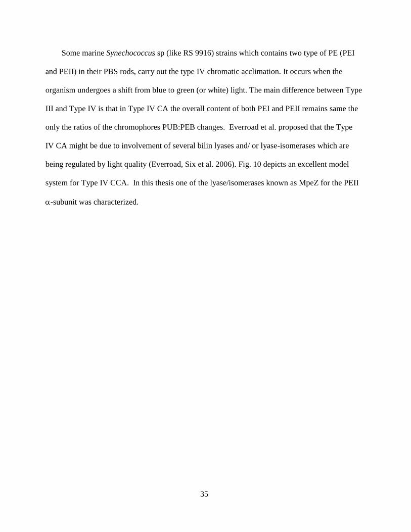
35
Some marine Synechococcus sp (like RS 9916) strains which contains two type of PE (PEI
and PEII) in their PBS rods, carry out the type IV chromatic acclimation. It occurs when the
organism undergoes a shift from blue to green (or white) light. The main difference between Type
III and Type IV is that in Type IV CA the overall content of both PEI and PEII remains same the
only the ratios of the chromophores PUB:PEB changes. Everroad et al. proposed that the Type
IV CA might be due to involvement of several bilin lyases and/ or lyase-isomerases which are
being regulated by light quality (Everroad, Six et al. 2006). Fig. 10 depicts an excellent model
system for Type IV CCA. In this thesis one of the lyase/isomerases known as MpeZ for the PEII
-subunit was characterized.

36
Fig. 8. The color phenotypes of F. diplosiphon filaments grown on agar plates and fully
acclimated to green light (left) and red light (right). The accumulation of different pigmented
proteins into phycobilisome (PBS) rods renders the cells brick red or blue green (Kehoe and Gutu
2006)
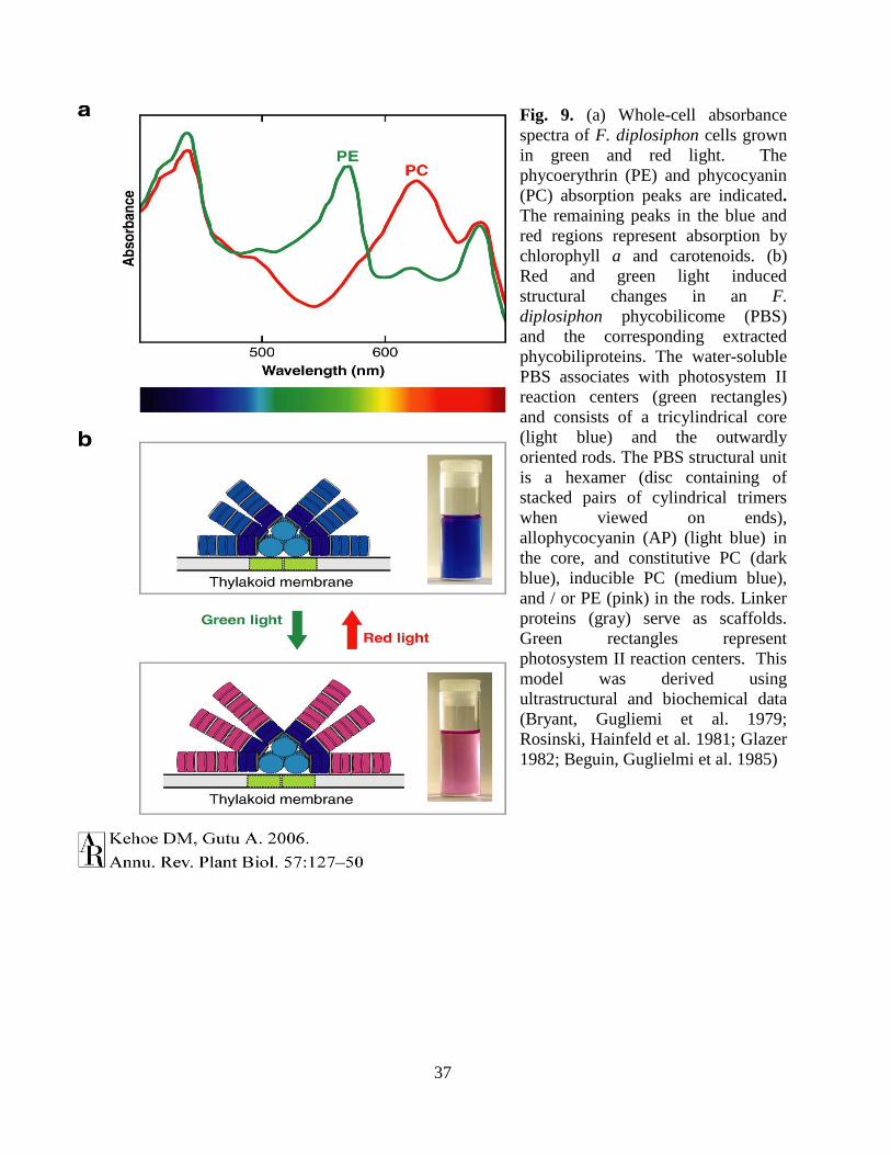
37
Fig. 9. (a) Whole-cell absorbance
spectra of F. diplosiphon cells grown
in green and red light. The
phycoerythrin (PE) and phycocyanin
(PC) absorption peaks are indicated.
The remaining peaks in the blue and
red regions represent absorption by
chlorophyll a and carotenoids. (b)
Red and green light induced
structural changes in an F.
diplosiphon phycobilicome (PBS)
and the corresponding extracted
phycobiliproteins. The water-soluble
PBS associates with photosystem II
reaction centers (green rectangles)
and consists of a tricylindrical core
(light blue) and the outwardly
oriented rods. The PBS structural unit
is a hexamer (disc containing of
stacked pairs of cylindrical trimers
when viewed on ends),
allophycocyanin (AP) (light blue) in
the core, and constitutive PC (dark
blue), inducible PC (medium blue),
and / or PE (pink) in the rods. Linker
proteins (gray) serve as scaffolds.
Green rectangles represent
photosystem II reaction centers. This
model was derived using
ultrastructural and biochemical data
(Bryant, Gugliemi et al. 1979;
Rosinski, Hainfeld et al. 1981; Glazer
1982; Beguin, Guglielmi et al. 1985)
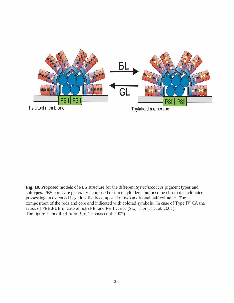
38
Fig. 10. Proposed models of PBS structure for the different Synechococcus pigment types and
subtypes. PBS cores are generally composed of three cylinders, but in some chromatic aclimaters
possessing an extended LCM, it is likely composed of two additional half cylinders. The
composition of the rods and core and indicated with colored symbols. In case of Type IV CA the
ratios of PEB:PUB in case of both PEI and PEII varies (Six, Thomas et al. 2007). The figure is modified from (Six, Thomas et al. 2007)
39
1.10. Purpose of this work:
The overall purpose of this thesis is to characterize the biochemical pathways involved in
the formation of the brilliantly colored phycobiliproteins in various cyanobacterial speices and to
demonstrate how they can be used for biotechnological applications. Here various lyase and/ or
lyase/isomerase present in different species of cyanobacteria were characterized in detail.
The first goal of the research work presented here was to develop an in vivo multiplasmid
heterologous expression system in E.coli by co-expressing three categories of genes: genes
encoding the phycobiliproteins substrate, genes for biosynthetic enzymes of the prosthetic groups,
and the enzymes which catalyze the ligation of the prosthetic groups.
This work included is identify the bilin lyases involved in ligating PCB on the less
abundant allophycocyanin subunits ApcD and ApcF in Synechococcus sp. PCC 7002 and also
elucidate intrinsic bilin lyase activity of N-terminal, allophycocyanin AP-like domain of ApcE
(Lcm99
).
The second goal of this work was to create unique phycobiliproteins in E.coli cells by
chromophorylating with unnatural occurring bilins, which may have potential biotechnological
applications.
The third goal of this work was to identify and characterize the bilin lyases for PE
subunits in Fremyella diplosiphon.
The fourth goal of this work was to elucidate the biochemical pathway for the less studied
bilin; phycourobilin (PUB) by identifying and characterizing the bilin lyase/ isomerase for PE II
subunits responsible for Type IV CA in the marine cyanobacterial species, Synechococcus sp. RS
9916.

40
The last goal of this thesis project was to characterize a new CpcS type lyase named as Ter
13 or TECpcS from Thermosynechococcus elongatus (Described in Appendix).
41
2.0. MATERIALS AND METHODS
2.1 Construction of expression vectors:
Plasmids used in this study are listed in Table 4. Some of the expression vectors used in this study
were previously described (Tooley, Cai et al. 2001; Shen, Saunee et al. 2006; Miller, Leonard et
al. 2008) or made by our collaborators in the Bryant lab (Pennsylvania State University) or in
the Kehoe lab (Indiana University) . All expression constructs newly produced for this study
were sequenced at the W. M. Keck Conservation and Molecular Genetics laboratory (University
of New Orleans) to confirm that no mutations had been introduced during PCR amplification and
cloning.
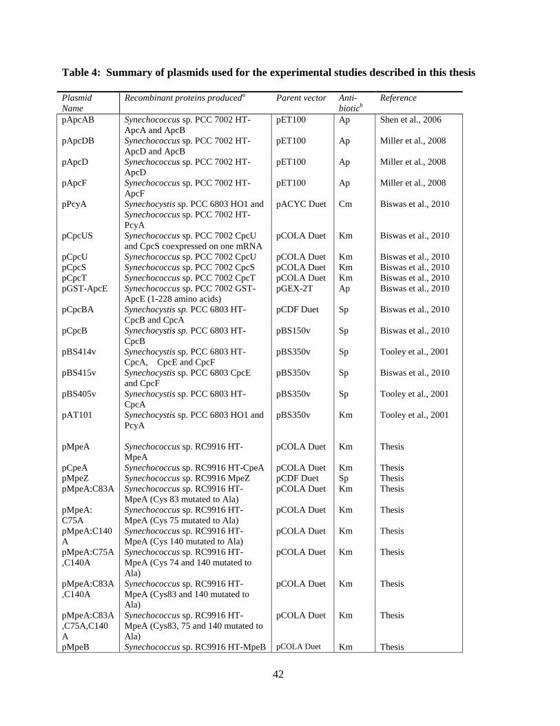
42
Table 4: Summary of plasmids used for the experimental studies described in this thesis
Plasmid
Name
Recombinant proteins produceda Parent vector Anti-
bioticb
Reference
pApcAB Synechococcus sp. PCC 7002 HT-
ApcA and ApcB
pET100 Ap Shen et al., 2006
pApcDB
Synechococcus sp. PCC 7002 HT-
ApcD and ApcB
pET100
Ap
Miller et al., 2008
pApcD
Synechococcus sp. PCC 7002 HT-
ApcD
pET100 Ap Miller et al., 2008
pApcF Synechococcus sp. PCC 7002 HT-
ApcF
pET100 Ap Miller et al., 2008
pPcyA Synechocystis sp. PCC 6803 HO1 and
Synechococcus sp. PCC 7002 HT-
PcyA
pACYC Duet Cm Biswas et al., 2010
pCpcUS Synechococcus sp. PCC 7002 CpcU
and CpcS coexpressed on one mRNA
pCOLA Duet Km Biswas et al., 2010
pCpcU Synechococcus sp. PCC 7002 CpcU pCOLA Duet Km Biswas et al., 2010
pCpcS Synechococcus sp. PCC 7002 CpcS pCOLA Duet Km Biswas et al., 2010
pCpcT Synechococcus sp. PCC 7002 CpcT pCOLA Duet Km Biswas et al., 2010
pGST-ApcE Synechococcus sp. PCC 7002 GST-
ApcE (1-228 amino acids)
pGEX-2T Ap Biswas et al., 2010
pCpcBA
Synechocystis sp. PCC 6803 HT-
CpcB and CpcA
pCDF Duet
Sp
Biswas et al., 2010
pCpcB Synechocystis sp. PCC 6803 HT-
CpcB
pBS150v Sp Biswas et al., 2010
pBS414v
Synechocystis sp. PCC 6803 HT-
CpcA, CpcE and CpcF
pBS350v Sp Tooley et al., 2001
pBS415v Synechocystis sp. PCC 6803 CpcE
and CpcF
pBS350v Sp Biswas et al., 2010
pBS405v Synechocystis sp. PCC 6803 HT-
CpcA
pBS350v
Sp Tooley et al., 2001
pAT101 Synechocystis sp. PCC 6803 HO1 and
PcyA
pBS350v
Km Tooley et al., 2001
pMpeA Synechococcus sp. RC9916 HT-
MpeA
pCOLA Duet Km Thesis
pCpeA Synechococcus sp. RC9916 HT-CpeA pCOLA Duet Km Thesis
pMpeZ Synechococcus sp. RC9916 MpeZ pCDF Duet Sp Thesis
pMpeA:C83A Synechococcus sp. RC9916 HT-
MpeA (Cys 83 mutated to Ala)
pCOLA Duet Km Thesis
pMpeA:
C75A
Synechococcus sp. RC9916 HT-
MpeA (Cys 75 mutated to Ala)
pCOLA Duet Km Thesis
pMpeA:C140
A
Synechococcus sp. RC9916 HT-
MpeA (Cys 140 mutated to Ala)
pCOLA Duet Km Thesis
pMpeA:C75A
,C140A
Synechococcus sp. RC9916 HT-
MpeA (Cys 74 and 140 mutated to
Ala)
pCOLA Duet Km Thesis
pMpeA:C83A
,C140A
Synechococcus sp. RC9916 HT-
MpeA (Cys83 and 140 mutated to
Ala)
pCOLA Duet Km Thesis
pMpeA:C83A
,C75A,C140
A
Synechococcus sp. RC9916 HT-
MpeA (Cys83, 75 and 140 mutated to
Ala)
pCOLA Duet Km Thesis
pMpeB Synechococcus sp. RC9916 HT-MpeB pCOLA Duet Km Thesis
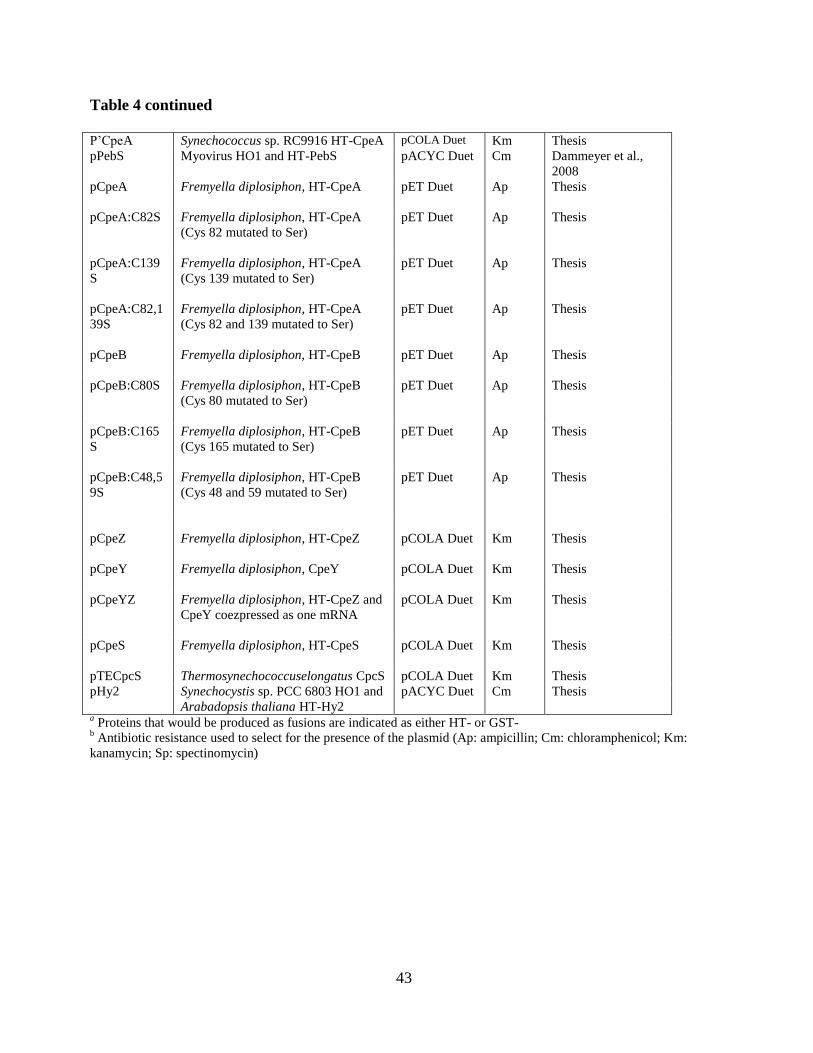
43
Table 4 continued
P’CpeA Synechococcus sp. RC9916 HT-CpeA pCOLA Duet Km Thesis
pPebS Myovirus HO1 and HT-PebS
pACYC Duet Cm Dammeyer et al.,
2008
pCpeA Fremyella diplosiphon, HT-CpeA
pET Duet
Ap Thesis
pCpeA:C82S Fremyella diplosiphon, HT-CpeA
(Cys 82 mutated to Ser)
pET Duet Ap Thesis
pCpeA:C139
S
Fremyella diplosiphon, HT-CpeA
(Cys 139 mutated to Ser)
pET Duet Ap Thesis
pCpeA:C82,1
39S
Fremyella diplosiphon, HT-CpeA
(Cys 82 and 139 mutated to Ser)
pET Duet Ap Thesis
pCpeB Fremyella diplosiphon, HT-CpeB
pET Duet Ap Thesis
pCpeB:C80S Fremyella diplosiphon, HT-CpeB
(Cys 80 mutated to Ser)
pET Duet Ap Thesis
pCpeB:C165
S
Fremyella diplosiphon, HT-CpeB
(Cys 165 mutated to Ser)
pET Duet Ap Thesis
pCpeB:C48,5
9S
Fremyella diplosiphon, HT-CpeB
(Cys 48 and 59 mutated to Ser)
pET Duet Ap Thesis
pCpeZ
Fremyella diplosiphon, HT-CpeZ
pCOLA Duet
Km
Thesis
pCpeY
Fremyella diplosiphon, CpeY
pCOLA Duet Km Thesis
pCpeYZ Fremyella diplosiphon, HT-CpeZ and
CpeY coezpressed as one mRNA
pCOLA Duet Km Thesis
pCpeS Fremyella diplosiphon, HT-CpeS
pCOLA Duet Km Thesis
pTECpcS Thermosynechococcuselongatus CpcS pCOLA Duet Km Thesis
pHy2 Synechocystis sp. PCC 6803 HO1 and
Arabadopsis thaliana HT-Hy2
pACYC Duet Cm Thesis
a Proteins that would be produced as fusions are indicated as either HT- or GST-
b Antibiotic resistance used to select for the presence of the plasmid (Ap: ampicillin; Cm: chloramphenicol; Km:
kanamycin; Sp: spectinomycin)
44
2.1.1. cpcS-I and cpcU expression construct:
The cpcS-I and cpcU genes were cloned in the pCOLA Duet vector (Novagen, Madison, WI) to
generate plasmid pCpcUS, from which these genes would be cotranscribed to produce HT-CpcU
and CpcS (see Table 4). The cpcS-I gene (SYNPCC7002_A1822) was amplified by PCR from
Synechococcus sp. strain PCC 7002 chromosomal DNA using primers cpcSF and cpcSR (Refer
to table 5) and cloned into pCOLA Duet after digestion with PstI and SalI (restriction sites in
primers are underlined, and the forward primer contains a ribosome binding site) to create
plasmid pCpcS. The cpcU gene (SYNPCC7002_A2053) was amplified by PCR from
Synechococcus sp. strain PCC 7002 chromosomal DNA using cpcUF and cpcUR (Refer to table
5) and cloned into the pCOLA Duet plasmid using the restriction enzymes BamHI and EcoRI
(restriction sites underlined in primers) to create the pCpcU plasmid. The cpcU gene was
subcloned into the pCpcS plasmid using BamHI and EcoRI to create plasmid pCpcUS (See Fig.
11). The CpcU protein will have an amino terminal with 6-Histidine tag.
2.1.2. cpcT expression construct:
The cpcT gene (SYNPCC7002_A1822) was amplified from Synechococcus sp. PCC 7002
chromosomal DNA using primers cpcTF and cpcTR (Refer to table 5) and cloned into pCOLA
Duet after digestion with NdeI and EcoRV (restriction sites are underlined in primers) to create
plasmid pCpcT (See Fig. 12).
2.1.3. pcyA/ho1 expression constructs:
The heme oxygenase 1 gene, ho1 (sll1184) was amplified by PCR from the chromosomal DNA of
Synechocystis sp. PCC 6803 using hox1F and hox1R (Refer to table 5) and cloned into pACYC
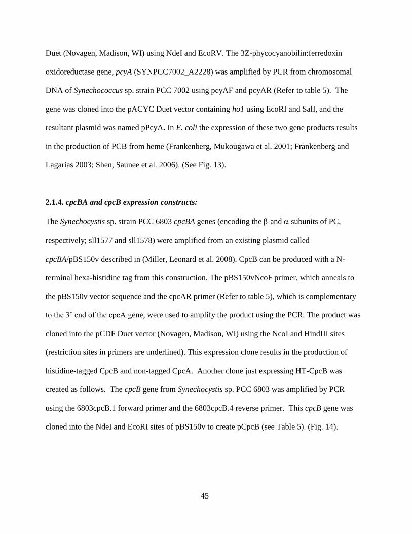
45
Duet (Novagen, Madison, WI) using NdeI and EcoRV. The 3Z-phycocyanobilin:ferredoxin
oxidoreductase gene, pcyA (SYNPCC7002_A2228) was amplified by PCR from chromosomal
DNA of Synechococcus sp. strain PCC 7002 using pcyAF and pcyAR (Refer to table 5). The
gene was cloned into the pACYC Duet vector containing ho1 using EcoRI and SalI, and the
resultant plasmid was named pPcyA. In E. coli the expression of these two gene products results
in the production of PCB from heme (Frankenberg, Mukougawa et al. 2001; Frankenberg and
Lagarias 2003; Shen, Saunee et al. 2006). (See Fig. 13).
2.1.4. cpcBA and cpcB expression constructs:
The Synechocystis sp. strain PCC 6803 cpcBA genes (encoding the and subunits of PC,
respectively; sll1577 and sll1578) were amplified from an existing plasmid called
cpcBA/pBS150v described in (Miller, Leonard et al. 2008). CpcB can be produced with a N-
terminal hexa-histidine tag from this construction. The pBS150vNcoF primer, which anneals to
the pBS150v vector sequence and the cpcAR primer (Refer to table 5), which is complementary
to the 3’ end of the cpcA gene, were used to amplify the product using the PCR. The product was
cloned into the pCDF Duet vector (Novagen, Madison, WI) using the NcoI and HindIII sites
(restriction sites in primers are underlined). This expression clone results in the production of
histidine-tagged CpcB and non-tagged CpcA. Another clone just expressing HT-CpcB was
created as follows. The cpcB gene from Synechocystis sp. PCC 6803 was amplified by PCR
using the 6803cpcB.1 forward primer and the 6803cpcB.4 reverse primer. This cpcB gene was
cloned into the NdeI and EcoRI sites of pBS150v to create pCpcB (see Table 5). (Fig. 14).
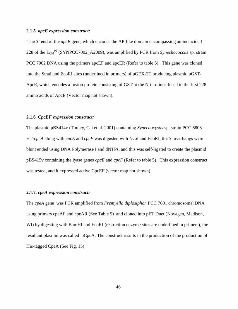
46
2.1.5. apcE expression construct:
The 5’ end of the apcE gene, which encodes the AP-like domain encompassing amino acids 1-
228 of the LCM99
(SYNPCC7002_A2009), was amplified by PCR from Synechococcus sp. strain
PCC 7002 DNA using the primers apcEF and apcER (Refer to table 5). This gene was cloned
into the SmaI and EcoRI sites (underlined in primers) of pGEX-2T producing plasmid pGST-
ApcE, which encodes a fusion protein consisting of GST at the N-terminus fused to the first 228
amino acids of ApcE (Vector map not shown).
2.1.6. CpcEF expression construct:
The plasmid pBS414v (Tooley, Cai et al. 2001) containing Synechocystis sp. strain PCC 6803
HT-cpcA along with cpcE and cpcF was digested with NcoI and EcoRI, the 5’ overhangs were
blunt ended using DNA Polymerase I and dNTPs, and this was self-ligated to create the plasmid
pBS415v containing the lyase genes cpcE and cpcF (Refer to table 5). This expression construct
was tested, and it expressed active CpcEF (vector map not shown).
2.1.7. cpeA expression construct:
The cpeA gene was PCR amplified from Fremyella diplosiphon PCC 7601 chromosomal DNA
using primers cpeAF and cpeAR (See Table 5) and cloned into pET Duet (Novagen, Madison,
WI) by digesting with BamHI and EcoRI (restriction enzyme sites are underlined in primers), the
resultant plasmid was called pCpeA. The construct results in the production of the production of
His-tagged CpeA (See Fig. 15)
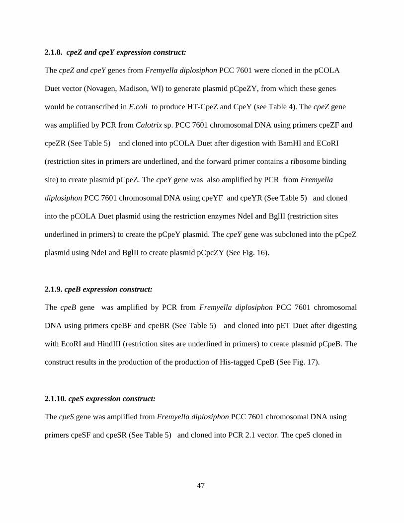
47
2.1.8. cpeZ and cpeY expression construct:
The cpeZ and cpeY genes from Fremyella diplosiphon PCC 7601 were cloned in the pCOLA
Duet vector (Novagen, Madison, WI) to generate plasmid pCpeZY, from which these genes
would be cotranscribed in E.coli to produce HT-CpeZ and CpeY (see Table 4). The cpeZ gene
was amplified by PCR from Calotrix sp. PCC 7601 chromosomal DNA using primers cpeZF and
cpeZR (See Table 5) and cloned into pCOLA Duet after digestion with BamHI and ECoRI
(restriction sites in primers are underlined, and the forward primer contains a ribosome binding
site) to create plasmid pCpeZ. The cpeY gene was also amplified by PCR from Fremyella
diplosiphon PCC 7601 chromosomal DNA using cpeYF and cpeYR (See Table 5) and cloned
into the pCOLA Duet plasmid using the restriction enzymes NdeI and BglII (restriction sites
underlined in primers) to create the pCpeY plasmid. The cpeY gene was subcloned into the pCpeZ
plasmid using NdeI and BglII to create plasmid pCpcZY (See Fig. 16).
2.1.9. cpeB expression construct:
The cpeB gene was amplified by PCR from Fremyella diplosiphon PCC 7601 chromosomal
DNA using primers cpeBF and cpeBR (See Table 5) and cloned into pET Duet after digesting
with EcoRI and HindIII (restriction sites are underlined in primers) to create plasmid pCpeB. The
construct results in the production of the production of His-tagged CpeB (See Fig. 17).
2.1.10. cpeS expression construct:
The cpeS gene was amplified from Fremyella diplosiphon PCC 7601 chromosomal DNA using
primers cpeSF and cpeSR (See Table 5) and cloned into PCR 2.1 vector. The cpeS cloned in
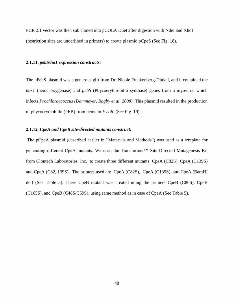
48
PCR 2.1 vector was then sub cloned into pCOLA Duet after digestion with NdeI and XhoI
(restriction sites are underlined in primers) to create plasmid pCpeS (See Fig. 18).
2.1.11. pebS/ho1 expression constructs:
The pPebS plasmid was a generous gift from Dr. Nicole Frankenberg-Dinkel, and it contained the
hox1 (heme oxygenase) and pebS (Phycoerythrobilin synthase) genes from a myovirus which
infects Prochloroccoccus (Dammeyer, Bagby et al. 2008). This plasmid resulted in the production
of phycoerythobilin (PEB) from heme in E.coli. (See Fig. 19)
2.1.12. CpeA and CpeB site-directed mutants construct:
The pCpeA plasmid (described earlier in “Materials and Methods”) was used as a template for
generating different CpeA mutants. We used the Transformer™ Site-Directed Mutagenesis Kit
from Clontech Laboratories, Inc. to create three different mutants; CpeA (C82S), CpeA (C139S)
and CpeA (C82, 139S). The primers used are CpeA (C82S), CpeA (C139S), and CpeA (BamHI
del) (See Table 5). There CpeB mutant was created uning the primers CpeB (C80S), CpeB
(C165S), and CpeB (C48S/C59S), using same method as in case of CpeA (See Table 5).
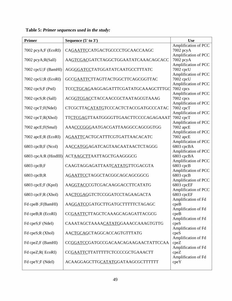
49
Table 5: Primer sequences used in the study:
Primer Sequence (5' to 3') Use
7002 pcyA;F (EcoRI) CAGAATTCCATGACTGCCCCTGCAACCAAGC
Amplification of PCC
7002 pcyA
7002 pcyA;R(SalI) AAGTCGACGATCTAGGCTGGAATATCAAACAGCACC
Amplification of PCC
7002 pcyA
7002 cpcU;F (BamHI) AGGGGATCCTATGGATATCAATGCCTTTATC
Amplification of PCC
7002 cpcU
7002 cpcU;R (EcoRI) GCCGAATTCTTAGTTACTGGCTTCAGCGGTTAC
Amplification of PCC
7002 cpcU
7002 cpcS;F (PstI) TCCCTGCAGAAGGAGATTTCGATATGCAAAGCTTTGC
Amplification of PCC
7002 cpcs
7002 cpcS;R (SalI) ACGGTCGACCTACCAACCGCTAATAGCGTAAAG
Amplification of PCC
7002 cpcs
7002 cpcT;F(NdeI) CTCGCTTACATATGTCCCACTCTACCGATGCCCATAC
Amplification of PCC
7002 cpcT
7002 cpcT;R(XhoI) TTCTCGAGTTAATGGGGTTGAACTTCCCCAGAGAAATT
Amplification of PCC
7002 cpcT
7002 apcE;F(SmaI) AAACCCGGGAATGACGATTAAGGCCAGCGGTGG
Amplification of PCC
7002 apcE
7002 apcE;R (EcoRI) AGAATTCACTGCATTTCGTGATTAACACATC
Amplification of PCC
7002 apcE
6803 cpcB;F (NcoI) AACCATGGAGATCAGTAACAATAACTCTAGGG
Amplification of PCC
6803 cpcBA
6803 cpcA:R (HindIII) ACTAAGCTTAATTAGCTGAAGGGCG
Amplification of PCC
6803 cpcBA
6803 cpcB;F CAAGTAGGAGATTAATCATATGTTCGACGTA
Amplification of PCC
6803 cpcB
6803 cpcB;R AGAATTCCTAGGCTACGGCAGCAGCGGCG
Amplification of PCC
6803 cpcB
6803 cpcE;F (KpnI) AAGGTACCCGTCGACAAGGACCTTCATATG
Amplification of PCC
6803 cpcEF
6803 cpcF;R (XhoI) AACTCGAGGTCTCCGGATCCTAGAAGACTA
Amplification of PCC
6803 cpcEF
Fd cpeB ;F(BamHI) AAGGATCCGATGCTTGATGCTTTTTCTAGAGC
Amplification of Fd
cpeB
Fd cpeB;R (EcoRI) CCGAATTCTTAGCTCAAAGCAGAGATTACGCG
Amplification of Fd
cpeB
Fd cpeS;F (NdeI) CAAATAGCTAAAACATATGGAAACCAAAGTGTTG
Amplification of Fd
cpeS
Fd cpeS;R (XhoI) AACTGCAGCTAGGCACCAGTGTTTATG
Amplification of Fd
cpeS
Fd cpeZ;F (BamHI) CCGGATCCGATGCCGACAACAGAAGAACTATTCCAA
Amplification of Fd
cpeZ
Fd cpeZ;R( EcoRI) CCGAATTCTTATTTTTCTCCCCGCTGAAACTT
Amplification of Fd
cpeZ
Fd cpeY;F (NdeI) ACAAGGAGCTTGCATATGGATAAGCGCTTTTTT
Amplification of Fd
cpeY
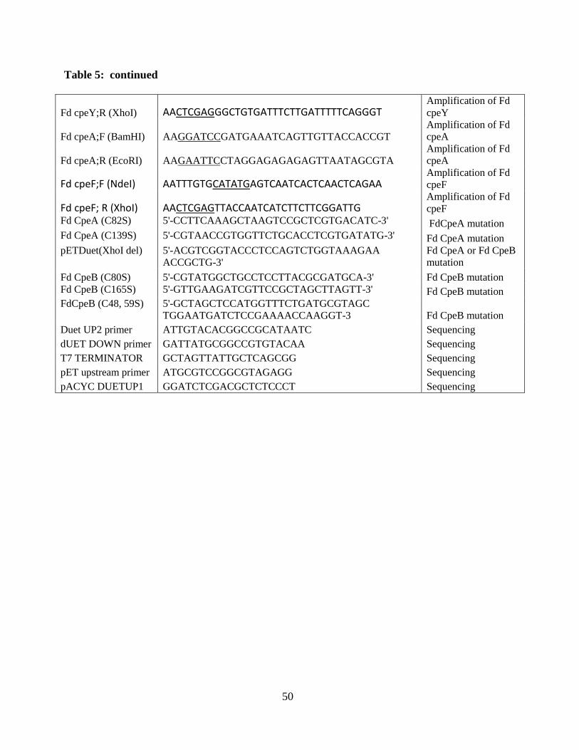
50
Table 5: continued
Fd cpeY;R (XhoI) AACTCGAGGGCTGTGATTTCTTGATTTTTCAGGGT Amplification of Fd
cpeY
Fd cpeA;F (BamHI) AAGGATCCGATGAAATCAGTTGTTACCACCGT
Amplification of Fd
cpeA
Fd cpeA;R (EcoRI) AAGAATTCCTAGGAGAGAGAGTTAATAGCGTA
Amplification of Fd
cpeA
Fd cpeF;F (NdeI) AATTTGTGCATATGAGTCAATCACTCAACTCAGAA Amplification of Fd
cpeF
Fd cpeF; R (XhoI) AACTCGAGTTACCAATCATCTTCTTCGGATTG Amplification of Fd
cpeF
Fd CpeA (C82S) 5'-CCTTCAAAGCTAAGTCCGCTCGTGACATC-3' FdCpeA mutation
Fd CpeA (C139S) 5'-CGTAACCGTGGTTCTGCACCTCGTGATATG-3' Fd CpeA mutation
pETDuet(XhoI del) 5'-ACGTCGGTACCCTCCAGTCTGGTAAAGAA
ACCGCTG-3'
Fd CpeA or Fd CpeB
mutation
Fd CpeB (C80S) 5'-CGTATGGCTGCCTCCTTACGCGATGCA-3' Fd CpeB mutation
Fd CpeB (C165S) 5'-GTTGAAGATCGTTCCGCTAGCTTAGTT-3' Fd CpeB mutation
FdCpeB (C48, 59S) 5'-GCTAGCTCCATGGTTTCTGATGCGTAGC
TGGAATGATCTCCGAAAACCAAGGT-3 Fd CpeB mutation
Duet UP2 primer ATTGTACACGGCCGCATAATC Sequencing
dUET DOWN primer GATTATGCGGCCGTGTACAA Sequencing
T7 TERMINATOR GCTAGTTATTGCTCAGCGG Sequencing
pET upstream primer ATGCGTCCGGCGTAGAGG Sequencing
pACYC DUETUP1 GGATCTCGACGCTCTCCCT Sequencing
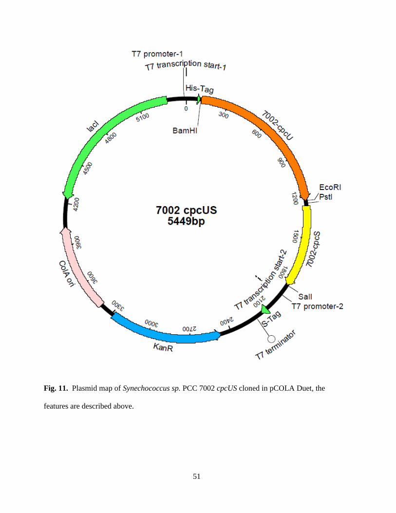
51
Fig. 11. Plasmid map of Synechococcus sp. PCC 7002 cpcUS cloned in pCOLA Duet, the
features are described above.
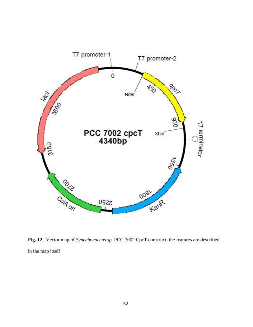
52
Fig. 12. Vector map of Synechococcus sp PCC 7002 CpcT construct, the features are described
in the map itself
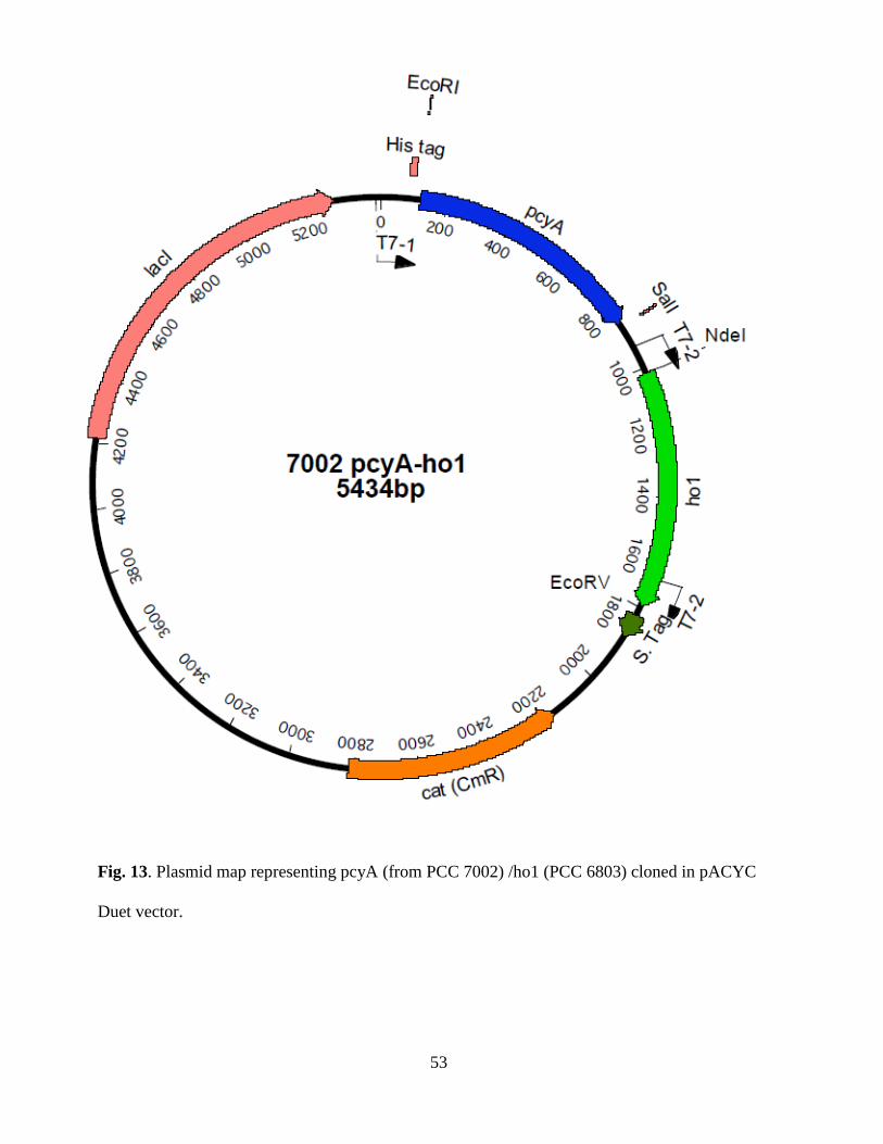
53
Fig. 13. Plasmid map representing pcyA (from PCC 7002) /ho1 (PCC 6803) cloned in pACYC
Duet vector.
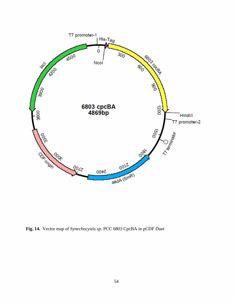
54
Fig. 14. Vector map of Synechocystis sp. PCC 6803 CpcBA in pCDF Duet
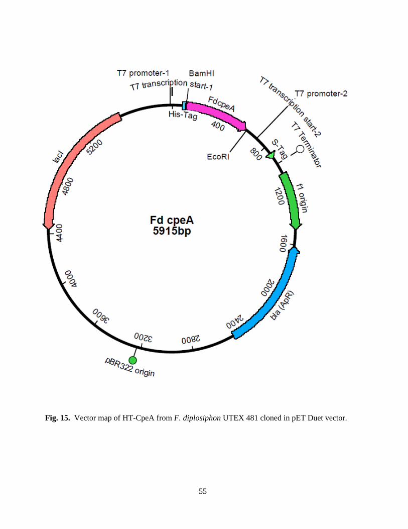
55
Fig. 15. Vector map of HT-CpeA from F. diplosiphon UTEX 481 cloned in pET Duet vector.
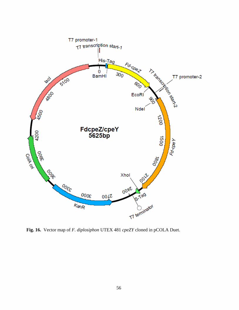
56
Fig. 16. Vector map of F. diplosiphon UTEX 481 cpeZY cloned in pCOLA Duet.
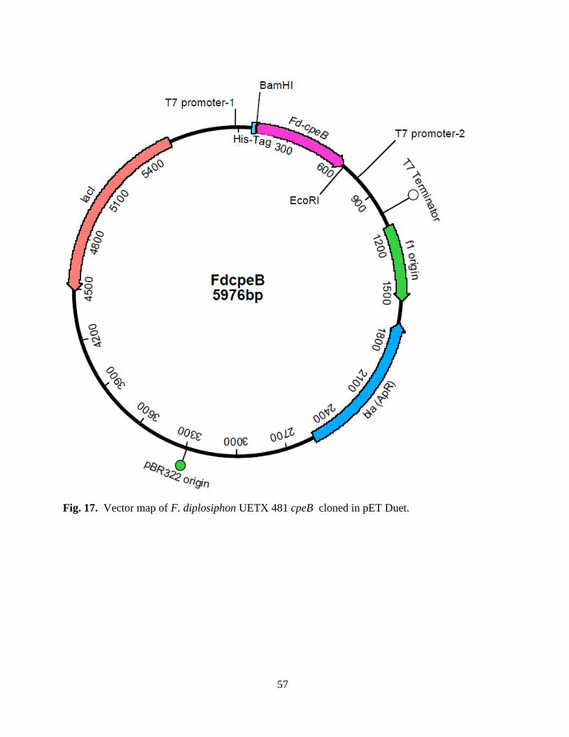
57
Fig. 17. Vector map of F. diplosiphon UETX 481 cpeB cloned in pET Duet.
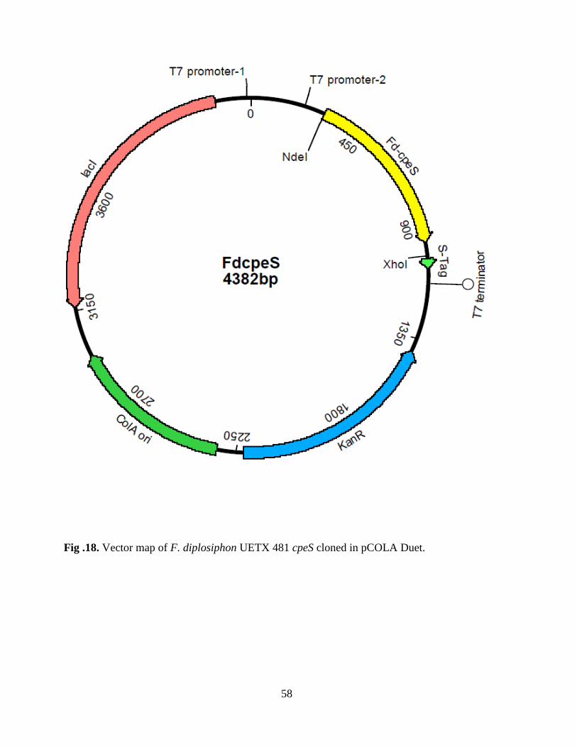
58
Fig .18. Vector map of F. diplosiphon UETX 481 cpeS cloned in pCOLA Duet.
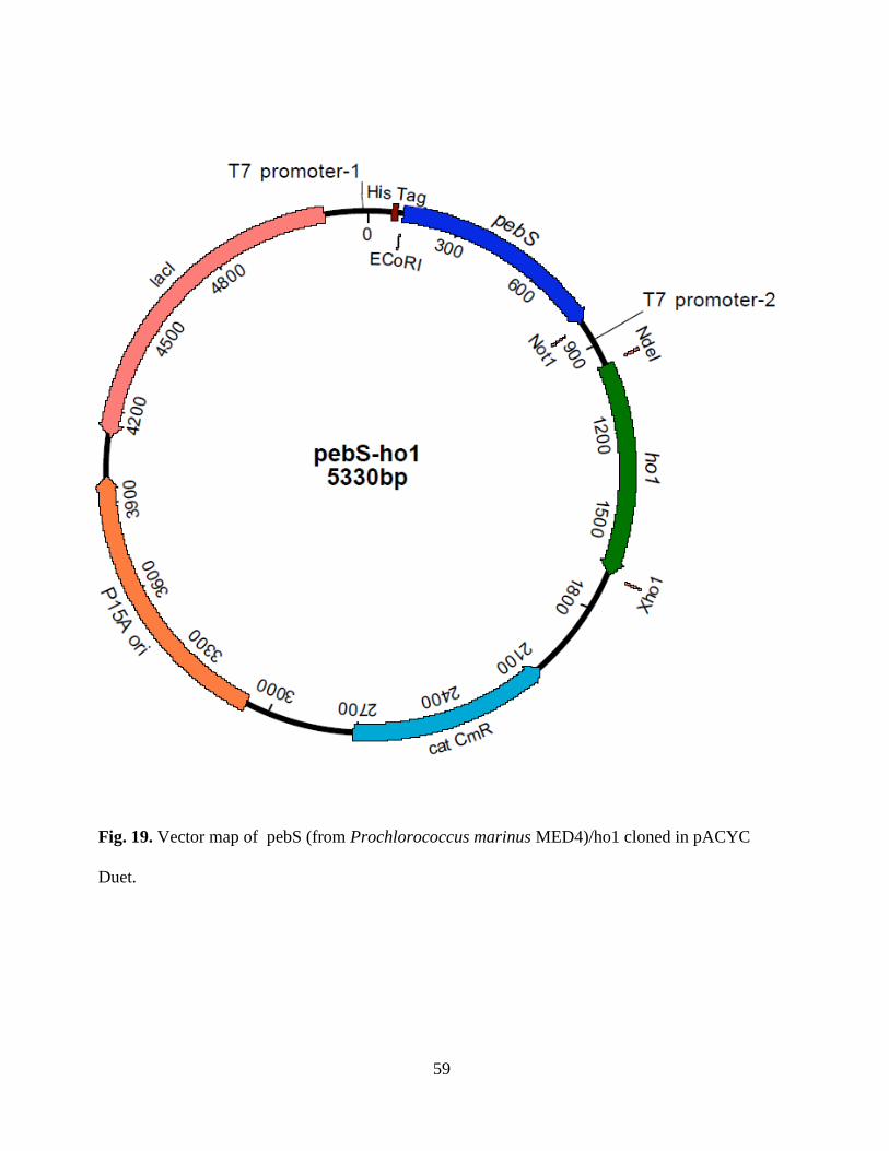
59
Fig. 19. Vector map of pebS (from Prochlorococcus marinus MED4)/ho1 cloned in pACYC
Duet.
60
2.2. In-vivo heterologous expression and purification of recombinant proteins: Expression
plasmids were co-transformed into E. coli BL21 DE3 cells, and colonies were
selected on Luria Bertani (LB) plates in the presence of the appropriate combination of antibiotics
(see Table 1) at the following concentrations: ampicillin (Ap: 100 μg ml-1
), chloramphenicol (Cm:
34 μg ml-1
), kanamycin (Km: 50 μg ml-1
), spectinomycin (Sp: 100 μg ml-1
). To produce PCB
using the pPcyA expression plasmid, a 50-ml overnight starter culture was added to 1 L of LB
medium with the appropriate combination of antibiotics. This culture was shaken at 37°C for 4 h
until the optical density reached OD600 nm = 0.6. Production of T7 RNA polymerase was induced
by the addition of 0.5 mM isopropyl β-D thiogalactoside (IPTG). Cells were incubated with
shaking at 225 rpm at 30°C for another 4 h before they were harvested by centrifugation at 10,000
g for 10 min. Cells were incubated at 30° C after induction with IPTG to limit the production of
inclusion bodies (data not shown). Cell pellets were stored at -20 °C until required.
E. coli cells containing recombinant proteins were thawed and resuspended in Buffer O (50 mM
Tris-HCl, 150 mM NaCl, pH 8.0) at 2.5 ml/g (wet weight) and lysed by three passages through a
chilled French pressure cell at 138 MPA. The lysed cell suspension was centrifuged for 20 min at
13,000 g to remove inclusion bodies, cell debris and unbroken cells. To purify hexa-histidine-
tagged recombinant proteins, the supernatant was passed over nickel-nitrilotriacetic acid-
Superflow-affinity column (Qiagen, Inc., Chatsworth, CA) containing 5 ml of resin, and proteins
were purified as previously described (Shen, Saunee et al. 2006). The recombinant protein(s) were
dialyzed with buffer O containing 10 mM 2-mercaptoethanol overnight at 4 °C to remove the
imidazole. For purification of GST-ApcE, cells were broken as described (Miller, Leonard et al.
2008) except that protease inhibitor cocktail tablets were added (“Complete Mini” purchased from
61
Roche, Mannheim, Germany). The clarified extract was passed over a 5-ml glutathione agarose
column (Sigma) as described previously (Miller, Leonard et al. 2008).
The expression conditions were little modified in case of chromophorylation study with
PEB (Biswas, Vasquez et al. 2010); to produce PEB using the pPebS expression plasmid, a 50-ml
overnight starter culture was added to 1 L of LB medium with the appropriate combination of
antibiotics. This culture was shaken at 37°C for 4 h until the optical density reached OD600 nm =
0.6. Production of T7 RNA polymerase was induced by the addition of 1 mM isopropyl β-D
thiogalactoside (IPTG). Cells were incubated with shaking at 190 rpm at 18°C for another 16 h
before they were harvested by centrifugation at 10,000 g for 10 min. Cells were incubated at
18° C after induction with IPTG to limit the production of inclusion bodies (data not shown). Cell
pellets were stored at -20 °C until required.
To produce holo-MpeA using the pPebS with and without pMpeZ expression plasmid, a
single colony was inoculated into a 200-ml overnight culture in LB medium with the appropriate
combination of antibiotics. This culture was shaken at 20 °C at 180 rpm for 30-48 h until the
optical density reached OD600 nm ~ 0.6. Production of T7 RNA polymerase was induced by the
addition of 1 mM isopropyl β-D thiogalactoside (IPTG). Cells were incubated with shaking at 180
rpm at 20 °C for another 48 h before they were harvested by centrifugation at 10,000 g for 10
min. Cell pellets were immediately processed for protein purification in dark, following the
procedure described earlier in “Methods and Materials”.
62
2.3. Fluorescence emission and absorbance spectra:
Fluorescence emission and excitation spectra were recorded with a Perkin Elmer LS55
fluorescence spectrophotometer (Waltham, MA) with slit widths set at 10 nm (excitation and
emission). For recombinant phycobiliproteins, the excitation wavelength was set depending on the
type of bilin bring used for in vivo assays; (PCB at 590 nm, PEB at 490 nm and PUB at 440 nm).
The chromophorylated samples were diluted to ~0.05 OD (at max) prior to obtaining their
fluorescence spectra whereas negative control samples (e. g., no lyase addition) with no obvious
chromophore attached were not diluted (their OD was generally less than 0.05). Absorbance
spectra were acquired using a lambda 35, dual-beam UV-Vis spectrophotometer (Perkin Elmer,
Waltham, MA). For calculating the fluorescence relative intensity the sample was diluted to 0.05
OD560nm. The fluorescence emission peak for wild type co-expression sample was set to 100%,
and relative emission peak was calculated accordingly using same diluted sample (based on the
equal protein concentration).
2.4. Protein and bilin analysis:
Polypeptides were resolved by polyacrylamide gel electrophoresis (PAGE; 15% w/v) in the
presence of sodium dodecyl sulfate (SDS), and polypeptides were visualized by staining with
Coomassie blue as described (Saunée, Williams et al. 2008). To visualize PCB linked to proteins,
gels were soaked in 100 mM ZnSO4 for approximately 5 min (Berkelman and Lagarias 1986;
Berkelman and Lagarias 1986; Raps 1990). Zinc-enhanced fluorescence of bilins was visualized
using an FX imaging system (BioRad, Hercules, CA) with excitation at 550 nm (whereas in the
case of phycobiliprotein subunits having PCB or PEB, but 488 nm in case of PUB). In order to
calculate % chromophorylation of each protein, BioRad’s Quantity One software was used to
63
determine the relative abundance of each polypeptide. (PcyA and CpcU were His-tagged but the
expression levels of HT-CpcU were very low, and the amount of this protein relative to the others
was very small.) This abundance percentage was multiplied by the extinction coefficient of the
protein at 280 nm (based upon Trp and Tyr content) to obtain the contribution of each protein to
the absorbance at 280 nm. A modified extinction coefficient for the total protein concentration
was used as shown below for purified HT-ApcA/ApcB.
280 nmtotal
= (%HT-ApcA*280 nmHT-ApcA
+ %ApcB*280 nmApcB
+ %HT-PcyA*280 nmHT-PcyA
)
Equation:1
280HT-ApcA/ApcB
= (% HT-ApcA + % ApcB)* 280 nmtotal
Equation 2.
The PCB concentration was calculated by denaturing the recombinant protein in 8 M urea, pH 2
and using 663 nm= 35.4 mM-1
cm-1
(Glazer 1988). The concentration of the bilin was divided by
the concentration of the PBP to give % chromophorylation (see Table 2). Holo-HT-CpcA
concentration was determined using 625 nm = 127.6 mM-1
cm-1
(Tooley, Cai et al. 2001). The yield
of chromophorylated PBP (expressed as mg of PBP l-1
E. coli culture) was estimated by
determining the concentration of PCB bound to protein (obtained in 8 M urea, pH 2); this value
was multiplied by the molecular weight of the PBP subunit(s) and the total volume of protein
solution purified from the cells grown in 1 l of culture medium.
The % chromophorylation of CpeA and CpeB was calculated as described earlier (Biswas,
Vasquez et al. 2010) with minor changes. BioRad’s Quantity One software was used to
determine the relative abundance of each polypeptide because PebS was also His-tagged. This
abundance percentage was multiplied by the extinction coefficient of the protein at 280 nm (based
upon Trp and Tyr content; mentioned below) to obtain the contribution of the protein to the
64
absorbance at 280 nm. A modified extinction coefficient for the total protein concentration was
used as shown below for purified HT-CpeA or HT-CpeB (represented by X).
280 nmtotal
= (%HT-X*280 nmHT-X
+ %PebS*280 nmPebS
) Equation 3.
280HT-X
= (% HT-X + % PebS)* 280 nmtotal
Equation 4
The PEB-peptide concentration (attached to HT-CpeA or HT-CpeB) was calculated by denaturing
the recombinant protein in 8 M urea, pH 2 and using 550 nm= 53.4 mM-1
cm-1
(Glazer 1988). The
concentration of the bilin was divided by the concentration of the PBP to give %
chromophorylation (see result).
For comparing the PCB yield obtained in expression cells containing pPcyA and pAT101, 50 ml
overnight cultures were transferred to 1 l of culture medium at 37 °C and grown until the OD600 nm
reached 0.6. IPTG (0.5 mM) was then added, and the cultures were incubated for an additional 4-
h period at 30°C. Cells were harvested and homogenized with 35 ml of acetone (Hu, Lee et al.
2006). The supernatant was vacuum dried to remove the acetone, and the dried pellet was
dissolved in methanol (1.0 ml). The total PCB from each construct was diluted 100-fold in
methanol/ 5% HCl and the concentration was calculated using the relationship 680 nmPCB
= 37.9
mM-1
cm-1
(Cole, Chapman et al. 1967). The PCB concentration was multiplied by the total
volume in MeOH and by the molecular mass of PCB (587 g/mol) (Fu, Friedman et al. 1979; Fu,
Friedman et al. 1979) to determine the grams of PCB produced per liter of culture.
2.5. Calculating fluorescence quantum yield: Fluorescent quantum yields of the holo-CpeA
was calculated as described (Parker and Rees 1960; Lakowicz 1983) using Perkin Elmer LS55
65
fluorescence spectrophotometer (Waltham, MA) with slit widths set at 10 nm (excitation and
emission), relative to cresyl violet (in ethanol Фf= 0.59; Sigma Aldich). Samples were diluted to
an absorbance of ~0.04-0.05 at λ560 nm and fluorescence emission was acquired from 570-800 nm.
Fluorescent quantum yield of the sample was calculating in comparison with the standard using
the following equation
A11/ A22= Фf1/ Фf2 Equation 5
Where A is the absorbance maxima, is the area of the fluorescence emission spectrum from 570
nm -800 nm, and Фf is fluorescence quantum yield.
2.6. Tryptic Digestion of Phycoerythrin: The partial holo-CpeA (pCpeA/pPebS,pCpeZY) and
partial holo CpeB (pCpeB/pPebS/pCpeS) retrieved from the nickel-nitrilotriacetic acid-
Superflow-affinity column was subjected to tryptic digestion following the protocol described in
(Arciero, Bryant et al. 1988). In short, the purified holoprotein was exhaustively dialyzed against
2mM sodium phosphate buffer, pH 7.0, 1 mM 2-mercaptoethanol and then concentrated by
ultrafiltration through an Amicon YM10 (Millipore, Billercia, MA). Concentrated sample was
diluted 1:3 and titrated to pH 2.0 with 1N HCl. The solution was incubated for 45min in dark at
room temperature for complete unfolding of protein. Trypsin was added to 2% (w/v) from a 5
mg/ml stock solution in 1 mM HCl. Ammonium bicarbonate was added in 0.1 M and the mixture
was titrated to pH 7.5 with 1N NaOH. The digested mixture was incubated at 30 °C for 2h in
dark. An additional aliquot of trypsin was added (2% w/v) and incubated for another 2h. The
reaction was quenched by adding 30% v/v glacial acetic acid. The mixture was passed through a
C-18 sep-pack cartridge. The eluted sample was vacuum dried and stored at -20⁰C for HPLC

66
separation as described (Shen, Saunee et al. 2006). The fraction peaks having absorbance maxima
at 560 nm were collected, vaccum dried and stored at -20⁰C for Mass spectrometry analysis.
2.7. Growth condition for Fremyella diplosiphon: The Fremyella diplosiphon cells are grown
in 250 ml conical flask in BG-11 medium (Allen 1968) with 10 mM HEPES (pH 8.0). The
cultures are grown on a shaker (100 rpm) illuminated with ~ 50-70 µE/m2/x cool white light
(encoded in green). The cell density was monitored with using UV-Vis spectrophotometer. The
cells are harvested by centrifugation at 10,000 g for 10 min, prior to proceeding with
phycobilisome isolation.
2.8. Separation of phycobilisome: Phycobilisomes from Fremyella diplosiphon were separated
following the protocol described by Glazer (Glazer 1988), with few minor changes. All steps
were performed at room temperature. Cells were collected by continuous centrifugation and
suspended at 1 g wet weight/ 15 ml in 0.75 M potassium phosphate buffer, pH 6.8, 1 % (w/v)
Triton X-100. After stirring for 1 hr the extract was centrifuged for 20 min at 27,000 g, and the
sedimented materials were discarded. Polyethylene glycol 6000 is added to the supernatant
solution 15 % (w/v), the mixture was stirred for 1 hr and then centrifuged for 20 min at 27,000 g,
and the supernatant wwas discarded. The sedimented material was suspended in 0.75 M
potassium phosphate buffer, pH 6.8, 1 % (w/v) Triton X-100, 15 %( w/v) polyethylene glycol
6000, the mixture was centrifuged for 20 min at 27,000 g, and the supernatant was discarded. The
purple sediment was suspended in 0.75 M potassium phosphate buffer, pH 6.8 centrifuged for 15
min at 27,000 g.
2.9. Isolation of Phycoerythrin: The phycoerythrin from Fremyella diplosiphon (both wild type
and mutant) cells (grown in David Kehoe’s lab) was purified following the protocol described by
67
Glazer (Glazer 1988) with minor changes. Cells (10 g wet weight) from both wide type and
mutants were suspended in 25 ml of 1 M sodium acetate, pH 5.0. The slurry was broken by
passing three times through a French pressure cells at 20,000 psi. The product was ultra
centrifuged at 81,000 g for 1 h (rotor-Ti64) to remove the chlorophyll and other cellular
membranes.
Solid ammonium sulphate was added to 35% of saturation in the case of wild type and to
45 % of saturation to the mutants extracts to the supernatants. The solution was allowed to stand
for 90 min and then centrifuged at 16,000 g for 15 min. The pellet was dissolved in 15 ml of 100
mM sodium acetate, pH 5.0 and dialyzed overnight against 2 L of same buffer.
The dialyzed sample was passed through a column (4.0 X 24 cm) of Sephadex G-100
preequlibrated with the 100 mM acetate buffer. After each run the peaks tubes from the early
eluting biliproteins peaks were pooled. The phycoerythrin containing solution was brought to 30
% of saturation and allowed to stand for 1 h prior to centrifugation. The pellets were resuspended
in 5 mM potassium phosphate, pH 7.0, and then dialyzed overnight against 2 l of same buffer.
The dialyzed phycoerythrin- containing solution was applied to a column (2.2 X 24 cm) of
DEAE-cellulose DE 52 (Watmann), preequilibrated with the 5 mM phosphate buffer. After
elution with 1 volume of starting buffer, the column is developed with a 550 ml linear 5-200 mM
potassium phosphate, pH 7.0, gradient. The peak tubes from the major phycoerythrin, were
pooled, brought to 45 % saturation with (NH4) 2SO4, permitted to stand for 1 h prior to
centrifugation. The pellet was suspended in a small volume of 100 mM sodium phosphate-1 mM
sodium azide at pH 7.0 and dialyzed against the same buffer.
2.10. Isolation of PEI and PEII from Synechococcus sp. RS 9916: RS9916 species was used to
study the bilin lyase isomerase for PE subunits. This is one of the cyanobacterial species which
68
can undergo Type IV chromatic acclimation. The cells were grown (David Kehoe’s lab) in
different light conditions (blue vs green) using LEDs to characterize the change in the
chromophore composition in PEI and PEII. The cell pellets were used to isolate PEI and PEII
proteins.
The separation was carried out following the protocol described earlier (Six, Thomas et al. 2007)
with a few modifications. The cell pellets were dissolved in 0.75 M Na-K phosphate buffer (pH
7.0). The slurry was lysed by passing three times through a French pressure cell at 15,000-18,000
psi. The product was ultra centrifuged at 35,000 g for 3 h to remove the chlorophyll and other
cellular debris. The supernatant was brought to 45 % saturation with (NH4) 2SO4, permitted to
stand for 1 h prior to centrifugation. The pellet was suspended in a small volume of 5 mM sodium
phosphate-1 mM sodium azide at pH 7.0 and dialyzed against the same buffer. The dialyzed
samples were concentrated 10 fold (using Amicron concentrator with a MW cut off of 10kDa)
and proceeded to next step of separation using IEF analysis.
The IEF (7 % acrylamide minigel containing ampholyte carrier pH 4 to 6.5, purchased from
BioRad, Hercules, CA) gel separation were carried out using the BioRad protocol. The sample
was diluted with equal amout of sample buffer (15 % glycerol) and loaded on a IEF gel. The
cathode tank was filled with 20 mM NaOH and the anode one contained 20 mM orthrophosphoric
acid. The PBPs were allowed to focus during approx. 2 h by increasing steps of 50 V every 10
min up to 350V.
The different color bands representing PEI and PEII were excised and then crushed in 10
mM Tricine buffer (pH 7.8) using a manual homogenizer. Acrylamide remnants were removed by
centrifugation for 10 min at 14,000 x g. The extracted proteins were vaccuum dried and stored at
-20⁰C until further use.
69
Separation of α and β subunits of PEI and PEII: The separation of the subunits were carried out
following the protocol described by Swanson and Glazer (Swanson and Glazer 1990) using C4
reverse-phase HPLC (Waters). In brief the vacuum dried samples were dissolved in 300 ul of
65% TFA (0.1%) and 35% 2:1 acetronitril: isopropanol containing 0.1 % TFA. The samples were
centrifuged for 10 mins to remove the undissolved proteins. Then the samples were separated on
a C4 column using the running conditions described earlier (Swanson and Glazer 1990) . The α
and β subunits of PEI and PEII peaks were collected based on the absorbance spectrum (490 and
550 nm). The separated subunits were vacuum dried and further analyzed by Mass spectrometry
(Mass Spectometry facility at Indiana University).
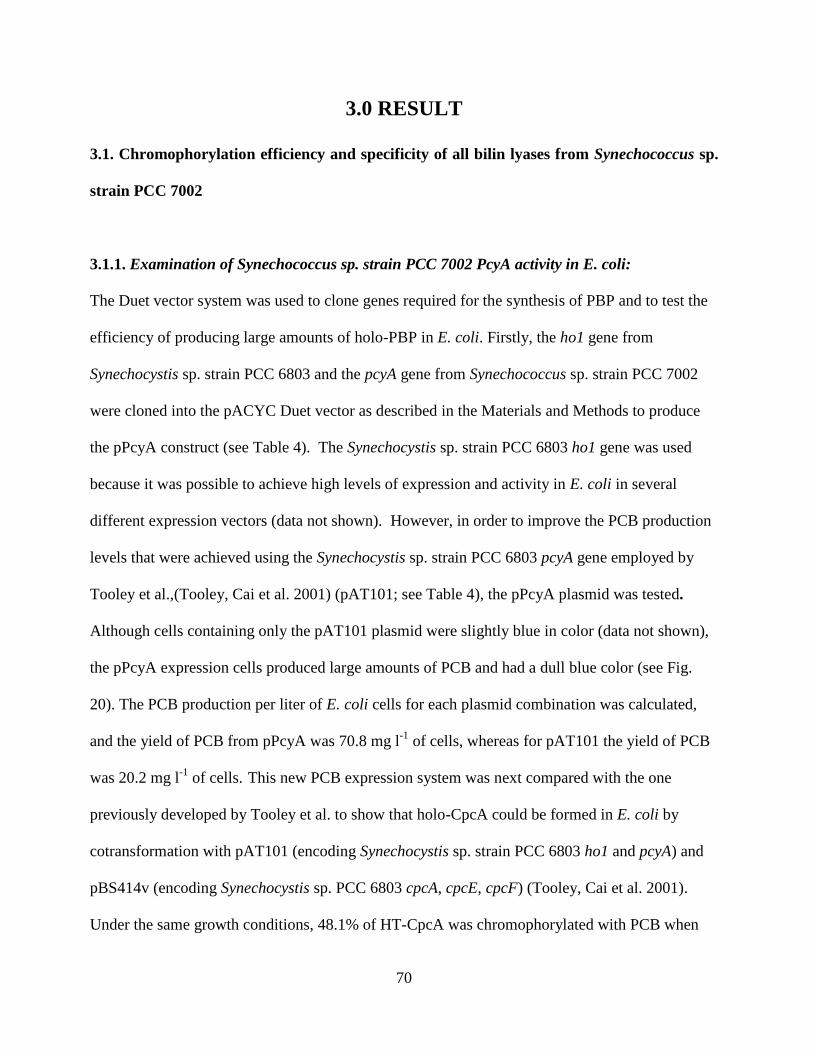
70
3.0 RESULT
3.1. Chromophorylation efficiency and specificity of all bilin lyases from Synechococcus sp.
strain PCC 7002
3.1.1. Examination of Synechococcus sp. strain PCC 7002 PcyA activity in E. coli:
The Duet vector system was used to clone genes required for the synthesis of PBP and to test the
efficiency of producing large amounts of holo-PBP in E. coli. Firstly, the ho1 gene from
Synechocystis sp. strain PCC 6803 and the pcyA gene from Synechococcus sp. strain PCC 7002
were cloned into the pACYC Duet vector as described in the Materials and Methods to produce
the pPcyA construct (see Table 4). The Synechocystis sp. strain PCC 6803 ho1 gene was used
because it was possible to achieve high levels of expression and activity in E. coli in several
different expression vectors (data not shown). However, in order to improve the PCB production
levels that were achieved using the Synechocystis sp. strain PCC 6803 pcyA gene employed by
Tooley et al.,(Tooley, Cai et al. 2001) (pAT101; see Table 4), the pPcyA plasmid was tested.
Although cells containing only the pAT101 plasmid were slightly blue in color (data not shown),
the pPcyA expression cells produced large amounts of PCB and had a dull blue color (see Fig.
20). The PCB production per liter of E. coli cells for each plasmid combination was calculated,
and the yield of PCB from pPcyA was 70.8 mg l-1
of cells, whereas for pAT101 the yield of PCB
was 20.2 mg l-1
of cells. This new PCB expression system was next compared with the one
previously developed by Tooley et al. to show that holo-CpcA could be formed in E. coli by
cotransformation with pAT101 (encoding Synechocystis sp. strain PCC 6803 ho1 and pcyA) and
pBS414v (encoding Synechocystis sp. PCC 6803 cpcA, cpcE, cpcF) (Tooley, Cai et al. 2001).
Under the same growth conditions, 48.1% of HT-CpcA was chromophorylated with PCB when
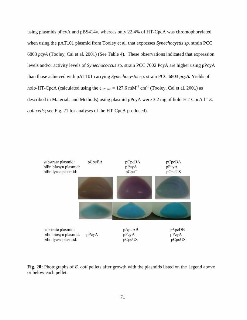
71
using plasmids pPcyA and pBS414v, whereas only 22.4% of HT-CpcA was chromophorylated
when using the pAT101 plasmid from Tooley et al. that expresses Synechocystis sp. strain PCC
6803 pcyA (Tooley, Cai et al. 2001) (See Table 4). These observations indicated that expression
levels and/or activity levels of Synechococcus sp. strain PCC 7002 PcyA are higher using pPcyA
than those achieved with pAT101 carrying Synechocystis sp. strain PCC 6803 pcyA. Yields of
holo-HT-CpcA (calculated using the 625 nm = 127.6 mM-1
cm-1
(Tooley, Cai et al. 2001) as
described in Materials and Methods) using plasmid pPcyA were 3.2 mg of holo-HT-CpcA l-1
E.
coli cells; see Fig. 21 for analyses of the HT-CpcA produced).
Fig. 20: Photographs of E. coli pellets after growth with the plasmids listed on the legend above
or below each pellet.
72
Fig. 21: Analyses of holo-HT-CpcA purified from E. coli cells. A. Absorbance
(solid) and fluorescence emission (dashed dotted) spectra of HT-CpcA purified from cells
containing pBS414v and pPcyA. B. Coomassie-stained SDS-polyacrylamide gel of HT-CpcA
purified from cells containing pBS414v and pPcyA (lane 1) or pBS414v and pAT101 (lane 2).
Molecular mass standards were loaded in the lane marked “S”; the position of the 21.5-kDa
standard is indicated at the left. C. Zinc-enhanced bilin fluorescence of the gel in panel B.
73
3.1.2. Development and use of a multi-plasmid system for expression of holo-AP:
E. coli cells containing plasmids pPcyA and pApcAB produced almost no colored and fluorescent
product (See Fig 21A; sample was not diluted prior to obtaining the fluorescence emission
shown), but E. coli cell2 containing plasmids pPcyA, pApcAB, pCpcUS had a brilliant blue color
(Fig. 20). HT-ApcAB purified from these cells had an absorbance maximum at 615 nm with a
small shoulder at 653 nm (see Fig. 22A). This product had a fluorescence emission maximum at
634 nm, consistent with that of monomeric () holo-AP (see Fig. 22A; the sample shown was
diluted to 0.05 OD615 prior to measuring the fluorescence emission). The ratio of the Vis:UV (615
nm:357 nm) maxima for HT-ApcA/ApcB was 2.53 (Table 6). Next, CpcS or CpcU alone was
tested within this heterologous E. coli system to confirm the mutagenesis results and previous in
vitro enzyme assay results (Shen, Saunee et al. 2006; Saunée, Williams et al. 2008). Cells
transformed with either pCpcU or pCpcS, and thus expressing only one of the subunits of this
heterodimeric lyase, were unable to produce a highly fluorescent product, similar to the results
obtained in cells containing only pApcAB and pPcyA (Fig 22B; the sample was not diluted prior
to obtaining the fluorescence emission). The HT-ApcA/ApcB samples purified from the different
E. coli cells were analyzed by SDS-PAGE (Fig. 22C). The bilin content of each protein was
examined by zinc-enhanced fluorescence of the gels as shown in Fig. 22D; protein content was
examined after staining the same gel with Coomassie Blue (Fig. 22C). When no bilin lyase
subunit was present, a small amount of covalent PCB addition to ApcB occurred (Fig. 22C, lane
2); this was similar to previous observations using in vitro reactions (Saunée, Williams et al.
2008). However, the yield of this covalent product was very low in this in-vivo E. coli system, and
the absorption and fluorescence properties suggested that the bilin attached auto-catalytically to
ApcB in the absence of a bilin lyase was not the biologically correct product (Arciero, Bryant et
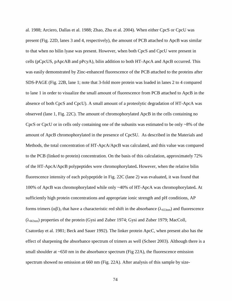
74
al. 1988; Arciero, Dallas et al. 1988; Zhao, Zhu et al. 2004). When either CpcS or CpcU was
present (Fig. 22D, lanes 3 and 4, respectively), the amount of PCB attached to ApcB was similar
to that when no bilin lyase was present. However, when both CpcS and CpcU were present in
cells (pCpcUS, pApcAB and pPcyA), bilin addition to both HT-ApcA and ApcB occurred. This
was easily demonstrated by Zinc-enhanced fluorescence of the PCB attached to the proteins after
SDS-PAGE (Fig. 22B, lane 1; note that 3-fold more protein was loaded in lanes 2 to 4 compared
to lane 1 in order to visualize the small amount of fluorescence from PCB attached to ApcB in the
absence of both CpcS and CpcU). A small amount of a proteolytic degradation of HT-ApcA was
observed (lane 1, Fig. 22C). The amount of chromophorylated ApcB in the cells containing no
CpcS or CpcU or in cells only containing one of the subunits was estimated to be only ~8% of the
amount of ApcB chromophorylated in the presence of CpcSU. As described in the Materials and
Methods, the total concentration of HT-ApcA/ApcB was calculated, and this value was compared
to the PCB (linked to protein) concentration. On the basis of this calculation, approximately 72%
of the HT-ApcA/ApcB polypeptides were chromophorylated. However, when the relative bilin
fluorescence intensity of each polypeptide in Fig. 22C (lane 2) was evaluated, it was found that
100% of ApcB was chromophorylated while only ~40% of HT-ApcA was chromophorylated. At
sufficiently high protein concentrations and appropriate ionic strength and pH conditions, AP
forms trimers ()3 that have a characteristic red shift in the absorbance (653nm) and fluorescence
(663nm) properties of the protein (Gysi and Zuber 1974; Gysi and Zuber 1979; MacColl,
Csatorday et al. 1981; Beck and Sauer 1992). The linker protein ApcC, when present also has the
effect of sharpening the absorbance spectrum of trimers as well (Scheer 2003). Although there is a
small shoulder at ~650 nm in the absorbance spectrum (Fig 22A), the fluorescence emission
spectrum showed no emission at 660 nm (Fig. 22A). After analysis of this sample by size-
75
exclusion HPLC, two peaks were observed for this the holo-HT-ApcA/ApcB. One had a
molecular mass consistent with single or subunits while the other had a mass consistent with
that of AP () protomers; no peak consistent with the elution properties for AP ()3 trimers
was observed (data not shown). Because of the presence of a significant proportion of apo-
subunits within the purified mixture, this was expected. Assuming a random association of
chromophorylated and non-chromophorylated subunits with a chromophorylation rate of 40% for
the subunit and 100% for the subunits, the probability of obtaining monomers with both
subunits chromophorylated is ~40% (1.0 * 0.40), and the probability of obtaining trimers in which
all subunits are chromophorylated would be ((1.0) 3
*(0.4)3) or ~6.4%.
The specificity of the other two known bilin lyases was also tested to see if either the CpcE/CpcF
lyase or the CpcT lyase could attach PCB to HT-ApcA/ApcB in this in-vivo system. As judged by
absorbance and fluorescence emission spectra (see Figure 23, panels A and B, respectively), no
holo-AP was produced when either pCpcEF or pCpcT was introduced together with the pApcAB
and pPcyA. In addition, only a small amount of PCB addition to ApcB was observed; this level
was similar to that observed when no lyase was present (compare to Fig 22D, lane 2). Therefore,
the specificity of the lyases for their PBP substrates was maintained within this E. coli system and
was completely consistent with previous in vitro biochemical and mutational analyses (Fairchild,
Zhao et al. 1992; Zhou, Gasparich et al. 1992; Fairchild and Glazer 1994; Shen, Saunee et al.
2006; Saunée, Williams et al. 2008; Shen, Schluchter et al. 2008).
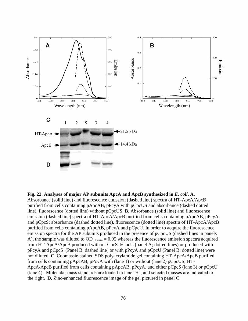
76
Fig. 22. Analyses of major AP subunits ApcA and ApcB synthesized in E. coli. A.
Absorbance (solid line) and fluorescence emission (dashed line) spectra of HT-ApcA/ApcB
purified from cells containing pApcAB, pPcyA with pCpcUS and absorbance (dashed dotted
line), fluorescence (dotted line) without pCpcUS. B. Absorbance (solid line) and fluorescence
emission (dashed line) spectra of HT-ApcA/ApcB purified from cells containing pApcAB, pPcyA
and pCpcS; absorbance (dashed dotted line), fluorescence (dotted line) spectra of HT-ApcA/ApcB
purified from cells containing pApcAB, pPcyA and pCpcU. In order to acquire the fluorescence
emission spectra for the AP subunits produced in the presence of pCpcUS (dashed lines in panels
A), the sample was diluted to OD615 nm = 0.05 whereas the fluorescence emission spectra acquired
from HT-ApcA/ApcB produced without CpcS-I/CpcU (panel A; dotted lines) or produced with
pPcyA and pCpcS (Panel B, dashed line) or with pPcyA and pCpcU (Panel B, dotted line) were
not diluted. C. Coomassie-stained SDS polyacrylamide gel containing HT-ApcA/ApcB purified
from cells containing pApcAB, pPcyA with (lane 1) or without (lane 2) pCpcUS; HT-
ApcA/ApcB purified from cells containing pApcAB, pPcyA, and either pCpcS (lane 3) or pCpcU
(lane 4). Molecular mass standards are loaded in lane “S”, and selected masses are indicated to
the right. D. Zinc-enhanced fluorescence image of the gel pictured in panel C.
77
Fig. 23: Analysis of HT-ApcAB purified from E. coli cells produced in the
presence of CpcEF or CpcT. A. Absorbance (solid) and fluorescence emission (dashed) spectra
of HT-ApcAB purified from cells containing pApcAB, pBS415v (CpcEF), and pPcyA. B.
Absorbance (solid) and fluorescence emission (dashed) spectra of HT-ApcAB purified from cells
containing pApcAB, pCpcT, and pPcyA. C. Coomassie-stained SDS-polyacrylamide gel of HT-
ApcA/ApcB purified from cells containing pApcAB, pCpcEF (lane 1), and pCpcT (lane 2) with
pPcyA. Molecular mass standards were loaded in the lane marked “S”; the position of the 21.5-
kDa mass standard is indicated. D. Zinc-enhanced bilin fluorescence of the gel in panel C.
78
3.1.3. Chromophorylation Requirements for HT-ApcD:
ApcD is a variant -AP subunit (AP-B) that pairs with -AP (ApcB) forming AP-B which has an
extremely red-shifted absorbance at 670 nm and is an important terminal emitter of the PBS
involved in energy transfer to Photosystem I; two copies of ApcD are present per PBS (Glazer
and Bryant 1975; Lundell and Glazer 1981; Fuglistaller, Mimuro et al. 1987; Maxson, Sauer et al.
1989; Zhao, Zhou et al. 1992; Ashby and Mullineaux 1999; Domg, Tang et al. 2009). An
alignment of ApcD with other allophycocyanin subunits is shown in Fig. 24. The apcD gene was
cloned together with apcB in order to produce HT-ApcD and ApcB, as this construct produced a
more soluble recombinant apo-protein than a construct expressing apcD alone (L. Harrison, Jr.
and W. M. Schluchter, unpublished data). E. coli cells containing plasmids pApcDB and pPcyA
had a faint, dull blue color (data not shown), whereas cells containing plasmids pApcDB, pPcyA,
and pCpcUS were a brilliant blue color (see Fig. 20). The absorbance and fluorescence emission
spectra for HT-ApcD/ApcB after purification of the proteins by metal-affinity chromatography
from cells containing these two plasmid combinations are shown in Fig. 25A. HT-ApcD/ApcB
produced in the absence of the pCpcUS plasmid had very little absorbance or fluorescence, but
HT-ApcD/ApcB produced in the presence of pCpcUS had absorbance peaks at 616 and 672 nm.
When excited at 590 nm, this recombinantly produced HT-ApcD/ApcB had fluorescence
emission maxima at 634 nm and 675 nm. When these proteins were separated by SDS-PAGE and
evaluated by zinc-enhanced fluorescence, both subunits carried PCB chromophores (see Fig 25C,
lane 1), but based upon quantitation of the fluorescence intensity (compare lane 1 in Figs. 25B
and 25C), the chromophorylation level for HT-ApcD was only ~40% (with ApcB subunit at
100%). However, in the absence of pCpcUS, no bilin addition to HT-ApcD occurred, and very
79
little bilin addition to ApcB was detected (lane 2 in Figs. 25B and 25C). A degradation product
of HT-ApcD is apparent in some preparations (indicated by a * in Fig. 25B).
In order to determine the absorbance spectrum of holo-HT-ApcD alone, HT-ApcD was produced
in the presence of pPcyA and pCpcUS. As evidenced by the absorbance and fluorescence spectra
shown in Fig. 25D and the SDS-PAGE and zinc-enhanced bilin fluorescence shown in Fig 25E
and F, a small amount of chromophorylated product was obtained. This holo-HT-ApcD had an
absorbance maximum at 642 nm and a fluorescence emission maximum at 653 nm.. This likely
means that when holo-HT-ApcD associates with holo-ApcB, there are interactions between
chromophores that red-shift the absorbance maximum to 672 nm. The 616 nm absorption
maximum seen in Fig. 25A is probably due to holo-ApcB (paired with apo-HT-ApcD), because
there is an excess of chromophorylated ApcB relative to holo-HT-ApcD (see Fig 25C).
Supporting this idea, the fluorescence excitation spectrum of the holo-HT-ApcD/ApcB with
emission at 675 nm, shows an excitation peak centered at 618 nm, indicating that some of the
ApcB-PCB chromophore is transferring energy to ApcD (see Fig. 25G). An alternative hypothesis
to explain the difference seen in the spectra of HT-ApcD produced with and without ApcB is that
the folding of ApcD was adversely affected in the absence of ApcB, leading to the differences
observed in the absorbance and fluorescence emission spectra.
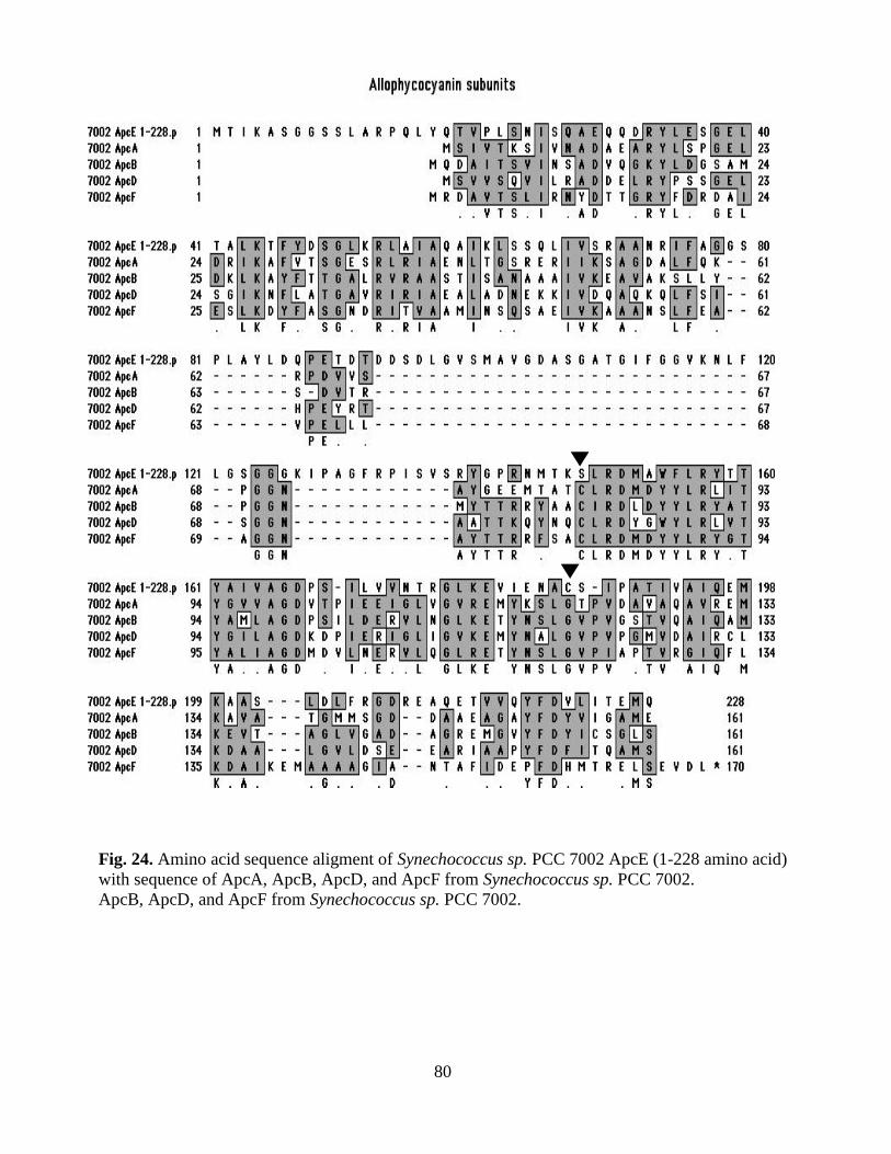
80
Fig. 24. Amino acid sequence aligment of Synechococcus sp. PCC 7002 ApcE (1-228 amino acid)
with sequence of ApcA, ApcB, ApcD, and ApcF from Synechococcus sp. PCC 7002.
ApcB, ApcD, and ApcF from Synechococcus sp. PCC 7002.
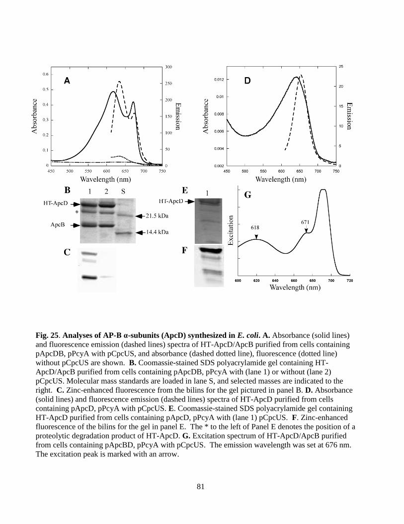
81
Fig. 25. Analyses of AP-B α-subunits (ApcD) synthesized in E. coli. A. Absorbance (solid lines)
and fluorescence emission (dashed lines) spectra of HT-ApcD/ApcB purified from cells containing
pApcDB, pPcyA with pCpcUS, and absorbance (dashed dotted line), fluorescence (dotted line)
without pCpcUS are shown. B. Coomassie-stained SDS polyacrylamide gel containing HT-
ApcD/ApcB purified from cells containing pApcDB, pPcyA with (lane 1) or without (lane 2)
pCpcUS. Molecular mass standards are loaded in lane S, and selected masses are indicated to the
right. C. Zinc-enhanced fluorescence from the bilins for the gel pictured in panel B. D. Absorbance
(solid lines) and fluorescence emission (dashed lines) spectra of HT-ApcD purified from cells
containing pApcD, pPcyA with pCpcUS. E. Coomassie-stained SDS polyacrylamide gel containing
HT-ApcD purified from cells containing pApcD, pPcyA with (lane 1) pCpcUS. F. Zinc-enhanced
fluorescence of the bilins for the gel in panel E. The * to the left of Panel E denotes the position of a
proteolytic degradation product of HT-ApcD. G. Excitation spectrum of HT-ApcD/ApcB purified
from cells containing pApcBD, pPcyA with pCpcUS. The emission wavelength was set at 676 nm.
The excitation peak is marked with an arrow.
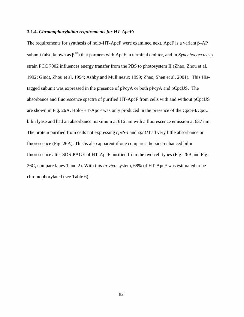
82
3.1.4. Chromophorylation requirements for HT-ApcF:
The requirements for synthesis of holo-HT-ApcF were examined next. ApcF is a variant -AP
subunit (also known as 18
) that partners with ApcE, a terminal emitter, and in Synechococcus sp.
strain PCC 7002 influences energy transfer from the PBS to photosystem II (Zhao, Zhou et al.
1992; Gindt, Zhou et al. 1994; Ashby and Mullineaux 1999; Zhao, Shen et al. 2001). This His-
tagged subunit was expressed in the presence of pPcyA or both pPcyA and pCpcUS. The
absorbance and fluorescence spectra of purified HT-ApcF from cells with and without pCpcUS
are shown in Fig. 26A. Holo-HT-ApcF was only produced in the presence of the CpcS-I/CpcU
bilin lyase and had an absorbance maximum at 616 nm with a fluorescence emission at 637 nm.
The protein purified from cells not expressing cpcS-I and cpcU had very little absorbance or
fluorescence (Fig. 26A). This is also apparent if one compares the zinc-enhanced bilin
fluorescence after SDS-PAGE of HT-ApcF purified from the two cell types (Fig. 26B and Fig.
26C, compare lanes 1 and 2). With this in-vivo system, 68% of HT-ApcF was estimated to be
chromophorylated (see Table 6).
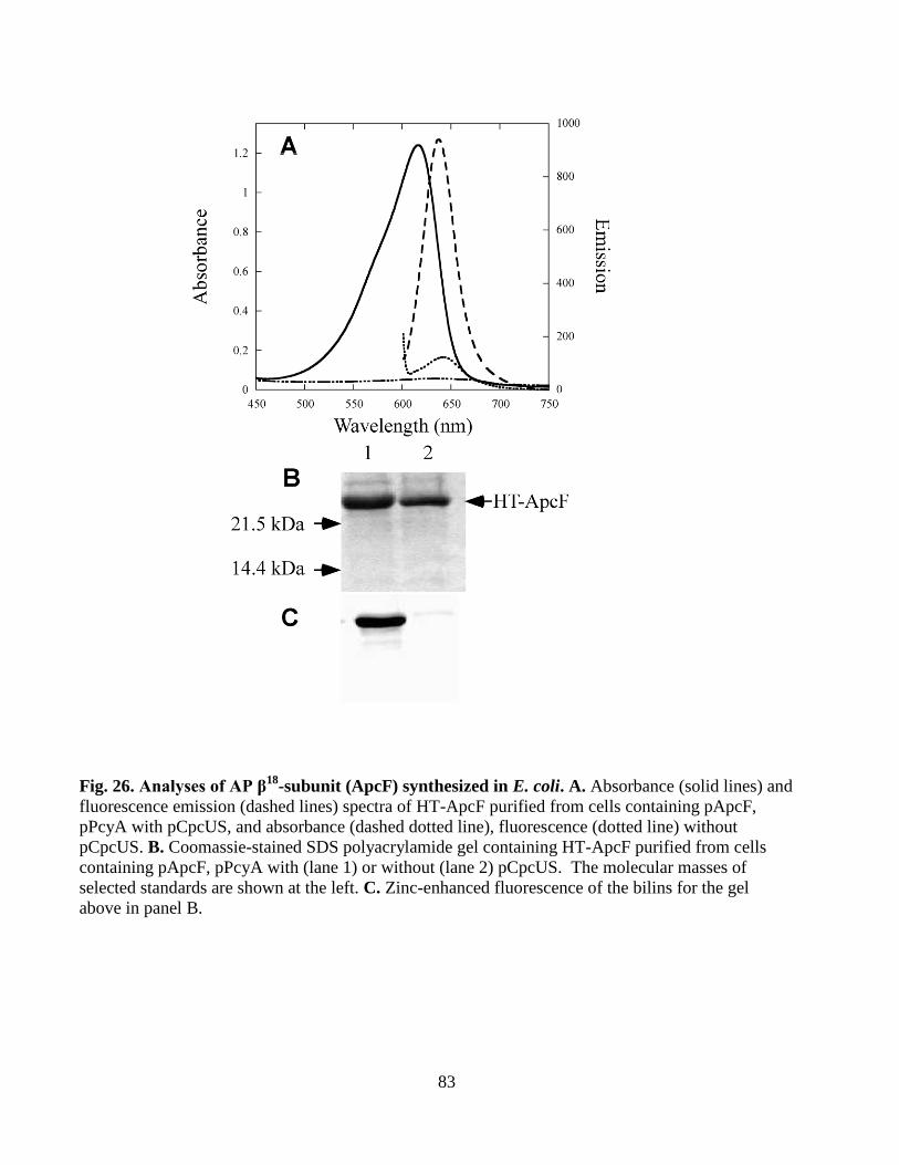
83
Fig. 26. Analyses of AP β18
-subunit (ApcF) synthesized in E. coli. A. Absorbance (solid lines) and
fluorescence emission (dashed lines) spectra of HT-ApcF purified from cells containing pApcF,
pPcyA with pCpcUS, and absorbance (dashed dotted line), fluorescence (dotted line) without
pCpcUS. B. Coomassie-stained SDS polyacrylamide gel containing HT-ApcF purified from cells
containing pApcF, pPcyA with (lane 1) or without (lane 2) pCpcUS. The molecular masses of
selected standards are shown at the left. C. Zinc-enhanced fluorescence of the bilins for the gel
above in panel B.
84
3.1.5. Chromophorylation requirements of ApcE:
The AP-like domain of ApcE from Synechococcus sp. strain PCC 7002 contains a PCB at Cys-
186 (see Fig. 27). The expression construct pGST-ApcE fused GST to amino acids 1-228 of the
AP-like domain of ApcE (Table 4). The construct pGST-ApcE was transformed into E. coli cells
with pPcyA, with or without pCpcUS, to determine whether the CpcS-I/CpcU bilin lyase was
required for correct and efficient PCB addition at Cys-186. Fig.27A shows the absorbance and
fluorescence spectra of the purified GST-ApcE from the two cell types. The ApcE domain alone
is sufficient to attach PCB in the correct manner to produce a strongly red-shifted PCB product
with an absorbance maximum at 662 nm and a fluorescence emission maximum at 675 nm. GST
alone did not react with PCB (data not shown). GST-ApcE synthesized in cells containing
pCpcUS also had absorbance and fluorescence properties similar to the native protein (Capuano,
Braux et al. 1991; Gindt, Zhou et al. 1994; Zhao, Su et al. 2006), but given that the correct
product can be formed in the absence of any additional enzyme, the CpcS-I/CpcU bilin lyase
apparently is not required for bilin ligation to ApcE. The GST-ApcE proteins were then separated
by SDS-PAGE and analyzed for the zinc-enhanced bilin fluorescence before staining for protein
with Coomassie blue staining. It was necessary to lyse the cells in the presence of a mixture of
protease inhibitors in order to avoid the formation of degradation products. The results of this
purification are shown in Fig. 27B and 27C (and correspond to the spectra shown in Fig. 27A).
Three polypeptides carried a bilin chromophore. One likely corresponded to the full-length fusion
(GST-ApcE: predicted mass of 51,445 Da), whereas the other two polypeptides had estimated
masses consistent with those of degradation products containing ApcE (amino acids 1-228) with
an expected mass of 24.5 kDa. As judged by Zinc-enhanced fluorescence (Fig. 27C, lanes 1 and
85
2) and supporting the conclusion that ApcE has intrinsic bilin lyase activity, approximately
equivalent PCB addition was observed whether pCpcUS was present or not.
Fig. 27: Analyses of GST-ApcE purified from E. coli cells. A. Absorbance (solid line) and
fluorescence emission (dashed line) spectra of GST-ApcE purified from E. coli cells containing
pGST-ApcE, pPcyA and pCpcUS, and absorbance (dashed dotted line) and fluorescence (dotted
line) spectra of GST-ApcE produced without pCpcUS. B. Coomassie-stained polyacrylamide gel
of purified GST-ApcE from E. coli cells containing pGST-ApcE, pPcyA with (lane 1) or without
pCpcUS (lane 2). Molecular mass standards are loaded in lane “S” and selected masses are
indicated to the right. Identities of polypeptides are indicated to the left. C. Zinc-enhanced bilin
fluorescence of the gel pictured in panel B.
86
3.1.6. Creation of partially chromophorylated PBPs in E. coli:
The efficiency of this in-vivo system in creating partially chromophorylated CpcB was examined
in order to determine the order for post-translational modifications on CpcB (Miller, Leonard et
al. 2008). The CpcS-I/CpcU lyase and the CpcT lyases were tested separately to confirm that each
could attach PCB to CpcB in this system, and the efficiency of PCB addition to Cys-82 and Cys-
153 in E. coli was estimated (see Table 6). Cells containing pCpcBA, pPcyA, and either pCpcUS
or pCpcT produced strongly colored PBPs (See Fig. 20). The spectra of these two partially
chromophorylated PCs are shown in Fig. 28 and 29. The PC produced by chromophorylation at
Cys-82 of CpcB by CpcS-I/CpcU was deep blue (see Fig. 20) and had an absorbance maximum at
620 nm and a fluorescence emission maximum at 642 nm for the CpcSU-product at Cys-82 (See
Fig. 28A, solid lines). The purple-colored PC (see Fig. 20) produced by chromophorylation of
Cys-153 by CpcT had an absorbance maximum at 592 nm and a fluorescence emission maxima at
618.5 nm (see Fig. 29A and Table 6). SDS-PAGE analyses of these two PCs were also
performed, and the Coomassie-stained gels of the purified proteins are shown in Figs. 28C and
28B, respectively. The zinc-enhanced bilin fluorescence analyses in Fig. 28D (lane 3) and 29C
(lane 1) showed that CpcB was the subunit to which PCB had been added. As has been seen
previously (Arciero, Bryant et al. 1988; Shen, Saunee et al. 2006; Zhao, Su et al. 2006; Saunée,
Williams et al. 2008; Shen, Schluchter et al. 2008), no significant bilin addition is seen in the
absence of the lyase. An estimated 37% of CpcB was chromophorylated by CpcS-I/CpcU,
whereas CpcT only chromophorylated ~17% of the CpcB (Table 6). Thus, although both bilin
lyases displayed activity toward CpcB in this in-vivo E. coli system, the chromophorylation levels
were much lower than those observed with CpcS-I/CpcU for AP subunit substrates. To determine
whether the presence of the CpcA subunit might be interfering with the chromophorylation by
87
CpcSU, cpcB was expressed from plasmid pCpcB construct (Table 4) together with pCpcUS and
pPcyA. Although the yields of holo-HT-CpcB were much less than when co-expressed with
CpcA (about 10-fold less), CpcSU was able to chromophorylate the HT-CpcB protein at a similar
level. Comparing equal amounts of HT-CpcB loaded on SDS-PAGE (See Fig. 29C, lanes 3 and
4) and examining the bilin fluorescence (See Fig. 29D), the bilin fluorescence was roughly equal
(within 10% as estimated by the Quantity One software). These observations indicated that
CpcA did not interfere with chromophorylation of CpcB by CpcSU.
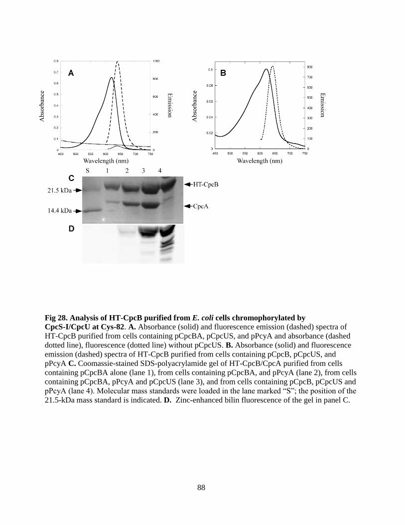
88
Fig 28. Analysis of HT-CpcB purified from E. coli cells chromophorylated by
CpcS-I/CpcU at Cys-82. A. Absorbance (solid) and fluorescence emission (dashed) spectra of
HT-CpcB purified from cells containing pCpcBA, pCpcUS, and pPcyA and absorbance (dashed
dotted line), fluorescence (dotted line) without pCpcUS. B. Absorbance (solid) and fluorescence
emission (dashed) spectra of HT-CpcB purified from cells containing pCpcB, pCpcUS, and
pPcyA C. Coomassie-stained SDS-polyacrylamide gel of HT-CpcB/CpcA purified from cells
containing pCpcBA alone (lane 1), from cells containing pCpcBA, and pPcyA (lane 2), from cells
containing pCpcBA, pPcyA and pCpcUS (lane 3), and from cells containing pCpcB, pCpcUS and
pPcyA (lane 4). Molecular mass standards were loaded in the lane marked “S”; the position of the
21.5-kDa mass standard is indicated. D. Zinc-enhanced bilin fluorescence of the gel in panel C.
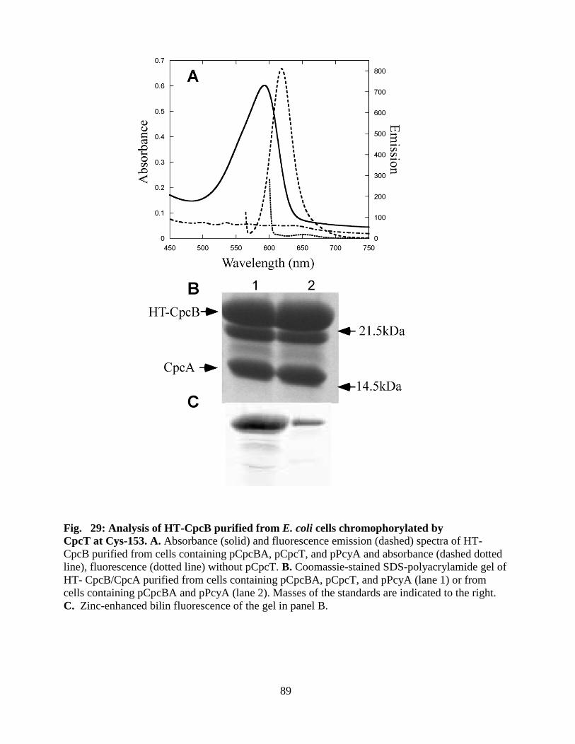
89
Fig. 29: Analysis of HT-CpcB purified from E. coli cells chromophorylated by
CpcT at Cys-153. A. Absorbance (solid) and fluorescence emission (dashed) spectra of HT-
CpcB purified from cells containing pCpcBA, pCpcT, and pPcyA and absorbance (dashed dotted
line), fluorescence (dotted line) without pCpcT. B. Coomassie-stained SDS-polyacrylamide gel of
HT- CpcB/CpcA purified from cells containing pCpcBA, pCpcT, and pPcyA (lane 1) or from
cells containing pCpcBA and pPcyA (lane 2). Masses of the standards are indicated to the right.
C. Zinc-enhanced bilin fluorescence of the gel in panel B.
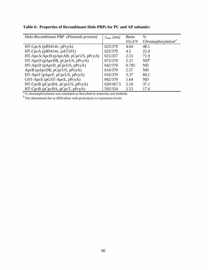
90
Table 6: Properties of Recombinant Holo-PBPs for PC and AP subunits:
Holo-Recombinant PBP (Plasmids present) max [nm] Ratio
Vis:UV
%
Chromophorylationa
HT-CpcA (pBS414v, pPcyA) 625/370 4.64 48.1
HT-CpcA (pBS414v, pAT101) 625/370 4.5 22.4
HT-ApcA/ApcB (pApcAB, pCpcUS, pPcyA) 615/357 2.53 71.9
HT-ApcD (pApcDB, pCpcUS, pPcyA) 672/370 2.21 NDb
HT-ApcD (pApcD, pCpcUS, pPcyA) 642/370 0.785 ND
ApcB (pApcDB, pCpcUS, pPcyA) 616/370 2.57 ND
HT-ApcF (pApcF, pCpcUS, pPcyA) 616/370 3.37 68.1
GST-ApcE (pGST-ApcE, pPcyA) 662/370 1.64 ND
HT-CpcB (pCpcBA, pCpcUS, pPcyA) 620/367.5 5.19 37.1
HT-CpcB (pCpcBA, pCpcT, pPcyA) 592/354 2.53 17.4 a % chromophorylation was estimated as described in materials and methods.
b Not determined due to difficulties with proteolysis or expression levels
91
3.2. Creation of Unique phycobiliproteins using PEB in E.coli for potential
Biotechnological applications
3.2.1. Creation of holo α-PC using PEB in E. coli:
For this part of the project, the goal was to see if the different classes of PCB lyases were able to
attach PEB to substrates using our in vivo heterologous system, as the ability to create
phycobiliproteins with different chromophore content in this system would be a great
biotechnological tool. PEb containing PBPs have higher quantum yield. First, the CpcE/CpcF
lyase which normally attaches PCB to CpcA (-PC) was used. The constructs we used had been
used by Tooley et al. to show that holo-CpcA could be formed in E. coli using cotransformation
with pAT101 (encoding Synechocystis sp. PCC 6803 hox1 and pcyA) (Tooley, Cai et al. 2001). E.
coli cells were transformed with the pPebS plasmid and either pBS414v (encoding cpcA, cpcE
and cpcF) or pBS405v (encoding cpcA). Cells were grown as described, harvested, and exhibited
a pink color as shown in Fig. 30D. The HT-CpcA was purified by metal affinity chromatography.
Absorption spectra of the two HT-CpcA showed that only the one produced in the presence of
CpcE and CpcF (pBS414v) contains significant PEB addition with an absorbance maximum at
560 nm (see Fig. 30A). The HT-CpcA produced in the presence of the bilin lyase CpcE/CpcF
was highly fluorescent with a single emission peak at 567 nm whereas the HT-CpcA produced in
cells with PEB but without the CpcE/CpcF bilin lyase (pBS405v) had two fluorescent emission
peaks at 567 nm and 630 nm. Fairchild and Glazer previously performed in vitro reactions with
apo-CpcA, PEB and with or without purified CpcE/CpcF; the fluorescence emission maxima for
the CpcE/CpcF-mediated CpcA product was at 571 nm whereas the non-enzyme mediated CpcA
product had a broad emission maxima in the range of 570-581 nm (Fairchild and Glazer 1994).
Interesting enough the Quantum yield for holo-CpcA (PEB) was 0.98, whereas in the case of
holo-CpcA (PCB) it was 0.85.
92
After separating the HT-CpcA proteins by SDS-PAGE and examining total protein
content (Coomassie Blue staining, Fig. 30B) versus bilin-bound content (Zn-enhanced
fluorescence in Fig. 30C), HT-CpcA contained PEB chromophore when purified from cells
containing CpcEF (lane 1) whereas there was almost no PEB attached to the HT-CpcA purified
from cells without CpcEF (lane 2). When the protein concentration of the HT-CpcA (OD280) is
divided by the PEB concentration of the sample (in 8 M urea, pH 2), 83.5% of HT-CpcA contains
PEB (see Table 7). The chromophorylated product yield for holo CpcA (CpcA-PEB) was 11.7
mg L-1
.
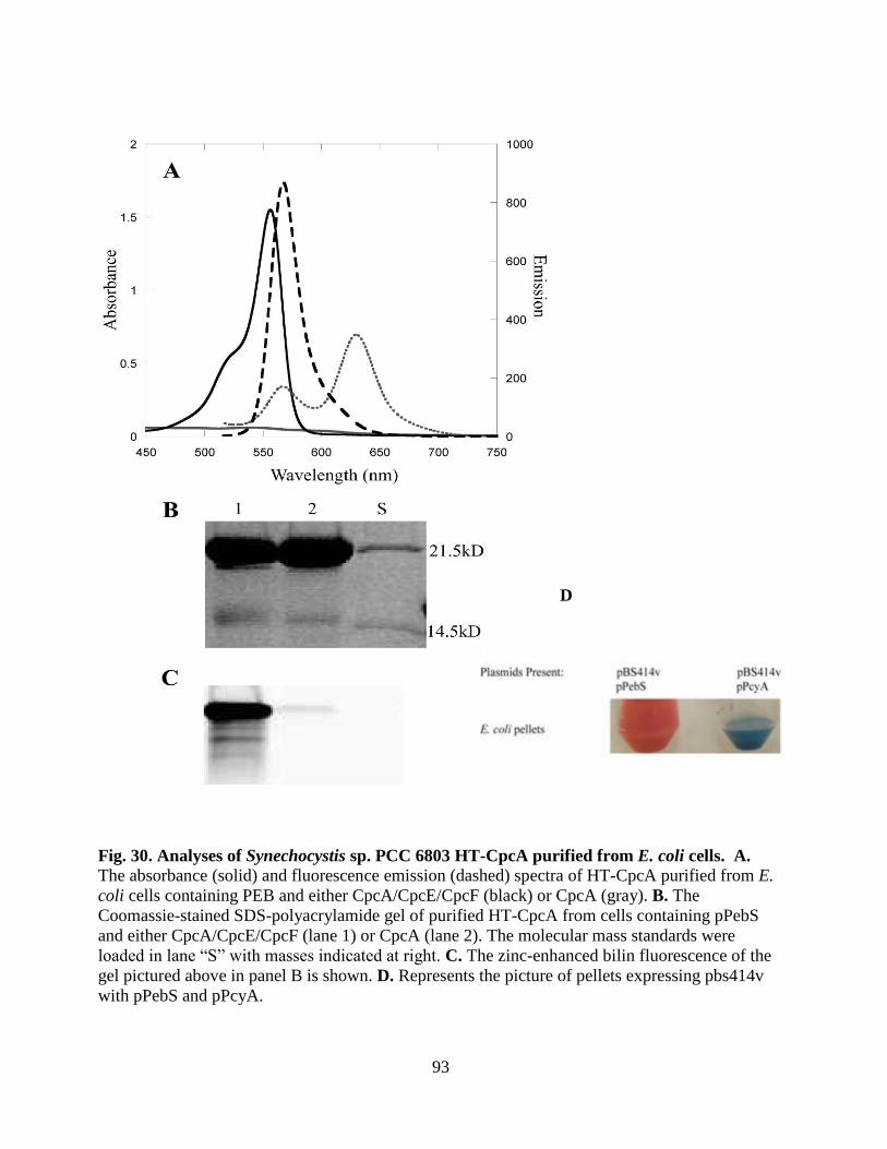
93
D
Fig. 30. Analyses of Synechocystis sp. PCC 6803 HT-CpcA purified from E. coli cells. A. The absorbance (solid) and fluorescence emission (dashed) spectra of HT-CpcA purified from E.
coli cells containing PEB and either CpcA/CpcE/CpcF (black) or CpcA (gray). B. The
Coomassie-stained SDS-polyacrylamide gel of purified HT-CpcA from cells containing pPebS
and either CpcA/CpcE/CpcF (lane 1) or CpcA (lane 2). The molecular mass standards were
loaded in lane “S” with masses indicated at right. C. The zinc-enhanced bilin fluorescence of the
gel pictured above in panel B is shown. D. Represents the picture of pellets expressing pbs414v
with pPebS and pPcyA.
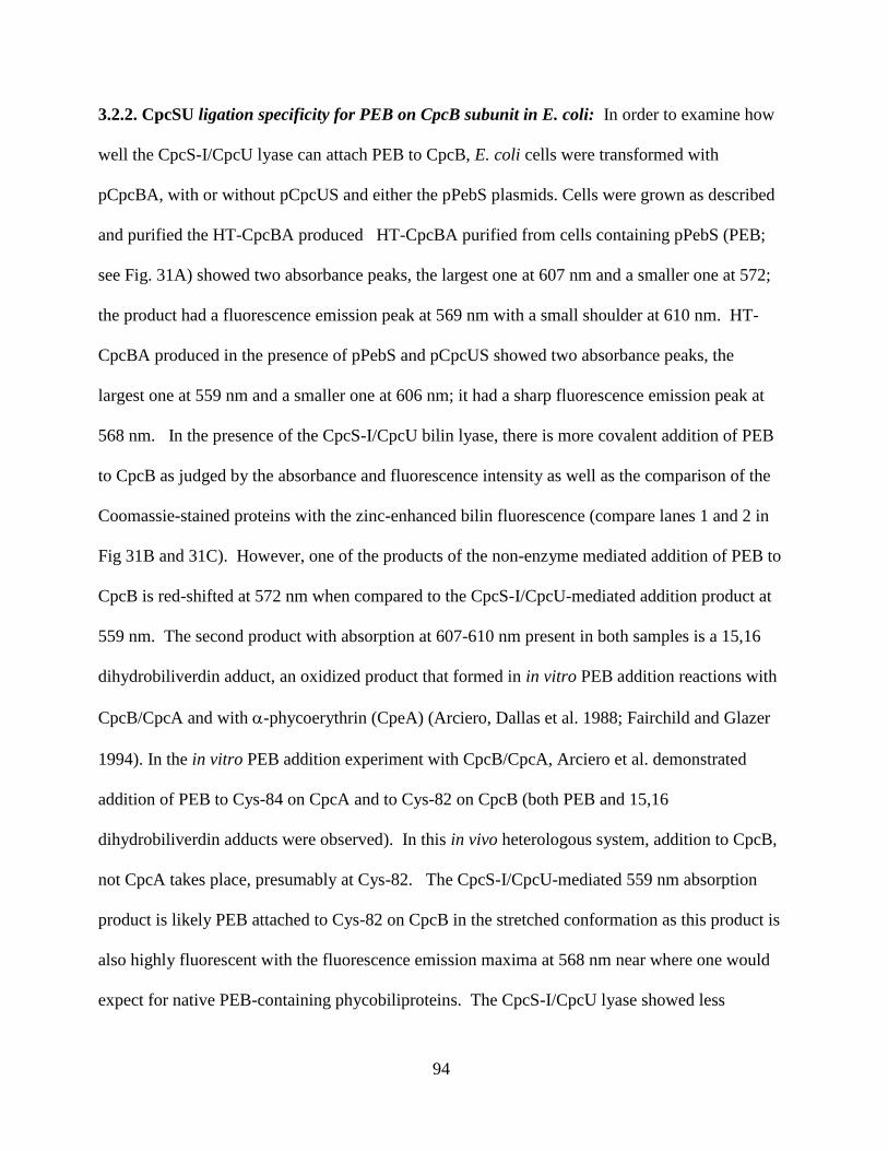
94
3.2.2. CpcSU ligation specificity for PEB on CpcB subunit in E. coli: In order to examine how
well the CpcS-I/CpcU lyase can attach PEB to CpcB, E. coli cells were transformed with
pCpcBA, with or without pCpcUS and either the pPebS plasmids. Cells were grown as described
and purified the HT-CpcBA produced HT-CpcBA purified from cells containing pPebS (PEB;
see Fig. 31A) showed two absorbance peaks, the largest one at 607 nm and a smaller one at 572;
the product had a fluorescence emission peak at 569 nm with a small shoulder at 610 nm. HT-
CpcBA produced in the presence of pPebS and pCpcUS showed two absorbance peaks, the
largest one at 559 nm and a smaller one at 606 nm; it had a sharp fluorescence emission peak at
568 nm. In the presence of the CpcS-I/CpcU bilin lyase, there is more covalent addition of PEB
to CpcB as judged by the absorbance and fluorescence intensity as well as the comparison of the
Coomassie-stained proteins with the zinc-enhanced bilin fluorescence (compare lanes 1 and 2 in
Fig 31B and 31C). However, one of the products of the non-enzyme mediated addition of PEB to
CpcB is red-shifted at 572 nm when compared to the CpcS-I/CpcU-mediated addition product at
559 nm. The second product with absorption at 607-610 nm present in both samples is a 15,16
dihydrobiliverdin adduct, an oxidized product that formed in in vitro PEB addition reactions with
CpcB/CpcA and with -phycoerythrin (CpeA) (Arciero, Dallas et al. 1988; Fairchild and Glazer
1994). In the in vitro PEB addition experiment with CpcB/CpcA, Arciero et al. demonstrated
addition of PEB to Cys-84 on CpcA and to Cys-82 on CpcB (both PEB and 15,16
dihydrobiliverdin adducts were observed). In this in vivo heterologous system, addition to CpcB,
not CpcA takes place, presumably at Cys-82. The CpcS-I/CpcU-mediated 559 nm absorption
product is likely PEB attached to Cys-82 on CpcB in the stretched conformation as this product is
also highly fluorescent with the fluorescence emission maxima at 568 nm near where one would
expect for native PEB-containing phycobiliproteins. The CpcS-I/CpcU lyase showed less
95
addition of PEB (6% chromophorylation; see Table 7) to CpcB than it did for PCB (37%
chromophorylation). The bilin at Cys-82 is the terminal energy acceptor within the PC trimer;
having PEB attached here would have negative consequences for efficient energy transfer from
PCB attached at Cys-153 on CpcB or from PCB attached to Cys-84 on CpcA. Energy absorbed
by these other peripheral bilins is transferred to Cys-82 on CpcB and eventually to the AP core.
Another explanation of the lower level of PEB chromophorylation to CpcB may be that the CpcS-
I/CpcU lyase is not as active at the lower temperatures required for activity of PebS (12 hours at
18 C), however the CpcE/CpcF lyase was able to achieve a high level of PEB chromophorylation
of CpcA under those same conditions.
96
Fig 31. Analyses of Synechocystis sp. PCC 6803 HT-CpcB/CpcA purified from E. coli cells. A. Absorbance (solid) and fluorescence emission (dashed) spectra of HT-CpcB/CpcA purified
from cells containing pCpcBA, pPebS, and with (black) or without (gray) pCpcUS.
B.The Coomassie-stained SDS-polyacrylamide gel containing HT-CpcB/CpcA purified from cells
containing pCpcBA, pPebS, and with (lane 1) our without (lane 2) pCpcUS. Molecular masses
of standards are indicated at right. C. The zinc-enhanced bilin fluorescence of the gel above in
panel B is shown here.

97
Table 7: Properties of Recombinant Holo-PBPs with non-cognate lyases
Holo-Recombinant PBP (Plasmids present) max [nm] Ratio
Vis:UV
%
Chromophorylationa
HT-CpcA(pBS414, pPebS) 571 8.69 83.5
HT-CpcBA (pCpcBA, pCpcSU, pPebS) 572 0.707 6.06 a % chromophorylation was estimated as described in materials and methods.
b Not determined due to difficulties with proteolysis or expression levels
98
3.3. Characterization of CpeY, CpeZ and CpeS bilin lyases involved in
phycoerythrin biosynthesis in Fremyella diplosiphon strain UTEX 481
3.3.1 Characterization of bilin lyase activity of CpeY and CpeZ with CpeA. The cpeY and cpeZ
genes occur downstream of the cpeBA genes encoding the and subunits of PE, respectively.
Based upon their sequence similarity (~32 %), CpeY and CpeZ belong to the CpcE/CpcF family
of bilin lyases (See Fig 32). Transposon mutants and complementation studies in Fremyella
diplosiphon UTEX 481 suggested that these two proteins might be involved in PE biogenesis, but
their specific roles were not elucidated (Kahn, Mazel et al. 1997). Recombinant CpeY and CpeZ
from the cyanobacterium F. diplosiphon were soluble (data not shown). An in vivo E. coli
heterologous co-expression system was used to test whether either of these genes encodes a bilin
lyase. Constructs made for this study (thesis) are listed in Table 4.
E.coli cells containing only plasmids pCpeA and pPebS (i. e., no lyase present) had no
significant color (data not shown), but cells containing these two plasmids and pCpeYZ were
bright pinkish-red in color (Fig. 33). Holo-HT-CpeA purified from these cells had an absorbance
maximum at 560 nm (See Fig. 34A) and a very high fluorescence emission maximum at 574 nm
(See Fig 34A; the sample was diluted 15-fold to 0.05 OD560 prior to obtaining the fluorescence
spectrum), whereas the cells containing pCpeA and pPebS only did not have any significant
absorbance or fluorescence emission (See Fig. 34A; the sample was not diluted prior to obtaining
the fluorescence emission spectrum). CpeZ and CpeY were also tested individually to determine
if the individual proteins could attach PEB to CpeA. The fluorescence emission spectrum
obtained from the HT-CpeA purified from cells containing pCpeA, pPebS and pCpeY showed
that only CpeY had significant activity by itself, but the amount of fluorescent product was lower
than when both CpeY and CpeZ were present (See Fig. 34B). The relative yields of holo-CpeA
produced when co-expressed with PebS along with either CpeY or CpeZ are given in Table 8.
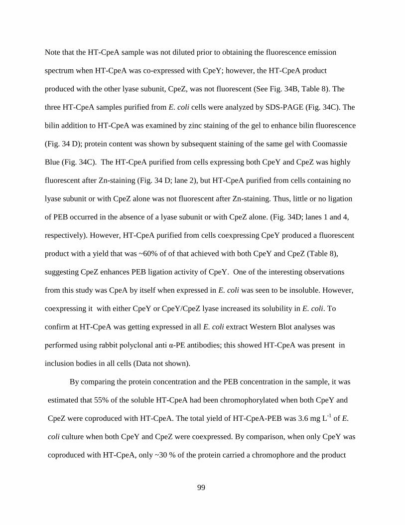
99
Note that the HT-CpeA sample was not diluted prior to obtaining the fluorescence emission
spectrum when HT-CpeA was co-expressed with CpeY; however, the HT-CpeA product
produced with the other lyase subunit, CpeZ, was not fluorescent (See Fig. 34B, Table 8). The
three HT-CpeA samples purified from E. coli cells were analyzed by SDS-PAGE (Fig. 34C). The
bilin addition to HT-CpeA was examined by zinc staining of the gel to enhance bilin fluorescence
(Fig. 34 D); protein content was shown by subsequent staining of the same gel with Coomassie
Blue (Fig. 34C). The HT-CpeA purified from cells expressing both CpeY and CpeZ was highly
fluorescent after Zn-staining (Fig. 34 D; lane 2), but HT-CpeA purified from cells containing no
lyase subunit or with CpeZ alone was not fluorescent after Zn-staining. Thus, little or no ligation
of PEB occurred in the absence of a lyase subunit or with CpeZ alone. (Fig. 34D; lanes 1 and 4,
respectively). However, HT-CpeA purified from cells coexpressing CpeY produced a fluorescent
product with a yield that was ~60% of of that achieved with both CpeY and CpeZ (Table 8),
suggesting CpeZ enhances PEB ligation activity of CpeY. One of the interesting observations
from this study was CpeA by itself when expressed in E. coli was seen to be insoluble. However,
coexpressing it with either CpeY or CpeY/CpeZ lyase increased its solubility in E. coli. To
confirm at HT-CpeA was getting expressed in all E. coli extract Western Blot analyses was
performed using rabbit polyclonal anti α-PE antibodies; this showed HT-CpeA was present in
inclusion bodies in all cells (Data not shown).
By comparing the protein concentration and the PEB concentration in the sample, it was
estimated that 55% of the soluble HT-CpeA had been chromophorylated when both CpeY and
CpeZ were coproduced with HT-CpeA. The total yield of HT-CpeA-PEB was 3.6 mg L-1
of E.
coli culture when both CpeY and CpeZ were coexpressed. By comparison, when only CpeY was
coproduced with HT-CpeA, only ~30 % of the protein carried a chromophore and the product

100
yield was 1.8 mg L-1
of culture. The fluorescence quantum yield for the partial holo-HT-CpeA
was 0.72, which is quite high for PE subunits.
CpeYZ is a CpcEF type lyase based on the sequence alignment. The CpcE and CpcF proteins
interact with each other (1:1) and copurify on a Ni-NTA column. However, in the case of CpeY
and CpeZ no interaction of CpeY in a pull-down assay with HT-CpeZ was detected (Data not
shown).
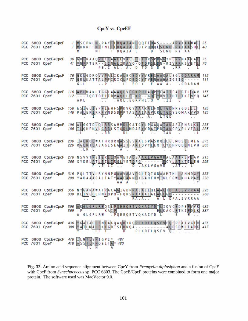
101
Fig. 32. Amino acid sequence alignment between CpeY from Fremyella diplosiphon and a fusion of CpcE
with CpcF from Synechococcus sp. PCC 6803. The CpcE/CpcF proteins were combined to form one major
protein. The software used was MacVector 9.0.
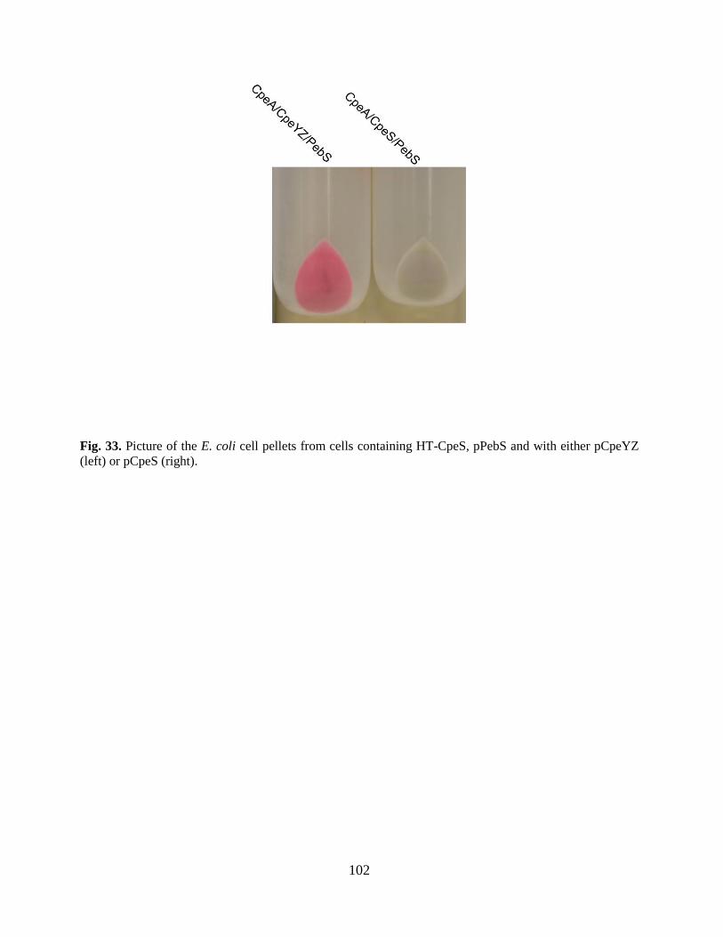
102
Fig. 33. Picture of the E. coli cell pellets from cells containing HT-CpeS, pPebS and with either pCpeYZ
(left) or pCpeS (right).
103
Fig. 34. Analyses of HT-CpeA produced with CpeY and CpeZ in E. coli. A. Absorbance
(solid line) and fluorescence emission (dashed line) spectra of HT-CpeA purified from cells
containing pCpeA, pPebS with pCpeYZ and absorbance (dashed dotted line), fluorescence (dotted
line) without pCpeYZ are shown. B. Absorbance (solid line) and fluorescence emission (dashed
line) spectra of HT-CpeA purified from cells containing pCpeA, pPebS with pCpeY and
absorbance (dashed dotted line), fluorescence (dotted line) with pCpeZ are shown. In order to
acquire the fluorescence emission spectra for the HT-CpeA produced in the presence of pCpeYZ
and pCpeY (dashed lines in panels A and B) the samples were diluted to OD560nm of 0.05;
however, no dilution was performed on HT-CpeA produced in the absence of a lyase or in the
presence of pCpeZ (dotted lines in panels A and B). C. This panel shows a Coomassie-stained
SDS polyacrylamide gel containing HT-CpeA purified from cells containing pCpeA, pPebS with
no lyase (lane 1) or with pCpeYZ (lane 2), or HT-CpeA purified from cells containing pCpeA,
pPebS, and either pCpeY (lane 3) or pCpeZ (lane 4). Molecular mass standards are loaded in lane
“S”, and masses are indicated to the right. D. The zinc-enhanced fluorescence of the gel pictured
in panel
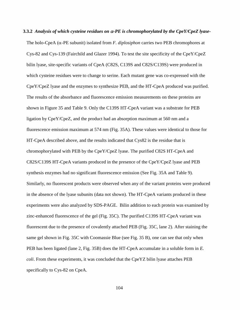
104
3.3.2 Analysis of which cysteine residues on -PE is chromophorylated by the CpeY/CpeZ lyase-
The holo-CpeA (-PE subunit) isolated from F. diplosiphon carries two PEB chromophores at
Cys-82 and Cys-139 (Fairchild and Glazer 1994). To test the site specificity of the CpeY/CpeZ
bilin lyase, site-specific variants of CpeA (C82S, C139S and C82S/C139S) were produced in
which cysteine residues were to change to serine. Each mutant gene was co-expressed with the
CpeY/CpeZ lyase and the enzymes to synthesize PEB, and the HT-CpeA produced was purified.
The results of the absorbance and fluorescence emission measurements on these proteins are
shown in Figure 35 and Table 9. Only the C139S HT-CpeA variant was a substrate for PEB
ligation by CpeY/CpeZ, and the product had an absorption maximum at 560 nm and a
fluorescence emission maximum at 574 nm (Fig. 35A). These values were identical to those for
HT-CpeA described above, and the results indicated that Cys82 is the residue that is
chromophorylated with PEB by the CpeY/CpeZ lyase. The purified C82S HT-CpeA and
C82S/C139S HT-CpeA variants produced in the presence of the CpeY/CpeZ lyase and PEB
synthesis enzymes had no significant fluorescence emission (See Fig. 35A and Table 9).
Similarly, no fluorescent products were observed when any of the variant proteins were produced
in the absence of the lyase subunits (data not shown). The HT-CpeA variants produced in these
experiments were also analyzed by SDS-PAGE. Bilin addition to each protein was examined by
zinc-enhanced fluorescence of the gel (Fig. 35C). The purified C139S HT-CpeA variant was
fluorescent due to the presence of covalently attached PEB (Fig. 35C, lane 2). After staining the
same gel shown in Fig. 35C with Coomassie Blue (see Fig. 35 B), one can see that only when
PEB has been ligated (lane 2, Fig. 35B) does the HT-CpeA accumulate in a soluble form in E.
coli. From these experiments, it was concluded that the CpeYZ bilin lyase attaches PEB
specifically to Cys-82 on CpeA.
105
Fig. 35. Analyses of the specific cysteine residue on HT-CpeA required for PEB addition by
CpeYZ. A. Absorbance (solid line) and fluorescence emission (dashed line) spectra of HT-
CpeA(C139S) purified from cells containing CpeA(C139S), pPebS with pCpeYZ and the
absorbance (dashed dotted line), fluorescence (dotted line) spectra from cells containing
CpeA(C82S), pPebS with pCpeYZ are shown. In order to acquire the fluorescence emission
spectra for the HTCpeA (C139S) produced in the presence of pCpeYZ (dashed lines in panel A),
the sample was diluted to OD560nm of 0.05; however, no dilution was performed on HT-
CpeA(C82S) (dotted line in panel A). B. Coomassie-stained SDS polyacrylamide gel containing
HTCpeA mutants purified from cells containing CpeA (C82S), pPebS, pCpeYZ with (lane 1) or
(lane 2) CpeA (C139S), pPebS, pCpeYZ and (lane 3) purified from the cells containing CpeA
(C82,139S), pPebS, pCpeYZ. Molecular mass standards are loaded in lane “S”, and masses are
indicated to the right. C. The zinc-enhanced fluorescence of the gel pictured in panel B is shown
here.
The above result was also confirmed using Mass spectrometry. The tryptic peptide from CpeA
containing PEB was identified (CAR) (Bhougatou and Cole, unpublished data).
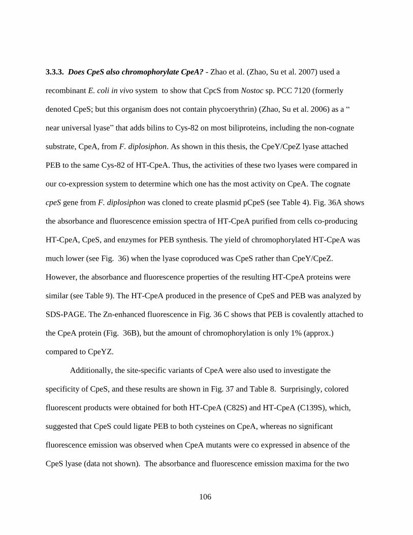
106
3.3.3. Does CpeS also chromophorylate CpeA? - Zhao et al. (Zhao, Su et al. 2007) used a
recombinant E. coli in vivo system to show that CpcS from Nostoc sp. PCC 7120 (formerly
denoted CpeS; but this organism does not contain phycoerythrin) (Zhao, Su et al. 2006) as a “
near universal lyase” that adds bilins to Cys-82 on most biliproteins, including the non-cognate
substrate, CpeA, from F. diplosiphon. As shown in this thesis, the CpeY/CpeZ lyase attached
PEB to the same Cys-82 of HT-CpeA. Thus, the activities of these two lyases were compared in
our co-expression system to determine which one has the most activity on CpeA. The cognate
cpeS gene from F. diplosiphon was cloned to create plasmid pCpeS (see Table 4). Fig. 36A shows
the absorbance and fluorescence emission spectra of HT-CpeA purified from cells co-producing
HT-CpeA, CpeS, and enzymes for PEB synthesis. The yield of chromophorylated HT-CpeA was
much lower (see Fig. 36) when the lyase coproduced was CpeS rather than CpeY/CpeZ.
However, the absorbance and fluorescence properties of the resulting HT-CpeA proteins were
similar (see Table 9). The HT-CpeA produced in the presence of CpeS and PEB was analyzed by
SDS-PAGE. The Zn-enhanced fluorescence in Fig. 36 C shows that PEB is covalently attached to
the CpeA protein (Fig. 36B), but the amount of chromophorylation is only 1% (approx.)
compared to CpeYZ.
Additionally, the site-specific variants of CpeA were also used to investigate the
specificity of CpeS, and these results are shown in Fig. 37 and Table 8. Surprisingly, colored
fluorescent products were obtained for both HT-CpeA (C82S) and HT-CpeA (C139S), which,
suggested that CpeS could ligate PEB to both cysteines on CpeA, whereas no significant
fluorescence emission was observed when CpeA mutants were co expressed in absence of the
CpeS lyase (data not shown). The absorbance and fluorescence emission maxima for the two

107
variants were different; this suggested the different protein environments for the PEB in the two
variants, affected its absorbance and emission properties (Table 8). In further support of this
interpretation, two PEB-containing peptides were observed after tryptic digestion of wild-type
HT-CpeA synthesized in the presence of PEB and CpeS (See Fig. 38).
108
Fig. 36. Analyses of HT-CpeA produced with CpeS in E. coli A. Absorbance (solid line) and
fluorescence emission (dashed line) spectra of HT-CpeA purified from cells containing pCpeA,
pPebS with pCpeS B. Coomassie-stained SDS polyacrylamide gel containing HT-CpeA purified
from cells containing pCpeA, pPebS, pCpeS. C. The zinc-enhanced fluorescence of the gel
pictured in panel B is shown here.
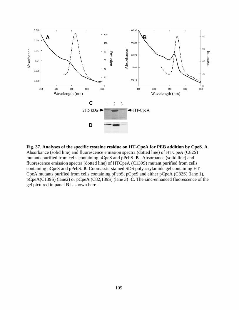
109
Fig. 37. Analyses of the specific cysteine residue on HT-CpeA for PEB addition by CpeS. A.
Absorbance (solid line) and fluorescence emission spectra (dotted line) of HTCpeA (C82S)
mutants purified from cells containing pCpeS and pPebS. B. Absorbance (solid line) and
fluorescence emission spectra (dotted line) of HTCpeA (C139S) mutant purified from cells
containing pCpeS and pPebS. B. Coomassie-stained SDS polyacrylamide gel containing HT-
CpeA mutants purified from cells containing pPebS, pCpeS and either pCpeA (C82S) (lane 1),
pCpeA(C139S) (lane2) or pCpeA (C82,139S) (lane 3) C. The zinc-enhanced fluorescence of the
gel pictured in panel B is shown here.
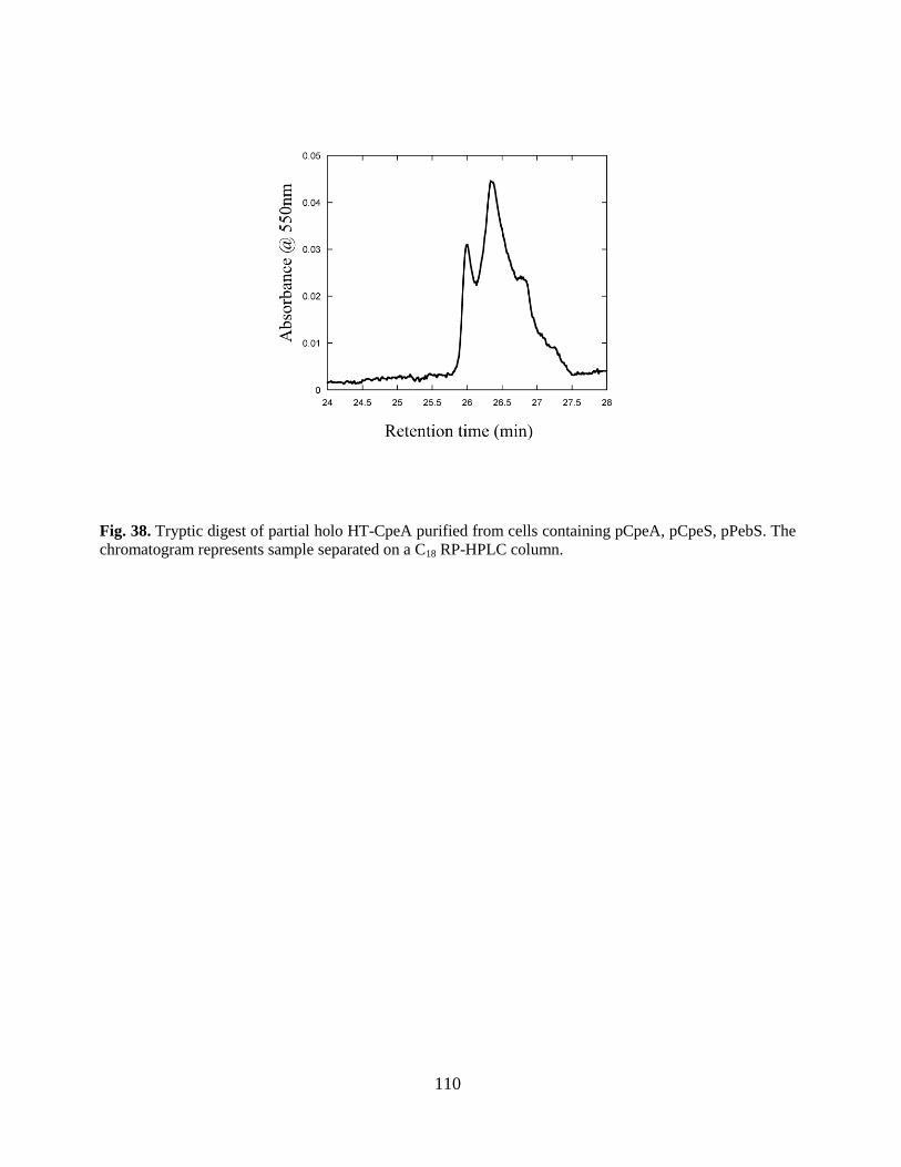
110
Fig. 38. Tryptic digest of partial holo HT-CpeA purified from cells containing pCpeA, pCpeS, pPebS. The
chromatogram represents sample separated on a C18 RP-HPLC column.
111
3.3.4. Comparison of the PEB ligation activity of CpeY/CpeZ and CpeS bilin lyases in CpeB-
CpeB (-PE) in F. diplosiphon has three PEB chromophores attached to four Cys residues: Cys-
80 and Cys-165 carry singly linked PEBs and Cys-48 and Cys-59 carry a doubly linked PEB
(Fairchild and Glazer 1994) (See Fig. 5). Two different sterioisomers occur in PE-β subunits ( C31
and C181 of the bilin ) (Schirmer, Huber et al. 1986). The R-isomer, the most common one is
found at Cys-80 and Cys-48, 59, whereas the S-isomer is present the Cys-165 on CpeB. Using the
in vivo co-expression system, HT-CpeB was coproduced with CpeS and enzymes for PEB
synthesis. Fig. 39A shows the absorbance and fluorescence emission spectra of the resulting HT-
CpeB product purified by metal-affinity chromatography. The absorbance maximum was 556 nm,
and the fluorescence emission maximum was 572 nm. No significant chromophorylated HT-
CpeB was synthesized in the absence of CpeS (Fig. 39A, dashed dotted line). The bilin content of
each protein was examined by Zn-enhanced fluorescence staining of an SDS-polyacrylamide gel
(Fig. 39 D) and the proteins were detected after staining the same gel with Coomassie Blue (Fig.
39C). PEB was covalently attached to CpeB only in the presence of CpeS (Fig. 39 D, lane 3).
However, it was also clear that very little CpeB remained soluble unless an attached bilin (or
lyase) was present (compare lanes 1, 2 and 3 in Fig. 39C). The HT-CpeB expression was
confirmed by Western Blot using rabbit polyclonal anti β-PE, and was found to be present only in
inclusion bodies.
The CpeY/CpeZ lyase was also coproduced with HT-CpeB; however, no absorbance or
fluorescence emission was observed (Fig. 39B). When the purified protein from
pCpeB/pCpeYZ/pPebS was separated on SDS-PAGE, no notable fluorescent band was observed
(Fig. 39D, lane 1), indicating that CpeYZ was unable to ligate PEB on β-PE. CpeY and CpeZ
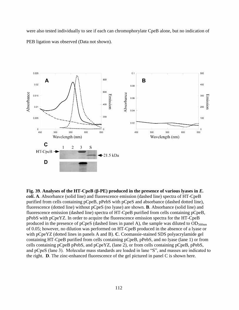
112
were also tested individually to see if each can chromophorylate CpeB alone, but no indication of
PEB ligation was observed (Data not shown).
Fig. 39. Analyses of the HT-CpeB (β-PE) produced in the presence of various lyases in E.
coli. A. Absorbance (solid line) and fluorescence emission (dashed line) spectra of HT-CpeB
purified from cells containing pCpeB, pPebS with pCpeS and absorbance (dashed dotted line),
fluorescence (dotted line) without pCpeS (no lyase) are shown. B. Absorbance (solid line) and
fluorescence emission (dashed line) spectra of HT-CpeB purified from cells containing pCpeB,
pPebS with pCpeYZ. In order to acquire the fluorescence emission spectra for the HT-CpeB
produced in the presence of pCpeS (dashed lines in panel A), the sample was diluted to OD560nm
of 0.05; however, no dilution was performed on HT-CpeB produced in the absence of a lyase or
with pCpeYZ (dotted lines in panels A and B). C. Coomassie-stained SDS polyacrylamide gel
containing HT-CpeB purified from cells containing pCpeB, pPebS, and no lyase (lane 1) or from
cells containing pCpeB pPebS, and pCpeYZ, (lane 2), or from cells containing pCpeB, pPebS,
and pCpeS (lane 3). Molecular mass standards are loaded in lane “S”, and masses are indicated to
the right. D. The zinc-enhanced fluorescence of the gel pictured in panel C is shown here.
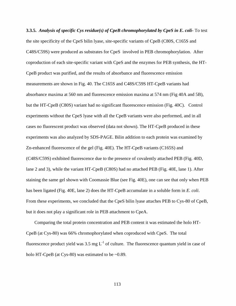
113
3.3.5. Analysis of specific Cys residue(s) of CpeB chromophorylated by CpeS in E. coli- To test
the site specificity of the CpeS bilin lyase, site-specific variants of CpeB (C80S, C165S and
C48S/C59S) were produced as substrates for CpeS involved in PEB chromophorylation. After
coproduction of each site-specific variant with CpeS and the enzymes for PEB synthesis, the HT-
CpeB product was purified, and the results of absorbance and fluorescence emission
measurements are shown in Fig. 40. The C165S and C48S/C59S HT-CpeB variants had
absorbance maxima at 560 nm and fluorescence emission maxima at 574 nm (Fig 40A and 5B),
but the HT-CpeB (C80S) variant had no significant fluorescence emission (Fig. 40C). Control
experiments without the CpeS lyase with all the CpeB variants were also performed, and in all
cases no fluorescent product was observed (data not shown). The HT-CpeB produced in these
experiments was also analyzed by SDS-PAGE. Bilin addition to each protein was examined by
Zn-enhanced fluorescence of the gel (Fig. 40E). The HT-CpeB variants (C165S) and
(C48S/C59S) exhibited fluorescence due to the presence of covalently attached PEB (Fig. 40D,
lane 2 and 3), while the variant HT-CpeB (C80S) had no attached PEB (Fig. 40E, lane 1). After
staining the same gel shown with Coomassie Blue (see Fig. 40E), one can see that only when PEB
has been ligated (Fig. 40E, lane 2) does the HT-CpeB accumulate in a soluble form in E. coli.
From these experiments, we concluded that the CpeS bilin lyase attaches PEB to Cys-80 of CpeB,
but it does not play a significant role in PEB attachment to CpeA.
Comparing the total protein concentration and PEB content it was estimated the holo HT-
CpeB (at Cys-80) was 66% chromophorylated when coproduced with CpeS. The total
fluorescence product yield was 3.5 mg L-1
of culture. The fluorescence quantum yield in case of
holo HT-CpeB (at Cys-80) was estimated to be ~0.89.

114
Maldi-Mass spectrometry(MS) and Tandem MS experiments on purified tryptic peptides
from CpeS generated HT CpeB that Cys 80 is the addition site for CpeS on CpeB (tryptic peptide
containing PEB was MAACLR) (Bhougatou, Cole unpublished data).
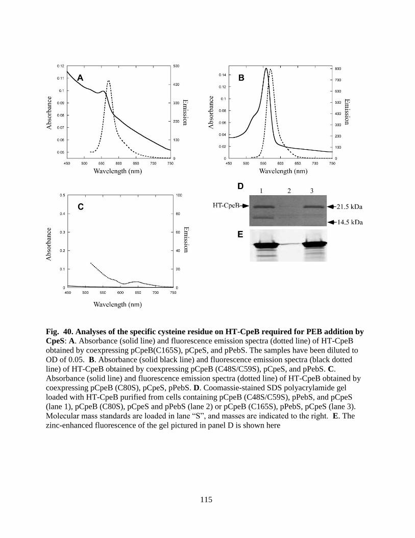
115
Fig. 40. Analyses of the specific cysteine residue on HT-CpeB required for PEB addition by
CpeS: A. Absorbance (solid line) and fluorescence emission spectra (dotted line) of HT-CpeB
obtained by coexpressing pCpeB(C165S), pCpeS, and pPebS. The samples have been diluted to
OD of 0.05. B. Absorbance (solid black line) and fluorescence emission spectra (black dotted
line) of HT-CpeB obtained by coexpressing pCpeB (C48S/C59S), pCpeS, and pPebS. C.
Absorbance (solid line) and fluorescence emission spectra (dotted line) of HT-CpeB obtained by
coexpressing pCpeB (C80S), pCpeS, pPebS. D. Coomassie-stained SDS polyacrylamide gel
loaded with HT-CpeB purified from cells containing pCpeB (C48S/C59S), pPebS, and pCpeS
(lane 1), pCpeB (C80S), pCpeS and pPebS (lane 2) or pCpeB (C165S), pPebS, pCpeS (lane 3).
Molecular mass standards are loaded in lane “S”, and masses are indicated to the right. E. The
zinc-enhanced fluorescence of the gel pictured in panel D is shown here
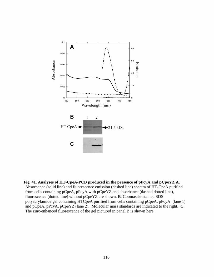
116
Fig. 41. Analyses of HT-CpeA-PCB produced in the presence of pPcyA and pCpeYZ A.
Absorbance (solid line) and fluorescence emission (dashed line) spectra of HT-CpeA purified
from cells containing pCpeA, pPcyA with pCpeYZ and absorbance (dashed dotted line),
fluorescence (dotted line) without pCpeYZ are shown. B. Coomassie-stained SDS
polyacrylamide gel containing HTCpeA purified from cells containing pCpeA, pPcyA (lane 1)
and pCpeA, pPcyA, pCpeYZ (lane 2). Molecular mass standards are indicated to the right. C.
The zinc-enhanced fluorescence of the gel pictured in panel B is shown here.
117
Table 8. Comparison of spectral properties for various PE subunits produced with bilin
lyases:
Name of recombinant plasmids max (nm)(Q Vis/UV) Fluorescence Emission max (nm)
pCpeA + pCpeYZ1 566/410 (5.4) 573.5
pCpeA(C82S) + pCpeYZ1 NA NA
pCpeA(C139S) + pCpeYZ1 566/410 (5.6) 574
pCpeA(C82, 139S) + pCpeYZ1 NA NA
pCpeA + pCpeZ 1
NA NA
pCpeA + pCpeY 1
566/410 (15.6) 573.5
pCpeA(C82S) + pCpeY 1
NA NA
pCpeA(C139S) + pCpeY 1 566/410 (14.8) 574
pCpeA(C82,139S) + pCpeY1 NA NA
pCpeA + pCpeS1 561/410 (0.315) 574
pCpeA(C82S) + pCpeS
1 550/398 (0.6) 562
pCpeA(C139S) + pCpeS
1 564/410 (0.6) 574
pCpeA(C82, 139S) + pCpeS
1 NA NA
pCpeB + pCpeS1 560/412 (5.2) 571
pCpeB(C80S) + pCpeS1 NA NA
pCpeB(C165S) + pCpeS1 560/412 (5.4) 571
pCpeB(C48,59S) + pCpeS1 560/398 (5.3) 571
Native PE ()6 3 563/374 (9.5) 573
1 Coexpressed with pPebS
2Q Vis/UV denoted the absorbance ratio of the visible and near- UV bands
2 Holo PE purified from F. diplosiphon.

118
Table 9. Comparing Fluorescence Intensity for various recombinant holo α-PE:
α-PE mutants * % Fluorescence emission
CpeA+pCpeYZ 100
CpeA+pCpeY 60
CpeA+pCpeZ 0
CpeA+CpeS 0.8
* coexpressed with pPebS
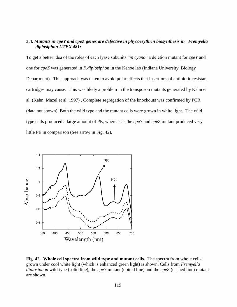
119
3.4. Mutants in cpeY and cpeZ genes are defective in phycoerythrin biosynthesis in Fremyella
diplosiphon UTEX 481:
To get a better idea of the roles of each lyase subunits “in cyano” a deletion mutant for cpeY and
one for cpeZ was generated in F.diplosiphon in the Kehoe lab (Indiana University, Biology
Department). This approach was taken to avoid polar effects that insertions of antibiotic resistant
cartridges may cause. This was likely a problem in the transposon mutants generated by Kahn et
al. (Kahn, Mazel et al. 1997) . Complete segregation of the knockouts was confirmed by PCR
(data not shown). Both the wild type and the mutant cells were grown in white light. The wild
type cells produced a large amount of PE, whereas as the cpeY and cpeZ mutant produced very
little PE in comparison (See arrow in Fig. 42).
Fig. 42. Whole cell spectra from wild type and mutant cells. The spectra from whole cells
grown under cool white light (which is enhanced green light) is shown. Cells from Fremyella
diplosiphon wild type (solid line), the cpeY mutant (dotted line) and the cpeZ (dashed line) mutant
are shown.
PE
PC
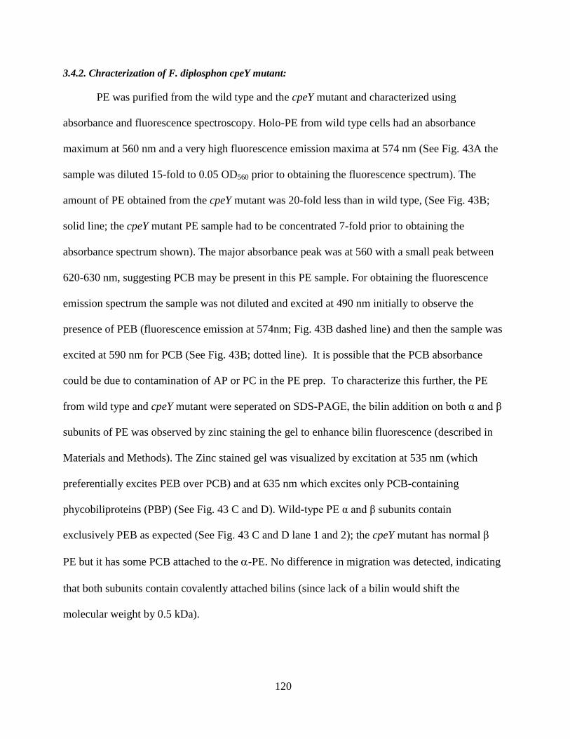
120
3.4.2. Chracterization of F. diplosphon cpeY mutant:
PE was purified from the wild type and the cpeY mutant and characterized using
absorbance and fluorescence spectroscopy. Holo-PE from wild type cells had an absorbance
maximum at 560 nm and a very high fluorescence emission maxima at 574 nm (See Fig. 43A the
sample was diluted 15-fold to 0.05 OD560 prior to obtaining the fluorescence spectrum). The
amount of PE obtained from the cpeY mutant was 20-fold less than in wild type, (See Fig. 43B;
solid line; the cpeY mutant PE sample had to be concentrated 7-fold prior to obtaining the
absorbance spectrum shown). The major absorbance peak was at 560 with a small peak between
620-630 nm, suggesting PCB may be present in this PE sample. For obtaining the fluorescence
emission spectrum the sample was not diluted and excited at 490 nm initially to observe the
presence of PEB (fluorescence emission at 574nm; Fig. 43B dashed line) and then the sample was
excited at 590 nm for PCB (See Fig. 43B; dotted line). It is possible that the PCB absorbance
could be due to contamination of AP or PC in the PE prep. To characterize this further, the PE
from wild type and cpeY mutant were seperated on SDS-PAGE, the bilin addition on both α and β
subunits of PE was observed by zinc staining the gel to enhance bilin fluorescence (described in
Materials and Methods). The Zinc stained gel was visualized by excitation at 535 nm (which
preferentially excites PEB over PCB) and at 635 nm which excites only PCB-containing
phycobiliproteins (PBP) (See Fig. 43 C and D). Wild-type PE α and β subunits contain
exclusively PEB as expected (See Fig. 43 C and D lane 1 and 2); the cpeY mutant has normal β
PE but it has some PCB attached to the -PE. No difference in migration was detected, indicating
that both subunits contain covalently attached bilins (since lack of a bilin would shift the
molecular weight by 0.5 kDa).

121
One explanation of the results of the cpeY mutant is that another lyase in the cells was
able to ligate PCB to CpeA in the absence of CpeY. The most likely choice would be CpcEF, the
lyase normally attaches PCB on CpcA. To examine if CpcEF can able to attach PCB on α-PE,
heterologous coexpression system in E. coli was used.
122
Fig. 43. Analysis of Phycoerythrin purified from wild type and the cpeY mutant cells: A. The
absorbance (solid line) and florescence emission (dashed line) spectra of PE purified from wild
type PCC 7601 is shown. The excitation wavelength used was 490 nm. B. The absorbance (solid
line) and florescence emission spectra of PE purified from the cpeY mutant. The excitation
wavelength used was 490 nm (dashed line) or 590 nm (dotted line). C. The Zn-stained SDS
polyacrylamide gel shown with fluorescence excitation at 535 nm for purified PE from wild type
(lane 1 and 2) lane 3-5 and lanes 6-8 represent the purified PE from the cpeY mutant. D. This is
the same gel as above but with the fluorescence excitation at 635 nm to observe fluorescence from
PCB.
123
3.4.3. PCC 6803 CpcEF lyase activity on PCB ligation on CpeA:
The pCpeA, pCpcEF, pPcyA were transformed into E.coli and was induced with IPTG for
protein production. The purified HT-CpeA obtained (See Materials and Methods section) was
characterized using absorbance and fluorescence emission spectrocopy. E.coli cells containing
pCpeA/pPcyA has no significant absorbance or fluorescence emission spectra (data not shown),
whereas cells containing pCpeA/pCpcEF/pPcyA was shown to have an absorbance and
fluorescence emission maxima at 625 nm and 640 nm, respectively which corresponds to native
PCB bound phycobiliproteins (See Fig. 44A; sample was concentrated 4 fold to obtain the
spectrum).
The HT-CpeA samples purified from E.coli cells were analyzed by SDS-PAGE (Fig 44B and C).
The bilin addition to CpeA was examined by zinc staining of the gel to enhance bilin
fluorescence (described in Materials and Methods; Biswas et al.) as shown in Fig 44C; protein
content was shown by subsequent staining of the same gel with Coomassie Blue (Fig. 44B).
Cells containing pCpeA, pCpcEF and pPcyA showed Zn-enhanced fluorescent protein (Fig. 44C,
lane 1; sample concentrated 4 fold) no fluorescent protein was seen in cells without CpcEF lyase
(data not shown). CpcEF can ligate as bilin PCB on CpeA, suggesting this is how PCB is
attached to CpeA in the cpeY mutant.
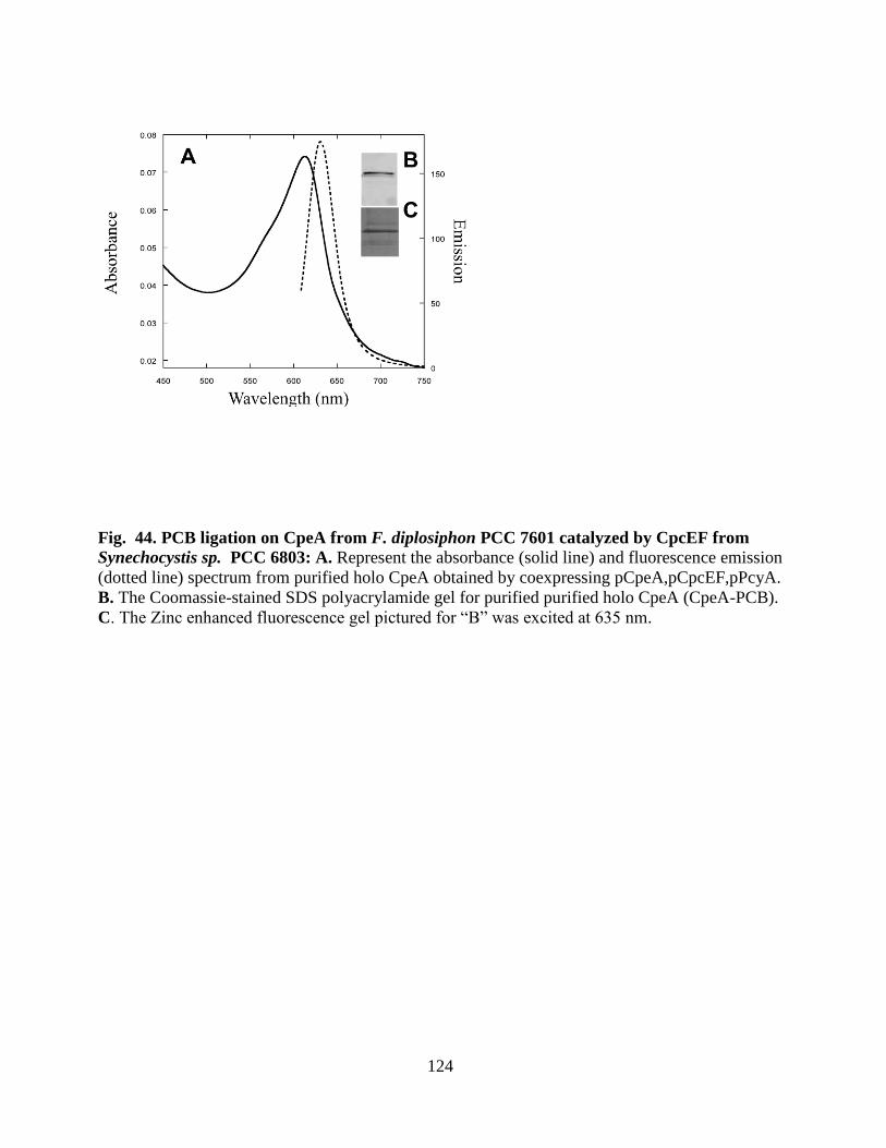
124
Fig. 44. PCB ligation on CpeA from F. diplosiphon PCC 7601 catalyzed by CpcEF from
Synechocystis sp. PCC 6803: A. Represent the absorbance (solid line) and fluorescence emission
(dotted line) spectrum from purified holo CpeA obtained by coexpressing pCpeA,pCpcEF,pPcyA.
B. The Coomassie-stained SDS polyacrylamide gel for purified purified holo CpeA (CpeA-PCB).
C. The Zinc enhanced fluorescence gel pictured for “B” was excited at 635 nm.
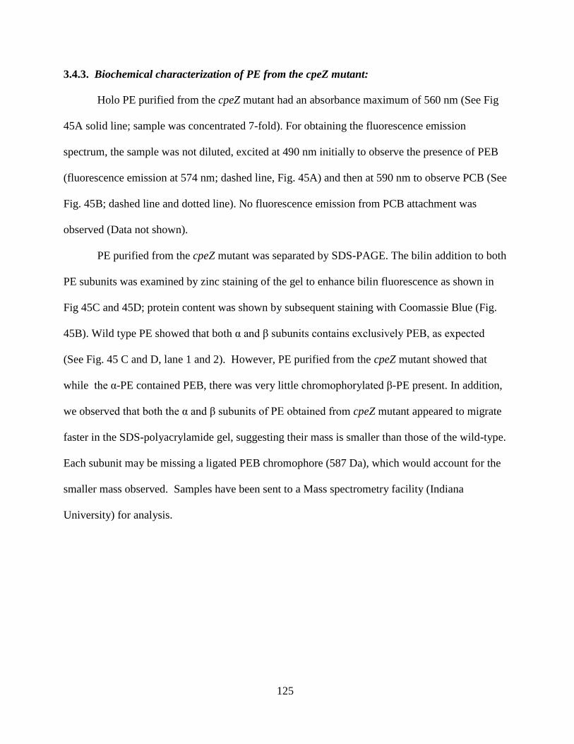
125
3.4.3. Biochemical characterization of PE from the cpeZ mutant:
Holo PE purified from the cpeZ mutant had an absorbance maximum of 560 nm (See Fig
45A solid line; sample was concentrated 7-fold). For obtaining the fluorescence emission
spectrum, the sample was not diluted, excited at 490 nm initially to observe the presence of PEB
(fluorescence emission at 574 nm; dashed line, Fig. 45A) and then at 590 nm to observe PCB (See
Fig. 45B; dashed line and dotted line). No fluorescence emission from PCB attachment was
observed (Data not shown).
PE purified from the cpeZ mutant was separated by SDS-PAGE. The bilin addition to both
PE subunits was examined by zinc staining of the gel to enhance bilin fluorescence as shown in
Fig 45C and 45D; protein content was shown by subsequent staining with Coomassie Blue (Fig.
45B). Wild type PE showed that both α and β subunits contains exclusively PEB, as expected
(See Fig. 45 C and D, lane 1 and 2). However, PE purified from the cpeZ mutant showed that
while the α-PE contained PEB, there was very little chromophorylated β-PE present. In addition,
we observed that both the α and β subunits of PE obtained from cpeZ mutant appeared to migrate
faster in the SDS-polyacrylamide gel, suggesting their mass is smaller than those of the wild-type.
Each subunit may be missing a ligated PEB chromophore (587 Da), which would account for the
smaller mass observed. Samples have been sent to a Mass spectrometry facility (Indiana
University) for analysis.
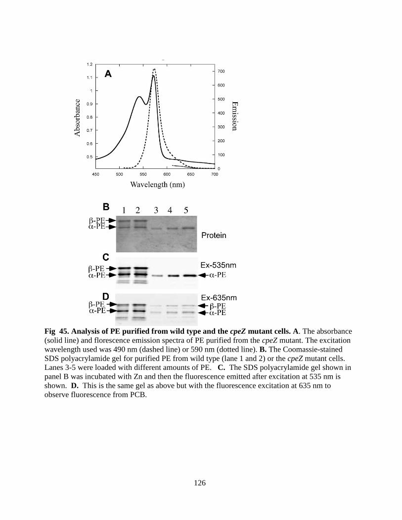
126
Fig 45. Analysis of PE purified from wild type and the cpeZ mutant cells. A. The absorbance
(solid line) and florescence emission spectra of PE purified from the cpeZ mutant. The excitation
wavelength used was 490 nm (dashed line) or 590 nm (dotted line). B. The Coomassie-stained
SDS polyacrylamide gel for purified PE from wild type (lane 1 and 2) or the cpeZ mutant cells.
Lanes 3-5 were loaded with different amounts of PE. C. The SDS polyacrylamide gel shown in
panel B was incubated with Zn and then the fluorescence emitted after excitation at 535 nm is
shown. D. This is the same gel as above but with the fluorescence excitation at 635 nm to
observe fluorescence from PCB.
127
3.5. A mpeZ gene is involved in Type IV chromatic adaptation in marine
Synechococcus cyanobacteria
Some of the marine Synechococcus sp. (WH 8020, WH 8103, and RS 9916) have a more complex
phycobiliprotein structure (See Fig 19). They have two types of PE: PEI and PEII, each with
multiple bilins (PEB or PUB) attached to the α and β subunits (See table 1). Some of these
species (WH 8020, and RS 9916) can undergo Type IV CA replacing the chomophores. Everroad
et al. (Everroad, Six et al. 2006) initially characterized the biochemical basis of Type IV CA in
few Synechococcus species including in RS 9916. This group observed a change in
chromophorylation on the α-PE-II subunit (MpeA). Under blue light, these sites are ligated with
PUB, whereas under green light there was both PEB and PUB pigments attached. The
phycobiliprotein themselves were same in both BL and GL. However, they were unable to
identify the genes involved in the Type IV CA. Recently, Kehoe’s group characterized the genes
that are upregulated or down- regulated in Synechococcus sp. RS 9916 (Shukla et al. unpublished
data) when shifted from GL to BL using a microarray. Based on the microarray data MpeZ was
upregulated 3 fold in BL compared to green light. MpeZ shared sequence similarity to CpeY
from F. diplosiphon (26%) (which is a lyase for α-PE, Biswas and Schluchter unpublished data)
and 64% to CpeY from Synechococcus sp CC 9311 (Data not shown). However, MpeZ was not
very similar to other E/F type lyase/ isomerase like RpcG from Synechococcus sp. RS 9916
(Blot, Wu et al. 2009) or Nostoc sp. PCC 7120 PecE/PecF (Zhao, Deng et al. 2000). These two
E/F type lyase/ isomerases had a conserved 6- amino acid domain motif (NHCQGN) but this was
not found in MpeZ (Blot, Wu et al. 2009) .
Based on this evidence we predicted that MpeZ might be a potential lyase for α-PEII (MpeA). I
used a heterologous coexpression system in E. coli to test the MpeZ lyase/ isomerase activity on
the PEII subunits.
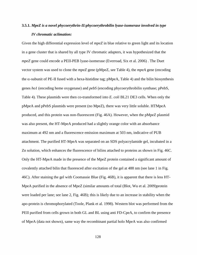
128
3.5.1. MpeZ is a novel phycoerythrin-II:phycoerythrobilin lyase-isomerase involved in type
IV chromatic aclimation:
Given the high differential expression level of mpeZ in blue relative to green light and its location
in a gene cluster that is shared by all type IV chromatic adapters, it was hypothesized that the
mpeZ gene could encode a PEII-PEB lyase-isomerase (Everroad, Six et al. 2006) . The Duet
vector system was used to clone the mpeZ gene (pMpeZ, see Table 4), the mpeA gene (encoding
the -subunit of PE-II fused with a hexa-histidine tag; pMpeA, Table 4) and the bilin biosynthesis
genes ho1 (encoding heme oxygenase) and pebS (encoding phycoerythrobilin synthase; pPebS,
Table 4). These plasmids were then co-transformed into E. coli BL21 DE3 cells. When only the
pMpeA and pPebS plasmids were present (no MpeZ), there was very little soluble. HTMpeA
produced, and this protein was non-fluorescent (Fig. 46A). However, when the pMpeZ plasmid
was also present, the HT-MpeA produced had a slightly orange color with an absorbance
maximum at 492 nm and a fluorescence emission maximum at 503 nm, indicative of PUB
attachment. The purified HT-MpeA was separated on an SDS polyacrylamide gel, incubated in a
Zn solution, which enhances the fluorescence of bilins attached to proteins as shown in Fig. 46C.
Only the HT-MpeA made in the presence of the MpeZ protein contained a significant amount of
covalently attached bilin that fluoresced after excitation of the gel at 488 nm (see lane 1 in Fig.
46C). After staining the gel with Coomassie Blue (Fig. 46B), it is apparent that there is less HT-
MpeA purified in the absence of MpeZ (similar amounts of total (Blot, Wu et al. 2009)protein
were loaded per lane; see lane 2, Fig. 46B); this is likely due to an increase in stability when the
apo-protein is chromophorylated (Toole, Plank et al. 1998). Western blot was performed from the
PEII purified from cells grown in both GL and BL using anti FD-CpeA, to confirm the presence
of MpeA (data not shown), same way the recombinant partial holo MpeA was also confirmed
129
using western blot with the same antibody (data not shown). Although several phycocyanin lyase-
isomerases have been described previously (Zhao, Deng et al. 2000; Blot, Wu et al. 2009), this is
the first report of a phycobilin lyase-isomerase specific for a phycoerythrin.
130
Fig. 46. Analyses of MpeZ lyase for PEB addition to Phycoerythrin α subunit (PEII) in E. coli. A.
Absorbance (solid line) and fluorescence emission (dashed line) spectra of HT-MpeA purified from cells
containing pMpeA, pPebS with pMpeZ and absorbance (dashed dotted line), fluorescence (dotted line)
without pMpeZ are shown. In order to acquire the fluorescence emission spectra for the phycoerythrin α-
subunit (PEII) produced in the presence of pMpeZ (black dashed lines in panels A), the sample was
diluted to OD490nm of 0.05; however, no dilution was performed on those produced in the absence of a
lyase. B. Coomassie-stained SDS polyacrylamide gel containing HT-MpeA purified from cells containing
pMpeA, pPebS with (lane 1) or without (lane 2) pMpeZ, Molecular mass standards are are indicated to
the left with arrow heads. C. The zinc-enhanced fluorescence of the gel pictured in panel C is shown here.
131
3.2.2. Specificity of MpeZ for cysteine residues on MpeA:
In green light, MpeA has three Cys which contain bilins: PUB at Cys 75, PEB at Cys 83, and PEB
at Cys 140 (Hammad, Shukla et al. 2011), whereas in blue light, PUB is located at all three
positions. In order to determine which Cys was the preferred attachment site for MpeZ, site-
directed mutants were created changing Cys to Ala at all three positions and in various
combinations. These recombinant proteins were co-expressed in E. coli in the presence of PEB
and MpeZ. The absorbance and fluorescence spectra for the purified HT-MpeA mutant proteins is
shown in Fig. 47. The C75A, the C140A and the C75A/C140A MpeA proteins all contained PUB,
as indicated by the strong absorbance at 492 nm and fluorescence emission at 503 nm
(summarized in Table 10). When the C83A MpeA protein was purified, it had no absorbance or
fluorescence emission (see Table 10); this indicates that MpeZ is a lyase isomerase specific for
Cys-83 on MpeA. However, using our in vivo coexpression system in E. coli we were unable to
show PUB chromophorylation on Cys-140 of MpeA.
Although there was no evidence that MpeB changes in GL-BL, it was also important to
check whether MpeB was a potential substrate for the MpeZ lyase-isomerase. The pMpeB,
pMpeZ and pPebS plasmids were co-transformed into E. coli and cultured as described. The HT-
MpeB produced in the presence of the MpeZ protein and PEB was non-fluorescent, indicating
that MpeB was not a potential substrate for MpeZ (see Table 10).
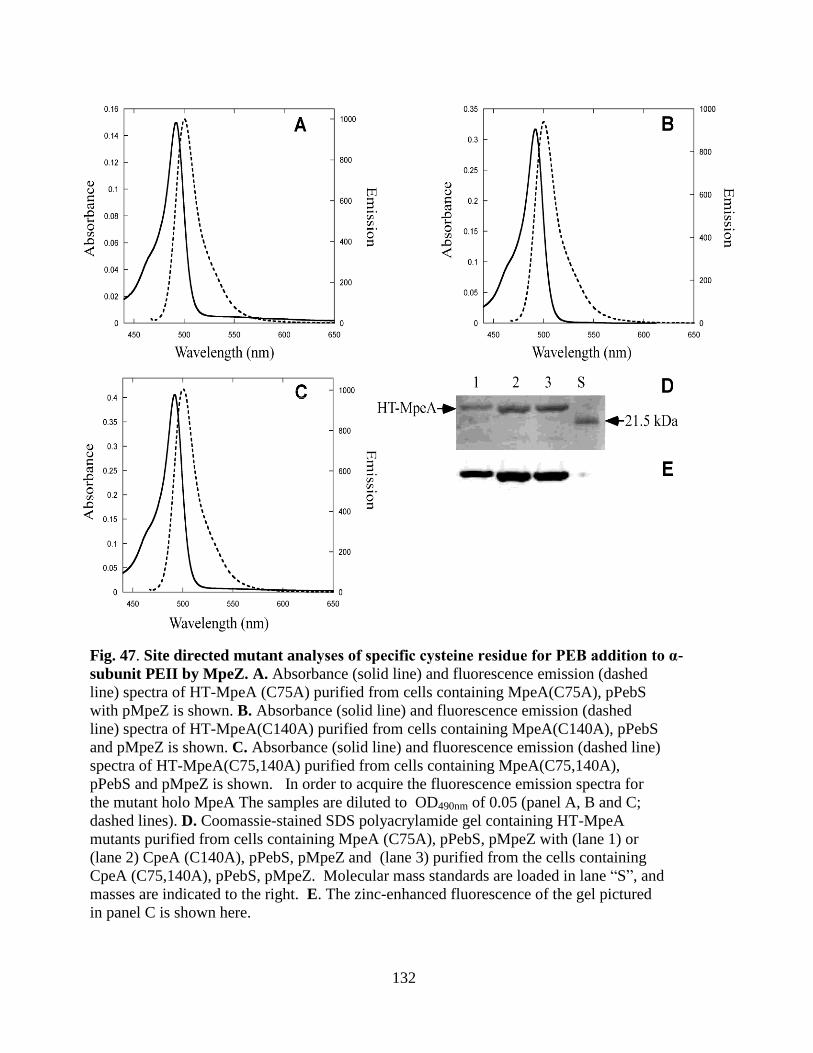
132
Fig. 47. Site directed mutant analyses of specific cysteine residue for PEB addition to α-
subunit PEII by MpeZ. A. Absorbance (solid line) and fluorescence emission (dashed
line) spectra of HT-MpeA (C75A) purified from cells containing MpeA(C75A), pPebS
with pMpeZ is shown. B. Absorbance (solid line) and fluorescence emission (dashed
line) spectra of HT-MpeA(C140A) purified from cells containing MpeA(C140A), pPebS
and pMpeZ is shown. C. Absorbance (solid line) and fluorescence emission (dashed line)
spectra of HT-MpeA(C75,140A) purified from cells containing MpeA(C75,140A),
pPebS and pMpeZ is shown. In order to acquire the fluorescence emission spectra for
the mutant holo MpeA The samples are diluted to OD490nm of 0.05 (panel A, B and C;
dashed lines). D. Coomassie-stained SDS polyacrylamide gel containing HT-MpeA
mutants purified from cells containing MpeA (C75A), pPebS, pMpeZ with (lane 1) or
(lane 2) CpeA (C140A), pPebS, pMpeZ and (lane 3) purified from the cells containing
CpeA (C75,140A), pPebS, pMpeZ. Molecular mass standards are loaded in lane “S”, and
masses are indicated to the right. E. The zinc-enhanced fluorescence of the gel pictured
in panel C is shown here.
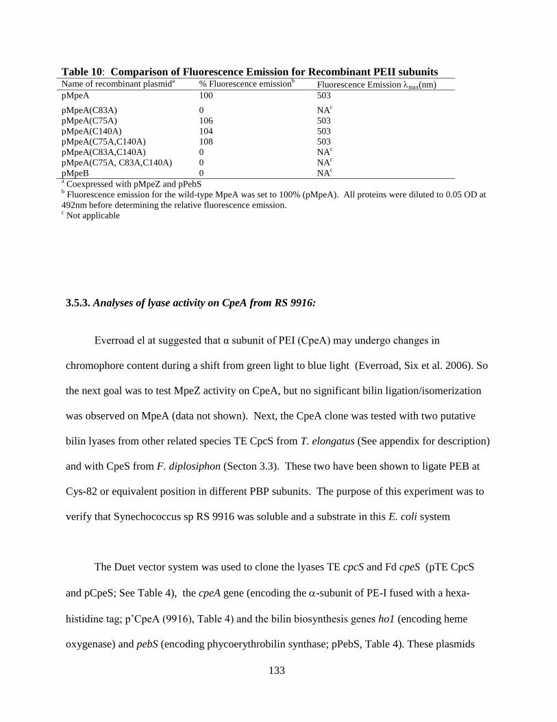
133
Table 10: Comparison of Fluorescence Emission for Recombinant PEII subunits Name of recombinant plasmid
a % Fluorescence emission
b Fluorescence Emission max(nm)
pMpeA 100 503
pMpeA(C83A) 0 NAc
pMpeA(C75A) 106 503
pMpeA(C140A) 104 503
pMpeA(C75A,C140A) 108 503
pMpeA(C83A,C140A) 0 NAc
pMpeA(C75A, C83A,C140A) 0 NAc
pMpeB 0 NAc
a Coexpressed with pMpeZ and pPebS
b Fluorescence emission for the wild-type MpeA was set to 100% (pMpeA). All proteins were diluted to 0.05 OD at
492nm before determining the relative fluorescence emission. c Not applicable
3.5.3. Analyses of lyase activity on CpeA from RS 9916:
Everroad el at suggested that α subunit of PEI (CpeA) may undergo changes in
chromophore content during a shift from green light to blue light (Everroad, Six et al. 2006). So
the next goal was to test MpeZ activity on CpeA, but no significant bilin ligation/isomerization
was observed on MpeA (data not shown). Next, the CpeA clone was tested with two putative
bilin lyases from other related species TE CpcS from T. elongatus (See appendix for description)
and with CpeS from F. diplosiphon (Secton 3.3). These two have been shown to ligate PEB at
Cys-82 or equivalent position in different PBP subunits. The purpose of this experiment was to
verify that Synechococcus sp RS 9916 was soluble and a substrate in this E. coli system
The Duet vector system was used to clone the lyases TE cpcS and Fd cpeS (pTE CpcS
and pCpeS; See Table 4), the cpeA gene (encoding the -subunit of PE-I fused with a hexa-
histidine tag; p’CpeA (9916), Table 4) and the bilin biosynthesis genes ho1 (encoding heme
oxygenase) and pebS (encoding phycoerythrobilin synthase; pPebS, Table 4). These plasmids
134
were then co-transformed into E. coli BL21 DE3 cells. When only pCpeA (9916) and pPebS
were present (no potential lyase), there was almost no solubility (Fig. 49B, lane 2). HT-CpeA
had no significant fluorescent or absorbance peaks (Fig 48 A). However, when pCpeA plasmid
was expressed along with either pTE CpcS or pCpeS, some chromophorylaed CpeA was
produced with an absorbance maximum of 561 nm and fluorescence emission of 570 nm,
indicative of PEB attachment (Fig. 48A and Fig. 49 A). The purified HT-CpeA from all the
experiments were separated on SDS-PAGE incubated with Zn solution, which enhances the
fluorescence of bilins attached to the protein (See Fig. 48C and Fig 49C). Only HT-CpeA made
in presence of bilin lyases TE CpcS or Fd CpeS contained covalently attached bilin that
fluoresced on the gel (See lane 1 and 2 in Fig. 48C and lane 2 in Fig. 49C).
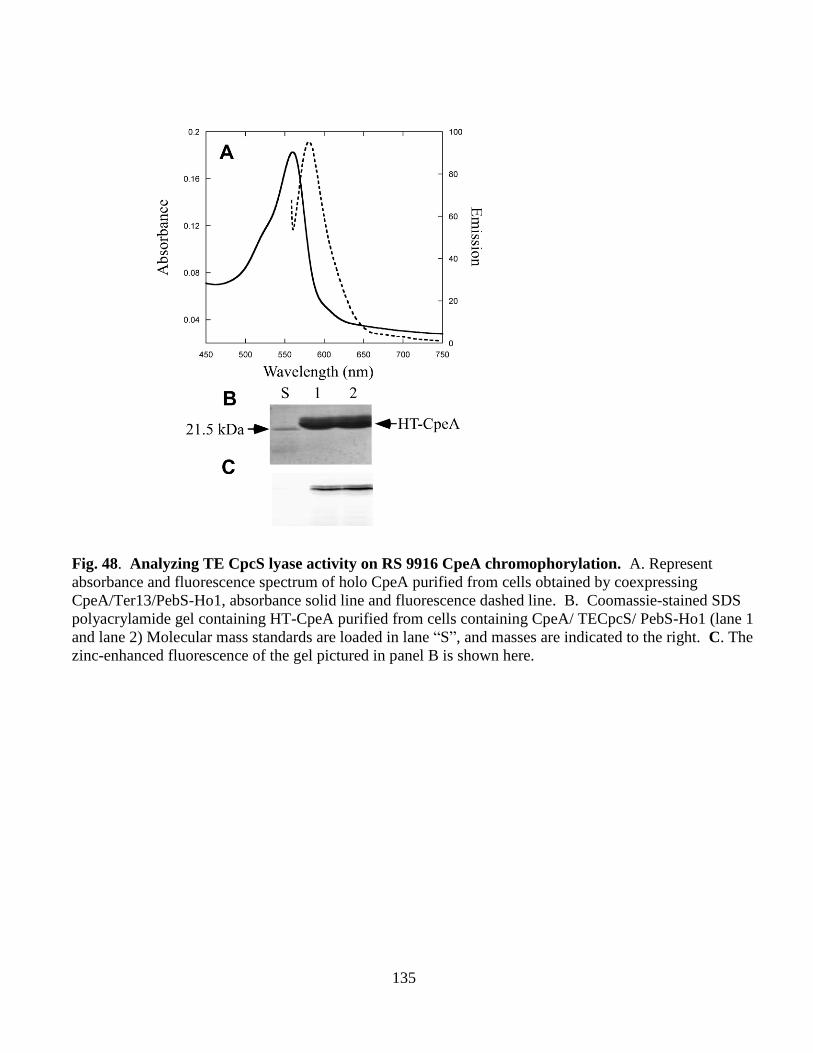
135
Fig. 48. Analyzing TE CpcS lyase activity on RS 9916 CpeA chromophorylation. A. Represent
absorbance and fluorescence spectrum of holo CpeA purified from cells obtained by coexpressing
CpeA/Ter13/PebS-Ho1, absorbance solid line and fluorescence dashed line. B. Coomassie-stained SDS
polyacrylamide gel containing HT-CpeA purified from cells containing CpeA/ TECpcS/ PebS-Ho1 (lane 1
and lane 2) Molecular mass standards are loaded in lane “S”, and masses are indicated to the right. C. The
zinc-enhanced fluorescence of the gel pictured in panel B is shown here.
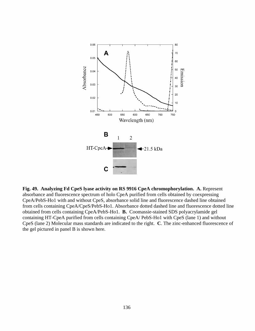
136
Fig. 49. Analyzing Fd CpeS lyase activity on RS 9916 CpeA chromophorylation. A. Represent
absorbance and fluorescence spectrum of holo CpeA purified from cells obtained by coexpressing
CpeA/PebS-Ho1 with and without CpeS, absorbance solid line and fluorescence dashed line obtained
from cells containing CpeA/CpeS/PebS-Ho1. Absorbance dotted dashed line and fluorescence dotted line
obtained from cells containing CpeA/PebS-Ho1. B. Coomassie-stained SDS polyacrylamide gel
containing HT-CpeA purified from cells containing CpeA/ PebS-Ho1 with CpeS (lane 1) and without
CpeS (lane 2) Molecular mass standards are indicated to the right. C. The zinc-enhanced fluorescence of
the gel pictured in panel B is shown here.
137
4.0 DISCUSSIONS:
4.1. Chromophorylation efficiency and specificity of all bilin lyases from Synechococcus sp.
strain PCC 7002:
In the studies reported here, an in-vivo, heterologous expression system was developed to
determine the biosynthetic requirements for synthesis of holo-ApcD, ApcF and ApcE, to test the
efficiency and specificity of various bilin lyases in chromophorylating substrates, and to produce
large quantities of holo-PBPs that might be used as fluorescent probes. Others have used a similar
approach to produce single subunits in E. coli (Tooley, Cai et al. 2001; Tooley and Glazer 2002;
Zhao, Su et al. 2007; Ge, Sun et al. 2009), but the system described here produced the best yield
of holo-PBPs in E. coli reported to date: 3 to 12 mg holo-PBP l-1
of E. coli culture (compared to
0.86-1.0 mg l-1
for AP subunits (Yang, Ge et al. 2008; Ge, Sun et al. 2009). When the system of
Tooley et al. (pAT101 encoding Synechocystis sp. strain PCC 6803 ho1 and pcyA) (Tooley, Cai et
al. 2001) was directly compared with the system (pPcyA encoding Synechocystis sp. strain PCC
6803 ho1 and Synechococcus sp. strain PCC 7002 pcyA) described here for the production of
PCB, 3.5-fold more PCB was achieved in the same culture volume using pPcyA (compared to
pAT101). This could be due to higher solubility, activity, and/or expression level of PcyA from
Synechococcus sp. strain PCC 7002. It may also be due to a difference in plasmid copy number or
the T7 promoter activity levels for the Duet plasmid pPcyA construct (pAT101 uses the trc (trp-
lac) promoter). The Synechocystis sp. strain PCC 6803 heme oxygenase was used in both
constructs, and the heme oxygenase enzyme produced by the Duet vector containing just ho1
(parent to the pPcyA vector) was assayed in vitro and was very active (Y. M. Vasquez and W. M.
Schluchter, unpublished results). It is possible that because Synechococcus sp. strain PCC 7002
138
grows optimally at nearly the same temperature as E. coli (38-40C), its enzymes may be more
stable and achieve higher activities when expressed in E. coli than those from Synechocystis sp.
strain PCC 6803, which has a lower optimum growth temperature. In fact, it may be possible to
improve the PCB production levels if the ho1 gene of Synechococcus sp. strain PCC 7002 is used
to replace the in Synechocystis sp. strain PCC 6803 ho1 gene.
It was also possible to produce large quantities (5 to 12.4 mg of holo-AP subunit l-1
of E.
coli culture) of holo-HT-ApcA/ApcB, holo-HT-ApcF, and holo-HTApcD/ApcB in E. coli. The
CpcS-I/CpcU bilin lyase was required for PCB addition to all of these proteins, but it appears that
holo--subunits (HT-ApcA and HT-ApcD) are produced in slightly lower quantities than their
holo--subunit counterparts (see Figs. 25B and 25D). When produced in E. coli, AP subunits
have previously been reported to have different solubility levels (Zhao, Su et al. 2007; Miller,
Leonard et al. 2008; Yang, Ge et al. 2008; Ge, Sun et al. 2009; Zhang, Guan et al. 2009).
Similarly, apo-HT-ApcF was not produced at very high levels unless ApcA was co-expressed
with it (A. Fletcher and W. M. Schluchter, unpublished results). However, HT-ApcF was
stabilized by chromophorylation, which permitted high levels of production to be achieved in the
absence of ApcA; up to ~12 mg of this PBP were produced per liter of E. coli culture. However,
HT-ApcD levels (produced alone) were low whether it was produced in the apo- or holo- form
(Fig. 25D). Other studies have reported that holo-ApcB can be produced in E. coli (0.86 mg l-1
)
(Ge, Sun et al. 2009) and in Streptomyces sp. (38 mg l-1
) (Hou, Qin et al. 2006) and that holo-
ApcA can be produced in E. coli (1 g l-1
) (Yang, Ge et al. 2008). Although one report claimed that
holo-ApcA could be produced without the need for any bilin lyase (Hu, Lee et al. 2006), the
results presented here and other studies show that either the heterologous CpcS-I/CpcU-type bilin
lyase or the CpcS-III single subunit-type bilin lyase (also called CpeS1) by Zhao et al. (Zhao, Su
139
et al. 2006) is required for maximal and correct addition of PCB to ApcA (Zhao, Su et al. 2006;
Zhao, Su et al. 2007; Saunée, Williams et al. 2008; Ge, Sun et al. 2009). Analyses of bilin lyase
mutants also strongly support the conclusion that bilin lyases are essential for AP biogenesis
(Shen, Schluchter et al. 2008). However, small amounts of AP subunits could be detected by
immunoblotting in a cpcSUT triple mutant (Shen, Schluchter et al. 2008). This suggests that
some autocatalytic bilin addition may occur in the absence of bilin lyases in cyanobacteria. In the
studies reported here, neither CpcE/CpcF nor CpcT attached PCB to ApcA or ApcB (data not
shown).
The synthesis of holo-HT-ApcD/ApcB produced a product with a sharp, red-shifted
absorption peak at 672 nm and a fluorescence emission maximum at 675 nm, both characteristic
of native allophycocyanin B () (Glazer and Bryant 1975; Lundell and Glazer 1981). In studies
by Zhao et al. (Zhao, Su et al. 2007) that showed that the single-subunit bilin lyase CpcS-III from
Nostoc sp. PCC 7120 was required for bilin addition to ApcD, the absorbance spectrum of the
product had a broad peak centered at 650 nm, possibly due to poor solubility. In the system
described here, holo-HT-ApcD had an absorbance maximum at 642 nm. Coproducing ApcD with
its partner subunit, ApcB, apparently improved its solubility, which resulted in a chromoprotein
with red-shifted absorbance and fluorescence emission maxima very similar to those of native
allophycocyanin B (Glazer and Bryant 1975; Lundell and Glazer 1981). ApcD may be
particularly useful as a fluorescent tag due to its far-red absorbance and fluorescence emission.
For the production of chromophorylated AP and PC subunits, it appears that a major
limitation may be the folding and/or conformation of the PBP subunit. For example, the
chromophorylation rates by the CpcS-I/CpcU bilin lyase for ApcA/ApcB are much higher than
those for CpcB, and the only difference is the PBP substrate. Because both substrates were
140
expressed as the and subunits, and because of the overall structural similarity of the two
proteins (Schirmer, Huber et al. 1986; Brejc, Ficner et al. 1995), presumably there is little
difference in the accessibility of the Cys-82 chromophorylation sites.
As first reported by Zhao et al (Zhao, Ping et al. 2005), the only PBP synthesized by
Synechococcus sp. strain PCC 7002 that is capable of autocatalytic PCB attachment is ApcE.
However, because of solubility problems with the truncation product of ApcE, Zhao et al. had to
perform in-vitro bilin addition reactions with the PBP domain of ApcE (residues 1-240) in 4 M
urea, which would not only affect the conformation of the protein substrate but also potentially of
that of the PCB chromophore. The addition of detergents, such as Triton X-100, has been shown
to affect chromophore conformation and to facilitate autocatalytic addition of bilins to PBPs in
the absence of lyases (Zhao, Zhu et al. 2004). Therefore, a critical role for the bilin lyases is to
bind the correct chromophore (in organisms producing more than one bilin) in the correct
conformation to achieve the appropriate stereochemical attachment of the bilin to the apoprotein
(Zhao, Zhu et al. 2004). At high bilin concentrations, that are probably never encountered inside
cyanobacterial cells, autocatalytic attachment does occur in vitro, but the products of these
reactions do not produce the naturally occurring holo-proteins (Arciero, Bryant et al. 1988;
Fairchild and Glazer 1994; Scheer and Zhao 2008). For all of these reasons, it was important to
show that a soluble form of ApcE, in the absence of detergents or urea, had an intrinsic bilin lyase
activity.
The amino-terminal domain of ApcE, including amino acids 1-228, contains an AP-like
domain that interacts with the AP--like subunit, ApcF, within an AP-trimer-like disc of the PBS
core (Fig. 49). This domain probably has a largely alpha-helical, globin-like fold that is similar to
141
the structures of other AP and PC subunits (Brejc, Ficner et al. 1995). This AP-like domain of
ApcE is interrupted by an insertion of 50 amino acid residues called the PB loop (Ajlani and
Vernotte 1998), and this insertion is readily apparent when one examines an alignment of the
amino-terminal domain with other AP subunits (Fig. 24). Because no other PBP subunit can
efficiently autocatalytically ligate PCB to Cys residues, it is possible that this insertion may
include residues that are important in the autocatalytic PCB ligation activity of ApcE. However,
Ajlani and Vernotte showed that a deletion of this PB loop within ApcE did not affect its ability
to attach a PCB chromophore (Ajlani and Vernotte 1998). Because the chromophore on ApcE is
bound to the opposing helix of the binding pocket relative to every other biliprotein, the
requirements for binding may be modified so that autocatalytic binding is possible. ApcE
(residues 1-200) from Synechococcus sp. strain PCC 7002 and Synechocystis sp. strain PCC 6803
were aligned with the bilin lyase containing domains of Cph1 and Cph2, two
cyanobacteriochromes (Wu and Lagarias 2000). One small motif that is conserved among these
four proteins is the sequence DXXLE, corresponding to amino acids 32-36, in ApcE from
Synechococcus sp. strain PCC 7002 and 185-189 in Cph1 or Slr0473 (see Fig. 50). The glutamic
acid at the end of this region (underlined in the sequence above) is the one that was shown by
mutagenesis to have a function in bilin binding in Cph1 (Wu and Lagarias 2000). The possibility
that E36 in ApcE is also responsible for PCB binding will be tested in future studies.
To summarize this portion of work, it has now been completely defined how each PBP in
Synechococcus sp. strain PCC 7002 is biosynthesized and has been established how one member
of each of the four families of bilin lyases is responsible for PCB addition to the eight, discrete
chromophore-binding sites on the seven different PBP subunits (Table 6) (Fairchild, Zhao et al.
1992; Fairchild and Glazer 1994; Shen, Saunee et al. 2006; Saunée, Williams et al. 2008; Shen,
142
Leonard et al. 2008). A heterologous, in-vivo expression system using PcyA from Synechococcus
sp. strain PCC 7002 and Ho1 from Synechocystis sp. strain PCC 6803 appears to be very efficient
and generates large amounts of PCB in E. coli. The use of a multi-plasmid system allows one to
test biosynthetic requirements of uncharacterized PBPs from other organisms rapidly, to produce
partially chromophorylated biliproteins for studies of the order of post-translational modifications
in in-vitro reactions, and to generate holo-PBPs for use as fluorescent probes for bioimaging.
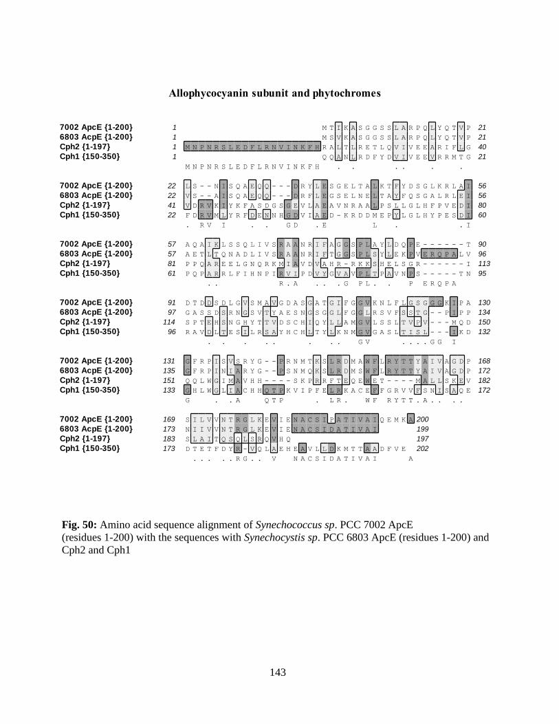
143
Fig. 50: Amino acid sequence alignment of Synechococcus sp. PCC 7002 ApcE
(residues 1-200) with the sequences with Synechocystis sp. PCC 6803 ApcE (residues 1-200) and
Cph2 and Cph1
Allophycocyanin subunit and phytochromes
7002 ApcE {1-200}
6803 AcpE {1-200}
Cph2 {1-197}
Cph1 {150-350}
1 21
1 21
1 40
1 21
M T I K A S G G S S L A R P Q L Y Q T V P
M S V K A S G G S S L A R P Q L Y Q T V P
M N P N R S L E D F L R N V I N K F H R A L T L R E T L Q V I V E E A R I F L G
Q Q A N L R D F Y D V I V E E V R R M T G
M N P N R S L E D F L R N V I N K F H . . . . . .
7002 ApcE {1-200}
6803 AcpE {1-200}
Cph2 {1-197}
Cph1 {150-350}
22 56
22 56
41 80
22 60
L S - - N I S Q A E Q Q - - - D R Y L E S G E L T A L K T F Y D S G L K R L A I
V S - - A I S Q A E Q Q - - - D R F L E G S E L N E L T A Y F Q S G A L R L E I
V D R V K I Y K F A S D G S G E V L A E A V N R A A L P S L L G L H F P V E D I
F D R V M L Y R F D E N N H G D V I A E D - K R D D M E P Y L G L H Y P E S D I
. R V I . . G D . E L . . I
7002 ApcE {1-200}
6803 AcpE {1-200}
Cph2 {1-197}
Cph1 {150-350}
57 90
57 96
81 113
61 95
A Q A I K L S S Q L I V S R A A N R I F A G G S P L A Y L D Q P E - - - - - - T
A E T L T Q N A D L I V S R A A N R I F T G G S P L S Y L E K P V E R Q P A L V
P P Q A R E E L G N Q R K M I A V D V A H R - R K K S H E L S G R - - - - - - I
P Q P A R R L F I H N P I R V I P D V Y G V A V P L T P A V N P S - - - - - T N
. . R . A . . . G P L . . P E R Q P A
7002 ApcE {1-200}
6803 AcpE {1-200}
Cph2 {1-197}
Cph1 {150-350}
91 130
97 134
114 150
96 132
D T D D S D L G V S M A V G D A S G A T G I F G G V K N L F L G S G G G K I P A
G A S S D S R N G S V T Y A E S N G S G G L F G G L R S V F S S T G - - P I P P
S P T E H S N G H Y T T V D S C H I Q Y L L A M G V L S S L T V P V - - - M Q D
R A V D L T E S I L R S A Y H C H L T Y L K N M G V G A S L T I S L - - - I K D
. . . . . . . . G V . . . . G G I
7002 ApcE {1-200}
6803 AcpE {1-200}
Cph2 {1-197}
Cph1 {150-350}
131 168
135 172
151 182
133 172
G F R P I S V S R Y G - - P R N M T K S L R D M A W F L R Y T T Y A I V A G D P
G F R P I N I A R Y G - - P S N M Q K S L R D M S W F L R Y T T Y A I V A G D P
Q Q L W G I M A V H H - - - - S K P R R F T E Q E W E T - - - - M A L L S K E V
G H L W G L I A C H H Q T P K V I P F E L R K A C E F F G R V V F S N I S A Q E
G . . A Q T P . L R . W F R Y T T . A . . . .
7002 ApcE {1-200}
6803 AcpE {1-200}
Cph2 {1-197}
Cph1 {150-350}
169 200
173 199
183 197
173 202
S I L V V N T R G L K E V I E N A C S I P A T I V A I Q E M K A
N I I V V N T R G L K E V I E N A C S I D A T I V A I
S L A I T Q S Q L S R Q V H Q
D T E T F D Y R - V Q L A E H E A V L L D K M T T A A D F V E
. . . . . R G . . V N A C S I D A T I V A I A
144
4.2. Creation of unique phycobiliproteins using PEB in E. coli for potential biotechnological
applications:
PBPs with PEB generally have higher quantum yields so the purpose of this project was
to create unique phycobiliproteins using different bilin lyases and PEB in our in vivo heterologous
system. Phycocyanin subunits are much more soluble in E. coli than PE subunits, and so CpcA
and CpcB were tested for their ability to accept PEB in this system. The PebS enzyme
(Dammeyer, Bagby et al. 2008), originally isolated from a phage that infects Prochlorococcus
species produced larger amounts of PEB in vivo than did the construct containing pebA and pebB
from Synechococcus WH8020 or from Fremyella diplosiphon (data not shown). The CpcE/CpcF
lyase has been shown to favor PCB over PEB in terms of turnover rate (Fairchild and Glazer
1994), but in this system, CpcE/CpcF was extremely efficient in adding PEB to HT-CpcA.
Greater levels of chromophorylation with PEB (83.5%) were achieved than with PCB (48.1%),
but the expression cells containing pPebS were allowed to grow an additional 12-15 hours at 18C
(for optimal activity/folding of PebS) than cells containing pPcyA (30C for 4 hr); this lower
temperature may have been better for CpcE/CpcF folding, and the longer incubation time
certainly allowed more time for PEB attachment. Therefore, this CpcEF class of bilin lyase is
very suitable for creating unique phycobiliproteins (e.g. not found in nature) to be used as
fluorescent labels. In fact CpcA was found to attach Phytochoromobilin (PφB) also (Alvey,
Biswas et al. 2011). CpcA could also be recognized by PecEF, which ligated PCB and
subsequently isomerized it to PVB (Alvey, Biswas et al. 2011).
The CpcS-I/CpcU and CpeS1 bilin lyases show broad PBP substrate specificity
recognizing various AP subunits and -PC as shown in the results presented here and in (Zhao, Su
145
et al. 2007; Saunée, Williams et al. 2008), but the CpcS-I/CpcU bilin lyase is quite specific for its
bilin substrate, showing much lower activity when attaching PEB compared to the CpcE/CpcF
bilin lyase. This makes sense when one thinks about the function of the Cys-82 on the -subunit
as the terminal energy acceptor within trimeric PC (See Fig. 3). In cyanobacterial cells that
contain both PCB and PEB, a mistake by the CpcS or CpeS bilin lyase in attaching PEB at Cys-82
would mean that energy from PCB at -Cys-153 or -Cys-84 would not be transferred to PEB at
-Cys-82. Likewise, the intrinsic bilin lyase activity of ApcE also exhibited selectivity as much
less activity was observed in the presence of PEB compared to PCB (data not shown). Therefore,
three of the four known bilin lyase families (CpcS/U, CpcE/F, and autocatalytic ApcE) exhibit
quite different specificities with respect to the bilin substrate. In the future, the CpcT bilin lyase
will be tested for its ability to attach PEB to CpcB. Some of the in vitro preliminary data suggest
CpcT can ligate PEB at Cys-153 on CpcB at very low levels compared to its activity with PCB
(data not shown).
It was found that PEB is a better choice of bilin in the cyanobacterial family for real world
applications since these have the highest quantum yield when ligated to CpcA (0.98), which is
much greater than any other fluorophore available (See table 7). Also in this system CpcA was
found to be the best choice of PBP subunit for creating fluorescent tag, because it is highly
soluble, well characterized, has a very high yield, and can be ligated with numerous bilins
(Alvey, Biswas et al. 2011).
In summary, unique phycobiliproteins not found naturally in E. coli systems have been
successfully generated. This contruct may have potential biotechnological applications as an ideal
fluorescent tag.
146
4.3. Characterization of CpeY, CpeZ and CpeS bilin lyases involved in phycoerythrin
biosynthesis in Fremyella diplosiphon strain UTEX 481
This study compared the activities of three different proteins with similarity to bilin
lyases on PE and subunit substrate derived from a filamentous cyanobacterium capable of
Type III complementary chromatic adaptation, F. diplosiphon. Zhao et al. suggested that CpcS
(also called CpeS1) from Nostoc sp. PCC 7120 had broad PBP substrate recognition and might
attach all chromophores at position Cys-82 except for those of CpcA, PecA, and RpcA (Ritter,
Hiller et al. 1999; Takano and Gusella 2002). However, because Nostoc sp. PCC 7120 does not
synthesize PE or PEB, a more thorough examination of the substrate specificity of bilin lyases for
PE subunits within one organism seemed necessary. The studies reported here showed that
CpeY/CpeZ, and not CpeS, is the principal bilin lyase responsible for attachment of PEB at Cys-
82 on CpeA. In fact, the PE produced in a F. diplosiphon cpeY mutant actually had PCB ligated
at the Cys-82 position, and the likely candidate for this activity is CpcE and CpcF. CpeS
performed poorly in attaching PCB to CpeA in this E. coli system (data not shown). Although
CpeS could ligate PEB to Cys-82 on CpeA, a comparison of the yields obtained with CpeS and
CpeY/CpeZ proteins in E. coli strongly suggested that the latter is more important in ligating PEB
to CpeA in vivo (see Fig. 34). In addition, it was shown that CpeS could ligate PEB to Cys-139 of
CpeA. However, based upon the very low levels of chromophorylation seen, it seems unlikely
that this lyase is the one that catalyzes this reaction in cyanobacteria. Several other lyase
candidates are currently being tested for PEB ligation at Cys-139.
At 429 amino acids, the CpeY protein is much larger than typical members of the E/F
family. However, there are examples of larger lyases that belong to this family that have been
147
demonstrated to be involved in chromophore ligation and isomerization, such as RpcG, and it is
likely these genes resulted from a fusion proteins (Blot, Wu et al. 2009). When the CpcE and
CpcF sequences of Synechocystis sp. PCC 6803 were combined, the resulting CpcE/CpcF fusion
protein aligned well with CpeY with 37% similarity (See Fig. 32). This could explain why CpeY
has significant activity (60%) in the absence of CpeZ. Individual CpcE and CpcF subunits usually
have low levels of ligation activity when assayed separately (Fairchild, Zhao et al. 1992; Fairchild
and Glazer 1994). For example, compared to PecE/PecF together, PecE from Mastigocladus
laminosus had only 10% PCB ligation activity on PecA (Bohm, Endres et al. 2007).
CpeZ (205 amino acids) is most similar to CpcE-like, HEAT-repeat proteins that are
found in cyanobacteria and other bacteria that do not contain PBPs. All CpcE/CpcF-type bilin
lyases contain 5-6 HEAT-repeat motifs; these motifs, which occur in many proteins from diverse
eukaryotic organisms, are generally believed to facilitate protein-protein interactions (Andrade,
Petosa et al. 2001; Takano and Gusella 2002). CpeZ increased the PEB ligation activity of CpeY,
but no evidence for a stable interaction between CpeY and CpeZ was detected using pull-down
assays (data not shown). Likewise, no demonstable interaction between HT-CpeA and either
CpeY or CpeZ was observed (data not shown). The cpeZ mutant results suggest this protein has
great importance for biosynthesis of CpeB and is less important for CpeA biosynthesis. The cpeZ
cyanobacterial deletion mutant produces very little PE under green light. When we purified this
PE, there was almost no chromophorylated CpeB present, but the CpeA produced in the mutant
appeared normal. The CpeZ may play a chaperone-type role in assisting or regulating CpeA and
CpeB’s interaction with other bilin lyases (Collier and Grossman 1994; Baier, Lehmann et al.
2004; Bienert, Baier et al. 2006; Dines, Sendersky et al. 2007).
148
Similar to the results of Fairchild and Glazer (Fairchild and Glazer 1994) in which CpeA
from F. diplosiphon was renatured from inclusion bodies, both CpeA (-PE) and CpeB (-PE )
from F. diplosiphon were insoluble when expressed in apo-form in E. coli (data not shown). Co-
expression of cpeB with cpeA did not increase the protein solubility as it often occurs with PC
subunits (Biswas, Vasquez et al. 2010). However, chromophorylation at the Cys-82 equivalent
position was obviously an important component in the solubility and accumulation of the folded
proteins in E. coli. This observation suggests that chromophorylation at Cys-82 might be an
important first step in PBP biosynthesis in cyanobacteria as well. Also, Zhao and Scheer (Scheer
and Zhao 2008) looked at the order of chromophorylation in post-translational modification ; they
suggested the T-type lyases first attach the bilin then the other lyases come into play. Here the
observations were different; my results suggest that the S-type lyase needed to add the bilin on the
central Cys residue, first in the order to make the subunits soluble, so that other lyases can
function.
Bilin deletion mutants in PC (where Cys were mutated to Ala) in cyanobacteria showed
lower stability in vivo (Toole, Plank et al. 1998). The absence of bilins at various positions
reduces the strength of α/β interactions in the heterodimers, and the authors suggested that these
mutants were diverted to a degradations pathway in cyanobateria (Anderson and Toole 1998;
Toole, Plank et al. 1998).
Recently published data by Weithaus et al. (Wiethaus, Busch et al. 2010) showed that
CpeS from Prochlorococcus marinus MED4 can ligate PEB on Cys-82 of -PE, but this species
is a little unusual in the sense it is devoid of regular phycobilisomes, and it lacks an subunit;
the function of β-PE in this species is unknown (Hess, Partensky et al. 1996; Steglich,
Frankenberg-Dinkel et al. 2005). The CpeS bilin lyase in F. diplosiphon was found to be specific
149
in attachment of PEB to Cys-80 on -PE. This is the first report to characterize the CpeS- type
lyase from cyanobacteria containing PE in their phycobilisome rods. The CpeS described here is
42 % similar to that from Prochlorococcus marinus MED4. Also, CpeS activity was examined on
CpeA as well. My results suggest that it is not the cognate lyase for CpeA, but it can act as a non-
specific lyase on CpeA (as it had 1% of the activity of CpeY/CpeZ). The site-directed
mutagenesis data showed that CpeS attached PEB to both Cys-82 and Cys-139 on CpeA (Fig. 43),
whereas Zhao et al. showed that the Nostoc CpcS could chromophorylate only Cys-82 of CpeA.
The data presented here tested the lyase for the CpeA and CpeB subunits all from F. diplosiphon
whereas Zhao et al. (Zhao, Su et al. 2007) used a lyase known as CpcS from an organism
(Nostoc) that does not produce PE. F. diplosiphon CpeS was also substrate specific, ligating only
PEB, not PCB (data not shown).
Why are different lyases needed for alpha vs beta subunits of PBPs at Cys-82? The EF
type lyase (CpcE/CpcF) was found to be specific for -PC (Fairchild, Zhao et al. 1992), however
some require the isomerizing activity of the bilin lyases that only EF types have, like PecE/PecF
and RpcG (Zhao, Deng et al. 2000; Storf, Parbel et al. 2001; Blot, Wu et al. 2009). The -
subunits of PC and PE tend to have more varied chromophore content in cyanobacteria. In
addition these chromophores are the ones that transfer energy to the terminal acceptor bilin
present in Cys-82 on β-subunits. This suggests that there is more flexibility of chromophore
content here and that seems to be provided by the E/F type lyases, some of which evolved the
isomerase activity (One of them is described in Section 3.5).
The fluorescence quantum yield of HT- CpeA (Cys 82) and HT-CpeB (Cys 80) was 0.72
and 0.89 respectively, which is much higher compared to that of the best mutants of green
fluorescent protein (GFP), which was 0.60 (Tsein 1998) (See Table 2). This property might be
150
useful in developing these as fluorescent tags. The disadvantage of using PBPs is the need to
express the genes for lyases and bilin biosynthesis together; GFP is autocatalytic. The higher
quantum yield and high chromophorylated product yield of CpeA might overcome the drawbacks
of PBP compared to GFP and may be useful in cell biology and other biological discipline. Also
CpeYZ was not as efficient in ligating the non-cognate bilin (PCB) (See Fig 44), whereas CpcEF
can ligate both PCB and PEB efficiently (See section 3.2).
Finally there are lots of unresolved questions; left to be examined in the phycoerythrin
biosynthesis, what the actual role of CpeZ? Is it a bilin lyase or does have other major role in PE
biosynthesis/assembly? What specific bilin lyases can ligate PEB to the other cysteine residues
like -Cys-139, -Cys-48, 59 and -Cys-165?
4.4. Mutants in cpeY and cpeZ genes are defective in phycoerythrin biosynthesis in Fremyella
diplosiphon sp. UTEX 481
My work showed that CpeY alone had 60% bilin binding activity compared to
CpeY/CpeZ together (100%,. but, CpeZ alone had no significant activity (Section 3.3). Therefore
to obtain more information about the roles of these two proteins, a reverse genetics approach was
used. In both cpeY and cpeZ deletion mutants, a decrease in the growth rate in GL compared to
the wild type was observed (data not shown), which corresponds with the earlier bilin lyases
knock out studies (Swanson, Zhou et al. 1992; Shen, Saunee et al. 2006; Shen, Schluchter et al.
2008). This is due to very low PE content in GL, in these mutants, decreasing the amount of light
absorbed for photosynthesis.
Khan et al. (Kahn, Mazel et al. 1997) suggested that CpeY and CpeZ are involved in the
biogenesis of PE, but they could not determine which subunit was the likely target. Their
151
transposon mutants may have had polar effects. In this report the knockouts for cpeY and cpeZ
were clean deletions, and the effect of each missing gene was examined.
It was observed that although CpeY is a specific lyase for -PE (CpeA), the cpeY
knockout mutant in cyanobacteria was only affected at on CpeA presumably at Cys-82. The small
amount of PE made contained PCB at this position, not the normal PEB. Jung et al showed that in
pecE/pecF knockouts, PCB was attached to PecA instead of PVB that is normally present (Jung,
Chan et al. 1995). The possible explanation for this phenomenon observed in the cpeY mutant in
F. diplosiphon might be that the CpcEF lyase that can ligate PCB on CpeA at very low levels (See
Fig. 44), since this activity was observed in the E. coli system. Although CpeA in the cpeY
mutants contains PCB, the amount of PE is much lower than seen in the wild type. Apo-CpcA is
soluble and can be incorporated a low amounts into PBS in a cpcE lyase mutant, but perhaps
missing PEB at Cys-82 on CpeA cannot fold properly, and hence it cannot accumulate. Further
studies using Mass spectrometry will be performed to get a clearer picture of which site on each
subunit of PE is affected in each mutant.
The real function of CpeZ is currently out of the scope of this thesis, but two different
results were obtained in the case of in vivo coexpression experiments compared to the reverse
genetics approach used here. In the case of in vivo coexpression experiments in E. coli no
significant function was detected for CpeZ except that it increased the bilin ligation activity of
CpeY on Cys-82 on CpeA (See Section 3.3). However, purified PE from the cpeZ mutant from F.
diplosiphon, had a reduction of -PE and probably loss of bilin on -PE. These results suggested
that CpeZ might have more of a function acting as a molecular chaperone during PE biosynthesis.
Other lyase subunits have shown such an activity (Bohm, Endres et al. 2007) .
152
Bohm et al. (Bohm, Endres et al. 2007) mentioned that PecE acts as molecular chaperone,
increasing the absorbance and fluorescence of PecA-PCB adducts. PecF is mostly needed for
conversion from PCB to PVB. Their result coincides with our cpeZ mutant data, and CpeZ may
act like PecE.
4.5. The mpeZ is gene involved in Type IV chromatic adaptation in marine Synechococcus sp.
RS 9916:
When the Synechococcus sp. RS 9916 culture is shifted from GL to BL the PEB on Cys-
83 and Cys-140 on MpeA get replaced with PUB (ASMS poster). During this shift, the only
potential lyase/isomerase gene that gets upregulated is mpeZ (Shukla and Kehoe unpublished
data). Using the hetrologous coexpression system in E. coli , the 414 amino acid MpeZ protein
was shown to be a specific PEB lyase-isomerase for the PEII subunit (MpeA), and that it was
specific for Cys-83 of MpeA but not for Cys-140. One of the possible explanations for these
results is that MpeA requires the central Cys-83 to be chromophorylated prior to ligating any
bilin to other residues (like Cys-140). Once Cys-83 gets mutated and cannot act as a bilin
ligation site, no bilin addition was observed in the recombinant system (data not shown). In this
experiment, when the Cys-83 site directed MpeA recombinant protein was used as a substrate, no
bilin addition by Mpe Z was observed (See Table 10). If ligation at Cys-83 is required for
stability/folding followed by Cys-140 gets chromophorylation, then this would explain the data
observed. Alternatively, it is possible that MpeZ interacts with and affects another lyase present
in Synechococcus sp. RS 9916 (not present in our E. coli system) to isomerize PEB to PUB at
Cys-140.
153
Earlier published data on the lyase-isomerase; PecE/PecF (Storf, Parbel et al. 2001) and
RpcG (Blot, Wu et al. 2009), mentioned the presence of a six amino acid motif (NHCQGN)
which is conserved in both PecF and RpcG (Zhao, Wu et al. 2005). These authors suggested that
this motif might be responsible for isomerase activity. There is no indication that this motif is
present in MpeZ. MpeZ has the most sequence similarity with CpeY (79% with CpeY from F.
diplosiphon, CC 9311, and 68% with CpeY from WH 8102) (See Section 3.3). Although
PecE/PecF and RpcG were designated as EF type lyase/ isomerases there is very little similiarity
(16%) when each is compared to MpeZ. Everroad et al. (Everroad, Six et al. 2006) proposed that
with the change of light conditions MpeA may change its chromophore content from 2:1, PEB:
PUB; in GL to all PUB (0:3) in BL. This group also suggested that this Type IV CA could be
controlled by only few lyase isomerase genes. In this report it was only possible to show that
MpeZ is a lyase/isomerase enzyme for Type IV CA, ligating a PUB chromophore on Cys-83 of
MpeA.
MpeA like other PBP subunits, is not soluble when expressed by itself, but once MpeA is
chromophorylated with PUB at Cys-83, it accumulated in E. coli in a soluble form. This was also
observed with CpeA and CpeB from F. diplosiphon.
Everroad et al also proposed that CpeA might be other subunit whose PUB content
increases when shifted to BL, but MpeZ showed no activity on CpeA in the E. coli system. I was
able to show that CpeA could be chromophorylated by other non-cognate lyases like TECpcS
and F. diplosiphon CpeS, showing that CpeA from RS 9916 can serve as a substrate for other
lyases in the E. coli system. Determination of cognate lyase or lyase/isomerase for CpeA is
outside the scope of this thesis.

154
There are lots of remaining questions: Which lyase/isomerase gene is involved for Cys-
140 on MpeA? Does CpeA also undergo Type IV CA, and if so what enzymes are involved?
Samples for PEI and PEII purified in GL vs BL are currently being analyzed to try and determine
whether chromophore content changes on each polypeptide in different light conditions.
155
5.0. Appendix
Characterization of bilin lyases have been done mostly on PC and APC subunits, but
unfortunately there were no the X-ray crystal structures of the lyases are available. In 2007, Jon
Hunt’s group have solved the crystal structure of CpcS-III type lyase from Thermosynechococcus
elongatus BPI (tl11696) as a part of a structural genomics project was made available in the
protein database and named as Ycf58 (Kuzin, Su et al. 2007). In this thesis it is designated as
TE-CpcS. The protein was crystallized as a dimer (See Fig. 51). The CpcS-III bilin lyase belongs
to the lipocalin structural family of proteins. These proteins are composed of an 8-stranded anti-
parallel, -barrel structure (similar to GFP) with an -helix; and can exist in different oligomeric
states; monomers, homodimers, heterodimers, or tetramers, and they bind a diverse set of ligands
including fatty acids, retinols, carotenoids, pheromones, prostaglandins, and biliverdin (Flower
1996; Bishop 2000; Hieber, Bugos et al. 2000; Newcomer and Ong 2000; Charron, Ouellet et al.
2005; Grzyb, Latowski et al. 2006). Kuzin et al. successfully co-crystallized TE CpcS along with
biliverdin (Kuzin et al., unpublished results), but these crystals did not diffract well. For the bilin
addition mechanism it seems as an initial step it needs to bind of the approproiate bilin before it
was ligated to the apo-PBPs (Schluchter and Glazer 1999; Scheer and Zhao 2008). It was
important to demonstrate that TE CpcS was a functional bilin lyase in Thermosynechococcus sp.
contains AP and PC proteins with only PCB.
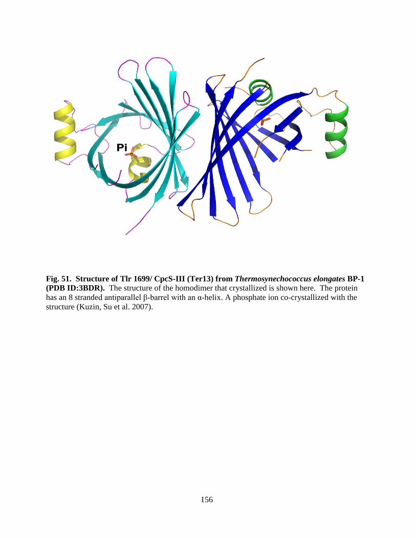
156
Fig. 51. Structure of Tlr 1699/ CpcS-III (Ter13) from Thermosynechococcus elongates BP-1
(PDB ID:3BDR). The structure of the homodimer that crystallized is shown here. The protein
has an 8 stranded antiparallel β-barrel with an α-helix. A phosphate ion co-crystallized with the
structure (Kuzin, Su et al. 2007).
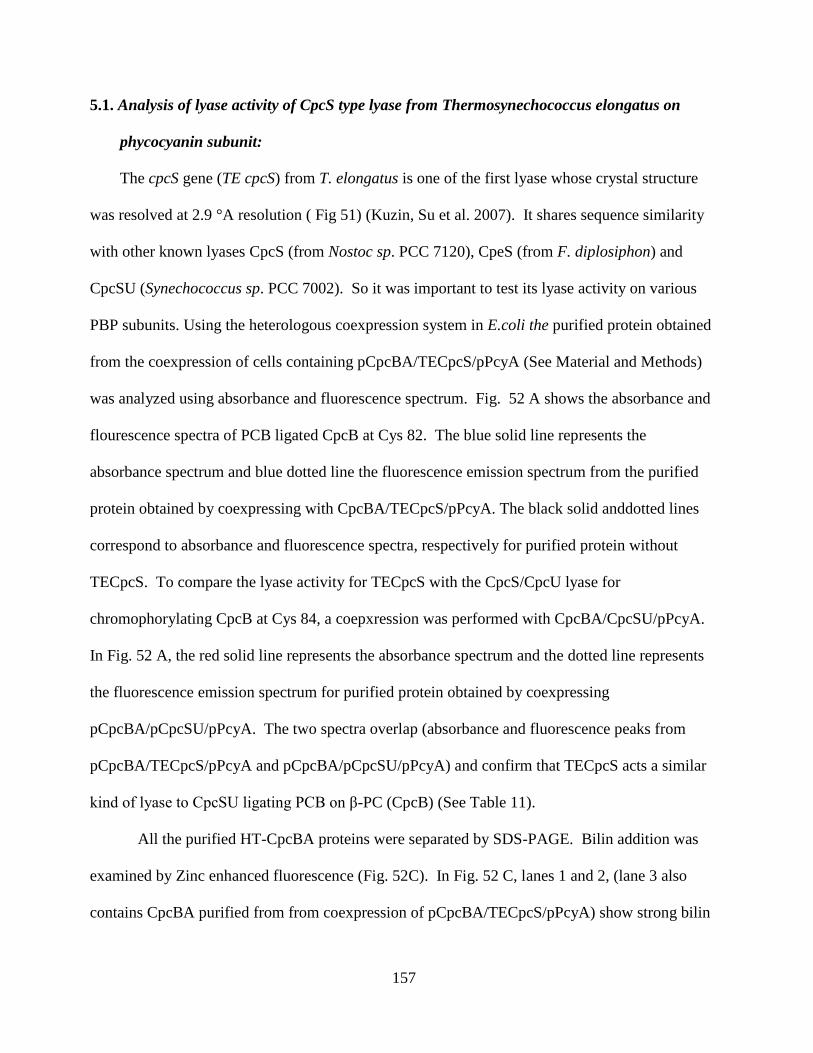
157
5.1. Analysis of lyase activity of CpcS type lyase from Thermosynechococcus elongatus on
phycocyanin subunit:
The cpcS gene (TE cpcS) from T. elongatus is one of the first lyase whose crystal structure
was resolved at 2.9 °A resolution ( Fig 51) (Kuzin, Su et al. 2007). It shares sequence similarity
with other known lyases CpcS (from Nostoc sp. PCC 7120), CpeS (from F. diplosiphon) and
CpcSU (Synechococcus sp. PCC 7002). So it was important to test its lyase activity on various
PBP subunits. Using the heterologous coexpression system in E.coli the purified protein obtained
from the coexpression of cells containing pCpcBA/TECpcS/pPcyA (See Material and Methods)
was analyzed using absorbance and fluorescence spectrum. Fig. 52 A shows the absorbance and
flourescence spectra of PCB ligated CpcB at Cys 82. The blue solid line represents the
absorbance spectrum and blue dotted line the fluorescence emission spectrum from the purified
protein obtained by coexpressing with CpcBA/TECpcS/pPcyA. The black solid anddotted lines
correspond to absorbance and fluorescence spectra, respectively for purified protein without
TECpcS. To compare the lyase activity for TECpcS with the CpcS/CpcU lyase for
chromophorylating CpcB at Cys 84, a coepxression was performed with CpcBA/CpcSU/pPcyA.
In Fig. 52 A, the red solid line represents the absorbance spectrum and the dotted line represents
the fluorescence emission spectrum for purified protein obtained by coexpressing
pCpcBA/pCpcSU/pPcyA. The two spectra overlap (absorbance and fluorescence peaks from
pCpcBA/TECpcS/pPcyA and pCpcBA/pCpcSU/pPcyA) and confirm that TECpcS acts a similar
kind of lyase to CpcSU ligating PCB on β-PC (CpcB) (See Table 11).
All the purified HT-CpcBA proteins were separated by SDS-PAGE. Bilin addition was
examined by Zinc enhanced fluorescence (Fig. 52C). In Fig. 52 C, lanes 1 and 2, (lane 3 also
contains CpcBA purified from from coexpression of pCpcBA/TECpcS/pPcyA) show strong bilin

158
fluorescence indicating that both CpcSU and TE CpcS act as bilin lyase for CpcB (-PC). The
protein content was confirmed by staining the gel with Comassie blue stain (Fig. 52 B, lanes 1
and 2). This section concludes TE CpcS acts as an single subunit S-type lyase involved in bilin
addition on Cys-84 of -PC, and confirming that we now know that the first structure of a
functional bilin lyase
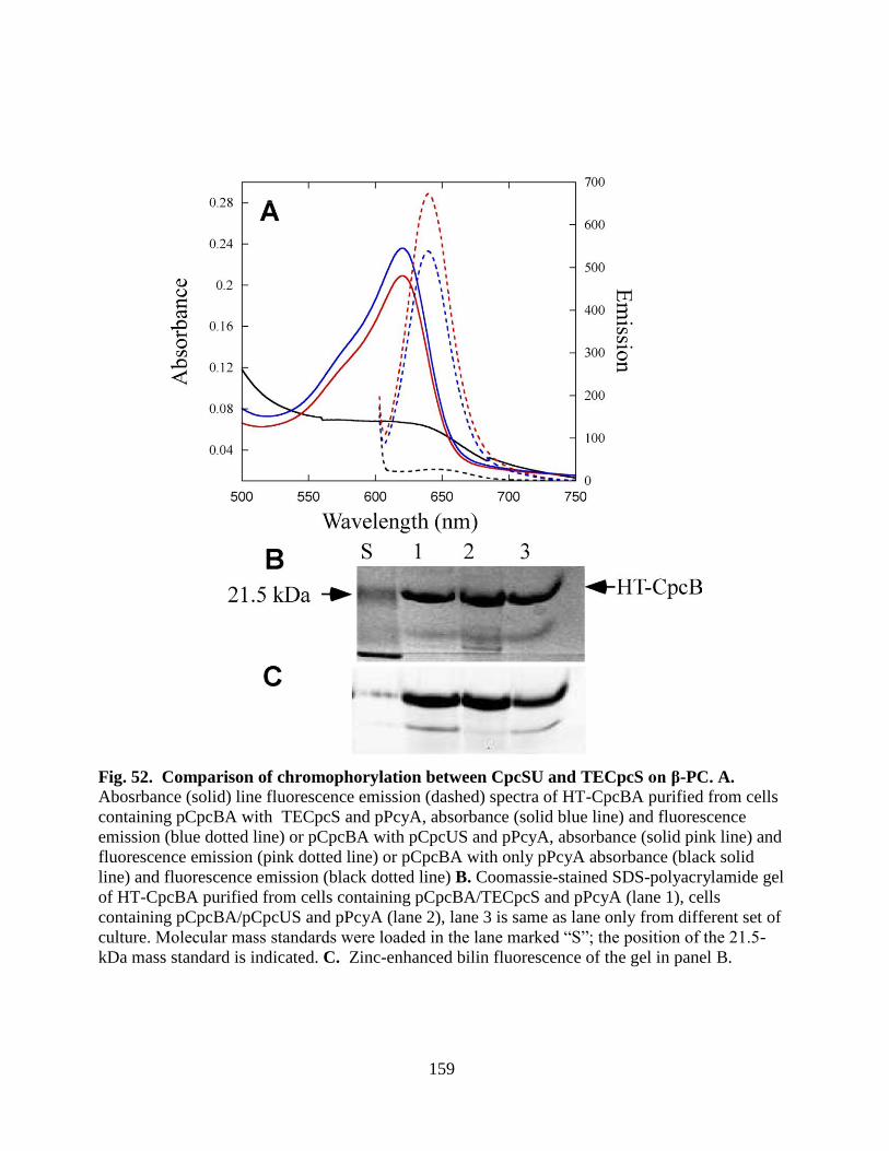
159
Fig. 52. Comparison of chromophorylation between CpcSU and TECpcS on β-PC. A. Abosrbance (solid) line fluorescence emission (dashed) spectra of HT-CpcBA purified from cells
containing pCpcBA with TECpcS and pPcyA, absorbance (solid blue line) and fluorescence
emission (blue dotted line) or pCpcBA with pCpcUS and pPcyA, absorbance (solid pink line) and
fluorescence emission (pink dotted line) or pCpcBA with only pPcyA absorbance (black solid
line) and fluorescence emission (black dotted line) B. Coomassie-stained SDS-polyacrylamide gel
of HT-CpcBA purified from cells containing pCpcBA/TECpcS and pPcyA (lane 1), cells
containing pCpcBA/pCpcUS and pPcyA (lane 2), lane 3 is same as lane only from different set of
culture. Molecular mass standards were loaded in the lane marked “S”; the position of the 21.5-
kDa mass standard is indicated. C. Zinc-enhanced bilin fluorescence of the gel in panel B.
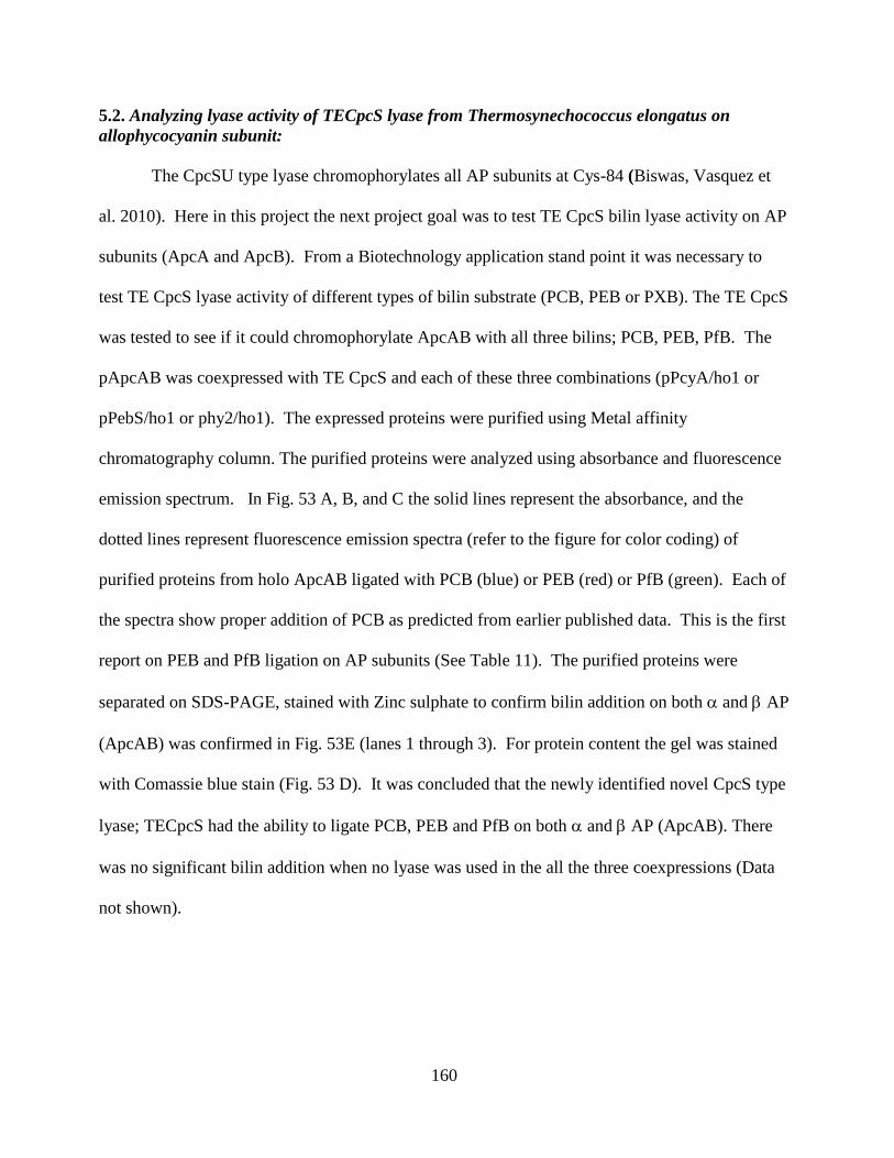
160
5.2. Analyzing lyase activity of TECpcS lyase from Thermosynechococcus elongatus on
allophycocyanin subunit:
The CpcSU type lyase chromophorylates all AP subunits at Cys-84 (Biswas, Vasquez et
al. 2010). Here in this project the next project goal was to test TE CpcS bilin lyase activity on AP
subunits (ApcA and ApcB). From a Biotechnology application stand point it was necessary to
test TE CpcS lyase activity of different types of bilin substrate (PCB, PEB or PXB). The TE CpcS
was tested to see if it could chromophorylate ApcAB with all three bilins; PCB, PEB, PfB. The
pApcAB was coexpressed with TE CpcS and each of these three combinations (pPcyA/ho1 or
pPebS/ho1 or phy2/ho1). The expressed proteins were purified using Metal affinity
chromatography column. The purified proteins were analyzed using absorbance and fluorescence
emission spectrum. In Fig. 53 A, B, and C the solid lines represent the absorbance, and the
dotted lines represent fluorescence emission spectra (refer to the figure for color coding) of
purified proteins from holo ApcAB ligated with PCB (blue) or PEB (red) or PfB (green). Each of
the spectra show proper addition of PCB as predicted from earlier published data. This is the first
report on PEB and PfB ligation on AP subunits (See Table 11). The purified proteins were
separated on SDS-PAGE, stained with Zinc sulphate to confirm bilin addition on both and AP
(ApcAB) was confirmed in Fig. 53E (lanes 1 through 3). For protein content the gel was stained
with Comassie blue stain (Fig. 53 D). It was concluded that the newly identified novel CpcS type
lyase; TECpcS had the ability to ligate PCB, PEB and PfB on both and AP (ApcAB). There
was no significant bilin addition when no lyase was used in the all the three coexpressions (Data
not shown).
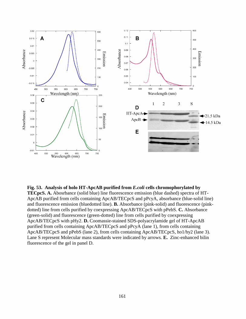
161
Fig. 53. Analysis of holo HT-ApcAB purified from E.coli cells chromophorylated by
TECpcS. A. Abosrbance (solid blue) line fluorescence emission (blue dashed) spectra of HT-
ApcAB purified from cells containing ApcAB/TECpcS and pPcyA, absorbance (blue-solid line)
and fluorescence emission (bluedotted line). B. Absorbance (pink-solid) and fluorescence (pink-
dotted) line from cells purified by coexpressing ApcAB/TECpcS with pPebS. C. Absorbance
(green-solid) and fluorescence (green-dotted) line from cells purified by coexpressing
ApcAB/TECpcS with pHy2. D. Coomassie-stained SDS-polyacrylamide gel of HT-ApcAB
purified from cells containing ApcAB/TECpcS and pPcyA (lane 1), from cells containing
ApcAB/TECpcS and pPebS (lane 2), from cells containing ApcAB/TECpcS, ho1/hy2 (lane 3).
Lane S represent Molecular mass standards were indicated by arrows. E. Zinc-enhanced bilin
fluorescence of the gel in panel D.
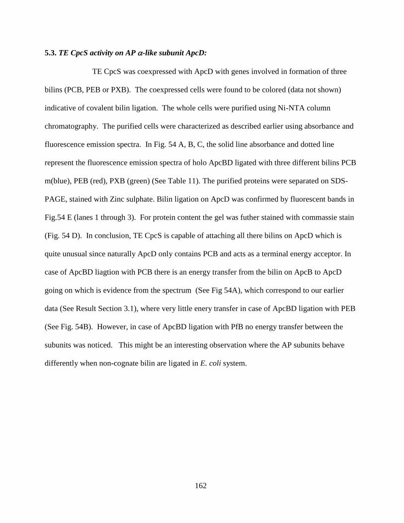
162
5.3. TE CpcS activity on AP -like subunit ApcD:
TE CpcS was coexpressed with ApcD with genes involved in formation of three
bilins (PCB, PEB or PXB). The coexpressed cells were found to be colored (data not shown)
indicative of covalent bilin ligation. The whole cells were purified using Ni-NTA column
chromatography. The purified cells were characterized as described earlier using absorbance and
fluorescence emission spectra. In Fig. 54 A, B, C, the solid line absorbance and dotted line
represent the fluorescence emission spectra of holo ApcBD ligated with three different bilins PCB
m(blue), PEB (red), PXB (green) (See Table 11). The purified proteins were separated on SDS-
PAGE, stained with Zinc sulphate. Bilin ligation on ApcD was confirmed by fluorescent bands in
Fig.54 E (lanes 1 through 3). For protein content the gel was futher stained with commassie stain
(Fig. 54 D). In conclusion, TE CpcS is capable of attaching all there bilins on ApcD which is
quite unusual since naturally ApcD only contains PCB and acts as a terminal energy acceptor. In
case of ApcBD liagtion with PCB there is an energy transfer from the bilin on ApcB to ApcD
going on which is evidence from the spectrum (See Fig 54A), which correspond to our earlier
data (See Result Section 3.1), where very little enery transfer in case of ApcBD ligation with PEB
(See Fig. 54B). However, in case of ApcBD ligation with PfB no energy transfer between the
subunits was noticed. This might be an interesting observation where the AP subunits behave
differently when non-cognate bilin are ligated in E. coli system.
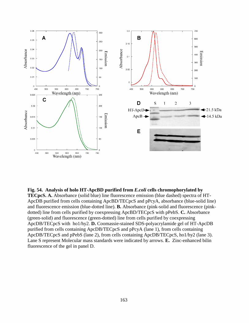
163
Fig. 54. Analysis of holo HT-ApcBD purified from E.coli cells chromophorylated by
TECpcS. A. Absorbance (solid blue) line fluorescence emission (blue dashed) spectra of HT-
ApcDB purified from cells containing ApcBD/TECpcS and pPcyA, absorbance (blue-solid line)
and fluorescence emission (blue-dotted line). B. Absorbance (pink-solid and fluorescence (pink-
dotted) line from cells purified by coexpressing ApcBD/TECpcS with pPebS. C. Absorbance
(green-solid) and fluorescence (green-dotted) line from cells purified by coexpressing
ApcDB/TECpcS with ho1/hy2. D. Coomassie-stained SDS-polyacrylamide gel of HT-ApcDB
purified from cells containing ApcDB/TECpcS and pPcyA (lane 1), from cells containing
ApcDB/TECpcS and pPebS (lane 2), from cells containing ApcDB/TECpcS, ho1/hy2 (lane 3).
Lane S represent Molecular mass standards were indicated by arrows. E. Zinc-enhanced bilin
fluorescence of the gel in panel D.
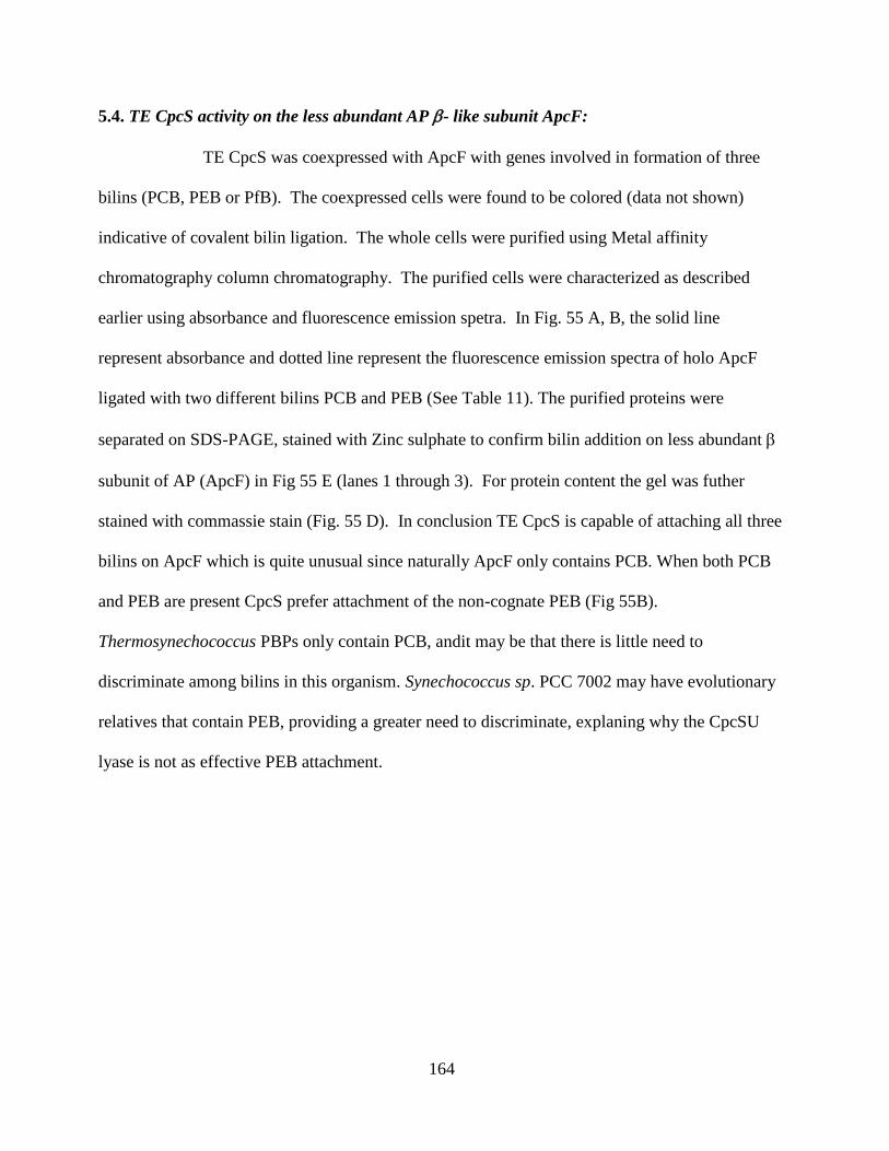
164
5.4. TE CpcS activity on the less abundant AP - like subunit ApcF:
TE CpcS was coexpressed with ApcF with genes involved in formation of three
bilins (PCB, PEB or PfB). The coexpressed cells were found to be colored (data not shown)
indicative of covalent bilin ligation. The whole cells were purified using Metal affinity
chromatography column chromatography. The purified cells were characterized as described
earlier using absorbance and fluorescence emission spetra. In Fig. 55 A, B, the solid line
represent absorbance and dotted line represent the fluorescence emission spectra of holo ApcF
ligated with two different bilins PCB and PEB (See Table 11). The purified proteins were
separated on SDS-PAGE, stained with Zinc sulphate to confirm bilin addition on less abundant
subunit of AP (ApcF) in Fig 55 E (lanes 1 through 3). For protein content the gel was futher
stained with commassie stain (Fig. 55 D). In conclusion TE CpcS is capable of attaching all three
bilins on ApcF which is quite unusual since naturally ApcF only contains PCB. When both PCB
and PEB are present CpcS prefer attachment of the non-cognate PEB (Fig 55B).
Thermosynechococcus PBPs only contain PCB, andit may be that there is little need to
discriminate among bilins in this organism. Synechococcus sp. PCC 7002 may have evolutionary
relatives that contain PEB, providing a greater need to discriminate, explaning why the CpcSU
lyase is not as effective PEB attachment.
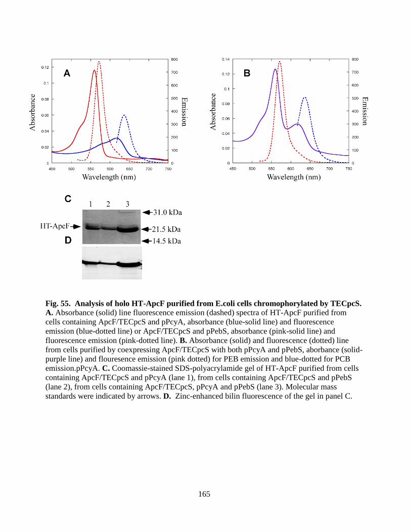
165
Fig. 55. Analysis of holo HT-ApcF purified from E.coli cells chromophorylated by TECpcS. A. Absorbance (solid) line fluorescence emission (dashed) spectra of HT-ApcF purified from
cells containing ApcF/TECpcS and pPcyA, absorbance (blue-solid line) and fluorescence
emission (blue-dotted line) or ApcF/TECpcS and pPebS, absorbance (pink-solid line) and
fluorescence emission (pink-dotted line). B. Absorbance (solid) and fluorescence (dotted) line
from cells purified by coexpressing ApcF/TECpcS with both pPcyA and pPebS, aborbance (solid-
purple line) and flouresence emission (pink dotted) for PEB emission and blue-dotted for PCB
emission.pPcyA. C. Coomassie-stained SDS-polyacrylamide gel of HT-ApcF purified from cells
containing ApcF/TECpcS and pPcyA (lane 1), from cells containing ApcF/TECpcS and pPebS
(lane 2), from cells containing ApcF/TECpcS, pPcyA and pPebS (lane 3). Molecular mass
standards were indicated by arrows. D. Zinc-enhanced bilin fluorescence of the gel in panel C.

166
Conclusion: The overall conclusion of this small project is that we have characterized one new
bilin lyase which shares 50-60 % sequence similarity with known CpcS type lyases. TE CpcS has
the capacity to ligate variable bilins to all AP subunits. Since the X-ray crystal structure is
available that may be helpful in using this lyase for designing various fluorescent tags for
biotechnology applications and this project has proven that this homodimeric structure indeed
functions as a bilin lyase.
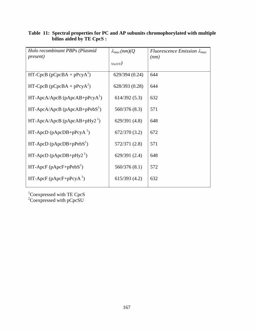
167
Table 11: Spectral properties for PC and AP subunits chromophorylated with multiple
bilins aided by TE CpcS :
1Coexpressed with TE CpcS
2Coexpressed with pCpcSU
Holo recombinant PBPs (Plasmid
present) max (nm)(Q
Vis/UV)
Fluorescence Emission max
(nm)
HT-CpcB (pCpcBA + pPcyA1) 629/394 (0.24) 644
HT-CpcB (pCpcBA + pPcyA2) 628/393 (0.28) 644
HT-ApcA/ApcB (pApcAB+pPcyA1) 614/392 (5.3) 632
HT-ApcA/ApcB (pApcAB+pPebS1) 560/376 (8.3) 571
HT-ApcA/ApcB (pApcAB+pHy2 1
) 629/391 (4.8) 648
HT-ApcD (pApcDB+pPcyA 1
) 672/370 (3.2) 672
HT-ApcD (pApcDB+pPebS1) 572/371 (2.8) 571
HT-ApcD (pApcDB+pHy2 1
) 629/391 (2.4) 648
HT-ApcF (pApcF+pPebS1) 560/376 (8.1) 572
HT-ApcF (pApcF+pPcyA 1
) 615/393 (4.2) 632
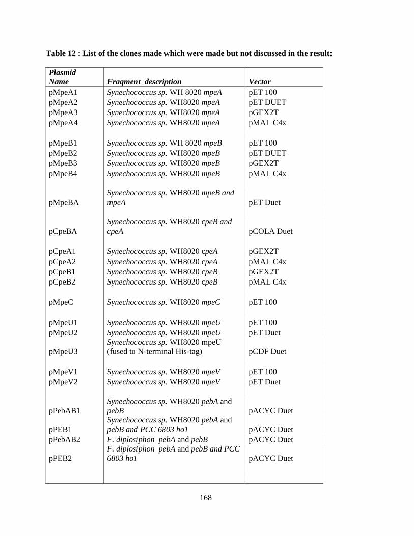
168
Table 12 : List of the clones made which were made but not discussed in the result:
Plasmid
Name Fragment description Vector
pMpeA1 Synechococcus sp. WH 8020 mpeA pET 100
pMpeA2 Synechococcus sp. WH8020 mpeA pET DUET
pMpeA3 Synechococcus sp. WH8020 mpeA pGEX2T
pMpeA4 Synechococcus sp. WH8020 mpeA pMAL C4x
pMpeB1 Synechococcus sp. WH 8020 mpeB pET 100
pMpeB2 Synechococcus sp. WH8020 mpeB pET DUET
pMpeB3 Synechococcus sp. WH8020 mpeB pGEX2T
pMpeB4 Synechococcus sp. WH8020 mpeB pMAL C4x
pMpeBA
Synechococcus sp. WH8020 mpeB and
mpeA pET Duet
pCpeBA
Synechococcus sp. WH8020 cpeB and
cpeA pCOLA Duet
pCpeA1 Synechococcus sp. WH8020 cpeA pGEX2T
pCpeA2 Synechococcus sp. WH8020 cpeA pMAL C4x
pCpeB1 Synechococcus sp. WH8020 cpeB pGEX2T
pCpeB2 Synechococcus sp. WH8020 cpeB pMAL C4x
pMpeC Synechococcus sp. WH8020 mpeC pET 100
pMpeU1 Synechococcus sp. WH8020 mpeU pET 100
pMpeU2 Synechococcus sp. WH8020 mpeU pET Duet
pMpeU3
Synechococcus sp. WH8020 mpeU
(fused to N-terminal His-tag) pCDF Duet
pMpeV1 Synechococcus sp. WH8020 mpeV pET 100
pMpeV2 Synechococcus sp. WH8020 mpeV pET Duet
pPebAB1
Synechococcus sp. WH8020 pebA and
pebB pACYC Duet
pPEB1
Synechococcus sp. WH8020 pebA and
pebB and PCC 6803 ho1 pACYC Duet
pPebAB2 F. diplosiphon pebA and pebB pACYC Duet
pPEB2
F. diplosiphon pebA and pebB and PCC
6803 ho1 pACYC Duet
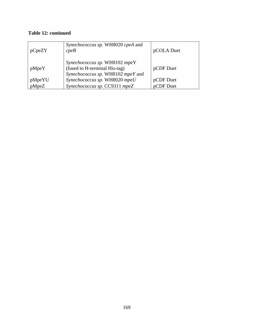
169
Table 12: continued
pCpeZY
Synechococcus sp. WH8020 cpeA and
cpeB pCOLA Duet
pMpeY
Synechococcus sp. WH8102 mpeY
(fused to H-terminal His-tag) pCDF Duet
pMpeYU
Synechococcus sp. WH8102 mpeY and
Synechococcus sp. WH8020 mpeU pCDF Duet
pMpeZ Synechococcus sp. CC9311 mpeZ pCDF Duet
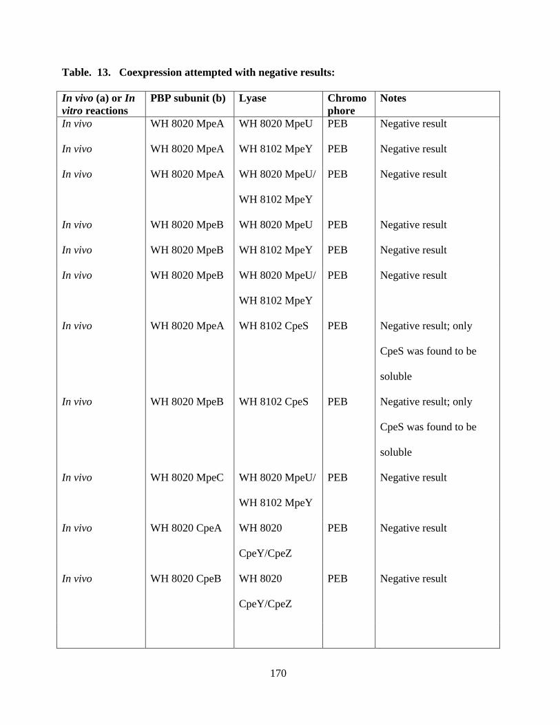
170
Table. 13. Coexpression attempted with negative results:
In vivo (a) or In
vitro reactions
PBP subunit (b) Lyase Chromo
phore
Notes
In vivo WH 8020 MpeA WH 8020 MpeU PEB Negative result
In vivo WH 8020 MpeA WH 8102 MpeY PEB Negative result
In vivo WH 8020 MpeA WH 8020 MpeU/
WH 8102 MpeY
PEB Negative result
In vivo WH 8020 MpeB WH 8020 MpeU PEB Negative result
In vivo WH 8020 MpeB WH 8102 MpeY PEB Negative result
In vivo WH 8020 MpeB WH 8020 MpeU/
WH 8102 MpeY
PEB Negative result
In vivo WH 8020 MpeA WH 8102 CpeS PEB Negative result; only
CpeS was found to be
soluble
In vivo WH 8020 MpeB WH 8102 CpeS PEB Negative result; only
CpeS was found to be
soluble
In vivo WH 8020 MpeC WH 8020 MpeU/
WH 8102 MpeY
PEB Negative result
In vivo WH 8020 CpeA WH 8020
CpeY/CpeZ
PEB Negative result
In vivo WH 8020 CpeB WH 8020
CpeY/CpeZ
PEB Negative result
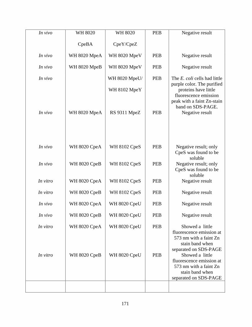
171
In vivo WH 8020
CpeBA
WH 8020
CpeY/CpeZ
PEB Negative result
In vivo WH 8020 MpeA WH 8020 MpeV PEB Negative result
In vivo WH 8020 MpeB WH 8020 MpeV PEB Negative result
In vivo WH 8020 MpeU/
WH 8102 MpeY
PEB The E. coli cells had little
purple color. The purified
proteins have little
fluorescence emission
peak with a faint Zn-stain
band on SDS-PAGE.
In vivo WH 8020 MpeA RS 9311 MpeZ PEB Negative result
In vivo WH 8020 CpeA WH 8102 CpeS PEB Negative result; only
CpeS was found to be
soluble
In vivo WH 8020 CpeB WH 8102 CpeS PEB Negative result; only
CpeS was found to be
soluble
In vitro WH 8020 CpeA WH 8102 CpeS PEB Negative result
In vitro WH 8020 CpeB WH 8102 CpeS PEB Negative result
In vivo WH 8020 CpeA WH 8020 CpeU PEB Negative result
In vivo WH 8020 CpeB WH 8020 CpeU PEB Negative result
In vitro WH 8020 CpeA WH 8020 CpeU PEB Showed a little
fluorescence emission at
573 nm with a faint Zn
stain band when
separated on SDS-PAGE
In vitro WH 8020 CpeB WH 8020 CpeU PEB Showed a little
fluorescence emission at
573 nm with a faint Zn
stain band when
separated on SDS-PAGE
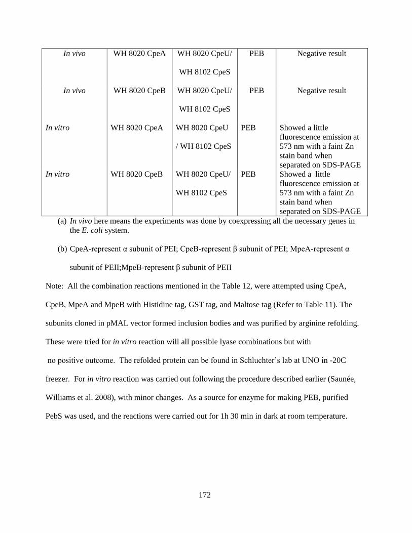
172
In vivo WH 8020 CpeA WH 8020 CpeU/
WH 8102 CpeS
PEB Negative result
In vivo WH 8020 CpeB WH 8020 CpeU/
WH 8102 CpeS
PEB Negative result
In vitro WH 8020 CpeA WH 8020 CpeU
/ WH 8102 CpeS
PEB Showed a little
fluorescence emission at
573 nm with a faint Zn
stain band when
separated on SDS-PAGE
In vitro WH 8020 CpeB WH 8020 CpeU/
WH 8102 CpeS
PEB Showed a little
fluorescence emission at
573 nm with a faint Zn
stain band when
separated on SDS-PAGE
(a) In vivo here means the experiments was done by coexpressing all the necessary genes in
the E. coli system.
(b) CpeA-represent α subunit of PEI; CpeB-represent β subunit of PEI; MpeA-represent α
subunit of PEII;MpeB-represent β subunit of PEII
Note: All the combination reactions mentioned in the Table 12, were attempted using CpeA,
CpeB, MpeA and MpeB with Histidine tag, GST tag, and Maltose tag (Refer to Table 11). The
subunits cloned in pMAL vector formed inclusion bodies and was purified by arginine refolding.
These were tried for in vitro reaction will all possible lyase combinations but with
no positive outcome. The refolded protein can be found in Schluchter’s lab at UNO in -20C
freezer. For in vitro reaction was carried out following the procedure described earlier (Saunée,
Williams et al. 2008), with minor changes. As a source for enzyme for making PEB, purified
PebS was used, and the reactions were carried out for 1h 30 min in dark at room temperature.
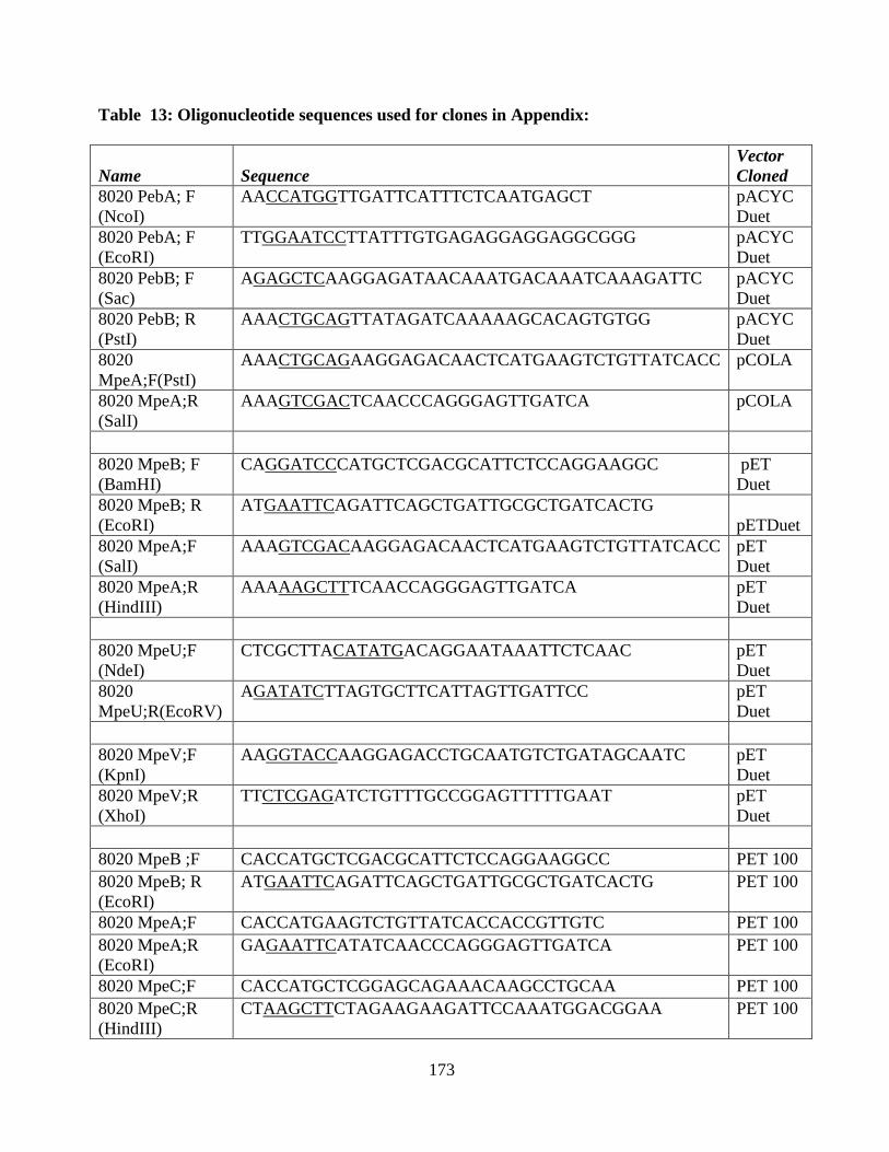
173
Table 13: Oligonucleotide sequences used for clones in Appendix:
Name Sequence
Vector
Cloned
8020 PebA; F
(NcoI)
AACCATGGTTGATTCATTTCTCAATGAGCT pACYC
Duet
8020 PebA; F
(EcoRI)
TTGGAATCCTTATTTGTGAGAGGAGGAGGCGGG pACYC
Duet
8020 PebB; F
(Sac)
AGAGCTCAAGGAGATAACAAATGACAAATCAAAGATTC pACYC
Duet
8020 PebB; R
(PstI)
AAACTGCAGTTATAGATCAAAAAGCACAGTGTGG pACYC
Duet
8020
MpeA;F(PstI)
AAACTGCAGAAGGAGACAACTCATGAAGTCTGTTATCACC pCOLA
8020 MpeA;R
(SalI)
AAAGTCGACTCAACCCAGGGAGTTGATCA pCOLA
8020 MpeB; F
(BamHI)
CAGGATCCCATGCTCGACGCATTCTCCAGGAAGGC pET
Duet
8020 MpeB; R
(EcoRI)
ATGAATTCAGATTCAGCTGATTGCGCTGATCACTG
pETDuet
8020 MpeA;F
(SalI)
AAAGTCGACAAGGAGACAACTCATGAAGTCTGTTATCACC pET
Duet
8020 MpeA;R
(HindIII)
AAAAAGCTTTCAACCAGGGAGTTGATCA pET
Duet
8020 MpeU;F
(NdeI)
CTCGCTTACATATGACAGGAATAAATTCTCAAC pET
Duet
8020
MpeU;R(EcoRV)
AGATATCTTAGTGCTTCATTAGTTGATTCC pET
Duet
8020 MpeV;F
(KpnI)
AAGGTACCAAGGAGACCTGCAATGTCTGATAGCAATC pET
Duet
8020 MpeV;R
(XhoI)
TTCTCGAGATCTGTTTGCCGGAGTTTTTGAAT pET
Duet
8020 MpeB ;F CACCATGCTCGACGCATTCTCCAGGAAGGCC PET 100
8020 MpeB; R
(EcoRI)
ATGAATTCAGATTCAGCTGATTGCGCTGATCACTG PET 100
8020 MpeA;F CACCATGAAGTCTGTTATCACCACCGTTGTC PET 100
8020 MpeA;R
(EcoRI)
GAGAATTCATATCAACCCAGGGAGTTGATCA PET 100
8020 MpeC;F CACCATGCTCGGAGCAGAAACAAGCCTGCAA PET 100
8020 MpeC;R
(HindIII)
CTAAGCTTCTAGAAGAAGATTCCAAATGGACGGAA PET 100
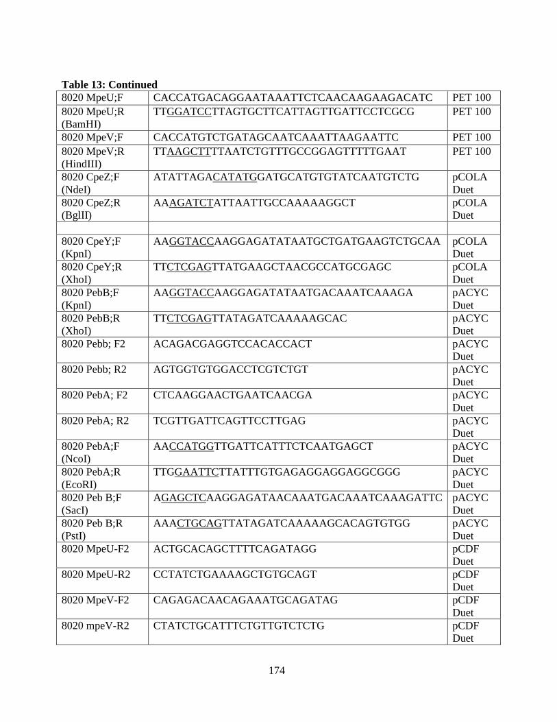
174
Table 13: Continued
8020 MpeU;F CACCATGACAGGAATAAATTCTCAACAAGAAGACATC PET 100
8020 MpeU;R
(BamHI)
TTGGATCCTTAGTGCTTCATTAGTTGATTCCTCGCG PET 100
8020 MpeV;F CACCATGTCTGATAGCAATCAAATTAAGAATTC PET 100
8020 MpeV;R
(HindIII)
TTAAGCTTTTAATCTGTTTGCCGGAGTTTTTGAAT PET 100
8020 CpeZ;F
(NdeI)
ATATTAGACATATGGATGCATGTGTATCAATGTCTG pCOLA
Duet
8020 CpeZ;R
(BglII)
AAAGATCTATTAATTGCCAAAAAGGCT pCOLA
Duet
8020 CpeY;F
(KpnI)
AAGGTACCAAGGAGATATAATGCTGATGAAGTCTGCAA pCOLA
Duet
8020 CpeY;R
(XhoI)
TTCTCGAGTTATGAAGCTAACGCCATGCGAGC pCOLA
Duet
8020 PebB;F
(KpnI)
AAGGTACCAAGGAGATATAATGACAAATCAAAGA pACYC
Duet
8020 PebB;R
(XhoI)
TTCTCGAGTTATAGATCAAAAAGCAC pACYC
Duet
8020 Pebb; F2 ACAGACGAGGTCCACACCACT pACYC
Duet
8020 Pebb; R2 AGTGGTGTGGACCTCGTCTGT pACYC
Duet
8020 PebA; F2 CTCAAGGAACTGAATCAACGA pACYC
Duet
8020 PebA; R2 TCGTTGATTCAGTTCCTTGAG pACYC
Duet
8020 PebA;F
(NcoI)
AACCATGGTTGATTCATTTCTCAATGAGCT pACYC
Duet
8020 PebA;R
(EcoRI)
TTGGAATTCTTATTTGTGAGAGGAGGAGGCGGG pACYC
Duet
8020 Peb B;F
(SacI)
AGAGCTCAAGGAGATAACAAATGACAAATCAAAGATTC pACYC
Duet
8020 Peb B;R
(PstI)
AAACTGCAGTTATAGATCAAAAAGCACAGTGTGG pACYC
Duet
8020 MpeU-F2 ACTGCACAGCTTTTCAGATAGG pCDF
Duet
8020 MpeU-R2 CCTATCTGAAAAGCTGTGCAGT pCDF
Duet
8020 MpeV-F2 CAGAGACAACAGAAATGCAGATAG pCDF
Duet
8020 mpeV-R2 CTATCTGCATTTCTGTTGTCTCTG pCDF
Duet
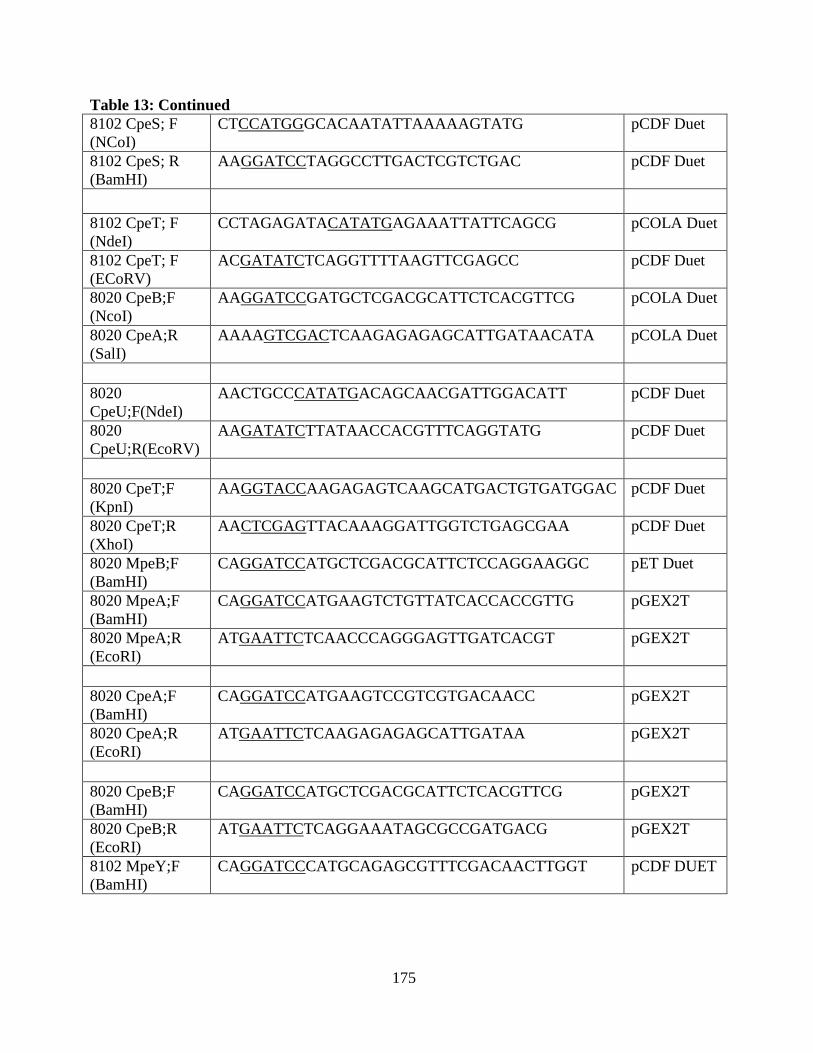
175
Table 13: Continued
8102 CpeS; F
(NCoI)
CTCCATGGGCACAATATTAAAAAGTATG pCDF Duet
8102 CpeS; R
(BamHI)
AAGGATCCTAGGCCTTGACTCGTCTGAC pCDF Duet
8102 CpeT; F
(NdeI)
CCTAGAGATACATATGAGAAATTATTCAGCG pCOLA Duet
8102 CpeT; F
(ECoRV)
ACGATATCTCAGGTTTTAAGTTCGAGCC pCDF Duet
8020 CpeB;F
(NcoI)
AAGGATCCGATGCTCGACGCATTCTCACGTTCG pCOLA Duet
8020 CpeA;R
(SalI)
AAAAGTCGACTCAAGAGAGAGCATTGATAACATA pCOLA Duet
8020
CpeU;F(NdeI)
AACTGCCCATATGACAGCAACGATTGGACATT pCDF Duet
8020
CpeU;R(EcoRV)
AAGATATCTTATAACCACGTTTCAGGTATG pCDF Duet
8020 CpeT;F
(KpnI)
AAGGTACCAAGAGAGTCAAGCATGACTGTGATGGAC pCDF Duet
8020 CpeT;R
(XhoI)
AACTCGAGTTACAAAGGATTGGTCTGAGCGAA pCDF Duet
8020 MpeB;F
(BamHI)
CAGGATCCATGCTCGACGCATTCTCCAGGAAGGC pET Duet
8020 MpeA;F
(BamHI)
CAGGATCCATGAAGTCTGTTATCACCACCGTTG pGEX2T
8020 MpeA;R
(EcoRI)
ATGAATTCTCAACCCAGGGAGTTGATCACGT pGEX2T
8020 CpeA;F
(BamHI)
CAGGATCCATGAAGTCCGTCGTGACAACC pGEX2T
8020 CpeA;R
(EcoRI)
ATGAATTCTCAAGAGAGAGCATTGATAA pGEX2T
8020 CpeB;F
(BamHI)
CAGGATCCATGCTCGACGCATTCTCACGTTCG pGEX2T
8020 CpeB;R
(EcoRI)
ATGAATTCTCAGGAAATAGCGCCGATGACG pGEX2T
8102 MpeY;F
(BamHI)
CAGGATCCCATGCAGAGCGTTTCGACAACTTGGT pCDF DUET
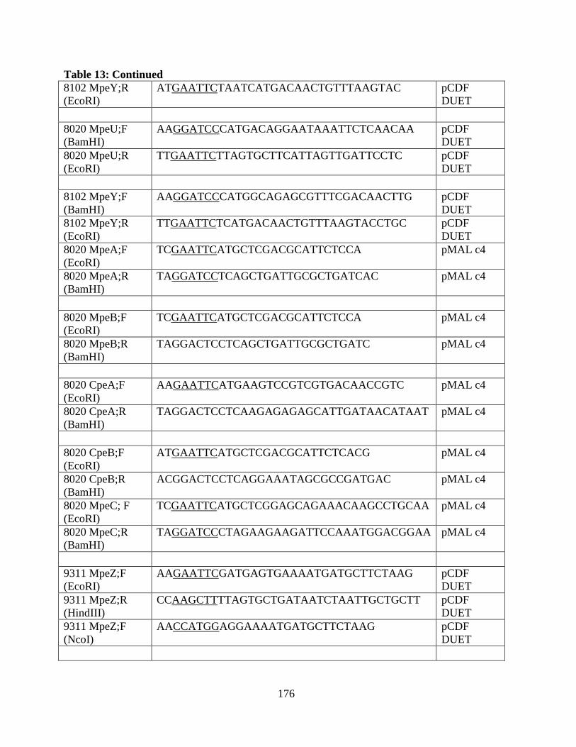
176
Table 13: Continued
8102 MpeY;R
(EcoRI)
ATGAATTCTAATCATGACAACTGTTTAAGTAC pCDF
DUET
8020 MpeU;F
(BamHI)
AAGGATCCCATGACAGGAATAAATTCTCAACAA pCDF
DUET
8020 MpeU;R
(EcoRI)
TTGAATTCTTAGTGCTTCATTAGTTGATTCCTC pCDF
DUET
8102 MpeY;F
(BamHI)
AAGGATCCCATGGCAGAGCGTTTCGACAACTTG pCDF
DUET
8102 MpeY;R
(EcoRI)
TTGAATTCTCATGACAACTGTTTAAGTACCTGC pCDF
DUET
8020 MpeA;F
(EcoRI)
TCGAATTCATGCTCGACGCATTCTCCA pMAL c4
8020 MpeA;R
(BamHI)
TAGGATCCTCAGCTGATTGCGCTGATCAC pMAL c4
8020 MpeB;F
(EcoRI)
TCGAATTCATGCTCGACGCATTCTCCA pMAL c4
8020 MpeB;R
(BamHI)
TAGGACTCCTCAGCTGATTGCGCTGATC pMAL c4
8020 CpeA;F
(EcoRI)
AAGAATTCATGAAGTCCGTCGTGACAACCGTC pMAL c4
8020 CpeA;R
(BamHI)
TAGGACTCCTCAAGAGAGAGCATTGATAACATAAT pMAL c4
8020 CpeB;F
(EcoRI)
ATGAATTCATGCTCGACGCATTCTCACG pMAL c4
8020 CpeB;R
(BamHI)
ACGGACTCCTCAGGAAATAGCGCCGATGAC pMAL c4
8020 MpeC; F
(EcoRI)
TCGAATTCATGCTCGGAGCAGAAACAAGCCTGCAA pMAL c4
8020 MpeC;R
(BamHI)
TAGGATCCCTAGAAGAAGATTCCAAATGGACGGAA pMAL c4
9311 MpeZ;F
(EcoRI)
AAGAATTCGATGAGTGAAAATGATGCTTCTAAG pCDF
DUET
9311 MpeZ;R
(HindIII)
CCAAGCTTTTAGTGCTGATAATCTAATTGCTGCTT pCDF
DUET
9311 MpeZ;F
(NcoI)
AACCATGGAGGAAAATGATGCTTCTAAG pCDF
DUET
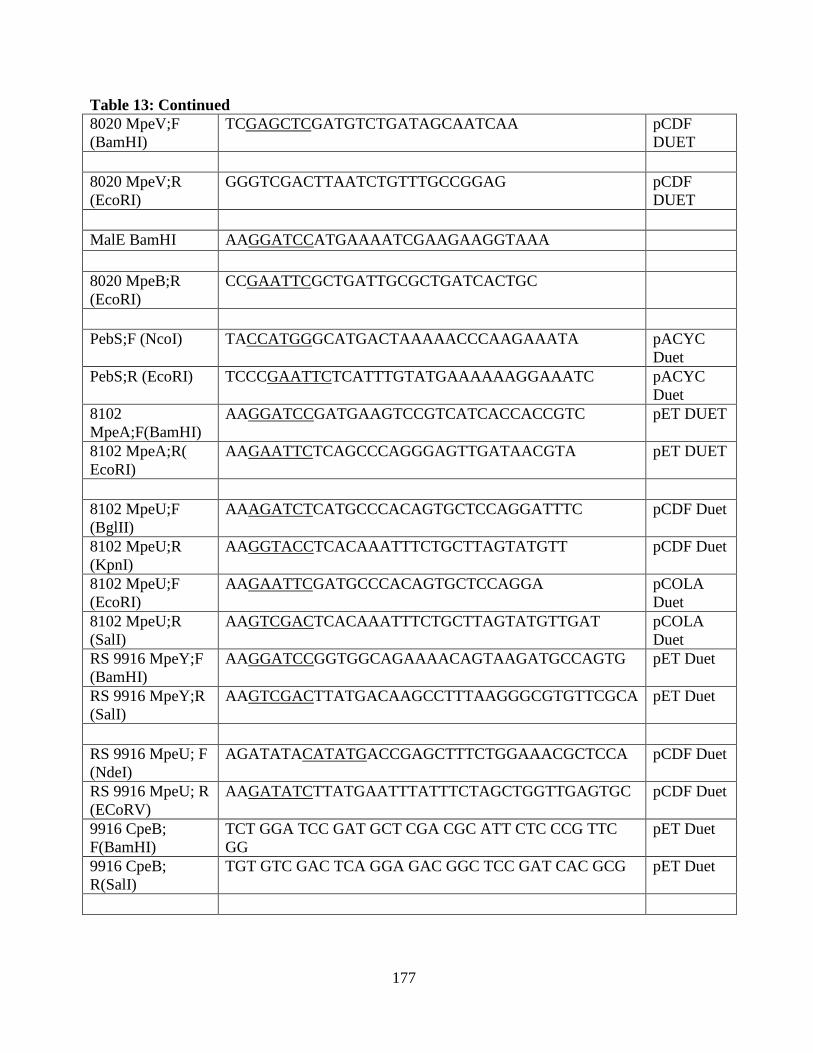
177
Table 13: Continued
8020 MpeV;F
(BamHI)
TCGAGCTCGATGTCTGATAGCAATCAA pCDF
DUET
8020 MpeV;R
(EcoRI)
GGGTCGACTTAATCTGTTTGCCGGAG pCDF
DUET
MalE BamHI AAGGATCCATGAAAATCGAAGAAGGTAAA
8020 MpeB;R
(EcoRI)
CCGAATTCGCTGATTGCGCTGATCACTGC
PebS;F (NcoI) TACCATGGGCATGACTAAAAACCCAAGAAATA pACYC
Duet
PebS;R (EcoRI) TCCCGAATTCTCATTTGTATGAAAAAAGGAAATC pACYC
Duet
8102
MpeA;F(BamHI)
AAGGATCCGATGAAGTCCGTCATCACCACCGTC pET DUET
8102 MpeA;R(
EcoRI)
AAGAATTCTCAGCCCAGGGAGTTGATAACGTA pET DUET
8102 MpeU;F
(BglII)
AAAGATCTCATGCCCACAGTGCTCCAGGATTTC pCDF Duet
8102 MpeU;R
(KpnI)
AAGGTACCTCACAAATTTCTGCTTAGTATGTT pCDF Duet
8102 MpeU;F
(EcoRI)
AAGAATTCGATGCCCACAGTGCTCCAGGA pCOLA
Duet
8102 MpeU;R
(SalI)
AAGTCGACTCACAAATTTCTGCTTAGTATGTTGAT pCOLA
Duet
RS 9916 MpeY;F
(BamHI)
AAGGATCCGGTGGCAGAAAACAGTAAGATGCCAGTG pET Duet
RS 9916 MpeY;R
(SalI)
AAGTCGACTTATGACAAGCCTTTAAGGGCGTGTTCGCA pET Duet
RS 9916 MpeU; F
(NdeI)
AGATATACATATGACCGAGCTTTCTGGAAACGCTCCA pCDF Duet
RS 9916 MpeU; R
(ECoRV)
AAGATATCTTATGAATTTATTTCTAGCTGGTTGAGTGC pCDF Duet
9916 CpeB;
F(BamHI)
TCT GGA TCC GAT GCT CGA CGC ATT CTC CCG TTC
GG
pET Duet
9916 CpeB;
R(SalI)
TGT GTC GAC TCA GGA GAC GGC TCC GAT CAC GCG pET Duet
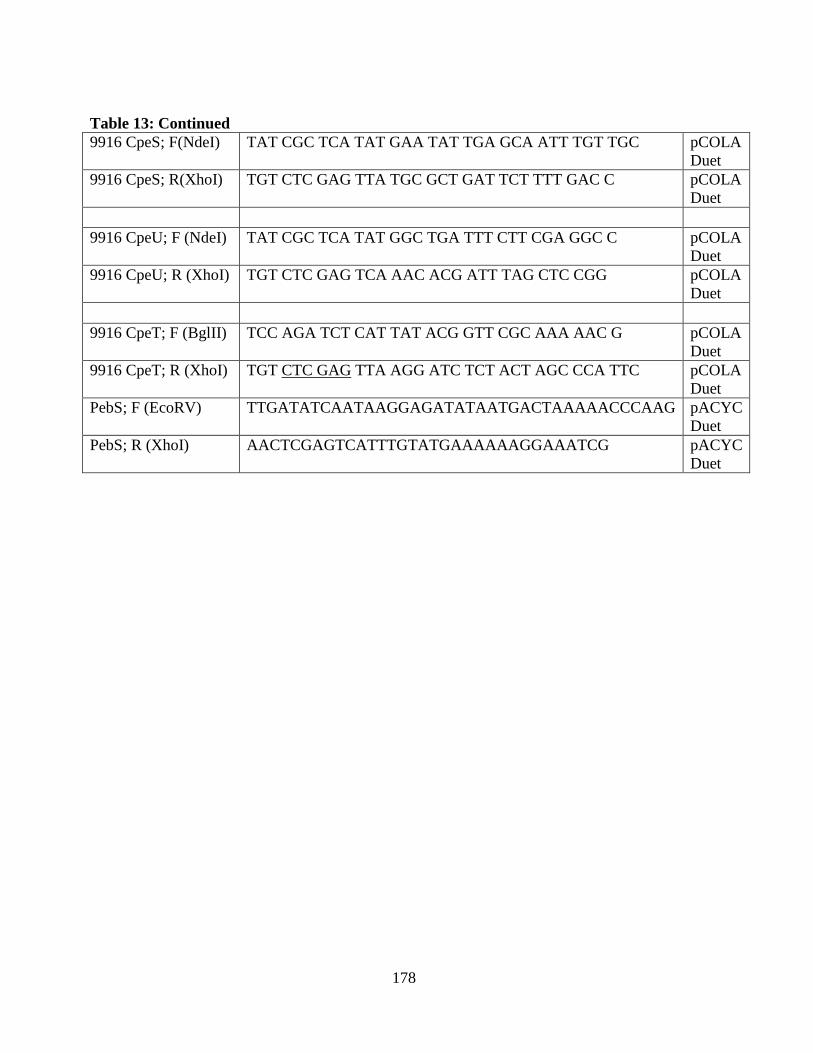
178
Table 13: Continued
9916 CpeS; F(NdeI) TAT CGC TCA TAT GAA TAT TGA GCA ATT TGT TGC pCOLA
Duet
9916 CpeS; R(XhoI) TGT CTC GAG TTA TGC GCT GAT TCT TTT GAC C pCOLA
Duet
9916 CpeU; F (NdeI) TAT CGC TCA TAT GGC TGA TTT CTT CGA GGC C pCOLA
Duet
9916 CpeU; R (XhoI) TGT CTC GAG TCA AAC ACG ATT TAG CTC CGG pCOLA
Duet
9916 CpeT; F (BglII) TCC AGA TCT CAT TAT ACG GTT CGC AAA AAC G pCOLA
Duet
9916 CpeT; R (XhoI) TGT CTC GAG TTA AGG ATC TCT ACT AGC CCA TTC pCOLA
Duet
PebS; F (EcoRV) TTGATATCAATAAGGAGATATAATGACTAAAAACCCAAG pACYC
Duet
PebS; R (XhoI) AACTCGAGTCATTTGTATGAAAAAAGGAAATCG pACYC
Duet
179
Reference:
Ajlani, G. and C. Vernotte (1998). "Construction and characterization of a phycobiliprotein-less
mutant of Synechocystis sp. PCC 6803." Plant Molecular Biology 37(3): 577-580.
Ajlani, G. and C. Vernotte (1998). "Deletion of the PB-loop in the LCM subunit does not affect
phycobilisome assembly or energy transfer functions in the cyanobacterium Synechocystis sp.
PCC6714." European Journal of Biochemistry 257(1): 154-159.
Alberte, R. S., A. M. Wood, et al. (1984). "Novel phycoerythrins in marine Synechococcus spp."
Plant Physiol 75: 732 - 739.
Allen, M. M. (1968). " Simple conditions for growth of unicellular blue-green algae on plates."
J.Phycol.
4: 1-4.
Alvey, R. M., A. Biswas, et al. (2011). "Attachment if Noncognate Chromophores to CpcA of
Synechocystis sp. PCC 6803 and Synechococcus sp. PCC 7002 bu Heterologous Expression in
Escherichia coli." Biochemistry 50(22): 4890-4902.
Alvey, R. M., J. A. Karty, et al. (2003). "Lesions in phycoerythrin chromophore biosynthesis in
Fremyelia diplosiphon reveal coordinated light regulation of apoprotein and pigment biosynthetic
enzyme gene expression." Plant Cell 15(10): 2448-2463.
Anderson, L. K. and C. M. Toole (1998). "A model for early events in the assembly pathway of
cyanobacterial phycobilisomes." Mol. Microbiol. 30(3): 467-474.
Andrade, M. A., C. Petosa, et al. (2001). "Comparison of ARM and HEAT Protein Repeats." J.
Mol. Biol. 309: 1-18.
Arciero, D. M., D. A. Bryant, et al. (1988). "In vitro attachment of bilins to apophycocyanin I:
specific covalent adduct formation at cysteinyl residues involved in phycocyanobilin binding in
C-phycocyanin." J. Biol. Chem. 263: 18343-18349.
Arciero, D. M., J. L. Dallas, et al. (1988). "In vitro attachment of bilins to apophycocyanin: II:
determination of the structures of tryptic bilin peptides derived from the phycocyanobilin adduct."
J. Biol. Chem. 263: 18350-18357.
Arciero, D. M., J. L. Dallas, et al. (1988). "In vitro attachment of bilins to apophycocyanin: III:
properties of the phycoerythrobilin adduct." J. Biol. Chem. 263: 18358-18363.
Ashby, M. K. and C. W. Mullineaux (1999). "The role of ApcD and ApcF in energy transfer from
phycobilisomes to PSI and PSII in a cyanobacterium." Photosynth. Res. 61: 169-179.
180
Baier, K., H. Lehmann, et al. (2004). "NblA is essential for phycobilisome degradation in
Anabaena sp strain PCC 7120 but not for development of functional heterocysts." Microbiology-
Sgm 150: 2739-2749.
Baker, B. J., H. Mutoh, et al. (2008). "Genetically encoded fluorescent sensors of membrane
potential." Brain Cell Biol 36(1-4): 53-67.
Beale, S. I. (1993). "Biosynthesis of Phycobilins." Chem. Revs. 93: 785-802.
Beale, S. I. (1999). "Enzymes for Chlorophyll biosynthesis." Photosynthesis Research 60: 43-73.
Beck, W. F. and K. Sauer (1992). "Energy-Transfer and Exciton-State Relaxation Processes in
Allophycocyanin." Journal of Physical Chemistry 96(11): 4658-4666.
Beguin, S., G. Guglielmi, et al. (1985). "Chromatic adaptation in a mutant of Fremyella
diplosiphon incapable of phycoerythrin synthesis." Biochimie 67: 109–117.
Bengston, S. (1994). "Early life on Earth." Nobel Symposium no. 84. Columbia Univ. Press: New
York
Bennett, A. and L. Bogorad (1971). "Properties of subunits and aggregates of blue-green alga
biliproteins." Biochmistry 10: 3625-3634.
Bennett, A. and L. Bogorad ( 1973). "Complementary chromatic adaptation in a filamentous
blue-green alga." J. Cell. Biol. 58: 419–435.
Berkelman, T. R. and J. C. Lagarias (1986). "Visualization of bilin-linked peptides and proteins in
polyacrylamide gels " Analytical Biochemistry 156(1): 194-201.
Berkelman, T. R. and J. C. Lagarias (1986). "Visualization of bilin-linked peptides peptides and
proteins in polyacrylamide gels." Analytical Biochemistry 156: 194-201.
Bienert, R., K. Baier, et al. (2006). "Crystal structure of NblA from Anabaena sp PCC 7120, a
small protein playing a key role in phycobilisome degradation." Journal of Biological Chemistry
281(8): 5216-5223.
Bishop, R. E. (2000). " The bacterial lipocalins." Biochimica Et Biophysica Acta-Protein
Structure and Molecular Enzymology 1482: 73-83.
Biswas, A., Y. M. Vasquez, et al. (2010). "Biosynthesis of cyanobacterial phycobiliproteins in
Escherichia coli: chromophorylation efficiency and specificity of all bilin lyases from
Synechococcus sp. strain PCC 7002." Appl Environ Microbiol 76(9): 2729-2739.
Biswas, A., Y. M. Vasquez, et al. (2010). "Biosynthesis of Cyanobacterial Phycobiliproteins in
Escherichia coli: Chromophorylation Efficiency and Specificity of All Bilin Lyases from

181
Synechococcus sp Strain PCC 7002." Applied and Environmental Microbiology 76(9): 2729-
2739.
Blot, N., X. J. Wu, et al. (2009). "Phycourobilin in Trichromatic Phycocyanin from Oceanic
Cyanobacteria Is Formed Post-translationally by a Phycoerythrobilin Lyase-Isomerase." Journal
of Biological Chemistry 284(14): 9290-9298.
Bogorad, L., S. M. Gendel, Haury, J.F., , et al. (1982). "Photomorphogenesis and complementary
chromatic adaptation in Fremyella diplosyphon." In Proceedings of the Special
FEBS Meeting on Cell Function and Differrentiation, ed. GC Papageorgiou, L Packer,Athens,
Greece, April 25–29: 119–126.
Bohm, S., S. Endres, et al. (2007). "Biliprotein chromophore attachment - Chaperone-like
function of the PecE subunit of alpha-phycoerythrocyanin lyase." Journal of Biological Chemistry
282(35): 25357-25366.
Boresch, K. (1922). "Die komplement¨are chromatic Adaptation." Arch. Protistenkd. 44: 1–70.
Brejc, K., R. Ficner, et al. (1995). "Isolation, crystallization, crystal structure analysis and
refinement of allophycocyanin form the cyanobacterium Spirulina platensis at 2.3 Å resolution."
J. Mol. Biol. 249: 424-440.
Brown, S. B. and R. F. Troxler (1982). "In Bilirbin." Heirweigh, K.P.M. and Brown, S.B., eds,
CRC Press, Boca-Raton, FL 2: 1-38.
Bryant, D. A., G. Gugliemi, et al. (1979). "The structure of cyanobacterial phycobilisomes: a
model." Archives of Microbiology 123: 113-127.
Bryant, D. A., V. L. Stirewalt, et al. (1991). "A SMALL MULTIGENE FAMILY ENCODES
THE ROD-CORE LINKER POLYPEPTIDES OF ANABAENA SP PCC7120
PHYCOBILISOMES." Gene 107(1): 91-99.
Capuano, V., A. S. Braux, et al. (1991). "The "anchor polypeptide" of cyanobacterial
phycobilisomes. Molecular characterization of the Synechococcus sp. PCC 6301 apcE gene." J
Biol Chem 266: 7239 - 7247.
Charron, J. B. F., F. Ouellet, et al. (2005). " Identification, expression, and evolutionary analyses
of plant lipocalins." Plant Physiology(139): 2017-2028.
Chudakov, D., S. Lukyanov, et al. (2005). "Fluorescent proteins as a toolkit for in vivo imaging."
Trends Biotechnol 23(12 ): 605-613.
Cody, W. C., D. Prasher, et al. (1993). "Chemical structure of the hexapeptide chromophore of the
Aequorea green-fluorescent protein." Biochemistry 32(5): 1212-1218.
Cole, W. J., D. J. Chapman, et al. (1967). J. Am. Chem. Soc. 89: 3643-3645.
182
Collier, J. L. and A. R. Grossman (1994). "A small polypeptide triggers complete degradation of
light-harvesting phycobiliproteins in nutrient-deprived cyanobacteria." EMBO J. 13: 1039-1047.
Cornejo, J., R. D. Willows, et al. (1998). "Phytobilin biosynthesis: cloning and expression of a
gene encoding soluble ferredoxin-dependent heme oxygenase from Synechocystis sp. PCC 6803."
Plant J. 15: 99-107.
Crossett, R. N., E. A. Drew, et al. (1965). "Chromatic adaptation in benthic marine
algae." Nature 207: 547–548.
Cubitt AB, M. Onno, et al. (1995). "Understanding, improving and using green fluorescent
proteins." Trends Biochem Sci 20: 448-455.
Cubitt AB, H. R., Woollenweber LA. and M. C. Biol (1997). " In Green Fluorescent. Protein:
Properties and Applications." Methods Cell Biol.
Damerval, T., G. Guglielmi, et al. (1991). "Hormogonium differentiation in the cyanobacterium
Calothrix - a photoregulated developmental process." Plant Cell 3: 191–201.
Dammeyer, T., S. C. Bagby, et al. (2008). "Efficient phage-mediated pigment biosynthesis in
oceanic cyanabacteria." Current Biology 18(6): 442-448.
Dammeyer, T. and N. Frankenberg-Dinkel (2006). "Insights into phycoerythrobilin biosynthesis
point toward metabolic channeling." Journal of Biological Chemistry 281(37): 27081-27089.
Dammeyer, T., E. Homann, et al. (2008). "Phycoerythrobilin Synthase (PebS) of a Marine Virus:
Crystal Structures of the Biliverdin Complex and the substrate-free form." J Biol Chem
283(41): 27547-27554.
Dammeyer, T., K. Michaelsen, et al. (2007). "Biosynthesis of open-chain tetrapyrroles in
Prochlorococcus marinus." Fems Microbiology Letters 271(2): 251-257.
DeGiorgi, F., M. Brini, et al. (1996). Gene 173: 113–17.
Desnik, H., R. Nicoll, ,A., , et al. (2005). "Photoinactivation of native AMPA receptors reveals
their real-time trafficking." Neuron 48(6): 977-685.
Dines, M., E. Sendersky, et al. (2007). "Crystallization of sparingly soluble stress-related proteins
from cyanobacteria by controlled urea solublization." Journal of Structural Biology 158(1): 116-
121.
Dolganov, N. and A. R. Grossman (1999). "A polypeptide with similarity to phycocyanin -
subunit phycocyanobilin lyase involved in degradation of phycobilisomes." J. Bacteriol. 181(2):
610-617.
183
Domg, C. A., J. Tang, et al. (2009). "ApcD necessary for efficient energy transfer from
phycobilisomes to photosystem I and helps to prevent photoinhibition in the cyanobacterium
Synechococcus sp. PCC 7002." Biochemical and Biophysical Acta.- Bioenergetics 1787(1122-
1128).
Ducret, A., W. Sidler, et al. (1994). "The complete amino acid sequence of R-Phycocyanin-I
and subunits from the red alga Porphyridium cruentum: structural and phylogenetic
relationships of the phycocyanins within the phycobiliproteins families." Eur. J. Biochem. 221:
563-580.
Edwards, M. R., C. Hauer, et al. (1997). "Thermophilic C-phycocyanin: Effect of temperature,
monomer stability and structure." Biochemical and Biophysical Acta. 1321: 157-164.
Eggen, M. and G. I. Georg (2002). "The cryptophycins: their synthesis and anticancer activity."
Med. Res. Rev. 22(2): 85-101.
Engelmann, T. W. (1883). "Farbe und Assimilation. I. Assimilation findet nur in den
farbstoffhaltigen
Plasmatheilchen statt. II N¨aherer Zusammenhang zwischen Lichtabsorbtion
und Assimilation. III.Weitere folgerungen." Bot. Z. 41: 1–29.
Engelmann, T. W. (1902). "Untersuchungen ¨ uber die qualitativen Beziehungen zwieschen
Absorbtion des Lichtes und Assimilation in Pflanzenzellen. I. Das Mikrospectraphotometer,
ein Apparat zur qualitativen Mikrospectralanalyse. II. Experimentelle Grundlangen
zur Ermittelung der quantitativen Beziehungen zwieschen Assimilationsenergie
und Absorptiongr ¨ osse. III. Bestimmung der Vertheilung der Energie im Spectrum von
Sonnenlicht mittels Bacterien-methode und quantitativen Mikrospectralanalyse." Bot. Z 42: 81–
105.
Evans, E. L. and M. M. Allen (1973). "Phycobilisome in Anacystis nidulans." J.Bacteriol. 113(1):
403-408.
Everroad, C., C. Six, et al. (2006). "Biochemical Bases of Type IV Chromatic Adaptation in
Marine Synechococcus spp." J. Bacteriol. 188(9): 3345-3356.
Fairchild, C. D. and A. N. Glazer (1994). "Nonenzymatic bilin addition to the subunit of an
apophycoerythrin." Journal of Biological Chemistry 269: 28988-28996.
Fairchild, C. D. and A. N. Glazer (1994). "Oligomeric structure, enzyme kinetics, and substrate
specificity of the phycocyanin alpha subunit phycocyanobilin lyase." J Biol Chem 269: 8686 -
8694.
Fairchild, C. D., J. Zhao, et al. (1992). "Phycocyanin subunit phycocyanobilin lyase." Proc.
Natl. Acad. Sci., USA 89: 7017-7021.
184
Ficner, R. and R. Huber (1993). "Refined crystal structure of phycoerythrin from Porphyridium
cruentum at 0.23 nm resolution and localization of the subunit." Eur. J. Biochem. 218: 103-106.
Flower, D. R. (1996). "The lipocalin protein family : structure and function." Biochem. J. 318: 1-
14.
Frankenberg, N. and J. C. Lagarias (2003). "Phycocyanobilin:Ferredoxin Oxidoreductase of
Anabaena sp. PCC 7120: Biochemical and Spectroscopic Characterization." J Biol. Chem.
278(11): 9219-9226.
Frankenberg, N., K. K. Mukougawa, et al. (2001). "Functional genomic analysis of the HY2
family of ferredoxin-dependent bilin reductases from oxygenic photosynthetic organisms." Plant
Cell 13: 965-978.
Fu, E., L. Friedman, et al. (1979). "Mass-spectral identification and purification of
phycoerythrobilin and phycocyanobilin." Biochem J. 179: 1–6.
Fu, E. L., L. Friedman, et al. (1979). "mass-spectral identification and purification of
phycoerythrobilin and phycocynaobilin." J Biochemistry 179: 1-6.
Fuglistaller, P., M. Mimuro, et al. (1987). "Allophycocyanin Complexes of the Phycobilisome
from Mastigocladus-Laminosus - Influence of the Linker Polypeptide-L-8.9c on the Spectral
Properties of the Phycobiliprotein Subunits." Biol. Chem. Hoppe-Seyler 368(4): 353-367.
Fukui, K., T. Saito, et al. (2004). "Relationship between color development and protein
conformation in the phycocyanin molecule." Dyes and Pigments 63(1): 89-94.
Gaiducov, N. (1902). "U¨ ber den Einfluss farbigen Lichtes auf die Fa¨rbung lebender
Oscillarien." Abh. Preuss. Akad. Wiss. 5: 1–36.
Gaiducov, N. (1903). "Die Farbenver¨anderung bei den Prozessen der komplement¨aren
chromatischen
Adaptation." Ber. Deutsch. Bot. Ges. 21: 517–522.
Gantt, E. and C. A. Lipschultz (1972). "Phycobilisomes of Porphyridium cruentum; isolation." J.
Cell Biology 54: 313-324.
Gantt, E. and H. Zuber (1974). "Phycobilisome of Porthyridum cruentum Pigment Analysis."
Biochemistry 13: 2960-2966.
Ge, B., H. Sun, et al. (2009). "Functional biosynthesis of an allophycocyanin beta subunit in
Escherichia coli." J. Biosci. Bioeng. 107(3): 246-249.
Ge, B., Y. N. Sun, et al. (2009). "Functional biosynthesis of an allophycocyanin beta-subunit in
Escheria coli." J. Biosci. Bioeng. 107: 246-249.
185
Gendel, S., I. Ohad, et al. (1979). "Control of phycoerythrin synthesis during chromatic
adaptation." Plant Physiol. 64: 786–790.
Gerwick, W. H., C. Mrozek, et al. (1989). "Novel cytotoxic peptides from the tropical marine
cyanobacterium Hormothamnion enteromorphoides. 1. Discovery, isolation and initial chemical
and biological characterization of the hormothamnins from wild and cultured materials.
." Experientia 45(2): 115-121.
Gindt, Y. M., J. Zhou, et al. (1994). "Spectroscopic studies of phycobilisome subcore preparations
lacking key core chromophores: assignment of excited state energies to the Lcm, 18
and AP-B
chromophores." Biochimica et Biophysica Acta 1186: 153-162.
Gindt, Y. M., J. H. Zhou, et al. (1994). "Spectroscopic Studies of Phycobilisome Subcore
Preparations Lacking Key Core Chromophores - Assignment of Excited-State Energies to the
L(Cm), Beta(18) and Alpha(Ap-B) Chromophores." Biochimica Et Biophysica Acta-
Bioenergetics 1186(3): 153-162.
Glazer, A. N. (1982). " Phycobilisomes: structure and dynamics." Annu. Rev. Microbiol. 36: 173–
98.
Glazer, A. N. (1985). "Light harvesting by phycobilisomes." Annu. Rev. Biophys. Biophys.
Chem. 14: 47-77.
Glazer, A. N. (1988). "Phycobiliproteins." Meth. Enzymol. 167: 291-303.
Glazer, A. N. (1988). "Phycobilisomes." Meth. Enzymol. 167: 304-312.
Glazer, A. N. (1989). "Light guides - directional energy transfer in a photosynthetic antenna." J
Biol Chem 264: 1 - 4.
Glazer, A. N. (1989). "Light guides: directional energy transfer in a photosynthetic antenna." J.
Biol. Chem 264: 1-4.
Glazer, A. N. (1994). "Phycobiliproteins - a family of valuable, widely used fluorophores." J.
Appl. Phycol. 6: 105-112.
Glazer, A. N. and D. A. Bryant (1975). "Allophycocyanin B (lambda max 671, 618 nm)- New
Cyanobacterial Phycobiliprotein." Arch Microbiol 104(12-22).
Glazer, A. N. and G. Cohen-Bazire (1971). "Subunit structure of the phycobiliproteins of blue-
green algae." Proc. Nat. Acad. Sci., USA 68(7): 1398-1401.
Glazer, A. N. and C. S. Hixson (1977). "Subunit structure and chromophore composition of
rhodophytan phycoerythrins: Porphyridium cruentum B-phycoerythrin and b-phycoerythrin." J.
Biol. Chem. 252(1): 32-42.
186
Glazer, A. N. and L. Stryer (1983 ). "Fluorescent tandem phycobiliprotein conjugates. Emission
wavelength shifting by energy transfer." Biophys J. 43(3): 383-6.
Glazer, A. N. and L. Stryer (1984). "Phycofuor probes." Trends Biochem Sci 9(10): 423-427.
Glazer, A. N. and G. J. Wedemayer (1995). "Cryptomonad biliproteins- an evolutionary
perspective." Photosyn. Res. 46: 93-105.
Grant, E. and S. F. Conti (1996). "Granules associated with the chloroplast lamellae Porphyridium
cruentum." J.Cell. Biol. 29: 423-430.
Grossman, A. R., D. Bhaya, et al. (2001). "Tracking the light environment by cyanobacteria and
the dynamic nature of light harvesting." J. Biol. Chem. 276(15): 11449-11452.
Grzyb, J., D. Latowski, et al. (2006). " Lipocalins - a family portrait." Journal of Plant Physiology
163: 895-915.
Gysi, J. and H. Zuber (1974). "Isolation and characterization of allophycocyanin from the
thermophilic blue-green alga Mastigocladus laminosus cohn." FEBS Lett. 48(2): 209-213.
Gysi, J. R. and H. Zuber (1979). "Properties of allophycocyanin II and its alpha- and beta-
subunits from the thermophilic blue-green alga Mastigocladus laminosus." Biochem. J. 181: 577-
583.
Hammad, L. A., A. Shukla, et al. (2011). "Combining UV Absorbance and Diagnostic CID
Fragment Ions to Identify and Distinguish Isobaric Chromophores on Phycobiliproteins." Poster
presentation for American Society of Mass Spectrometry.
Hanakam, F., R. Albrecht, et al. (1996). "Binding of Hisactophilin I and II to Lipid Membranes Is
Controlled by a pH-Dependent Myristoyl−Histidine Switch." EMBO J. 15: 2935-2943.
Heim, R., D. C. Prasher, et al. (1994). "Wavelength mutations and posttranslational autoxidation
of green fluorescent protein." Proc Natl Acad Sci USA 91(26): 12501-12504.
Hess, W. R., F. Partensky, et al. (1996). "Coexistence of phycoerythrin and a chlorophyll a/b
antenna in a marine prokaryote." Proc. Natl. Acad. Sci. USA 93: 11126-11130.
Hieber, A. D., R. C. Bugos, et al. (2000). "Plant lipocalins: violaxanthin de-epoxidase and
zeaxanthin epoxidase." Biochimica Et Biophysica Acta-Protein Structure and Molecular
Enzymology 1482: 84-91.
Hou, Y. H., S. Qin, et al. (2006). "Heterologous expression and purification of recombinant
allophycocyanin in marine Streptomyces sp isolate M097." World Journal of Microbiology &
Biotechnology 22(5): 525-529.
187
Hu, I. C., T. R. Lee, et al. (2006). "Biosynthesis of fluorescent allophycocyanin alpha-subunits by
autocatalytic bilin attachment." Biochemistry 45(23): 7092-7099.
Jensen, G. S., D. I. Ginsberg, et al. (2001). "Blue-green algae as an immuno-enhancer and
biomodulator." J. AM. Nutraceut. Ass. 3: 24-30.
Joo, K. I. and P. Wang (2008). "Visualization of targeted transduction by engineered lentiviral
vectors." Gene Ther. 15(20): 1384–1396.
Jung, L. J., C. F. Chan, et al. (1995). "Candidate genes for the phycoerythrocyanin subunit
lyase: biochemical analysis of pecE and pecF interposon mutants." J. Biol. Chem. 270: 12877-
12884.
Kahn, K., D. Mazel, et al. (1997). "A role for cpeYZ in cyanobacterial phycoerythrin
biosynthesis." J. Bacteriol. 179: 998-1006.
Kapitulnik, J. and M. D. Maines (2009). "Pleitropic functions of biliverdin reductase: cellular
signaling and generation of cytoprotective and cytotoxic bilirubin." Trends Pharmacol Sci 3: 129-
137.
Kehoe, D. M. and A. Gutu (2006). "Responding to color: The regulation of complementary
chromatic adaptation." Ann Rev Plant Biol 57: 127 - 150.
Klotz, A. V. and A. N. Glazer (1985). "Characterization of the bilin attachment sites in R-
phycoerythrin." J. Biol. Chem. 260: 4856-4863.
Klotz, A. V. and A. N. Glazer (1987). "gamma-N-methylasparagine in phycobiliproteins.
Occurrence, location, and biosynthesis." J. Biol. Chem. 262: 17350-17355.
Klotz, A. V., J. A. Leary, et al. (1986). "Post-translational methylation of asparaginyl residues.
Identification of beta-71 gamma-N-methylasparagine in allophycocyanin." J. Biol. Chem. 261:
15891-15894.
Kupka, M., J. Jhang, et al. (2009). "Catalytic Mechanism of S-type Phycobiliprotein Lyase
CHAPERONE-LIKE ACTION AND FUNCTIONAL AMINO ACID RESIDUES." J. Biological
Chemistry 284(52): 36405-36141.
Kuzin, A. P., M. Su, et al. (2007). "Crystal Structure Of Fatty Acid-Binding Protein-Like Ycf58
From Thermosynechoccus elongatus." Northeast Structural Genomics Consortium Target Ter13.
In: pdb ID: 3BDR.
Lagarias, J. C., A. V. Klotz, et al. (1988). "Exclusive A-ring linkage for singly attached
phycocyanobilins and phycoerythrobilins in phycobiliproteins." J. Biol. Chem. 263: 12977-12985.
Lakadamyali, M., M. J. Rust, et al. (2003). "Visualizing infection of individual influenza viruses."
Proc Natl Acad Sci USA 100(16): 9280–9285.
188
Lakowicz, J. R. (1983). "Principles of Flourescence Spectroscopy." Plentum, New York.
Lamparter, T. (2004). "Evolution of cyanobacterial and plant phytochromes." Febs Letters 573(1-
3): 1-5.
Lazaroff, N. and J. Schiff (1962). "Action spectrum for developmental photo-induction of
bluegreen alga Nostoc muscorum." Science 137: 603–604.
Li, L. and D. M. Kehoe (2005). "In vivo analysis of the roles of conserved aspartate and histidine
residues within a complex response regulator." Molecular Microbiology 55(5): 1538-1552.
Lim, C. R., Y. Kimata, et al. (1995). "Thermosensitivity of green fluorescent protein fluorescence
utilized to reveal nuclear-like compartments in a mutant nucleoporin NSPI." J.Biochem. 118: 13-
17.
Lluisma, A. O., B. P. Karmacharya, et al. (2001). "Suitability of Anabena PCC 7120 expressing
mosquitocidal toxin genes from Bacillus thuringiensis sub sp. Israelensis for biotechnological
application." Appl Environ Microbiol 57(1): 161-166.
Lundell, D. J. and A. N. Glazer (1981). "Allophycocyanin B: a common subunit in
Synechococcus allophycocyanin B (max 670 nm) and allophycocyanin (max 650 nm)." J. Biol.
Chem. 246(23): 12600-12606.
MacColl, R., D. Csatorday, et al. (1981). "The relationship of the quaternary structure of
allophycocyanin to its spectrum." Arch. Biochem. Biophys. 208(1): 42-48.
Maxson, P., K. Sauer, et al. (1989). "Spectroscopic studies of cyanobacterial phycobilisomes
lacking core polypeptides." Biochim. Biophys. Acta 977: 40-51.
Miller, C. A., H. S. Leonard, et al. (2008). "Biogenesis of phycobiliproteins.III. CpcM is the
Asparagine Methyltransferase for Phycobiliprotein β-subunits in cyanobacteria." J. Biol. Chem.
283 19293-19300.
Miyawaki, A., J. Llopis, et al. (1997). "Fluorescent indicators for Ca2+ based on green
fluorescent proteins and calmodulin." Nature 338(882-887).
Mohammed, H. E. and W. Vermaas (2004). "Slr 1293 in Synechocystis sp. strain PCC 6803; Is
the C-3', 4' desaturase (CrtD) involved in myxoxanthophyll biosynthesis." J Bacteriol 186(17):
5621-5628.
Montgomery, B. and J. Lagarias (2002). "Phytochrome ancestry: sensors of bilins and light."
Trends in Plant Science 7(8): 357-366.
Morrise, H., S. O, et al. (1974). "Intermolecular energy transfer in the bioluminescent system of
Aequorea." Biochemistry 13: 2656-2662.
189
Murray, A. W. and M. W. Kirschner (1989). "Dominoes and clocks: the union of two views of the
cell cycle." Science 246: 614-621.
Newcomer, M. E. and D. E. Ong (2000). "Plasma retinol binding protein: structure and function
of the prototypic lipocalin." Biochimica Et Biophysica Acta-Protein Structure and Molecular
Enzymology(1482): 57-64.
O'Donnel, C. M. and S. C. Suffin (1979). "Fluorescence Immunoassays." Anal. Chem. 51: 33A-
40A.
Oi, V. T., A. N. Glazer, et al. (1982). "Fluorescent Phycobiliprotein Conjugates for analyses of
cells and molecules." 93: 981-986.
Ong, L. E. (1988). "Phycobiliproteins from Marine Cyanobacteria: Bilin Distribution and the
Identification of the Terminal Acceptor Bilin in Phycocyanins and Phycoerythrins." PhD Thesis,
University of California, Berkley.
Ong, L. J. and A. N. Glazer (1988). "Structural studies of phycobiliproteins in unicellular marine
cyanobacteria." Light-energy Transduction in Photosynthesis: Higher Plant and Bacterial Models:
102 - 121.
Ong, L. J. and A. N. Glazer (1991). "Phycoerythrins of marine unicellular cyanobacteria. I. Bilin
types and locations and energy transfer pathways in Synechococcus spp. phycoerythrins." J Biol
Chem 266: 9515 - 9527.
Ong, L. J., A. N. Glazer, et al. (1984). "An unusual phycoerythrin from a marine
cyanobacterium." Science 224: 80-83.
Ormo, M., A. B. Cubitt, et al. (1996). "Crystal structure of the Aequorea victoria green
fluorescent protein. ." Science. 273 1392-1395.
P., O. C. and S. D. Killilea (1971). "Subunit structure of C-phycocyanin and C-phycorythrin."
Biochemical and Biophysical Research Communications 45: 1192-1197
.
Padyana, A. K., V. B. Bhat, et al. (2001). "Crystal structure of a light-harvesting protein C-
phycocyanin from Spirulina platensis." Biochem Biophys Res Commun 282(4): 893-898.
Parker, C. A. and W. T. Rees (1960). "Correction of Fluorescence. Spectra and Measurement of
Fluorescence Quantum Efficiency." Analyst 85(8): 587-600.
Patterson, G. H., S. M. Knobel, et al. (1997). "Use of the green fluorescent protein and its mutants
in quantitative fluorescence microscopy." Biophys J. 73(2782-2790).
190
Perozzo, M. A., K. B. Ward, et al. (1988). "X-ray diffraction and time- resolved fluorescence
analyses of Aequorea green flourescent protein crystal." J. Biol. Chem. 263: 7713–16.
Pomati, F., B. P. Burns, et al. (2004). "Identification of an Na(+) dependent transporter associated
with saxitoxin-producing strains of the cyanobacterium Anabena circinalis." Appl Environ
Microbiol 70(8): 4711-4719.
Ponkratov, V. V., J. Friedrich, et al. (2004). "Spectral diffusion experiment with a denatured
protein." Journal of Physical Chemistry B 108(3): 1109-1114.
Prasher, D., R. O. McCann, et al. (1985). "Cloning and expression of cDNA coding for aequorin,
a bioluminescent calcium-binding protein." Biochem Biophys Res Commun 126: 1259-1268.
Prendergast, F. and K. Mann (1978). "Chemical and physical properties of aequorin and the green
fluorescent protein isolated from Aequorea forskålea." Biochemistry 17(7): 3448-3453.
Presley, J. F., N. B. Cole, et al. (1997). "ER-to-Golgi transport visualized in living cells." Nature
389: 81-85.
Ramus, J. (1983). "A physiological test of the theory of complementary chromatic adaptation.
II. Brown, green and red seaweeds." J. Phycol. 19: 173–178.
Ramus. J. and J. P. van der Meer (1983). "A physiological test of the theory of complementary
chromatic adaptation. I. Color mutants of a red seaweed." J. Phycol. 19: 86–91.
Raps, S. (1990). "Differentiation between phycobiliprotein and colorless linker polypeptides by
fluorescence in the presence of of ZnSO4." Plant Physiology 92: 358-362.
Ritter, S., R. G. Hiller, et al. (1999). "Crystal structure of a phycourobilin-containing
phycoerythrin at 1.9 Å resolution." Journal of Structural Biology 126: 86-97.
Rockwell, N. C., S. L. Njuguna, et al. (2008). "A second conserved GAF domain cysteine is
required for the blue/green photoreversibility of cyanobacteriochrome Tlr0924 from
Thermosynechococcus elongatus." Biochemistry 47(27): 7304-7316.
Rosinski, J., J. F. Hainfeld, et al. (1981). "Phycobilisome ultrastructure and chromatic adaptation
in Fremyella diplosiphon." Ann. Bot. 47: 1-12.
Rümbeli, R., F. Suter, et al. (1987). "[gamma]-N-Methylasparagine in phycobiliproteins from the
cyanobacteria Mastigocladus laminosus and Calothrix." FEBS Letters 221(1): 1-2.
Saffo, M. B. (1987). " New light on seaweeds." Bioscience 37: 654–664.
Sakamoto, H., M. Sugishima, et al. (2002). "Structure and mechanism of heme oxygenase-1."
International Congress Series 1233: 177-183.
191
Saunée, N. A., S. R. Williams, et al. (2008). "Biogenesis of phycobiliproteins. II. CpcS-I and
CpcU comprise the heterodimeric bilin lyase that attaches phycocyanobilin to Cys-82 of beta -
phycocyanin and Cys-81 of allophycocyanin subunits in Synechococcus sp. PCC 7002." J. Biol.
Chem. 283: 7513-7522.
Scheer, H. (2003). The Pigments. Light-Harvesting Antennas in Photosynthesis. B. R. Green and
W. W. Parson. Dordrecht, Kluwer Academic Publishers. 13: 29-81.
Scheer, H. and K. H. Zhao (2008). "Biliprotein maturation: the chromophore attachment."
Molecular Microbiology 68(2): 263-276.
Schirmer, T., W. Bode, et al. (1985). "X-ray crystallographyic structure of the light-harvesting
biliprotein C-phycocyanin from the thermophilic cyanobacterium Mastigocladus laminosus and
its resemblance to globin structures." J. Mol. Biol. 184: 257-277.
Schirmer, T., R. Huber, et al. (1986). "Crystal structure analysis and refinement at 2.5 Å of
hexameric C-phycocyanin from the cyanobacterium Agmenellum quadruplicatum: the molecular
model and its implications from light-harvesting." J. Mol. Biol. 188: 651-676.
Schluchter, W. M. and A. N. Glazer (1997). "Characterization of Cyanobacterial Biliverdin
Reductase: Conversion of Biliverdin to Bilirubin is Important for Normal Phycobiliprotein
Biosynthesis." J. Biol. Chem. 272: 13562-13569.
Schluchter, W. M. and A. N. Glazer, Eds. (1999). Biosynthesis of Phycobiliproteins in
Cyanobacteria. The Phototrophic Prokaryotes. New York, Plenum.
Schopf, J. W. (1983). "Earth's earliest biosphere." Princeton Uni. Press, NJ. USA.
Sekar, R. B. and A. Periasamy (2003). "Fluorescence resonance energy transfer (FRET)
microscopy imaging of live cell protein localizations." Proc Natl Acad Sci USA 160(5): 629-633.
Shen, G., H. S. Leonard, et al. (2008). "CpcM post-translationally methylates asparagine-71/72 of
phycobiliprotein beta subunits in Synechococcus sp. PCC 7002 and Synechocystis sp. PCC 6803."
J. Bacteriol. in press: doi:10.1128/JB.00436-08.
Shen, G., N. A. Saunee, et al. (2006). "Identification and characterization of a new class of bilin
lyase: the cpcT gene encodes a bilin lyase responsible for attachment of phycocyanobilin to Cys-
153 on the beta subunit of phycocyanin in Synechococcus sp. PCC 7002." J Biol Chem 281:
17768 - 17778.
Shen, G., W. M. Schluchter, et al. (2008). "Biogenesis of phycobiliproteins. I. cpcS-I and cpcU
mutants of the cyanobacterium Synechococcus sp. PCC 7002 define a heterodimeric
phycocaynobilin lyase specific for beta -phycocyanin and allophycocyanin subunits." J. Biol.
Chem. 28: 7503-7512.
192
Shen, G. Z., N. A. Saunee, et al. (2004). Identification of novel phycobiliprotein lyases in
cyanobacteria. Photosynthesis 2004 Light-Harvesting Systems Workshop, Saint Adele, Quebec,
Canada.
Shimomura, O. (1979). "Structure of the chromophore of Aequorea green fluorescent protein."
FEBS Letters 104: 220-222.
Shimomura, O., M. Chalfie, et al. (2008). "The green fluorescent protein: discovery, expression
and development." The Royal Swedish Academy of Science: 1-18.
Shimomura, O., F. H. Johnson, et al. (1962). "Extraction, purification and properties of aequorin,
a bioluminescent protein from the luminous hydromedusan, Aequorea." J Cell Comp Physiol.:
223-239.
Sidler, W. A. (1994). Phycobilisome and phycobiliprotein structure. The Molecular Biology of
Cyanobacteria. D. A. Bryant. Dordrecht, The Netherlands, Kluwer Academic. 1: 139-216.
Sidler, W. A. (1994). "Phycobilisome and phycobiliprotein structure." The Molecular Biology of
Cyanobacteria: 139 - 216.
Six, C., J.-C. Thomas, et al. (2007). "Diversity and evolution of phycobilisomes in marine
Synechococcus spp.: a comparative genomics study." Genome Biology 8(12): R259.
Six, C., J. C. Thomas, et al. (2005). "Two novel phycoerythrin-associated linker proteins in the
marine cyanobacterium Synechococcus sp strain WH8102." Journal of Bacteriology 187(5):
1685-1694.
Sobczyk, A., G. Schyns, et al. (1993). "Transduction of the light signal during complementary
chromatic adaptation in the cyanobacterium Calothrix sp PCC 7601: DNA-binding proteins and
modulation by phosphorylation." EMBO 12: 997-1004.
Soini, E. and I. Hemmila (1979). "Flouroimmunoassay: Present status adm key problems.
Review." Clin. Chem. 25(353-361).
Sorby, H. C. (1877). J.Linn.Soc. Lond. Bot. 15: 34-40.
Steglich, C., N. Frankenberg-Dinkel, et al. (2005). "A green light-absorbing phycoerythrin is
present in the high-light-adapted marine cyanobacterium Prochlorococcus sp MED4."
Environmental Microbiology 7(10): 1611-1618.
Stocker, R., A. N. Glazer, et al. (1987). "Antioxidant activity of albumin-bound bilirubin." Proc
Natl Acad Sci USA 84: 5918-5922.
Storf, M., A. Parbel, et al. (2001). "Chromophore attachment to biliproteins: specificity of
PecE/PecF, a lyase-isomerase for the photoactive 3(1)-cys-alpha 84-phycoviolobilin chromophore
of phycoerythrocyanin." Biochemistry 40(41): 12444-56.
193
Stowe-Evans, E. L., J. Ford, et al. (2004). "Genomic DNA microarray analysis: Identification of
new genes regulated by light color in the cyanobacterium Fremyella diplosiphon." Journal of
Bacteriology 186(13): 4338-4349.
Subramanian, K. and T. Meyer (1997). "Calcium-induced restructuring of nuclear envelope and
endoplasmic reticulum calcium stores." Cell 89(963-971).
Sun, L., S. Wang, et al. (2003). "Promising flourescent probes from phycobiliproteins." IEEE J.
Sel. Top Quantum Electron.
9: 177-188.
Sun, L., S. M. Wang, et al. (2006). "Chemical stabilization of the phycocyanin from
cyanobacterium Spirulina platensis." Journal of Biotechnology 121(4): 563-569.
Swanson, R. and A. Glazer (1990). "Phycobiliprotein methylation: effect of the γ-N-
methylasparagine residue on energy transfer in phycocyanin and the phycobilisome." J. Mol. Biol
214: 787-796.
Swanson, R. V. and A. N. Glazer (1990). "Separation of phycobiliprotein subunits by reverse-
phase high pressure liquid chromatography." Anal. Biochem. 188: 295-299.
Swanson, R. V., J. Zhou, et al. (1992). "Characterization of phycocyanin produced by cpcE and
cpcF mutants and identification of an intergenic suppressor of the defect in bilin attachment." J.
Biol. Chem. 267: 16146-16154.
Takano, H. and J. Gusella (2002). "The predominantly HEAT-like motif structure of huntingtin
and its association and coincident nuclear entry with dorsal, an NF-kB/Rel/dorsal family
transcription factor." BMC Neurosci 14: 15.
Tandeau de Marsac, N. (1994). "In: Bryant, D.A. (ed). The Molecular Biology of Cyanobacteria.
Kluwer Academic Publisher. Amsterdam, The Netherlands."
Tandeau de Marsac, N. ( 1977). "Occurrence and nature of chromatic adaptation in
cyanobacteria." J. Bacteriol. 130: 82–91.
Telford, W. G., M. W. Moss, et al. (2001 ). "Cyanobacterial stabilized phycobilisomes as
fluorochromes for extracellular antigen detection by flow cytometry." J. Immunol. Methods 254:
13-30.
Terauchi, K., B. L. Montgomery, et al. (2004). "RcaE is a complementary chromatic adaptation
photoreceptor required for green and red light responsiveness." Molecular Microbiology 51(2):
567-577.
Thomas, B. A., T. M. Bricker, et al. (1993). "Post-translational methylation of phycobilisomes
and oxygen evolution efficiency in cyanobacteria." Biochim. Biophys. Acta 1143: 104-108.
194
Thomas, B. A., L. P. McMahon, et al. (1994). "GAMMA-N-METHYLASPARAGINE - A
POSTTRANSLATIONAL MODIFICATION THAT IMPROVES ENERGY-TRANSFER
EFFICIENCY IN PHYCOBILIPROTEINS." Biophysical Journal 66(2): A164-A164.
Thoren, K. L., K. B. Connell, et al. (2006). "The free energy of dissociation of oligomeric
structure in phycocyanin is not linear with denaturant." Biochemistry 45(39): 12050-12059.
Toole, C. M., T. L. Plank, et al. (1998). "Bilin deletions and subunit stability in cyanobacterial
light-harvesting proteins." Mol. Microbiol. 30(3): 475-486.
Tooley, A., Y. Cai, et al. (2001). "Biosynthesis of a fluorescent cyanobacterial C-Phycocyanin
holo-a subunit in a heterologous host." Proceedings of the National Academy of Sciences of the
United States of America 98(19): 10560-10565.
Tooley, A. and A. Glazer (2002). "Biosynthesis of the cyanobacterial light-harvesting polypeptide
phycoerythrocyanin holo-alpha subunit in a heterologous host." Journal of Bacteriology 184(17):
4666-4671.
Tsein, R. Y. (1998). "Green Flourescent Protein." Annu. Rev. Biochem. 67: 509-544.
Tsein, R. Y. (1998). "The Green flourescent protein." Annu. Rev. Biochem. 67: 509-544.
Tu, S. L., A. Gunn, et al. (2004). "Biliverdin reduction by cyanobacterial phycocyanobilin :
ferredoxin oxidoreductase (PcyA) proceeds via linear tetrapyrrole radical intermediates." Journal
of the American Chemical Society 126(28): 8682-8693.
Wagner, J. R., J. S. Brunzelle, et al. (2005). "A light-sensing knot revealed by the structure of the
chromophore-binding domain of phytochrome." Nature 438: 325-331.
Ward, W. W. (1997). "In Green Fluorescent Protein: Properties, Applications, and Protocols." Ed
M Chalfie, S Kain. New York: John Wiley & Sons.
Wemmer, D. E., G. J. Wedemayer, et al. (1993). "Phycobilins of cryptophycean algae: novel
linkage of dihydrobiliverdin in a phycoerythrin 555 and a phycocyanin 645." J. Biol. Chem. 268:
1658-1669.
Whitaker, M. J., B. Pattanaik, et al. (2011). "Characterization of green mutants in Fremyella
diplosiphon provides insight into the impact of phycoerythrin deficiency and linker function on
complementary chromatic adaptation." Biochem Biophys Res Commun 404(1): 52-56.
Wiethaus, J., A. W. Busch, et al. (2010). "CpeS is a lyase specific for attachment of 3Z-PEB to
Cys82 of β-phycoerythrin from Prochlorococcus marinus MED4." J. Biological Chemistry
285(44): 172619-172626.
195
Wilbanks, S. M. and A. N. Glazer (1993). "Rod structure of a phycoerythrin II-containing
phycobilisome I: organization and sequence of the gene cluster encoding the major
phycobiliprotein rod components in the genome of marine Synechococcus sp. WH8020." J. Biol.
Chem. 268: 1226-1235.
Wu, S.-H. and J. C. Lagarias (1996). "The Methylotrophic yeast Pichia pastoris synthesizes a
functionally active chromophore precursor of the plant photoreceptor phytochrome." Proc. Natl.
Acad. Sci. USA 93: 8989-8994.
Wu, S.-H. and J. C. Lagarias (2000). "Defining the Bilin Lyase Domain: Lessons from the
Extended Phytochrome Superfamily." Biochemistry 39: 13487-13495.
Yang, Y., B. S. Ge, et al. (2008). "Combinational biosynthesis of a fluorescent cyanobacterial
holo-alpha-allophycocyanin in Escherichia coli." Biotechnology Letters 30(6): 1001-1004.
Yoshikawa, K., K. Adachi, et al. (2000). "B-Cyanoalanine production by marine bacteria on
cyanode-free medium and its specific inhibitory activity toward cyanobacteria." Appl Environ
Microbiol 66(2): 718-722.
Zhang, W. J., X. Y. Guan, et al. (2009). "Biosynthesis of flourescent allophycocyanin alpha-
subunits by autocatalysis in Escherichia coli." Biotechnology and Applied Biochemistry 52: 135-
140.
Zhao, J., G. Shen, et al. (2001). "Photosystem stoichiometry and state transitions in a mutant of
the cyanobacterium Synechococcus sp. PCC 7002 lacking phycocyanin." Biochim. Biosphys.
Acta 1505: 248-257.
Zhao, J., J. Zhou, et al. (1992). Energy transfer processes in phycobilisomes as deduced from
analyses of mutants of Synechococcus sp. PCC 7002. Research in Photosynthesis. N. Murata.
Dordrecht, Kluwer. I: 25-32.
Zhao, K., P. Su, et al. (2007). "Phycobilin:cystein-84 biliprotein lyase, a near-universal lyase for
cysteine-84-binding sites in cyanobacterial phycobiliproteins." Proc Natl Acad Sci USA 104:
14300 - 14305.
Zhao, K. H., M. G. Deng, et al. (2000). "Novel activity of a phycobiliprotein lyase: both the
attachment of phycocyanobilin and the isomerization to phycobiliviolin are catalyzed by the
proteins PecE and PecF encoded by the phycoerythrocyanin operon." FEBS Lett 469: 9-13.
Zhao, K. H., S. Ping, et al. (2005). "Reconstitution of phycobilisome core-membrane linker, L-
CM, by autocatalytic chromophore binding to ApcE." Biochimica Et Biophysica Acta-
Bioenergetics 1706(1-2): 81-87.
196
Zhao, K. H., P. Su, et al. (2006). "Chromophore attachment to phycobiliprotein beta-subunits:
phycocyanobilin:cystein-beta84 phycobiliprotein lyase activity of CpeS-like protein from
Anabaena sp. PCC7120." J Biol Chem 281: 8573 - 8581.
Zhao, K. H., D. Wu, et al. (2002). "Characterization of phycoviolobilin phycoerythrocyanin-alpha
84-cystein-lyase-(isomerizing) from Mastigocladus laminosus." Eur J Biochem 269: 4542-50.
Zhao, K. H., D. Wu, et al. (2005). "Amino acid residues associated with enzymatic activities of
the isomerizing phycoviolobilin-lyase PecE/F." Biochemistry 44(22): 8126-8137.
Zhao, K. H., J. Zhang, et al. (2007). "Lyase activities of CpcS- and CpcT-like proteins from
Nostoc PCC7120 and sequential reconstitution of binding sites of phycoerythrocyanin and
phycocyanin beta-subunits." Journal of Biological Chemistry 282(47): 34093-34103.
Zhao, K. H., J. P. Zhu, et al. (2004). "Nonenzymatic chromophore attachment in biliproteins:
conformational control by the detergent Triton X-100." Biochimica Et Biophysica Acta-
Bioenergetics 1657(2-3): 131-145.
Zhou, J., G. E. Gasparich, et al. (1992). "The cpcE and cpcF genes of Synechococcus sp. PCC
7002: construction and phenotypic characterization of interposon mutants." J. Biol. Chem. 267:
16138-16145.
Zhou, J. and Y. X. Li (2010). "Engineering cyanobacteria for fuels and chemical production."
Protein Cell 3: 207-210.
Zuber, H. (1987). "The light Reactions." Elsevier Amsterdam, Barber J, ed.: 197-259.
197
Vita
Avijit Biswas was born in Calcutta and metropolitan city in Eastern India. He graduated with a BS
degree in Pharmacy in 2001from Utkal University, India. He went to Rutgers, The State
University of New Jersey and earned his Master’s degree in Biology in 2006. He joined
University of New Orleans, Chemistry department in 2006 to pursue his PhD degree. In
December of same year he joined Dr. Wendy Schluchter’s group to pursue research in the field of
Biochemistry for the PhD candidacy.
Related Documents


