Page 1/17 Identication, Pathogenicity, and Fungicide Sensitivity of Colletotrichum Spaethianum Isolates, the Causal Agents of Anthracnose of Daylily in Shanghai, China Dongmei Huang ( [email protected] ) Shanghai Institute of Technolohy https://orcid.org/0000-0001-7966-3453 Mei Li Chinese Academy of Agricultural Sciences QP Qin Shanghai Institute of Technology Research Article Keywords: Daylily (Hemerocallis fulva), Anthracnose, Colletotrichum spaethianum, Fungicides susceptibility, Pathogenicity Posted Date: January 21st, 2022 DOI: https://doi.org/10.21203/rs.3.rs-1137482/v1 License: This work is licensed under a Creative Commons Attribution 4.0 International License. Read Full License

Welcome message from author
This document is posted to help you gain knowledge. Please leave a comment to let me know what you think about it! Share it to your friends and learn new things together.
Transcript
Page 1/17
Identi�cation, Pathogenicity, and Fungicide Sensitivity ofColletotrichum Spaethianum Isolates, the Causal Agents ofAnthracnose of Daylily in Shanghai, ChinaDongmei Huang ( [email protected] )
Shanghai Institute of Technolohy https://orcid.org/0000-0001-7966-3453Mei Li
Chinese Academy of Agricultural SciencesQP Qin
Shanghai Institute of Technology
Research Article
Keywords: Daylily (Hemerocallis fulva), Anthracnose, Colletotrichum spaethianum, Fungicides susceptibility, Pathogenicity
Posted Date: January 21st, 2022
DOI: https://doi.org/10.21203/rs.3.rs-1137482/v1
License: This work is licensed under a Creative Commons Attribution 4.0 International License. Read Full License
Page 2/17
AbstractAnthracnose caused by Colletotrichum spp. is a common disease of daylily (Hemerocallis fulva) and results in large economic losses. In thisstudy, typical leaves suspected to be infected with anthracnose were collected, and two isolates were obtained by tissue separation combinedwith single spore separation. Based on the analysis of morphological characteristics, multi-gene phylogenetic analyses (ITS, GAPDH, ACT,and TUB2) and pathogenicity tests, both isolates were identi�ed as Colletotrichum spaethianum. Pathogenicity tests showed that bothisolates were able to cause typical daylily anthracnose symptoms. Fungicide susceptibility analyses showed that the two isolates were highlysensitive to fungicides, including zineb, difenoconazole, captan, and carbendazim, based on the inhibition of mycelial growth with EC50
values ranged from 0.0903 to 3.402 µg/mL. This is the �rst report of C. spaethianum causing daylily anthracnose in China. This studyprovided a comprehensive reference for the pathogen, symptoms and pesticide prevention of daylily anthracnose, which may help thesustainable management of daylily anthracnose.
IntroductionDaylily is a herbaceous perennial plant belonging to the family Asphodelaceae, subfamily Hemerocallidoideae (Byng et al., 2016). Daylily is apopular plant that is widely used in landscaping. In recent years, daylily has been more widely used. The Asian market demand for daylily isgradually increasing, especially in China. Daylily is often affected by various diseases, such as rust disease, leaf blight, leaf spot disease andanthracnose (Mostert et al., 2008; Bai et al., 2012; Vieira et al., 2014; Gao et al., 2020a). Daylily anthracnose is common, especially inHemerocallis middendor�i and Hemerocallis fulva kwanso. Severe infection can result in the death of foliage and the reduced marketability ofthe plants.
Anthracnose is a disease caused by the Colletotrichum fungus, which has more than 190 recognized species (Jayawardena et al., 2016). Thehost range and characteristics of each species are different, but anthracnose on many host plants is often caused by compound infectionwith multiple species (Liu et al., 2016). In preliminary study, Colletotrichum species were distinguished by spores, bristle size and shape, orhost plants (Sutton, 1992). However, the symptom of anthracnose is simple, and the morphological differences between species are small. Itis di�cult to accurately distinguish different Colletotrichum spp. through morphological identi�cation alone because of the variablemorphology and the small differences of some conidia (Moriwaki et al., 2002). For example, Liu et al. (2016) isolated 173 Colletotrichumisolates from peppers, and Liu et al. (2020) isolated 78 Colletotrichum isolates from blueberries, all of which had similar morphologicalcharacteristics and caused the same symptoms. The conidia of all Colletotrichum are oval- or crescent-shaped, and transparent or pale yellowin color.
With the development of molecular biology, classi�cation based on rDNA internal transcribed spaces (ITS) sequences becomes a commonlyused method for fungal classi�cation. The ITS sequence is a highly tandem repeat sequence with a conserved length, generally between650–750 bp. In addition, the evolution rate of ITS is fast, which can provide abundant mutation sites and information sites, and can be usedto effectively distinguish fungi (Elder and Turner, 1995; Schoch et al., 2012). Hu and Zhang (2009) identi�ed C. higginsianum a causal agentof anthracnose on Arabidopsis thaliana, through ITS. Since many Colletotrichum are complex species, including many biologicallyindependent species, the ITS sequence alone cannot effectively distinguish the cryptic species of the complex species (Crouch et al., 2009;Weir et al., 2012). At present, the combination of morphological characteristics and multi-gene phylogenetic analyses to distinguishColletotrichum has been widely used to identify pathogens and solve classi�cation problems (Sharma et al., 2015). Cara et al. (2020)identi�ed the fungal pathogen that causes olive anthracnose as C. acutatum through ITS, glyceraldehyde-3-phosphate dehydrogenase(GAPDH), actin (ACT), chitin synthase (CHS-1), and beta-tubulin (TUB2) genes. Li et al. (2021) identi�ed the fungal pathogen that causespepper anthracnose as C. gloeosporioides using a similar method.
In 2011, daylily anthracnose was �rst reported in nurseries in Vitória da Conquista, northeastern Brazil, and it was con�rmed that C.spaethianum could cause the disease (Vieira et al., 2014). However, the understanding of the disease is inadequate. The management ofdaylily anthracnose in production has mainly been conducted through the use of fungicides. Due to the lack of knowledge about the disease,the improper use of fungicides affects the environment. At present, there is little available information about the prevention and control ofdaylily anthracnose disease. The objective of the current study was to identify fungal isolates that associated with anthracnose of daylilyusing a combination of morphological characteristics and multi-gene phylogenetic analyses, and to analyze the pathogenicity and fungicidessusceptibility of the fungal isolates, providing information for daylily anthracnose prevention and control.
Materials And MethodsSample collection and isolation

Page 3/17
Ten daylily leaves with anthracnose disease symptoms were collected from the Shanghai Institute of Technology, China. According to themethod used by Zhou et al, (2015), the characteristics of daylily leaves suspected to be infected with anthracnose were observed andrecorded. The samples were stored in sterile polythene bags and brought to the laboratory for stereomicroscope (Koppace, 3.5X-180X, China)examination. The isolation and puri�cation of fungal isolates were performed based on the tissue separation method combined with thesingle spore separation method. The lesion margin pieces were cut into square pieces with sides of 2–3 mm and sterilized with 75% ethanol(CH3CH2OH) for 30 s, and rinsed three times with distilled sterile water in an ultra-clean workbench. Excess moisture was blotted fromdiseased patches with �lter papers, and they were placed on the PDA medium (potato dextrose agar medium: potato 200g, glucose 10g, agarpowder 10g, and added distilled water constant volume to 1L) plate, which was then placed in an incubator at 25 ± 1°C in the dark for sevendays. Spores were removed from the lesion margin pieces using sterilized dissect needle, transferred to clear WA medium (water agarmedium: agar powder 10g, and added distilled water constant volume to 1L), and then scattered single spores were transferred to another WAmedium slant and puri�ed until single spores were obtained. The pure cultures were obtained and stored on PDA slants at 4℃.
Morphological analysis
After culturing on PDA at 25 ± 1 ℃ in the dark for 7 d, the macroscopic colony characters and microscopic characteristics of two fungalisolates, including the colony, mycelial appressoria, conidia, and conidial appressoria, were initially observed. The conidia were placed on theglass coverslip of a microscope slide. Mycelial appressoria were observed using the slide culture technique. The morphologicalcharacteristics of conidia were observed and a morphological map was constructed under the light microscope (Leica, DM4 B, Germany). Thecolony diameter of isolates from 1 d to 7 d after inoculation was assessed using the cross measurement method (Sun et al., 2020b). Fiftyconidia were randomly selected to measure their size at 60-fold magni�cation.
DNA extraction, PCR ampli�cation, and sequencing
The genomic DNA of two fungal isolates was extracted from fresh mycelia grown on PDA for 7 d using the UNIQ-10 Column Fungal GenomicDNA Isolation Kit, following the manufacturer’s instructions (Sangon, Shanghai, China). The quality of DNA was analyzed by 1.5% (w/v)agarose gel electrophoresis and Nanodrop One, and stored at −20℃ for PCR analysis.
ITS, ACT, GAPDH, and TUB2 genes of the isolated DNA were ampli�ed and sequenced using their primer pairs: ITS1/ITS4 (ITS, Gardes andBruns, 1993), GDF1/GDR1 (GAPDH, Guerber et al., 2003), ACT-512F/ACT-783R (ACT, Carbone and Kohn 1999), and T1/T2b (TUB2, Glass andDonaldson, 1995), respectively (Table S1). PCR ampli�cation was carried out using Taq DNA polymerase in a PCR Thermal Cycler (Bio-Rad,S1000, America) in a total volume of 30 μL. The reaction conditions were pre-denaturation at 94 ℃ for 5 min; denaturation at 94 ℃ for 30 s,annealing at a temperature depending on the primers for 30 s, extension at 72 ℃ for 2 min, cycling for 35 times; and extension at 72 ℃ foranother 5 min. All PCR products were puri�ed with the anPrep Column PCR Product Puri�cation Kit, according to the manufacturer’sinstructions. All the above kits and primers were provided by Sangon, Shanghai, China. The puri�ed PCR products were sequenced (Sangon,Shanghai, China). The consensus sequences were deposited in GenBank (Table 1).
Sequence analysis and phylogenetic tree construction
A multigene phylogenetic tree was constructed using the method described by Liu et al. (2020). The homology of sequences from the isolateswas compared with the NCBI database (https://blast.ncbi.nlm.nih.gov/Blast.cgi) by blastn and further analyzed using Clustal(https://www.ebi.ac.uk/Tools/msa/clustalo/). The maximum likelihood method was used to construct the multigene phylogenetic tree basedon ITS, GAPDH, ACT, and TUB2 genes by MEGA7.0 with 1000 bootstrap replicates. Alternaria alternata (strain: AA2-8) was used as theoutgroup. GenBank accession numbers of all the isolates used in the phylogenetic analyses are listed in Table 1.
Pathogenicity tests
The pathogenicity of the two fungal isolates was tested on one-year-old healthy daylily ‘Rosy Rhino’ × ‘Spacecoast Storm’ hybrids. Green, full-length leaves from living daylily plants were collected by cutting the leaves at the base of a division. The cut leaves were immediately rinsedunder running water and air-dried in an upright position in a glass beaker with the cut ends immersed in distilled water. In an ultra-cleanworkbench, distilled water was used to wash the impurities on the surfaces of the leaves. The leaves was then sterilized with 70 % ethanol for30 s to sterilize the surface, and rinsed three times with distilled sterile water to remove the residual alcohol on the leaf surfaces. To help delayleaves senescence, leaves were cut into 4-5 cm sections and placed in 9 cm borosilicate glass culture dishes covered with �lter papermoistened with 0.1 µg/mL gibberellic acid solution (Buck, 2013). The daylily leaves were treated three ways: non-wound, stab wound, andexcised wound treatments. In the non-wound treatment, no mechanical damage was done to the leaves. In the stab treatment, the leaves werestabbed using a self-made 5-needle plum-blossom needle to stab the central position of two side of the leaves veins. In the excised woundtreatment, �eld pruning was applied by cutting a small opening about 2 cm long on one side of the leaves.
Page 4/17
A mycelial plug with an area of about 0.25 cm2 was taken from the edge of the active growing colony of each isolate with a hole punch (Gaoet al., 2020b), and the mycelial plug was inoculated on the leaf wounds with the fungal mycelia facing the leaf. A moist �lter paper larger thanthe mycelial plug was placed on the surface of the it to keep the moisture. For the controls, only the PDA medium and wet �lter paper wereplaced on the leaves without mycelial plug. The experimental design consisted of six replicate dishes (each with a single leaf segment) foreach treament and each isolate of daylily. Borosilicate glass culture dishes were placed in a light incubator and moisturized at 25 ℃ with 12h photoperiod. All experiments were repeated twice. The onset time and pathogenicity of leaves were observed and recorded every day. If thetypical symptoms produced by arti�cial inoculation were the same as the original symptoms under natural conditions, and the control leavesdid not develop the disease, then the infected plants were inoculated for the re-separation of pathogenic fungi, and Koch’s postulates wereveri�ed (Liu et al., 2020).
Fungicide susceptibility analysis
Fungicide susceptibility analysis of the two fungal isolates from daylily was carried out (Arikan, 2007). The sensitivity of the isolates to fourfungicides was determined by the mycelial growth rate method (Liang et al., 2015). All fungicides were provided by Chinese companies, andthe detailed information is shown in Table 2. According to the concentrations of fungicides used in production, the experimentalconcentrations of the fungicides were designed by the fractional dose method. The fungicides were prepared into a solution and added to thePDA medium. The solution was spread to the whole medium by gently shaking the dishes.
A mycelial plug (0.25 cm2) was obtained by a hole punch, and it was inoculated on the middle of a borosilicate glass culture dish with adiameter of 9 cm that was �lled with PDA medium with fungicides, with the fungal mycelia facing down the agar. PDA plates with the sameamount of sterile water were used as controls, and each fungicide and concentration had three independent replicates. PDA plates wereincubated in a constant temperature incubator at 25 ± 1 ℃ for 7 d in the dark. For each plate, the colony diameter from 1 d to 7 d wasassessed by using the cross measurement method. The percentage of growth inhibition in response to each fungicide at varyingconcentrations was calculated with the control plate as a reference (Liang et al., 2015). The calculation was as follows:
inhibitory effect (%) = [(Fungal colony diameter of the control − fungal colony diameter of the treatment) / (Fungal colony diameter of thecontrol − 0.5)] ×100%
Through the linear regression relationship between the mass concentration pair value (X) and the probability value of inhibition rate (Y), thetoxicity regression equation was Y= a X + b. The mass concentration EC50 (effective inhibiting concentration) values in the effective inhibitionwere obtained (Ma et al., 2009), then the sensitivity of each fungicide to the pathogen was compared. This experiment was conducted twice.
ResultsSymptomatology
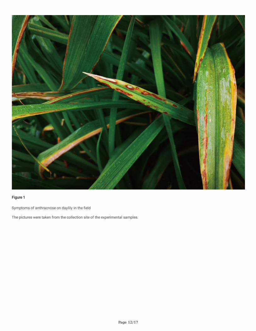
Shanghai Institute of Technology, as the sample collection site of this experiment, has 33,000 square meters of daylily planting area, wherethe incidence of anthracnose reach 25 %. Based on �eld observations, anthracnose symptoms on daylily leaves were recorded. The initialstage of leaf disease was reddish-brown lesions with a yellow halo, which later expanded into reddish brown stripes. If the disease spotsoccurred at the tip of the leaf, the tip died and then spread down the vein to kill the upper part of the leaf. If the disease spots occurred in theleaf margin, the leaves were twisted and deformed, and the leaves changed from green to greyish yellow. The junction of the disease wasreddish brown, and the center was gray-brown. When the disease was serious, many disease spots connected into large spots, whicheventually led to the death of the whole leaf and dense black spots on the leaf surface (Figure 1).
Morphological characterization
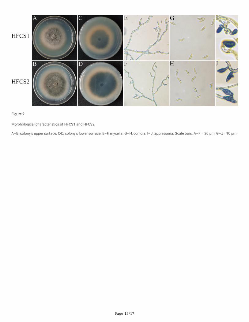
Two isolates were obtained by the tissue separation method combined with the single spore separation method. These isolates were namedHFCS1 and HFCS2. After 7 d of culture on PDA medium at 25 ± 1 ℃, the two isolates had similar morphology. They produced colonies withuniform edges. The colonies appeared felted with aerial mycelia that were light gray to grayish white on the upper surface and dark gray toolive gray to light gray on the lower surface, with a cottony texture, high density, and low presence of sectors (Figure 2A–D). The hyphaeproduced were initially pale yellow in color, smooth, and cylindrical at the bases with rounded apexes. Later mycelia were twisted, branchedmore, separated, and were pale in color (Figure 2E, F). The average mycelial growth rate of HFCS1 was 9.3 ± 2.4 mm day-1, while that ofHFCS2 was 10.5 ± 3.1 mm day-1. Conidia were both hyaline, aseptate, curved or slightly curved, and had a round or somewhat acute apex andbase (Figure 2G, H). The conidia of HFCS1 had lengths of 15.5–21.6 (M ± SD = 19.1 ± 4.71) µm, widths of 3.6–5.4 (4.7 ± 0.42) µm, andlength/width (L/W) ratios of 3.34–5.61, while those of HFCS2 had lengths of 12.3–25.5 (M ± SD = 20.1 ± 3.16) µm, widths of 4.2–5.7 (5.19 ±0.51) µm, and length/width (L/W) ratios of 2.87–5.89. The conidia of HFCS1 were more elongated than those of HFCS2. The appressoria of
Page 5/17
two isolates were both solitary, mostly simple, rarely lobed, and dark brown in color. The appressoria of HFCS1 were ovate (Figure 2I), whilethose of HFCS2 were clavate (Figure 2J).
The morphological characteristics of the two isolates were similar to those of the species in the C. spaethianum, C. graminicola, andC. truncatum species complexes, which have curved and falcate conidia and ovoid, dark brown spores (Du et al., 2005; Bonacci et al., 2021).However, this was not enough to distinguish the Colletotrichum species of the two isolates based on morphology alone, and furtheridenti�cation was needed.
Phylogenetic analysis
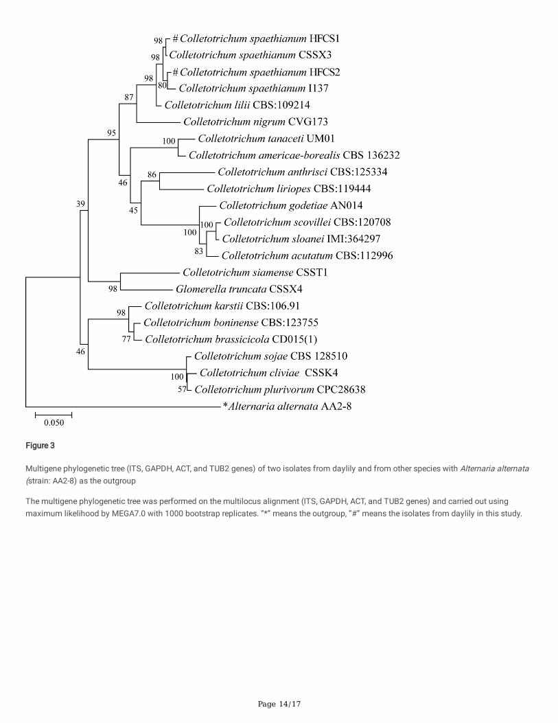
Approximately 591-bp (HFCS1) and 550-bp (HFCS2) DNA fragments were obtained using the primer pairs ITS1 and ITS4. In addition, PCRampli�cation and sequencing were carried out on the GAPDH gene with about 262-bp and 264-bp sequences, the ACT gene with 234-bp and247-bp sequences, and the TUB2 gene with 755-bp and 606-bp sequences for HFCS1 and HFCS2, respectively. The sequence analysis showsthat HFCS1 and HFCS2 could only be distinguished by TUB2 sequences and not by ITS, GAPDH or CHS-1 sequences. The similarity betweenthe two isolates and the reference sequence of C. spaethianum was greater than 97 %. The multigene phylogenetic tree was constructed byMEGA software with A. alternata (strain: AA2-8) as the outgroup based on multi-gene phylogenetic analyses (ITS, GAPDH, ACT and TUB2genes). The two isolates isolated in this study were closely related to C. spaethianum on the same branch (Figure 3).
Pathogenicity evaluation
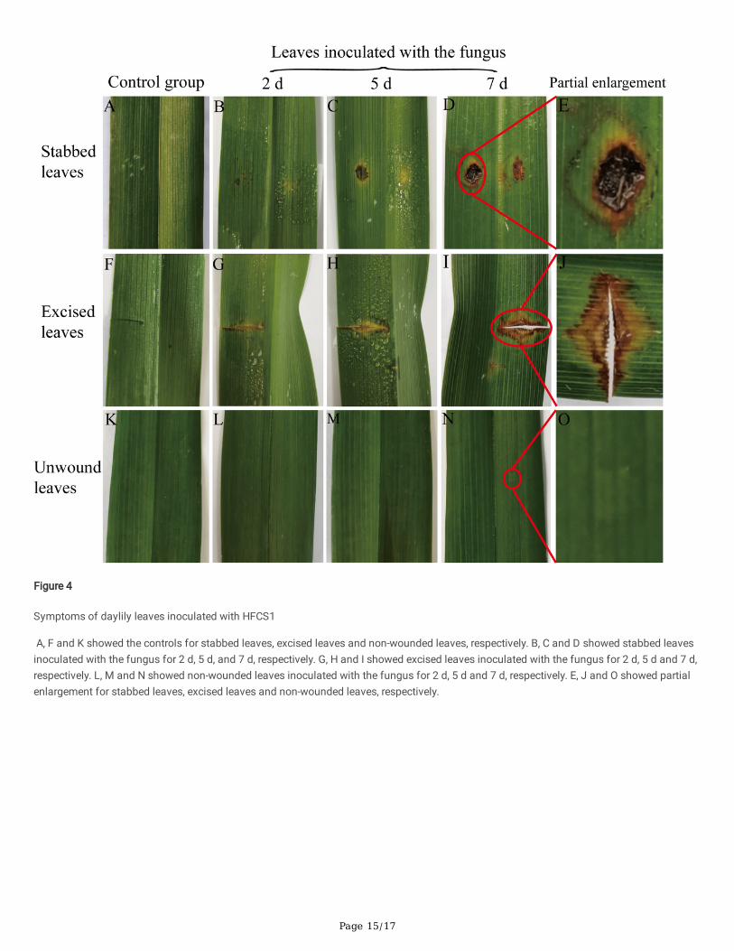
After inoculation on wounded daylily leaves, all the leaves showed anthracnose on inoculated leaves, which exhibited symptoms similar tothe symptoms of leaf samples collected from �eld (Figure 1). Initially, the stabbed leaves showed small and yellowish halo symptoms afterinoculation for 2 d (Figure 4B). Five days after inoculation, the water-soaked spots appeared that expanded rapidly and irregularly (Figure 4C).Seven days after inoculation, as the disease progressed, the enlarged spots merged, and severe leaf blight with concentric black rings formedin the enlarged lesions (Figure 4D, E). The symptoms of excised leaves were similar to those of stabbed leaves in the early stage (Figure 4G–H), but the two isolates exhibited higher aggressiveness in stabbed leaves (Figure 4I–J). There were no symptoms in the non-wounded leavesinoculated with the fungus (Figure 4L–O). None of the controls showed symptoms (Figure 4A, F, K). The symptoms caused by HFCS1 andHFCS2 were similar, although the symptoms caused by HFCS2 spread faster and caused larger wounds (on average, each wound was 0.2 cmlarger 7 d after the inoculation).
The typical symptoms produced by arti�cial inoculation con�rmed with the original symptoms under natural conditions, and themorphological characteristics and ITS sequences of the re-isolated fungi were consistent with those of the �rst isolated fungi. Therefore,Koch’ s postulates were con�rmed. The results proved that the inoculated fungus was the pathogen of daylily anthracnose. Combined withmorphological characteristics, multigene phylogenetic analysis, and pathogenicity analysis, the two fungal isolates from daylily wereidenti�ed as C. spaethianum.
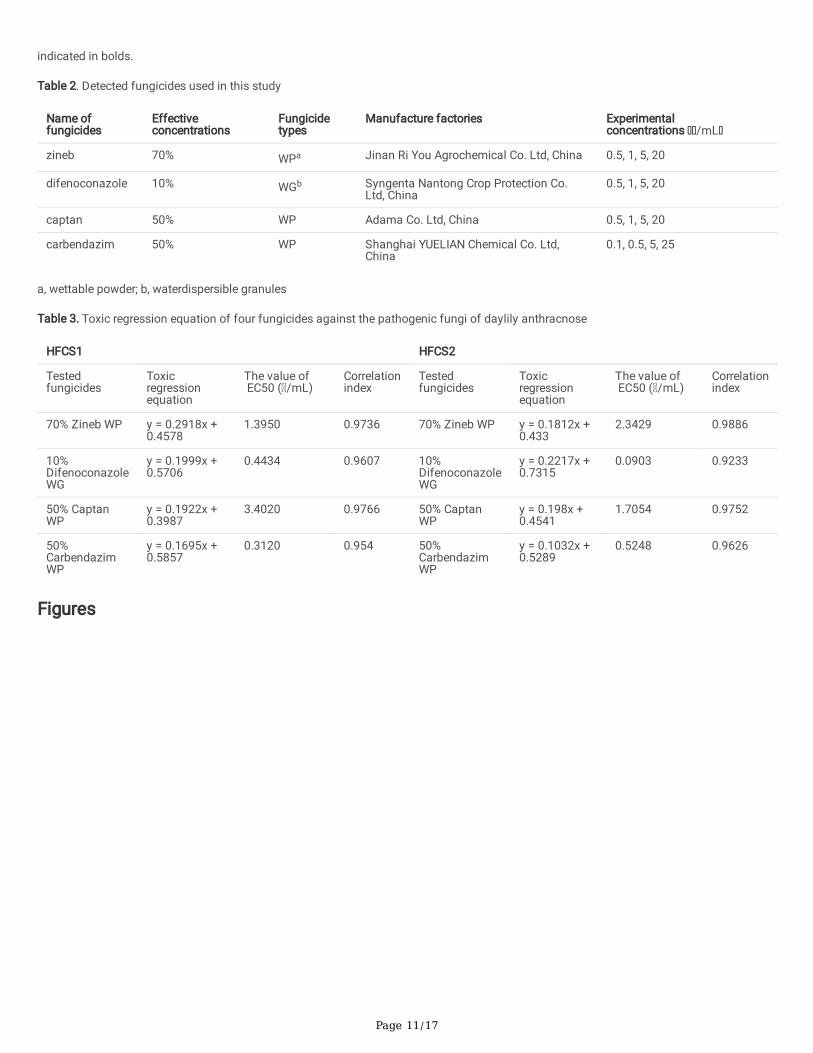
Evaluation of fungicide susceptibility
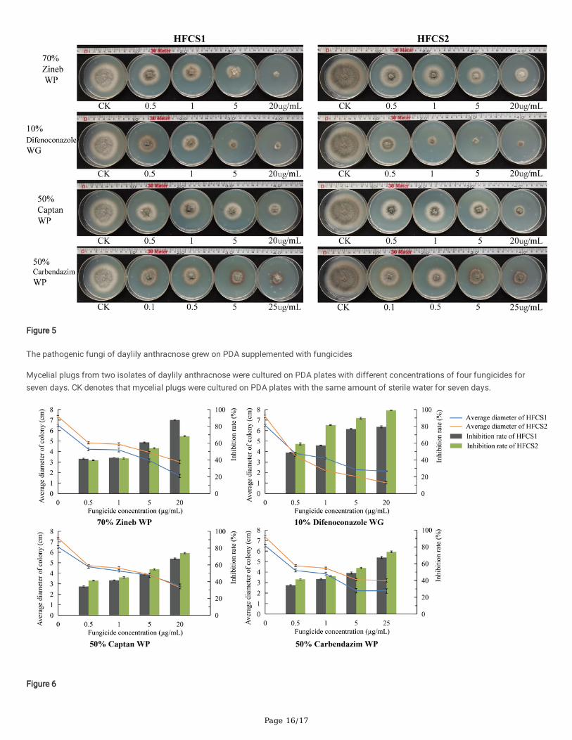
The effect of fungicides was assessed on the qualitative growth of mycelium after the inoculation of mycelial plug on in PDA medium for 7 d.The results showed that all four fungicides had inhibitory effects on the two isolates from daylily (Figure 5–6). The EC50 of mycelial growthwas calculated to determine the level of fungicide sensitivity of the pathogen (Table 3). Difenoconazole and carbendazim presentedremarkable antifungal activity against the two isolates tested with EC50 values lower than 1 μg/mL. The inhibitory effects of zineb andcaptan were the second-higher, with EC50 values higher than 1 μg/mL and lower than 5 μg/mL. From the control groups of the two isolates,the mycelial growth rate of HFCS2 was higher than that of HFCS1, and the EC50 values of HFCS2 treated with zineb were 1.6 times as muchas those of HFCS1. However, the EC50 value of HFCS2 treated with captan was half as much as that of HFCS1, and the EC50 value of HFCS1treated with difenoconazole of HFCS1 was 4.9 times as much as that of HFCS2. The results showed that the susceptibility of the two isolatesfrom daylily to four fungicides was signi�cantly different.
DiscussionAnthracnose causes extensive yield losses in many plants, including daylily, especially in warm and rainy seasons. In thisinvestigation, daylily with suspected anthracnose disease symptoms were sampled from the Shanghai Institute of Technology, Shanghai,China. The incidence of anthracnose, along with other diseases such as rust disease, is particularly prevalent during the rainy season.
According to the standard of morpho-taxonomic criteria, the fungal isolates in this study were classi�ed as Colletotrichum species. Themorphological characteristics of them were similar to those of the species in the C. spaethianum, C. graminicola, and C. truncatum speciescomplexes. Then the two isolates were further identi�ed by the analysis of restricted polymorphisms in the DNA ampli�cation region.
Page 6/17
According to the result of sequence analysis, the ITS, GAPDH and CHS-1 sequences of HFCS1 and HFCS2 were not signi�cantly different, thesequence similarity was up to 97 %. And the TUB2 sequence (77 % sequence similarity) was the one that really distinguishes the two isolates.The multigene phylogenetic tree based on multi-locus genes including ITS, GAPDH, CHS-1, and TUB2 sequences provided a good method ofdistinguishing of the two isolates, and they were clustered together with C. spaethianum with high bootstrap support (98 % of HFCS1 and C.spaethianum CCSX3, 80 % of HFCS2 and C. spaethianum I137).
Pathogenicity tests of two isolates from daylily in this study showed that the isolates were pathogenic on daylily leaves after wounding thesurface, but they were produced no apparent infection on non-wounded leaves. This might illustrate the importance of the cuticle acting as abarrier to infection by C. spaethianum Alkan et al., 2015; Fukada et al., 2019). Combined with morphological characteristics, multi-genephylogenetic analysis, and pathogenicity tests, the two isolates from daylily leaves were identi�ed as C. spaethianum.
C. spaethianum is a very common causal agent of anthracnose worldwide, and it has been found in a number of other hosts, including Liliumbrownii, Allium ledebourianum, and Polygonatum cyrtonema (Yang et al., 2009; Salunkhe et al., 2018; Ma et al., 2020). In addition toanthracnose, C. spaethianum has also been identi�ed as a causal agent of leaf spot (Guo et al., 2013; Zhong et al., 2019; Sun et al., 2020a).The present study identi�ed C. spaethianum isolates as the pathogenic fungi of daylily anthracnose after the species was previously reportedin Brazil (Vieira et al., 2014). However, none of the systematic studies of the disease have been conducted. This survey focused on thepathogenic fungi causing anthracnose-like symptoms in �elds, and the two isolates collected in the current study could have been primary orsecondary invaders. The isolates tested in this study were con�rmed to be capable of causing anthracnose-like symptoms on woundedleaves of daylily, but no obvious symptoms were shown on non-wounded leaves. This may indicated that the two fungal isolates in this studywere wound pathogens on daylily.
For the effective development and deployment of host resistance, and the reduction of fungicide application and risks to the environment, it isnecessary to understand the sensitivity of the pathogens causing daylily anthracnose to those fungicides commonly used in daylily diseasecontrol. Zineb, captan, difenoconazole, and carbendazim are all commonly used in production, these compounds are currently labeled for thecontrol of anthracnose (Patiño-Vera et al., 2005; Abbott and Beckerman, 2018; Zhang et al., 2020b; Wang et al., 2020). In this study, the two C.spaethianum isolates from daylily leaves showed different sensitivity to zineb, difenoconazole, captan and carbendazim, with EC50 valuesranging from 0.0903 to 3.402 µg/mL. The EC50 values of C. spaethianum varied greatly among the fungicides, which should be furtherinvestigated to improve the e�cacy of daylily anthracnose management strategies.
In this study, based on the resistance ratio and EC50 values, difenoconazole and carbendazim would be expected to provide better controlthan zineb and captan, and the sensitivity of HFCS1 to carbendazim was higher than that of difenoconazole with a 0.13 difference in EC50
values. The sensitivity of HFCS2 to difenoconazole was 5.81 times higher than its sensitivity to carbendazim. Studies showed thatColletotrichum is highly sensitive to carbendazim, with EC50 value lower than 10 μg/mL (Kim et al., 2020; Wang et al., 2020; Zhong et al.,2021). However, study of Gama et al. (2020) showed that there is no relationship between C. acutatum inhibition and fungicide concentration,and EC50 values for carbendazim could not be determined. The above results indicate that the susceptibility of Colletotrichum to fungicides isprobably related to species, and there are differences among different strains from the same species.
DeclarationsAcknowledgements
This study was supported by Science and Technology agriculture Project of Shanghai (No. 2019-02-08-00-08-F01107), funded by ShanghaiAgricultural Commission. We thank LetPub (www.letpub.com) for its linguistic assistance during the preparation of this manuscript.
Author contributions
QQ was responsible for the guidance of experiments, the revision of manuscript, the purchase of experimental samples and the supervision ofexperiments.. DH was responsible for the designed and performed of experiments, carried out data analysis and wrote manuscript. ML wasresponsible for the culture of experimental samples.
Data availability
DNA sequences used in this present study are available in GenBank.
Con�ict of Interest
The authors declare no con�ict of interest in this study.
Page 7/17
Ethics approval
Not applicable
Con�ict of interest
The authors declare no competing interests.
Open Access
This article is licensed under a Creative Commons Attribution 4.0 International License, which permits use, sharing, adaptation, distributionand reproduction in any medium or format, as long as you give appropriate credit to the original author(s) and the source, provide a link to theCreative Commons licence, and indicate if changes were made. The images or other third party material in this article are included in thearticle's Creative Commons licence, unless indicated otherwise in a credit line to the material. If material is not included in the article's CreativeCommons licence and your intended use is not permitted by statutory regulation or exceeds the permitted use, you will need to obtainpermission directly from the copyright holder. To view a copy of this licence, visit http://creativecommons.org/licenses/by/4.0/.
References1. Abbott, C.P., & Beckerman, J.L. (2018). Incorporating adjuvants with captan to manage common apple diseases. Plant Disease,
102(1),231-236. https://doi.org/10.1094/PDIS-05-17-0629-RE
2. Afanador-Kafuri, L., González, A., Gañán, L., Mejía, J.F., Cardona,, N., & Alvarez, E. (2014). Characterization of the Colletotrichum speciescausing anthracnose in Andean Blackberry in Colombia. Plant Disease, 98(11),1503-1513. https://doi.org/10.1094/PDIS-07-13-0752-RE
3. Alkan, N., Friedlander, G., Ment, D., Prusky, D., & Fluhr, R. (2015). Simultaneous transcriptome analysis of Colletotrichum gloeosporioidesand tomato fruit pathosystem reveals novel fungal pathogenicity and fruit defense strategies. New Phytologist, 205(2),801-15.https://doi.org/10.1111/nph.13087
4. Arikan, S. (2007). Current status of antifungal susceptibility testing methods. Medical Mycology, 45(7),569-87.https://doi.org/10.1080/13693780701436794
5. Bai, Q.R., Han, S., Xie, Y.Y., Dong, R., Gao, J., & Li, Y. (2012). First report of Daylily leaf streak caused by Kabatiella microsticta in China.Plant Disease, 96(10),1579. https://doi.org/10.1094/PDIS-04-12-0381-PDN
�. Barimani, M., Pethybridge, S.J., Vaghe�, N., Hay, F.S., & Taylor, P.W.J. (2013). A new anthracnose disease of pyrethrum caused byColletotrichum tanaceti sp. Nov, Plant Pathology, 62(6),1248-1257. https://doi.org/10.1111/ppa.12054
7. Bonacci, M., Formento, Á.N., Morales, M.C., Orlando, J., Ibáñez, F., Sartori, M., Etcheverry, M., Nesci, A., & Barros, G. (2021). Conyzabonariensis as an alternative host for Colletotrichum species in Argentina. Journal of Applied Microbiology, 130(5),1656-1670.https://doi.org/10.1111/jam.14879
�. Buck, J.W. (2013). Identi�cation of pathotypes in the daylily rust pathogen Puccinia hemerocallidis. Journal of Phytopathology, 161(11-12),784-790. https://doi.org/10.1111/jph.12134
9. Byng, J.W., Chase, M.W., Fay, M.F., Other members of the angiosperm phylogeny group. (2016). An update of the angiosperm phylogenygroup classi�cation for the orders and families of �owering plants: APG IV. Botanical Journal of the Linnean Society, 181(1),1-20.https://doi.org/10.1111/boj.12385
10. Crouch, J. A., Tredway, L.P., Clarke, B.B., & Hillman, B.I. (2009). Phylogenetic and population genetic divergence correspond with habitatfor the pathogen Colletotrichum cereale and allied taxa across diverse grass communities. Molecular Ecology, 18(1),123-35.https://doi.org/10.1111/j.1365-294X.2008.04008.x
11. Cara, M., Iliadi, M.K., Lagogianni, C.S., Paplomatas, E., Merkuri, J., & Tsitsigiannis, D.I. (2020). First report of Colletotrichum acutatumcausing anthracnose on olives in Albania. Plant Disease, 14,495-499. https://doi.org/10.1094/PDIS-04-20-0774-PDN
12. Carbone I., & Kohn L.M.(1999.) A method for designing primer sets for speciation studies in �lamentous ascomycetes. Mycologia,91(3),553-556. https://doi.org/
13. Damm, U., Cannon, P.F., Woudenberg, J.H., & Crous, P.W. (2012). The Colletotrichum acutatum species complex. Studies in Mycology,73(1),37-113. https://doi.org/10.3114/sim0010
14. Damm, U., Woudenberg, J.H.C., Cannon, P.F., & Crous, P.W. (2009). Colletotrichum species with curved conidia from herbaceous hosts.Fungal Diversity, 39,45-87.
15. Damm, U., O'Connell, R.J., Groenewald, J.Z., & Crous, P.W. (2014). The Colletotrichum destructivum species complex - hemibiotrophicpathogens of forage and �eld crops. Studies in Mycology, 79,49-84. https://doi.org/10.1016/j.simyco.2014.09.003
Page 8/17
1�. Du, M., Schardl, C.L., Nuckles, E.M., & Vaillancourt, L.J. (2005). Using mating-type gene sequences for improved phylogenetic resolutionof Colletotrichum species complexes. Mycologia, 97(3), 641-658. https://doi.org/10.1080/15572536.2006.11832795
17. Elder, J.F., & Turner, B.J. (1995). Concerted evolution of repetitive DNA sequences in eukaryotes. Quarterly Review of Biology, 70(3), 297-320. https://doi.org/10.1086/419073
1�. Fukada, F., Kodama, S., Nishiuchi, T., Kajikawa, N., & Kubo, Y. (2019). Plant pathogenic fungi Colletotrichum and Magnaporthe share acommon G1 phase monitoring strategy for proper appressorium development. New Phytologist, 222(4),1909-1923.https://doi.org/10.1111/nph.15728
19. Gama, A.B., Baggio, J.S., Rebello, C.S., Lourenço, S.A., Gasparoto, M.C.G., da Silva Junior, G.J., Peres, N.A., & Amorim, L. (2020). Sensitivityof Colletotrichum acutatum isolates from citrus to carbendazim, difenoconazole, tebuconazole, and tri�oxystrobin. Plant Disease,104(6),1621-1628. https://doi.org/10.1094/PDIS-10-19-2195-RE
20. Gao, J., Zeng, G.P., & Song, L.S. (2020a). Identi�cation and biological characteristics of pathogen causing leaf spot disease ofHemerocallis fulva. Acta Horticulturae Sinica, 47(01),169-178. In Chinese. https://doi.org/10.16420/j.issn.0513-353x.2019-0343
21. Gao, Y.Y., Li, X.X., He, L.F., Li, B.X., Mu, W., & Liu, F. (2020b). Effect of Pyrisoxazole on Colletotrichum scovillei Infection and Anthracnoseon Chili. Plant Disease, 104(2),551-559. https://doi.org/10.1094/PDIS-06-19-1291-RE
22. Gardes, M., & Bruns, T.D. (1993). ITS primers with enhanced speci�city for basidiomycetes--application to the identi�cation ofmycorrhizae and rusts. Molecular. Ecology, 2(2),113-8. https://doi.org/10.1111/j.1365-294X.1993.tb00005.x
23. Glass, N.L., & Donaldson, G.C. (1995). Development of primer sets designed for use with the PCR to amplify conserved genes from�lamentous ascomycetes. Applied & Environmental Microbiology, 61(4),1323-30. https://doi.org/10.1002/bit.260460112
24. Guarnaccia, V., Gilardi, G., Martino, I., Garibaldi, A., & Gullino, M.L. (2019). Species diversity in Colletotrichum causing anthracnose ofaromatic and ornamental lamiaceae in Italy. Agronomy, 9(10),613-628. https://doi.org/10.3390/agronomy9100613
25. Guerber, J.C., Liu, B., Correll, J.C., & Johnston, P.R. (2003). Characterization of diversity in Colletotrichum acutatum sensu lato bysequence analysis of two gene introns, mtDNA and intron RFLPs, and mating compatibility. Mycologia, 95(5),872-95.https://doi.org/10.1080/15572536.2004.11833047
2�. Guo, M., Pan, Y.M., Dai, Y.L., & Gao, Z.M. (2013). First report of leaf spot caused by Colletotrichum spaethianum on Peucedanumpraeruptorum in China. Plant Disease, 97(10),1380. https://doi.org/10.1094/PDIS-03-13-0246-PDN
27. Hu, S., & Zhang, J.Z. (2009). Colletotrichum destructivum from cowpea infecting Arabidopsis thaliana and its identity to C. higginsianum.European Journal of Plant Pathology, 125(3),459-469. https://doi.org/10.1007/s10658-009-9495-2
2�. Jayawardena, R.S., Hyde, K.D., Damm, U., Cai, L., & Yan J.Y. (2016). Notes on currently accepted species of Colletotrichum.Mycosphere, 7(8), 1192-1260. https://doi.org/10.5943/mycosphere/si/2c/9
29. Kim, C.H., Hassan, O., & Chang, T. (2020). Diversity, pathogenicity, and fungicide sensitivity of Colletotrichum species associated withapple anthracnose in South Korea. Plant Disease. 104(11),2866-2874. https://doi.org/10.1094/PDIS-01-20-0050-RE
30. Li, Y., Ma, X., Gai, W., Xiao, L., & Gong, Z. (2021). First report of Colletotrichum gloeosporioides causing anthracnose on pepper in ShaanxiProvince, China. Plant Disease, 23,1-4. https://doi.org/10.1094/PDIS-01-21-0123-PDN
31. Liang, H. J., Di, Y.L., Li, J.L., & Zhu, F.X. (2015). Baseline sensitivity and control e�cacy of �uazinam against Sclerotinia sclerotiorum.European Journal of Plant Pathology, 142(4),691-699. https://doi.org/10.1007/s10658-015-0644-5
32. Liu, F., Tang, G., Zheng, X., Li, Y., Sun, X., Qi, X., Zhou, Y., Xu, J., Chen, H., Chang, X., Zhang, S., & Gong, G. (2016). Molecular and phenotypiccharacterization of Colletotrichum species associated with anthracnose disease in peppers from Sichuan province, China. Scienti�cReports, 6,32761. https://doi.org/10.1038/srep32761
33. Liu, X., Zheng, X., Khaskheli, M.I., Sun, X., Chang, X., & Gong, G. (2020). Identi�cation of Colletotrichum species associated with blueberryanthracnose in Sichuan, China. Pathogens, 9(9),718. https://doi.org/10.3390/pathogens9090718
34. Ma, H.X., Feng, X.J., Chen, Y., Chen, C.J., & Zhou, M.G. (2009). Occurrence and characterization of dimethachlon insensitivity in Sclerotiniasclerotiorum in Jiangsu province of China. Plant Disease, 93(1),36-42. https://doi.org/10.1094/PDIS-93-1-0036
35. Ma, J., Xiao, X., Wang, X., & Guo, M. (2020). Colletotrichum spaethianum causing anthracnose on Polygonatum cyrtonema in Anhuiprovince, China. Plant Disease. 20,509. https://doi.org/10.1094/PDIS-04-20-0778-PDN
3�. Moriwaki, J., Tsukiboshi, T., & Sato, T. (2002). Grouping of Colletotrichum species in Japan based on rDNA sequences. Journal of GeneralPlant Pathology, 68(4),307-320. https://doi.org/10.1007/PL00013096
37. Mostert, L., Bester, W., Coertze, S., & Wood, A.R. (2008). First report of daylily rust caused by Puccinia hemerocallidis in the western capeprovince of South Africa. Plant Disease, 92(7),1133. https://doi.org/10.1094/PDIS-92-7-1133A
3�. O'Connell, R.J., Thon, M.R., Hacquard, S., Amyotte, S.G., Kleemann, J., Torres, M.F., Damm, U., Buiate, E.A., Epstein, L., Alkan, N., Altmüller,J., Alvarado-Balderrama, L., Bauser, C.A., Becker, C., Birren, B.W., Chen, Z., Choi, J., Crouch, J.A., Duvick, J.P., Farman, M.A., & Vaillancourt,
Page 9/17
L.J. (2012). Lifestyle transitions in plant pathogenic Colletotrichum fungi deciphered by genome and transcriptome analyses. NatureGenetics, 44(9),1060-1065. https://doi.org/10.1038/ng.2372
39. Patiño-Vera, M., Jiménez, B., Balderas, K., Ortiz, M., Allende, R., Carrillo, A., & Galindo, E. (2005). Pilot-scale production and liquidformulation of Rhodotorula minuta, a potential biocontrol agent of mango anthracnose. Journal of Applied Microbiology, 99(3),540-50.https://doi.org/10.1111/j.1365-2672.2005.02646.x
40. Salunkhe, V.N., Anandhan, S., Gawande, S.J., Ikkar, R.B., & Singh, M. (2018). First report of anthracnose caused by Colletotrichumspaethianum on Allium ledebourianum in India. Plant Disease. 102(10). https://doi.org/10.1094/PDIS-01-18-0104-PDN
41. Schoch, C.L., Seifert, K.A., Huhndorf, S., Robert, V., Spouge, J.L., Levesque, C.A., Chen, W., Fungal Barcoding Consortium, Fungal BarcodingConsortium Author List. (2012). Nuclear ribosomal internal transcribed spacer (ITS) region as a universal DNA barcode marker for Fungi.Proceedings of the National Academy of ences of the United States of America, 109(16),6241-6246.https://doi.org/10.1073/pnas.1117018109
42. Sharma, G., Pinnaka, A.K., & Shenoy, B.D. (2015). Resolving the Colletotrichum siamense species complex using ApMat marker. FungalDiversity, 71(1),247-264. https://doi.org/10.1007/s13225-014-0312-7
43. Silva, D.D., Groenewald, J.Z., Crous, P.W., Ades, P.K., Nasruddin, A., Mongkolporn, O., & Taylor, P.W.J. (2019). Identi�cation, prevalence andpathogenicity of Colletotrichum species causing anthracnose of Capsicum annuum in Asia. IMA Fungus, 10(1),1-32.https://doi.org/10.1186/s43008-019-0001-y
44. Sun, H., Sun, L., Hong, Y., & Liang, Y. (2020a). First report of anthracnose on Hosta ventricosa caused by Colletotrichum spaethianum inChina. Crop Protection, 131,105-104. https://doi.org/10.1016/j.cropro.2020.105104
45. Sun, X., Qi, X., Wang, W., Liu, X., Zhao, H., Wu, C., Chang, X., Zhang, M., Chen, H., & Gong, G. (2020b). Etiology and symptoms of maize leafspot caused by Bipolaris spp. in Sichuan, China. Pathogens, 9(3),229-247. https://doi.org/10.3390/pathogens9030229
4�. Sutton, B.C. (1992). The genus Glomerella and its anamorph Colletotrichum. Colletorichum biology, pathology and control, 1992:1-26.
47. Vieira, W.A.S., Michereff, S.J., Oliveira, A.C., Santos, A., & Câmara, M.P.S. (2014). First report of anthracnose caused by Colletotrichumspaethianum on Hemerocallis �ava in Brazil. Plant Disease. 98(7),997. https://doi.org/10.1094/PDIS-10-13-1026-PDN
4�. Wang, Q.H., Fan, K., Li, D.W., Han, C.M., Qu, Y.Y., Qi, Y.K., & Wu, X.Q. (2020). Identi�cation, virulence and fungicide sensitivity ofColletotrichum gloeosporioides s.s. responsible for walnut anthracnose disease in China. Plant Disease. 104(5), 1358-1368.https://doi.org/10.1094/PDIS-12-19-2569-RE
49. Wang, Q.T., Liu, F., Hou, C.L., & Cai, L. (2021). Species of Colletotrichum on bamboos from China. Mycologia, 113(2), 450-458.https://doi.org/10.1080/00275514.2020.1837567
50. Weir, B.S., Johnston, P.R., & Damm, U. (2012). The Colletotrichum gloeosporioides species complex. Studies in Mycology, 73(1), 115–180.https://doi.org/10.3114/sim0011
51. Yang, Y.L., Liu, Z.Y., Cai, L., Hyde, K.D., & Ehc, M.K. (2009). Colletotrichum anthracnose of amaryllidaceae. Fungal Diversity, 39(2), 123-146.
52. Zhang, J., Kang, Y., Xu, X., Xu, T., Li, C., Chen, K., & Cao, H. (2020a). First report of leaf spot caused by Alternaria alternata on Sonchusasper in china. Plant Disease, 105(2),1-5. https://doi.org/10.1094/PDIS-05-19-0940-PDN
53. Zhang, L., Song, L., Xu, X., Zou, X., Duan, K., & Gao, Q. (2020b). Characterization and fungicide sensitivity of Colletotrichum speciescausing strawberry anthracnose in Eastern China. Plant Disease, 104(7), 1960-1968. https://doi.org/10.1094/PDIS-10-19-2241-RE
54. Zhong, J., Li, C.X., Zhong, S.Y., & Hu, Z. (2019). First report of leaf spot caused by Colletotrichum spaethianum on Paris polyphylla inchina. Plant Disease, 104(3). https://doi.org/10.1094/PDIS-09-19-1844-PDN
55. Zhong, S., Miao, J., Liu, X., & Zhang, G. (2021). Characterization of Colletotrichum spp. sensitivity to carbendazim for isolates causingstrawberry anthracnose in China. Plant Disease, 105(1),87-95. https://doi.org/10.1094/PDIS-04-20-0875-RE
5�. Zhou, Y., Gong, G., Cui, Y., Zhang, D., Chang, X., Hu, R., Liu, N., & Sun, X. (2015). Identi�cation of Botryosphaeriaceae species causingkiwifruit rot in Sichuan province, China. Plant Disease, 99(5), 699-708. https://doi.org/10.1094/PDIS-07-14-0727-RE
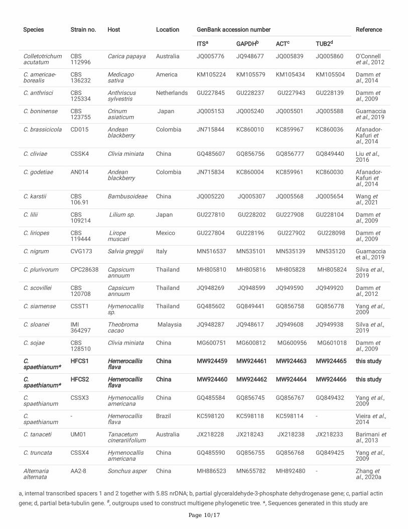
TablesTable 1. Strain details of Colletotrichum species and outgroup used in the multi-gene phylogenetic analysis
Page 10/17
Species Strain no. Host Location GenBank accession number Reference
ITSa GAPDHb ACTc TUB2d
Colletotrichumacutatum
CBS112996
Carica papaya Australia JQ005776 JQ948677 JQ005839 JQ005860 O'Connellet al., 2012
C. americae-borealis
CBS136232
Medicagosativa
America KM105224 KM105579 KM105434 KM105504 Damm etal., 2014
C. anthrisci CBS125334
Anthriscussylvestris
Netherlands GU227845 GU228237 GU227943 GU228139 Damm etal., 2009
C. boninense CBS123755
Crinumasiaticum
Japan JQ005153 JQ005240 JQ005501 JQ005588 Guarnacciaet al., 2019
C. brassicicola CD015 Andeanblackberry
Colombia JN715844 KC860010 KC859967 KC860036 Afanador-Kafuri etal., 2014
C. cliviae CSSK4 Clivia miniata China GQ485607 GQ856756 GQ856777 GQ849440 Liu et al.,2016
C. godetiae AN014 Andeanblackberry
Colombia JN715834 KC860004 KC859961 KC860030 Afanador-Kafuri etal., 2014
C. karstii CBS106.91
Bambusoideae China JQ005220 JQ005307 JQ005568 JQ005654 Wang etal., 2021
C. lilii CBS109214
Lilium sp. Japan GU227810 GU228202 GU227908 GU228104 Damm etal., 2009
C. liriopes CBS119444
Liropemuscari
Mexico GU227804 GU228196 GU227902 GU228098 Damm etal., 2009
C. nigrum CVG173 Salvia greggii Italy MN516537 MN535101 MN535139 MN535120 Guarnacciaet al., 2019
C. plurivorum CPC28638 Capsicumannuum
Thailand MH805810 MH805816 MH805828 MH805824 Silva et al.,2019
C. scovillei CBS120708
Capsicumannuum
Thailand JQ948269 JQ948599 JQ949590 JQ949920 Damm etal., 2012
C. siamense CSST1 Hymenocallissp.
Thailand GQ485602 GQ849441 GQ856758 GQ856778 Yang et al.,2009
C. sloanei IMI364297
Theobromacacao
Malaysia JQ948287 JQ948617 JQ949608 JQ949938 Silva et al.,2019
C. sojae CBS128510
Clivia miniata China MG600751 MG600812 MG600956 MG601018 Damm etal., 2009
C.spaethianum*
HFCS1 Hemerocallis�ava
China MW924459 MW924461 MW924463 MW924465 this study
C.spaethianum*
HFCS2 Hemerocallis�ava
China MW924460 MW924462 MW924464 MW924466 this study
C.spaethianum
CSSX3 Hymenocallisamericana
China GQ485584 GQ856745 GQ856767 GQ849432 Yang et al.,2009
C.spaethianum
- Hemerocallis�ava
Brazil KC598120 KC598118 KC598114 - Vieira et al.,2014
C. tanaceti UM01 Tanacetumcinerariifolium
Australia JX218228 JX218243 JX218238 JX218233 Barimani etal., 2013
C. truncata CSSX4 Hymenocallisamericana
China GQ485590 GQ856755 GQ856768 GQ849425 Yang et al.,2009
Alternariaalternata
AA2-8 Sonchus asper China MH886523 MN655782 MH892480 - Zhang etal., 2020a
a, internal transcribed spacers 1 and 2 together with 5.8S nrDNA; b, partial glyceraldehyde-3-phosphate dehydrogenase gene; c, partial actingene; d, partial beta-tubulin gene. #, outgroups used to construct multigene phylogenetic tree. *, Sequences generated in this study are
Page 11/17
indicated in bolds.
Table 2. Detected fungicides used in this study
Name offungicides
Effectiveconcentrations
Fungicidetypes
Manufacture factories Experimentalconcentrations /mL
zineb 70% WPa Jinan Ri You Agrochemical Co. Ltd, China 0.5, 1, 5, 20
difenoconazole 10% WGb Syngenta Nantong Crop Protection Co.Ltd, China
0.5, 1, 5, 20
captan 50% WP Adama Co. Ltd, China 0.5, 1, 5, 20
carbendazim 50% WP Shanghai YUELIAN Chemical Co. Ltd,China
0.1, 0.5, 5, 25
a, wettable powder; b, waterdispersible granules
Table 3. Toxic regression equation of four fungicides against the pathogenic fungi of daylily anthracnose
HFCS1 HFCS2
Testedfungicides
Toxicregressionequation
The value of EC50 ( /mL)
Correlationindex
Testedfungicides
Toxicregressionequation
The value of EC50 ( /mL)
Correlationindex
70% Zineb WP y = 0.2918x +0.4578
1.3950 0.9736 70% Zineb WP y = 0.1812x +0.433
2.3429 0.9886
10%DifenoconazoleWG
y = 0.1999x +0.5706
0.4434 0.9607 10%DifenoconazoleWG
y = 0.2217x +0.7315
0.0903 0.9233
50% CaptanWP
y = 0.1922x +0.3987
3.4020 0.9766 50% CaptanWP
y = 0.198x +0.4541
1.7054 0.9752
50%CarbendazimWP
y = 0.1695x +0.5857
0.3120 0.954 50%CarbendazimWP
y = 0.1032x +0.5289
0.5248 0.9626
Figures
Page 12/17
Figure 1
Symptoms of anthracnose on daylily in the �eld
The pictures were taken from the collection site of the experimental samples.
Page 13/17
Figure 2
Morphological characteristics of HFCS1 and HFCS2
A–B, colony’s upper surface. C-D, colony’s lower surface. E–F, mycelia. G–H, conidia. I–J, appressoria. Scale bars: A–F = 20 µm, G–J= 10 µm.
Page 14/17
Figure 3
Multigene phylogenetic tree (ITS, GAPDH, ACT, and TUB2 genes) of two isolates from daylily and from other species with Alternaria alternata(strain: AA2-8) as the outgroup
The multigene phylogenetic tree was performed on the multilocus alignment (ITS, GAPDH, ACT, and TUB2 genes) and carried out usingmaximum likelihood by MEGA7.0 with 1000 bootstrap replicates. “*” means the outgroup, “#” means the isolates from daylily in this study.
Page 15/17
Figure 4
Symptoms of daylily leaves inoculated with HFCS1
A, F and K showed the controls for stabbed leaves, excised leaves and non-wounded leaves, respectively. B, C and D showed stabbed leavesinoculated with the fungus for 2 d, 5 d, and 7 d, respectively. G, H and I showed excised leaves inoculated with the fungus for 2 d, 5 d and 7 d,respectively. L, M and N showed non-wounded leaves inoculated with the fungus for 2 d, 5 d and 7 d, respectively. E, J and O showed partialenlargement for stabbed leaves, excised leaves and non-wounded leaves, respectively.
Page 16/17
Figure 5
The pathogenic fungi of daylily anthracnose grew on PDA supplemented with fungicides
Mycelial plugs from two isolates of daylily anthracnose were cultured on PDA plates with different concentrations of four fungicides forseven days. CK denotes that mycelial plugs were cultured on PDA plates with the same amount of sterile water for seven days.
Figure 6
Page 17/17
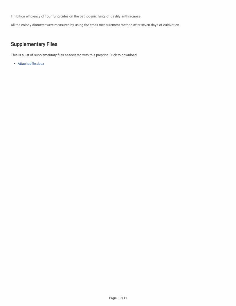
Inhibition e�ciency of four fungicides on the pathogenic fungi of daylily anthracnose
All the colony diameter were measured by using the cross measurement method after seven days of cultivation.
Supplementary Files
This is a list of supplementary �les associated with this preprint. Click to download.
Attached�le.docx
Related Documents