cHAPter one Ideal occlusion Question . A How would you classify this incisor relationship? B What are the other occlusal features you can see? C What are Andrew’s six keys to normal static occlusion? D What is the difference between an Angle’s and an Andrew’s class I molar relationship? Figure . From 'Illustrated Questions in Orthodontics' by Nightingale and Sandy, 2014, Copyright Oxford University Press

Welcome message from author
This document is posted to help you gain knowledge. Please leave a comment to let me know what you think about it! Share it to your friends and learn new things together.
Transcript

cHAPter oneIdeal occlusion
Question .A How would you classify this incisor relationship?B What are the other occlusal features you can see?C What are Andrew’s six keys to normal static occlusion?D What is the difference between an Angle’s and an Andrew’s class I molar
relationship?
Figure .
From 'Illustrated Questions in Orthodontics' by Nightingale and Sandy, 2014, Copyright Oxford University Press

42 Illustrated Questions in Orthodontics
Question 3.9This is a tracing of a cephalometric radiograph of a patient. The following values were obtained from the tracing:SNA = 8.5ºSNB = 93.5ºANB = –2ºMaxillary mandibular plane = 5ºOverjet = –2 mmUpper incisors to maxillary plane = 33ºLower incisors to mandibular plane = 80º
A Describe the skeletal relationship both vertically and anteroposteriorly.B Compare the incisor angulations to normal Caucasian values and account for
the differences.C Describe the overjet. Is this what you would expect to find in a patient with
this skeletal relationship?D Is the maxilla normally positioned?E Will this patient’s malocclusion be manageable by orthodontics alone?
Figure 3.9
From 'Illustrated Questions in Orthodontics' by Nightingale and Sandy, 2014, Copyright Oxford University Press
Pathology 55
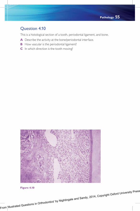
Question 4.0This is a histological section of a tooth, periodontal ligament, and bone.
A Describe the activity at the bone/periodontal interface.B How vascular is the periodontal ligament?C In which direction is the tooth moving?
Figure 4.0
From 'Illustrated Questions in Orthodontics' by Nightingale and Sandy, 2014, Copyright Oxford University Press

Removable appliances 63
Question 5.6A What is this appliance?B What is it suitable for?C What is the process for its manufacture and supply?D What additions aid tooth movement?E How much tooth movement can be achieved?F What instructions would you give for its use?G What adjunctive mechanics can you see?
Figure 5.6
From 'Illustrated Questions in Orthodontics' by Nightingale and Sandy, 2014, Copyright Oxford University Press

Functional appliances 69
Question 6.4A What is this?B When might it be useful?C How is it constructed and retained?
Figure 6.4
From 'Illustrated Questions in Orthodontics' by Nightingale and Sandy, 2014, Copyright Oxford University Press

80 Illustrated Questions in Orthodontics
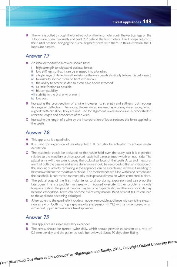
Question 7.9A What is this appliance called?B How often should the screw be turned and when should the patient be
reviewed after fitting?C Of what should the patient be warned?D What is the difference between the expansion achieved using this appliance
compared to other appliances?
Figure 7.9
From 'Illustrated Questions in Orthodontics' by Nightingale and Sandy, 2014, Copyright Oxford University Press

Anchorage 89
Question 8.6A What problem can you identify here?B How is the patient being managed?
Figure 8.6
From 'Illustrated Questions in Orthodontics' by Nightingale and Sandy, 2014, Copyright Oxford University Press

cHAPter oneIdeal occlusion
Answer .A Incisor relationship is classified as class I, II, or III, depending where the lower incisors
occlude relative to the cingulum plateau of the upper central incisors. This is a class I inci-sor relationship where the lower incisors occlude with or lie directly below the cingulum plateau of the upper central incisors. The overjet usually measures 2–4 mm.
B The cusp of the upper canine occludes in the embrasure between the lower canine and first premolar and this is called a class I canine relationship. The mesiobuccal cusp of the upper first molar occludes with the mesiobuccal groove of the lower first molar, which is an Angle’s class I molar relationship. There are no crossbites or scissorbites as the upper buccal segment teeth occlude just buccally to the opposing mandibular buccal teeth. Additionally, on maximum intercuspation, the mandibular condyles must be cor-rectly seated in the articular fossae.
C Andrew’s six keys to occlusion are based on his assessment of the study casts of 20 non-orthodontically created class I occlusions. He concluded that the six dental features common to all were:
i correct molar relationship ii correct crown angulation iii correct crown inclination iv tight interproximal contacts v absence of rotations vi a flat curve of Spee.
Additionally, tooth size must be correctly proportioned to achieve a perfect class I occlu-sion. Most of these features are visible in this photograph.
D An Angle’s class I molar relationship is such that the mesiobuccal cusp of the upper first molar occludes in the mesiobuccal groove of the lower first molar. To satisfy an Andrew’s class I molar relationship, the distal cusp of the upper first molar occludes with the mesial marginal ridge of the lower second molar, i.e. supra class I Angle relationship.
Answer .2A This photograph demonstrates canine guidance during lateral mandibular excursion.B This functional occlusal relationship is called the mutually protected occlusion (MPO) or
the canine guided occlusion.
From 'Illustrated Questions in Orthodontics' by Nightingale and Sandy, 2014, Copyright Oxford University Press

126 Illustrated Questions in Orthodontics
C There are no rigid guidelines to follow when considering space maintenance and it should be planned for the benefit of the individual. However, in general, space should be main-tained for avulsed upper central incisors and impacted canines following the loss of the deciduous canine. When deciduous molars are lost, a decision should be based on the child’s likely need for extractions of permanent teeth in the future. If they will not need extractions or will be severely crowded and need every millimetre of extraction space to treat the malocclusion, then space maintenance will be helpful to avoid further space loss. However, if extractions will be needed and the case is not severely crowded, then space maintenance is not necessary. Loss of deciduous molars is usually due to caries. If this is the case, long-term space maintenance in a caries-prone mouth may cause further harm.
Answer 3.8A A lateral cephalometric radiograph is a sagittal view of the patient’s skeletal and facial
structures taken in a standardized manner, with the patient’s head placed in a cephalostat. The patient is positioned 2 m from the X-ray beam and less than 0.3 m from the film, with the Frankfort plane parallel to the floor and the head held in position by the ear rods of the cephalostat. The mid-sagittal plane of the patient is parallel with the plane of the film and the patient must bite together in undisplaced centric occlusion. The central X-ray beam is perpendicular to the sagittal plane and is centred to the middle of the film. If these distances are varied, then the magnification of the final image is distorted. There is always some magnification, but this should be standardized at 5–2%. Some operators prefer to position the patient using natural head posture rather than the Frankfort plane, as this may allow greater reproducibility for subsequent images of the same patient.
B Lateral cephalometric radiographs supplement the clinical examination. They are used as a means of assessing the hard and soft tissue relationships of the patient’s facial struc-ture. The upper and lower incisor and interincisor angulation is also assessed. Various analyses have been created which compare the patient’s values to normal values for the appropriate population. This enables the clinician to assess the severity of the patient’s malocclusion compared with the normal population group and aids treatment planning. In addition to diagnosis and treatment planning, cephalometry may be used to predict the likely growth pattern of the patient and may also be used as a research tool.
C Care should be taken to minimize radiation dosage and ensure a good-quality film. As the image is taken, appropriate exposure and radiation dose settings should be used. Rare earth screens must be used to reduce the intensity of radiation needed to generate an image. An aluminium wedge between the patient and the X-ray tube will enhance the soft tissue profile by minimizing ‘burn out’ of the soft tissues. X-ray scatter can be reduced by placing a grid over the film cassette. However, this tends not to be needed with modern machines. Finally, careful developing techniques will maximize image quality, if film images are still in use. However, digital imaging has eliminated potential errors introduced during development.
Answer 3.9A Anteroposteriorly, the skeletal relationship is severely class III, demonstrated by the ANB
difference of –2º. The maxillary mandibular planes angle is 5º, which is significantly reduced compared to the norm of 27º and shows that the patient’s skeletal relationship is reduced in the vertical plane.
B The lower incisors are upright at 80º to the mandibular plane. The normal angulation is 93º +/– 5º. The upper incisors are very proclined at 33º relative to the norm of 09º +/– 5º. These normal values depend on which populations they are drawn from. In this case, Caucasian norms were appropriate, but this patient’s incisor angulations are
From 'Illustrated Questions in Orthodontics' by Nightingale and Sandy, 2014, Copyright Oxford University Press
Treatment planning 127
extreme for any population group. The incisors have tipped beyond normal values in an attempt to overcome the severe class III skeletal relationship. This is an example of den-toalveolar compensation.
C The overjet is reversed but the incisors are almost edge to edge; therefore, the reverse overjet is small. This is surprising in view of the severity of the underlying skeletal relation-ship and is because the incisors have attempted to compensate for this.
D The value for SNA is 8.5º and this is essentially normal, showing that the maxilla is cor-rectly positioned. Therefore, much of the skeletal III relationship can be attributed to a large mandible.
E The severity of the skeletal relationship and the degree of dentoalveolar compensa-tion already present indicates that a normal overjet and overbite will not be achievable by orthodontic means alone. Therefore, a combined orthodontic and orthognathic approach will be required if the patient wishes to have treatment.
Answer 3.0A The incisor relationship is a class II division 2, where the lower incisors occlude palatal to
the cingulum plateau of retroclined upper incisors. The overbite is increased.B The maxillary position is normal with an SNA of 8.5º. This suggests that much of the
malocclusion is due to a small or retropositioned (retrognathic) mandible.C The upper incisors are retroclined at 99º but the lower incisors are slightly proclined
at 98º. Therefore, there has been some dentoalveolar compensation for a moderately severe skeletal II relationship. In a class II division 2 type malocclusion, the overjet is usually within normal limits or reduced. Here, it is slightly increased at 6 mm (the norm for the population is 3 mm +/– mm) due to the underlying anteroposterior skeletal discrep-ancy. In fact, the overjet should be even greater but has been minimized by the compen-sated incisor positions. This can lead to underestimation of the severity of the skeletal relationship and demonstrates that incisor angulation must be carefully considered at diagnosis.
Answer 3.A IOTN stands for the Index of Treatment Need and PAR stands for Peer Assessment
Rating.B The difference between these indices is that the IOTN assesses the need for treatment
whilst the PAR index is a means of measuring the improvement achieved by a course of orthodontic treatment.
C The IOTN is divided into a dental health component (DHC), which is further subdivided into five categories, and an aesthetic component (AC), which has ten categories. The DHC is the main guide to determining treatment need. A Grade has no need for treat-ment and a Grade 5 has the greatest need. Within this latter group are patients with cleft lip and/or palate, overjets greater than 9 mm, reverse overjets greater than 3.5 mm, miss-ing teeth with restorative implications, supernumerary teeth, and impacted teeth (not third molars).
From 'Illustrated Questions in Orthodontics' by Nightingale and Sandy, 2014, Copyright Oxford University Press
132 Illustrated Questions in Orthodontics
B Tooth gemination occurs more frequently in the deciduous dentition than the permanent dentition, with figures of 0.6% and 0.% respectively.
C Geminated teeth occur when two teeth develop from a single tooth bud, as opposed to fusion where two adjacent tooth buds unite. Fused teeth may have two independent pulp chambers and root canals evident, in which case it may be possible to divide the fused tooth into two. However, geminated teeth normally have a single root canal, in which case the crown is effectively macrodont and extraction, as part of a restorative treatment plan, may be a more appropriate option.
D If the number of teeth is normal, a macrodont crown causes a tooth size discrepancy and will result in an increased overjet, if all teeth are to be aligned within the arch. However, in this case, it was decided to extract the geminated tooth and reduce the anterior space to provide a normal sized restorative replacement for the upper right central incisor.
Answer 4.9A This is an occlusal view of a child in the early mixed dentition, with a well aligned arch,
apart from mild irregularity of the lower central incisors. The lower left first molar is fis-sure sealed, but the lower right first molar is mottled and restored with glass ionomer cement (a glass ionomer ‘bandage’). This condition is called molar incisor hypomineraliza-tion (MIH) because the first molars and central incisors are commonly affected.
B The cause is not understood, but some form of trauma at the time of enamel for-mation is thought to be associated, e.g. febrile illness, respiratory problems, birth by Caesarian section, have all been proposed as possible causes.
C The deficient enamel can cause the teeth to be sensitive to cold and tooth brushing, and to be unaesthetic.
D Mild cases can be managed with the application of fluoride and fissure sealants when the tooth is fully erupted. Badly affected molars may need to be extracted and incisors may need to be veneered.
E First molars are ideally extracted at the time of root bifurcation of the lower second molars, i.e. around the age of 9–0 years old. Ideally, the tooth germ of third molars will be visible radiographically. This will give the best chance of the lower second molars erupting alongside the second premolars, i.e. replacing the position of the first molars.
Answer 4.0A The bone/periodontal interface demonstrates bone-forming activity. The bone is lined by
osteoblasts and there is relatively recently formed bone subjacent to these cells.B The periodontal ligament has large blood vessels close to the bone. There are fewer
vessels adjacent to the cementum. Some have postulated that anti-angiogenesis factors produced by the cementum may be responsible for this.
C Bone is being laid down here, not resorbed. Therefore, the tooth is moving away from this area.
Answer 4.A This image demonstrates hyalinization, caused by excessive force being applied to the
tooth, which compresses the blood vessels in the periodontal ligament.
From 'Illustrated Questions in Orthodontics' by Nightingale and Sandy, 2014, Copyright Oxford University Press

Removable appliances 139
Note: expansion screw orientated anteroposteriorly; Adams cribs in 0.7 mm stain-less steel wire on the upper first molars and upper first premolars; Southend clasp in 0.7 mm stainless steel wire on the upper central incisors; posterior bite capping; baseplate, split between the upper left canine and first premolar.
B In accordance with Newton’s third law of motion, all forces have a resultant equivalent force which acts in the opposite direction. Therefore, forces used to distalize teeth will also encourage the unwanted proclination of teeth anterior to the point of force applica-tion. This is called anchorage loss. If the overjet increases during treatment, this indicates anchorage loss, and overjet measurement at each visit is a good means of monitoring anchorage management. Anchorage can be controlled by the addition of headgear worn at nights.
Answer 5.6A This is the Invisalign® appliance. It is an orthodontic appliance which requires a succession
of plastic aligners to be worn over a period of time. Each aligner delivers a subtle progres-sive tooth movement over a two-week period until the treatment goals are achieved. On completion of the first phase of treatment, it is common that a second cycle of treatment, called refinement, is provided.
B It is suitable for the treatment of patients with mild malocclusions such as lower incisor imbrications, closure of minor spaces, and minor rotations. It can be used to treat patients with more difficult malocclusions, providing that the treatment goals are modified accord-ingly. Whilst tooth intrusion is achievable, it is not good at extruding teeth.
C Silicone-based impressions of both arches are taken and sent to the commercial labo-ratory, where they are scanned. Alternatively, a scan of the dental arches may be sub-mitted digitally. Radiographs and photographs are submitted either as hard copies with the impressions or as digital images online. A treatment planning form also needs to be submitted, with the plan based on conventional orthodontic treatment principles. A 3D virtual set-up of the proposed treatment plan, called a Clincheck, is prepared by the Invisalign® team and viewed via the Invisalign® website. This is modified by the treating clinician until the proposed treatment plan is satisfactory in outcome and viability. At this point, the orthodontist instructs manufacture and the aligners are laser engineered from medical quality plastic, before despatch to the clinician.
D Composite attachments to the enamel tooth surfaces are designed to enhance tooth movements. These are added by using an attachment template, also created from plastic. Additionally, precision cuts in the aligners can be requested to allow the application of elastics and buttons for interarch elastic traction use.
E There is little doubt that Invisalign® treatment can move teeth but the appliance intrinsi-cally struggles with tooth movement in all three dimensions. It is popular with patients but, in the hands of inexperienced clinicians, can create difficulties which require correc-tion with conventional orthodontic approaches. The Invisalign® default maximum speed is approximately 0.25 mm of translation (approximately 0.5 mm a month if each aligner is worn for 2 weeks) and degree rotation per stage.
F Aligners should be worn for 22 hours per day, for two weeks, before progressing to the next aligner. They should be removed for eating and drinking anything other than water and cleaned using toothpaste and cold water.
G Class III traction is being used to correct an anterior crossbite. The elastic is attached to a standard molar bracket and a cut-out in the aligner in the canine region. A button could be used instead of a bracket.
From 'Illustrated Questions in Orthodontics' by Nightingale and Sandy, 2014, Copyright Oxford University Press

Functional appliances 143
can be achieved concurrently with overjet reduction. Speech is normalized more rapidly with the wearing of the appliance 24 hours a day.
E Headgear can be attached to soldered extraoral traction (EOT) tubes on the bridges of the Adams cribs or EOT tubes embedded in the acrylic. The use of EOT will help reduce the overjet and aid restraint of maxillary growth in the anteroposterior and verti-cal planes.
Answer 6.3A This is a Frankel functional appliance. In total, there are four variants of the Frankel appli-
ance to treat class I, II, and III cases and cases with anterior open bite or bimaxillary proclination.
B The Frankel appliance was invented by Dr Frankel in former East Germany. Limited access to fixed appliance components after the Second World War hindered their use and Dr Frankel created appliances that could be more readily manufactured using available mate-rials. He termed his group of appliances ‘function regulators’.
C Unlike the other functional appliances that use a postured bite to access orofacial muscu-lar forces, the Frankel appliance has carefully positioned buccal and labial acrylic shields. These alter the balance of muscular forces by lifting the soft tissues away from the teeth, enabling them to tip into the space created. Therefore, the correction of a class III maloc-clusion with this appliance is by dentoalveolar means, not by skeletal modification. The Frankel 2 appliance, used to correct class II malocclusions, has both lingual and labial acrylic pads in the lower labial segment, which encourage the mandible to be held in a postured position. The buccal shields are extended to fill the buccal sulcus, which Dr Frankel theo-rized would induce periosteal stretch, therefore stimulating bone formation and altering lip behaviour. There is no evidence to support this theory of skeletal correction.
D This appliance is particularly useful for orthodontic correction in the mixed dentition. This is because the appliance is primarily soft tissue borne, rather than tooth borne, and, therefore, wear is minimally affected by exfoliation of the primary dentition.
E Alternative methods of correcting a developing class III malocclusion include the reverse Twin Block functional appliance, in which the bite blocks displace the mandibular con-dyles backwards in the glenoid fossae. Chin cup therapy also attempts to limit mandibular development, but studies have shown that its effects are limited to causing the lower inci-sors to retrocline, without an effect on mandibular growth. Finally, protraction headgear, often in conjunction with maxillary expansion, can be used to pull the maxillary denti-tion forwards. Whilst there is little evidence to show that growth modification can be achieved for skeletal II cases, there is even less evidence to support growth modification in skeletal III cases. The majority of change is achieved by dentoalveolar means and may be transient whilst active facial growth is continuing. Moderate to severe skeletal III cases may only be correctable with orthognathic surgery.
Answer 6.4A This appliance is a clip-over Twin Block.B It is useful for the correction of a large overjet during fixed appliance treatment, since it
may be worn concurrently. This is helpful if a patient has a relapse of overjet reduction during the fixed appliance phase of treatment following a previous course of functional appliance treatment, or if it is desirable to use the two appliances simultaneously as a means of trying to reduce the length of treatment.
C At the outset, the patient should have completed orthodontic alignment and be wearing heavy archwires (ideally, 0.09” x 0.025” stainless steel). Alginate impressions are taken
From 'Illustrated Questions in Orthodontics' by Nightingale and Sandy, 2014, Copyright Oxford University Press

144 Illustrated Questions in Orthodontics
of the dental arches without the archwires in situ, together with a postured bite. The clip-over Twin Block is constructed on articulated study models in the orthodontic labo-ratory. It is retained using ball clasps between the premolars and molars, and has acrylic bite blocks as the active component, which posture the mandible forwards.
Answer 6.5A This is a Herbst appliance.B It is a fixed functional appliance, attached to upper first molars and lower first premolars.
The active component is the sliding piston, which causes the mandible to protrude.C The principal advantage is that the appliance cannot be removed by the patient, therefore
compliance is ensured.D It cannot be removed for eating and there is no means of the patient getting used to the
appliance gradually.E This patient has a reverse overjet because the active pistons are pushing the mandible
forwards. The magnitude of the reverse overjet will increase as the appliance becomes effective and can be measured to assess progress.
From 'Illustrated Questions in Orthodontics' by Nightingale and Sandy, 2014, Copyright Oxford University Press
Fixed appliances 149
B The wire is pulled through the bracket slot on the first molars until the vertical legs on the T loops are open maximally and bent 90° behind the first molars. The T loops return to their intial position, bringing the buccal segment teeth with them. In this illustration, the T loops are passive.
Answer 7.7A An ideal orthodontic archwire should have: i high strength to withstand occlusal forces ii low stiffness so that it can be engaged into a bracket iii a high range of deflection (the distance the wire bends elastically before it is deformed) iv formability so that it can be bent into hooks v the ability to accept solder so it can have hooks attached vi as little friction as possible vii biocompatibility viii stability in the oral environment ix low cost.
B Increasing the cross-section of a wire increases its strength and stiffness, but reduces its range of deflection. Therefore, thicker wires are used as working wires, along which aligned teeth can slide. They are not used for alignment, unless loops are incorporated to alter the length and properties of the wire.
C Increasing the length of a wire by the incorporation of loops reduces the force applied to the teeth.
Answer 7.8A This appliance is a quadhelix.B It is used for expansion of maxillary teeth. It can also be activated to achieve molar
derotation.C The quadhelix should be activated so that when held over the study cast it is expanded
relative to the maxillary arch by approximately half a molar tooth width on each side. The palatal arms will then extend along the occlusal surfaces of the teeth. A careful measure-ment of both the passive and active dimensions should be recorded so that an indication of the amount of activity remaining in the appliance can be ascertained without it needing to be removed from the mouth at each visit. The molar bands are filled with band cement and the quadhelix is contracted momentarily to its passive dimension whilst cemented in place.
D The palatal cusp of the first molar tends to drop during expansion and can prop the bite open. This is a problem in cases with reduced overbite. Other problems include tongue irritation, the palatal mucosa may become hyperplastic, and the anterior coils may become embedded. Teeth can become excessively mobile. Band cement failure can lead to the appliance becoming dislodged.
E Alternatives to the quadhelix include an upper removable appliance with a midline expan-sion screw or Coffin spring, rapid maxillary expansion (RME) with a hyrax screw, or an expanded upper archwire in a fixed appliance.
Answer 7.9A This appliance is a rapid maxillary expander.B The screw should be turned twice daily, which should provide expansion at a rate of
0.5 mm per day, and the patient should be reviewed about 0 days after fitting.
From 'Illustrated Questions in Orthodontics' by Nightingale and Sandy, 2014, Copyright Oxford University Press

150 Illustrated Questions in Orthodontics
C The patient should be warned that a midline diastema will open between their maxillary central incisors.
D This appliance is used to open the midpalatal suture, across which new bone then forms. Therefore, this appliance has an orthopaedic effect, inducing bone formation and a den-toalveolar effect by displacing teeth buccally. The other expansion appliances have limited orthopaedic effects and work primarily by tipping maxillary buccal segment teeth buccally.
Answer 7.0A This appliance is a nickel titanium molar derotator and is derotating the upper first molars
distally about the palatal root. It is a proprietary removable appliance and inserts into palatal sheaths on the first molar bands. It is compressed on insertion and returns to its original dimension, causing the molars to derotate as it does so.
B The most likely cause of the upper first molars being rotated is premature loss of the upper second deciduous molars. In this instance, the upper right second premolar was palatally impacted and the upper left second premolar was palatally displaced. The first molars were being derotated in order to create space to accommodate the second pre-molars. This patient had a class III malocclusion and it was desirable to avoid upper arch extractions, otherwise the upper second premolars could simply have been extracted.
C Alternatively, a transpalatal arch with a distal facing loop that could be progressively opened could also have been used.
Answer 7.A This is a hayrake appliance and is used to stop a persistent digit-sucking habit. The advan-
tage of this style over the one shown in Figure 5.4 (Section , Chapter 5, Question 5.4) is that it cannot be removed by the patient.
B Orthodontic bands are selected to fit the upper first molars and an alginate impression is taken. The hayrake appliance is constructed by an orthodontic laboratory technician and then cemented in place by the clinician.
From 'Illustrated Questions in Orthodontics' by Nightingale and Sandy, 2014, Copyright Oxford University Press

154 Illustrated Questions in Orthodontics
these can lead to a skin rash. Cotton linings may help overcome this or, alternatively, per-forations in these areas can be introduced. Traction is usually by elastics and, if these chafe at the angle of the mouth, significant discomfort can arise. Furthermore, saliva passing down the elastics can exacerbate this problem.
D Elastics attached to hooks on the facemask are used to apply traction either to a remov-able appliance or fixed appliance. Such traction also keeps the facemask in place on the chin and forehead. The elastics should be attached to the middle of the frame to prevent them chafing at the angle of the mouth.
Answer 8.5A This patient has an overerupted upper right second premolar, due to the opposing
lower right second deciduous molar being retained and infraoccluded. The lower right second premolar is developmentally absent. An implant-retained restoration is ultimately planned to replace it, but the upper right second premolar needs to be intruded first. A temporary orthodontic implant has been positioned both in the buc-cal and palatal alveolus, and elastomeric traction has been placed between the tooth and the implants. These mechanics have been supported by a sectional archwire.
B The orthodontic implant must be placed in attached mucosa, avoiding adjacent structures such as tooth roots and unerupted teeth.
C The implant can be loaded immediately after placement.D Orthodontic implants can be used to support anchorage, to apply directional force to
impacted teeth, and to distalize and intrude/extrude teeth.
Answer 8.6A This patient has two impacted teeth, namely the upper right lateral incisor and the upper
right permanent canine.B A temporary orthodontic implant has been placed in the buccal alveolus and the upper
right canine has been bonded with a gold chain. Elastic traction has been applied to move the crown of the upper right canine distally, in the hope this will allow the upper right lateral incisor to erupt.
From 'Illustrated Questions in Orthodontics' by Nightingale and Sandy, 2014, Copyright Oxford University Press
Related Documents