Class X Biology www.vedantu.com 1 ICSE Selina Solutions for Class 10 Science (Biology) Chapter 8 - The Circulatory system A. Multiple choice questions 1. Non-granular WBCs are: (a) lymphocytes and monocytes (b) lymphocytes and basophils (c) eosinophils and basophils (d) eosinophils and monocytes Ans: (a) lymphocytes and monocytes 2. White blood cells engulf bacteria in a process called: (a) diapedesis (b) phagocytosis (c) active transport (d) passive transport Ans: (b) phagocytosis 3. The nearest organ to which the heart supplies oxygenated blood is

Welcome message from author
This document is posted to help you gain knowledge. Please leave a comment to let me know what you think about it! Share it to your friends and learn new things together.
Transcript
Class X Biology www.vedantu.com 1
ICSE Selina Solutions for Class 10
Science (Biology)
Chapter 8 - The Circulatory system
A. Multiple choice questions
1. Non-granular WBCs are:
(a) lymphocytes and monocytes
(b) lymphocytes and basophils
(c) eosinophils and basophils
(d) eosinophils and monocytes
Ans: (a) lymphocytes and monocytes
2. White blood cells engulf bacteria in a process called:
(a) diapedesis
(b) phagocytosis
(c) active transport
(d) passive transport
Ans: (b) phagocytosis
3. The nearest organ to which the heart supplies oxygenated blood is
Class X Biology www.vedantu.com 2
(a) Lung
(b) Stomach
(c) Intestine
(d) Heart itself
Ans: (d) Heart itself
4. When a doctor is recording your pulse, he is pressing on your wrist exactly
on a
(a) vein
(b) capillary
(c) artery
(d) arteriole
Ans: (c) artery
5. Angina Pectoris is due to
(a) defective nutrition
(b) chest pain due to inadequate supply of oxygen to the heart muscle
(c) defective functioning of mitral valve
(d) infection by a virus
Ans: (b) chest pain due to inadequate supply of oxygen to the heart muscle
Class X Biology www.vedantu.com 3
6. The chief function of lymph nodes is to
(a) produce WBCs
(b) produce hormones
(c) destroy old RBCs
(d) destroy pathogens
Ans: (d) destroy pathogens
7. Heart sounds are produced due to
(a) closure of tricuspid and bicuspid valves
(b) rushing of blood through valves producing turbulence
(c) closure of atrioventricular and semilunar valves
(d) entry of blood into auricles
Ans: (a) closure of tricuspid and bicuspid valves
B. Very Short Answer Type
1. Given below are certain structures, write their chief functional activity.
(a) Blood platelets ------
Ans: (a) Blood platelets and blood coagulation
(b) Neutrophils -----
Ans: (b) Neutrophils and phagocytosis
Class X Biology www.vedantu.com 4
(c) Erythrocytes -----
Ans: (c) Erythrocytes and transportation of gases
(d) Lymphocytes -----
Ans: (d) Lymphocytes and Produce antibodies
(e) Bone marrow -----
Ans: (e) Bone marrow and destruction of old and weak RBC's/production of RBCs
and WBCs.
2. Name the following:
(a) The cells transport oxygen to the different parts of the human body.
Ans: (a) Red Blood Cells
(b) The cells that initiate blood clotting.
Ans: (b) Blood Platelets
3. Name the following:
(a) Any one vein which starts from an organ and ends in another organ besides
the heart.
Ans: (a) Hepatic portal vein
(b) The kind of blood vessels which have no muscular walls.
Ans: (b) Blood Capillaries
(c) Any artery which carries impure (deoxygenated) blood.
Class X Biology www.vedantu.com 5
Ans: (c) Pulmonary artery
(d) The kind of blood cells which can squeeze out through the walls of one
category of blood vessels.
Ans: (d) White blood cells
(e) The smallest common blood vessels formed by the union of capillaries.
Ans: (e) Venules
(f) The category of blood vessels which start from capillaries and end in
capillaries.
Ans: (f) Portal vein
(g) The phase of the cardiac cycle in which the auricles contract.
Ans: (g) Atrial systole
(h) The valve is present in between the chambers on the right side of the human
heart.
Ans: (h) Tricuspid valve
(i) The phase of the cardiac cycle in which the ventricles get filled with blood
from the atrium.
Ans: (i) Atrial systole
(j) The fluid found between the membranes of the heart.
Ans: (j) Pericardial fluid
4. Complete the following statements by filling in the blanks from the choices
given in the brackets.
Class X Biology www.vedantu.com 6
(a) The blood vessel that begins and ends in capillaries is the ______. (hepatic
artery, hepatic portal vein, hepatic vein)
Ans: (a) The blood vessel that begins and ends in capillaries is the hepatic portal
vein.
(b) A blood vessel that has a small lumen and a thick wall is _______. (capillary,
lymphatic duct, artery, venule)
Ans: (b) A blood vessel that has a small lumen and the thick wall is an artery.
(c) The valve which prevents the backflow of blood in the veins and lymph
vessels ______. (mitral valve, tricuspid valve, semilunar valve)
Ans: (c) The valve which prevents the backflow of blood in the veins and lymph
vessels is a semilunar valve.
(d) An anticoagulant present in the blood is _______.(heparin, hirudin,
thromboplastin, calcium)
Ans: (d) An anticoagulant present in the blood is heparin.
5. Note the relationship between the first two words and suggest the suitable
word/words for the fourth place:
(a) Lubb: Atrio-ventricular valve:: Dub:_______
Ans: (a) Lubb: Atrio-ventricular valve:: Dub: Semilunar valves
(b) Coronary artery: Heart::Hepatic artery:______
Ans: (b) Coronary artery: Heart::Hepatic artery: Liver
6. Given the reason, why does a matured mammalian erythrocyte lacks a
nucleus and mitochondria?
Class X Biology www.vedantu.com 7
Ans: A matured mammalian erythrocyte lacks a nucleus and mitochondria. The lack
of a nucleus increases the surface area-volume ratio of RBCs, thus increasing the
area for oxygen absorption. Also, the lack of a nucleus reduces the size of the cell,
making it easy to flow through the blood vessels and more cells can be
accommodated in a small area.
The lack of mitochondria implies that the cell does not use any oxygen absorbed for
respiration, thus increasing the efficiency of the cell to transport oxygen as all the
oxygen absorbed is transported without any loss.
C. Short Answer type
1. Enumerate the structural differences between white blood cells and red blood
cells.
Ans: Structural Differences between White Blood Cells and Red Blood Cells:
White Blood Cells Red Blood Cells
1. White blood cells are amoeboid
in shape.
Red blood cells are minute biconcave disc-like
structures.
2. Nucleus is present in these cells. No nucleus is present in these cells.
3. Absence of hemoglobin. Hemoglobin is present.
2. Why is it necessary to know the blood groups before giving a transfusion?
Ans: During a blood transfusion, it is necessary to know the blood groups before
transfusion because of the compatibility issues of the donor and recipient blood. In
case of an incompatible blood transfusion, the recipient develops antibodies that
attack the antigens present on the RBCs of the donor causing the blood cells to clump
together which may result in death.
Class X Biology www.vedantu.com 8
3. Differentiate between members of each of the following pairs with reference
to phrases in brackets:
(a) Antibodies and Antibiotics (Source)
(b) Serum and Vaccine (Composition)
(c) Erythrocytes and leucocytes (function)
(d) Tricuspid and bicuspid valves (location)
Ans: (a) Differences between antibodies and antibiotics based on their source:
Antibodies Antibiotics
Lymphocytes produce them in response to
the entry of pathogens into the
bloodstream.
They are medicines extracted from some
bacteria and fungi. Antibiotics destroy or
inhibit the growth of pathogens.
(b) Differences between serum and vaccine based on their composition:
Serum Vaccine
The plasma from which the protein
fibrinogen has been removed is called
serum.
The vaccine is killed or living weakened
germs that are introduced in the body to
stimulate the production of antibodies
against pathogens for a particular disease.
(c) Differences between erythrocytes and leucocytes based on their function:
Erythrocytes Leucocytes
They function in the transport of oxygen
throughout the body and in the removal of
carbon dioxide from the body.
They help in the defense of the body
against disease-causing pathogens.
Class X Biology www.vedantu.com 9
(d) Differences between tricuspid valve and bicuspid valve based on their location:
Tricuspid valve Bicuspid valve
It is located between the right atrium and
right ventricle of the heart.
It is located between the left atrium and
left ventricle of the heart.
4. Why do people have a common belief that the heart is located on the left side
of the chest?
Ans: Actually the heart is not present on the left side of the heart. It is present in the
middle of the chest and is slightly tilted towards the left. This tilt makes us feel that
the heart is present on the left side of the stomach.
5. Match the items in column A with those in column B. Rewrite the correct
matching pair.
Column A Column B
(a) SA node Plasma
(b) Defective hemoglobin in
RBC
Serum
(c) Muscle fibers located in
the heart
Pacemaker
(d) The liquid squeezed out of
blood during the clotting
Sickle cell anemia
(e) Never tires, keep on
contracting and relaxing
Purkinje fibers
(f) Cardiac cycle Cardiac muscles
Class X Biology www.vedantu.com 10
(g) Liquid part of the blood
without corpuscles
0.85 sec
Ans: The answers to column A are written in front of column B.
Column A Column B
(a) SA node Pacemaker
(b) Defective hemoglobin in
RBC
Sickle cell anemia
(c) Muscle fibers located in the
heart
Purkinje fibers
(d) The liquid squeezed out of
blood during the clotting
Serum
(e) Never tires, keep on
contracting and relaxing
Cardiac muscles
(f) Cardiac cycle 0.85 sec
(g) Liquid part of the blood
without corpuscles
Plasma
D. Descriptive type
1. Define the following terms:
(a) Circulatory system: It is composed of the heart, arteries, veins, and blood
capillaries. It is responsible for the transport of various substances into the body.
(b) Blood: Blood is the circulating fluid that is present in the heart and in the blood
vessels such as arteries, veins, and capillaries of the circulatory system.
Class X Biology www.vedantu.com 11
(c) Heart: The heart is the muscular pumping organ that pushes the blood around
the body and has different chambers such as the right atrium, left atrium, right
ventricle, left ventricle to prevent the mixing of oxygenated blood and carbon
dioxide-rich blood.
(d) Diapedesis: Diapedesis is the squeezing of leucocytes through the wall of
capillaries into the tissues.
(e) Phagocytosis: Phagocytosis is the process in which most WBCs and particularly
the neutrophils engulf particle-like solid substances, especially bacteria.
(f) Rh factor: Rh factor is an inherited antigen often found on the blood cells. Some
individuals have these antigens and are thus Rh positive (Rh+) while others who do
not have this antigen are Rh negative (Rh-)
2. Distinguish between the following pairs:
(a) Systole and Diastole
(b) Arteriole and Venule
(c) Universal donor and Universal recipient
(d) Arteries and Veins
(e) Haemoglobin and Chlorophyll
Ans: (a) Differences between systole and diastole:
Systole Diastole
1. The contraction of cardiac (heart)
chambers is called systole.
1. The relaxation of cardiac (heart)
chambers is called diastole.
2. Blood is pumped out of the cardiac
chambers.
2. Blood is received in the cardiac
chambers.
Class X Biology www.vedantu.com 12
3. The valves are closed to prevent
backflow.
3. The valves are opened to allow entry
of blood.
(b) Differences between arteriole and venule:
Arteriole Venule
1. The smallest or final branch of an
artery is called an arteriole.
1. The smallest united branch of
capillaries is called a venule.
2. They are highly muscular. 2. They are less muscular.
3. Arteriole breaks up into capillaries. 3. They unite to form larger veins.
(c) Differences between the universal donor and universal recipient:
Universal donor Universal recipient
1. It can donate blood to any ABO blood
group.
1. It can receive blood from any ABO
blood group.
2. The universal donor is Type O. 2. The universal donor is Type AB.
3. It lacks antigen A and antigen B on its
red blood cells.
3. It contains both types of antigens on
their red blood cells.
(d) Differences between arteries and veins:
Arteries Veins
Class X Biology www.vedantu.com 13
1. An artery is a vessel that carries blood
away from the heart towards any organ.
1. A vein is a vessel that conveys the
blood away from an organ towards the
heart.
2. Artery has thick muscular walls. 2. Vein has thin muscular walls.
3. It has a narrow lumen. 3. It has a broad lumen.
4. There are no valves. 4. Thin pocket-shaped valves are
present in the veins.
(e) Differences between hemoglobin and chlorophyll:
Hemoglobin Chlorophyll
1. It is a respiratory pigment that
transports oxygen from the lungs across
the body parts.
1. It is a light-capturing pigment that
facilitates light absorption during
photosynthesis.
2. It is red in color. 2. It is green in color.
3. The central ion is iron. 3. The central ion is magnesium.
4. It is found in human blood. 4. It is found in green plants and algae.
3. Give reasons/explain:
(a) The left ventricle has thicker walls than the right ventricle.
Ans: (a) The left ventricle pumps blood to the farthest points in the body such as the
feet, the toes, and the brain against gravity while the right ventricle pumps the blood
only up to the lungs. Therefore, the left ventricle has thicker walls than the right
ventricle.
Class X Biology www.vedantu.com 14
(b) The walls of the right ventricle are thicker than those of the right auricle.
Ans: (b) The right ventricle pumps blood to the lungs for oxygenation whereas the
right auricle receives the blood from vena cavae and passes it to the right ventricle.
Therefore, the walls of the right ventricle are thicker than those of the right auricle.
(c) Vitamin K is essential for the process of blood clotting.
Ans: (c) The mechanism of blood clotting involves the presence of calcium and
other clotting factors. Thrombokinase activates an enzyme called prothrombin
activator. The enzyme prothrombin activator then converts plasma protein
prothrombin into thrombin. Thrombin is the enzyme that in turn converts fibrinogen
into fibrin. Polymerized fibrin together with platelets forms a clot at the wound site.
Prothrombin is a plasma protein synthesized in the liver. Vitamin K is essential for
the synthesis of prothrombin. Hence, Vitamin K is essential for the process of blood
clotting.
4. Write important role/roles of the following:
Ans: (a) Tonsils: They are located on the sides of the neck. They are lymph glands.
They tend to localize the infection and prevent it from spreading in the body as a
whole.
Ans: (b) Spleen: The spleen is a large lymphatic organ. It is known as the graveyard
of the RBCs.
Ans: (c) Hepatic portal vein: The hepatic portal vein is a blood vessel that carries
blood from the gastrointestinal tract, gallbladder, pancreas, and spleen to the liver.
This blood contains nutrients and toxins extracted from digested contents.
Ans: (d) Basophils: They are a type of granular WBCs that release chemicals called
histamine for inflammation which dilate blood vessels.
Ans: (e) S.A.N.: They are known as Sinoatrial nodes. It is a group of cells located
in the wall of the right atrium of the heart. The action potential required for the
Class X Biology www.vedantu.com 15
rhythmic contractile activity of the heart is generated at the SA node. When the
impulse is initiated, it results in the atrial systole.
5. What is meant by the term 'Double circulation'? Distinguish between the two
types of circulation in our body.
Ans: Double circulation is a process during which blood passes twice through the
heart during one complete cycle. The flow of blood in the heart consists of two
phases-
1. The short pulmonary (lung) circulation
2. The long systemic (general body) circulation
Differences between pulmonary and systemic circulation:
Pulmonary circulation Systemic circulation
1. It involves the circulation of blood
between the heart and the lungs.
1. It involves the circulation of blood
between the heart and the body organs
(except lungs).
2. It carries deoxygenated blood to the
lungs to receive oxygen.
2. It carries oxygenated blood to the
body organs.
3. It returns oxygenated blood back to
the heart.
3. It returns deoxygenated blood back to
the heart.
6. What are the main steps in coagulation of blood in their correct sequence?
Ans: Blood clotting or coagulation occurs in a series of the following steps:
(a) The injured tissue cells and the platelets disintegrate at the site of the wound to
release thromboplastin.
Class X Biology www.vedantu.com 16
(b) The thromboplastin with the help of calcium ions converts inactive prothrombin
into active thrombin.
(c) Thrombin in the presence of calcium ions converts soluble fibrinogen into
insoluble fibrin which forms a mesh or network at the site of the wound.
(d) The blood cells trapped in this network shrink and squeeze out the plasma to
leave behind a solid mass known as the clot.
7. What are the functions of blood plasma?
Ans: The functions of blood plasma are:
(a) Transports of digested food from the alimentary canal to tissues.
(b) Transports excretory materials from tissues to excretory organs.
(c) Distributes hormones from the glands to their target site.
(d) Distributes heat in the body to maintain the body temperature.
8. State any five functions of the blood.
Ans: The functions of the blood are:
(a) Transport of digested food from the alimentary canal to tissues. These substances
are simple sugars like glucose, amino acids, vitamins, mineral salts, etc.
(b) Transport of oxygen in the form of an unstable compound 'oxyhemoglobin' from
the lungs to the tissues.
(c) Transport of carbon dioxide from the tissues to the lungs.
(d) Transport of excretory materials from the tissues to the liver, kidney, or the skin
for elimination.
Class X Biology www.vedantu.com 17
(e) Distribution of hormones from glands to the target sites.
(f) Distribution of heat to keep the body temperature uniform.
9. How do you account for the following differences?
(a) The left ventricle has thicker walls than the right ventricle.
Ans: The left ventricle pumps blood to the farthest points in the body such as the
feet, the toes, and the brain against gravity while the right ventricle pumps the blood
only up to the lungs. Therefore, the left ventricle has thicker walls than the right
ventricle.
(b) The walls of the right ventricle are thicker than those of the right auricle.
Ans: The right ventricle pumps blood to the lungs for oxygenation whereas the right
auricle receives the blood from the vena cavae and passes it to the right ventricle.
Therefore, the walls of the right ventricle are thicker than those of the right auricle.
11. What is meant by the term 'double circulation of blood in mammals? What
is diastole?
Ans: Blood flows twice in the heart before it completes one full cycle. This process
of blood circulation in the human body is called double circulation.
The expansion or relaxation phase of the atria is called diastole.
E. Structure/application/skill type
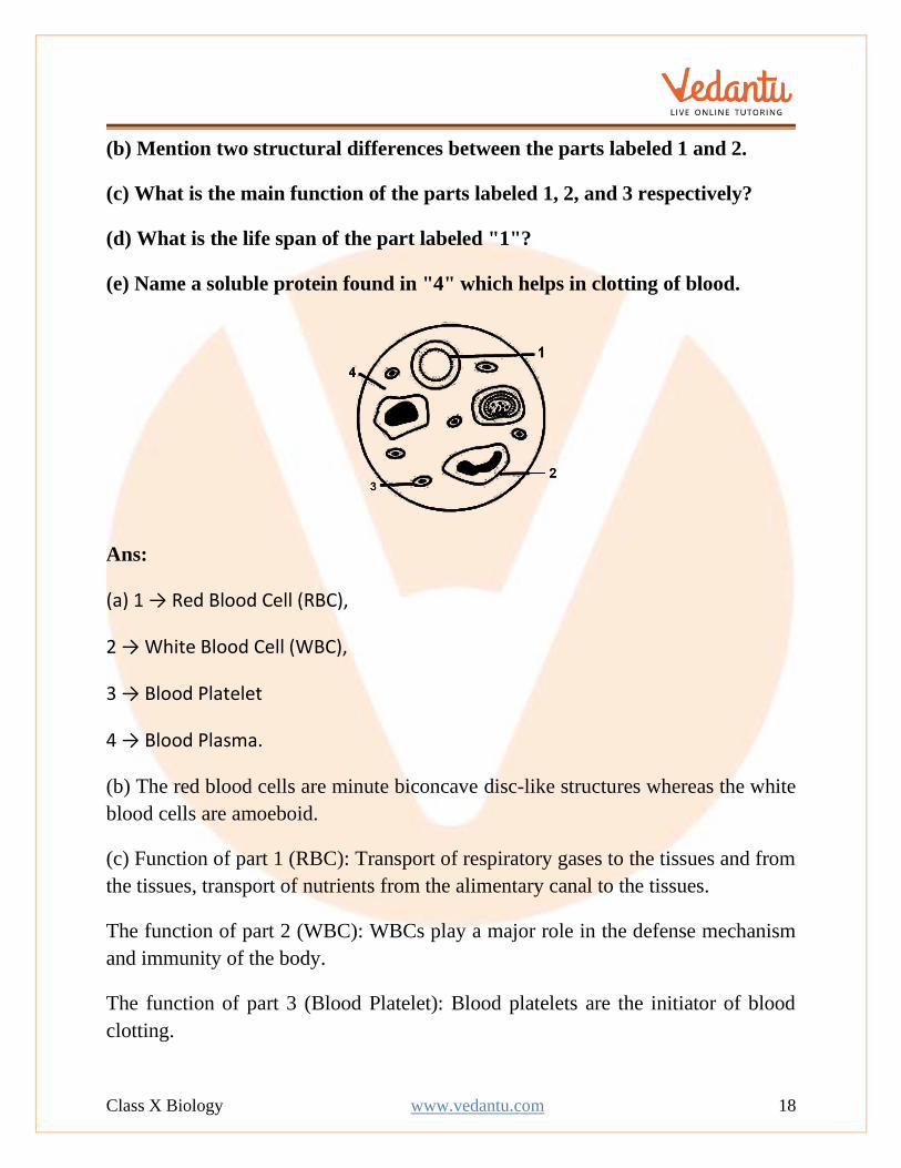
1. Given below is a diagram of a smear of human blood. Study the same and
answer the questions that follow:
(a) Name the parts 1, 2, 3, and 4 indicated by guidelines.
Class X Biology www.vedantu.com 18
(b) Mention two structural differences between the parts labeled 1 and 2.
(c) What is the main function of the parts labeled 1, 2, and 3 respectively?
(d) What is the life span of the part labeled "1"?
(e) Name a soluble protein found in "4" which helps in clotting of blood.
Ans:
(a) 1 → Red Blood Cell (RBC),
2 → White Blood Cell (WBC),
3 → Blood Platelet
4 → Blood Plasma.
(b) The red blood cells are minute biconcave disc-like structures whereas the white
blood cells are amoeboid.
(c) Function of part 1 (RBC): Transport of respiratory gases to the tissues and from
the tissues, transport of nutrients from the alimentary canal to the tissues.
The function of part 2 (WBC): WBCs play a major role in the defense mechanism
and immunity of the body.
The function of part 3 (Blood Platelet): Blood platelets are the initiator of blood
clotting.
Class X Biology www.vedantu.com 19
(d) The average life span of a red blood cell (RBC) is about 120 days.
(e) Thromboplastin
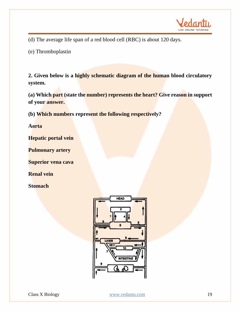
2. Given below is a highly schematic diagram of the human blood circulatory
system.
(a) Which part (state the number) represents the heart? Give reason in support
of your answer.
(b) Which numbers represent the following respectively?
Aorta
Hepatic portal vein
Pulmonary artery
Superior vena cava
Renal vein
Stomach
Class X Biology www.vedantu.com 20
Ans:
(a) structure 3 represents the heart. It forms the center of double circulation and is
located between the liver and the head (as per the diagram). Also, the blood
circulation (indicated by 1) begins from the heart to the lungs.
(b) The numbers that represent the following are:
Aorta 5
Hepatic portal vein 7
Pulmonary artery 1
Superior vena cava 9
Renal vein 8
Stomach 10
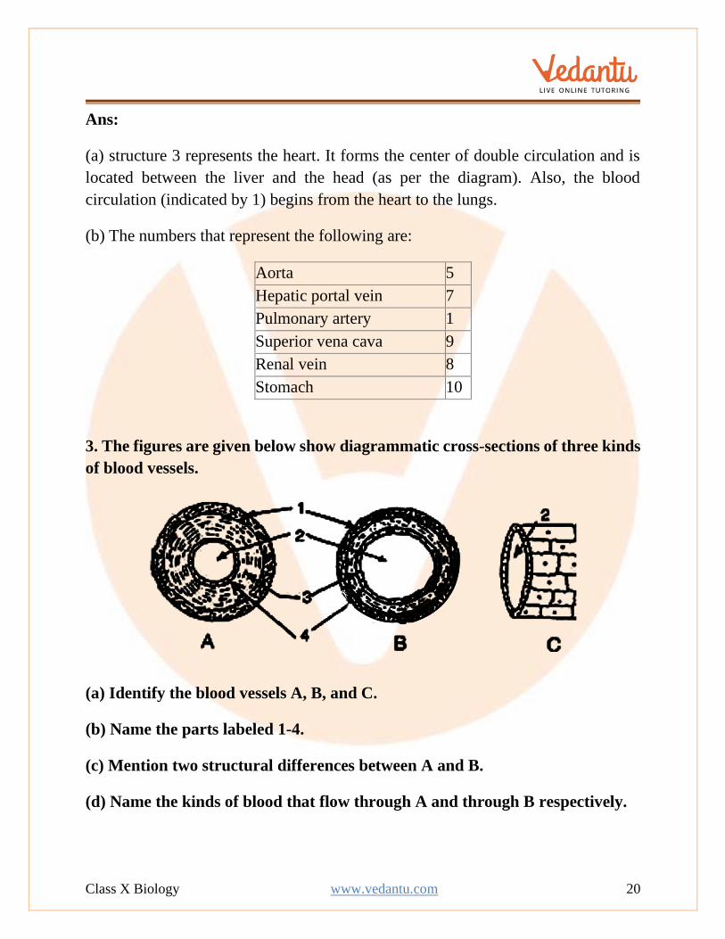
3. The figures are given below show diagrammatic cross-sections of three kinds
of blood vessels.
(a) Identify the blood vessels A, B, and C.
(b) Name the parts labeled 1-4.
(c) Mention two structural differences between A and B.
(d) Name the kinds of blood that flow through A and through B respectively.
Class X Biology www.vedantu.com 21
(e) In which one of the above vessels referred to in (a) above does the exchanges
of gases actually take place?
Ans:
(a) A- Artery, B-Vein, C-Capillary
(b) 1 - External layer made of connective tissue
2 - Lumen
3 - Middle layer of smooth muscles and elastic fibers
4 - Endothelium
(c) An artery has thick muscular walls and a narrow lumen. It does not have any
valve. A vein on the other hand has thin muscular walls and a wider lumen. It has
valves to prevent the backflow of blood.
(d) A (Artery)- Oxygenated blood, B (Vein)- Deoxygenated blood
(e) At the capillary level, the actual exchange of gases takes place.
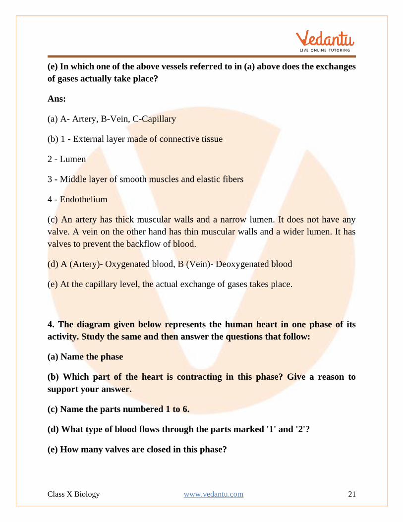
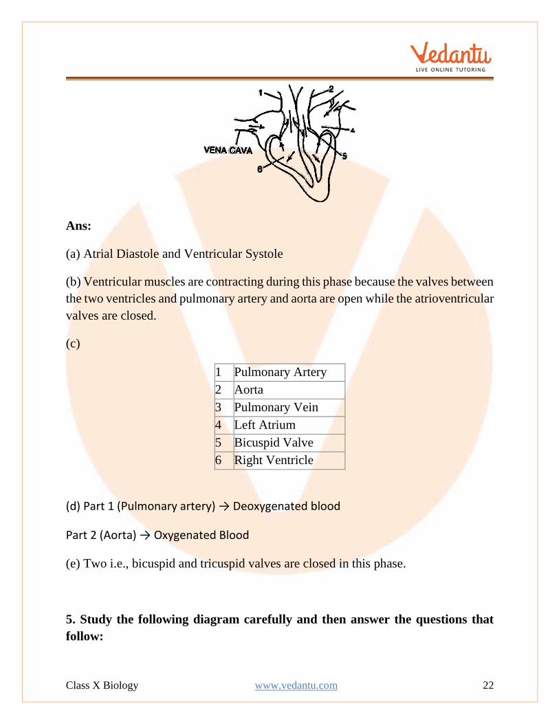
4. The diagram given below represents the human heart in one phase of its
activity. Study the same and then answer the questions that follow:
(a) Name the phase
(b) Which part of the heart is contracting in this phase? Give a reason to
support your answer.
(c) Name the parts numbered 1 to 6.
(d) What type of blood flows through the parts marked '1' and '2'?
(e) How many valves are closed in this phase?
Class X Biology www.vedantu.com 22
Ans:
(a) Atrial Diastole and Ventricular Systole
(b) Ventricular muscles are contracting during this phase because the valves between
the two ventricles and pulmonary artery and aorta are open while the atrioventricular
valves are closed.
(c)
1 Pulmonary Artery
2 Aorta
3 Pulmonary Vein
4 Left Atrium
5 Bicuspid Valve
6 Right Ventricle
(d) Part 1 (Pulmonary artery) → Deoxygenated blood
Part 2 (Aorta) → Oxygenated Blood
(e) Two i.e., bicuspid and tricuspid valves are closed in this phase.
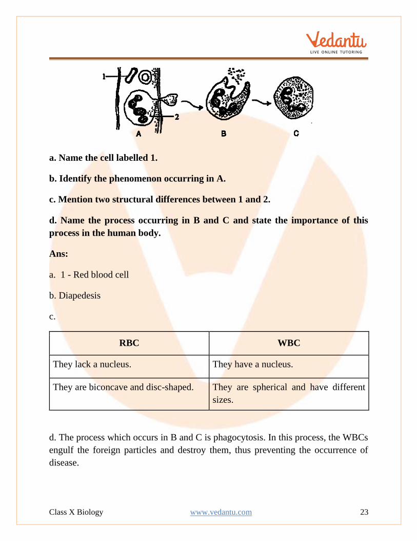
5. Study the following diagram carefully and then answer the questions that
follow:
Class X Biology www.vedantu.com 23
a. Name the cell labelled 1.
b. Identify the phenomenon occurring in A.
c. Mention two structural differences between 1 and 2.
d. Name the process occurring in B and C and state the importance of this
process in the human body.
Ans:
a. 1 - Red blood cell
b. Diapedesis
c.
RBC WBC
They lack a nucleus. They have a nucleus.
They are biconcave and disc-shaped. They are spherical and have different
sizes.
d. The process which occurs in B and C is phagocytosis. In this process, the WBCs
engulf the foreign particles and destroy them, thus preventing the occurrence of
disease.
Class X Biology www.vedantu.com 24
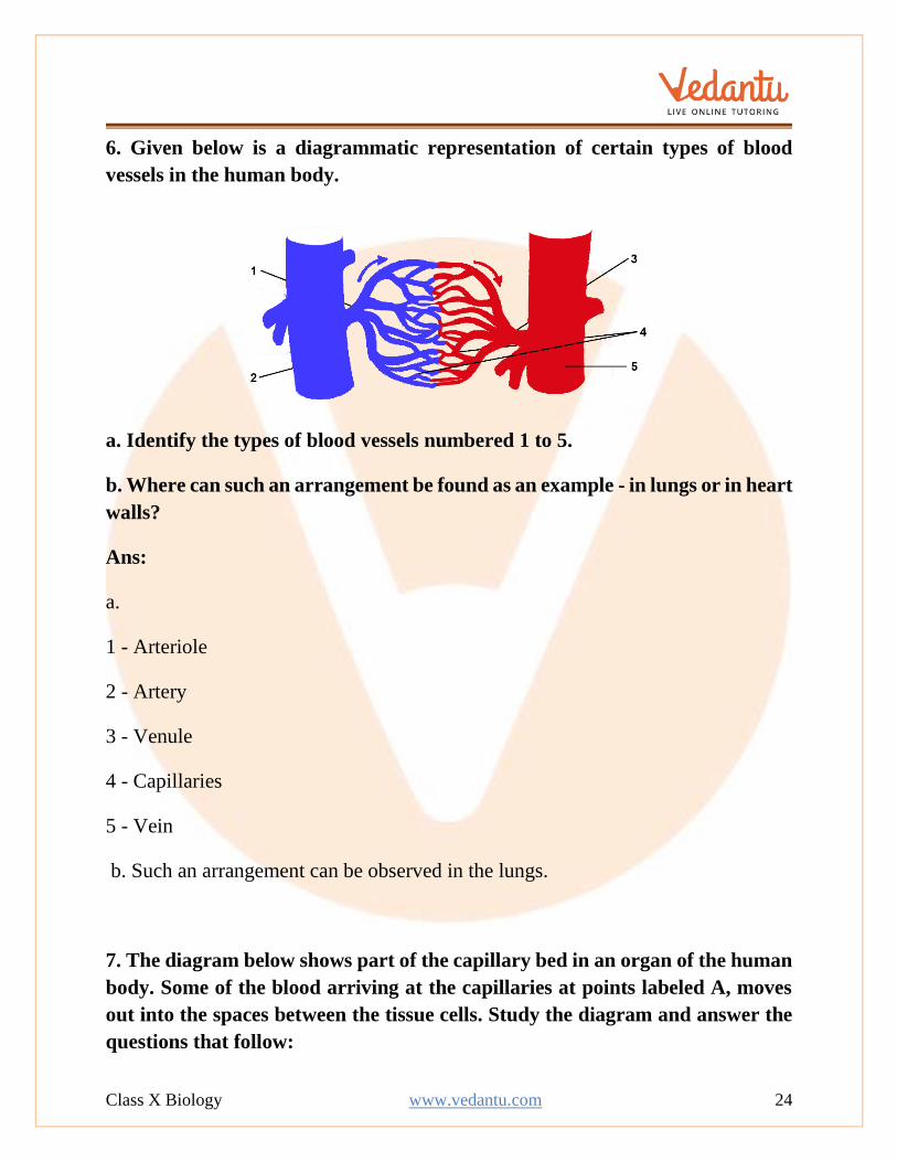
6. Given below is a diagrammatic representation of certain types of blood
vessels in the human body.
a. Identify the types of blood vessels numbered 1 to 5.
b. Where can such an arrangement be found as an example - in lungs or in heart
walls?
Ans:
a.
1 - Arteriole
2 - Artery
3 - Venule
4 - Capillaries
5 - Vein
b. Such an arrangement can be observed in the lungs.
7. The diagram below shows part of the capillary bed in an organ of the human
body. Some of the blood arriving at the capillaries at points labeled A, moves
out into the spaces between the tissue cells. Study the diagram and answer the
questions that follow:
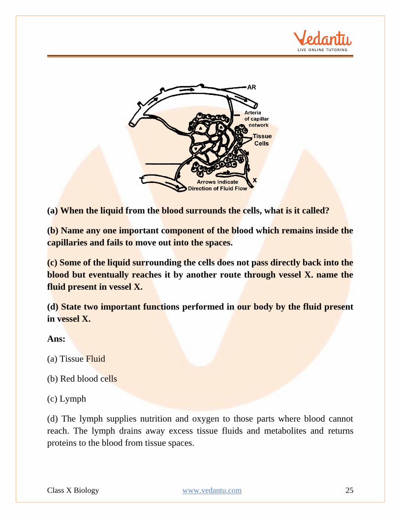
Class X Biology www.vedantu.com 25
(a) When the liquid from the blood surrounds the cells, what is it called?
(b) Name any one important component of the blood which remains inside the
capillaries and fails to move out into the spaces.
(c) Some of the liquid surrounding the cells does not pass directly back into the
blood but eventually reaches it by another route through vessel X. name the
fluid present in vessel X.
(d) State two important functions performed in our body by the fluid present
in vessel X.
Ans:
(a) Tissue Fluid
(b) Red blood cells
(c) Lymph
(d) The lymph supplies nutrition and oxygen to those parts where blood cannot
reach. The lymph drains away excess tissue fluids and metabolites and returns
proteins to the blood from tissue spaces.
Related Documents