Topic 6: Human physiology 6.1 Digestion and absorption U1 The contraction of circular and longitudinal muscle of the small intestine mixes the food with enzymes and moves it along the gut U2 The pancreas secretes enzymes into the lumen of the small intestine U3 Enzymes digest most macromolecules in food into monomers in the small intestine U4 Villi increase the surface area of epithelium over which absorption is carried out U5 Villi absorb monomers formed by digestion as well as mineral ions and vitamins U6 Different methods of membrane transport are required to absorb different nutrients A1 Processes occurring in the small intestine that results in the digestion of starch and transport of the products of digestion to the liver A2 Use of dialysis tubing to model absorption of digested food in the intestine S1 Production of an annotated diagram of the digestive system S2 Identification of tissue layers in transverse sections of the small intestine viewed with a microscope or in a micrograph Digestion of food molecules Large food molecules need to be digested before the nutrients can be absorbed Therefore, the purpose of digestion is to break complex molecules into simpler molecules to transport around the blood stream . When we ingested food we ingest the four types of macromolecules: 1. Nucleic acids (from cells): Ingested as DNA or RNA and broken down into nucleotides 2. Proteins: Ingested as proteins and broken down into amino acids 3. Lipids: Ingested as triglycerides and broken down into glycerol and fatty acids 4. Carbohydrates: Ingested as monosaccharides, disaccharides, or polysaccharides and broken down into monosaccharides Role of Enzymes During Digestion Enzymes are globular proteins that increase the rate of reaction by lowering activation energy By lowering activation energy, reactions happen faster and don’t require high temperatures (as high temperatures can increase reaction time). This is ideal as high temperatures would cause damage to cells and proteins Definitions Ingestion – The taking in of food into the body Digestion – Chemical breakdown of food into smaller molecules Absorption – Passage of smaller molecules from the digestive system into the bloodstream Transport – Delivery of small molecules to tissues by the circulatory system

Welcome message from author
This document is posted to help you gain knowledge. Please leave a comment to let me know what you think about it! Share it to your friends and learn new things together.
Transcript
Topic 6: Human physiology6.1 Digestion and absorptionU1 The contraction of circular and longitudinal muscle of the small intestine mixes the food with enzymes and moves it along
the gut
U2 The pancreas secretes enzymes into the lumen of the small intestine
U3 Enzymes digest most macromolecules in food into monomers in the small intestine
U4 Villi increase the surface area of epithelium over which absorption is carried out
U5 Villi absorb monomers formed by digestion as well as mineral ions and vitamins
U6 Different methods of membrane transport are required to absorb different nutrients
A1 Processes occurring in the small intestine that results in the digestion of starch and transport of the products of digestion to the liver
A2 Use of dialysis tubing to model absorption of digested food in the intestine
S1 Production of an annotated diagram of the digestive system
S2 Identification of tissue layers in transverse sections of the small intestine viewed with a microscope or in a micrograph
Digestion of food molecules
Large food molecules need to be digested before the nutrients can be absorbed Therefore, the purpose of digestion is to break complex molecules into simpler molecules to transport around the blood
stream. When we ingested food we ingest the four types of macromolecules:1. Nucleic acids (from cells): Ingested as DNA or RNA and broken down into nucleotides2. Proteins: Ingested as proteins and broken down into amino acids3. Lipids: Ingested as triglycerides and broken down into glycerol and fatty acids4. Carbohydrates: Ingested as monosaccharides, disaccharides, or polysaccharides and broken down into monosaccharides
Role of Enzymes During Digestion
Enzymes are globular proteins that increase the rate of reaction by lowering activation energy By lowering activation energy, reactions happen faster and don’t require high temperatures (as high temperatures can
increase reaction time). This is ideal as high temperatures would cause damage to cells and proteins To sum up, by using enzymes reactions can occur more quickly at body temperature without harming the body Digestive enzymes are released from glands into the gut and are used in catabolic reactions Although, organisms like humans can’t digest cellulose as they can’t produce the enzyme cellulase
Anatomy of the Human Digestive System
There are two major groups of organs which comprise the human digestive system:o The alimentary canal consists of organs which food passes through (esophagus, stomach, small/large intestine) o The accessory organs aid in digestion but do not actually transfer food (salivary glands, pancreas, liver, gall bladder)
Definitions
Ingestion – The taking in of food into the body
Digestion – Chemical breakdown of food into smaller molecules
Absorption – Passage of smaller molecules from the digestive system into the bloodstream
Transport – Delivery of small molecules to tissues by the circulatory system
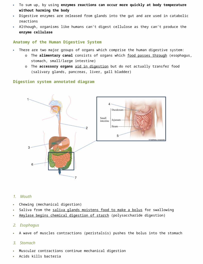
Digestion system annotated diagram
1. Mouth
Chewing (mechanical digestion) Saliva from the saliva glands moistens food to make a bolus for swallowing Amylase begins chemical digestion of starch (polysaccharide digestion)
2. Esophagus
A wave of muscles contractions (peristalsis) pushes the bolus into the stomach
3. Stomach
Muscular contractions continue mechanical digestion Acids kills bacteria Pepsin begins digestion of proteins
4. Duodenum (small intestine)
Bile from the liver and gall bladder neutralizes acid and emulsifies fats Pancreatic amylase and lipase digest carbohydrates and fats Trypsin digests polypeptides to amino acids
5. Ileum (small intestine)
Lower half of small intestine absorbs nutrients into the blood, via the villi
6. Large intestine
Water is absorbed and returned to the blood, leaving semi-solid feces. This is stored in the rectum
7. Egestion
Fasces (containing undigested food, dead cells and other waste) is forced out of the anus

Mechanical Digestion
In mechanical digestion, food is physical broken down into smaller fragments via: Chewing (Mouth)
o Food is initially broken down in the mouth by the grinding action of teetho The tongue pushes the food towards the back of the throat, where it travels down the esophaguso The epiglottis prevents the bolus from entering the trachea, which the uvula prevents the bolus from entering the
nasal cavity Churning (Stomach)
o The stomach lining contains muscles which physically squeeze and mix the food with strong digestive juices o Food is digested within the stomach for several hours and is turned into a creamy paste called chimeo Eventually the chime enters the small intestine (duodenum) where absorption will occur
Chemical Digestion
In chemical digestion, food is chemically broken down by chemical agents such as: Stomach Acids
o The stomach contains gastric glands which release digestive acids to create a low pH environment (pH ~2)o The acidic environment denatures proteins and other macromolecules, aiding in their overall digestiono The stomach epithelium contains a mucous membrane which prevents the acids from damaging the gastric liningo The pancreas releases alkaline compounds (e.g. bicarbonate ions), which neutralize the acids as they enter the
intestine Bile
o The liver produces a fluid called bile which is stored within the gall bladder prior to release into the intestineo Bile helps emulsify (mix) lipids
Enzymes (Also refer to first page)o The pancreas produces the three main types of digestive enzymeso The pancreas empties pancreatic juices into the small intestineo Remember: Enzymes are specific to their substrates and each enzyme has its own optimum pHo The three types of enzymes in human digestion
1. Amylases breaks down carbohydrates and hydrolyzes starch into maltose (a disaccharide)2. Pepsin breaks long polypeptides into shorter polypeptides. Proteases breaks down proteins into amino acid3. Lipases break down lipids and hydrolyzes them into fatty acids and glycerol
# Enzymes Substrate Product Source Optimum pH
1 Amylase Starch Maltose Salivary glands or pancreas 7 – 7.8
2 Pepsin Polypeptides Amino Acids Stomach 2
3 Pancreatic lipase Triglycerides Fatty acids & glycerol
Pancreas, delivered into the small intestine
7.2 – 7.5
Movement of Food
Peristalsis: Muscular contractions (both around and down the alimentary canal) that moves food through the digestive tract1. Contraction of smooth muscles behind the bolus forces it forward2. Waves of muscle contractions move bolus towards the stomach
This is important because:o Food travels in one direction only. This ensures that it only moves forwardo In the intestine it enables the chyme to mix and churn with enzymes. Although it is
slow at a few centimeters/contraction it enables

Small Intestine
The small intestine completes digestion of food molecules Ducts connect to the duodenum (start of small intestine) Enzymes will be used from the liver to split macromolecules with hydrolysis After enzymes are secreted by the walls of the small intestine Enzymes are further released into the jejunum The ileum is the last stage of the small intestine Here, absorption of digested food molecules takes place Villi increase the surface area for absorption and have a rich blood supply A wave of muscle contractions (peristalsis) keeps the mixture of digested and undigested food moving This can take from 8 – 24 hours
Structure of the small intestine
The small intestine contains four distinct tissue layers from the lumeno Mucosa: Inner lining, includes villio Submucosa: Connective tissue (between the mucosa and muscle)o Muscular layer: Inner circular and outer longitudinal muscle perform peristalsiso Serosa: Protective outer layer
The inner epithelial cells: Single outer layer of cells on each villus Many villi will protrude into the intestinal lumen, greatly increasing the available surface area for material absorption
Features of Villi
Villi absorb monomers formed by digestion as well as mineral ions and vitamins The role of the villus and the villi is to increase surface area for absorption This is because many villi protrude into the lumen, greatly increasing the surface area
for absorption Although, it is not the villi doesn’t absorbs itself, it is the epithelial cells that cover the
villi that do the absorption A lacteal is a lymphatic capillary that absorbs dietary fats in the villi of the small intestine Features that allow for increased SA:Vol absorption:
o Single-cell layers of epithelial cells allow a short path for diffusiono Microvilli on the surface of each cell increases SA even furthero Lymph vessels: Allows for rapid absorption and transport of lipidso Also has a rich blood supply which helps maintain concentration gradients between the lumen and blood
Absorption and assimilation
Once absorption occurs (uptake of broken down molecules into the blood) and the molecules are in the blood, they are carried to the tissues where they are assimilated (taken in to be used)
Membrane Transport mechanisms

Different methods of membrane transport are required to absorb different nutrients Two types of passive transport (simple/facilitated diffusion) are used to get materials from the lumen into the epithelial cells
of the villi. Remember, these do not require energy as they move molecules down the concentration gradient Two types of active transport (protein pumps and endocytosis) are used to get materials from the lumen into the epithelial
cells of the villi. Remember, these do require energy as they move molecules up the concentration gradient
Starch Digestion
Starch is a polysaccharide composed of glucose monomers It accounts for ~60% of the carbohydrates consumed by humans Starch digestion begins in the mouth with the enzyme called salivary amylase This doesn’t completely breakdown the starch because the enzyme is destroyed by the acidic environment of the stomach The pancreases then secretes pancreatic amylase into the small intestine which finishes breaking down the starch into
maltose (a disaccharide) Within the small intestine, there is another enzyme (maltase) that finishes breaking down maltose into 2 glucoses
6.2 The Blood SystemU1 Arteries convey blood at high pressure from the ventricles to the tissue of the body
U2 Arteries have muscle cells and elastic fibers in their walls
U3 The muscles and elastic fibers assist in maintaining blood pressure between pump cycles
U4 Blood flows through tissues in capillaries. Capillaries have permeable walls that allow exchange of materials between cells in the tissue and the blood in the capillary
U5 Veins collect blood at low pressure from the tissues of the body and return it to the atria of the heart
U6 Valves in veins and the heart ensure circulation of blood by preventing backflow
U7 There is a separate circulation for the lungs
U8 The heart beat is initiated by a group of specialized muscle cells in the right atrium called the sinoatrial node
U9 The sinoatrial node acts as a pacemaker
U10 The sinoatrial node sends out an electrical signal that stimulates contraction as it is propagated through the walls of the atria and then the walls of the ventricles
U11 The heart rate can be increased or decreased by impulses brought to the heart through two nerves from the medulla of
Method of transport Nutrients Example Outline
Simple Diffusion Fatty acids Fatty acids can pass freely through the plasma membrane into the epithelial cells (down the concentration gradient)
Facilitated Diffusion Glucose When the concentration of glucose is higher in the lumen than in the cells they can use facilitated diffusion to pass phospholipid bilayer and enter the epithelial cells (down the concentration gradient)
Membrane (Protein) Pumps
Glucose When the concentration in the lumen is lower than in the cells but the cells need more glucose protein pumps use ATP to move molecules against the concentration gradient into epithelial cells
Endocytosis (pinocytosis)
Larger molecules Larger molecules that haven’t been fully digested yet can be transported by endocytosis
Osmosis Water, dissolve ions
The absorption of water and dissolved molecules occurs in both the small and large intestine

the brain
U12 Epinephrine increases the heart rate to prepare for vigorous physical activity
A1 William Harvey’s discovery of the circulation of the blood with the heart acting as the pump
A2 Pressure changes in the left atrium, left ventricle and aorta during the cardiac cycle
A3 Causes and consequences of occlusion of the coronary arteries
S1 Identification of blood vessels as arteries, capillaries or veins from the structure of their walls
S2 Recognition of the chambers and valves of the heart and the blood vessels connected to it in dissected hearts or in diagrams of heart structure
The Blood System
The following are transported around the body in blood: oxygen, heat, carbon dioxide, nutrients, antibodies, hormones, This is achieved through the beating of the heat pumping the blood through a network of arteries veins, and capillaries
Arteries
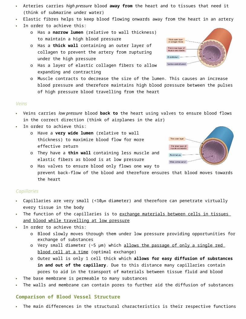
Arteries carries high pressure blood away from the heart and to tissues that need it (think of submarine under water) Elastic fibres helps to keep blood flowing onwards away from the heart in an artery In order to achieve this:
o Has a narrow lumen (relative to wall thickness) to maintain a high blood pressure
o Has a thick wall containing an outer layer of collagen to prevent the artery from rupturing under the high pressure
o Has a layer of elastic collagen fibers to allow expanding and contractingo Muscle contracts to decrease the size of the lumen. This causes an
increase blood pressure and therefore maintains high blood pressure between the pulses of high pressure blood travelling from the heart
Veins
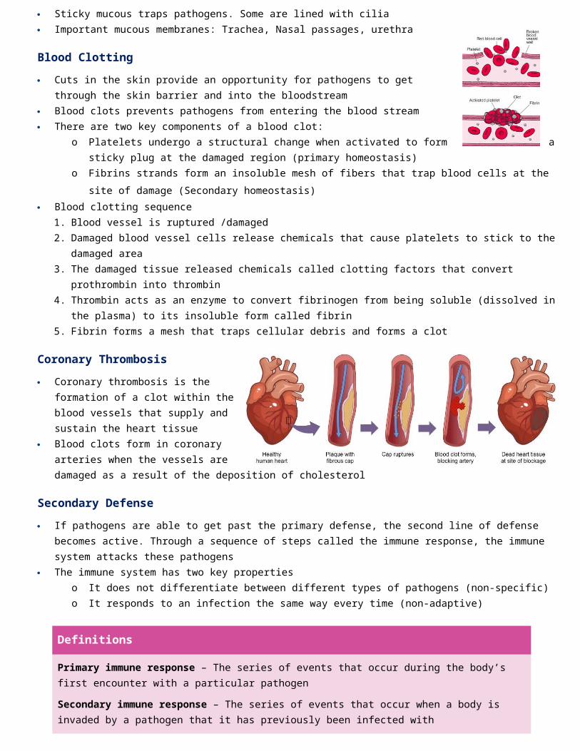
Veins carries low pressure blood back to the heart using valves to ensure blood flows in the correct direction (think of airplanes in the air)
In order to achieve this:o Have a very wide lumen (relative to wall thickness) to maximize blood
flow for more effective return o They have a thin wall containing less muscle and elastic fibers as blood is
at low pressureo Has valves to ensure blood only flows one way to prevent back-flow of
the blood and therefore ensures that blood moves towards the heart
Capillaries
Capillaries are very small (<10μm diameter) and therefore can penetrate virtually every tissue in the body The function of the capillaries is to exchange materials between cells in tissues and blood while travelling at low pressure In order to achieve this:
o Blood slowly moves through them under low pressure providing opportunities for exchange of substanceso Very small diameter (~5 μm) which allows the passage of only a single red blood cell at a time (optimal exchange) o Outer wall is only 1 cell thick which allows for easy diffusion of substances in and out of the capillary. Due to this
distance many capillaries contain pores to aid in the transport of materials between tissue fluid and blood The base membrane is permeable to many substances The walls and membrane can contain pores to further aid the diffusion of substances

Comparison of Blood Vessel Structure
The main differences in the structural characteristics is their respective functionso Arteries have thick walls and narrow lumens because they transport blood at high pressureo Veins have thin walls with wide lumens and valves because they transport blood at low pressureo Capillaries have walls that are only a single cell thick because they exchange materials between blood and tissues
Arteries Veins Capillaries
Function Send blood away from heart Send blood to heart For material exchange with tissues
Pressure High Low Low
Lumen Diameter
Narrow Wide Extremely Narrow (one cell wide)
Wall thickness
Thick Thin Extremely thin (single cell thick)
Wall layers - Tunica adventitia- Tunica media- Tunica intima
- Tunica adventitia- Tunica media- Tunica intima
- Tunica intima
Muscle & Elastic Fibers
Large amounts Small amounts None
Valves No Yes No
Identification of Blood Vessels
Blood vessels can be identified from histological slides or images according to the thickness of their walls:o Arteries have thick walls composed of three distinct layerso Veins have thin walls but wider lumeno Capillaries are very small and will not be easily detected under the same magnification as the others
Heart Structure
Blood flows through capillaries very slowly and at very low pressure in order to allow for maximal material exchange The structure of the human heart includes the following key components: Chambers
o Two atria (receives blood)o Two ventricles (pumps blood)
Heart valveso Atrioventricular valves o Semilunar valves
Blood Vesselso Pulmonary artery: Sends blood away from heart, high O2 concentration,o Pulmonary vein: Sends blood to heart, low O2 concentrationo Vena cavao Aorta
Ventricle vs Atria
The atrium is a chamber in where blood enters the heart, the ventricle is a chamber where blood is pushed out of the heart The walls of the ventricle are thicker than the atria because:
o They have to pump blood all the way from the heart to the whole bodyo A strong muscle contraction is needed to produce enough pressure to carry the blood the whole wayo The right ventricle only has to pump to the lungs, which is closer, and the left ventricle has to pump all the way to
the rest of the body
Cardiac Cycles
A cardiac cycle is a series of events from the beginning of one heart beat to the beginning of the next, commonly referred to as one heartbeat. This includes atrial and ventricular contractions
On average there are about 72 cardiac cycles per minute, meaning that one happens around .8 seconds It is comprised of a period of contraction (systole) and relaxation (diastole)
Heart Valves
Valves keep blood moving in a single direction to prevent backflow . Valves are like doors that only open one way When a chamber is in diastole, blood is allowed into that chamber because it is moving in a direction that allows the
“doors” of the valves to “flap” open When a chamber is in systole, the pressure of the blood forces the flaps to close and blood can’t enter the chamber where
it just came from Also notice that the heartbeat isn’t a single sound, but a lub-dub sounds. These sounds are the semilunar valves causing the
lub and the atrioventricular valves closing the dub
Control of heart rate
Myogenic muscle contraction: a muscle contraction that spontaneously contracts and relaxes without any control by the nervous system. The heart is myogenic, however the heartbeat does need to be controlled
The sinoatrial node is a specialized tissue inside the right atrium that acts as a pacemaker for the heart by sending out a signal that causes both atria to contract
Definitions
Diastole – When a chamber is relaxed, it causes a decrease in pressure and allows blood to fill the chamber
Systole – When a chamber contracts, it causes an increase in pressure and forces blood out of the chamber through any available opening

The atrial ventricular node is another group of specialized tissue also located in the right atrium and its job is to receive a signal from the SA node and then relay the signal to the ventricles causing them to contract
Control of heart beat
Heat beat is controlled the autonomic nervous system. This is the part of the nervous system that responds automatically to changes in body condition. It is not a conscious process: 1. Carbon dioxide accumulates in the blood (waste produce of cellular respiration)2. A part of the brain called the medulla oblongata senses the increase CO2 levels3. The medulla oblongata sends a message (through nerves) to the SA node to increase the heart rate. Once the CO2 levels
returns the normal, the medulla oblongata sends another message to tell the SA node to return to a resting heart rate
William Harvey
People use to think that blood was produced by the heart and was slowly used up by tissues in the body Therefore is a person had a disease many doctors would use leeches to such out the diseased blood Although William discovered that the blood is circulated and recirculated through the body (not used up) and
that the heart is a double pump In one circuit, the blood goes through the heart twice (once to be pumped to the lungs, the second to be
pumped to the body). This is why it’s called the double pump
Heat Disease
Coronary arteries are the blood vessels that surround the heart and nourish the cardiac tissue to keep the heart working. If coronary arteries become occluded, the region of heart tissue nourished by the blocked artery will die and cease to function
Blood pumped through the heart is at high pressure and cannot be used to supply the heart muscle with oxygen and nutrients
Cause of Coronary Occlusion:
Atherosclerosis
Atherosclerosis is the hardening and narrowing of the arteries. This happens due to the build up of cholesterol in damaged areas which forms a plaque
As build-ups of cholesterol and plaque form, the lumen narrows, restricting blood flow. If plaque ruptures, blood clotting is triggered. Blood clots are known as coronary thrombosis
Therefore, if atherosclerosis can lead to blood clots, and if these clots occur in myocardial tissue, it is called coronary heart disease. Coronary muscle tissue dies as a result of a lack of blood and oxygen
Risk Factors
Age: Blood vessels become less flexible with advancing age Genetics: Having hypertension predispose individuals Obesity: Increasing in blood pressure/Leads to plaque formation in arteries Diet: Increases fat/cholesterol/LDL in blood/leads to plaque formation in arteries Exercise: Lack of exercise increases risk que to weakened circulation Obesity: Increase in blood pressure/Leads to plaque formation in arteries Stress: Stress has been linked to increased cortisol hormones in the blood, causing increased atherosclerosis
6.3 Defense against infectious diseaseU1 The skin and mucous membranes form a primary defense against pathogens that cause infectious disease
U2 Cuts in the skin are sealed by blood clotting
U3 Clotting factors are released from platelets
U4 The cascade results in the rapid conversion of fibrinogen to fibrin by thrombin

U5 Ingestion of pathogens by phagocytic white blood cells gives non-specific immunity to diseases
U6 Production of antibodies by lymphocytes in response to particular pathogens gives specific immunity
U7 Antibiotics block processes that occur in prokaryotic cells but not in eukaryotic cells
U8 Viruses lack a metabolism and cannot therefore be treated with antibiotics. Some strains of bacteria have evolved with genes that confer resistance
A1 Causes and consequences of blood clot formation in coronary arteries
A2 Florey and Chain’s experiments to test penicillin on bacterial infections in mice
A3 Effects of HIV on the immune system and methods of transmission
Transmission of pathogens
Direct contact: Herpes (virus) Bodily Fluids: Strep throat, HIV Animal vectors: Rabies (virus), malaria (protozoa) Blood contact: Hepatitis B virus Ingested/Swallowed: Salmonella bacteria
Primary Defense
The best way to prevent diseases is to prevent pathogens from entering the body in the first place The skin and mucous membranes are a primary immune defense. Their
job is to prevent pathogens from entering tissues and/or the bloodstream
The Skin
The skin is made of several layers. The top layer is mostly dead cells, which provides a good barrier
The skin also secretes lactic acid and fatty acids to lower the pH (5.6 – 6.4 depending on region)
Mucous membranes
Sticky mucous traps pathogens. Some are lined with cilia Important mucous membranes: Trachea, Nasal passages, urethra
Blood Clotting
Cuts in the skin provide an opportunity for pathogens to get through the skin barrier and into the bloodstream
Blood clots prevents pathogens from entering the blood stream There are two key components of a blood clot:
Definitions
Pathogen – Any living organism or virus capable of causing a disease
Bacteria Diseases – E. coli, tetanus, strep throat
Viral Diseases – flu, cold, HIV/AIDS
Fungal Diseases – Athletes foot, ringworm, aspergillus
Protozoal Diseases – malaria, toxoplasmosis, giardia

o Platelets undergo a structural change when activated to form a sticky plug at the damaged region (primary homeostasis)
o Fibrins strands form an insoluble mesh of fibers that trap blood cells at the site of damage (Secondary homeostasis) Blood clotting sequence
1. Blood vessel is ruptured /damaged2. Damaged blood vessel cells release chemicals that cause platelets to stick to the damaged area3. The damaged tissue released chemicals called clotting factors that convert prothrombin into thrombin4. Thrombin acts as an enzyme to convert fibrinogen from being soluble (dissolved in the plasma) to its insoluble form
called fibrin5. Fibrin forms a mesh that traps cellular debris and forms a clot
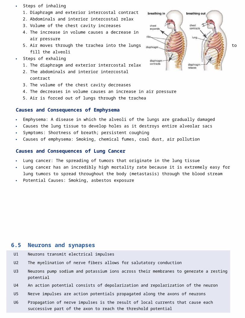
Coronary Thrombosis
Coronary thrombosis is the formation of a clot within the blood vessels that supply and sustain the heart tissue
Blood clots form in coronary arteries when the vessels are damaged as a result of the deposition of cholesterol
Secondary Defense
If pathogens are able to get past the primary defense, the second line of defense becomes active. Through a sequence of steps called the immune response, the immune system attacks these pathogens
The immune system has two key propertieso It does not differentiate between different types of pathogens (non-specific)o It responds to an infection the same way every time (non-adaptive)
Phagocytes
Phagocytosis is the process by which solid materials (such as pathogens) are ingested by a cell Phagocytic leukocytes (also called macrophages) are usually the first type of cell to encounter a pathogen. They circulate in the blood and move into the body tissue in response to infection
1. The macrophage reads the protein on the outside of the pathogen and recognize that the pathogen isn’t suppose to be there
2. The macrophage then engulfs the pathogen via phagocytosis3. The many enzymes found in the lysosomes of the macrophage destroy the pathogen4. Pathogen fragments (antigens) may be present on the surface of the phagocyte in order to stimulate the 3rd line of
defense This is a non-specific response. The macrophage doesn’t know what the pathogen is, just that it doesn’t belong there
Definitions
Primary immune response – The series of events that occur during the body’s first encounter with a particular pathogen
Secondary immune response – The series of events that occur when a body is invaded by a pathogen that it has previously been infected with

Tertiary Defense
A tertiary line of defense are substances like antigens or any invaders that pass through the first and second line of defense The third line of defense against infection disease is the adaptive immune system, which is specific in the production of
antibodieso It can differentiate between particular pathogens and target a response that is specific to a given pathogeno It can respond rapidly upon re-exposure to a specific pathogen, preventing symptoms from developing
(immunological memory)
Antibodies
Antigen: Membrane proteins on the outside of a pathogen that the immune cells can use to recognize that the pathogen is not part of the body (not self)
Antibody: A protein produced by B lymphocytes that is specific to a given pathogen Antibodies are y-shaped proteins (polypeptides) that attach to specific pathogens Antibodies are specific to each pathogen. So the antibody for the flu virus is different than
the one for syphilis The antibody sticks to the antigen like a lock and key Specific immunity: The production of antibodies that are specific to a single pathogen
Lymphocytes
The adaptive immune system is coordinated by lymphocytes and results in the production of antibodieso B lymphocytes (B cells) are antibody-producing cells that can recognize and target a particular pathogen fragment
(antigen)o Helper T lymphocytes (TH cells) are regulator cells that release chemicals to activate specific B lymphocytes
Antibiotics
Antibiotics are compounds that kill or inhibit the growth of microbes (specifically bacteria) by targeting prokaryotic metabolism
Metabolic features that may be targeted by antibiotics include key enzymes, 70s ribosomes and components of the cell wall
Because eukaryotic cells do not possess these features, antibiotics will target the pathogenic bacteria and not the infected host
Antibiotics may either kill the invading bacteria or suppress its potential to reproduce
However, since viruses do not possess a metabolism (they are not alive) they must be treated with specific antiviral agents opposed to antibiotics
Penicillin was the first chemical compound found to have antibiotic properties, which was identified by Alexander Fleming in 1928
HIV
HIV stands for human immunodeficiency virus HIV specifically targets the helper T lymphocytes which regulate the adaptive immune system Following infection, the virus undergoes a period of inactivity during which infected helper T cells reproduce Eventually, the virus becomes active again and begins the spread, destroying the T lymphocytes in the process With a reduction in the number of helper T cells, antibodies are unable to be produced resulting in a lowered immunity When all of the plasma cells have been destroyed and a person’s ability to produce a specific immune response is gone, then
its classified as AIDS (acquired immune deficiency syndrome) HIV is transmitted through, sexual contact, pregnancy, childbirth, injection drugs, blood transufion or organ transplant

6.4 Gas ExchangeU1 Ventilation maintains concentration gradients of oxygen and carbon dioxide between air in alveoli and blood flowing in
adjacent capillaries
U2 Type I pneumocytes are extremely thin alveolar cells that are adapted to carry out gas exchange
U3 Type II pnenumocytes secrete a solution containing surfactant that creates a moist surface inside the alveoli to prevent the sides of the alveolus adhering to each other by reducing surface tension
U4 Air is carried to the lungs in the trachea and bronchi and then to the alveoli in bronchioles
U5 Muscle contractions cause the pressure changes inside the thorax that force air in and out of the lungs to ventilate them
U6 Different muscles are required for inspiration and expiration because muscles only do work when they contract
A1 Causes and consequences of lung cancer
A2 Causes and consequences of emphysema
A3 External and internal intercostal muscles, and diaphragm and abdominal muscles as examples of antagonistic muscle action
S1 Monitoring of ventilation in humans at rest and after mild and vigorous exercise
Physiological Respiration
Physiological respiration involves the transport of oxygen to cells within tissues, where energy production occurs It can be divided into three distinct processes The processes involved in physiological respiration are:
o Ventilation: The physical process of air entering and exiting the lungs/The pumping of air into and out of the alveoli of the lungs
o Gas Exchange: The diffusion of oxygen and carbon dioxide into/out of the capillaries and alveoli/The exchange of CO2 and O2 between the air in the alveoli and the blood in the capillaries. Occurs in type I pneumocytes
o Respiration: The biochemical process during which the chemical energy in food (glucose) is converted into chemical energy in the form of ATP
Respiratory System
Ventilation helps rid the blood of carbon dioxide and absorb new oxygen molecules needed by the cells
1. Trachea
Air enters the respiratory system through the nose or mouth and passes through the pharynx to the trachea
2. Bronchus
The air then travels down the trachea until it divides into two bronchi (singular: bronchus)
3. Lungs/Bronchioles
The right lung is composed of three lobes, while the left lung is only comprise of two (smaller due to the position of the heart)
Inside each lung, the bronchi divide into many smaller airways called bronchioles, greatly increasing surface area
4. Alveoli
Each bronchiole terminates with a cluster of air sacs called alveoli, where gas exchange with the bloodstream occurs
Structure of Alveolus
Alveoli: Clusters of small sacs found as the end of the bronchioles Alveoli function as the site of gas exchange, and hence have specialized structural features to help fulfil this role:
Thin wall
The walls of alveolus are only 1 cell thick. This makes diffusion of oxygen and carbon dioxide easy and efficient Pneumocytes are the cells that line the alveoli and are the majority of the inner surface of the lungs. There are two types:
Spherical shape
They are roughly spherical in shape, in order to maximize the available surface area for gas exchange Their internal surface is covered with a layer of fluid, as dissolved gases are better able to diffuse into the bloodstream
Rich capillary network
They are surrounded by a rich capillary network to increase the capacity for gas exchange with the blood Surrounding each alveoli is a capillary network that brings deoxygenated blood to the alveoli and leaves with oxygenated
blood They are surrounded by a rich capillary network to increase the capacity for gas exchange with the blood For oxygen to be absorbed by the cells in your bloodstream the following must occur:
1. Air enters your trachea2. Air flows through your bronchi3. Air flows into the smaller branches of the bronchi4. Air flows into the bronchioles5. Air enters the alveoli sacs6. Oxygen diffuses out of the alveoli, through the capillary, and into a red blood cell
Their internal surface is covered with a layer of fluid, as dissolved gases are better able to diffuse into the bloodstream
Cell Type Shape Function
Type 1 Pheumocytes
Thin and flatMakes up the alveolus wall, easy for
diffusion
Type II Pheumocytes
Cube-shapedProduces a surfactant that keeps the alveoli moist and prevents them from
sticking together

Fluid Layer
Alveoli are lined with a layer of liquid in order to create a moist surface conducive to gas exchange with the capillaries It is easier for oxygen to diffuse across the alveolar and capillary membranes when dissolve in liquid While this moist lining assists with gas exchange Alveoli’s internal surface is covered with a layer of fluid, as dissolved gases are better to diffuse into the bloodstream This is due to surface tension: the elastic force created by a fluid surface
that minimizes the surface area Type II pneumocytes secrete a liquid known as pulmonary surfactant which
reduces the surface tension As an alveolus expands with gas intake, the surfactant becomes more
spread out across the moist alveolar lining This increases the surface tension and slows the rate of expansion, ensuring
all alveoli inflate at roughly the same time
Mechanism of Ventilation
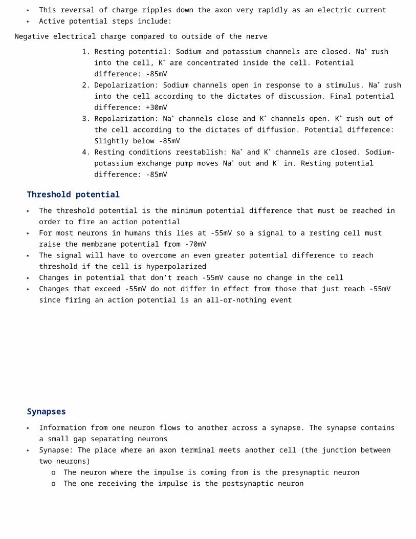
Steps of inhaling1. Diaphragm and exterior intercostal contract2. Abdominals and interior intercostal relax3. Volume of the chest cavity increases4. The increase in volume causes a decrease in air pressure5. Air moves through the trachea into the lungs to fill the alveoli
Steps of exhaling1. The diaphragm and exterior intercostal relax2. The abdominals and interior intercostal contract3. The volume of the chest cavity decreases4. The decreases in volume causes an increase in air pressure5. Air is forced out of lungs through the trachea
Causes and Consequences of Emphysema
Emphysema: A disease in which the alveoli of the lungs are gradually damaged Causes the lung tissue to develop holes as it destroys entire alveolar sacs Symptoms: Shortness of breath; persistent coughing Causes of emphysema: Smoking, chemical fumes, coal dust, air pollution
Causes and Consequences of Lung Cancer
Lung cancer: The spreading of tumors that originate in the lung tissue Lung cancer has an incredibly high mortality rate because it is extremely easy for lung tumors to spread throughout the body
(metastasis) through the blood stream Potential Causes: Smoking, asbestos exposure
6.5 Neurons and synapsesU1 Neurons transmit electrical impulses

U2 The myelination of nerve fibers allows for salutatory conduction
U3 Neurons pump sodium and potassium ions across their membranes to generate a resting potential
U4 An action potential consists of depolarization and repolarization of the neuron
U5 Nerve impulses are action potentials propagated along the axons of neurons
U6 Propagation of nerve impulses is the result of local currents that cause each successive part of the axon to reach the threshold potential
U7 Synapses are junctions between neurons and between neurons and receptor or effector cells
U8 When presynaptic neurons are depolarized they release a neurotransmitter into the synapse
U9 A nerve impulse is only initiated if the threshold potential is reached
A1 Secretion and reabsorption of acetylcholine by neurons at synapses
A2 Blocking of synaptic transmission at cholinergic synapses in insects by binding of neonicotinoid pesticides to acetylcholine receptors
S1 Analysis of oscilloscope traces showing resting potentials and action potentials
Nervous System
The nervous system is a complex network of nerves and cells that carry messages to and from the brain and spinal cord to various parts of the body
The nervous system includes both the central nervous system (CNS) and peripheral nervous system (PNS)
The CNS is made up of the brain and spinal chord The PNS is made up of the somatic and automatic nervous systems
Neurons
The nervous system is a complex collection of nerves and specialized cells known as neurons Neurons are specialized cells that function to transmit electrical impulses within the nervous system There are three classes of neurons: sensory, motor and interneurons. All these neurons have the following functions:
o Receive signals (or information)o Integrate incoming signals (to determine whether or not the information should be passed along)o Communicate signals to target cells (other neurons or muscles or glands)
The components of a neuron are as follows:
Axons and dendrites are bundled with axons or dendrites from other neurons to form nerves
Definitions
Soma (Cell Body) – A cell body containing the nucleus and organelles, where essential metabolic processes occur to maintain cell survival
Dendrites – Short branched fibers that convert chemical information from other neurons or receptor cells into electrical signals. Dendrites bring information to the cell body
Axon – An elongated fiber that transmits electrical signals to terminal regions for communication with other neurons or effectors. Axons take information away from the cell body
Synaptic terminal – Neurotransmitters are manufactured in the cell body but released from synaptic terminals. The neurotransmitters stimulate other neutrons
Synapse – A synapse is the junction between the synaptic terminal and another cell. The other cell is called a postsynaptic cell

In some neurons, the axon may be surrounded by an insulating layer known as a myelin sheath. This improves the conduction speed of electrical impulses along the axon, but require additional space and energy
Nerve impulses are carried from sensory neurons to the central nervous system
Myelination
In certain neurons, the axon may be covered by a fatty white substance called myelin which functions as an insulating layer
Myelin is a mixture of protein and phospholipids that is produced by glial cells
The main purpose of the myelin sheath is to increase the speed of electrical transmissions via salutatory conduction
The advantage of myelination is that is improves the speed of electrical transmission via salutatory conduction
The disadvantage of myelination is that is takes up significant space within an enclosed environment
Membrane potential
In all types of cells there is an electrical potential difference between the inside of the cell and the surrounding extracellular fluid. This is known as the membrane potential
o Electrical potential difference: When there is a net separation of charge between the two locationso Electrical potentials are measured in units of volts
While this phenomenon is present in all cells it is especially important in nerve and muscle cells because changes in their membrane potentials are used to code and transmit information
Resting Potential
Resting potential: The negative charge maintained when a nerve is not conducting a nerve impulse The resting potential is created by a transport protein called the sodium-potassium pump During the resting state the sodium potassium pump maintains a difference in charge across the cell membrane. This is
maintained by active transport as it uses energy in ATP to pump positive sodium ions out of the cell and potassium ions into the cell:
o Sodium ions are pumped outo Potassium ions are pumped back in
Some potassium ions also diffuse back out, leaving the outside more positive and the inside more negative Because the number of sodium ions is greater outside the cell the number of potassium ions moved inside the cell is more
positive outside than inside This is known as polarization as the cytoplasm inside the cell has a negative electrical charge and the fluid outside the cell
has a positive charge
Active Potential
Action potential: The reversal (depolarization) and restoration (repolarization) of the electrical potential across a plasma membrane as a nerve impulse passes along a neuron
A nerve impulse is a sudden reversal of the electrical charge across the membrane of a resting neuron
Definitions
Resting potential – Negative charge registered when the nerve is “at rest” and not conducting a nerve impulse
Action potential – The positive electrochemical charge generated at the nerve impulse
Depolarization – The change from the negative resting potential to the positive action potential
Re-polarization – The change in the electrical potential from the positive action potential back to the negative resting potential

The reversal of charge is called an action potential It begins when the neuron receives a chemical signal from another cell The signal causes gates in sodium ion channel to open, allowing positive sodium ions to flow back into the cell As a result the inside of the cell becomes positively charged compared to the outside of the cell This reversal of charge ripples down the axon very rapidly as an electric current Active potential steps include:
Negative electrical charge compared to outside of the nerve
1. Resting potential: Sodium and potassium channels are closed. Na+ rush into the cell, K+ are concentrated inside the cell. Potential difference: -85mV
2. Depolarization: Sodium channels open in response to a stimulus. Na+ rush into the cell according to the dictates of discussion. Final potential difference: +30mV
3. Repolarization: Na+ channels close and K+ channels open. K+ rush out of the cell according to the dictates of diffusion. Potential difference: Slightly below -85mV
4. Resting conditions reestablish: Na+ and K+ channels are closed. Sodium-potassium exchange pump moves Na+ out and K+ in. Resting potential difference: -85mV
Threshold potential
The threshold potential is the minimum potential difference that must be reached in order to fire an action potential For most neurons in humans this lies at -55mV so a signal to a resting cell must raise the membrane potential from -70mV The signal will have to overcome an even greater potential difference to reach threshold if the cell is hyperpolarized Changes in potential that don’t reach -55mV cause no change in the cell Changes that exceed -55mV do not differ in effect from those that just reach -55mV since firing an action potential is an all-
or-nothing event
Synapses
Information from one neuron flows to another across a synapse. The synapse contains a small gap separating neurons
Synapse: The place where an axon terminal meets another cell (the junction between two neurons)
o The neuron where the impulse is coming from is the presynaptic neuron
o The one receiving the impulse is the postsynaptic neuron The impulse is passed across the synapse using a chemical called a
neutron transmitter which is stored in vesicles at the end of the presynaptic neuron
Steps in Synaptic Transmission:1. Action potential arrives at axon terminal2. Voltage-gated Ca2+ channels open3. Ca2+ enters the presynaptic neuron4. Ca2+ signals to neurotransmitters vesicles5. Vesicles move to the membrane and dock6. Neurotransmitters released via exocytosis

7. Neurotransmitters bind to receptors8. Signal initiated in postsynaptic cell
6.6 Hormones, homeostasis and reproductionU1 Insulin and glucagon are secreted by beta and alpha cells of the pancreas respectively to control blood glucose
concentration
U2 Thyroxin is secreted by the thyroid gland to regulate the metabolic rate and help control body temperature
U3 Leptin is secreted by cells in adipose tissue and acts on the hypothalamus of the brain to inhibit appetite
U4 Melatonin is secreted by the pineal gland to control circadian rhythms
U5 A gene on the Y chromosome causes embryonic gonads to develop as testes and secrete testosterone
U6 Testosterone causes pre-natal development of male genitalia and both sperm production and development of male secondary sexual characteristics during puberty
U7 Estrogen and progesterone cause pre-natal development of female reproductive organs and female secondary sexual characteristics during puberty
U8 The menstrual cycle is controlled by negative and positive feedback mechanisms involving ovarian and pituitary hormones
A1 Causes and treatment of Type I and Type II diabetes
A2 Testing of leptin on patients with clinical obesity and reasons for the failure to control the disease
A3 Causes of jet lag and use of melatonin to alleviate it
A4 The use of IVF of drugs to suspend the normal secretion of hormones, followed by the use of artificial doses of hormones to induce superovulation and establish a pregnancy
A5 William Harvey’s investigation of sexual reproduction in deer
S1 Annotate diagrams of the male and female reproductive system to show names of structures and their functions
Homeostasis
Homeostasis: The process in which organ systems work to maintain a stable internal environment inside the body All the organs/organ systems work well together because they are closely regulated by the nervous and endocrine systems
o The nervous system controls virtually all body activitieso The endocrine system secretes hormones that regulate these activities
These two systems try to maintain a stable internal environment by maintaining temperature, pH and other conditions at just the right levels to support life processes while also suppling the body with all required substances and eliminates wastes
Furthermore, keeping a stable internal environment requires constant adjustments. There are many processes in which organ systems help maintain homeostasis including:
The regulation of the internal environment is done primarily through negative feedbacko Negative feedback is a response to a stimulus that keeps a variable close to a set value. Essentially it “shuts off” or
“turns on” a system when it varies from a set valueo However, some processes are regulated by positive feedback. Positive feedback is when a response to an event
increases the likelihood of the event to continue If homeostasis fails death or disease may result
Endocrine system
The endocrine system includes all of the glands of the body and the hormones produced by those glands These glands are controlled directly by stimulation from the nervous system as well as chemical receptors in the blood
and hormones produced by other glands Sensory neurons send messages to the brain, brain interprets the message, brain sends messages to the endocrine glands to
release hormones. By regulating the function of organs in the body, these glands help to maintain the body’s homeostasis Cellular metabolism, reproduction, sexual development, sugar and mineral homeostasis, heart rate, and digestion are among
the many processes regulated by the action of hormones Hormone: A lipid-based molecule that is secreted into the bloodstream by an endocrine gland, only affects targeted
bloodstreams. However, problems can arise if too much hormone is excreted. Example hormones include:
Hormone Source Function Problem
Thyroxin Thyroid glandControls metabolism rate and
body temperature
Too much: Hyperthyroidism. Leads to increased metabolism and temperature
Too little: Hypothyroidism. Leads to decreased metabolism
LeptinFat tissue in
the bodyTo decrease appetite
Can become desensitized to leptin where it doesn’t work in controlling appetite anymore
MelatoninPineal gland in
the brain
Regulates day/night cycle
(melatonin levels are highest at night)
If you fly through time zones, you may experience jet lag because your body is still producing melatonin during your
usual sleep hours. Taking melatonin pills at night in the new time zone can help with jet lag
Insulin and Glucagon
Example: Respiratory System Excretory System Endocrine System
Environment
A high concentration of CO2 in the blood triggers fast breathing
A low level of water in the blood triggers retention of
water by the kidneys
A high concentration of sugars in the blood
AdjustmentThe lungs will exhale more
frequently to remove CO2 faster
The kidneys produce more concentrated urine, so less water is lost from the body
Triggers secretion of insulin by an endocrine gland called the pancreas

Insulin and glucagon are hormones that help regulate the levels of blood glucose, or sugar in your body
The pancreas is both an endocrine and an exocrine gland There are 2 hormones produced by the endocrine cells of the pancreas
o Insulin which is produced in the beta cells of the pancreaso Glucagon which is produced in the alpha cells of the pancreas
Insulin and glucagon work together to balance the bodies blood sugar levels keeping them in the required range As blood glucose levels decline, alpha cells releases glucagon to raise the blood glucose levels by increasing rates of glycogen
breakdown and glucose released by the liver When blood glucose levels rise, such as after a meal, beta cells release insulin to lower blood glucose levels by increasing the
rate of glucose uptake in most body cells, and by increasing glycogen synthesis in skeletal muscles and the liver
Soil and Seed Theory
Aristotle proposed the “soil and seed” theory where a male will produce a “seed” which forms an “egg” when mixed with menstrual blood “the soil”. The “Egg” will then develop into a fetus inside the mother with the information contained within the male “seed” alone
William Harvey debunked this theory by studying the sexual organs of female deer after mating in an effort to identify the developing embryo. He was unable to detect a growing embryo until approximately 6 – 7 weeks after mating had occurred
William Harvey concluded that Aristotle’s theory was incorrect and that menstrual blood did not contribute to the development of a fetus. However, Harvey was unable to identify the correct mechanism of sexual reproduction and incorrectly asserted that the fetus did not develop from a mixture of male and female “seeds”
It is now known that a fetus forms from a combination of both male and female “seeds” (gametes)
SRY Gene
Virtually all X chromosomes are unrelated to sex, unlike the Y chromosomes which contains genes that do determine sex A single Y chromosome gene, called SRY (Sex-determining region) triggers an embryo to develop into a male Without a Y chromosome, an individual develops into a female, so you can think of female as the default sex of the human
species
Male Reproductive Hormones
The main male reproductive hormone is testosterone, which is secreted by the testes and serves a number of roleso It is responsible for the pre-natal development of male genitaliao It is involved in sperm production following the onset of pubertyo It aids in the development of secondary sex characteristics (including body hair, muscle mass, deepening of voice)o It helps maintain sex drive
The main female reproductive hormones (secreted by the ovaries) are estrogen and progesterone, which server several roles:
o They promote the pre-natal development of the female reproductive organs o They are responsible for the development of secondary sex characteristics (including body hair and breast
development)
Definitions
Glucose – A monosaccharide sugar that is primarily used for cellular respiration
Glycogen – A substance made from glucose that’s stored in the liver and muscle cells that can later be used for energy
Insulin – A hormone that tells the body’s cells either to take glucose from the blood for energy, or to store it for later use (Hormone to reduce blood glucose levels)
Glucagon – A hormone that tells cells in the liver and muscles to converg glycogen into glucose and release it into the blood so the cells can use it for energy (Hormone to increase blood glucose levels)
Pancreas – An organ in your abdomen that makes and releases insulin and glucagon

o They are involved in monthly preparation of egg release following puberty (via the menstrual cycle)
Male Reproductive system
The male reproductive system has two goals: to produce and deliver sperm and to secrete testosterone The male reproductive systems has two main functions: To produce sperm, the male gamete, and to release the male sex
hormone, testosterone into the body It includes all the organs responsible for the production of sperm. The following structures all contribute to the production of
sperm and semen as part of the reproductive process in males. The general order follows:1. Testis: The testis is responsible for the production of sperm and
testosterone2. Epididymis: Site where sperm matures and develops the ability
to be motile. Mature sperm is stored here until ejaculation3. Vas Deferens: Long tube which conducts sperm from the testes
to the prostrate gland4. Seminal Vesicle: Secretes fluid containing fructose (to nourish
sperm), and prostaglandin (triggers uterine contractions)5. Prostate Gland: Secretes an alkaline fluid to neutralize vaginal
acids (necessary to maintain sperm viability)6. Urethra: Conducts sperm/seman from the prostrate gland to the
outside of the body via the penis
Female Reproductive system
The female reproductive system consists of structures that produce female gametes called eggs and secrete te=he female sex hormone estrogen. The female reproductive system has several other functions as well:
o It receives sperm during sexual intercourseo It supports the development of a fetuso It delivers a baby during birtho It breast feeds a baby after birth
The following structures all contribute to the production and maintenance of an egg:1. Ovary: The ovary is where oocytes mature prior to release (ovulation). It is also responsible for estrogen and progesterone
secretion2. Fimbria: Fimbria are a fringe of tissue adjacent to an ovary that
sweep an oocyte into the oviduct3. Oviduct: The oviduct (or fallopian tube) transports the oocyte to
the uterus. It is also typically where fertilization occurs4. Uterus: The uterus is the organ where a fertilized egg will implant
and develop (becoming an embryo)5. Endometrium: The mucous membrane lining of the uterus. It
thickens in preparation for implantation or is otherwise lost (via menstruation)
6. Vagina: Passage leading to the uterus by which the penis can enter (uterus protected by a muscular opening called the cervix)
Menstrual cycle
The menstrual cycle describes recurring changes that occur within the female reproductive system to make pregnancy possible
Each menstrual cycle lasts roughly one month (~28 days) and begins at puberty before ending with menopause There are two key groups of hormones which control and coordinate the menstrual cycle

Pituitary hormones are released from the anterior pituitary gland and act on the ovaries to develop follicles Ovarian hormones (estrogen and progesterone) are released from the ovaries and act on the uterus to prepare for
pregnancy There are four key events that comprise a typical menstrual cycle. These events are distinguished by changes to hormonal
levels, follicular development and the status of the endometrium
1. Follicular Phase
Follicle stimulating hormone (FSH) is secreted from the anterior pituitary and stimulates growth of ovarian follicles
The dominant follicle produces estrogen, which inhibits FSH secretion (negative feedback) to prevent other follicles growing
Estrogen acts on the uterus to stimulate the thickening of the endometrial layer
2. Ovulation
Midway through the cycle (~ day 12), estrogen stimulates the anterior pituitary to secrete hormones (positive feedback)
This positive feedback results in a large surge of luteinizing hormone (LH) and a lesser surge of FSH
LH causes the dominant follicle to rupture and release an egg (secondary oocyte) – this is called ovulation
3. Luteal Phase
The ruptured follicle develops into a slowly degenerating corpus luteum
The corpus luteum secretes high levels of progesterone, as well as lower levels of oestrogen
Estrogen and progesterone act on the uterus to thicken the endometrial lining (in preparation for pregnancy)
Estrogen and progesterone also inhibit secretion of FSH and LH, preventing any follicles from developing
4. Menstruation
If fertilisation occurs, the developing embryo will implant in the endometrium and release hormones to sustain the corpus luteum
If fertilisation doesn’t occur, the corpus luteum eventually degenerates (forming a corpus albicans after ~ 2 weeks)
When the corpus luteum degenerates, estrogen and progesteron levels drop and the endometrium can no longer be maintained
The endometrial layer is sloughed away and eliminated from the body as menstrual blood (i.e. a woman’s period)
As estrogen and progesterone levels are too now low to inhibit the anterior pituitary, the cycle can now begin again
In Vitro fertilization (IVF)
Vitro Fertilization: The process of developing a fertilized egg outside a body It involes:
o Harvesting mature eggs from the mother. This is not an easy process. The mother must undergo hormonal treatment to produce multiple eggs, which then must be removed from her ovaries
o Harvesting sperm from the father. Harvesting is usualy no problem, but often the sperm are defective in their ability to fertilize
o Mixing sperm and eggs in a culture vessel (in vitro)o Culturing the fertilized eggs for several days until they have developed to at least the 8-cell stage
o Placing two or more of these into the mother’s uteruso This will result in 1/3 success
Related Documents