UNIVERSITI PUTRA MALAYSIA IN VITRO STUDIES ON THE ADHESION OF FIBROBACTER SUCCINOGENES STRAIN D3 TO MACROCRYSTALLINE CELLULOSE SIEO CHIN CHIN IB 1997 1

Welcome message from author
This document is posted to help you gain knowledge. Please leave a comment to let me know what you think about it! Share it to your friends and learn new things together.
Transcript
UNIVERSITI PUTRA MALAYSIA
IN VITRO STUDIES ON THE ADHESION OF FIBROBACTER SUCCINOGENES STRAIN D3 TO MACROCRYSTALLINE
CELLULOSE
SIEO CHIN CHIN
IB 1997 1
IN VITRO STUDIES ON THE ADHESION OF FIBROBACTER
SUCCINOGENES STRAIN D3 TO MICROCRYSTALLINE
CELLULOSE
By
SIEO CHIN CHIN
Thesis Submitted in Fulfilment of the Requirements for
the Degree of Master of Science in the
Institute of Bioscience
Universiti Putra Malaysia
June 1997
ACKNOWLEDGEMENTS
I would like to express my appreciation and sincere gratitude to the chairman
of the supervisory committee, Associate Professor Dr. Norhani Abdullah for her
guidance and advice throughout the course of this study and in the preparation of this
thesis. Sincere thanks are also extended to the other members of the supervisory
committee, Professor Dr. Ho Yin Wan, and Professor Dato' Dr. Syed Jalaludin Syed
Salim, for their invaluable advices and suggestions, and to Dr. Mohd. Ridzwan,
Department of Agronomy and Horticulture, Faculty of Agriculture, for his advice on
the statistical analysis of the data.
I would also like to extend my sincere thanks to all the staff of the Rumen
Microbiology Laboratory, especially Khairul and Jivanathan and staff of the Electron
Microscope Unit, especially Mr. Ho Oi Kuan, Puan Aminah Jusoh and Cik Azilah
Abdullah Jalil. To my fellow labmates and friends, Geok Yong, Wan Zuhainis, Dr.
Jin LiZhi, Thongsuk, Michael and Foong Yee, my grateful thanks for their
companionship, humour, support and cooperation.
Finally, I wish to extend my heartfelt thanks and gratitude to my family and
my husband, Lai Kok Loong, for their unending support, encouragement and
understanding throughout my study.
11

TABLE OF CONTENTS
Page
ACKNOWLEDGEMENTS. . . . . . . . . . . . . . .. . . . . . . . . . . . . . . . . . . . . . . . . . . . . . . . . . . . . . . . . . . . . . . . . . . . . . . . . . . . . 11
LIST OF TABLES. . . . . . . . . . . . ................... ........ ....... ................... ................... . . . ... VI
LIST OF FIGURES. .............................................................................. . . . . . ... Vll
LIST OF PLATES . . . . . . ... . . . . . . . . . . . . . . . . . . . . . . . . . . . . . . . . . . . . . . . . . . . . . . . . . . . . . . . . . . . . . . . . . . . . . . . . . . . . . . ... Vll1
LIST OF ABBREVIATIONS. . . . . . . . . . . . . . . . . . . . . . . . . . . . . . . . . . . . . . . . . . . . . . . . . . . . . . . . . . . . . . . . . . . . . . . . x ABSTRACT. . . . . . . . . .. . . . . . . . . . . . . . . . . . . . . . . . . . . . . . . . . . . . . . . . . . . . . . . . . . . . . . . . . . . . . . . . . . . . . . . . . . . . . . . . . . . . . . . . . Xl
ABSTRAK. . . . . . . . . . . . . . . . . . . . . . . . . . . . . . . . . . . . . . . . . . . . . . . . . . . . . . . . . . . . . . . . . . . . . . . . . . . . . . . . . . . . . . . . . . . . . . . . . . . . . xiv
CHAPTER
I INTRODUCTION. . . . .. . . . . . . . . . . . . . . . . . . . . . . .. . . . . . . . . . . . . . . . . . . . . . . . . . . . . . . . . . . . . 1
II LITERATURE REVIEW.. . . . . . . . . . . . . . . . . . . . . . . . . . . . . . . . . . . . . . . . . . . . . . . . . . . . . . 4 The Rumen Microorganisms and Their Activity . . . . . . . . . . . . . . . . . . . 4
Rumen Protozoa. . . . . . . . . . . . . . . . . . . . . . . . . . . . . . . . . . . . . . . . . . . . . . . . . . . . . . . . . . 4 Rumen Fungi . . . . . . . . . . . . . . . . . . . . . . . . . . . . . . . . . . . . . . . . . . . . . . . . . . . . . . . . . . . . ... . 6 Rumen Bacteria. . . . . . . . . . . . . . . . . . . . . . . . . . . . . . . . . . . . . . . . . . . . . . . . . . . . . . . . . . . . 8
Adhesion of Rumen Microbes on Insoluble Nutrients. . . . . . . . . . 1 0 Electron Microscopic Observation on the Adhesion of Bacteria. . . . . . . . . . . . . . . . . . . . . . . . . . . . . . . . . . . . . . . . . . . . . . . . . . . . .. . . . . . . . . . . . . . . . . . . . . . . . 12 Specificity in the Adhesion of Rumen Microbes to Solid Substrates . . . . . . . . . . . . . . . . . . . . . . . . . . . . . . . . . . . . . . . . : . . . . . . . . . . . . . . . . . . . . . . . . . . . 1 5
The Cellulosome. . . . . . . . . . . . . . . . . . . . . . . . . . . . . . . . . . . . . . . . . . . . . . . . . . . . . . . . . . . . . . . . . . . . 1 7
Cellulosome and Rumen Microorganisms. . . . . . . . . . . . . . . . . . . . . . . . . . . . 18 Strategies to Improve Cellulose Digestion. . . . . . . . . . . . . . . . . . . . . . . . . . . . 1 9
III EFFECT OF PHYSICOCHEMICAL FACTORS ON THE ADHESION OF F SUCCINOGENES STRAIN D3 TO AVICEL. . . . . . . . . . . . . ... . . . . . . . . . . . . . . . . . . . . . . . . . . . . . . . . . . . . . . . . . . . . . . . . . . 23
Materials and Methods. . . . . . . . . . . . . . . . . . . . . . . . . . . . . . . . . . . . . . . . . . . . . . . . . . . . . . . . 24 Source of Chemicals . . . . . . . . . . . . . . . . . . . . . . . . . . . . . . . . . . . . . . . . . . .. . . . . . . . 24 Source of Bacteria. . . . . . . . . . . . . . . . . . . . . . . . . . . . . . . . . . . . . . . . . . . . . . . . . . . . . . 24 Experimental Conditions. . . . . . . . . . . . . . . . . . . . .. . . . . . . . . . . . . . . . . . . . . . . 24 Preparation of Modified Scott and Dehority Medium (MOD-SD) . . . . . . . . . . . . . . . . . . . . . . . . . . . . . . . . . . . . . . . . . . . . . . . . . . 25 Bryant's Solution. . . . . . . . . . . . . . .. . . . . . . . . . . . . . . . . . . . . . . . . . . . . . . . . . . . . . . . . 27 Preparation of Bacteria Culture. . . . . . . . . . . . . . . . . . . . . . . . . . . . . . . . . . 28 Studies on Growth Curve of F succinogenes......... .... 28 Adhesion Test. . . . . . . . . . . . . . . . . . . . . . . . . . . . . . . . . . . . . . . . . . . . . . . . . . 29
111
Adhering Ability of F succinogenes to A vice I at Different Growth Phase . . . . . . . . . . . . . . . . . . . . . . . . . . . . . . . . . . . . . . . . . . 3 1 Effect of pH and Temperature on Adhesion . . . . . . . . . . . . . . . . 3 1 Effect of Enzyme Treatment of Cells on Adhesion. . . . . . . . . .. . . . . . . . . . . . . . . . . . . . . . . . . . . . . . . . . . . . . . . . . . . . . . . . . . . . . . . . . 32 Statistical Analysis. . . . . . . . . . . . . . . . . . . . . . ............... . ...... . . . . ...... . 34
Results . . . . . . . . . . . . . . . . . . . . . . . . . . . . . . . . . . . . . . . . . . . . . . . . . . . . . . . . . . . . ......... ... . . . . . .... 34 Growth Pattern of F succinogenes. . . . . . . . . . . . . . . . . . . . . . . . . . . 34 Influence of Growth Phase on Adhesion of F. succinogenes to A vicel . . . . . . . . . ... . . . . . . . . . . . . . . .... . . . . . 37 Effect of pH and Temperature on the Adhesion of F succinogenes . . . . . . . . . . . . . . . . . . . . . . . . . . . . . . . '" 40 Effect of Enzyme Treatments on Adhesion of F. succinogenes. . . . . . . . . . .. . . .. . . . . . . . . . . . . . . . . . . . . . . . . . . . . . . . . . 42
Discussion. . . . . . . . . . . . .. . . . . . . . . . . . . . . . . . . . . . . . . . . . . . . . . . . . . . . . . . . . . . . . . . .. . . . ... . . . ... 46
IV ELECTRON MICROSCOPIC STUDY ON THE ADHESION OF F SUCCINOGENES STRAIN D3 TO AVICEL . . . . . . . . . . . . . . . . . . . . . . . . . . . . . . . . . . . . . . . . . . . . . . . . . . . . . . . . . . . . . . . . . 52
Materials and Methods. . . . . . . . . . . . . . . . . . . . . . . . . . . . . . . . . . . . . . . . . . . . . . . . ... . .... . 52 Scanning Electron Microscopy. . . . . . . . . . . . . . . . . . . . . . . . . . . . . . . . . .... 52 Transmission Electron Microscopy. . . . . . . . . . . . . . . . . . . . . . . . ... . . . 54
Results . . . . . . . . . . . . . . ... . . . . . . . . . . . . . . . . . . . . . . . . . . . . . . . . . . . . . . . . . . . . . . . . . . . . . . . . . ........ 55 Scanning Electron Microscopy. . . . . . . . . . . . . . . . . . . . . .... . . . . . . . .... 5 5 Transmission Electron Microscopy. . . . . . . . . . . . . . . . . . . . . . . . . . . . . . 57
Discussion.. . . . . . . . . . . . . . . . . . . . . . . . . . . . . . . . . . . . . . . . . ... . . . ....... ..... . . . . . . . . . . . . ...... 68
V DETECTION OF CELLULOSE-BINDING PROTEINS (CBPs) OF F. SUCCINOGENES STRAIN D3. . . . . . . . . . . . . . . . . . . 74
Materials and Methods. . . ... . . . ..... ............ ................. . . . . . . . . . . . .... 74 Detection of CBPs . . . . . . . . . . . . . . . . . . . . . . . . . . . . . . . . . . . . . . . . . . . . . . 74 Elution of CBPs . . . . . . . . . . . . . . . . . . . . . . . . . . . . . . . . . . . . . . . . . . . . . . . . . . . 75 Binding Ability of Eluted Proteins . . . . . . . . . . . . . . . . . . . . . . . . . . . . . . . CBPs of Enzymes Treated Cells . . . . . . . . . . . . . . . . . . . . . . . . . . . . . . SDS-PAGE Electrophoresis . . . . . . . . . . . . . . . . . . . . . . . . . . . . . . . . . . . . . . . . .
Detection of Carboxymethy1cellulase (CMCase) and Xylanase Activity of CBPs . . . . . . . . . . . . . . . . . . . . . . . . . . . . . . . . . . .
iv
76 76 77
78
'ERPlIJS'fA1<MN !D1\lJ\1ERSITI PUTRA MALA YSlA
Results. . . . . . . . . . . . . . . . . . . . . . . . . . .. . . . . . . . . . . . . . . . . . . . . . . . . . . . . . . . . . . . . . . . . . . . . . . . .. . . . . 79 Detection of CBPs . . . . . . . . . . . . . . . . . . . . . . . . . . . . . ... . . . . . . . . . . . . . 79 Elution of CBPs. . . . . . . . . . . . . . . . . . . . . . . . . . . . . . . . . .. . . . . . . . . . . . . . . 79
Binding Ability of CBPs. . . . . . . . . . . . . . . . . . . . . . . . . . . . . . . . . . . . . . . . . . . . . 86 Detection of CBPs on Cells Treated with
Enzymes. . . . . . . . . . .. . . . . . . . . . . . . . . . . . . . . . . . . . . . . . . . . . . . . . . . . . . . . .. 86 CMCase and Xylanase Activity of CBPs. . . . . . . . . . . . . . . . . . . 86
Discussion. . . . . . . . . . . . . . . . . . . . . . . . . . . . . . . . . . . . . ... . . . . . . . . . . . . . . . . . . . . . . . . . . . . . . . . . . . . 9 1
VI GENERAL DISCUSSION. . . . . . . . . . . . . . . . . . . . . . . . . . . . . . . . . . . . . . . . . . . . . . . . . . . . . 99
VII CONCLUSION. . . . . . . . . . . . . . . . . . . . . . . . . . . . . . . . . . . . . . . . . . . . . . . .. .. . . . . . .. . . . .. . . . . . . 1 06
BIBLIOGRAPHy... . . . . . . . . . . . . . . . . . . . . . . . . . . .... . . . . . . . . . . .. . . . . . . . . . . . . . . . . . . . . . . . . . . . .. . . . . . . . . . . . . . . . 1 08
APPENDIX. . . . . . . . . . . . . .. . . . . . . . . . . . . . . . . . . . . . . .. . . . . . . . .. . . . . . . . . . . . . . . . . . . . . . . . . . . . . . . . . . . . . . . . . . . . . . . . . .. 1 20
BIOGRAPHICAL VITA. . . . . . . . . . . . . . . . . . . . . . . . . . . . . . . . . . . . . . ... . . . . . . . . . . . . . . . . . . . . . . . . . . . . . . . . . . . . . 1 28
v
LIST OF TABLES
Table
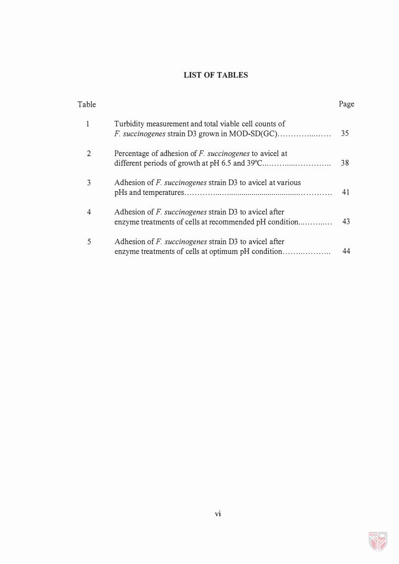
1 Turbidity measurement and total viable cell counts of
Page
F. succinogenes strain D3 grown in MOD-SD(GC)......... . . ....... . .. 35
2 Percentage of adhesion of F. succinogenes to avicel at different periods of growth at pH 6.5 and 39°C......... ................... 3 8
3 Adhesion of F. succinogenes strain D3 to avicel at various pHs and temperatures....................................................... . . . . . . . . . . 4 1
4 Adhesion of F. succinogenes strain D3 to avicel after enzyme treatments of cells at recommended pH condition........ ...... 43
5 Adhesion of F. succinogenes strain D3 to avicel after enzyme treatments of cells at optimum pH condition. . . . . . ... . . . . . . . . .. 44
VI

Figure
2
LIST OF FIGURES
Growth pattern of F. succinogenes strain D3 in MOD-SD(GC) evaluated by turbidity measurement and total viable cell
Page
counts . . . . . . . . . . . . . . . . . . . . . . . . . . . . . . . . . . . . . . . . . . . . . . . . . . . . . . . . . . . . . . . . . . . . . . . . . . . . . . 36
Adhesion of F. succinogenes to avicel at various stages of growth . . . . ...... ......... ................ . . . . . . . . . ......... . . . . . . . . . : . . . . . . . . . . . .. . . . . . . . . . . . . 39
Vll
LIST OF PLATES
Plate
1 Scanning electron micrographs showing the adhesion of F. succinogenes (late exponential phase) to avicel after
Page
1 0 min of incubation. . . . . . . . . . . . . . . . . . . . . . . .. . . . . . . . . . . . . . . . . . . . . . . . . . . . . . . . . . . . . . . . . . . 56
2 Scanning electron micrograph of F. succinogenes showing the spike-like structures (S) for adhesion of the bacteria to the substrate and thicker structures (T) that joined the bacteria
together. The bacteria were grown for 8 h in MOD-SD medium containing avicel (V) . . . . . . . . . . . . . . . . . . . . . . . . . . . . . . . . . . . . . . . . . . . . 58
3 Digestion of avicel by F. succinogenes after 30 h of incubation . . . . . 59
4 Pit of digestion at higher magnification. . . . . . . . . . . . . . . . . . . . . . .. . . . . . . . . . . . . . . . . 60
5 Transmission electron micrograph of late-exponential-phase F. succinogenes strain D3 grown for 8 h in soluble
carbohydrate medium. . . . . . . . . . . . . . . . . . . . . . . . .. . . . . ... . . .. . . . . . . . . . . . . . . . . . . . . .. . . . 6 1
6 Transmission electron micrograph of F. succinogenes showing initial adhesion to avicel (V) ( 1 0 min after incubation with avicel) . . . . . . . . . . . . . . . . . . . . . . . . . . . . . . . . . . . . . . . . . . . . . . . . . . . . . . . . .. . . . . .. . . . . . . . . . . . . . . . . . . . 63
7 Transmission electron micrograph showing adhesion of F. succinogenes to avicel (V) after 18 h of incubation. . . . . . . . . . . . . . . . 64
8 Transmission electron micrograph showing adhesion of F. succinogenes to avicel (V) after 18 h of incubation in
MOD-SD(A). . . . . . . . . . . . . . . . .. . . . . . . . . . .. . . . . . . . . . . . . . . . . . . . . . . . . . . .. . . . .. . . . . . . . . . . . . . . . .. 65
9 Transmission electron micrograph showing the digestion pits or zones in avicel (V) made by F. succinogenes after 36 h of incubation in MOD-SD(A). . . . . . . . . . . . . . . . . . . . . . . . . . . . . . . . . . . . . . . . . . . . . . . . . . . . . . 66
1 0 Transmission electron micrograph showing the digestion zones in avicel (V) made by F. succinogenes after 56 h of incubation in MOD-SD(A). . . . . . . . . . . . . . . . . . . . . . . . . . . . . . . . . . . . . .. . . . . . . . . . . . . . .. . . . . . . . . . . . . 67
1 1 Transmission electron micrograph showing the surface material of F. succinogenes involved in adhesion of the cells to avicel at different incubation period . . . . . . . . . . . . . . . . . . . . . . . . . . . . . . . . . . . . . . . . . . . . . . 69
Vlll

1 2 SDS-PAGE protein profiles of degraded F. succinogenes
strain D3 before and after incubation with avicel, and control samples of medium and buffer used . . .. . . . . . . . . . . . . . . . . .. . . . . . . . 80
1 3 SDS-PAGE analysis of washing buffers of avicel incubated with cell lysate ofF. succinogenes. . . . . . . . . . . . . . . . . .. ... . ... . . . . . . . . . . . . . . . . . . 8 1
1 4 SDS-PAGE of proteins eluted with 1 % (w/v) CMC, performed in 7.5% acrylamide separating gel and 4% acrylamide stacking geL. . . . . ........ . . . . . . . . . . . . . . . . . . . . . . . . . . . . . . . . . . . . . . . . . . . . . ... . . . . . . . . . . . . . . 83
1 5 SDS-PAGE of proteins eluted with 1 0% (w/v) cellobiose and 5% (w/v) SDS from avicel incubated with cell lysate of F. succinogenes. . . . . . . . . . . . . . . . . . . . . . . . . . . . . . . . . . . . .. . . . . . . . . . . . . . . . . . . . . . . . . . . 84
1 6 SDS-PAGE analysis of proteins eluted with 5% (w/v) SDS. . . . . . . . ... 85
1 7 SDS-PAGE analysis of proteins eluted from avicel treated with CBPs of F. succinogenes strain D3.... ... ................ . . . . . . . . . . . . . . . 87
1 8 SDS-PAGE of CBPs from cells treated with or without enzymes........... . . . . . . . ... . . . . . . . . . . . . . . . . . . . . . . . . . . . . . .. . . . . . .. .. . . . . . . . . . . . . . . . 88
1 9A Zymogram analysis of xylanase in cell lysate and CBPs of F. succinogenes. . . . . . . . . . . . . . . . . . . . . . . . . . . . . . . . .. . . . . . . . . . . . . . . . . . . . . . . . . . . . . . . . . . . . ... 89
1 9B Zymograms analysis of CMCase in cell lysate and CBPs of F. succinogenes.. . . . . . . . . . . . . ... . . . . . ............ ... ... . . . . . .. . . . . . . . .. . . . . . . . . . . . . . . .. 90
lX
LIST OF ABBREVATIONS
CMC carboxymethylcellulose
CMCase - carboxymethyl cellulase
CBPs cellulose-binding proteins
EDTA ethylenediaminetetraacetic acid
kDa kilodalton
O.D. optical density
PAGE polyacrylamide gel electrophoresis
SEM scanning electron microscopy
SDS sodium dodecyl sulphate
TEM transmission electron microscopy
x
Abstract of thesis presented to the Senate of Universiti Putra Malaysia in fulfilment of the requirements for the degree of Master of Science.
IN VITRO STUDIES ON THE ADHESION OF FIBROBACTER SUCCINOGENES STRAIN D3 TO MICROCRYSTALLINE CELLULOSE
By
SIEO CHIN CHIN
June 1997
Chairman Associate Professor Dr. Norhani Abdullah
Faculty Institute of Bioscience
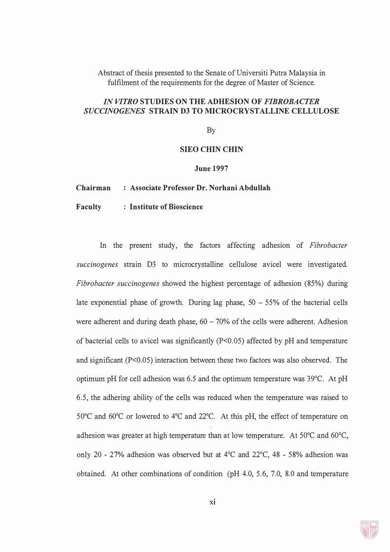
In the present study, the factors affecting adhesion of Fibrobacter
succinogenes strain D3 to microcrystalline cellulose avicel were investigated.
Fibrobacter succinogenes showed the highest percentage of adhesion (85%) during
late exponential phase of growth. During lag phase, 50 - 55% of the bacterial cells
were adherent and during death phase, 60 - 70% of the cells were adherent. Adhesion
of bacterial cells to avicel was significantly (P<0.05) affected by pH and temperature
and significant (P<0.05) interaction between these two factors was also observed. The
optimum pH for cell adhesion was 6.5 and the optimum temperature was 39°C. At pH
6 .5 , the adhering ability of the cells was reduced when the temperature was raised to
50°C and 60°C or lowered to 4°C and 22°C. At this pH, the effect of temperature on
adhesion was greater at high temperature than at low temperature. At 50°C and 60°C,
only 20 - 27% adhesion was observed but at 4°C and 22°C, 48 - 58% adhesion was
obtained. At other combinations of condition (PH 4.0, 5.6, 7.0, 8.0 and temperature
Xl
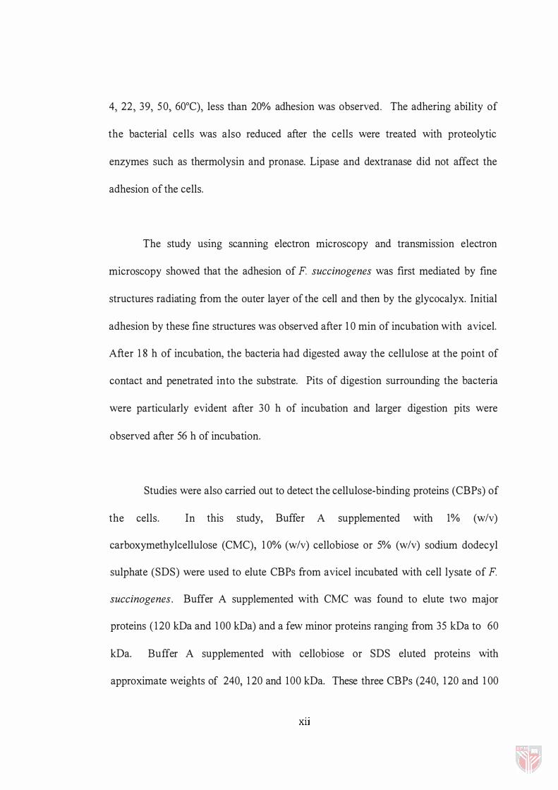
4, 22, 39, 50, 60°C), less than 20% adhesion was observed. The adhering ability of
the bacterial cells was also reduced after the cells were treated with proteolytic
enzymes such as thermolysin and pronase. Lipase and dextranase did not affect the
adhesion of the cells.
The study usmg scannmg electron microscopy and transmission electron
microscopy showed that the adhesion of F. succinogenes was first mediated by fine
structures radiating from the outer layer of the cell and then by the glycocalyx. Initial
adhesion by these fine structures was observed after 1 0 min of incubation with avicel.
After 1 8 h of incubation, the bacteria had digested away the cellulose at the point of
contact and penetrated into the substrate. Pits of digestion surrounding the bacteria
were particularly evident after 30 h of incubation and larger digestion pits were
observed after 56 h of incubation.
Studies were also carried out to detect the cellulose-binding proteins (CBPs) of
the cells. In this study, Buffer A supplemented with 1% (w/v)
carboxymethylcellulose (CMC), 1 0% (w/v) cellobiose or 5% (w/v) sodium dodecyl
sulphate (SDS) were used to elute CBPs from avicel incubated with cell lysate of F.
succinogenes. Buffer A supplemented with CMC was found to elute two major
proteins ( 1 20 kDa and 1 00 kDa) and a few minor proteins ranging from 35 kDa to 60
kDa. Buffer A supplemented with cellobiose or SDS eluted proteins with
approximate weights of 240, 1 20 and 100 kDa. These three CBPs (240, 1 20 and 1 00
XlI
kDa) were involved in the adhesion process of the cells as cells with reduced adhering
ability after being treated with proteolytic enzymes such as thermo lysin and pronase
did not show these CBPs. Other than possessing the ability to bind, the 240 kDa CBP
showed xylanase activity and the 1 20 kDa protein showed carboxymethylcellulase
(CMCase) activity in the cell lysate. The 1 00 kDa CBP did not show any of the two
enzyme activities shown by 240 kDa and 120 kDa CBPs.
Xlll
Abstrak tesis yang dikemukakan kepada Senat Universiti Putra Malaysia sebagai memenuhi keperluan untuk ijazah Master Sains.
KAJIAN IN VITRO KE AT AS PELEKATAN FIBROBACTER SUCCINOGENES STRAIN D3 P ADA SELULOSA MIKROHABLUR
OLEH
SIEO CHIN CHIN
Jun 1997
Pengerusi : Prof. Madya Dr. Norhani Abdullah
Fakulti : Institut Biosains
Kajian ke atas faktor-faktor yang mempengaruhi pelekatan Fibrobacter
succinogenes strain D3 pada selulosa mikrokristal avisel telah dijalankan.
Fibrobacter succinogenes menunjukkan peratusan pelekatan yang tertinggi (85%)
pada fasa pertumbuhan eksponen. Pada fasa pertumbuhan 'lag', 50 - 55% daripada
sel bakteria bersifat melekat dan pada fasa kematian, 60 - 70% daripada sel bakteria
bersifat melekat. Pelekatan sel bakteria pada avisel dipengaruhi oleh pH dan suhu,
dan kedua-dua faktor ini berinteraksi secara signifikan (P<0.05). Keadaan optimum
untuk pelekatan adalah pH 6.5 dan 39°C. Pada pH 6.5, keupayaan sel untuk melekat
berkurangan apabila suhu ditingkatkan ke 50°C dan 60°C atau diturunkan ke 4°C dan
22°C. Pada pH ini, suhu tinggi lebih mempengaruhi pelekatan sel berbanding dengan
suhu rendah. Pada 50°C and 60°C, hanya 20 - 27% pelekatan diperhatikan tetapi pada
4°C dan 22°C, 48 - 58% pelekatan diperolehi. Pada keadaan kombinasi yang lain (PH
4.0, 5.6, 7.0, 8.0 dan suhu 4, 22, 39, 50, 60°C), kurang daripada 20% pelekatan
XIV
diperolehi. Keupayaan untuk sel bakteria melekat juga berkurangan setelah sel
ditindak dengan enzim proteolitik seperti termolisin dan pronase. Lipase dan
dekstranase tidak mempengaruhi pelekatan sel.
Kajian dengan mikroskop elektron imbasan dan mikroskop elektron transmisi
menunjukkan peringkat awal proses pelekatan melibatkan struktur-struktur halus yang
berasal dari lapisan luar sel dan seterusnya dilakukan oleh glikokaliks. Pelekatan oleh
struktur-struktur hal us ini diperhatikan selepas sel dieram selama 1 0 min dengan
avisel. Selepas 1 8 jam pengeraman, bakteria mendegradasi substrat di tapak pelekatan
dan menembusi substrat tersebut. Zon degradasi di sekeliling bakteria diperhati
selepas 30 jam pengeraman dan bertambah besar selepas 56 jam pengeraman.
Kajian juga dijalankan untuk mengesan protein pelekat-selulosa pada sel.
Dalam kajian ini, larutan penimbal A yang ditambah dengan 1 % (w/v)
karboksimetilselulosa (CMC), 1 0% (w/v) selobiosa dan 5% (w/v) sodium dodesil
sulfat (SDS) telah diguna untuk menanggalkan protein pelekat-selulosa daripada
avisel yang dieram dengan sel lisat F succinogenes. Larutan penimbal A yang
ditambah dengan CMC berjaya menanggalkan dua protein utama ( 120 kDa dan 1 00
kDa) dan beberapa protein lain yang mempunyai berat molekul antara 35 kDa hingga
60 kDa. Selobiosa dan SDS menanggalkan protein yang mempunyai berat molekul
240 kDa, 1 20 kDa dan 100 kDa. Ketiga-tiga protein pelekat-selulosa (240 kDa, 1 20
kDa dan 1 00 kDa) ini terlibat dalam proses pelekatan kerana sel yang mempunyai
xv
keupayaan untuk melekat yang rendah setelah ditindak dengan enZlm proteolitik
seperti termolisin dan pronase tidak menunjukkan protein-protein pelekat-selulosa ini.
Selain daripada berupaya untuk melekat, protein pelekat-selulosa 240 kDa
menunjukkan aktiviti enzim xilanase dan protein pelekat-selulosa 1 20 kDa
menunjukkan aktiviti enzim karboksimetilselulase (CMCase) dalam sel lisat. Protein
pelekat-selulosa 1 00 kDa tidak mempunyai aktiviti enzim seperti yang ditunjukkan
oleh protein pelekat-selulosa 240 kDa dan 1 20 kDa.
xvi
CHAPTER I
INTRODUCTION
For thousand of years, ruminants have played a major role in farming
production and have provided mankind with meat, milk and clothing. Unlike man,
ruminants feed on fibrous plant materials and utilise the carbohydrate components of
the plant cell wall as major source of energy. The ability to digest and utilise plant
materials is made possible through a unique relationship between the microbes in the
rumen and the host animal.
The rumen, which is the largest compartment of the fore-stomach, is inhabited
by a complex microbial population and the ability of ruminants to utilise fibrous
materials for energy depends very much on the microbial activity and the symbiotic
interaction between the microbial population and the host animal.
Among the rumen microorganisms which consist of bacteria, protozoa, fungi
and probably other unknown microorganisms (Hungate, 1966), bacteria play a major
role in the degradation of cellulosic materials. Early studies using light microscopy
have shown that cavities developed in plant particles undergoing digestion in the
rumen contain many bacteria (Baker and Harris, 1947). Later, studies using
transmission electron microscopy and scanning electron microscopy revealed the

2
presence of many different morphological types of rumen bacteria adhering to the
plant particles while degradation was in process (Akin et aI ., 1 974; Akin and Amos,
1 975; Akin, 1 976). Digestion of plant cell walls in the rumen is predominantly by
cellulolytic bacteria such as F. succinogenes, Ruminococcus albus and Ruminococcus
flavefaciens (Amos and Akin, 1 978; Windham and Akin, 1984). The close association
between the rumen bacteria and the plant tissues often observed during digestion of
plant materials (Cheng et aI ., 1 983) suggests that bacterial adhesion is an important
aspect of fibre degradation. Loose and irregular pattern of bacterial cell adherence on
plant materials often results in less cell wall degradation (Kudo et aI., 1 987).
Adhesion has also been reported to be substrate-specific and it has been postulated
that a substrate-binding factor is involved in the recognition of the substrate (Gong
and Forsberg, 1 989).
Since bacterial adhesion is a prerequisite for the breakdown of cellulosic
materials, the mechanisms of adhesion should be studied in order to improve and
enhance the degradation of cellulose. Further characterisation of the cellular adhesion
mechanisms may enable genetic engineering to play a dominant role in the
manipUlation of feed digestion through regulation of the genes responsible for the
adhesion of microorganisms to the feed materials.
In view of this, an in vitro study on the adhesion of F. succinogenes, which is
one of the maj or cellulolytic bacteria in the rumen (Bryant and Doetsch, 1 954;
Malburg and Forsberg., 1 993), to crystalline cellulose avicel was carried out to

3
determine the factors involved in the mechanism of adhesion. The specific objectives
of this study are:
a) to study the effects of several physiochemical factors such as the growth phase of
bacteria, pH, temperature and enzyme treatments on the adhesion of F.
succinogenes;
b) to observe the adhesion of F. succinogenes on avicel using scanning electron
microscopy and transmission electron microscopy; and
c) to detect the substrate-binding factor i.e. cellulose-binding factor of F.
succinogenes.
CHAPTER II
LITERATURE REVIEW
The Rumen Microorganisms and Their Activity
The rumen, which is one of the most important compartment of the ruminant's
stomach, is inhabited by a complex microbial population. It contains one of the most
varied and dense microbial populations known in nature. Of these microorganisms,
fungi, protozoa and bacteria are known to be the main inhabitants in the rumen
(Czerkawski, 1986). They play a role in the colonisation and degradation of feed
material in the rumen and their presence significantly affects the performance of the
animal in the utilisation of feed materials. The animal provides a suitable niche for
the microorganisms and, in return, the microorganisms degrade the feed ingested by
the animal and provide valuable metabolites to the host animal. The population of
these microorganisms often fluctuate with the intake of feed and types of diet of the
animal (Eadie, 1962).
Rumen Protozoa
The population of rumen protozoa has been estimated to be about 105-1 06/ml
of rumen contents (Tsuda, 1976), and they have been observed to play a significant
role in the primary degradation of plant fragments (Williams, 1989). About 25 - 30%
of the total fibre degradation in the rumen is contributed by the protozoa (Williams
4
5
and Coleman, 1992). The population density of rumen protozoa is dependent on the
frequency of feeding. Higher population of protozoa is observed in animals which are
fed regularly and frequently throughout the day (Bonhomme, 1990).
Generally, protozoa can be divided into two classes: ciliates and flagellates.
Cilliates are the predominant protozoa in the rumen (Bonhomme, 1990). They are
made up of two groups, namely, the holotrich and the entodiniomorphid (Williams,
1986). The entodiniomorphid ingests and digests particulate plant materials, while the
holotrich uses mainly soluble carbohydrates (Williams, 1986). Hence, the occurrence
of holotrich and entodiniomorphid is very much determined by the type of diet of the
host animal. For instance, the numbers of holotrich are higher when the diet contains
readily available soluble carbohydrates such as sugar and molasses (Valdez et al.,
1977).
Rumen protozoa have been reported to play a number of roles in the rumen.
Other than being able to degrade cellulose (Coleman, 1985), protozoa are able to
degrade and metabolise the principle protein, carbohydrate and lipid components of
the feed materials ingested by the host animal (Bonhomme, 1990). Protozoa also
contributes to the microbial turnover in the rumen by predation process where
bacteria, which are the nitrogen source for the protozoa, are engulfed by the protozoa.
Protozoa play an important role especially in ruminants fed high sugar. The holotrich
assimilates soluble sugar and store them as amylopectin (Coleman, 1979). This
reduces the rate of fermentation and thus prevents accumulation of high level of
6
lactate which may cause a rapid lowering of pH in the rumen. In addition to that,
rumen protozoa have been implicated to be involved in copper (Cu) metabolism
where their presence may alleviate Cu toxicity of the animal (I van et aI . , 1 991 ).
In spite of the roles the protozoa play in the rumen, the protozoa have been
considered to be unimportant in ruminant production. Protozoa are found to be
unnecessary in improving animal performance since defaunation results in higher
growth rate of the animal (Romulo et aI. , 1 988). Furthermore, the unavoidable
predatory behaviour of protozoa reduces bacterial growth efficiency and hence reduce
the net yield of bacterial amino acids available for intestinal absorption. As 60 - 80%
of the protozoa were lysed and degraded in the rumen (Leng, 1 988), the protozoa are
not considered as an important source of protein to the animal.
Rumen Fungi
Anaerobic rumen fungi are discovered in the rumen only about two decades
ago (Orpin, 1 975; Bauchop, 1 979). Five genera of anaerobic rumen fungi have been
identified and they are divided generally into two groups, the monocentric and the
polycentric. The monocentric fungi produce a single sporangium and an anucleate
rhizoidal system while the polycentric fungi produce an extensive network of
rhizomycelium with many sporangiophores on which sporangia develop. Out of the
five genera of anaerobic rumen fungi, three genera are in the monocentric group, viz. ,
Neocallimastix, Piromyces and Caecomyces (Orpin, 1 975; 1976; 1 977), and two in the
7
polycentric group, viz. , Orpinomyces (Barr et ai. , 1989) and Anaeromyces (Breton et
ai. , 1 99 1 ) [= Ruminomyces (Ho et aI. , 1 990)].
Rumen fungi contribute significantly to the prime function of the rumen. The
fungi are actively involved in the digestion of plant cell walls to provide fermentation
products for the nutrition of the host animal (Orpin and Ho, 1991 ). All the species of
rumen fungi isolated so far are capable of fermenting structural carbohydrates of plant
cell walls, especially lignocellulosic tissues (Orpin, 1 98 1 ; Akin et aI . , 1 983) . In vitro
studies of some species of rumen fungi on the digestion of wheat straw showed that
about 40 - 50% of the dry weight of wheat straw fragments was lost in four days.
About 50% of the cellulose and hemicellulose was digested and approximately 1 6%
ofthe lignin in the plant fragments was degraded (Orpin, 1984).
The manner in which rumen fungi colonise the plant materials in the rumen
differs markedly from that of the rumen bacteria. The fungus uses its rhizoidal system
or hyphae to attach to plant fragments. It was observed that initial colonisation often
occurs at the sites of damaged tissues and at the stomata (Orpin, 1 977; Akin et aI.,
1 983) . Lignified cell walls or recalcitrant tissues such as schlerenchyma and vascular
tissues are preferentially colonised (Orpin and Joblin, 1 988). Upon attachment of the
fungus, the rhizoids or hyphae penetrate into the tissue, colonising the sclerenchyma
and vascular tissues, and eventually breaking the tissues into smaller particles (Ho et
aI., 1 988) which in tum will enhance colonisation and digestion by bacteria. The
anaerobic fungi penetrate plant cell walls with the help of structures called appresoria
8
which help the rhizoids in piercing the plant cell wall (Ho et. aI., 1988). It has been
observed that the penetration of cell walls by the rhizoids not only caused degradation
of tissues, which provide surfaces for secondary attack of other rumen
microorganisms, but it also prevent the fungus from being washed out by the liquid
phase of the rumen contents.
Although the population of fungi ( 103 - 105 Iml of rumen content) is the lowest
among the rumen microorganisms, the presence of fungi in the rumen is essential
especially in animal fed low quality forage. Orpin and Joblin ( 1988) suggested that the
presence of fungi is not necessary in animal fed low fibre diet as the rumen bacteria,
with a higher population, are capable of digesting this low fibre materials efficiently.
Rumen Bacteria
Rumen bacteria is considered to be one of the most important microbial
groups in terms of number, total activity, diversity, consistency in fibre degradation
and their ability to survive unfavourable conditions (Akin et aI., 1993). In fact, it has
been reported that cellulolysis in the rumen is primarily due to the activities of the
rumen cellulolytic bacteria (Weimer, 1996).
It is estimated that the population density of rumen bacteria is about 1010 - lOll
bacteria Iml of rumen content (Trinci et aI., 1994). The bacteria range from as small
as 1 - 2 I-Lm to as large as 3 - 6 I-Lm in diameter (Czerkawski, 1986), and are made up
of all the major morphological forms of bacteria. They may be Gram positive or
Related Documents