I28. STUDIES IN IMMUNOCHEMISTRY 6. THE USE OF PHENOL AND OF ALKALI IN THE DEGRADATION OF ANTIGENIC MATERIAL ISOLATED FROM BACT. DYSENTERIAE (SHIGA)l BY W. T. J. MORGAN AND S. M. PARTRIDGE2 From the Biochemical Department, Lister Institute of Preventive Medicine, London (Received 31 October 1941) EARLIER work has shown [Morgan, 1937; Morgan & Partridge, 1939, 1, 2; 1940, 1; Partridge & Morgan, 1940] that the antigenic substance which is responsible for the dominant serological properties of the 'smooth' form of Bact. dysenteriae (Shiga) can be extracted from the micro-organism by means of diethyleneglycol. The properties of the antigenic complex were described in some detail and three major components were isolated and identified as polysaccharide, phospholipin and a protein or polypeptide-like substance. By the action of cold, neutral formamide, the phospholipin component was removed and there remained a complex of specific polysaccharide and the polypeptide-like component which still possesged powerful antigenic properties. The material dissolved in phosphate buffer (pH 7-5-8.0) solution containing 0Q85 % NaCl to yield a clear 0*2 % solution which could readily be passed through a sterile Berkefeld candle without appreciable loss. The protein component of the original antigenic complex on the other hand could be largely removed by the action of trypsin but the resulting polysaccharide-phospholipin complex possessed no significant antigenic capacity, as measured by the production of 'Shiga' agglutinins and precipitins. A number of other experimental procedures designed to separate the com- ponents of the antigenic complex were also mentioned. The material was shown to yield clear solutions in cold anhydrous formic acid and in concentrated solutions of phenol. From these solvents the antigenic material could be largely recovered by precipitation with alcohol or acetone. Details of our work with the latter solvent are now given with some observations on the action of alkali on the antigenic complex. EXPERIMENTAL It has already been shown [Morgan & Partridge, 1940] that the protein or polypeptide-like component of the antigenic complex of Bact. dysenteriae (Shiga) can be removed by the action of trypsin or by the dissociating action of anhydrous formamide. The experimental work described below extends these observations and indicates that this component can also be eliminated from the antigenic comnplex by the action of phenol or by repeated precipitation of the complex from alkaline solution by alcohol. All these procedures, however, destroy the capacity of the resulting substance to induce the formation of 'Shiga' agglu- tinins and precipitins.- As a result of the work to be described in this paper the so-called polypeptide-like component of.the antigenic complex is now con- sidered to be a conjugated protein and therefore the former term 'polypeptide- like' component will no longer be used. For Part 5 see Partridge & Morgan [1940]. 2 Beit Memorial Research Fellow. 1140

Welcome message from author
This document is posted to help you gain knowledge. Please leave a comment to let me know what you think about it! Share it to your friends and learn new things together.
Transcript

I28. STUDIES IN IMMUNOCHEMISTRY6. THE USE OF PHENOL AND OF ALKALI INTHE DEGRADATION OF ANTIGENIC MATERIALISOLATED FROM BACT. DYSENTERIAE (SHIGA)l
BY W. T. J. MORGAN AND S. M. PARTRIDGE2From the Biochemical Department, Lister Institute of Preventive
Medicine, London
(Received 31 October 1941)
EARLIER work has shown [Morgan, 1937; Morgan & Partridge, 1939, 1, 2;1940, 1; Partridge & Morgan, 1940] that the antigenic substance which isresponsible for the dominant serological properties of the 'smooth' form ofBact. dysenteriae (Shiga) can be extracted from the micro-organism by means ofdiethyleneglycol. The properties of the antigenic complex were described in somedetail and three major components were isolated and identified as polysaccharide,phospholipin and a protein or polypeptide-like substance. By the action of cold,neutral formamide, the phospholipin component was removed and there remaineda complex of specific polysaccharide and the polypeptide-like component whichstill possesged powerful antigenic properties. The material dissolved in phosphatebuffer (pH 7-5-8.0) solution containing 0Q85 % NaCl to yield a clear 0*2 %solution which could readily be passed through a sterile Berkefeld candle withoutappreciable loss. The protein component of the original antigenic complex onthe other hand could be largely removed by the action of trypsin but theresulting polysaccharide-phospholipin complex possessed no significant antigeniccapacity, as measured by the production of 'Shiga' agglutinins and precipitins.
A number of other experimental procedures designed to separate the com-ponents of the antigenic complex were also mentioned. The material was shownto yield clear solutions in cold anhydrous formic acid and in concentratedsolutions of phenol. From these solvents the antigenic material could be largelyrecovered by precipitation with alcohol or acetone. Details of our work with thelatter solvent are now given with some observations on the action of alkali onthe antigenic complex.
EXPERIMENTALIt has already been shown [Morgan & Partridge, 1940] that the protein or
polypeptide-like component of the antigenic complex of Bact. dysenteriae (Shiga)can be removed by the action oftrypsin or by the dissociating action of anhydrousformamide. The experimental work described below extends these observationsand indicates that this component can also be eliminated from the antigeniccomnplex by the action of phenol or by repeated precipitation of the complexfrom alkaline solution by alcohol. All these procedures, however, destroy thecapacity of the resulting substance to induce the formation of 'Shiga' agglu-tinins and precipitins.- As a result of the work to be described in this paper theso-called polypeptide-like component of.the antigenic complex is now con-sidered to be a conjugated protein and therefore the former term 'polypeptide-like' component will no longer be used.
For Part 5 see Partridge & Morgan [1940]. 2 Beit Memorial Research Fellow.1140

STUDIES IN IMMUNOCHEMISTRY
The action of phenol on the antigenic complex
In some early experiments attempts were made to remove the protein com-ponent of the antigenic complex by thoroughly extracting a 2-3 % aqueoussolution of the primary diethyleneglycol extraction product [Morgan & Partridge,1940, 1] with a strong solution of phenol. The upper aqueous layer was removedand again treated with liquid phenol but the product finally recovered from theaqueous layer still contained protein and represented a large proportion of theoriginal material. Examination of the resulting product revealed, however, thatthe phospholipin component had been removed during the phenol extraction.
During some attempts to separate from crude preparations of the antigen,agar or agar hydrolysis products, such as kanten, by means of their insolubilityin 90 % liquid phenol, it was observed that the antigenic complex was readilysoluble in phenol of this strength and could be recovered from phenol solution byprecipitation with alcohol. At first no attempt was made to fractionate thephenol-soluble material but it was noticed that after several treatments withphenol in this manner there was a significant decrease in the nitrogen contentof the product finally isolated. Examination of the successive alcoholic phenolmother liquors showed that the amino-acid complex was being graduallyeliminated. It was also observed that the antigenicity of the complex recoveredafter phenol treatment was considerably reduced, presumably owing to theelimination of the protein, and for this reason the degradation process was notextended, at that time, to eliminate the whole of the protein component. Atthis stage in the investigation Palmer & Gerlough [1940] described a methodwhereby the 'O' antigenic complex of Bact. typhosum could be recovered fromthe dry bacterial cells or from antigenic preparations by extraction of thematerial with 88 or 95 % liquid phenol. The antigenic complex remained in-soluble in phenol of this strength. Our earlier experiments on the action ofphenol on the antigenic complex of Bact. dysenteriae (Shiga) were continued,using a technique very similar to the one described by these authors.
Dissociation of the antigenic complex. A preparation (1.25 g.) of the antigeniccomplex of Bact. dysenteriae (Shiga), [oc] 61+ 65+ 50, N, 4.4 %, after treatmentwith formamide to remove the phospholipin component, was treated with twoquantities of 30 ml. of 90 % phenol. The material dissolved completely and couldbe recovered from the combined phenol solutions by the addition of alcohol.The greater part (0-95 g.) of the dissolved substance was thrown out of solutionbetween the alcohol levels 0 and 33 % by, volume. There was no precipitateformation on increasing the alcohol concentration from 33 to 50 %. The alcoholicsupernatant was dialysed to remove phenol and alcohol and the resulting aqueoussolution was taken to dryness in vacuo. This fraction gave strong biuret andSakaguchi tests. The material thrown out of 90 % phenol solution between 0and 33 % alcohol levels contained 3-4 % N and 3-8 % ash. It was again treatedwith 90 % phenol but the volume of phenol solution was reduced so that a smallamount (5-10 %) of the material was left undissolved. Once again the mainportion of the substance was recovered from the phenol solution by the additionof alcohol to 33 %. The material showed [X]M61+ 68 + 40, N, 3-0 %, P, 0-64 %.The. fraction was then extracted with 92 % phenol (30 ml.) in an attempt toseparate a larger phenol-insoluble portion. The insoluble material weighed 0-32 g.and contained 31 % N, 0-64 %.total P and no inorganic P. The composition isseen to be very similar to that of the 90 %° phenol-soluble material which wasprecipitated by the addition of alcohol up to 33 O/ . A 0-5 % aqueous solution ofthe material insoluble in 92% phenol was-too opalescent for accurate deter-
1141
W. T. J. MORGAN AND S. M. PARTRIDGE
mination of specifiQ rotation. The material precipitated from solution in 92 %phenol by 33 % alcohol gave the following analytical data: [ocLx61+72+30;N, 2-97 %; P, 0 77 %; inorganic P, nil. A quantitative Sakaguchi test carriedout according to the method of Jorpes & Thoren [1932] showed the material tocontain at least 0415 % arginine. The phenol extraction of the material insolublein 92 % phenol was repeated twice but each successive phenol-insolfible portionshowed a similar rotation and N content and the Sakaguchi colour reactionindicated that in each preparation about 0 15-0-30 % of arginine still remainedin the phenol-insoluble fractions. The analytical figures for the material showedix]5461+76 +40 (c, 0.5) N, 2.95 %; P, 0.75 %.
After the above phenol extraction experiments had been completed work onthe recombination of the non-antigenic specific polysaccharide and protein com-ponents of the 'Shiga' antigenic complex [Partridge & Morgan, 1940] indicatedthat from solution in certain organic solvents these components could be pre-cipitated in the form of an antigenic complex that possessed chemical, physicaland immunological properties very similar to those of the original polysac-charide-protein complex derived from the native antigen. These observationssuggested that the recovery of phenol-soluble and dissociated material byprecipitation with alcohol caused the free polysaccharide and protein componentsto recombine for the most part, and thus the material precipitated from phenolsolution by alcohol was composed to a considerable extent of a complex of thesecomponents and not of a simple mixture of polysaccharide and protein. Thealcoholic phenol supernatant fluid, however, usually contained some of the freeprotein component of the antigenic complex which could be readily recoveredafter dialysis of the fluid to remove alcohol and phenol. The existence of theuncombined amino-acid complex in the alcoholic phenol offers an explanationfor the lower protein content of the material precipitated from phenol solution byalcohol.
The experiments described above failed to show, in a clear cut manner,whether the protein component could be preferentially extracted from theantigenic complex by simple leaching with phenol solution. In order to obtainfurther information on this point, the experiment was repeated using a 95%phenol solution for extraction. The antigenic material was only sparingly solublein this solvent and thus, even after several extractions with phenol, a con-siderable part of the complex remained undissolved and was therefore availablefor analysis and for antigenicity tests.
A preparation of the primary diethyleneglycol extraction product wasthoroughly treated with cold anhydrous formamide (M.P. 2°) to remove thephospholipin component and, as a result of the thorough treatment, the productwas found to have lost part of its polysaccharide as well as the phospholipincomponent. The analytical figures were 6-4% N; 1-4 % P; inorganic P, nil;ash, 5*0 %. A similar polysaccharide-protein complex containing a high pro-portion of the latter component has already been described in detail [Morgan &Partridge, 1940, 1]. The specific rotation of the material could not be deter-mined with accuracy owing to a heavy bluish opalescence but was estimated tobe + 30+5°. It has frequently been observed that preparations of antigenicmaterial possessing a high content of the protein component show an intensebluish opalescence when in aqueous alkaline solution.
The phospholipin-free material (1.0 g.) was treated with 30 ml. of 95%phenol solution and after thorough shaking was set aside for 30 min. at roomtemperature. The material swelled up to a thin transparent jelly but wasonly sparingly soluble in the solvent. The suspension was centrifuged and the
1142
STUDIES IN IMMUNOCHEMISTRY 1143
insoluble material was resuspended in fresh 95 % phenol. After an hour thebulk of the preparation was again recovered and resuspended in fresh phenolsolution. The process of extraction with phenol was repeated seven times.Methyl alcohol was added to each phenol supernatant fluid to make a concen-tration of 3 % and each mixture was dialysed separately until free from phenol.The addition of alcohol produced no precipitate and was made to prevent thestrong phenol solution from crystallizing during manipulation and to increasethe rate of diffusion of phenol through the cellophane. Some material insolublein dilute phenol' solution separated during the later stages of dialysis and finallyremained insoluble after the phenol had been completely removed. The contentsof each of the cellophane bags were centrifuged separately to remove insolublematter, the clear solutions were evaporated in vacuo to a- small volume, frozenat -10° and taken to dryness in vacuo from the frozen state.
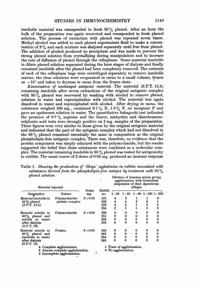
Examination of unchanged antigenic material. The material (S.P.T. 12A)remaining insoluble after seven extractions of the original antigenic complexwith 95 % phenol was recovered by washing with alcohol to remove phenol,solution in water and reprecipitation with alcohol. The material was againdissolved in water'and reprecipitated with alcohol. After drying in vacuo, thesubstance weighed 350 mg., contained 6.1% N, 1V % P, no inorganic P andgave an opalescent solution in water. The quantitative Sakaguchi test indicatedthe presence of 0*7 % arginine and the 'biuret, ninhydrin and diazobenzene-sulphonic acid tests were strongly positive on 2 mg. samples of the preparation.These figures werie very similar to those given by the original antigenic materialand indicated that the part of the antigenic complex which had not dissolved inthe 95 %'phenol remained essentially the same in'composition as the originalphospholipin-free antigenic complex. There was, therefore, no evidence that theprotein component was simply admixed with the polysaccharide, but the resultssupported the belief that these substances were combined as a molecular com-plex. The material remaining insoluble in 95% phenol was tested for antigenicityin rabbits. The usual course of 3 doses of 0-05 mg. produced an immune response
Table 1. Showing the production of 'Shiga' agglutinins in rabbits inoculated withsubstances derived from the phospholipin-free antigen by treatment with 95%
Material injected
Des,riptionMaterial insoluble in95% phenol(S.P.T. 12A)
NaturePolysaccharide-protein complex
Doim3
3 x C
Material soluble in Polysaccharide 6 xC95 /% phenol andsoluble in waterafter dialysis(S.P.T. 16)
Material soluble in Protein 6 x(
95% phenol andinsoluble in waterafter dialysis(S.P.T. 13)
4 Complete agglutination.3 Almost complete agglutination.2 Incomplete agglutination.
ses RabbitLg. no.
0)05 231232233234
a 05 250251252253
Dilution of immune serum givingagglutination with formolizedsuspension of Bact. dy8enteriae
(Shiga)
1: 20 1: 40 1: 80 1: 160 1:3204 3 1 1 04 4 3 2 14 2 1 0 02 1 1 0 00 0 0 0 0
0 0 0 0 0
0 0 0 0 0
0 0 0 0 0
0 05 242 0 0 0 0 0243 0 0 v 0 0 0244 0 0 0 0 0245 0 0 0 0 0
1 Trace of agglutination.0 No agglutination.
phenol solution

W. T. J. MORGAN AND S. M. PARTRIDGE
in each of the four animals immunized (Table 1, animals 231, 232, 233 and 234).After a further course of immunization the sera of all rabbits gave precipitationwhen mixed with equal volume of 1 :100,000 dilution of the 'Shiga' specificpolysaccharide.
Isolation of the polysaccharide component. The results of an examination ofthe phenol-soluble fraction that was also soluble in water after dialysis are givenin Table 2. It appears that the water-soluble material extracted by phenol from
Table 2.- Showing the amount and composition of material extracted from theantigen of Bact. dysenteriae (Shiga) by strong solutions of phenol
Total*Phenol Volume Weight Nitrogen phosphorus Arginineextract ml. mg. % % %
I 30 54-2 1-15 -II 30 75-3 1-68 0-55 005-001III 30 70 3 1-78 0*74 0-05-0*1
IV and V 2 x20 86-4 2-27 1.00 010VI 30 78-4 2-38 0-87 007VII 30 67-0 2-10 0-98 0 01
* Inorganic P, 0.0%.
the antigenic complex was almost entirely polysaccharide in nature. Traces ofarginine remained in the soluble material but this was to be expected in view ofthe very simple treatment involved. The combined extracts weighed 431-6 mg.,and 5 mg. portions of the material failed to give a positive biuret, ninhydrin,oc-nitroso-3-naphthol or diazobenzenesulphonic acid test. The Molisch test wasstrongly positive. Fractions II-VII after solution in water were combined,made distinctly acid with acetic acid; spun to remove a small amount of in-soluble matter and precipitated by the addition of several volumes of alcohol.The preparation showed an opalescence which rendered the determination ofspecific rotation difficult. The substance contained 2-05% N, 0-81 % P and noinorganic P. The material was tested in rabbits for antigenicity; 3 animalsreceived 6 doses each of 0-05 mg. intravenously. There was slight productionof specific agglutinins and precipitins although the immune response wasnegligible when compared with that elicited by the original phospholipin-freeantigenic complex. The material was again dissolved in phenol solution (92 %)and after the addition of 3 % methyl alcohol, was dialysed in a cellophane baguntil free from phenol. The solution was freed from a small amount of insolublematter, concentrated to 10 ml. and taken to dryness in vacuo. A solution (1 %)of the substance (S.P.T. 16) possessed slight opalescence, but the material(10 mg.) gave a negative Sakaguchi test. Arginine (0.01 mg.) under the sameconditions gave a definite rose-pink coloration. Analytical data: [ogC5461+ 84±3'(0-5 %O in H20); N, 1-82 %; P, 0 75 %, inorganic P, 0-0 %. The material was againtested for antigenicity in 4 rabbits; there was no detectable immune response(Table 1, animals 250, 251, 252 and 253).
Isolation of the protein component. The material which separated duringdialysis of the phenol extracts (I-VII) was collected and extracted with 2 ml.N NaOH. The insoluble residue was again extracted with 1 ml. 0 1N NaOH andfinally washed with several 1 ml. quantities of distilled water. The residueappeared to be largely inorganic, weighed 60 mg. and contained 0-38% N. Thealkali-soluble material and the water washings were mixed together, acidifiedwith acetic acid and the resulting precipitate 'was collected, dissolved by theaddition of alkali to pH 8-0 and again recovered by the addition of acid. The
1144
STUDIES. IN IMMUNOCHEMISTRY 1
precipitate was thoroughly washed with 0-05N acetic acid, alcohol and dried invacuo. The substance (S.P.T. 13) weighed 120 mg., gave an intense biuret test andaccording to an arginine determination made by Jorpes & Thoren's [1932]modification of Sakaguchi's test, contained about 5 % of this amino-acid. Thematerial gave an almost colourless solution (0-5 % in 0- N NaOH), possessed astrong negative rotation, [OC]546,-60 + 30 (c, 1 in 0O N NaOH), contained 12-3 %N, no appreciable quantity of P, and a 5 mg. sample gave a very faint nitro-prusside test for S after Na fusion. A diazobenzenesulphonic acid test on 1 mg.of the preparation gave a strong red coloration in 2-3 sec.; a control test em-ploying the reagents alone gave a pale yellow colour only during the first 1-2 min.The fraction consists, therefore, of the protein that was originally associated withthe polysaccharide that had been recovered in the phenol-soluble fractions I-VII.The results of antigenicity tests on this material are shown in Table 1 (animals242, 243, 244 and 245); there was no detectable immune response. The figuresgiven in Table 2 indicate that the total weight of the antigenic material dis-solved in each 30 ml. of 95 % phenol used for extraction remained approximatelyconstant and that in all 530 mg. of the antigenic complex dissolved in 190 ml. ofphenol solution. -
An examination of the protein isolated by this method showed that thesubstance was more-soluble in dilute acids than samples of protein prepared byacetic acid hydrolysis of the 'Shiga' antigen and that in this property the twoprotein preparations appeared to be different. For further investigation aquantity of the material was therefore prepared by the following simplifiedprocess. The diethyleneglycol primary extraction product- (400 mg.) was dis-solved in 90 % phenol solution (20 ml.) and a small insoluble residue was removed.The clear solution was dialysed against running tap water- in a cellophane baguntil free from phenol and the precipitate which formed in the bag was collected.After extraction, first with alcohol and then with alcohol-ether mixture, toremove phospholipin, the material was dissolved in dilute NaOH and the smallinsoluble residue was removed by centrifuging. The substance was then subjectedto three successive precipitations from dilute alkaline solution with acetic acidat pH 4-6, the protein thrown out of solution being redissolved each time inwater by the addition of a minimum quantity of dilute NaOH. It was thennoticed that the protein exhibited a marked solubility in N acetic acid and inHCI at about pH 2. Addition of further HOl, however, produced a heavy pre-cipitate presumably due to the production 'of an insoluble hydrochloride.- Theprotein was further purified by precipitating twice from an alkaline solution withHC1 to make a final concentration of N HCI. The insoluble hydrochloride waswashed with absolute alcohol t9 remove the excess of HCI, and the substancewas then found to' dissolve in distilled water to yield a clear colourless solutionshowing an acid reaction. The protein was finally precipitated from solution by-the addition of sufficient solid sodium acetate to raise the pH to about 4-5. Theprotein component recovered in this manner was found to be free from P,showed [ao]M61-74+20. (c, 1 in 0-1N 'NaOH) and contained 130% N. Theprotein could be precipitated from solution in N acetic acid by means of tri-chloroacetic acid, metaphosphoric acid, salicylsulphonic acid and other proteinprecipitants. Addition of common salt or other soluble chlorides, evoked theprecipitation of the insoluble hydrochloride.
The analytical figures which we have previously given for the 'protein'component obtained from the diethyleneglycol primary extraction product byacid hydrolysis are significantly different from those obtained for the phenol-dissociated protein component. The former substance could be reprecipitated
1145

W. T. J. MORGAN AND S. M. PARTRIDGE
many times from variable concentrations of the acid without appreciable loss,whereas the phenol-dissociated protein was readily soluble at about pH 2-5. It isapparent, therefore, that the proteins isolated from the same antigenic materialby two different methods are substantially different in character and it appearsthat one product is most probably a degradation product of the other. Thepossibility that denaturation might be responsible for the difference was firstconsidered, but the hypothesis appeared unlikely in view of the considerabledifference in the analytical figures. Furthermore, it was found that the proteinprepared by the phenol method, when dissolved to yield a clear solution in Nacetic acid, could be heated for many hours at 950 without causing any pre-cipitation to occur. The following experiment, however, indicates that thesimple protein obtained by the phenol dissociation of the antigenic complex, isa derivative of the conjugated protein which is obtained by heating the antigeniccomplex with dilute acetic acid.
Isolation of the simple protein from the conjugated proteinA sample of the crude conjugated protein, prepared by hydrolysis of the
antigenic complex with dilute acetic acid according to the usual method, wasfreed from traces of fatty impurities by extraction with an alcohol-ether mixtureand was subsequently precipitated from faintly alkaline solution with aceticacid. The procedure was designed to limit the manipulations to the minimumconsistent with the isolation of an apparently homogeneous material. The sub-stance contained 11-2% N, 1-07 % P, and possessed [oc],461-48 +30 (c, in 0 1NNaOH). The material was not soluble in N acetic acid, even on warming. Theconjugated protein (400 mg.) thus obtained dissolved readily in 90 % phenolsolution (10 ml.) and was largely thrown out of solution by the addition of ethylalcohol (10 ml.). The precipitate was redissolved in phenol (10 ml.), again thrownout of solution by the addition of an equal volume of absolute alcohol, thoroughlywashed with alcohol and finally dissolved in water by the addition of a few dropsof NaOH solution. A small insoluble residue was removed and an equal volumeof 2N HC1 was added to precipitate the protein from solution. The precipitatewas washed with alcohol to remove the excess of acid. On addition of distilledwater the precipitate slowly dissolved to a rather opalescent solution. Theprotein was again recovered by addition of sodium acetate to reduce the acidityof the solution to pH 4*5. After washing with alcohol the protein was taken upin 90% phenol, a small fraction which remained undissolved was removed andthe clear supernatant fluid was precipitated by addition of ethyl alcohol to 50%concentration. The protein was once again precipitated from phenol solution byalcohol and again recovered from aqueous solution. The protein was then foundto dissolve completely in N acetic acid on warming (35-40') to yield a clear.,colourless solution. The protein was finally recovered as the insoluble hydro-chloride. After washing with alcohol the hydrochloride yielded a clear solutionof acid reaction on addition of distilled water, from which it was precipitated atpH 4*5 by the addition of sodium acetate. The material recovered (120 mg.)contained 13-8 % N, no P, and showed a specific rotation [O]5461-88±2° (1 %solution in 01N NaOH). The analytical figures and general properties of thepurified protein are distinctly different from those of the starting material, butshow a close agreement with those of the substance prepared by phenol dis-sociation of the whole antigenic complex. It is not considered that either of thepreparations isolated in this manner are pure but the simple amphoteric proteinhas been obtained in larger quantities by other methods and is described in moredetail later in this paper.
1146

STUDIES IN IMMUNOCHEMISTRY
Owing to the difficulty of establishing the purity of the conjugated protein itis not clear whether all the changes in the analytical data that accompany thechanged solubility properties of the protein during the rather long series ofmanipulations described above can be correlated with the removal of a singlechemical component that might be termed the prosthetic group. In previouspurification procedures the conjugated protein was dissolved in a small volumeof cold 0.1N NaOH and reprecipitated with dilute HCI. This procedure wasusually carried out five or six times and the substance was sometimes furtherpurified by precipitation as sodium salt by the addition of alcohol to the alkalinesolution of the material. Isolated in this manner, the protein preparationscontained 11-12 % N and showed [OC]w61-45 to -50°. The P content fell steadilyduring purification and the final product usually contained not more than 0*2 % P.From these observations it appears that the P of the protein component obtainedby acetic acid hydrolysis of the antigenic complex can be largely removedwithout causing a significant increase in the specific rotation or nitrogen contentof the protein substance. On the other hand repeated phenol treatment of theprotein component of the antigen liberated by acetic acid hydrolysis not onlyremoves completely any P present but also causes the resulting protein topossess a higher laevorotation (-82 + 59), a higher N content (13-5-14-0 %) andto show a changed solubility at pH 2-0-2-5. The influence of these changes on theimmunological properties of the preparations will be described later in the paper.
Extraction of Bact. dysenteriae (Shiga) with 90% phenol solution\ In view of the marked solubility-of the 'Shiga' antigenic material in 90 O/o
phenol solution, and the observed insolubility of agar and of nucleic acid in thissolvent, it was decided to attempt a phenol extraction, in the first instance, of asample of organisms that had already been repeatedly extracted with diethylene-glycol, but which contained a further quantity of antigenic material. Organismsthat had already been in contact with anhydrous diethyleneglycol at 0-2° formany months and which had been repeatedly extracted with fresh diethylene-glycol and had yielded about 7 % of their dry weight as antigenic material wereobserved to have remained morphologically intact.
The heavy paste of organisms collected from the bowl of the Sharples super-centrifuge after removing the excess of diethyleneglycol was repeatedly trituratedwith acetone, washed with this solvent and dried in-vacuo. The fine white powder(132 g.) so obtained was shaken with 90% phenol solution (800 ml.) and afterstanding for 96 hr. with occasional shaking, the bacterial residue was againcollected in a Sharples centrifuge. The clear supernatant was passed through aDoulton filter candle and mixed with 3 vol. of alcohol. The precipitate whichformed was then suspended in water and dialysed against water in a cellophanebag until free from phenol. The bag finally contained a heavy white precipitateand a strongly opalescent supernatant solution. The precipitate .was removed bycentrifuging, dried after washing with water and alcohol and weighed. Thesupernatant was evaporated to dryness in vacuo from the frozen state. Table 4shows the yields obtained with six successive extractions of the bacteria with90% phenol. As the extraction proceeded the organisms disintegrated untilfinally the bacterial residue appeared as a clear gelatinous mass.
The material soluble in waterThe white fibrous material prepared by drying the supernatant fluid from
the dialysis bags, dissolved readily in water to yield heavily opalescent solutions.The extracts were pooled, suspended in water to yield a 1% solution and the
1147

W. T. J. MORGAN AND S. M. PARTRIDGE
00
02--
* C:)
o
0
CC
bID 0
..~*e o
w0r
*C) C)*+i ~-Cio0 L
o -Ci)CCi)c:
C)4;C)o
cO coC 00
c = =
oo:00o
C01 = --Jt] "t1 -I
bID
cC~~~~~~~c0-4-
-~ ~ ~ ~ ~~~1
Il l
0> 000
00= 00
00 00=
Il l
00 0 0 0
00 00 0
= C = (=
"men
1 I CO
"I'
I I I
O0 0
0 0 0
0 0 o
bID
0
o~~~~~~~~~~~. VoMM C O o
C" q cq C9 CA c cqCi)+
T-0
:CO O :C X X n n U < Da
SDo
CO~~~~~~~~~
-
cC
C)ICCC
-o
C) e"oSn
Ca
c
.boo C)
0)s1:;
-
C)
0
c0 *
CZ
2C)
"0.0
00
C)CC0
-o0
4Q,
;~4
C)
IC*
.00o
1148
s-0
-0
CD
*C o
Oc')
o o
^C)
't tI -lil
1.14 It -t
't It 1-t
"t 't Nt

STUDIES IN IMMUNOCHEMISTRY
Table 4. Material extractable with 90% phenol from 'smooth' Bact.dysenteriae (Shiga)*
Volume of Water-soluble Water-insolubleExtract extract material material
no. ml. g. g.1 315 3-10 3 502 320 2-07 2-993 324 1*10 2-984 372 0-14 1-205 344 0-08 0-886 410 0-46
Total weight of extract 6-49 12-01Percentage of dry bacteria 4-9 9-1
* After previous treatment with diethyleneglycol [see Morgan, 1937].
material was precipitated with acetone. The substance thrown out of solutionbetween the levels 50 and 65 % acetone was collected, redissolved in water andagain precipitated with acetone. The material obtained in this manner repre-sented the bulk of the substance present in the supernatant fluid from thedialysis bags. The purified material was tested in rabbits and was found to be apowerful 'Shiga' antigen. Three doses of the substance, when given intravenouslyat intervals of 3 days, yield strongly agglutinating sera (Table 3, animals 228, 229,253 and 256). The antigenic substance contained 5-8 % N, and 0-80 % P. Inorder to show that this material was similar to the phospzolipin-free diethylene-glycol primary extraction product the following experiments were carried out.
Hydrolysis with 1 % acetic acid. The antigenic material (1 g.) was dissolved inwater (66 ml.) and glacial acetic acid (0-66 ml.) added. The solution was treatedin exactly the same manner as for the acid hydrolysis of the primary extractionproduct [Morgan & Partridge, 1940, 1]. The water-insoluble precipitate (293 mg.)after extraction with ether gave an almost clear solution in dilute alkali andproved to be insoluble at all concentrations of acetic acid to 2N. After pre-cipitation from slightly alkaline solution (pH 8.0) by addition of acetic acid thematerial contained 10 8 % N, 0 8 % P ahd showed [IO]6,-55 + 5°. The substancewas further purified by solution in ice-cold 0.1N NaOH to make a 1 % solutionand precipitated by the addition of alcohol to yield 80% by vol. and a fewdrops of acetic acid. The substance then contained 11-2 % N and 0-65 % P, andshowed [IOc]j6,-56 + 5°. The analytical figures are in good agreement with thoseobtained for the conjugated protein isolated in a similar manner from thediethyleneglycol primary extraction product. The substance was tested in animalsfor antigenicity and was found to behave like the so-called 'polypeptide' pre-viously isolated [Partridge & Morgan, 1940]; specific Bact. dysenteriae (Shiga)precipitins and agglutinins were not produced but the formation of homologousprecipitins was observed.
The clear supernatant fluid from the hydrolysis mixture after removal of thewater-insoluble protein component was evaporated under reduced pressure to asmall volume and precipitated by addition of 15 vol. of absolute alcohol to whicha few crystals of sodium acetate were added to ensure complete precipitation.The material obtained weighed 505 mg. Specific precipitation with Bact.dysenteriae (Shiga) immune rabbit serum was given by dilutions of the substanceout to 1: 106. The material gave a colour which matched closely in tint andintensity that produced by 'Shiga' specific polysaccharide when the two werecompared according to the orcinol method of S0rensen & Haugaard [1933]. After
1149
W. T. J. MORGAN AND S. M. PARTRIDGE
purification in the usual manner [Morgan, 1936] the polysaccharide contained1-68% N and showed [oc]M615+ 106 + 20.
Dissociation in phenol solution. The antigenic material (1 g.) was dissolved in90% phenol solution (33 ml.) and after removing a small residue was dialysedagainst frequent changes of distilled water. A heavy precipitate of materialinsoluble in water gradually formed during the removal of the phenol. Pre-cipitation of the substance was complete when the phenol had been entirelyeliminated. The contents of the bag were then centrifuged, the insoluble materialcollected, dissolved in dilute alkali and precipitated by addition of acetic acidto pH 4-6. The fraction weighed 290 mg. The substance formed a clear solutionin N acetic acid and in HCI aft pH 2-0-2-5 and thus behaved like the proteinobtained in this manner from the diethylenegiycol primary extraction product.The addition of further HCI, or a soluble chloride precipitated the protein ashydrochloride. The protein contained 14-5 % N and P was absent; [O]5461-78 + 5°.The somewhat opalescent supernatant from the dialysis bag was concentratedand precipitated by addition of 4 vol. of ethyl alcohol in the presence of a littlesodium acetate. The precipitate (559 mg.) proved to be largely undegraded'Shiga' polysaccharide. The material contained 2-1 % N and 1-33 % P. Theviscosity of a 0 5% solution in 0 9 % NaCl solution was 1-32 (solvent, 1).
The above experiments show the antigenic material isolated by phenolextraction of the organism to be similar in chemical constitution and physicalproperties to the diethyleneglycol primary extraction product except for theexpected absence of the phospholipin component.
The material insoluble in waterFrom the yield of this fraction obtained by phenol extraction and subsequent
dialysis (Table 4) it will be seen that it represents a major coifstituent of thesomatic substance of Bact. dysenteriae (Shiga). The substance dissolved, exceptfor a small residue, in dilute NaOH, showed all the usual qualitative tests forproteins and was precipitated quantitatively from alkaline solution by additionof acetic acid to pH 4-6.
A sample of the protein (2.5 g.) was dissolved in dilute NaOH (40 ml.) anda fraction (0.59 g.) removed at 75% alcohol level, This material was set on oneside to be worked up later with a further batch of material. A second precipitatewas obtained by increasin, the alcohol concentration to 80 %. A small thirdfraction was recovered by neutralizing the 80 % alcoholic supernatant fluid withacetic acid. It was observed that the original substance showed a considerablesolubility in 90% alcohol in the presence of alkali or of excess acid, especiallywhen salts were present. The second and third fractions were mixed, dissolvedin a few ml. dilute NaOH and yielded a perfectly clear; colourless solution. Theprotein was finally precipitated as the hydrochloride by the addition of HCI to Nconcentration. The precipitate dissolved in distilled water, presumably owing tohydrolysis of the acid salt, and the addition of solid sodium acetate to reduce theacidity of the solution to about pH 4 caused the material to precipitate com-pletely. The substance (1 g.) was washed several times with alcohol and driedin vacuo. The purified protein was free from P and contained 13*7 % N (un-corrected for ash). Specific rotation [O]541-81 + 20 (in 0-1N NaOH); 79 + 2°(in N acetic acid). The protein developed no colour with S0rensen & Haugaard'sorcinol reagent for the detection of carbohydrate.
Electrophoresis. A sample of the protein was studied in the Tiselius electro-phoresis apparatus through the kindness of Dr R. A. Kekwick. The protein wasdissolved to make a 2% solution in phosphate buffer ofpH 8, u 0*1, containing
1150

STUDIES IN IMMUNOCHEMISTRY
a drop of dilute NaOH and dialysed against the buffer solution at O and atconstant volume until equilibrium was attained. The solution was then clear andcolourless. The Schlieren picture showed the substance to be electrophoreticallyhomogeneous.
Phenol extraction of a 'rough' strain of Bact. dysenteriae (Shiga)The rough organisms, which remained as a glycol-moist paste (83 g.) after
a diethyleneglycol extraction experiment, were taken up in 90 % phenol andtreated in the same way as for the 'smooth' organisms. The clear phenol extractafter dialysis against distilled water to remove phenol contained a heavy depositof water-insoluble material. The supernatant solution after the precipitate wasremoved was quite clear and on evaporation to dryness was found to be practicallyfree from dissolved material, thus confirming the absence of the specific antigenfrom this variant of the organism. The insoluble material appeared to beessentially protein in nature and after purification it dissolved readily to formclear solutions in dilute NaOH and also in N acetic acid. The material contained13-5 % N (uncorrected for ash), no P and showed [O]5461 -800 (c, 0.5). The sub-stance appeared to be identical with the phenol-soluble amphoteric protein ofthe 'smooth' organism.
Dissociation of the antigenic complex by means of alkaliThe amino-acid-containing component can readily be removed from the
phospholipin-free antigenic complex by repeated precipitation of the materialfrom alkaline solution by alcohol. It is essential that during the process thesolution is kept as near 00 as possible since it has been found that on standingat room temperature or 370 alkaline solutions of the undegraded polysaccharidetend to lose their high viscosity and give rise to a degraded form of the specificpolysaccharide. In one experiment a preparation (0.75 g.) of phospholipin-freeantigenic material ([O]M61 + 65+50; N, 4.4 %) was dissolved in distilled water(75 ml.) cooled to 0° and NaOH cautiously added to make a 0-05N solution.Cold alcohol (300 ml.) was then added slowly with vigorous shaking and thesolution was left at 00 for an hour. The precipitate was collected, thoroughlydialysed at 00 and the resulting solution dried in vacuo from the frozen state.The preparation showed [O]461 + 79 +3; N, 3-5 %. The alkaline alcoholic super-natant was acidified with acetic acid, concentrated in vacuo, dialysed and takento dryness. The material obtained showed a negative rotation and gave a strongbiuret test for protein. It was not further purified but was put aside to bemixed with similar fractions obtained from the subsequent alkaline supernatantfluids.
The main fraction that was precipitated by 80 % alcohol was again dissolvedin water at 00 and the alkaline precipitation process was repeated. The N contentof the material (0.43 g.) again fell and the specific rotation increased to + 82 + 50.A further alkaline treatment gave a product showing [OC]461 + 84 + 50; N, 2-2 %;P; 0-56 %. Arginine determined by Sakaguchi's method was 0 05 % and thebiuret test was negative on 10 mg. of the substance. Two further precipitationsfrom alkaline solution by alcohol gave a product that showed [oc]MS61+ 84+20;N, 2-07 %; P, 0*57 %. The material dissolved in water to give a clear viscoussolution. A 0.5 % solution in 0-9 % NaCl gave a viscosity at 370 of 1-96 com-pared with the solvent as unity (Ostwald viscosimeter). The substance was givenintravenously to a group of eight rabbits. Six doses of 0 05 mg. at 2-3 dayintervals gave rise to no demonstrable 'Shiga' agglutinins or precipitins in anyof the animals immunized whereas the original phospholipin-free complex btefore
Biochem. 1941, 35 74
1151
W. T. J. MORGAN AND S. M. PARTRIDGE
alkaline treatment induced the formation of high titre agglutinins and pre-cipitins when given in similar doses. The results obtained with four only of therabbits immunized are shown in Table 3 (animals 200, 201, 202 and 203).
The protein component recovered by acidifying the alkaline alcoholic super-natant liquids was freed from admixed polysaccharide by repeated precipitationsfrom alkaline solution by the addition of acetic acid to pH 4.5. On warminggently, the precipitate dissolved to yield a clear, colourless solution in N aceticacid. The protein was recovered from the acetic acid solution by the addition ofNaOH to bring the solution topH 4 5, and, after washing the precipitated proteinthoroughly with alcohol to remove sodium acetate, was found to contain 13 8 % N.The substance was free from P and showed [1]5461-74 + 40. The analytical figuresare similar to those for the simple protein prepared by phenol dissociation of theantigenic complex and indicate that the protein component of the antigen isobtained as a result of dissociation in alkaline alcoholic solution, largely ini theamphoteric form.A preparation of antigenic material that had been obtained by phenol
extraction of a dry culture of Bact. dysenteriae (Shiga) was separated into itscomponents by fractionation from alkaline solution as described above. Thematerial (2 g.) was dissolved in 100 ml. of ice-cold water and made N with NaOH.Alcohol (200.ml.) at - 100 was then slowly added with vigorous shaking, thesolution was allowed to stand at 00 for about 30 min., and the precipitate wascollected and redissolved in 100 ml. of cold water. The process of precipitationfrom alkaline solution was repeated twice, the combined alkaline-alcoholicsolutions being acidified with acetic acid and the protein component, which wasthrown out of solution, was recovered in the usual manner. The polysaccharidefinally isolated analysed as follows: []5461 + 85 + 20 (c, 0.5); N, 2-01 %; P, 0-55 %,inorganic P, nil. The viscosity of a 0 5 % solution in 0-85 NaCl was 1-36 relativeto 0 85 % saline as 1 (Ostwald viscosimeter). The protein component showed[C]5461-80 + 30 (c, 0-5% in0-1NNaOH); N, 13-9%; P, nil; and arginine estimated bySakaguchi's method as modified by Jorpes & Thoren [1932], 6-8 %. The analyticalfigures obtained show that the antigenic material extracted from Bact. dysen-teriae (Shiga) by phenol contains the same polysaccharide and protein componentsas the antigenic complex extracted from the organisms by diethyleneglycol.
In a series of experiments the reaction conditions were varied in order thatsome estimate might be made of the, most suitable conditions to effect themaximum dissociation. A preparation of phospholipin-free antigenic materialwas dissolved in water and NaOH added to a final concentration of 0-1 N. Afterstanding at 20° for 1 hr. the solution was treated with alcohol to make a finalconcentration of 75 % by vol. The precipitate, which contained most of thepolysaccharide present, represented 77 % of the material used and contained4.1 % N. The protein recovered from the 75 % alcoholic solution contained 11-5%N and represented 12-5 % of the complex. Tke experiment was then repeated, butalcohol was added immediately after the addition of the alkali, so that the latterwas only in contact with the, complex for 10-15 sec. The distribution of thepolysaccharide and protein components was found not to be significantly differentfrom that observed in the previous experiment. The material precipitated by75 % alcohol represented about 75 % of the complex and contained 3.7 % Nwhile the crude protein recovered from the supernatant contained 10-9 % N andrepresented 20% of the starting material. From these results it appears that theseparation of the components from an alkaline solution of the antigenic materialis a process that depends upon dissociation of the complex at high pH values andis not due to alkaline hydrolysis which would presumably depend largely upon
1152

STUDIES IN IMMUNOCHEMISTRY 1
the time during which the alkali is in contact with the complex. In all experi-ments, repeated fractionation from alkaline solution has been necessary toproduce a sample of polysaccharide uncontaminated with protein.
An experiment was also undertaken in order to form a rough estimate of theminimum conditions of alkalinity required to produce a useful degree of dis-sociation. The antigenic material was made up to a concentration of 1 % in(a) 10 % potassium acetate solution, and (b) 0 N ammonia solution (pH 11.f0)and after a few minutes alcohol was added to make a final concentration of 75%by vol. The thoroughly washed precipitates contained 5-5 % and 5-3% Nrespectively and in each instance practically the whole of the material wasrecovered as precipitate. Acidification of the alcoholic filtrate yielded noappreciable precipitate of protein. It appears, therefore, that strong alkali isessential in order to induce a degree of dissociation sufficiently marked to be ofuse for the purpose of separating the constituents by alcoholic fractionation.
The formation of complexes between the specific polysaccharide ofBact. dysenteriae (Shiga) and proteins
It has already been mentioned that degradation of the 'Shiga' polysaccharidetakes place readily in the presence of strong alkali at 370, or in neutral or acidsolution at higher temperatures, the result being a reduction in the viscosityof the aqueous solution, and it was subsequently found that the degraded poly-saccharide no longer possesses the capacity to form soluble complexes withproteins.
The undegraded specific polysaccharide, on the other hand, prepared byphenol or alkali dissociation of the antigenic substance forms acid-soluble com-plexes with proteins that are themselves, like the 'Shiga' protein component,normally insoluble in aqueous solution at acid reaction. If a solution of thespecific polysaccharide and an alkaline solution of a protein are mixed andacidified with-acetic acid a soluble, stable complex is formed and the protein isfound to be no longer precipitated by the addition of trichloroacetic acid or theother acid protein precipitants, provided that an excess of the protein above thatfound in the native antigenic complex has not been used.
In one experiment a number of complexes of various proteins with theundegraded polysaccharide prepared by phenol dissociation of the antigenicsubstance were formed as follows. The protein, 0-,5 ml. of a 1% solution in0.1N NaOH, was added to 2 ml. of an 0*5% solution of the specific polysaccharide.The mixture was kept for 5-10 min. at 25-30° and acidified to pH 45 bycautious addition of 0-1N acetic acid. The proteins employed were hen's eggvitellin, stromatin, prepared from human erythrocytes according to the methodof Jorpes [1932], electrophoretically pure human serum-y-globulin, 'Shiga' con-jugated protein and the simple protein prepared from- it by phenol treatment.In each instance the solution developed a bluish opalescence on acidification,but no precipitate was formed on the addition of 10% trichloroacetic acidsolution (2 ml.). CoAtrol experiments were made in which the 'Shiga' poly-saccharide was replaced by gum acacia, gum tragacanth and a sample ofdegraded (low viscosity) 'Shiga' polysaccharide, but in each instance a heavyprecipitate of protein appeared on acidification of the mixed protein-poly-saccharide solution with acetic acid or trichloroacetic acid. If the protein contentof the alkaline mixture of the undegraded specific polysaccharide and any of theproteins mentioned was raised above 30% a considerable opalescence developedupon acidification, while a large protein excess in the mixture gave rise to aprecipitate on addition of trichloroacetic acid.
74-2
1153

W. T. J. MORGAN AND S. M. PARTRIDGE
Although the complexes formed by the undegraded polysaccharide with allthe proteins investigated are alike in their behaviour with protein precipitantsand in the appearance of their aqueous solutions, a considerable variation intheir stability toward heat treatment was noticed. The complex formed fromthe polysaccharide and 'Shiga' conjugated protein behaves, on heating inacetate buffer, in a similar way to the antigenic diethyleneglycol primaryextraction product [Morgan & Partridge, 1940, 1], that is to say, it withstandsheating to 1000 at pH 4-6 for 2-3 hr. before flocculation occurs and the con-jugated protein is thrown out of solution. Under these experimental conditionsthe polysaccharide itself becomes degraded and loses its power to associate withthe protein. On the other hand, the complexes formed with the other proteinsmentioned, including the simple amphoteric protein derived from the 'Shiga'antigen or from the organism itself, are heat-labile. On raising the te;mperatureof a solution of the complexes in acetate buffer at pH 4-6 by immersion in aboiling water bath, flocculation commences almost at once and is complete in4-5 min., the protein separating as an insoluble precipitate from the clearsupernatant fluid. These experiments indicate that whereas the complex formedby recombination of the 'Shiga' polysaccharide and the conjugated protein issimilar in constitution and physical properties to the antigenic substanceobtained by extraction of the micro-organism with diethyleneglycol, the complexformed by the polysaccharide with other proteins cannot be regarded as a truereversal of the dissociation of the 'Shiga' antigen in alkaline solution. It is to benoted that the production of the latter type of complex indicates the danger ofplacing too much reliance on the use of the usual acid protein precipitants toindicate the absence of protein from material containing a large percentage ofundegraded bacterial polysaccharide.
Antigenicity of the artificial complexesThe complex formed by acidifying an alkaline solution of purified, undegraded
polysaccharide and the 'Shiga' conjugated protein is a powerful antigen whentested in rabbits. In one experiment the complex was formed as follows: Asample (25 mg.) of the conjugated protein (11.3% N, 0-58 % P, [OC]5461-52 + 20)was dissolved in 2 5 ml. of 0 1N NaOH and added to 7-5 ml. of a 1 % solution ofthe undegraded 'Shiga' polysaccharide prepared by the phenol method. Themixture was kept 5 min. at 250 and then cautiously acidified to about pH 4 byaddition of acetic acid. The solution, which had a faint bluish opalescence, wasevaporated to dryness in vacuo from the frozen state. Three doses of the artificialcomplex, amounting to 0'08 mg., were sufficient to produce strongly precipitatingsera which also agglutinated killed suspensions of Bact. dysenteriae (Shiga) toabout the same dilution as sera prepared against similar doses of the diethylene-glycol primary extraction product (Table 3, animals 237, 238 and 239). Thesamples of conjugated, protein and undegraded polysaccharide used were bothtested in rabbits under similar conditions and were found tb be inactive (Table 3,rabbits 243, 248, 257, 260, 261 and 238, 239, 240, 241 respectively).
The same sample-of polysaccharide (S 27) was combined in a similar way withthe proteins, vitellin, stromatin, gelatin and serum-y-globulin, together, with thesimple protein prepared by phenol dissociation of the antigenic complex and asample of protein prepared from a 'rough' strain of Bact. dysenteriae (Shiga)which is described later in this paper. Two rabbits were used for each of thepreparations and the immunization was continued for a total of six doses, eachof 0-05 mg. Jn no case was any 'Shiga' precipitin or agglutinin formation
1154

STUDIES IN IMMUNOCHEMISTRY 1155
observed. Similar unsuccessful attempts to produce artificial antigens by com-bining the undegraded polysaccharide with such proteins as casein and denaturedrabbit serum protein have already been reported [Partridge & Morgan, 1940].On the other hand, the conjugated protein obtained from the 'Shiga' organism,and a similar substance obtained from the 'O' antigen of Bact. typhosum bothyield polysaccharide-specific antigens on combination with a number of poly-saccharides of vegetable and animal origin [Morgan & Partridge, 1940, 2;Morgan, 1941 and unpublished work]. These results, takejl together, lend sup-port to the view that in antigen formation a specific grouping in the conjugatedprotein is involved and that this grouping is not contained in the other proteinsubstances examined.
The ultra-violet absorption spectrum of the amphoteric proteinand the conjugated protein
The ultra-violet absorption spectra of a number of the bacterial protein pre-parations were determined for us by Dr R. A. Morton, to whom we express ourthanks. -In Fig. 1 the spectrum of a sample of the simple protein prepared by
60
55
50-
45-
40-
30-
25
20 -
5-
I I05-
220 240 260 280 300 320A in mP
Fig. 1. Ultra-violet absorption spectrum of simple protein in 0-1N NaOH.
phenol dissociation of the diethyleneglycol primary extraction product and dis-solved in 0-1N NaOH is recorded and is of the general type expected for sub-stances of protein or peptide nature. The absorption at 280-290 mu is normalfor a protein containing tyrosine [Stenstr6m & Reinhard, 1925, 1, 2], but thesignificance of the absorption at 235-245 mp is not understood. Spiegel-Adolf &Seibert [1934] have shown that the pseudoglobulin in alkaline solution absorbslight selectively between 276 and 289 mp whereas various nucleic acid pre-parations and their constituent pyrimidines under similar conditions absorbwithin the range 240-276 mp with maximum absorption at about 265 m,.Rough estimates of the tyrosine and tryptophan contents of the protein pre-parations examined have been made from the absorption data according to themethod of Holiday [1936].
W. T. J. MORGAN AND S. M. PARTRIDGE
Using the equations given by Holiday,M, (tyrosine) = (1.0 Em-0-092 E280) x 10-3,
the tyrosine content of one particular sample of amphoteric protein was calcu-lated as 7 2 %. An estimate of the tryptophan content was also derived fromthe equation
M (tryptophan) = (0.21 E280-0-288 E305) x 10-,and led to a tryptophan value of about 2 %. In an earlier paper the tryptophancontent determined by a colorimetric method was incorrectly given as 6 % . Theabsorption spectrum of a sample of simple protein derived from a 'rough'variant of Bact. dysenteriae (Shiga) by means of phenol extraction was almostidentical with that obtained for the protein substance prepared in a similarmanner from a 'smooth' strain of the organism. t
The ultra-violet absorption spectra of several samples of the conjugatedprotein were also examined and were found to be very similar to those of thesimple amphoteric protein. A strong absorption at 280-290 mjl indicated thepresence in the molecule of approximately the same amount of tyrosine. Theintensity of the absorption within the range 230-250 m/i, however, vari-edconsiderably with different preparations of the conjugated protein, but mostspecimens examined gave more intense absorption within this range than pre-parations of the simple amphoteric protein. It has also been observed thatpreparations of conjugated protein contain a yellow pigment and it is not yetknown whether the prosthetic group of the conjugated protein is to be identifiedwith this chromophoric substance or with the absorption within the range230-250 mu. Further work on this aspect of the problem is being undertaken.
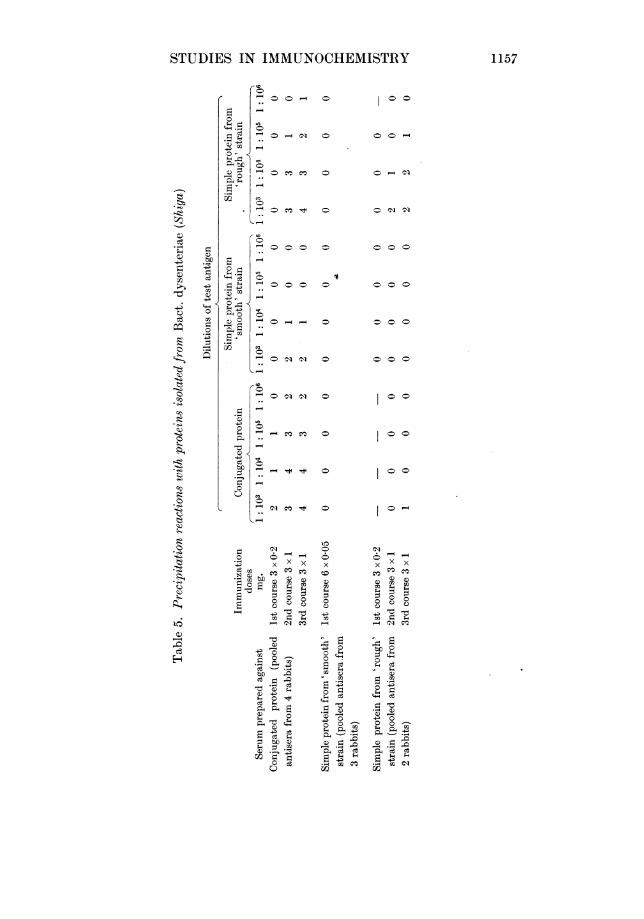
Serological comparison of the 'Shiga' proteinsIt has already been demonstrated that the conjugated protein gives rise to
homologous precipitins when injected into rabbits in small doses by the in-travenous route [Partridge & Morgan, 1940]. Stronger sera than those previouslyprepared have now been obtained by giving larger doses of the protein and bylimiting the operations involved in its purification. The more recent samples ofconjugated protein have been prepared with the minimum of manipulationnecessary to free the material from contaminating specific polysaccharide andare characterized by a higher phosphorus content. Table 5 shows the result ofimmunizing a group of four rabbits with a sample of conjugated protein thatcontained 11-3 % N, 0-58% P and showed [C]5461,-52 +20. Further groups ofrabbits were also immunized with samples of the electrophoretically pure simpleprotein prepared from the 'smooth' organism, and with the simple proteinprepared from the 'rough' strain. When immune sera collected after the firstcourse of antigen are employed, it appears that the anti-conjugated proteinserum reacts with the homologous antigen only, whereas, after a further courseof injections the strength of the conjugated protein antiserum is considerablyincreased and its specificity is spread so that it then reacts with the preparationsof simple protein. The amphoteric proteins themselves appear to be relativelypoor antigens, and the sample' prepared from the 'rough' strain of Bact. dysen-teriae (Shiga) alone engendered the formation of weak homologous precipitins.On the other hand, the antiserum to the 'rough' protein does not react with theconjugated protein or with the simple protein from the corresponding 'smooth'strain. For brevity the results obtained with pooled sera are given in Table 5,but each serum was also tested separately, and each gave similar results to thoseof the corresponding pooled serum samples. A sample of antitoxin prepared in
1156
STUDIES IN IMMUNOCHEMISTRY
0
0.0
S ji
oC)
0
*50X0o,
0 o 0
o~
- 0
L..0
- o
Li
1- 0
C CO
CO m
00
CO m
H40 X t X
O-
-
bt0cd
0Le
0so
1157
0
C130
0
c)
0o
*')
* 0z
.0
*0t
*0)s
0t
0
0
0
0
0
0
0
0
0
xCO
C)
-ou
0
0-0V
0 o
-Q .5 0
0zc c3
*-{2CO
0 0 -
0-01
00101
0 0
0 0
0-4
0- 4
- 01 CO
FCt0oo
40
40 0
00-
o~ _ o
o o
0ICo o
o
W. T. J. MORGAN AND S. M. PARTRIDGE
the rabbit against the toxic filtrates of the 'rough' organism [Neisser & Shiga,1903; Conradi, 1903; Todd, 1904] gave no reaction with the test-antigens usedin Table 5. A sample of toxic protein prepared by the trichloroacetic acidmethod of Boivin & Mesrobeanu [1937; Mesrobeanu & Boivin, 1937] was alsotested against the three types of antisera given in Table 5 but no significantprecipitation was observed. From these results it seems that the prostheticgroup plays some role in determining the specificity of the conjugated protein.
DISCUSSIONAs a result of our earlier investigations on the chemical nature of the specific
antigen of Bact. dysenteriae (Shiga) evidence was obtained for the view thatantigenic substances prepared from Bact. dysenteriae (Shiga) by various methodsowe their activity to the presence of two major components, a specific poly-saccharide and an acidic substance that contains amino-acids and is largelypolypeptide or protein in nature. Other components undoubtedly occur in thenative antigen but it was found that these could be eliminated if suitablemeans were employed, without seriously impairing the antigenic qualities of theresulting material. The removal of these components, however, caused analteration in the chemical and physical properties of the native antigenic com-plex. A method was described for the stepwise degradation of the antigeniccomplex which was based on the dissociating action of formamide. The non-antigenic specific polysaccharide isolated by this means could be recombinedwith the polypeptide-like component, obtained by acid hydrolysis of the nativeantigenic complex, to yield a powerful antigen that induced in rabbits theformation of immune-body specific for Bact. dysenteriae (Shiga).
The polypeptide-like component previously isolated from the antigenic sub-stance by hydrolysis with dilute acetic acid at 1000 contains 11F5-12-5 % N and0-6-8 % P, and is readily soluble in dilute alkali but is insoluble throughout theentire acid pH range. An extensive amino-acid analysis of the material was notattempted, but it was shown to contain tyrosine, arginine, glutamic acid andtryptophan. The substance is digested by pepsin and by trypsin and, after acidhydrolysis, about half the total nitrogen appears as free amino-nitrogen. Thelow nitrogen content of the material, the p or yield of ac-amino groups afteracid hydrolysis and its unusual insolubility under acid conditions were con-sidered sufficient reason for rejecting its classification as a simple protein withoutfurther investigation, and the material was at that time therefore described as a'polypeptide-like' substance.
The results of the experiments that are described in the present paperindicate that the phospholipin-free antigenic complex can also be dissociatedinto its two major components by solution in 90 % phenol. The addition ofalcohol to the phenol solution causes the liberated polysaccharide and proteinmolecules to recombine and the resulting complex that is thrown out of solutionis found to be antigenic; Direct dialysis of the phenol solution of the antigeniccomplex, however, enables the separated components to be readily isolated. Thepolysaccharide and protein constituents obtained by this means are unable toinduce the formation of 'Shiga' agglutinins and precipitins.
In earlier attempts to purify the 'polypeptide', it. was repeatedly dissolvedin dilute alkali and reprecipitated by addition of acid and it was found thatduring this process the phosphorus content was slowly reduced while the specificrotation and nitrogen content of the substance showed only slight increases, butthere was some indication that during the process the capacity of the substanceto recombine with 'Shiga' polysaccharide to form an antigenic complex was
1 158
STUDIES IN IMMUNOCHEMISTRY
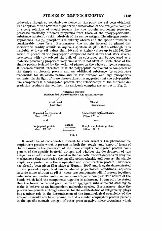
reduced, although no conclusive evidence on this point has yet been obtained:.The adoption of the new technique for the dissociation of the antigenic complexin strong solutions of phenol reveals that the protein component recoveredpossesses markedly different properties from those of the 'polypeptide-like'substance isolated by acid hydrolysis of the native antigen. The nitrogen contentapproaches 14-5 %, phosphorus is entirely absent and the specific rotation isconsiderably more laevo. Furthermore, the protein isolated by phenol dis-sociation is readily soluble in aqueous solution at pH 2-0-2*5 although it isinsoluble at lower pH values than 2-0 and at higher values up to pH 7*0. Theaction of phenol on the polypeptide component itself shows that after severaltreatments with this solvent the bulk of the substance can be recovered as amaterial possessing properties very similar to, if not identical with, those of thesimple protein isolated by the action of phenol on the whole antigenic complex.It becomes evident, therefore, that the polypeptide component is composed ofthe simple amphoteric protein and an additional substance (or substances)responsible for its acidic nature and its low nitrogen and high phosphoruscontents. In the light of these observations it is suggested that the polypeptide-like component is a conjugated protein. The relationships of the different de-gradation products derived from the antigenic complex are set out in Fig. 2.
Antigenic complex(undegraded polysaccharide + conjugated protein)
Acetic acid Phenolhydrolysis dissociation/\
Degraded polysaccharide Undegraded polysaccharide[a]5461 + 108 ±20 [aC]5461 + 850 ±20
and and
Conjugated protein Phenol Simple amphoteric protein[oc]461 - 50 ±50 dissociation [oc]54 - 82 ±50
Fig. 2
It would be of considerable interest to know whether the phenol-solubleamphoteric protein which is present in both the 'rough' and 'smooth' forms ofthe organism is the precursor of the more complex conjugated protein com-ponent of the specific bacterial antigen and whether the development of thisantigen as an additional component in the 'smooth' variant depends on enzymicmechanisms that synthesize the specific polysaccharide and convert the simpleamphoteric protein into the conjugated and more reactive protein. Evidencehas already been given [Partridge & Morgan, 1940] and is again demonstratedin the present paper, that under almost physiological conditions-aqueousisotonic saline solution at pH 8-these two components will, if present together,enter into combination and give rise to an antigenic complex. The nature of thebonds which hold the components together is unknown. It can only be statedthat the forces concerned give rise to an aggregate with sufficient stability tomake it behave as an independent molecular species. Furthermore, since theprotein component, although essential for the manifestation of antigenicity, playsbut a minor role in the determination of the immunological specificity of theantigen it would not be surprising to find a similar conjugated protein presentin the specific somatic antigen of other gram-negative micro-organisms which
1159
W. T. J. MORGAN AND S. M. PARTRIDGE
possess the polysaccharide-protein type of antigen. Evidence for the existenceof a conjugated protein common to the antigenic complexes present in othertypes of gram-negative bacteria is being sought and it is of interest in thisconnexion to record that 'Shiga' undegraded polysaccharide combines with theconjugated protein component of the 'O' antigen of Bact. typhosum to form apowerful antigenic complex that induces the formation of specific 'Shiga'agglutinins and precipitins. Thus, the capacity of a specific polysaccharide ofone organism to form an antigenic complex when combined with the conjugatedprotein component derived from the antigen of another organism that belongs toan entirely different bacterial species, has been demonstrated.
An additional method for the isolation of the polysaccharide and proteincomponents of the native antigenic complex is also described. Antigenic materialdissolved in dilute NaOH is dissociated into its two main components. If thealkaline treatment is carried out quickly and at 00 an undegraded polysac-charide is recovered on the addition of alcohol whereas the protein componentcan be subsequently obtained from the alcoholic supernatant fluid by theaddition of acetic acid. The protein recovered appears to be identical with thesimple amphoteric substance obtained by phenol treatment of the whole anti-genic complex.
The polysaccharide obtained by either phenol or alkali dissociation is foundto yield viscous solutions in water and to contain 0-5-10 % of P in organiccombination. The P is not removed by the action of bone phosphatase, pro-longed dialysis or by precipitation of the material from solution in ice-cold0-05N HCI by alcohol, from which it is inferred that the P is most probably anintegral part of the polysaccharide molecule. Preparations of the degradedpolysaccharide [Morgan, 1936] contain no appreciable quantity of P.
The viscosity values obtained for the undegraded polysaccharide exhibit aconsiderable variation according to the method of preparation used. It was atfirst thought that a determination of viscosity alone would furnish a reliableguide to the degree of degradation of the sample of polysaccharide, but it hasbeen found that samples prepared by the phenol method occasionally haveviscosity values only slightly greater than the degraded polysaccharide preparedby heating the antigenic complex in dilute acetic acid. It is to be noted, however,that phenol-treated preparations still contain 0.5-1.0 % P in organic com-bination and that in spite of the low viscosity, the material enters into unionwith the conjugated protein to yield an antigenic complex. A rapid test forascertaining the potential capacity of polysaccharide preparations to form anti-genic complexes is obviously of considerable practical value, and the mostreliable guide so far found depends on the formation of soluble complexes withproteins at pH 4-5. On the addition of trichloroacetic acid to a mixture ofconjugated protein and degraded polysaccharide a heavy precipitate of theprotein immediately appears, whereas if the polysaccharide is in a form that willgive rise to an antigen in combination with the conjugated protein, the solutionremains clear, or, at most, develops a bluish opalescence.
It was earlier reported [Morgan & Partridge, 1940, 1; Partridge & Morgan,1940] that the undegraded specific polysaccharide hapten of Bact. dysenteriae(Shiga) could be recombinedwith the non-antigenic conjugated protein ('poly-peptide-like') component of the native antigen to form an antigenic complexthat was able to induce the formation of specific 'Shiga' precipitins and agglu-tinins. Similarly, the conjugated protein component derived from the 'O'antigen of Bact. typhosum also combines with the 'Shiga' polysaccharide andforms an antigenic complex with 'Shiga' specificity. The simple protein derived
1160
STUDIES IN IMMUNOCHEMISTRY
from the conjugated protein by the action of phenol, however, does not give riseto an antigenic complex in this manner and, in this respect, the simple amphotericprotein behaves like the proteins, stromatin, gelatin, human serum-y-globulin,hen's egg vitellin and heat-denatured normal rabbit serum proteins. Theseobservations suggest that a part of the conjugated protein molecule-mostprobably the prosthetic group-is necessary in order that the molecule shouldbe capable of combining with the polysaccharide to form an antigen. The forma-tion of a complex; between undegraded specific polysaccharide and the differentproteins examined certainly occurs as is shown by the behaviour of mixedsolutions of the protein and polysaccharide on the addition of trichloroaceticacid. It appears unlikely that the stability of the protein under these conditionsis due to the specific polysaccharide acting simply as a protective colloid, forgum acacia and other gums, which have low gold numbers [Schultz & Zsigmondy,1903] and might be expected to function as powerful protective colloids, fail toprevent precipitation of the protein in the presence of trichloroacetic acid. Inthe light of these observations on the stability of proteins in the presence of anundegraded bacterial polysaccharide it is clear that the absence of a precipitateon the addition of the usual acid precipitants cannot be accepted as evidencethat protein is not present in the material examined, and thus many reportedobservations on bacterial and tissue antigens stated to be 'protein-free' on thebasis of negative tests given by trichloroacetic acid must be open to doubt. Amore detailed study of these soluble and stable polysaccharide-protein complexesis being undertaken in collaboration with Dr R. A. Kekwick.
The possibility of evolving a differential extraction method whereby severalorganic solvents are utilized in turn for the solution and isolation of specificconstituents of the bacterial cell, has already been discussed in an earlier paper[Morgan, 1937]. The results of some simple experiments along these lines arenow described and it has been shown that after 6-7 % of the dry weight of theorganisms employed has been removed by extraction with diethyleneglycol, it ispossible by means of further extraction with strong solutions of phenol, torecover an additional 4-5 % of antigenic material together with about 10% ofa bacterial somatic protein. The antigenic material extracted from dry bacteriaby 90 % phenol solution and recovered by precipitation with alcohol appears tobe very similar in composition to the phospholipin-free primary extractionproduct obtained by means of diethyleneglycol. The free protein recovered fromthe bacilli, on the other hand, does not appear to be identical with the con-jugated protein component of the antigenic complex. Preparations of proteinwhich have been extracted by phenol from both 'rough' and 'smooth' culturesof Bact. dysenteriae (Shiga) contain 13-5-14-5 % N, are free from P and show astrongly negative rotation [oc],461-82 + 5°. The protein isolated in each instanceis electrophoretically homogeneous and represents at least 10% of the dryweight of the bacterial cell.
Finally, it may be said that in common with most workers we have deemed apreparation derived from bacteria to be devoid of antigenicity when it failed toyield demonstrable homolbgous specific agglutinins and precipitins after it hadbeen injected intravenously into each of a group of rabbits. Usually six doses of0-05 mg. were given. This amount of the native antigen invariably elicits apowerful immune retponse with the resulting production of high titre agglu-tinating sera in all rabbits inoculated. For the purposes of direct comparisonwith other preparations from the same organism this method of measuring theantigenicity of a substance has yielded useful results, but it is necessary toemphasize that in the absence of adequate criteria of exactly what constitutes
1161

W. T. J. MORGAN AND S. M. PARTRIDGE
an antigenic response it is advisable to keep in mind the results of other workers[Avery & Goebel, 1933; Chow, 1936; and Topley, 1937] who claim to havedemonstrated the presence of protective immune-body in the serum of animalsafter imifunization while being unable to detect the presence ofthe correspondingagglutinins or precipitins. In the absence of the latter immune-bodies thedifficulty of deciding whether a preparation retains a part of the original anti-genicity is still further increased when the material under test 'has been isolatedfrom an organism such as Shiga's bacillus, for which the usual active andpassive immunity tests that employ infection of the test animal with the livingbacterium cannot readily be performed. Furthermore, it is bnly when adequatemethods of assay exist that it is possibleto detect certain changes in the qualita-tive nature of the immune-bodies formed. For example, Felix & Bhatnagar[1935] found that the Vi antibody elaborated in response to immunization withformolized Vi antigen is not identical with that resulting- from immunizationwith native antigen as it occurs in the living, virulent Bact. typhosum. Thequantitative production of Vi agglutinins appears to be normal when either typeof vaccine is employed for immunization, but when sera of equal agglutinincontents are used, the phagocytosis-promoting activity and the protective powerof the immune serum produced against the formolized Vi culture are muchinferior to that induced after immunization with the living organism. Similarresults were obtained by Henderson & Morgan [1938] who immunited rabbitswith antigenic material isolated from Bact. typhosu'm. Once again, Vi agglutininsappeared in the immune serum but the protective power of the immune body asmeasured in passive protection tests was found to be much inferior to that givenby an equal quantity of serum of equal Vi agglutinin titre prepared by im-munization with living 'rough' Vi bacilli.
SUMMARY
1. The antigenic complex of Bact. dysenteriae (Shiga) is soluble in concen-trated (90 %) solutions of phenol. In phenol solution the complex is largelydissociated into its protein and polysaccharide components which can then beisolated in a free state by dialysis of the phenol solution.
2. The antigenic complex is partly dissociated in aqueous alkaline solutionand the components can be separated by repeated fractionation of the antigeniccomplex from alkaline solution by alcohol.
3. The protein-free polysaccharide component prepared by either methodfails to induce the formation of specific 'Shiga' agglutinins or precipitins in therabbit.
4. Some evidence is given for the view that the acidic protein componentpreviously described by the term 'polypeptide-like' is a conjugated protein. Itis devoid of antigenicity as measured by the productioiuof 'Shiga' immune-body, but can be recombined in aqueous solution with undegraded 'Shiga'specific polysaccharide to form a powerful antigen.
5. The protein isolated from the antigenic complhx by phenol or alkali dis-sociation is amphoteric and has a higher nitrogen content and optical rotationthan the conjugated protein. The simple amphoteric protein can also be isolateddirectly from the conjugated protein component of the antigenic complex. Thesimple protein does not form an antigenic complex when mixed with 'Shiga'polysaccharide in aqueous solution.
6. The extraction of a 'smooth' culture of Bact. dysenteriae (Shiga) with 90%phenol yields the characteristic water-soluble; antigenic complex and about an
1162

STUDIES IN IMMUNOCHEMISTRY 1163
equal amount of the non-specific, water-insoluble simple protein. Similar ex-traction of the corresponding 'rough' variant yields no antigenic material but aconsiderable quantity of the simple protein.
REFERENCES
Avery & Goebel (1933). J. exp. Med. 58, 731.Boivin & Mesrobeanu (1937). C.R. Soc. Biol., Pare, 124, 442.Chow (1936). J. exp. Med. 64, 843.Conradi (1903). Dt8ch. med. W8chr. 29,26.Felix & Blhatnagar (1935). Brit. J. exp. Path. 16, 422.Henderson & Morgan (1938). Brit. J. exp. Path. 19, 82.Holiday (1936). Biochem. J. 30, 1795.Jorpes (1932). Biochem. J. 26, 1488.
& Thoren (1932). Biochem. J. 26, 1504.Mesrobeanu & Boivin (1937). C.R. Soc. Biol., Pari8, 124, 439.Morgan (1936). Biochem. J. 30, 909.
(1937). Biochem. J. 31, 2003.(1941). Chem. I-nd. 60, 722..& Partridge (1939, 1). Nature, Lond., 143, 1025.
(1939, 2). Chem. Ind. 58, 1020.(1940, 1). Biochem. J. 34, 169.(1940, 2). Chem. Ind. 59, 849.
Neisser & Shiga (1903). Dtech. med. W8chr. 29, 61.Palmer & Gerlough (1940). Science, 92, 155.Partridge & Morgan (1940). Brit. J. exp. Path. 21, 180.Schultz & Zsigmondy (1903). Beitr. chem. phy8iol. Path. (Hofmeister), 3, 137.S0rensen & Haugaard (1933). C.R. Lab. Carlsberg, 19, no. 12, 1.Spiegel-Adolf & Seibert (1934). J. biol. Chem. 106, 373.Stenstr6m & Reinhard (1925, 1). J. phy8. Chem. 29, 1477.- (1925, 2). J. biol. Chem. 66, 819.Todd (1904). J. Hyg., Camb., 4, 480.Topley (1937). Proc. 2nd Int. Congr. Microbiol. Lond., 354.
Related Documents


