12] I\.1UNGBEAN YELLOW MOSAIC VIRUS Yohachiro Honda* ABSTRACT The whitefly-borne mungbean yellow mosaic virus (MYMVi ,Jbserved in Thailand was transmitted by mechanical inoculation. The optima! incubation temperatures for symptom expression on plant ranged from 25 to 30°C in the growth chamber. Host range of MYMV was limited to seven plant species in the family Leguminosae. Determination of the stability of the virus in plant sap gave the following results : thermal inactivation point : 40-5(J'C for HJ min, dilution end point : JO 2 -10·1, and longevity in vitro : 1-2 days at 20'C. Purified virus preparations consisted of geminate particles about 18 x :-m nm in s11e with an ultrai'iolet light absorption spectrum typical of that of nucleoprotein (A,"',/A,;c1 value (Jf i .'.1-· ! .4\. Infrctivity \Vas associated with the presence of purified virus particles. In ultrathin sections, the \·i.rus particles were isometric, about 15-20 nm in diameteL and aggregates of virus particles sc,rnetimes filled almost completely the nuclei of infected phloem cells as early as 2 days before symptom appearance. Nucleic acid isolated from MYMV particles was identified as circular single· stranded DNA with a molecular weight of 8.0 x 10 '. MYlV1V was assigned to the Geminivirus group. Introduction In 1977, the first report on the occurrence of mungbean yellow mosaic disease outbreak in Thailand was received from Kamphaeng Phet Province in northern Thailand (Thongmeearkom et al., 1981). The disease also occurred in five other nearby provinces and caused almost total yield loss in infected mungbean(Vigna radiata) plants in the field. Laboratory tests indicated that the causal agent of the disease was transmitted by the tobacco whitefly, Bemisia tabaci Genn. (Thongmeearkom et al .. 1981). Prior to that report, a similar whitefly-borne disease had been observed in India (Nariani, 1960). The disease in India was very severe in blackgram (V mzmgo) and caused as much as 100% yield loss when plants were infected at the seedling stage (Nene, 1973). Despite the severity of the disease in both countries, very !itt le was known previously about the nature of the causal agent except that it was whitefly-borne and was not transmissible by mechanical inoculation (Nene, 1973). Isolation and characterization of the causal agent of mungbean yellow mosaic disease have not been reported. In this paper, I report the mechanical transmission, purification and ultrastructural studies of mungbean yellow mosaic virus (MYMV) in Thailand and describe some of the properties indicating that MYMV is a member of the Geminiviruses. Virus source and maintenace Mungbean plants showing yellow mosaic symptoms were collected from fields in the Kamphaeng Phet Province of northern Thailand in 1979. The virus was isolated from naturally infected mungbean plants by whitefly (B. tabaci) transmission and maintained in mungbean plants by whitefly and grafting transmission in a greenhouse. Mechanical inoculation Seedlings of mungbean used for mechanical inoculation tests were 5-7 days old. All inocula were prepared by grinding infected young mungbean leaves in buffers (about 4 ml/g tissue) with * Plant Pathologist. National Agriculture Research Center, Tsukuba Science City, Yatabe, Tsukuba, lbaraki 305. Japan.

Welcome message from author
This document is posted to help you gain knowledge. Please leave a comment to let me know what you think about it! Share it to your friends and learn new things together.
Transcript
12]
I\.1UNGBEAN YELLOW MOSAIC VIRUS
Yohachiro Honda*
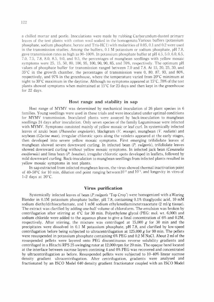
ABSTRACT The whitefly-borne mungbean yellow mosaic virus (MYMVi ,Jbserved in Thailand was
transmitted by mechanical inoculation. The optima! incubation temperatures for symptom expression on plant ranged from 25 to 30°C in the growth chamber. Host range of MYMV was limited to seven plant species in the family Leguminosae. Determination of the stability of the virus in plant sap gave the following results : thermal inactivation point : 40-5(J'C for HJ min, dilution end point : JO 2-10·1, and longevity in vitro : 1-2 days at 20'C. Purified virus preparations consisted of geminate particles about 18 x :-m nm in s11e with an ultrai'iolet light absorption spectrum typical of that of nucleoprotein (A,"',/A,;c1 value (Jf i .'.1-· ! .4\. Infrctivity \Vas associated with the presence of purified virus particles. In ultrathin sections, the \·i.rus particles were isometric, about 15-20 nm in diameteL and aggregates of virus particles sc,rnetimes filled almost completely the nuclei of infected phloem cells as early as 2 days before symptom appearance. Nucleic acid isolated from MYMV particles was identified as circular single· stranded DNA with a molecular weight of 8.0 x 10 '. MYlV1V was assigned to the Geminivirus group.
Introduction
In 1977, the first report on the occurrence of mungbean yellow mosaic disease outbreak in Thailand was received from Kamphaeng Phet Province in northern Thailand (Thongmeearkom et al., 1981). The disease also occurred in five other nearby provinces and caused almost total yield loss in infected mungbean(Vigna radiata) plants in the field. Laboratory tests indicated that the causal agent of the disease was transmitted by the tobacco whitefly, Bemisia tabaci Genn. (Thongmeearkom et al .. 1981). Prior to that report, a similar whitefly-borne disease had been observed in India (Nariani, 1960). The disease in India was very severe in blackgram (V mzmgo) and caused as much as 100% yield loss when plants were infected at the seedling stage (Nene, 1973). Despite the severity of the disease in both countries, very !itt le was known previously about the nature of the causal agent except that it was whitefly-borne and was not transmissible by mechanical inoculation (Nene, 1973). Isolation and characterization of the causal agent of mungbean yellow mosaic disease have not been reported.
In this paper, I report the mechanical transmission, purification and ultrastructural studies of mungbean yellow mosaic virus (MYMV) in Thailand and describe some of the properties indicating that MYMV is a member of the Geminiviruses.
Virus source and maintenace
Mungbean plants showing yellow mosaic symptoms were collected from fields in the Kamphaeng Phet Province of northern Thailand in 1979. The virus was isolated from naturally infected mungbean plants by whitefly (B. tabaci) transmission and maintained in mungbean plants by whitefly and grafting transmission in a greenhouse.
Mechanical inoculation
Seedlings of mungbean used for mechanical inoculation tests were 5-7 days old. All inocula were prepared by grinding infected young mungbean leaves in buffers (about 4 ml/g tissue) with
* Plant Pathologist. National Agriculture Research Center, Tsukuba Science City, Yatabe, Tsukuba, lbaraki 305. Japan.
a chilled ;nortar and pestlt·, lnroculations were made by rubbing Carborundum-dusted primaryleaves ot the test plan1s wifr, cqtton "· c.o\ soak,,d ::, the hnmogenateVari,m,, buffer::' ,potassium phosphate. sodium phosphate. bon11 t, and 'f ris-HCl) witb molariries uf 0.05, OJ and 0,2 w,,re used in 1hc transmission studies Among thP bufler,:, OJ !1.1 potassmrn or ,;odium phosphate, pH 7,8, gave trnnsn1issior> Files as bigh as 90- J 00%, In \Kii a5.sium phm,phate buffer at pH 45, 5,0, 6.0, 6.5. 7,0 75. 7.8. 8.0, M,5, 9,0, and 9,:i, the percentages .if mungbe:m seedlings \Vith ::ellow mosaic symptoms were 2fj, E. 50. 80, !00. 95, 100, 90, 90, 85, and 70'/'ii, respectively. The optimum pH ,alues of phosphatt: buffer for transmis;;.ion rnnged betv .. ·een i·,o and 7.8. At 15, 20, 25, 30, and 35"C in the growth chamber, the percentages of transmission were 0, 80, 87, 93, and 80%, re:c:pectively, and 97% in the greenhouse, where the temperature varied from 20°C minimum at night to 30°C maximum in the daytime, Ai though no symptoms appeared at 15"(, 70% of the test plants showed symptoms when maintained at l'.'/C for 23 days and then kept in the greenhouse for 22 day:s.
Host range and stability in sap
Host range of MYMV was determined by mechanical inoculation of 26 plant species in 6 families. Young seedlings were used in these trials and ,vere inoculated under optimal conditions for MYMV transmission. Inoculated plants were assayed by hack-inoculation to mungbean seedlings 24 days after inoculation. Only seven spt~cies of the family Leguminosae were infected with MYMV. Symptoms consisted mainly of yellow mosaic or lt'af curl. In systemically infected leaves of azuki bean (Phaseolus angularis). blackgram (V ,mmgo), mungbean nr. radiata) and soybean \Glycine max), irregular chlorotic spots along the veinlets appeared at the early stages, then developed into severe yellow mosaic symptoms. First emerging trifoliolate leaves of mungbean showed severe downward curling. In infected bean (P vulgaris), trifolio!ate leaves showed downward curling without yellow mosaic symptoms. In infected jack bean (Canavalia ensiformis) and lima bean (A lunatus), irregular chlorotic spots developed in leaflets, followed by mild dO\vnward curling. Back-inoculation to mungbean seedlings from infected plants resulted in yellow mosaic symptoms in test plants,
In sap extracted from infected mungbean leaves, the virus showed thermal inactivation point of 40-G0°C for 10 min, dilution end p:lint ranging hetweenlff' and 10:l, and longevity in vitro of 1-2 days at 20°C.
Virus purification
Systemically infected leaves of bean (P. uulgaris 'Top Crop') were homogenized with a \'laring Blender in 0.1M potassium phosphate buffer, pH 7,8, containing 0.1% thioglycolic acid, 10 mM sodium diethyldithiocarbamate, and 1 mM sodium ethylenediaminetetraacetate (2 ml/g tissue). The extract was clarified by adding one-half volume of chloroform. The emulsion ,vas broken by centrifugation after stirring at 4°C for 30 min. Polyethylene glycol (PEG: mo!. wt. 6,000) and sodium chloride were added to the aqueous phase to give a final concentration of 6% and 0.2M, respectively, After stirring, the mixture was centrifuged at 15,000 g for 30 min and the precipitates were dissolved in 0.1 M potassium phosphate, pH 7.8, and clarified by low-speed centrifugation before being subjected to ultracentrifugation at 125,000 g for 90 min. The pellets were resuspended in potassium phosphate containing 6% PEG and 0.2 M NaCL About 2 ml of the resuspended peilets were layered onto PEG discontinuous reverse solubility gradients and centrifuged in a Hitachi RPS 25 swinging rotor at 12,000 rpm for 20 min. The opaque band located at the interface between sucrose layers con tining 4 and 0% PEG was recovered and concentrated by ultracentrifugation as before. Resuspended pellets were subjected to 10-40(}(1 linear sucrose density gradient ultracentrifugation, After centrifugation, gradients were analysed and fractionated by an ISCO Model 640 density gradient fractionator coupled with an ISCO Model
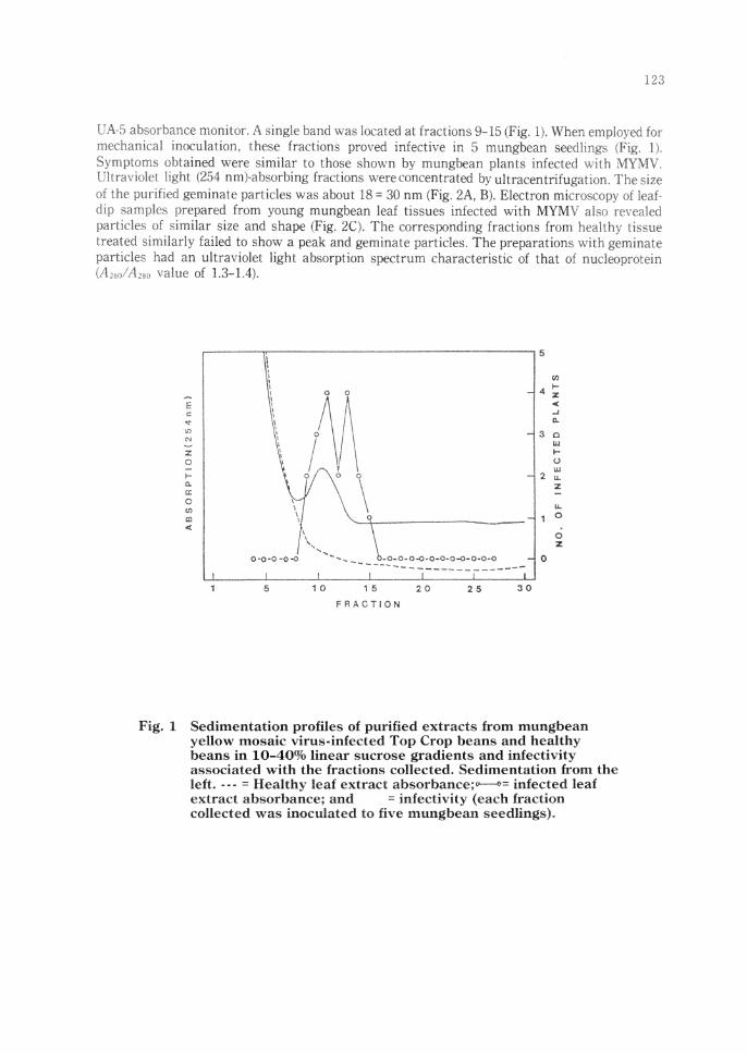
UA-5 absorbance monitor. A single band was located at fractions 9- b (Fig. lJ. When employed fot mechanical inoculation, these fractions proved infective in 5 mungbean seedlings (Fig. l L
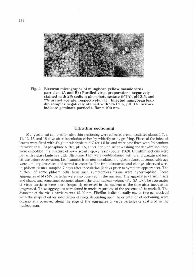
Symptoms obtained were similar to those shown by mungbean plants infecte(: with MYMV. Ultraviolet light (254 nm)-absorbing fractions were concentrated by ult racentrifuga11u,~ The ~i1e of the purified geminate particles was about 18 = 30 nm (Fig. 2A, B). Electron mk-roscopy of leaf dip samples prepared from young mungbean leaf tissues infected with MYMV aiso n.•,·e;iled particles of similar size and shape (Fig. 2C). The corresponding fractions from healthy tisrne treated similarly failed to sho,v a peak and geminate particles. The preparations v,ith geminate particles had an ultraviolet light absorption spectrum characteristic of that of nudeop:-otein \A2,nl A,8, value of 1.3-L4) .
Fig. l
.: 0
0 if)
OJ <
5 i 0 1 5
FRACTION
20 25 30
0
0 z
Sedimentation profiles of purified extracts from mungbean yellow mosaic virus-infected Top Crop beans and healthy beans in 10-40% linear sucrose gradients and infectivity associated with the fractions collected, Sedimentation from the left •••• = Healthv leaf extract absorbam::e;.,..------s:•cc infected leaf extract. absorba~ce; and = infectivity (each fraction collected was inoculated to five mungbean seedlings).
124
Fig. 2 Electron micrographs of mungbean yellow mosaic virus particles. (A and B) : Purified virus preparations negatively stained with 2% sodium phosphotungstate (PTA), pH 3.5, and 2 11/o uranyl acetate, respectively. (C): Infected mungbean leafdip samples negatively stained with 2% PT A, pH 3.5. Arrows indicate geminate particels. Bar = 100 nm.
Ultrathin sectioning
Mungbean leaf samples for ultra thin sectioning were collected from inoculated plants 5, 7, 9, 11, 13, 15, and 18 days after inoculation either by whitefly or by grafting. Pieces of the infected leaves were fixed with 4% glutaraldehyde at 5°C for 1.5 hr, and \Vere post-fixed with 2% osmium tetroxide in OJ M phosphate buffer, pH 7.5, at 5°C for 5 hr. After washing and dehydration, they were embedded in a mixture of low-viscosity epoxy resin (Spurr, 1969). Ultrathin sections were cut with a glass knife in a LKB Ultrotome. They were double-stained with uranyl acetate and lead citrate before observation. Leaf samples from non inoculated mungbean plants at comparable age were similary processed and served as controls. The first ultrastructural changes observed were in phloem tissues sampled 7 days after inoculation (2 days prior to symptom appearance). The nucleoli of some phloem cells from such symptomless tissue were hypertrophied. Loose aggregates of MYMV particles were also observed in the nucleus. The aggregates varied in size and shape, and sometimes occupied almost the total nuclear volume (Fig. 3A, B). The aggregates of virus particles were more frequently observed in the nucleus as the time after inoculation progressed. These aggregates were found in nuclei regardless of the presence of the nucleoli. The diameter of the virus particles was 15-20 nm. Fibrillar bodies (usually one or two per nucleus) with the shape of either solid circles of rings, depending upon the orientation of sectioning, were occasionally observed along the edge of the aggregates of virus particles or scattered in the nucleoplasm.
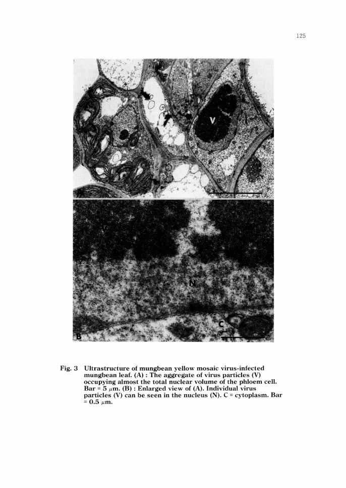
Fig. 3 Ultrastructure of mungbean yellow mosaic virus-infected mungbean leaf. (A) : The aggregate of virus particles (V) occupying almost the total nuclear volume of the phloem cell. Bar = 5 µm. (B) : Enlarged view of (A). Individual virus particles (V) can be seen in the nucleus (N). C = cytoplasm. Bar = 0.5 µm.
126
Nucleic acid of virus
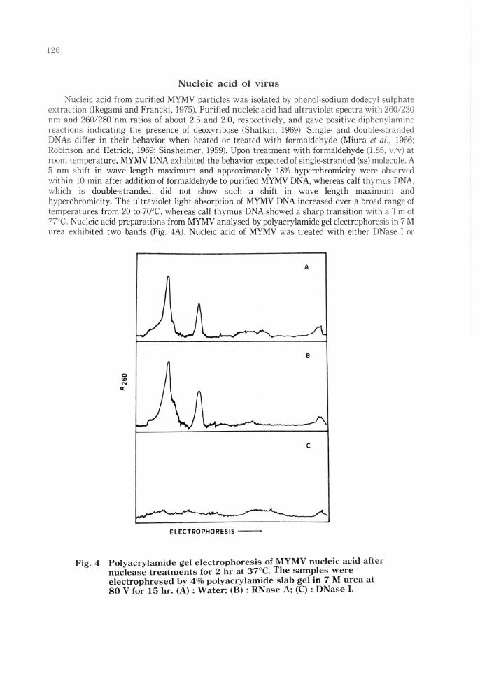
Nucleic acid from purified MYMV particles was isolated by phenol-sodium dodecvl ,,,u]ph;.;te extraction (Ikegami and Francki, 1975). Purified nucleic acid had ultraviolet spectra with :?G0,230 nm and 260/280 nm ratios of about 2.5 and 2.0, respectively, and gave positive cliphvnylarnine reactions indicating the presence of deoxyribose (Shatkin, 1969). Single- and doublt'Slranded DNAs differ in their behavior when heated or treated with formaldehyde (Miura ct al., 1966; Robinson and Hetrick, 1969; Sinsheimer, 1959). Upon treatment with formaldehyde (1.8:-i, v/v) at room temperature, MYMV DNA exhibited the behavior expected of single-stranded (ss) rnoleculE'. A 5 nm shift in wave length maximum and approximately 18% hyperchromicity were ob~.erved within 10 min after addition of formaldehyde to purified MYMV DNA, whereas calf thyrnus DNA, which is double-stranded, did not show such a shift in wave length maximum and hyperchromicity. The ultraviolet light absorption of MYMV DNA increased over a broad range of temperatures from 20 to 70°C, whereas calf thymus DNA showed a sharp transition with a Tm of 77°C. Nucleic acid preparations from MYMV analysed by polyacrylamide gel electrophoresi;:; in 7 M urea exhibited two bands (Fig. 4A). Nucleic acid of MYMV was treated ,vith either DNase I or
Fig. 4
B
C
ELECTROPHORESIS --
Polyacrylamide gel electrophoresis of MYMV nucleic acid after nuclease treatments for 2 hr at 37°C. The sall!ples were electrophresed by 4% polyacrylamide slab gel in 7 M urea at 80 V for 15 hr. (A): Water; (B): RNase A; (C): DNase L
bmme pancreaci,: RNase A. ,ind analysed by poly,!crylamide gel electrnphoresis. N1JC!eic acid of MYMV was hydrolyzed hr DNase I but not bovine µancreatic RNase (Fig. 4B. C). When the susceptibility of MYMV nucleic acid to nuclease S1 ,, a~ tested, the digestion rate of l\IYMV nucleic acid was comparable to tha\ of heat dem1tured calf thymus DNA.
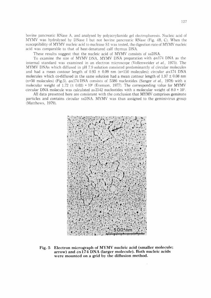
These results suggest thal the nucleic acid of MYMV consist;; of ,,sDNA. To examine the size of MYMV DNA, I\TYMV DNA preparation with ,t,x174 DNA as the
mterna! standard was examined in an electron microscclpe (V,Jllenweider et al., 1975). The MYMV DNAs ,vhich diffused in pH 7.9 .solution consisted predominantly of circular molecules and had a mean contour length of 0.93 :t 0.09 nm (ff-'150 molecules); circular ,t,xl74 DNA molecules which co-diffused in the same solution had a mean contour length of L97 ±. 0.08 nm (n=50 molecules) (Fig.5). ,f>xl74 DNA consists of 5386 nucleotides 1Sanger et al., 1978) with a molecular weight of L72 (± 0.03) x 10'; (Evenson. 1977). The corresponding value for MYMV circular DNA molecule was calculated as2542 nucleotides v;ith a molecular weight of 8.0 x 1(}•.
All data presented here are consistent with the conclusion that MYMV comprises gerninate particles and contains circular ssDNA. MYMV was thus assigned to the geminivirus group (~fatthews, 1979).
Fig. 5 Electron micrograph of MYMV nucleic acid (smaller molecule; arrow) and ,t,xl 74 DNA (larger molecule). Both nucleic acids were mounted on a grid by the diffusion method.
128
Acknowledgement
I am most grateful to P. Thongmeearkom*, K. Kiratiya-angul and N. Deema (Department Agriculture. Ministry of Agriculture and Cooperatives, Thailand), M. Iwaki (Nat10nal Institute of Agro-Environmental Sciences, Japan), and M. Ikegami (NODAi Research Institute, Tokyo University of Agriculture, Japan) for their cooperation and helpful suggestions. * Present address: Monsanto Thailand, Bangkok, Thailand.
References
I) Evenson, D.P. (1977): Electron microscopy of viral nucleic acids. In: Methods in Virology. Edited by: K. Maramorosch and Koprowski, H. Vol. II, Academic Press, New York. p. 219-264.
2) Ikegami, M. and Francki, R.I.B. (1975): Some properties of RNA from Fiji disease subviral particles. Virology, 64, 464-470.
3) Matthews, R.E.F. (1979): Classification and nomenclature of viruses. Intervirology, 12, 129-2%
4) Miura, K., Kimura, L and Suzuki, N. (1966): Double-stranded ribonucleic acid from rice dwarf virus. Virology, 28, 571-579.
5) Nariani, T.K. (1960): Yellow mosaic of mung (Phaseolus aureus L.). Indian Phytopathol., 13, 24-29.
6) Nene, Y.L. (1973): Viral diseases of some warm weather pulse crops in India. Plant Dis. Rep., 57, 463-367.
7) Robinson, D.M. and Hetrick, F.M. (1969): Single-stranded DNA from the ki!ham rat virus. J. gen. Virol., 4, 269-281.
8) Sanger, F., Coulson, R.R., Friedman, T., Air, G.M., Barrell, B.G., Brown, N.L., Fiddes, J.C., Hutchinson III, C.A., Slocombe, P.M. and Smith, M. (1978): The nucleotide sequence of bacteriophge ¢xl74. J. Mo!. Biol., 125, 225-246.
9) Shatkin, A.J. (1969): Colorimetric reactions for DNA, RNA, and protein determinations. In: Fundamental Techniques in Virology. Edited by: K. Habel and Salzman, N.P. Academic Press, New York. p. 231-237.
10) Sinsheimer, R.L. (1959): Purification and properties of bacteriophage ¢xl 74. J. Mol. Biol., 1, 37-42.
11) Spurr, A.R. (1969): A low-viscosity epoxy resin embedding medium for electron microscopy.]. Ultrastruc. Res., 26, 31-43.
12) Thongmeearkom, P., Kittipakorn, K. and Surin, P. (1981): Outbreak of mungbean yellow mosaic disease in Thailand. Thai. J. Agric. Sci., 14, 201-206.
13) Vollenweider, H.J., Songo, J.M. and Koller, T.H. (1975): A routine method for protein-free spreading of double- and single-stranded nucleic acid molecules. Proc. Nat. Acad. Scie., USA, 72, 83-87.
Discussion
Reddy, D.V.R. (ICRISAT): I believe that the mungbean yellow mosaic virus from India is different from that you observed in Thailand. The virus from India has been purified and found to be a geminivirus. It is not transmitted mechanically but is transmitted by whitefly and infects peanut.
Answer: There are differences between the Thai and Indian isolates of MYMV. The former is transmissible by mechanical inoculation unlike the latter. The host range of the Indian isolate is wider than that of the Thai isolate and includes Gramineae and Compositae in addition to Leguminosae. I believe that we are dealing with different strains of the same virus.
Related Documents