I. Microscopy • magnification • resolving power Ocular x Objective

I. Microscopy magnification resolving power Ocular x Objective.
Dec 13, 2015
Welcome message from author
This document is posted to help you gain knowledge. Please leave a comment to let me know what you think about it! Share it to your friends and learn new things together.
Transcript

I. Microscopy
• magnification
• resolving power
Ocular x Objective

Electron Microscopes:
Better resolving power than light microscopes
TEM aims an electron beam at a thin stained section
Used to study internal cell structure
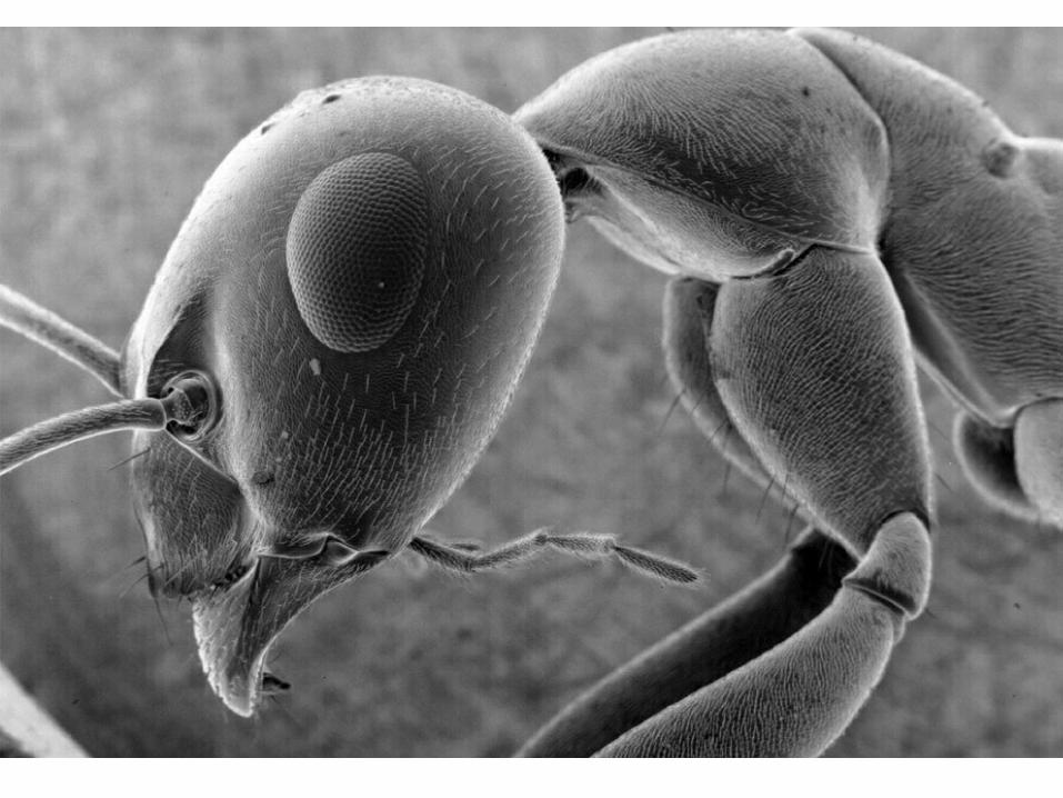

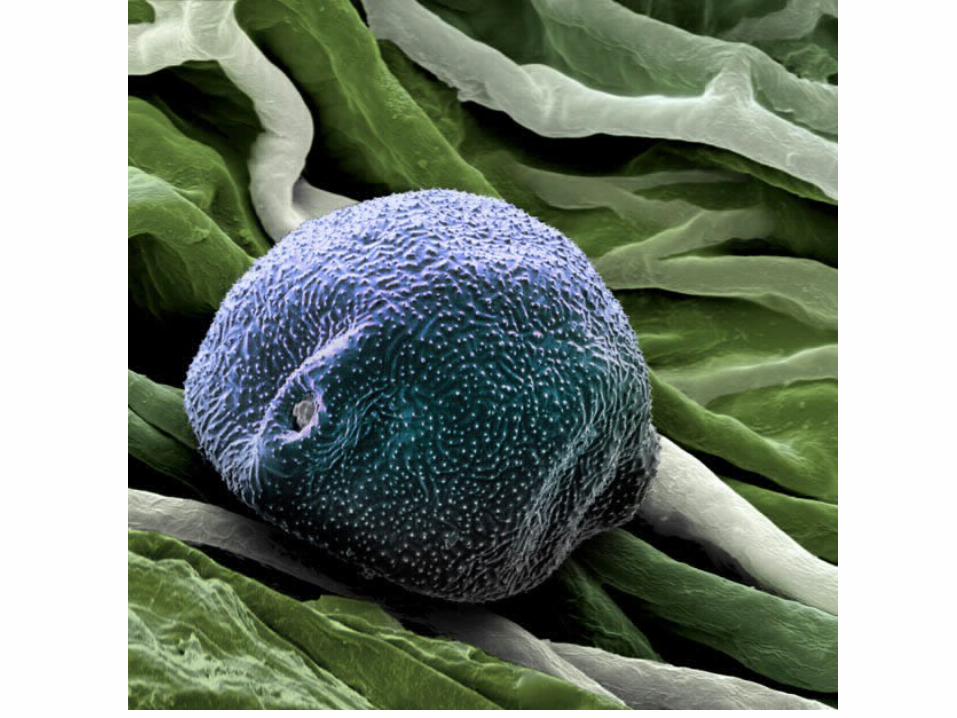
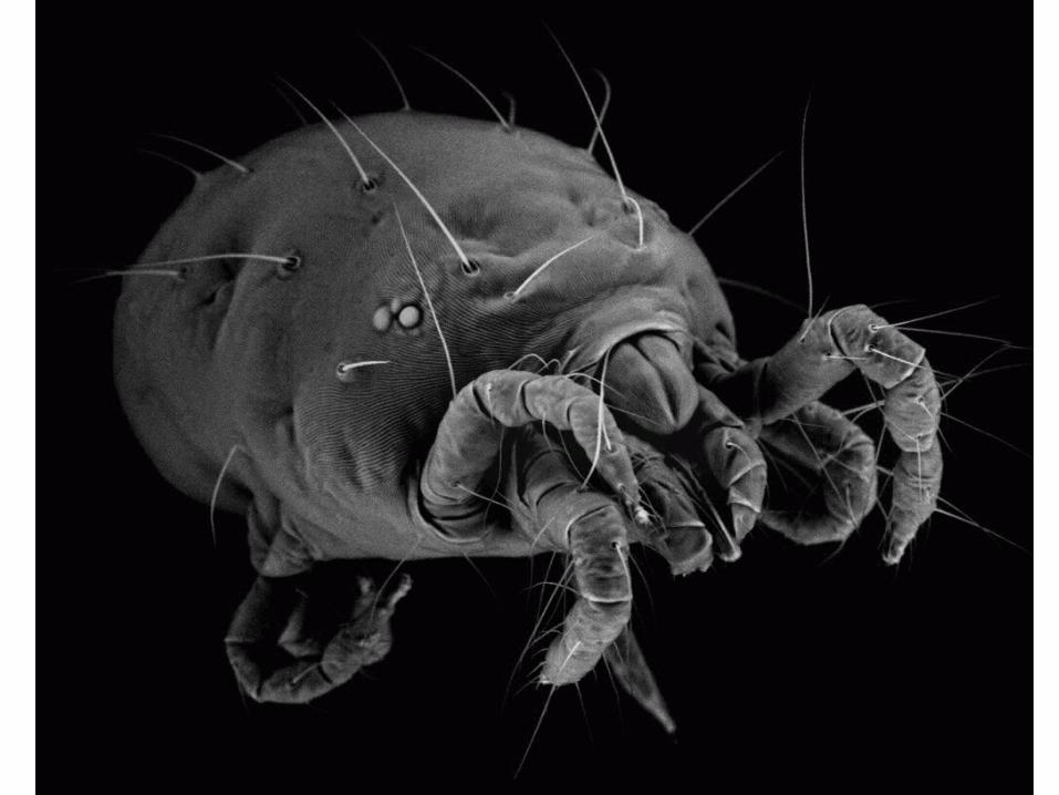
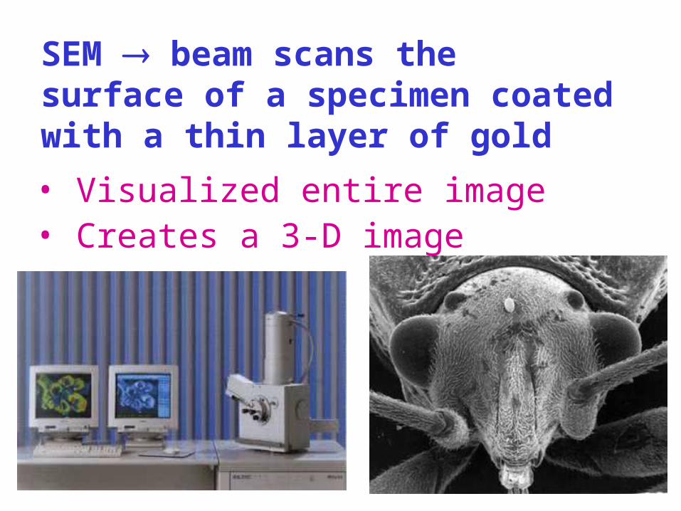
SEM beam scans the surface of a specimen coated with a thin layer of gold
• Visualized entire image• Creates a 3-D image
BUT, EM’s can only view dead cells
PHASE CONTRAST allow unstained, living tissues to be observed
CELL FRACTIONATION using a centrifuge to separate cell components based on
DENSITY
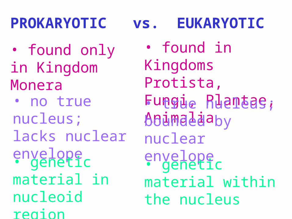
PROKARYOTIC vs. EUKARYOTIC
• found only in Kingdom Monera
• found in Kingdoms Protista, Fungi, Plantae, Animalia• no true
nucleus; lacks nuclear envelope
• true nucleus; bounded by nuclear envelope
• genetic material in nucleoid region
• genetic material within the nucleus
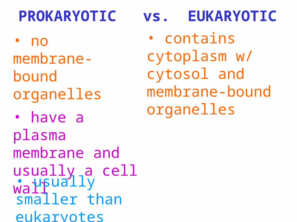
PROKARYOTIC vs. EUKARYOTIC
• no membrane-bound organelles
• contains cytoplasm w/ cytosol and membrane-bound organelles• have a plasma
membrane and usually a cell wall• usually smaller than eukaryotes
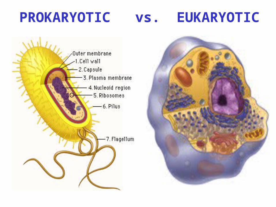
PROKARYOTIC vs. EUKARYOTIC

CELL SIZE is limited by:
• metabolic requirements
• surface area to volume ratio
II. PARTS of the CELL
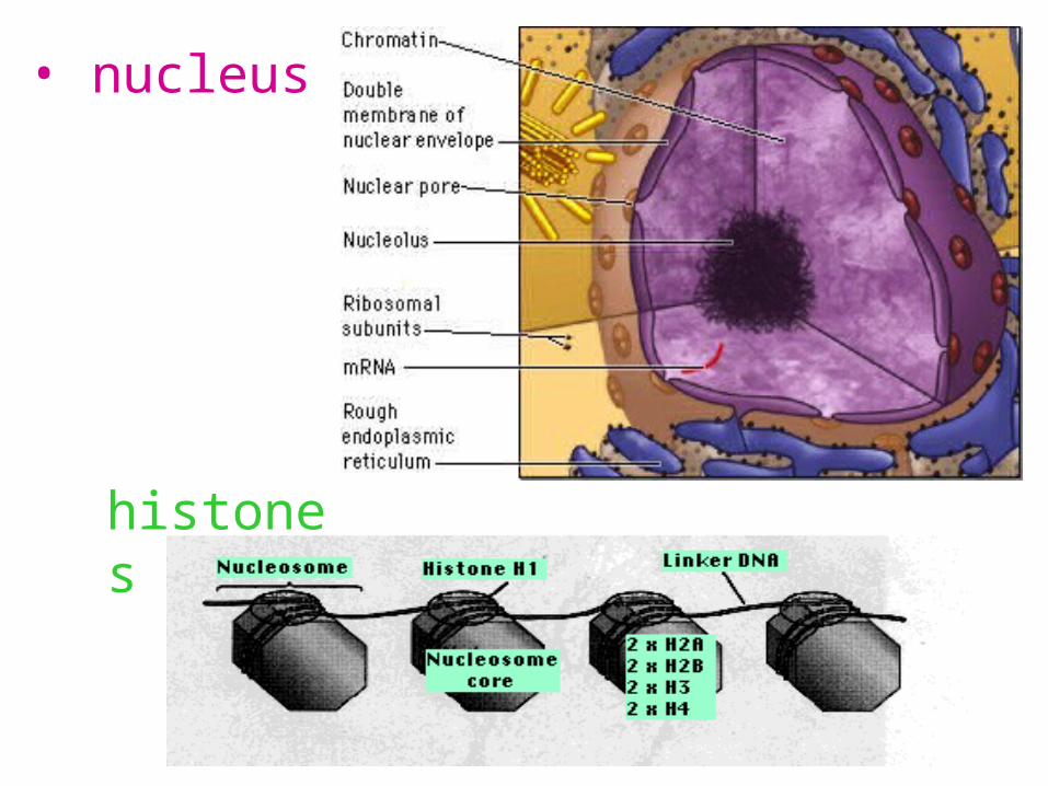
• nucleus
histones
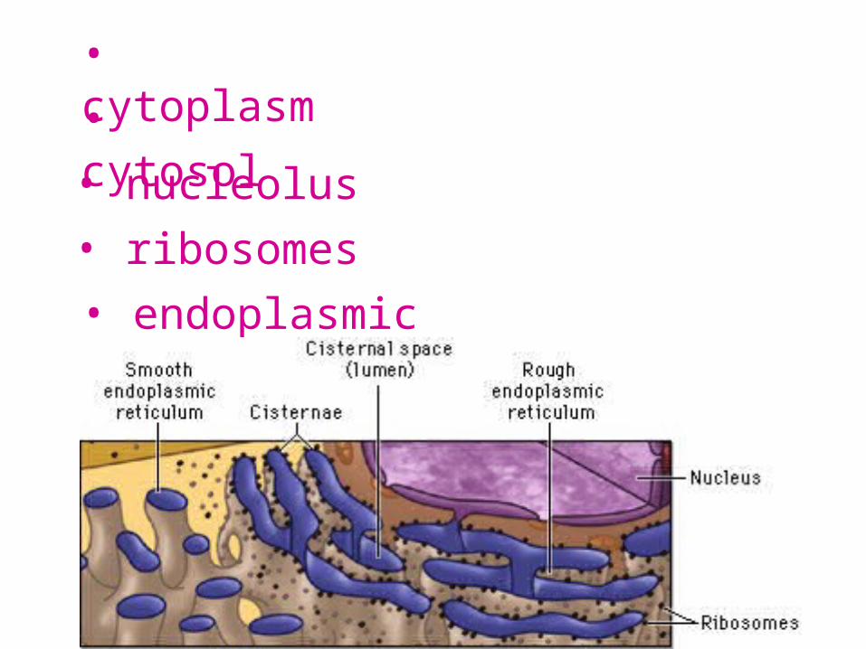
• cytoplasm• cytosol• nucleolus• ribosomes• endoplasmic reticulum

• Golgi apparatus
• lysosomes• intracellular digestion• apoptosis
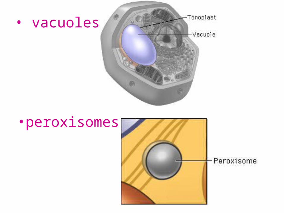
• vacuoles
•peroxisomes

• mitochondria
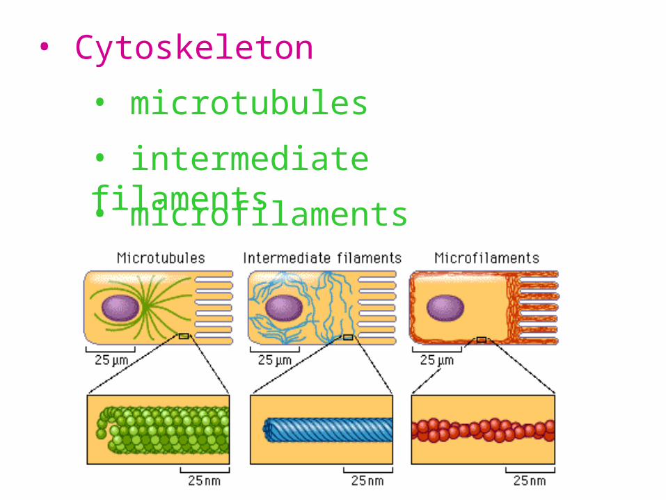
• Cytoskeleton
• microtubules
• intermediate filaments
• microfilaments

• cell walls
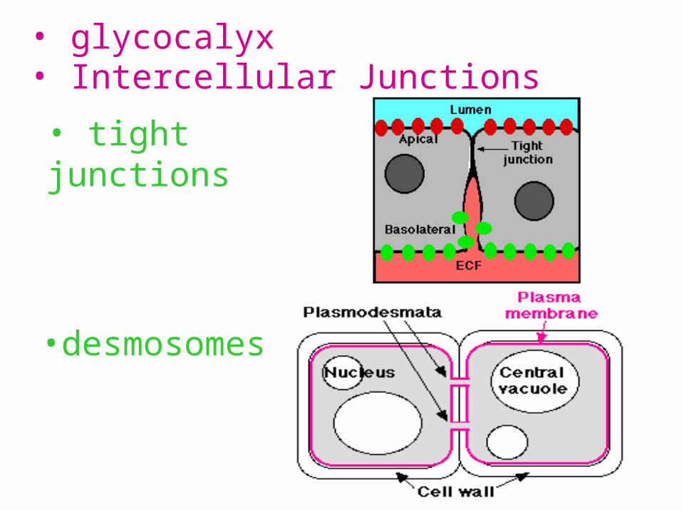
• glycocalyx• Intercellular Junctions
• tight junctions
•desmosomes

• gap junctions
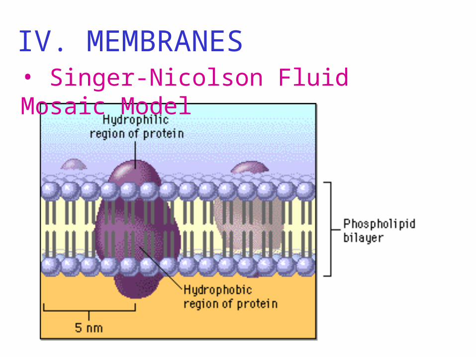
IV. MEMBRANES• Singer-Nicolson Fluid Mosaic Model

• Selective Permeability
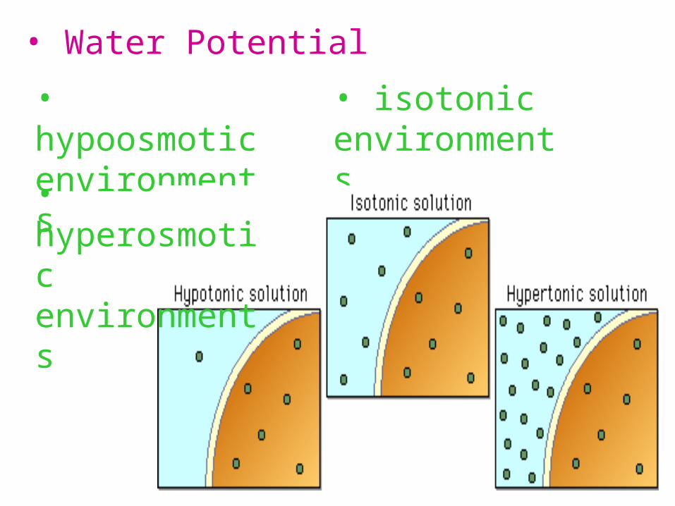
• Water Potential
• hypoosmotic environments
• isotonic environments
• hyperosmotic environments

•Diffusion and Osmosis

• Facilitated Diffusion
•Active Transport

• Forms of Active Transport• ATP Pump• Symport• Antiport

• Receptor-mediated endocytosis
Related Documents