I. Diagnostic Casts

I. Diagnostic Casts
Dec 30, 2015
I. Diagnostic Casts. A. Diagnostic CR Casts. Most patients have a slight discrepancy between tooth contact in CR and IP Therefore the CR record is made at an open vertical to eliminate the tendency for the patient to posture towards IP with tooth contact. 1. Wax interocclusal record. - PowerPoint PPT Presentation
Welcome message from author
This document is posted to help you gain knowledge. Please leave a comment to let me know what you think about it! Share it to your friends and learn new things together.
Transcript

I. Diagnostic Casts

A. Diagnostic CR Casts
• Most patients have a slight discrepancy between tooth contact in CR and IP
• Therefore the CR record is made at an open vertical to eliminate the tendency for the patient to posture towards IP with tooth contact

1. Wax interocclusal record
• Wax begins to set up immediately as it cools, therefore helps to stabilize jaw at open vertical dimension
• A stiff, brittle wax required to prevent distortion during mounting procedure
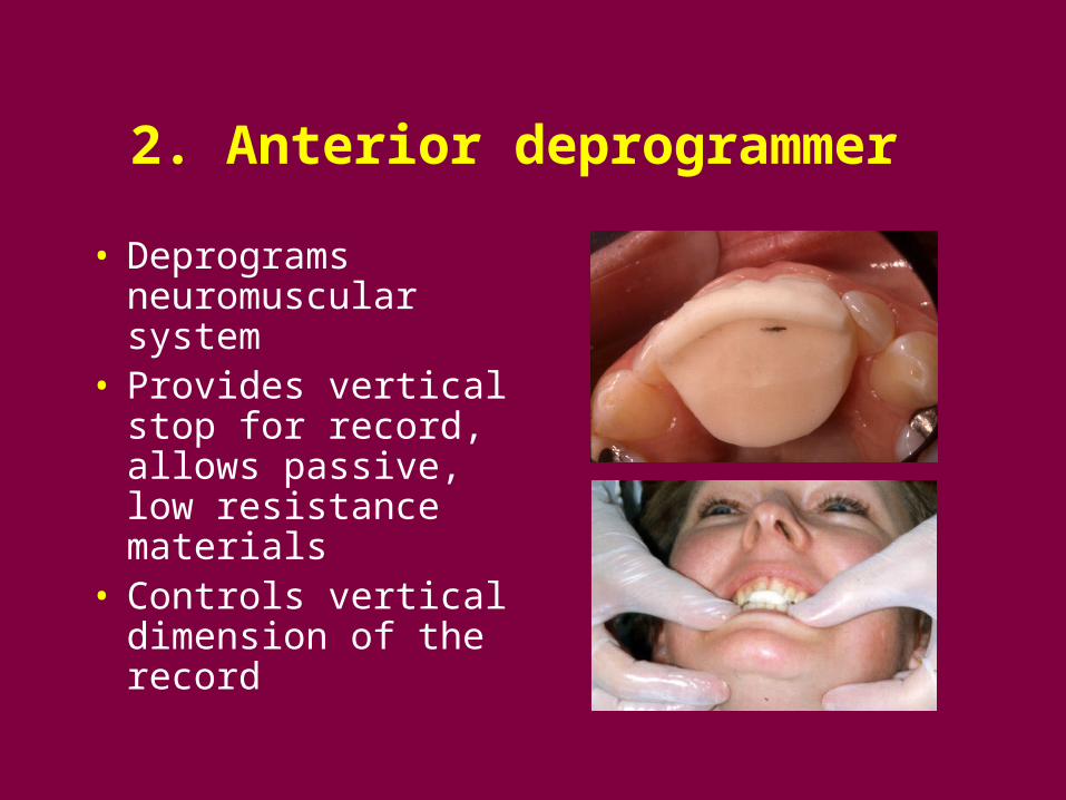
2. Anterior deprogrammer
• Deprograms neuromuscular system
• Provides vertical stop for record, allows passive, low resistance materials
• Controls vertical dimension of the record

B. Diagnostic IP Casts
Walls AWG et al., J Oral Rehabil 1991;18:43-48
• Record at tooth contact/ VDO
• 70% most accurately through hand articulation

B. Diagnostic IP Casts
Walls AWG et al., J Oral Rehabil 1991;18:43-48
• 30% need registration material w little resistance to closure, E.g. addition silicone

II. Working Casts

II. Working Casts
• Record at the Vertical Dimension of Occlusion
• If CR treatment position, new IP first in CR (occlusal adjustment). Then mount cast in new IP at VDO.
• Exception: final remount complete dentures

II. Working Casts
A. Hand-Articulated: IF unprepared teeth provide a stable IP
B. Registration Material Placement: > record stability needed, add only over prepared teeth
C. Closed Mouth Record: Have patient close into IP, then inject material over prepared teeth.

III. Interocclusal Records: KEEP IN MIND!

A. NO soft tissue contact
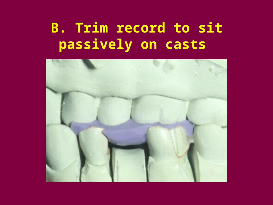
B. Trim record to sit passively on casts

C. “Arc of Closure”
• Hinge Axis Locator: allows a “hinge axis” facebow transfer, which duplicates the patients arc of opening & closing
• Arbitrary Facebow: orients the axis based on anatomic landmarks resulting in a discrepancy between the patient’s and the articulator’s arc of opening & closing

C. Minimize “Arc of Closure Error”
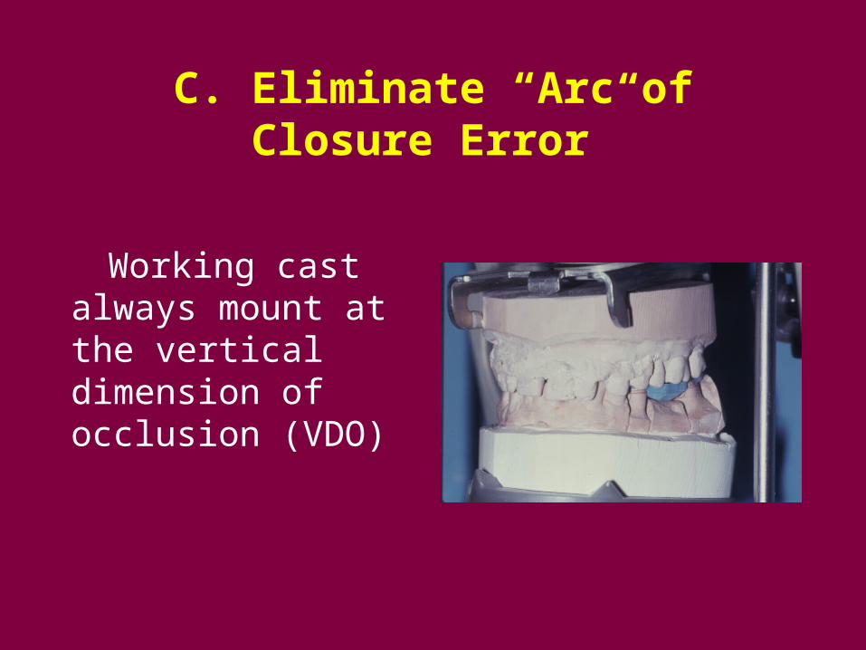
C. Eliminate “Arc of Closure Error”
Working cast always mount at the vertical dimension of occlusion (VDO)

C. Minimize “Arc of Closure Error”
Exception: Complete denture
records and remounts as thin as possible without perforation of registration

Centric Relation
• Typodont with no “rock” of teeth in hinged position
• Patient “hinged position” is called Centric Relation which is determined by the horizontal axis of the TM jts.

CENTRIC RELATION: Bimanual Clinical Method

CR Clinical Technique: Bimanual Guidance
• A cooperative effort between the patient and the clinician
• “Slow and deliberate” • Begin with light
pressure on the mandible.

A. Position of Clinician: Directly behind the patient’s head

B. Position of Patient: 1. Chair back parallel with the floor

B. Position of Patient: 2. Chin point raised/ Neck extended

C. Hand position: 1. Fingers lower border of the mandible, 5th finger on ascending ramus

C. Hand position: 2. Thumbs at symphysis of mandible, Hand in “C-Shape” cradling the jaw
C

C. Hand position: 2. Force application, slowly increase during
opening & closing to verify CR position

D. Rotation from the elbows, wrists fixed: SLOW AND DELIBERATE, a cooperative effort!

D. Rotation from the elbows: SLOW AND DELIBERATE, a cooperative effort!

II. Repeatability
This method has shown greater reliability than other methods of recording centric relation.
• Kantor ME, et al.: JPD 1975• Hobo S, Iwata T: JPD 1985• McKee JR: JPD 1997

III. Clinical Recording
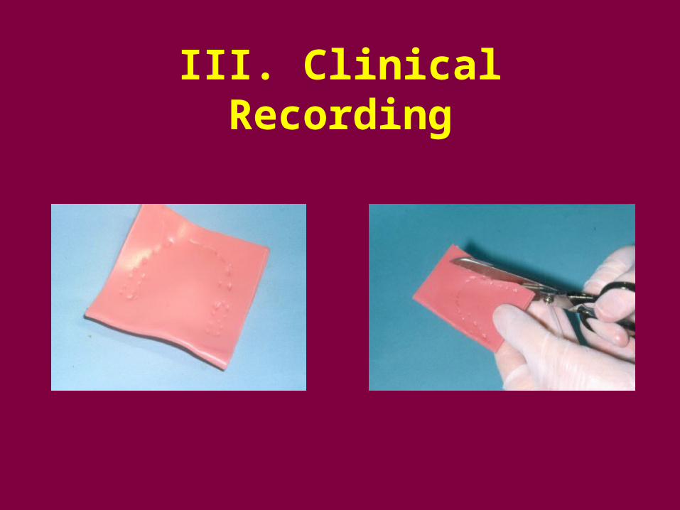
III. Clinical Recording
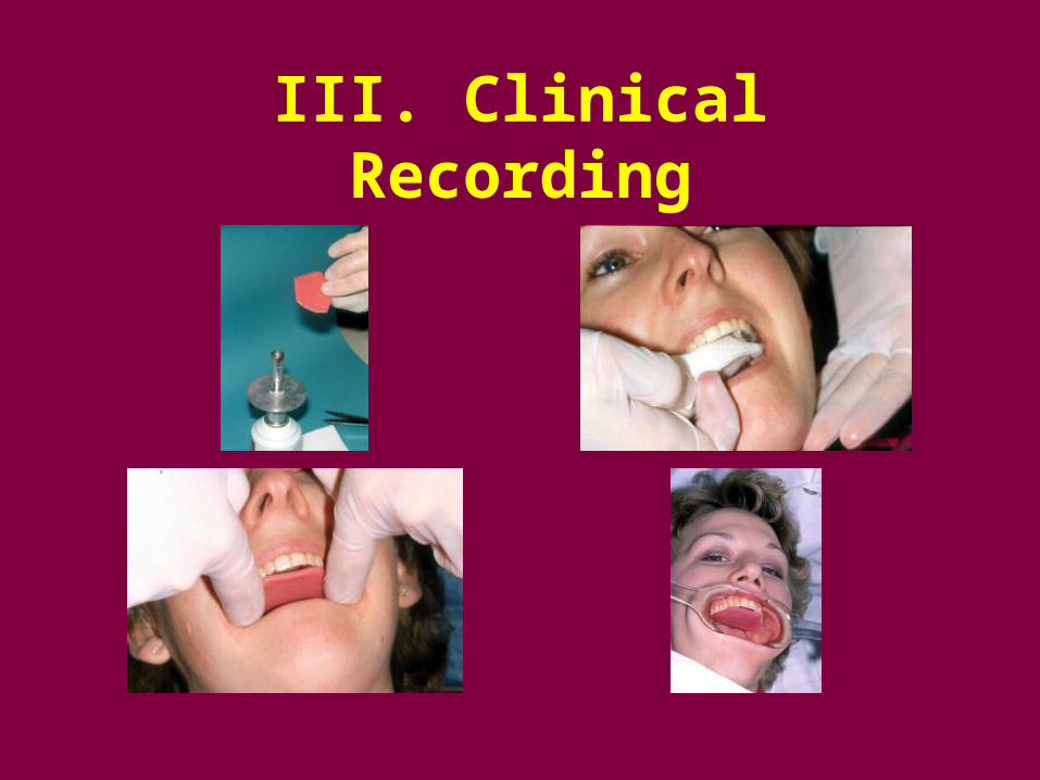
III. Clinical Recording

III. Clinical Recording

III. Clinical Recording

IV. CR Recording with Complete Dentures
A. Need to stabilize the trial or denture bases, therefore can not use bimanual method
B. Minimal thickness record without perforation, to prevent shift of trial or denture base
Related Documents


