miR-210 and myogenic differentiation 1 Hypoxia Inducible Factor 1- alpha induces miR-210 in normoxic differentiating myoblasts* Lucia Cicchillitti 1 , Valeria Di Stefano 1,2 , Eleonora Isaia 1 , Luca Crimaldi 1# , Pasquale Fasanaro 2 , Valeria Ambrosino 3 , Annalisa Antonini 2 , Maurizio C. Capogrossi 2 , Carlo Gaetano 4 , Giulia Piaggio 3 , and Fabio Martelli 1 1 IRCCS-Policlinico San Donato, 20097, Milan, Italy 2 Istituto Dermopatico dell’Immacolata-IRCCS, 00167, Rome, Italy 3 Istituto Nazionale dei Tumori Regina Elena, 00158 Rome, Italy 4 Uniklinikum-Goethe University, 60590, Frankfurt am Main, Germany *Running title: miR-210 and myogenic differentiation To whom correspondence should be addressed: Fabio Martelli, Molecular Cardiology Laboratory, IRCCS-Policlinico San Donato, via Morandi 30, San Donato Milanese, Milan, Italy, ZIP:20097, Tel:+390252774533; FAX: +390252774666; E-mail: [email protected] # Current address: Istituto Europeo di Oncologia, Milan, Italy Keywords: skeletal muscle, miR-210, promoter, Hif1a, cell death, oxidative stress Background: miR-210 hypoxamir is induced by Hif1a in hypoxic cells. Results: miR-210 expression increased during myogenic differentiation in normoxia, with a Hif1a-dependent mechanism. Moreover, miR- 210 displayed a cytoprotective role in response to mitochondrial dysfunction and oxidative stress. Conclusion: miR-210 regulation and function extend beyond cell response to hypoxia. Significance: Identifying miR-210 as a potential target in skeletal muscle disorders. SUMMARY microRNA-210 (miR-210) induction is a virtually constant feature of the hypoxic response in both normal and transformed cells, regulating several key aspects of cardiovascular diseases and cancer. We found that miR-210 was induced in normoxic myoblasts upon myogenic differentiation, both in vitro and in vivo. miR-210 transcription was activated in an Hypoxia Inducible Factor 1-alpha (Hif1a) dependent manner and Chromatin Immunoprecipitation experiments show that Hif1a bound to miR- 210 promoter only in differentiated myotubes. Accordingly, luciferase reporter assays demonstrated the functional relevance of Hif1a binding site for miR-210 promoter activation in differentiating myoblasts. To investigate the functional relevance of increased miR-210 levels in differentiated myofibers, we blocked miR-210 with complementary Locked Nucleic Acid oligonucleotides (anti-miR-210). We found that C2C12 myoblast cell line differentiation was largely unaffected by anti-210. Likewise, miR-210 inhibition did not affect skeletal muscle regeneration following cardiotoxin damage. However, we found that miR-210 blockade greatly increased myotube sensitivity to oxidative stress and mitochondrial dysfunction. In conclusion, miR-210 is induced in normoxic myofibers playing a cytoprotective role. microRNA-210 (miR-210) induction is a virtually constant feature of the hypoxic response in both normal and transformed cells, regulating several key aspects of health and disease (1-4). Indeed, miR-210 is a robust target of Hypoxia Inducible Factor (HIF): HIF1A isoform-specific induction has been reported in several cell types (5-9), albeit regulation by HIF2A was observed as well (10). Given the wide impact of miR-210 on hypoxic cell metabolism, redox balance, survival and angiogenesis, it has been also named as a master miRNA of the hypoxia response (hypoxamiR) (11). In keeping with its pleiotropic function, a broad spectrum of miR- 210 targets have been identified (1,2,12). Reduced survival of cells devoid of miR- 210 in normoxia and/or hypoxia is well documented (7-9,13-16). Moreover, miR-210 http://www.jbc.org/cgi/doi/10.1074/jbc.M112.421255 The latest version is at JBC Papers in Press. Published on November 12, 2012 as Manuscript M112.421255 Copyright 2012 by The American Society for Biochemistry and Molecular Biology, Inc. by guest on December 3, 2015 http://www.jbc.org/ Downloaded from by guest on December 3, 2015 http://www.jbc.org/ Downloaded from by guest on December 3, 2015 http://www.jbc.org/ Downloaded from by guest on December 3, 2015 http://www.jbc.org/ Downloaded from by guest on December 3, 2015 http://www.jbc.org/ Downloaded from by guest on December 3, 2015 http://www.jbc.org/ Downloaded from by guest on December 3, 2015 http://www.jbc.org/ Downloaded from by guest on December 3, 2015 http://www.jbc.org/ Downloaded from by guest on December 3, 2015 http://www.jbc.org/ Downloaded from by guest on December 3, 2015 http://www.jbc.org/ Downloaded from by guest on December 3, 2015 http://www.jbc.org/ Downloaded from by guest on December 3, 2015 http://www.jbc.org/ Downloaded from by guest on December 3, 2015 http://www.jbc.org/ Downloaded from by guest on December 3, 2015 http://www.jbc.org/ Downloaded from

Welcome message from author
This document is posted to help you gain knowledge. Please leave a comment to let me know what you think about it! Share it to your friends and learn new things together.
Transcript
miR-210 and myogenic differentiation
1
Hypoxia Inducible Factor 1- alpha induces miR-210 in normoxic differentiating myoblasts* Lucia Cicchillitti1, Valeria Di Stefano1,2, Eleonora Isaia1, Luca Crimaldi1#, Pasquale Fasanaro2,
Valeria Ambrosino3, Annalisa Antonini2, Maurizio C. Capogrossi2, Carlo Gaetano4, Giulia Piaggio3, and Fabio Martelli1
1IRCCS-Policlinico San Donato, 20097, Milan, Italy
2Istituto Dermopatico dell’Immacolata-IRCCS, 00167, Rome, Italy
3Istituto Nazionale dei Tumori Regina Elena, 00158 Rome, Italy
4Uniklinikum-Goethe University, 60590, Frankfurt am Main, Germany
*Running title: miR-210 and myogenic differentiation
To whom correspondence should be addressed: Fabio Martelli, Molecular Cardiology Laboratory, IRCCS-Policlinico San Donato, via Morandi 30, San Donato Milanese, Milan, Italy, ZIP:20097, Tel:+390252774533; FAX: +390252774666; E-mail: [email protected] # Current address: Istituto Europeo di Oncologia, Milan, Italy Keywords: skeletal muscle, miR-210, promoter, Hif1a, cell death, oxidative stress Background: miR-210 hypoxamir is induced by Hif1a in hypoxic cells. Results: miR-210 expression increased during myogenic differentiation in normoxia, with a Hif1a-dependent mechanism. Moreover, miR-210 displayed a cytoprotective role in response to mitochondrial dysfunction and oxidative stress. Conclusion: miR-210 regulation and function extend beyond cell response to hypoxia. Significance: Identifying miR-210 as a potential target in skeletal muscle disorders. SUMMARY
microRNA-210 (miR-210) induction is a virtually constant feature of the hypoxic response in both normal and transformed cells, regulating several key aspects of cardiovascular diseases and cancer. We found that miR-210 was induced in normoxic myoblasts upon myogenic differentiation, both in vitro and in vivo. miR-210 transcription was activated in an Hypoxia Inducible Factor 1-alpha (Hif1a) dependent manner and Chromatin Immunoprecipitation experiments show that Hif1a bound to miR-210 promoter only in differentiated myotubes. Accordingly, luciferase reporter assays demonstrated the functional relevance of Hif1a binding site for miR-210 promoter activation in differentiating myoblasts. To investigate the functional relevance of increased miR-210 levels in differentiated
myofibers, we blocked miR-210 with complementary Locked Nucleic Acid oligonucleotides (anti-miR-210). We found that C2C12 myoblast cell line differentiation was largely unaffected by anti-210. Likewise, miR-210 inhibition did not affect skeletal muscle regeneration following cardiotoxin damage. However, we found that miR-210 blockade greatly increased myotube sensitivity to oxidative stress and mitochondrial dysfunction. In conclusion, miR-210 is induced in normoxic myofibers playing a cytoprotective role.
microRNA-210 (miR-210) induction is a virtually constant feature of the hypoxic response in both normal and transformed cells, regulating several key aspects of health and disease (1-4). Indeed, miR-210 is a robust target of Hypoxia Inducible Factor (HIF): HIF1A isoform-specific induction has been reported in several cell types (5-9), albeit regulation by HIF2A was observed as well (10).
Given the wide impact of miR-210 on hypoxic cell metabolism, redox balance, survival and angiogenesis, it has been also named as a master miRNA of the hypoxia response (hypoxamiR) (11). In keeping with its pleiotropic function, a broad spectrum of miR-210 targets have been identified (1,2,12).
Reduced survival of cells devoid of miR-210 in normoxia and/or hypoxia is well documented (7-9,13-16). Moreover, miR-210
http://www.jbc.org/cgi/doi/10.1074/jbc.M112.421255The latest version is at JBC Papers in Press. Published on November 12, 2012 as Manuscript M112.421255
Copyright 2012 by The American Society for Biochemistry and Molecular Biology, Inc.
by guest on Decem
ber 3, 2015http://w
ww
.jbc.org/D
ownloaded from
by guest on D
ecember 3, 2015
http://ww
w.jbc.org/
Dow
nloaded from
by guest on Decem
ber 3, 2015http://w
ww
.jbc.org/D
ownloaded from
by guest on D
ecember 3, 2015
http://ww
w.jbc.org/
Dow
nloaded from
by guest on Decem
ber 3, 2015http://w
ww
.jbc.org/D
ownloaded from
by guest on D
ecember 3, 2015
http://ww
w.jbc.org/
Dow
nloaded from
by guest on Decem
ber 3, 2015http://w
ww
.jbc.org/D
ownloaded from
by guest on D
ecember 3, 2015
http://ww
w.jbc.org/
Dow
nloaded from
by guest on Decem
ber 3, 2015http://w
ww
.jbc.org/D
ownloaded from
by guest on D
ecember 3, 2015
http://ww
w.jbc.org/
Dow
nloaded from
by guest on Decem
ber 3, 2015http://w
ww
.jbc.org/D
ownloaded from
by guest on D
ecember 3, 2015
http://ww
w.jbc.org/
Dow
nloaded from
by guest on Decem
ber 3, 2015http://w
ww
.jbc.org/D
ownloaded from
by guest on D
ecember 3, 2015
http://ww
w.jbc.org/
Dow
nloaded from
miR-210 and myogenic differentiation
2
plays an instrumental role in the cytoprotection afforded to bone marrow-derived mesenchymal stem cells by ischemia/reoxygenation, supporting their survival under subsequent longer exposure to anoxia and following engraftment in the infarcted heart (17). While the molecular mechanisms underpinning these events are complex, miR-210 directly represses the apoptotic component CASP8AP2 (17), as well as other apoptosis-related genes, such as DAPK1 and, at least in humans, AIFM3 (14,18,19). However, miR-210 function seems to be complex and context dependent, since increased apoptosis upon miR-210 overexpression in normoxia has been reported as well (8,9).
miR-210 also influences mitochondrial metabolism: By down-regulating the expression of iron-sulfur cluster scaffold protein ISCU1/2, miR-210 disrupts the mitochondrial electron transport activity, providing a mechanistic insight into the shift from mitochondrial respiration to glycolysis observed in hypoxia (8,12,20-22). Accordingly, miR-210 can also modulate the generation of reactive oxygen species (ROS) (8,20,22). Moreover, expression of miR-210 in human umbilical vein endothelial cells (HUVEC) results in more tubulogenesis and increased vascular endothelial growth factor (VEGF)-induced cell migration through the repression of the receptor tyrosine kinase ligand Ephrin-A3 (7,23,24). Confirming miR-210 pro-angiogenic role, up-regulation of miR-210 in CD34+ cells enhanced tissue perfusion and capillary density in a mouse model of hindlimb ischemia (25).
In keeping with its hypoxamir role, orchestrating cell and tissue response to ischemia, miR-210 expression is also increased during erythropoietin-induced erythroid differentiation and miR-210 inhibition in this context leads to apoptosis (13). Moreover, miR-210 might be involved in increased expression of gamma-globin genes in differentiating erythroid cells (26).
miR-210 seems to have functions independent of its hypoxamir role: miR-210 regulates normoxic gene expression involved in tumor initiation (5) and promotes both osteoblastic and adipocyte differentiation (27,28).
Given the complexity of miR-210 action, it is not surprising miR-210 involvement in several key aspects of health and disease. Indeed, miR-210 overexpression has been detected in a variety of cardiovascular diseases,
including myocardial infarction, heart failure and stroke, as well as in solid tumors, such as breast and pancreatic cancer (1,2,4,29).
In this study, we found that miR-210 expression levels increased during myogenic differentiation, both in vitro and in vivo, and that this regulation occurred at the transcriptional level with a Hif1a-dependent mechanism. We also identified the Hif1a binding site responsible for the robust induction of the miR-210 promoter. Moreover, we demonstrated a pivotal cytoprotective role of miR-210 in response to mitochondrial dysfunction and oxidative stress.
EXPERIMENTAL PROCEDURES
Cell culture- Mouse C2C12 myoblast cell line (ATCC) was cultured in DMEM containing 20% FCS, as previously described (30). Differentiation was induced by plating the cells in DMEM containing 2% horse serum (differentiation medium, DM). Polyclonal populations of quail myoblasts transformed by the temperature-sensitive mutant of the Rous Sarcoma Virus LA29 were established and cultured as described (31). Briefly, quail myoblast line was propagated at 35°C in DMEM supplemented with 10% FCS and 10% tryptose phosphate broth (GM conditions). Differentiation was induced by plating the cells on collagen-coated dishes in GM and, the following day, incubating the cells with DMEM supplemented with 2% FCS at 41°C (DM conditions). L6C5 rat myoblasts were maintained in DMEM supplemented with 15% FCS (GM). Differentiation was induced by plating the cells in DMEM containing 1% horse serum and 1mg/ml of insulin (DM). To induce hypoxia, cells were cultured in a hypoxic incubator at 1% oxygen tension (Forma series II incubator mod.3131). For drug treatments, C2C12 cells were grown for 24 hours (hrs) in DM and then treated with either solvent alone or the following drugs: 200µM H2O2, 40mM NaN3, 2µM Rotenone, and 6µM Antimycin A (all from Sigma-Aldrich).
Immunoblotting- Cells were lysed in 2x Laemmli buffer and boiled for 5 minutes. Equal amounts of proteins were separated by SDS-PAGE and transferred to nitrocellulose by standard procedures. Proteins of interest were detected with the following specific antibodies: anti-Hif1a (H1alpha6, Novus Biological), and anti-α-Tubulin (Ab-1, Oncogene Research Prodouct).
by guest on Decem
ber 3, 2015http://w
ww
.jbc.org/D
ownloaded from
miR-210 and myogenic differentiation
3
Indirect Immunofluorescence- Cells were fixed with 4% paraformaldehyde and immunofluorescence staining was performed as previously described (32). Myotubes were identified using a Myosin Heavy Chain, Myh2 antibody (MF 20 mouse hybridoma). Cells were counter-stained with Hoechst 33342 (Sigma-Aldrich). Images were acquired by a fluorescence microscope (Axioplan2; Carl Zeiss), analyzed by IAS-software (Delta System). Myotubes and nuclei were counted by two blinded readers obtaining similar results. Fusion index was calculated as the percentage of nuclei belonging to MHC-positive cells with three or more nuclei (33).
Oxidative stress- Cell cultures were incubated at 37°C with 2μM Dihydroethidium (DHE, Sigma-Aldrich) or 5μM MitoSOX (Molecular Probes, Invitrogen) for 15 min and 10 min, respectively. Then, cells were fixed with 4% paraformaldehyde, nuclei were stained with Hoechst 33342, and fluorescence was revealed by fluorescence microscopy, using the same exposure conditions for each sample and quantified using Scion Image software as described previously (7). DHE and MitoSOX fluorescence was normalized for the number of Hoechst 33342 positive nuclei.
miRNA down-modulation and RNAi- Locked Nucleic Acid (LNA) oligonucleotides against miR-210 (in vivo LNA miR-210 inhibitor) or a control scramble LNA sequence (40 nM, both from Exiqon) were transfected using siRNA transfection reagent (SC 29528, Santa Cruz Biotechnology) in C2C12 in GM. After 16 hrs, cells were washed and fresh medium was added, and experiments were performed 24 hrs later.
For siRNA silencing, C2C12 cells were transfected with the HiPerfect transfection reagent (QIAGEN) complexed with siRNAs according to manufacturer instructions. Briefly, in C2C12 RNAi for a single molecule was performed by complexing 6 μl HiPerfect and 25nM si RNA targeting Hif1a (SC 35562, Santa Cruz Biotechnology) or a scramble sequence per 35-mm dish. After 16 hrs, cells were washed and fresh medium was added.
For shRNA-mediated knockdown, the following shRNA pLKO.1 plasmids were employed: pLKO.1 empty (Addgene 8543), pLKO.1 scrambled shRNA (Addgene 1864), pLKO.1 Hif1a shRNA (Sigma-Aldrich TRCN0000232220, TRCN0000232222,
TRCN0000232223). Lentiviral generation and infection were performed as previously described (34). Uninfected cells were selected out by puromycin selection.
miRNA and mRNA quantification- Total RNA was extracted using TRIzol (Invitrogen). miRNA levels were analyzed using Applied Biosystems TaqMan quantitative real-time PCR method (qPCR; 1ng /assay) performed according to the manufacturer’s instruction and quantified with the ABI Prism 7000 SDS (Applied Biosystems). Mature miRNA levels were normalized to miR-16, while pri-miR-210 levels were normalized to Gapdh.
Hif1a mRNA levels were analyzed using Applied Biosystems TaqMan quantitative real-time PCR method (qPCR; 1ng/assay).
mRNAs levels were analyzed using the SYBR-GREEN qPCR method (5ng/assay, Qiagen) performed according to the manufacturer’s instruction, using Table S1 primers and quantified with ABI Prism 7000 SDS (Applied Biosystems). mRNA expression was normalized for Beta-2-microglobulin (B2m) or Rpl13 levels. For both miRNAs and mRNAs, relative expression was calculated using the comparative Ct method (2–ΔΔCt).
Chromatin Immunoprecipitation (ChIP) assay- The procedure for ChIP was performed as described previously (35). Briefly, 1% formaldehyde was added directly to the cells and incubated at 22°C for 10 min. The reaction was stopped adding 0.125M glycine. Then, the cells were rinsed with cold 1x PBS, incubated with 0.2x trypsin-EDTA in 1x PBS, and scraped. Cells were centrifuged, washed in cold 1x PBS plus 0.5mM PMSF, and resuspended in lysis buffer (5 mM piperazine N,N bis zethone sulfonic acid, pH 8.85mM KCl, 0.5% NP-40). Next, nuclei were solicited in the sonication buffer (0.1% SDS, 10mM EDTA, 50mM Tris-HCl, pH 8, 0.5% deoxycholic acid) for 10 min by using microultrasonic cell disruptor. The chromatin was sheared to an average size of 500 base pairs, and immunoprecipitation was performed with protein G-agarose (KPL). The chromatin solution was precleared by adding protein G for 1 hr at 4°C and incubated at 4°C overnight with 4μg of Hif1a antibody (H1alpha6, Novus Biological) or not specific immunoglobulins (IgGs, Santa Cruz Biotechnology) as negative control. Input was collected from a control sample supernatant (not antibody immunoprecipitated). Immunoprecipitates were recovered by incubation for 2 hrs at 4 °C with Protein G-
by guest on Decem
ber 3, 2015http://w
ww
.jbc.org/D
ownloaded from
miR-210 and myogenic differentiation
4
agarose previously precleared in IP buffer (1μg/μl bovine serum albumin, 1μg/μl salmon testis DNA, protease inhibitors, and PMSF). Reversal of formaldehyde cross-linking, RNase A, and Proteinase K treatments were performed as previously described (35). DNA was phenol-extracted, ethanol precipitated, and analyzed by PCR. DNA representing 0.005-0.01% of the total chromatin sample (Input) or 1-10% of the immunoprecipitates was amplified using specific primers indicated in Table S2.
Reporter assay- miR-210 promoter constructs were generated using pGL2 firefly luciferase vector (Promega) adopting standard techniques. Relevant mouse genomic DNA fragments were amplified using Table S3 primers and cloned between BglII and HindIII sites. For the HRE mutational analysis, HRE 1 and 2 were mutated within the D construct promoter sequence, either alone (D mut 1 and D mut 2) or in combination (D mut 1+2). The HREs (A/GCGTG) were mutated to A/GAAAG. The corresponding DNA fragments were obtained from the IDT gene synthesis service (TEMA ricerca) and then subcloned in the pGL2 luciferase plasmid. pGL2 luciferase plasmids were cotransfected with pRL-null plasmid encoding renilla luciferase (Promega). All transfections were carried out using Fugene 6 Transfection Reagent (Roche) and luciferase activity was measured using the Dual Luciferase assay system (Promega) according to the manufacturers’ instructions.
Analysis of miR-210 CpG island methylation- To identify CpG islands in the 2.373 kb upstream mature miR-210, DNA sequence was submitted to the CpG Island Searcher (http://www.uscnorris.com/cpgislands2/cpg.aspx) using the following parameters: length=300bp, ObsCpG/ExpCpG=70% and %GC=60%. C2C12 DNA bisulfite conversion was performed directly using the EZ DNA Methylation-Direct kit following the manufacturer’s recommendations (Zymo Research). Products of the bisulfite reactions PCR amplified using the HotMaster Taq DNA polymerase kit (5 PRIME) using Table S4 primers. The products were resolved on a 1.6% agarose gel, purified using MinElute Gel Extraction Kit (Qiagen), subcloned into pDRIVE vector (using Qiagen PCR Cloning kit) and sequenced.
Mouse model of skeletal muscle regeneration- All experimental procedures complied with the Guidelines of the Italian
National Institutes of Health and with the Guide for the Care and Use of Laboratory Animals (Institute of Laboratory Animal Resources, National Academy of Sciences, Bethesda, Md) and were approved by the institutional Animal Care and Use Committee. Two months old CD1 male mice were used. In vivo down-modulation of miR-210 was carried out by tail vein injection of 12mg/Kg “in vivo LNA miR-210 inhibitor” (Exiqon) or a LNA scrambled sequence (SCR). For longer time courses, a second LNA oligonucleotide dose was administered at day 7. Two days after the first LNA administration, tibialis anterior muscle injury was induced by cardiotoxin (CTX, Calbiochem) injection as previously described (32). Next, both treated and controlateral muscles were harvested and split in 2 parts: half was fixed, paraffin embedded and used to histological analysis; half was used for RNA extraction. To measure CDX induced damage, myofibers permeability was measured by intraperitoneal injection of Evans Blue Dye (EBD) (Sigma- Aldrich) 16 hrs before sacrifice as previously described (36). Thereafter muscles were harvested and split in 2 parts: half was frozen in OCT embedding medium and half was used for RNA extraction.
Histology and morphometric analysis- H&E sections of paraffin embedded muscles were prepared as previously described (32) and regenerating centrally nucleated myofibers were counted. Frozen sections of EBD stained muscles were fixed in cold acetone at –20°C for 10 min, washed in PBS and mounted with fluorescence mounting medium. A Zeiss Axioplan 2 fluorescence microscope with image analyzer KS300 software was used to acquire images and to measure areas. All histological and morphometric analyses were carried out by two blinded readers with comparable results.
Statistical Analysis- Variables were analyzed by both Student's t test and one-way analysis of variance and a of p ≤ 0.05 was deemed statistically significant. Values are expressed as ±standard error (S.E.).
RESULTS
miR-210 expression is enhanced during myogenic differentiation- To test whether miR-210 is modulated upon myogenic differentiation, we used C2C12 mouse myoblast cell line, a widely accepted myogenesis model. C2C12 cells were cultured in Growing Medium (GM) and then switched to Differentiation Medium (DM) for 24, 48 and 72 hrs. When miR-210 levels were measured, we found that miR-210
by guest on Decem
ber 3, 2015http://w
ww
.jbc.org/D
ownloaded from
miR-210 and myogenic differentiation
5
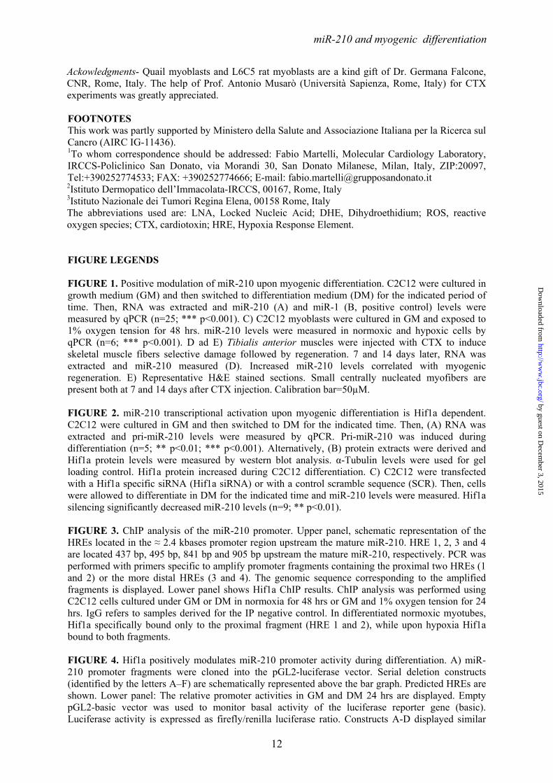
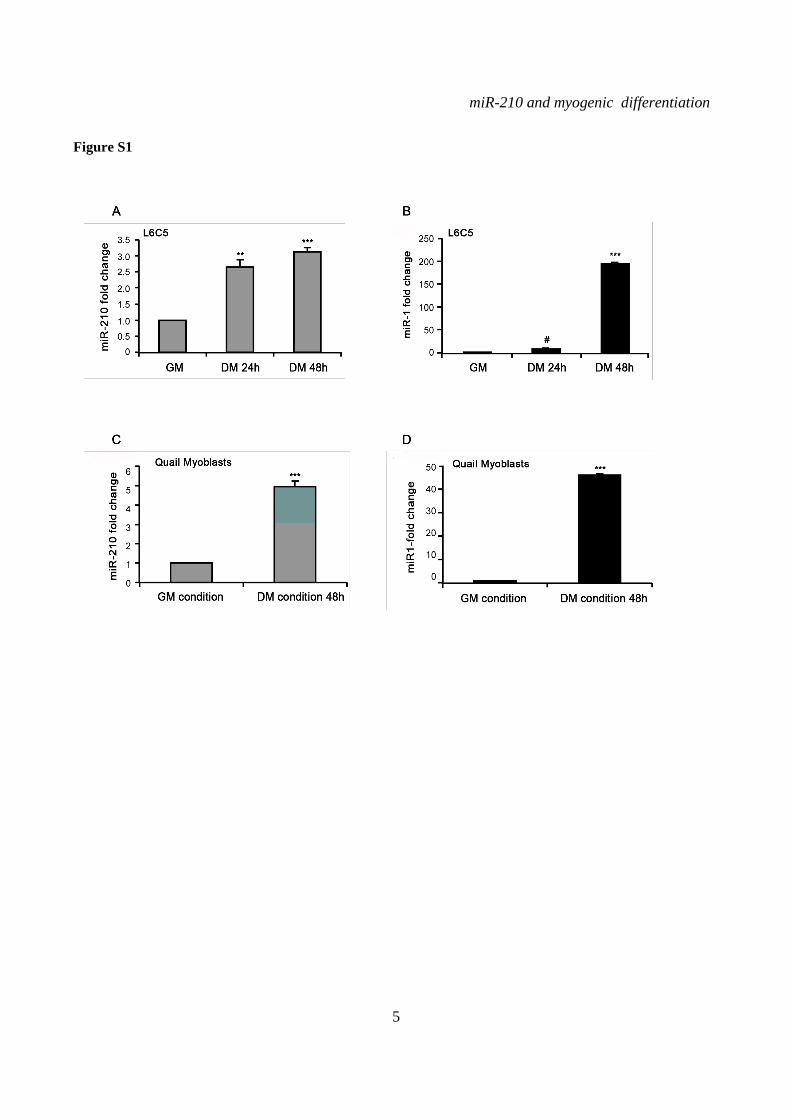
expression increased during myogenic differentiation (Fig.1A). As expected, muscle specific miR-1 levels increased steadily during differentiation, as well (Fig.1B). As a comparison, miR-210 levels after 72 hrs in DM were similar to these observed when GM cultures of C2C12 were exposed to hypoxia for 48 hrs (Fig.1C). miR-210 increase following muscle differentiation was not limited to C2C12, since miR-210 was similarly induced also in independent myogenesis cell models, such as rat myoblasts L6C5 (Fig. S1A) and quail myoblast cell lines (Fig.S1C). Again, positive control was represented by increased miR-1 levels (Fig.S1B and D).
Next, we investigated whether miR-210 was induced also during myogenesis in vivo. To this aim, myonecrosis was induced by cardiotoxin (CTX) injection of tibialis anterior muscles. Fig. 1D and E time course experiment showed that at both day 7 and 14 after injection, skeletal muscle regeneration was associated with increased miR-210 levels.
These data suggest that miR-210 up-regulation during myogenesis was likely functionally relevant.
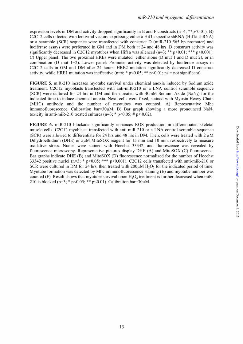
miR-210 induction during differentiation occurs at the transcriptional level and is Hif1a dependent- We sought to determine how miR-210 is regulated during differentiation. Figure 2A shows that pri-miR-210 was induced upon myogenic differentiation to levels similar to these of the corresponding mature miRNA, indicating a transcriptional regulation of miR-210 during differentiation. These data were confirmed also with an independent primer couple (not shown). Next, given the importance of HIF1A for both miR-210 induction following hypoxia (1) and skeletal muscle physio-pathology (37-40), we tested whether Hif1a silencing affected miR-210 induction in differentiating C2C12 cells.
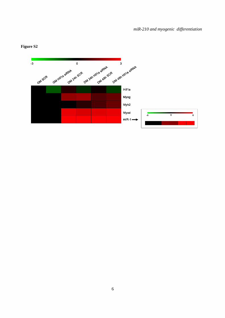
Western blot analysis showed that Hif1a protein increased upon C2C12 differentiation (Fig.2B). Next we assayed the effect of Hif1a silencing on miR-210 levels in differentiating myoblasts. Hif1a transcript was not modulated during C2C12 differentiation and was significantly knocked-down following specific siRNA transfection (Fig.S2). When miR-210 expression was measured, we found that miR-210 levels were significantly decreased at both 24 and 48 hrs differentiation timepoints in Hif1a silenced cells (Fig.2C). miR-210 expression was decreased in myoblasts as well, but the difference did not reach statistical significance.
Decreased miR-210 levels in DM were not simply due to an impairment of the differentiation process since the expression of myogenic markers such as miR-1, myogenin and Myosin Heavy Chain was unaffected (Fig.S2).
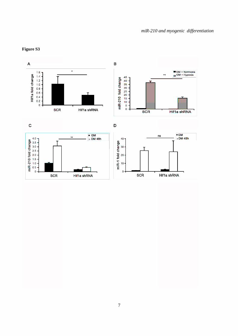
To confirm these findings, Hif1a was also knocked down transducing C2C12 cells with a lentiviral vector expressing a specific shRNA (Fig.S3A). The adopted vector also carried a puromycin resistance gene, allowing the selection of infected cells. As expected, Hif1a silencing decreased miR-210 levels in hypoxic myoblasts (Fig.S3B). When normoxic differentiating myoblasts were measured, we found that miR-210 levels were significantly decreased by Hif1a shRNA, both in GM and in DM; indeed, after 48 hrs of differentiation, miR-210 levels were still lower than these observed in myoblasts expressing a scrambled control (Fig.S3C). This stronger inhibition of miR-210 expression was likely due to the higher efficiency of transduction yielded by infection followed by puromycin selection, compared to transient transfection. Again, unaffected miR-1 induction upon differentiation indicated that myogenic differentiation was not grossly inhibited by Hif1a knock-down (Fig.S3D). These findings were further confirmed by two independent shRNA targeting Hif1a (not shown).
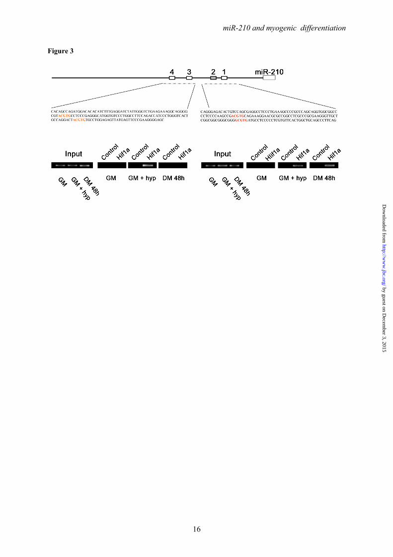
Hif1a binds to miR-210 promoter activating its expression in differentiating myoblasts- Analysis of the ≈2.4 kbases promoter sequence upstream the mature miR-210 sequence identified four potential Hypoxia Response Element (HRE) sites. In order to elucidate whether these sites were functionally relevant, a chromatin immunoprecipitation experiment was performed. As shown in Figure 3, Hif1a antibody (but not control IgGs) immunoprecipitated the miR-210 promoter fragments in hypoxic and in differentiated C2C12 cells, but very little in the normoxic undifferentiated controls. Specifically, two primer couples were used, encompassing the two proximal HRE (HRE1 and 2) and the two distal sites (HRE3 and 4). A more defined analysis was precluded by the close proximity of the HREs. We found that, while Hif1a bound to both fragments upon hypoxia, in differentiated myotubes Hif1a preferentially bound to the proximal fragment.
To better identify and characterize the functional HRE, we amplified ≈2.4 kbases of the genomic regions upstream of miR-210
by guest on Decem
ber 3, 2015http://w
ww
.jbc.org/D
ownloaded from
miR-210 and myogenic differentiation
6
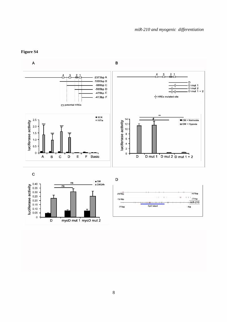
sequence and generated serial deletion constructs (Fig.4A). The promoter fragments were cloned into a luciferase vector and the effect of the deletions on reporter gene activity during C2C12 myogenic differentiation was analyzed. Luciferase assays showed that miR-210 promoter was induced in DM and that a 565 bases fragment (D construct) that harbors the two proximal HRE sites (HRE1 and 2) was essential for sustained luciferase activity in differentiated myotubes (Fig.4A). We also found that miR-210 D-construct activity was significantly decreased in C2C12 myotubes where Hif1a was silenced, further corroborating Hif1a role in miR-210 promoter modulation (Fig.4B). Further deletions encompassing HRE2 (construct E), significantly decreased miR-210 promoter activity, albeit some residual functionality and inducibility were present (Fig.4A). Interestingly, a similar pattern was observed when Hif1a overexpressing myoblasts were assayed, indicating that HRE1 was not functional (Fig.S4A). To confirm these findings, HRE1 and HRE2 were mutated, either alone or in combination. Fig.4C shows that HRE2 mutation decreased the activity of D construct, while HRE1 mutation was ineffective. Again, similar results were obtained when hypoxic myoblasts were assayed, albeit promoter activity was much stronger (Fig.S4B).
Finally, promoter analysis indicated the presence of a potential Myod binding site 523 nucleotides upstream of miR-210. However, mutational analysis showed that this sequence did not affect miR-210 promoter expression, either in GM or in DM (Fig.S4C and table S3).
Bioinformatic analysis of the ≈2.4 kbases upstream of miR-210 mature sequence also indicated a potential CpG island from base -548 to base -280 (Fig.S4D), suggesting that DNA methylation may contribute to miR-210 promoter regulation during muscle differentiation. Indeed, a CpG island has been identified upstream of human miR-210 as well (41). When the cytosine methylation of this CpG rich region was analyzed using the bisulfite technique, we found that methylation was almost undetectable in myoblasts and the methylation status did not change during differentiation (not shown). In keeping with these findings, C2C12 treatment with 5-azacytidine de-methylating drug for 24 to 72 hrs did not affect miR-210 expression (not shown). Thus, cytosine methylation does not seem to play a major role in miR-210 regulation during myogenic differentiation.
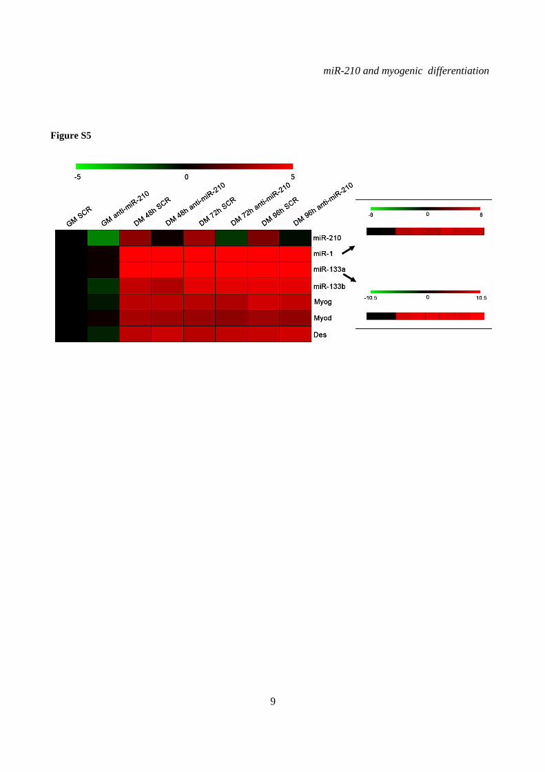
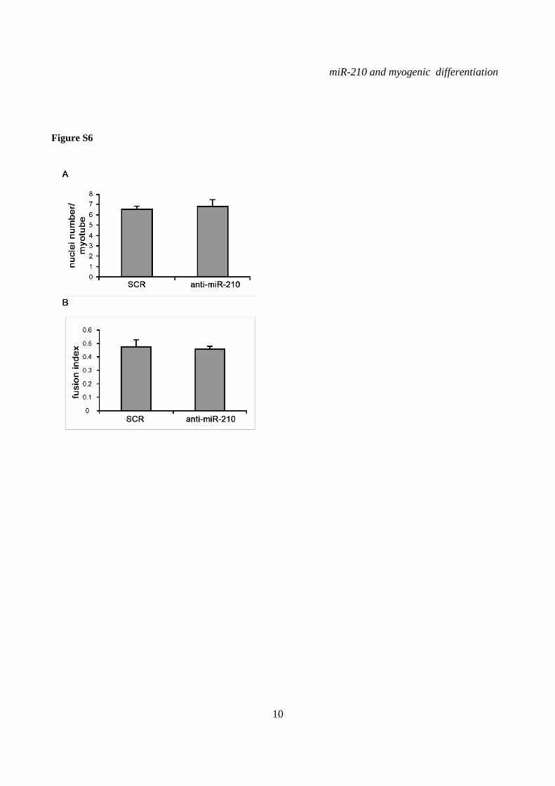
miR-210 blockade does not affect myogenic differentiation- To assess the functional role of miR-210 in skeletal muscle differentiation, we inhibited miR-210 in C2C12 cells using a LNA-modified antisense oligonucleotide (anti-miR-210). Albeit miR-210 levels were decreased more than 10 fold in differentiated cells (Fig.S5), we did not observe any overt alteration in myotube morphology (Fig.5A, left panel). In keeping with this observation, the expression levels of myogenesis markers such as Myogenin, Desmin, Myod, miR-1, miR-133a and miR-133b were unaffected by miR-210 blockade (Fig.S5). Also myotube number (not shown), fusion index and the average number of nuclei per myotube after 48 hrs of differentiation did not change significantly (Fig.S6).
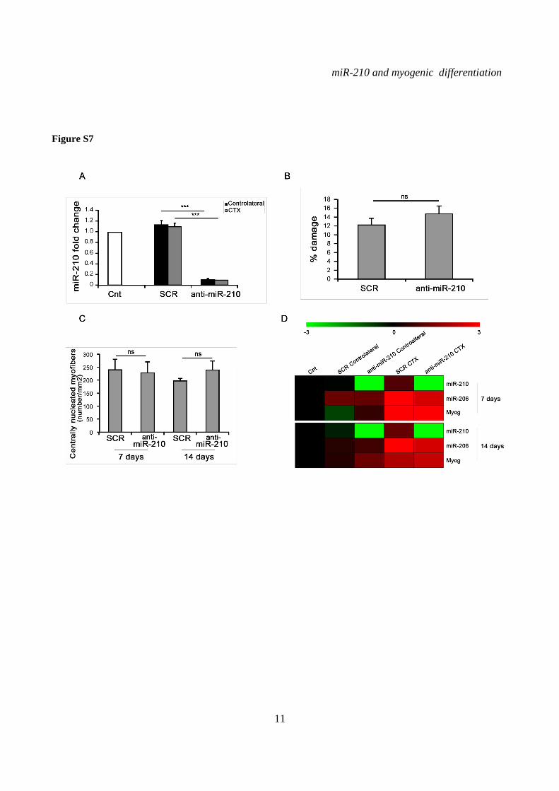
We also assayed whether miR-210 blockade in vivo affected skeletal muscle regeneration. miR-210 was inhibited by systemic anti-miR-210 administration, decreasing miR-210 levels >10 fold (Fig.S7A). Skeletal muscle injury was induced by cardiotoxin (CTX) injection and Evans Blue staining showed that similar damage was induced in the two groups (Fig.S7B). Skeletal muscle regeneration was assayed 7 and 14 days after CTX injection. Although miR-210 was efficiently decreased, the number of centrally nucleated regenerating myofibers was similar in anti-miR-210 and LNA-scrambled groups (Fig.S7C). In keeping with this finding, regeneration markers such as miR-206 and myogenin were similarly induced in the two groups both at 7 and 14 days (Fig.S7D).
In conclusion, at least in the adopted experimental conditions, we found no evidence that miR-210 was necessary for myogenic differentiation.
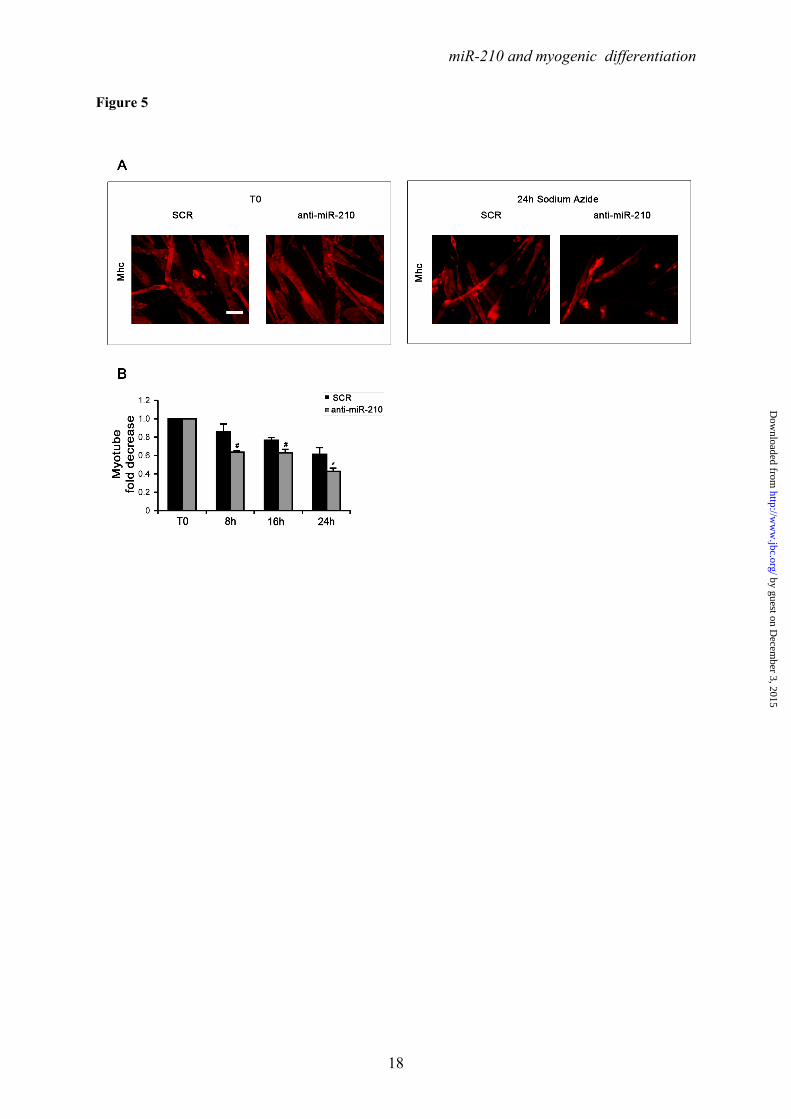
miR-210 increases myotube survival in the presence of mitochondrial dysfunction and oxidative stress- During skeletal muscle intense workload, oxygen supply may not match the metabolic needs. This mismatch between oxygen supply and its demand at the cellular level may result in a hypoxic condition. miR-210 is induced by hypoxia, contributing to the adaptive mechanisms allowing cell survival. Albeit miR-210 induction by low oxygen tension is rapid, we asked whether high miR-210 levels observed in differentiated myotubes may boost cell survival, allowing a prompter response. In order to evaluate the role of miR-210 when mitochondrial function is impaired, we blocked the electron transport chain by sodium azide, antimycin A and rotenone (42).
by guest on Decem
ber 3, 2015http://w
ww
.jbc.org/D
ownloaded from
miR-210 and myogenic differentiation
7
Albeit many differences exist, these drugs inhibit mitochondrial function, modeling insufficient oxygen supply, without the confounding effect of miR-210 induction (not shown). We found that sodium azide (Fig.5), antimycin A (Fig.S8A and B) and rotenone (Fig.S8C and D) all decreased myotube survival, as expected. However, myotube number further decreased when miR-210 was inhibited.
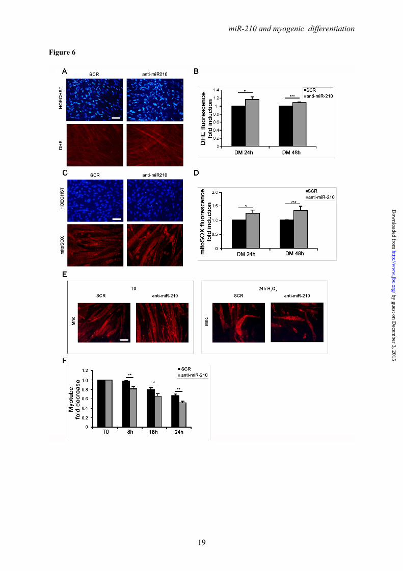
One of the consequences of mitochondrial electron transport chain uncoupling is an increase of ROS production (43). Given miR-210 role in mitochondrial metabolism (1), we assayed whether miR-210 blockade increased C2C12 myotube levels of oxidative stress. miR-210 was inhibited by anti-miR-210 transfection and oxidative stress was measured using DHE and MitoSOX fluorescent dyes, that are particularly sensitive to intracellular and mitochondrial superoxide anion levels, respectively. We found that both DHE (Fig.6A and B) and MitoSOX (Fig.6C and D) associated fluorescence was significantly increased when miR-210 was blocked. Next, we assayed whether miR-210 levels affected myotube resistance to oxidative stress. After anti-miR-210 and LNA-scrambled transfection, C2C12 cells were allowed to differentiate for 48 hrs and then were challenged with a 200μM H2O2 bolus. We found that myotube survival was further decreased when miR-210 was blocked (Fig.6E and F).
We conclude that high levels of miR-210 in differentiated skeletal muscle contribute to the maintenance of the redox balance and to cell survival in the presence of mitochondrial and oxidative stresses.
DISCUSSION
miR-210 has been studied mostly for its role in cell and tissue response to hypoxia. Here, we demonstrated that miR-210 is induced during normoxic myogenesis in different cell culture models. The relevance of these findings confirmed that miR-210 levels increased in regenerating skeletal muscles. We adopted the CTX model for this experiment since this drug targets muscle cells specifically, while vascular cells are largely unaffected (44). Thus, miR-210 levels were not increased by hypoxia due to a lack of perfusion. Accordingly, we observed that CTX treatment did not induce miR-210 acutely, and miR-210 levels increased only in the presence of concomitant muscle regeneration.
miR-210 is involved in other differentiating systems as well, independently of its
hypoxamiR role: miR-210 promotes osteoblastic differentiation by inhibiting the expression of a type 1B receptor of activin A (AcvR1b) (27) and miR-210 also stimulates adipogenesis by repressing WNT signaling through targeting Tcf7l2 (28). Moreover, albeit its function in these systems is not known yet, miR-210 is also induced during epithelial cell and keratinocyte differentiation (45,46), as well as in embryonic stem cells differentiating towards the endothelial lineage (47).
The induction of miR-210 precursor (pri-miR-210) in differentiating myoblasts showed that its regulation occurred at the transcriptional level. We characterized the promoter region involved in this modulation, showing that Hif1a is a major player in the robust induction of the miR-210 promoter during C2C12 myogenic differentiation. We also identified a functional HRE site, localized at –495bp from mature mouse miR-210. Interestingly, this HRE site is localized in a genomic region highly conserved across species. Indeed, the 45bp encompassing mouse HRE2 displays 89% identity with the human miR-210 promoter sequence containing an HRE that Huang et al. (5) showed to be important for miR-210 induction upon hypoxia. Thus, these two binding sites are likely homologs. Mutation of mouse HRE2 site greatly diminished promoter activity both during myogenic differentiation and hypoxia. In keeping with these data, Hif1a knockdown was associated with a significant down-modulation of miR-210 levels and promoter activity supporting the importance of Hif1a in the regulation of miR-210 expression in normoxic myofibers.
We also noticed that, while Hif1a knock-down greatly decreased miR-210 promoter activity upon myogenic differentiation, some residual activity was present. Further investigations are needed to explore additional regulators of miR-210 expression during myogenesis. However, it is worth noting that Akt increases miR-210 levels through a Hif-independent mechanism in hypoxic cardiomyocytes (19), suggesting that a similar mechanism may be at work in skeletal muscles. Among the potential miR-210 regulators, a conserved NF-κB binding site has also been mapped in the 200-bp proximal promoter region (48). However, since myogenic differentiation correlates with loss of NF-κB transactivation function, it was not further explored (49). Finally, in most cell types, upon exposure to hypoxia, miR-210 is activated by HIF1A, but
by guest on Decem
ber 3, 2015http://w
ww
.jbc.org/D
ownloaded from
miR-210 and myogenic differentiation
8
not by HIF2A (5-9). Thus, we speculate that the same HIF isoform selection may likely be in place in differentiating myogenic cells. Nonetheless, Hif2a in normoxic myotubes cannot be ruled out.
Albeit Hif1a has been mainly characterized for its role in hypoxia response, Hif1a involvement in normoxic skeletal muscle physiology is not without precedent. Indeed, contrary to most tissues, it has been shown that Hif1a is present under normoxic conditions in a few mouse tissues, including skeletal muscle (39), where its level increases in response to systemic hypoxia (40) or ischaemia (50).
Targeted deletion of Hif1a in mouse skeletal muscle affects exercise-induced gene regulatory responses of glycolytic enzymes. Skeletal muscle devoid of Hif1a also displays an increase in the activity of mitochondrial rate-limiting enzymes and the formation of excessive mitochondrial ROS. Conversely, development is largely unaffected (38). We found that Hif1a knock-down did not impair myogenic differentiation, but blunted miR-210 expression, that in turn is a demonstrated modulator of mitochondrial metabolism and of intracellular ROS production (1). However, contrary to us, Ono et. al found that Hif1a knockdown had a negative impact on myoblast differentiation (37), possibly due to differences in the adopted experimental conditions.
To explore the functional role of increased miR-210 levels in differentiated myofibers, miR-210 was inhibited, both in vitro and in vivo. Albeit several features of myogenic differentiation were measured, we failed to show that decreased miR-210 activity impairs myogenic differentiation significantly. However, it is possible that the effects of miR-210 blockade are subtle, and the adopted experimental conditions inadequate to make them evident. Conversely, we found that miR-210 impairment affected myotube survival and oxidative stress levels. Myofibers, and in particular type I ones, mostly rely on oxidative
metabolism, efficiently deriving ATP from glucose to match the intense energy consumption associated with muscle contraction. Oxidative metabolism however, comes with a price, represented by ROS production by mitochondria (51). Paradoxically, ROS production is particularly significant in hypoxic conditions, when electron transport chain is uncoupled (43,52). Workload type also seems to be important, with eccentric contractions inducing greater oxidative stress than concentric contractions (53).
Skeletal muscle is particularly prone to hypoxia and oxidative damage since during intense workload oxygen supply may not match the need. In this respect, increased miR-210 levels seem to be instrumental to increase myofiber survival under metabolic stress conditions. Albeit many differences apply, it may be considered as a sort of “constitutive preconditioning”, protecting metabolically active myofibers from mitochondrial dysfunction and oxidative stress. In keeping with this model, Kim et al. demonstrated a crucial role of miR-210 in ischemia-reperfusion preconditioning (17). Furthermore, miR-210 inhibition increased oxidative stress levels of normoxic differentiated myoblasts, as previously observed in cardiac myocytes (19) and in hypoxic endothelial cells (20). Likewise, miR-210 also displayed a cytoprotective role in hypoxic cardiomyocytes (19).
It is also worth noting that the magnitude of myofiber cytoprotection found in our studies was relatively modest, although similar to that found by others (18,19).
In conclusion, we found that Hif1a induces miR-210 in normoxic differentiating myoblasts by direct transactivation of its promoter. miR-210 cytoprotective role indicates future potential therapeutic applications in skeletal muscle ischemic and non-ischemic diseases. In this respect, particularly encouraging are preclinical evidences indicating miR-210 as a novel therapy for treatment of ischemic heart disease (18).
by guest on Decem
ber 3, 2015http://w
ww
.jbc.org/D
ownloaded from
miR-210 and myogenic differentiation
9
REFERENCES 1. Devlin, C., Greco, S., Martelli, F., and Ivan, M. (2011) IUBMB Life 63, 94-100 2. Huang, X., Le, Q. T., and Giaccia, A. J. (2010) Trends Mol Med 16, 230-237 3. Voellenkle, C., van Rooij, J., Guffanti, A., Brini, E., Fasanaro, P., Isaia, E., Croft, L.,
David, M., Capogrossi, M. C., Moles, A., Felsani, A., and Martelli, F. (2012) Rna 18, 472-484
4. Ivan, M., Harris, A. L., Martelli, F., and Kulshreshtha, R. (2008) J Cell Mol Med 12, 1426-1431
5. Huang, X., Ding, L., Bennewith, K. L., Tong, R. T., Welford, S. M., Ang, K. K., Story, M., Le, Q. T., and Giaccia, A. J. (2009) Mol Cell 35, 856-867
6. Camps, C., Buffa, F. M., Colella, S., Moore, J., Sotiriou, C., Sheldon, H., Harris, A. L., Gleadle, J. M., and Ragoussis, J. (2008) Clin Cancer Res 14, 1340-1348
7. Fasanaro, P., D'Alessandra, Y., Di Stefano, V., Melchionna, R., Romani, S., Pompilio, G., Capogrossi, M. C., and Martelli, F. (2008) J Biol Chem 283, 15878-15883
8. Favaro, E., Ramachandran, A., McCormick, R., Gee, H., Blancher, C., Crosby, M., Devlin, C., Blick, C., Buffa, F., Li, J. L., Vojnovic, B., Pires das Neves, R., Glazer, P., Iborra, F., Ivan, M., Ragoussis, J., and Harris, A. L. (2010) PLoS One 5, e10345
9. Puissegur, M. P., Mazure, N. M., Bertero, T., Pradelli, L., Grosso, S., Robbe-Sermesant, K., Maurin, T., Lebrigand, K., Cardinaud, B., Hofman, V., Fourre, S., Magnone, V., Ricci, J. E., Pouyssegur, J., Gounon, P., Hofman, P., Barbry, P., and Mari, B. (2011) Cell Death Differ 18, 465-478
10. Zhang, Z., Sun, H., Dai, H., Walsh, R. M., Imakura, M., Schelter, J., Burchard, J., Dai, X., Chang, A. N., Diaz, R. L., Marszalek, J. R., Bartz, S. R., Carleton, M., Cleary, M. A., Linsley, P. S., and Grandori, C. (2009) Cell Cycle 8, aheadofprint
11. Chan, Y. C., Banerjee, J., Choi, S. Y., and Sen, C. K. (2011) Microcirculation, aheadofprint
12. Fasanaro, P., Greco, S., Lorenzi, M., Pescatori, M., Brioschi, M., Kulshreshtha, R., Banfi, C., Stubbs, A., Calin, G. A., Ivan, M., Capogrossi, M. C., and Martelli, F. (2009) J Biol Chem 284, 35134-35143
13. Kosaka, N., Sugiura, K., Yamamoto, Y., Yoshioka, Y., Miyazaki, H., Komatsu, N., Ochiya, T., and Kato, T. (2008) Br J Haematol 142, 293-300
14. Yang, W., Sun, T., Cao, J., Liu, F., Tian, Y., and Zhu, W. (2012) Exp Cell Res 318, 944-954
15. Gou, D., Ramchandran, R., Peng, X., Yao, L., Kang, K., Sarkar, J., Wang, Z., Zhou, G., and Raj, J. U. (2012) Am J Physiol Lung Cell Mol Physiol, aheadofprint
16. Quero, L., Dubois, L., Lieuwes, N. G., Hennequin, C., and Lambin, P. (2011) Radiother Oncol 101, 203-208
17. Kim, H. W., Haider, H. K., Jiang, S., and Ashraf, M. (2009) J Biol Chem 284, 33161-33168
18. Hu, S., Huang, M., Li, Z., Jia, F., Ghosh, Z., Lijkwan, M. A., Fasanaro, P., Sun, N., Wang, X., Martelli, F., Robbins, R. C., and Wu, J. C. (2010) Circulation 122, S124-131
19. Mutharasan, R. K., Nagpal, V., Ichikawa, Y., and Ardehali, H. (2011) Am J Physiol Heart Circ Physiol 301, H1519-1530
20. Chan, S. Y., Zhang, Y. Y., Hemann, C., Mahoney, C. E., Zweier, J. L., and Loscalzo, J. (2009) Cell Metab 10, 273-284
21. Yoshioka, Y., Kosaka, N., Ochiya, T., and Kato, T. (2012) J Biol Chem, aheadofprint 22. Chen, Z., Li, Y., Zhang, H., Huang, P., and Luthra, R. (2010) Oncogene 29, 4362-
4368
by guest on Decem
ber 3, 2015http://w
ww
.jbc.org/D
ownloaded from
miR-210 and myogenic differentiation
10
23. Liu, F., Lou, Y. L., Wu, J., Ruan, Q. F., Xie, A., Guo, F., Cui, S. P., Deng, Z. F., and Wang, Y. (2011) Kidney Blood Press Res 35, 182-191
24. Lou, Y. L., Guo, F., Liu, F., Gao, F. L., Zhang, P. Q., Niu, X., Guo, S. C., Yin, J. H., Wang, Y., and Deng, Z. F. (2012) Mol Cell Biochem, aheadofprint
25. Alaiti, M. A., Ishikawa, M., Masuda, H., Simon, D. I., Jain, M. K., Asahara, T., and Costa, M. A. (2012) J Cell Mol Med, aheadofprint
26. Bianchi, N., Zuccato, C., Lampronti, I., Borgatti, M., and Gambari, R. (2009) BMB Rep 42, 493-499
27. Mizuno, Y., Tokuzawa, Y., Ninomiya, Y., Yagi, K., Yatsuka-Kanesaki, Y., Suda, T., Fukuda, T., Katagiri, T., Kondoh, Y., Amemiya, T., Tashiro, H., and Okazaki, Y. (2009) FEBS Lett, aheadofprint
28. Qin, L., Chen, Y., Niu, Y., Chen, W., Wang, Q., Xiao, S., Li, A., Xie, Y., Li, J., Zhao, X., He, Z., and Mo, D. (2010) BMC Genomics 11, 320
29. Greco, S., Fasanaro, P., Castelvecchio, S., D'Alessandra, Y., Arcelli, D., Di Donato, M., Malavazos, A., Capogrossi, M. C., Menicanti, L., and Martelli, F. (2012) Diabetes 61, 1633-1641
30. Martelli, F., Cenciarelli, C., Santarelli, G., Polikar, B., Felsani, A., and Caruso, M. (1994) Oncogene 9, 3579-3590
31. Cardinali, B., Castellani, L., Fasanaro, P., Basso, A., Alema, S., Martelli, F., and Falcone, G. (2009) PLoS One 4, e7607
32. Zaccagnini, G., Martelli, F., Magenta, A., Cencioni, C., Fasanaro, P., Nicoletti, C., Biglioli, P., Pelicci, P. G., and Capogrossi, M. C. (2007) J Biol Chem 282, 31453-31459
33. Travaglione, S., Messina, G., Fabbri, A., Falzano, L., Giammarioli, A. M., Grossi, M., Rufini, S., and Fiorentini, C. (2005) Cell Death Differ 12, 78-86
34. Fasanaro, P., Romani, S., Voellenkle, C., Maimone, B., Capogrossi, M. C., and Martelli, F. (2012) PLoS One 7, e44651
35. Gurtner, C., Tu, E., Jamshidi, N., Haigis, R. W., Onofrey, T. J., Edman, C. F., Sosnowski, R., Wallace, B., and Heller, M. J. (2002) Electrophoresis 23, 1543-1550
36. Musaro, A., Giacinti, C., Borsellino, G., Dobrowolny, G., Pelosi, L., Cairns, L., Ottolenghi, S., Cossu, G., Bernardi, G., Battistini, L., Molinaro, M., and Rosenthal, N. (2004) Proc Natl Acad Sci U S A 101, 1206-1210
37. Ono, Y., Sensui, H., Sakamoto, Y., and Nagatomi, R. (2006) J Cell Biochem 98, 642-649
38. Mason, S. D., Howlett, R. A., Kim, M. J., Olfert, I. M., Hogan, M. C., McNulty, W., Hickey, R. P., Wagner, P. D., Kahn, C. R., Giordano, F. J., and Johnson, R. S. (2004) PLoS Biol 2, e288
39. Pisani, D. F., and Dechesne, C. A. (2005) J Gen Physiol 126, 173-178 40. Stroka, D. M., Burkhardt, T., Desbaillets, I., Wenger, R. H., Neil, D. A., Bauer, C.,
Gassmann, M., and Candinas, D. (2001) Faseb J 15, 2445-2453 41. Du, Y., Liu, Z., Gu, L., Zhou, J., Zhu, B. D., Ji, J., and Deng, D. (2012) BMC Cancer
12, 249 42. Scatena, R. (2012) Adv Exp Med Biol 942, 329-346 43. Guzy, R. D., and Schumacker, P. T. (2006) Exp Physiol 91, 807-819 44. d'Albis, A., Couteaux, R., Janmot, C., Roulet, A., and Mira, J. C. (1988) Eur J
Biochem 174, 103-110 45. Tsuchiya, S., Oku, M., Imanaka, Y., Kunimoto, R., Okuno, Y., Terasawa, K., Sato, F.,
Tsujimoto, G., and Shimizu, K. (2009) Nucleic Acids Res, aheadofprint 46. Hildebrand, J., Rutze, M., Walz, N., Gallinat, S., Wenck, H., Deppert, W., Grundhoff,
A., and Knott, A. (2011) J Invest Dermatol 131, 20-29
by guest on Decem
ber 3, 2015http://w
ww
.jbc.org/D
ownloaded from

miR-210 and myogenic differentiation
11
47. Kane, N. M., Meloni, M., Spencer, H. L., Craig, M. A., Strehl, R., Milligan, G., Houslay, M. D., Mountford, J. C., Emanueli, C., and Baker, A. H. (2010) Arterioscler Thromb Vasc Biol 30, 1389-1397
48. Zhang, Y., Fei, M., Xue, G., Zhou, Q., Jia, Y., Li, L., Xin, H., and Sun, S. (2012) J Cell Mol Med 16, 249-259
49. Guttridge, D. C., Albanese, C., Reuther, J. Y., Pestell, R. G., and Baldwin, A. S., Jr. (1999) Mol Cell Biol 19, 5785-5799
50. Milkiewicz, M., Pugh, C. W., and Egginton, S. (2004) J Physiol 560, 21-26 51. Bloomer, R. J., and Goldfarb, A. H. (2004) Can J Appl Physiol 29, 245-263 52. Musaro, A., Fulle, S., and Fano, G. (2010) Curr Opin Clin Nutr Metab Care 13, 236-
242 53. Kon, M., Tanabe, K., Lee, H., Kimura, F., Akimoto, T., and Kono, I. (2007) Appl
Physiol Nutr Metab 32, 273-281
by guest on Decem
ber 3, 2015http://w
ww
.jbc.org/D
ownloaded from
miR-210 and myogenic differentiation
12
Ackowledgments- Quail myoblasts and L6C5 rat myoblasts are a kind gift of Dr. Germana Falcone, CNR, Rome, Italy. The help of Prof. Antonio Musarò (Università Sapienza, Rome, Italy) for CTX experiments was greatly appreciated. FOOTNOTES This work was partly supported by Ministero della Salute and Associazione Italiana per la Ricerca sul Cancro (AIRC IG-11436). 1To whom correspondence should be addressed: Fabio Martelli, Molecular Cardiology Laboratory, IRCCS-Policlinico San Donato, via Morandi 30, San Donato Milanese, Milan, Italy, ZIP:20097, Tel:+390252774533; FAX: +390252774666; E-mail: [email protected] 2Istituto Dermopatico dell’Immacolata-IRCCS, 00167, Rome, Italy 3Istituto Nazionale dei Tumori Regina Elena, 00158 Rome, Italy The abbreviations used are: LNA, Locked Nucleic Acid; DHE, Dihydroethidium; ROS, reactive oxygen species; CTX, cardiotoxin; HRE, Hypoxia Response Element. FIGURE LEGENDS FIGURE 1. Positive modulation of miR-210 upon myogenic differentiation. C2C12 were cultured in growth medium (GM) and then switched to differentiation medium (DM) for the indicated period of time. Then, RNA was extracted and miR-210 (A) and miR-1 (B, positive control) levels were measured by qPCR (n=25; *** p<0.001). C) C2C12 myoblasts were cultured in GM and exposed to 1% oxygen tension for 48 hrs. miR-210 levels were measured in normoxic and hypoxic cells by qPCR (n=6; *** p<0.001). D ad E) Tibialis anterior muscles were injected with CTX to induce skeletal muscle fibers selective damage followed by regeneration. 7 and 14 days later, RNA was extracted and miR-210 measured (D). Increased miR-210 levels correlated with myogenic regeneration. E) Representative H&E stained sections. Small centrally nucleated myofibers are present both at 7 and 14 days after CTX injection. Calibration bar=50µM. FIGURE 2. miR-210 transcriptional activation upon myogenic differentiation is Hif1a dependent. C2C12 were cultured in GM and then switched to DM for the indicated time. Then, (A) RNA was extracted and pri-miR-210 levels were measured by qPCR. Pri-miR-210 was induced during differentiation (n=5; ** p<0.01; *** p<0.001). Alternatively, (B) protein extracts were derived and Hif1a protein levels were measured by western blot analysis. α-Tubulin levels were used for gel loading control. Hif1a protein increased during C2C12 differentiation. C) C2C12 were transfected with a Hif1a specific siRNA (Hif1a siRNA) or with a control scramble sequence (SCR). Then, cells were allowed to differentiate in DM for the indicated time and miR-210 levels were measured. Hif1a silencing significantly decreased miR-210 levels (n=9; ** p<0.01). FIGURE 3. ChIP analysis of the miR-210 promoter. Upper panel, schematic representation of the HREs located in the ≈ 2.4 kbases promoter region upstream the mature miR-210. HRE 1, 2, 3 and 4 are located 437 bp, 495 bp, 841 bp and 905 bp upstream the mature miR-210, respectively. PCR was performed with primers specific to amplify promoter fragments containing the proximal two HREs (1 and 2) or the more distal HREs (3 and 4). The genomic sequence corresponding to the amplified fragments is displayed. Lower panel shows Hif1a ChIP results. ChIP analysis was performed using C2C12 cells cultured under GM or DM in normoxia for 48 hrs or GM and 1% oxygen tension for 24 hrs. IgG refers to samples derived for the IP negative control. In differentiated normoxic myotubes, Hif1a specifically bound only to the proximal fragment (HRE 1 and 2), while upon hypoxia Hif1a bound to both fragments. FIGURE 4. Hif1a positively modulates miR-210 promoter activity during differentiation. A) miR-210 promoter fragments were cloned into the pGL2-luciferase vector. Serial deletion constructs (identified by the letters A–F) are schematically represented above the bar graph. Predicted HREs are shown. Lower panel: The relative promoter activities in GM and DM 24 hrs are displayed. Empty pGL2-basic vector was used to monitor basal activity of the luciferase reporter gene (basic). Luciferase activity is expressed as firefly/renilla luciferase ratio. Constructs A-D displayed similar
by guest on Decem
ber 3, 2015http://w
ww
.jbc.org/D
ownloaded from
miR-210 and myogenic differentiation
13
expression levels in DM and activity dropped significantly in E and F constructs (n=4; **p<0.01). B) C2C12 cells infected with lentiviral vectors expressing either a Hif1a specific shRNA (Hif1a shRNA) or a scramble (SCR) sequence were transfected with construct D (miR-210 565 bp promoter) and luciferase assays were performed in GM and in DM both at 24 and 48 hrs. D construct activity was significantly decreased in C2C12 myotubes when Hif1a was silenced (n=3; ** p<0.01; *** p<0.001). C) Upper panel: The two proximal HREs were mutated either alone (D mut 1 and D mut 2), or in combination (D mut 1+2). Lower panel: Promoter activity was detected by luciferase assays in C2C12 cells in GM and DM after 24 hours. HRE2 mutation significantly decreased D construct activity, while HRE1 mutation was ineffective (n=6; * p<0.05; ** p<0.01; ns = not significant). FIGURE 5. miR-210 increases myotube survival under chemical anoxia induced by Sodium azide treatment. C2C12 myoblasts transfected with anti-miR-210 or a LNA control scramble sequence (SCR) were cultured for 24 hrs in DM and then treated with 40mM Sodium Azide (NaN3) for the indicated time to induce chemical anoxia. Next, cells were fixed, stained with Myosin Heavy Chain (MHC) antibody and the number of myotubes was counted. A) Representative Mhc immunofluorescence. Calibration bar=30µM. B) Bar graph showing a more pronounced NaN3 toxicity in anti-miR-210 treated cultures (n=3; * p<0.05; # p< 0.02). FIGURE 6. miR-210 blockade significantly enhances ROS production in differentiated skeletal muscle cells. C2C12 myoblasts transfected with anti-miR-210 or a LNA control scramble sequence (SCR) were allowed to differentiate for 24 hrs and 48 hrs in DM. Then, cells were treated with 2 μM Dihydroethidium (DHE) or 5μM MitoSOX reagent for 15 min and 10 min, respectively to measure oxidative stress. Nuclei were stained with Hoechst 33342, and fluorescence was revealed by fluorescence microscopy. Representative pictures display DHE (A) and MitoSOX (C) fluorescence. Bar graphs indicate DHE (B) and MitoSOX (D) fluorescence normalized for the number of Hoechst 33342 positive nuclei (n=3; * p<0.05; *** p<0.001). C2C12 cells transfected with anti-miR-210 or SCR were cultured in DM for 24 hrs, then treated with 200µM H2O2 for the indicated period of time. Myotube formation was detected by Mhc immunofluorescence staining (E) and myotube number was counted (F). Result shows that myotube survival upon H2O2 treatment is further decreased when miR-210 is blocked (n=3; * p<0.05; ** p<0.01). Calibration bar=30µM.
by guest on Decem
ber 3, 2015http://w
ww
.jbc.org/D
ownloaded from
miR-210 and myogenic differentiation
14
Figure 1
by guest on Decem
ber 3, 2015http://w
ww
.jbc.org/D
ownloaded from
miR-210 and myogenic differentiation
15
Figure 2
by guest on Decem
ber 3, 2015http://w
ww
.jbc.org/D
ownloaded from
miR-210 and myogenic differentiation
16
Figure 3
by guest on Decem
ber 3, 2015http://w
ww
.jbc.org/D
ownloaded from

miR-210 and myogenic differentiation
17
Figure 4
by guest on Decem
ber 3, 2015http://w
ww
.jbc.org/D
ownloaded from
miR-210 and myogenic differentiation
18
Figure 5
by guest on Decem
ber 3, 2015http://w
ww
.jbc.org/D
ownloaded from
miR-210 and myogenic differentiation
19
Figure 6
by guest on Decem
ber 3, 2015http://w
ww
.jbc.org/D
ownloaded from
miR-210 and myogenic differentiation
1
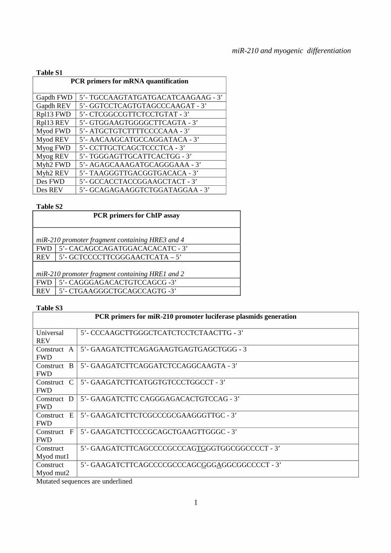
Table S1 PCR primers for mRNA quantification
Gapdh FWD 5’- TGCCAAGTATGATGACATCAAGAAG - 3’ Gapdh REV 5’- GGTCCTCAGTGTAGCCCAAGAT - 3’ Rpl13 FWD 5’- CTCGGCCGTTCTCCTGTAT - 3’ Rpl13 REV 5’- GTGGAAGTGGGGCTTCAGTA - 3’ Myod FWD 5’- ATGCTGTCTTTTCCCCAAA - 3’ Myod REV 5’- AACAAGCATGCCAGGATACA - 3’ Myog FWD 5’- CCTTGCTCAGCTCCCTCA - 3’ Myog REV 5’- TGGGAGTTGCATTCACTGG - 3’ Myh2 FWD 5’- AGAGCAAAGATGCAGGGAAA - 3’ Myh2 REV 5’- TAAGGGTTGACGGTGACACA - 3’ Des FWD 5’- GCCACCTACCGGAAGCTACT - 3’ Des REV 5’- GCAGAGAAGGTCTGGATAGGAA - 3’ Table S2
PCR primers for ChIP assay miR-210 promoter fragment containing HRE3 and 4 FWD 5’- CACAGCCAGATGGACACACATC - 3’ REV 5’- GCTCCCCTTCGGGAACTCATA – 5’ miR-210 promoter fragment containing HRE1 and 2 FWD 5’- CAGGGAGACACTGTCCAGCG -3’ REV 5’- CTGAAGGGCTGCAGCCAGTG -3’ Table S3
PCR primers for miR-210 promoter luciferase plasmids generation Universal REV
5’- CCCAAGCTTGGGCTCATCTCCTCTAACTTG - 3’
Construct A FWD
5’- GAAGATCTTCAGAGAAGTGAGTGAGCTGGG - 3
Construct B FWD
5’- GAAGATCTTCAGGATCTCCAGGCAAGTA - 3’
Construct C FWD
5’- GAAGATCTTCATGGTGTCCCTGGCCT - 3’
Construct D FWD
5’- GAAGATCTTC CAGGGAGACACTGTCCAG - 3’
Construct E FWD
5’- GAAGATCTTCTCGCCCGCGAAGGGTTGC - 3’
Construct F FWD
5’- GAAGATCTTCCCGCAGCTGAAGTTGGGC - 3’
Construct Myod mut1
5’- GAAGATCTTCAGCCCCGCCCAGTGGGTGGCGGCCCCT - 3’
Construct Myod mut2
5’- GAAGATCTTCAGCCCCGCCCAGCGGGAGGCGGCCCCT - 3’
Mutated sequences are underlined

miR-210 and myogenic differentiation
2
Table S4
PCR primers for miR-210 promoter methylation study FWD 1 5’ – GAGGAAAAGGTATGTTTTGGGTTGTA – 3’ REV 1 5’ - CCCCCACCCTCATCTCCTCTAACTT – 3’ FWD 2 5’ - GAGGAAAAGGTATGTTTTGGGTTGTA – 3’ REV 2 5’ -CCCCATATACAAATAATCTATCTT – 3’ FWD 3 5’ - AAGATAGATTATTTGTATATGGGG – 3’ REV 3 5’ - CCCCCACCCTCATCTCCTCTAACTT – 3’
miR-210 and myogenic differentiation
3
SUPPLEMENTARY FIGURE LEGENDS Figure S1. miR-210 expression is induced upon rat and quail myoblast differentiation. L6C5 rat myoblasts were cultured in GM and then switched to DM for the indicated time. Then, RNA was extracted and miR-210 (A) and miR-1 (B) levels were measured by qPCR (n=4; # p< 0.02; ** p<0.01; *** p<0.001). Polyclonal populations of quail myoblasts transformed by the temperature-sensitive mutant of the Rous Sarcoma Virus LA29 were grown in GM conditions (high serum and 35°C allowing src activity) and moved to DM conditions (low serum and 41°C, inactivating src) for 48 hrs. Then, RNA was extracted and miR-210 (C) and miR-1 (D) levels were measured by qPCR (n=3; *** p<0.001). Figure S2. A specific siRNA knocks-down Hif1a efficiently and does not affect skeletal muscle markers. C2C12 were transfected with a Hif1a specific siRNA (Hif1a siRNA) or with a control scramble (SCR) sequence (n=8). Then, cells were allowed to differentiate in DM for 24 and 48 hrs, and the indicated mRNA and miRNA levels were measured by qPCR. Average values are expressed as fold change in a log2 scale. Green and red colors indicate down- or up-regulation, respectively. Hif1a silencing significantly decreased Hif1a transcript levels at both differentiation timepoints (p<0.01). Differentiation was monitored detecting the following markers by qPCR: miR-1, myogenin (Myog), Myod and Myosin Heavy Chain (Myh2). Differences observed between control and Hif1a silenced cells, at each timepoint, were not statistically significant. The inset shows miR-1 levels using a different scale. Figure S3. A specific shRNA knocks-down Hif1a efficiently, attenuates miR-210 hypoxic induction and does not affect miR-1. C2C12 myoblasts were infected with lentiviral vectors expressing either a Hif1a specific shRNA (Hif1 shRNA) or a scramble sequence (SCR) and, after puromycin selection, RNA was extracted. A) Hif1a mRNA levels were analyzed by qPCR. (n=4; * p<0.05). Hif1a knockdown significantly decreased Hif1a transcript levels. B) miR-210 levels were measured in both normoxic (black bars) and hypoxic (grey bars) myoblasts. Hif1a silencing significantly decreased miR-210 levels (n=4; ** p<0.01). C) miR-210 and miR-1 (D) levels were measured in C2C12 myoblasts (black bars) and in cells switched to DM for 48 hrs (white bars). Hif1a shRNA significantly decreased miR-210 levels, while miR-1 was unaffected (n=4; ** p<0.01; ns = not significant). Figure S4. miR-210 promoter mutational analysis. A) miR-210 promoter fragments were cloned into the pGL2-luciferase vector. Serial deletion constructs (identified by the letters A–F) are schematically represented above the bar graph. Potential HREs are shown. Lower panel: GM C2C12 cells were cotransfected with reporter vectors and a plasmid encoding Hif1a or vector alone. The relative promoter activities are displayed. Empty pGL2-basic vector was used to monitor basal activity of the luciferase reporter gene (basic). Luciferase activity is expressed as firefly/renilla luciferase ratio. Constructs A-D display similar expression levels in the presence of Hif1a and activity drops significantly in E and F constructs (n=3; ***p<0.001). B) Upper panel: The two proximal HREs were mutated either alone (D mut 1 and D mut 2), or in combination (D mut 1+2). Lower panel: Promoter activity was detected by luciferase assays in GM C2C12 cells cultured either in normoxia or hypoxia for 24 hrs. HRE2 mutation significantly decreased D construct activity in hypoxia, while HRE1 mutation was ineffective (n=3; ** p<0.01; # p< 0.02). C). o different constructs were generated to mutate a potential Myod binding site 523 nucleotides upstream of mature miR-210 (Myod mut1 and Myod mut2). Luciferase assays were performed in C2C12 cells cultured either in GM or in DM for 24 hrs. Results show that these mutations did not affect miR-210 promoter expression, either in GM or in DM (n=6; ns= not significant). D) Bioinformatic analysis of the ≈2.4 kbases upstream of mouse miR-210 revealed the presence of a potential CpG island from -548 bp to -280 bp upstream mature miR-210. Figure S5. miR-210 blockade does not affect myogenic differentiation. C2C12 were transfected with anti-miR-210 or a control scramble sequence (SCR) in GM and then switched to DM for the indicated time (n=8). Next, total RNA was extracted and the expression of miR-210, -1, 133a and -133b, myogenin (Myog), Myod, and desmin (Des) was measured by qPCR. Average values are expressed as fold change in a log2 scale. Green and
miR-210 and myogenic differentiation
4
red colors indicate down- or up-regulation, respectively. Insets show miR-1 and miR-133a levels using a different scale. miR-210 blockade significantly decreased miR-210 levels at all differentiation timepoints (p<0.001). Differences in the expression levels of all myogenesis markers tested in C2C12 cells treated with anti-miR-210 compared with SCR are not statistically significant. Figure S6. miR-210 blockade does not affect myoblast fusion. C2C12 were transfected with anti-miR-210 or a control scramble sequence (SCR) in GM and then switched to DM for 48 hrs (n=5). Cells were stained for MHC and the number of nuclei/myofiber was evaluated (A). B) The bar graph shows myoblast fusion index. Differences between SCR and anti-miR-210 are not statistically different. Figure S7. No evidence that miR-210 is necessary for skeletal muscle regeneration. Anti-miR-210 or a control scramble LNA sequence (SCR) were injected in the tail vein of CD1 mice. Two days after LNA administration, tibialis anterior muscle injury was induced by CTX. A) miR-210 levels in both CTX treated and contralateral muscles 2 days after CTX injection. Anti-miR-210 decreased miR-210 levels efficiently (n=3; *** p< 0.001). B) CTX induced damage measured by EBD assay. CTX induced damage was similar in anti-miR-210 and SCR mice (n=3; ns= not significant). C) The number of centrally nucleated regenerating myofibers was assayed by histological analysis of H&E stained sections 7 and 14 days after CTX injection. A similar number of regenerating myofibers was observed in anti-miR-210 and SCR mice, at both timepoints (n=6; ns= not significant). D) The indicated mRNA and miRNA levels were measured by qPCR (n=6). Average values are expressed as fold change in a log2 scale. Green and red colors indicate down- or up-regulation, respectively. Anti-miR-210 significantly decreased miR-210 levels both at 7 and 14 days timepoints (p<0.005). Differences in miR-206 and myogenin regeneration markers observed between anti-miR-210 and SCR CTX-treated muscles were not statistically significant. Figure S8. miR-210 increases myotube survival upon antimycin A and rotenone treatment. C2C12 myoblasts transfected with anti-miR-210 or a control scramble sLNA equence (SCR) were cultured for 24 hrs in DM and then treated with 6µM antimycin A (A and B) or 2µM rotenone (C and D). After 8, 16 and 24 hrs of treatment, MHC immunofluorescence was performed (A and C) and myotubes were counted (B and D). The bar graphs show that miR-210 blockade significantly decreased myotube survival when mitochondrial function was impaired (n=3; * p<0.05; ** p<0.01; *** p<0.001). Calibration bar=30µM.
miR-210 and myogenic differentiation
5
Figure S1
miR-210 and myogenic differentiation
6
Figure S2
miR-210 and myogenic differentiation
7
Figure S3
miR-210 and myogenic differentiation
8
Figure S4
miR-210 and myogenic differentiation
9
Figure S5
miR-210 and myogenic differentiation
10
Figure S6
miR-210 and myogenic differentiation
11
Figure S7

miR-210 and myogenic differentiation
12
Figure S8

Gaetano, Giulia Piaggio and Fabio MartelliAntonini, Maurizio C. Capogrossi, Carlo Fasanaro, Valeria Ambrosino, AnnalisaEleonora Isaia, Luca Crimaldi, Pasquale Lucia Cicchillitti, Valeria Di Stefano, myoblastsmiR-210 in normoxic differentiating Hypoxia Inducible Factor 1- alpha inducesGene Regulation:
published online November 12, 2012J. Biol. Chem.
10.1074/jbc.M112.421255Access the most updated version of this article at doi:
.JBC Affinity SitesFind articles, minireviews, Reflections and Classics on similar topics on the
Alerts:
When a correction for this article is posted•
When this article is cited•
to choose from all of JBC's e-mail alertsClick here
Supplemental material:
http://www.jbc.org/content/suppl/2012/11/12/M112.421255.DC1.html
http://www.jbc.org/content/early/2012/11/12/jbc.M112.421255.full.html#ref-list-1
This article cites 0 references, 0 of which can be accessed free at
by guest on Decem
ber 3, 2015http://w
ww
.jbc.org/D
ownloaded from
Related Documents