© 2015 d’Anglemont de Tassigny et al. This work is published by Dove Medical Press Limited, and licensed under Creative Commons Attribution – Non Commercial (unported, v3.0) License. The full terms of the License are available at http://creativecommons.org/licenses/by-nc/3.0/. Non-commercial uses of the work are permitted without any further permission from Dove Medical Press Limited, provided the work is properly attributed. Permissions beyond the scope of the License are administered by Dove Medical Press Limited. Information on how to request permission may be found at: http://www.dovepress.com/permissions.php Hypoxia 2015:3 15–33 Hypoxia Dovepress submit your manuscript | www.dovepress.com Dovepress 15 ORIGINAL RESEARCH open access to scientific and medical research Open Access Full Text Article http://dx.doi.org/10.2147/HP.S78248 Resistance of subventricular neural stem cells to chronic hypoxemia despite structural disorganization of the germinal center and impairment of neuronal and oligodendrocyte survival Xavier d’Anglemont de Tassigny 1, * M Salomé Sirerol-Piquer 2,3, * Ulises Gómez-Pinedo 4 Ricardo Pardal 1 Sonia Bonilla 1 Vivian Capilla-Gonzalez 2 Ivette López-López 1 Francisco Javier De la Torre- Laviana 1 José Manuel García-Verdugo 2,3 José López-Barneo 1,3 1 Medical Physiology and Biophysics Department, Institute of Biomedicine of Seville (IBiS), Virgen del Rocío University Hospital/CSIC/University of Seville, Seville, Spain; 2 Cavanilles Institute of Biodiversity and Evolutionary Biology, University of Valencia, Valencia, Spain; 3 Network Center of Biomedical Research on Neurodegenerative Diseases (CIBERNED), Spain; 4 Laboratory of Regenerative Medicine, San Carlos Institute of Health Investigation, Madrid, Spain *These authors contributed equally to this work Correspondence: José López-Barneo Institute of Biomedicine of Seville (IBiS), Virgen del Rocío University Hospital, Avenida Manuel Siurot s/n, 41013 Sevilla, Spain Tel +34 95 592 3001 Fax +34 95 592 3101 Email [email protected] José Manuel García-Verdugo Department of Comparative Neurobiology, Cavanilles Institute of Biodiversity and Evolutionary Biology, University of Valencia, Polígono La Coma s/n, 46980 Paterna, Valencia, Spain Tel +34 96 354 3769 Fax +34 96 354 3670 Email [email protected] Abstract: Chronic hypoxemia, as evidenced in de-acclimatized high-altitude residents or in patients with chronic obstructive respiratory disorders, is a common medical condition that can produce serious neurological alterations. However, the pathogenesis of this phenomenon is unknown. We have found that adult rodents exposed for several days/weeks to hypoxia, with an arterial oxygen tension similar to that of chronically hypoxemic patients, manifest a partially irreversible structural disarrangement of the subventricular neurogenic niche (subventricular zone) characterized by displacement of neurons and myelinated axons, flattening of the ependy- mal cell layer, and thinning of capillary walls. Despite these abnormalities, the number of neuronal and oligodendrocyte progenitors, neuroblasts, and neurosphere-forming cells as well as the proliferative activity in subventricular zone was unchanged. These results suggest that neural stem cells and their undifferentiated progeny are resistant to hypoxia. However, in vivo and in vitro experiments indicate that severe chronic hypoxia decreases the survival of newly generated neurons and oligodendrocytes, with damage of myelin sheaths. These findings help explain the effects of hypoxia on adult neurogenesis and provide new perspectives on brain responsiveness to persistent hypoxemia. Keywords: neural stem cells, chronic hypoxemia, subventricular germinal niche, ultrastructure, neuronal differentiation, oligodendrocyte survival Introduction Chronic hypoxemia is a frequent condition in the human population. Millions of people live or travel at high altitudes and are thus exposed to low atmospheric air pressure and decreased oxygen (O 2 ) diffusion into the blood. 1,2 In addition, highly prevalent medical disorders such as chronic obstructive pulmonary disease (COPD) can cause severe systemic hypoxia due to reduction of the O 2 exchange capacity between the alveolar gas and the pulmonary capillaries. 3,4 The O 2 tension (pO 2 ) in some regions of the brain parenchyma can as such reach low values (∼10 mmHg or less), 5 which during hypoxemia decrease further to levels that could eventually be deleterious for neuronal function. Indeed, cumulative evidence indicates that COPD patients with marked decrease in blood pO 2 can suffer serious neurological alterations. 4,6–8 In addi- tion, a significant number of high-altitude residents do not acclimatize to hypoxia and develop chronic mountain sickness, presenting sensorimotor alterations, dizziness, and cognitive impairment. 1,9–11

Welcome message from author
This document is posted to help you gain knowledge. Please leave a comment to let me know what you think about it! Share it to your friends and learn new things together.
Transcript

© 2015 d’Anglemont de Tassigny et al. This work is published by Dove Medical Press Limited, and licensed under Creative Commons Attribution – Non Commercial (unported, v3.0) License. The full terms of the License are available at http://creativecommons.org/licenses/by-nc/3.0/. Non-commercial uses of the work are permitted without any
further permission from Dove Medical Press Limited, provided the work is properly attributed. Permissions beyond the scope of the License are administered by Dove Medical Press Limited. Information on how to request permission may be found at: http://www.dovepress.com/permissions.php
Hypoxia 2015:3 15–33
Hypoxia Dovepress
submit your manuscript | www.dovepress.com
Dovepress 15
O r i g i n a l r e s e a r c H
open access to scientific and medical research
Open access Full Text article
http://dx.doi.org/10.2147/HP.S78248
resistance of subventricular neural stem cells to chronic hypoxemia despite structural disorganization of the germinal center and impairment of neuronal and oligodendrocyte survival
Xavier d’anglemont de Tassigny1,*M salomé sirerol-Piquer2,3,*Ulises gómez-Pinedo4
ricardo Pardal1
sonia Bonilla1
Vivian capilla-gonzalez2
ivette lópez-lópez1
Francisco Javier De la Torre-laviana1
José Manuel garcía-Verdugo2,3
José lópez-Barneo1,3
1Medical Physiology and Biophysics Department, institute of Biomedicine of seville (iBis), Virgen del rocío University Hospital/csic/University of seville, seville, spain; 2cavanilles institute of Biodiversity and evolutionary Biology, University of Valencia, Valencia, spain; 3network center of Biomedical research on neurodegenerative Diseases (ciBerneD), spain; 4laboratory of regenerative Medicine, san carlos institute of Health investigation, Madrid, spain
*These authors contributed equally to this work
correspondence: José lópez-Barneo institute of Biomedicine of seville (iBis), Virgen del rocío University Hospital, avenida Manuel siurot s/n, 41013 sevilla, spain Tel +34 95 592 3001 Fax +34 95 592 3101 email [email protected] José Manuel garcía-Verdugo Department of comparative neurobiology, cavanilles institute of Biodiversity and evolutionary Biology, University of Valencia, Polígono la coma s/n, 46980 Paterna, Valencia, spain Tel +34 96 354 3769 Fax +34 96 354 3670 email [email protected]
Abstract: Chronic hypoxemia, as evidenced in de-acclimatized high-altitude residents or in
patients with chronic obstructive respiratory disorders, is a common medical condition that
can produce serious neurological alterations. However, the pathogenesis of this phenomenon is
unknown. We have found that adult rodents exposed for several days/weeks to hypoxia, with an
arterial oxygen tension similar to that of chronically hypoxemic patients, manifest a partially
irreversible structural disarrangement of the subventricular neurogenic niche (subventricular
zone) characterized by displacement of neurons and myelinated axons, flattening of the ependy-
mal cell layer, and thinning of capillary walls. Despite these abnormalities, the number of
neuronal and oligodendrocyte progenitors, neuroblasts, and neurosphere-forming cells as well
as the proliferative activity in subventricular zone was unchanged. These results suggest that
neural stem cells and their undifferentiated progeny are resistant to hypoxia. However, in vivo
and in vitro experiments indicate that severe chronic hypoxia decreases the survival of newly
generated neurons and oligodendrocytes, with damage of myelin sheaths. These findings help
explain the effects of hypoxia on adult neurogenesis and provide new perspectives on brain
responsiveness to persistent hypoxemia.
Keywords: neural stem cells, chronic hypoxemia, subventricular germinal niche, ultrastructure,
neuronal differentiation, oligodendrocyte survival
IntroductionChronic hypoxemia is a frequent condition in the human population. Millions of people
live or travel at high altitudes and are thus exposed to low atmospheric air pressure
and decreased oxygen (O2) diffusion into the blood.1,2 In addition, highly prevalent
medical disorders such as chronic obstructive pulmonary disease (COPD) can cause
severe systemic hypoxia due to reduction of the O2 exchange capacity between the
alveolar gas and the pulmonary capillaries.3,4 The O2 tension (pO
2) in some regions
of the brain parenchyma can as such reach low values (∼10 mmHg or less),5 which
during hypoxemia decrease further to levels that could eventually be deleterious for
neuronal function. Indeed, cumulative evidence indicates that COPD patients with
marked decrease in blood pO2 can suffer serious neurological alterations.4,6–8 In addi-
tion, a significant number of high-altitude residents do not acclimatize to hypoxia and
develop chronic mountain sickness, presenting sensorimotor alterations, dizziness,
and cognitive impairment.1,9–11
Hypoxia 2015:3submit your manuscript | www.dovepress.com
Dovepress
Dovepress
16
d’anglemont de Tassigny et al
Despite its clinical relevance in humans, the pathogenesis
of brain dysfunction induced by chronic hypoxemia is poorly
known. In particular, the impact of sustained low blood pO2
on areas of the adult brain with a high cellular turnover, such
as the neurogenic centers at the subventricular zone (SVZ) or
hippocampus, has yet to be elucidated. These regions contain
subpopulations of neural stem cells (NSCs) able to differenti-
ate into new neurons and glial cells throughout life.12 O2 is
gaining increased recognition as a critical component of stem
cell niches.13,14 Quiescent somatic stem cells have a predomi-
nantly anaerobic metabolism, which helps to preserve them
from excessive production of reactive oxygen species (ROS)
and other stressors.15 Recently, we have shown that peripheral
NSCs in vitro are unaffected by hypoxia.16,17 However, the
actual effect of maintained hypoxic pO2 values, such as those
reached during extreme in vivo pathophysiological condi-
tions, on brain NSC maintenance and differentiation is barely
studied. Herein, we report the effect of chronic hypoxemia
on the adult SVZ, a germinal layer lining the walls of the
lateral ventricles in the brain,18,19 and the adjacent striatum.
NSCs in the SVZ generate large numbers of migrating neu-
roblasts and oligodendrocyte progenitors that could play a
role in recovery after brain ischemia or myelin damage.20–25
We show that NSCs as well as intermediate neuronal and
oligodendrocyte progenitors at the SVZ are quite resistant to
chronic hypoxia. Nonetheless, in this condition, the germinal
layer undergoes a marked structural disarrangement that is
accompanied by impairment of neuron and oligodendrocyte
survival. Our data thus provide a new perspective on brain
responsiveness to hypoxia, which is likely to be of significant
medical relevance.
Materials and methodsanimals and hypoxic treatmentsAll experiments were performed according to institutional
guidelines approved by the ethics committee of the Hospital
Universitario Virgen del Rocío, the Animal Research Commit-
tee of the University of Seville, and the European Community
(Council Directive 2010/63/EU). Wistar rats (2–3 months
old) were used for the gasometry experiments. C57BL/6J
mice (2–3 months old) were used in the other experiments
presented in this study. Both were obtained from Charles River
France and housed under temperature-controlled conditions
(22°C) in a 12-hour light/dark cycle with free access to food
and water. Animals were maintained in room air (normoxia,
21% O2) or chronically exposed to hypoxia (10% or 8% O
2
environment) for 12–23 days by using a hermetic chamber
enabling control of O2 and CO
2 tensions as well as temperature
and humidity (Coy Laboratory Products, Inc., Grass Lake, MI,
USA). Control experiments were also performed with animals
maintained within the chamber but at 21% O2 (normoxic
conditions). At the end of the experiment, each animal was
deeply anesthetized with thiobarbital 0.6 g/kg body weight
(B. Braun, Jaén, Spain). Systematic hematocrit analysis was
performed with blood withdrawn from the vena cava. Then,
the animals were either sacrificed by fixative perfusion for
electron microscopy or immunohistochemistry or sacrificed
by decapitation for tissue cell culture. For in vivo prolifera-
tion studies, the mice received a single intraperitoneal (ip)
injection of 5-bromo-2’-deoxyuridine (BrdU) (50 mg/kg bw)
after 12 days in the experimental conditions (Nx, 10% or 8%)
and were sacrificed 1 hour later by perfusion. In the migra-
tion protocol, mice were ip injected with 50 mg/kg three
times every 2 hours and were left in the normoxic or hypoxic
conditions. These same mice were sacrificed by perfusion
11 days later, which is the delay for the cells generated in
the SVZ to migrate to the olfactory bulb (OB) via the rostral
migratory stream (RMS).
gasometry and arterial blood parametersArterial blood was removed from the aorta of anesthetized
normally breathing mice and rats using specific gasometry
capillary tubes (catalog number 942-882; Radiometer Medi-
cal ApS, Brønshøj, Denmark) and immediately placed in a
blood gas analyzer (ABL800 FLEX; Radiometer Medical
ApS) to determine values for arterial pO2, O
2 hemoglobin
saturation, pH, pCO2, and hemoglobin content. A hematocrit
capillary (7311; DeltaLab, Barcelona, Spain) was also filled
with arterial blood from the same animals. After 5 minutes of
centrifugation in a small centrifuge (JP Selecta, Barcelona,
Spain), hematocrit was determined by measuring cell volume
as a percentage of the total blood volume.
antibodies, special reagents, and immunostainingFor immunohistochemical and immunocytochemical studies,
we followed procedures used before in our laboratory.16,17,26
Details are given in the Supplementary materials.
neurosphere assaysAfter 12 days in hypoxia or normoxia, the bilateral SVZ
region (comprising surrounding striatal tissue) of freshly
dissected brains was removed and kept in ice-cold sterile
phosphate-buffered saline (PBS) until completing the dis-
sections of the other animals in the experimental group.
Each explant was cut into five to six smaller pieces and
incubated for 30 minutes at 37°C and a 5% CO2 atmosphere
in Earle’s balanced salt solution (24010, Gibco) containing
Hypoxia 2015:3 submit your manuscript | www.dovepress.com
Dovepress
Dovepress
17
Hypoxia and adult brain neurogenesis
papain (30 U/mL, P4762, Sigma), supplemented with 1 mM
l-cysteine and 0.5 mM ethylenediaminetetraacetic acid.
Papain-incubated tissues were washed twice in Dulbecco’s
Modified Eagle’s Medium-F12 (21331, Gibco) containing
100 U/mL penicillin–streptomycin, 1% N2, and 2% B27
supplements (Gibco), and cells were dissociated by pass-
ing through a tip-blushed glass pipette, centrifuged at 300×
g for 5 minutes, and resuspended in neurosphere culture
medium: Dulbecco’s Modified Eagle’s Medium-F12 with
penicillin–streptomycin, N2, B27, 10 ng/mL basic fibroblast
growth factor (bFGF, R&D Systems), 20 ng/mL epidermal
growth factor (EGF, R&D Systems), and 0.7 U/mL heparin.
After counting using a hemocytometer, cells were plated
in ultralow-attachment six-well plates (Costar 3471, Corn-
ing) at a 2.5 cells/µL clonal density, that is, 500 cells/cm2,
to prevent neurosphere fusion.27 Four technical replicates
per animal were performed. Seven days after plating, the
number of floating spheres per well was counted in each
well, and the percentage of neurosphere-forming cells was
calculated. For self-renewal assessment of primary neuro-
spheres, SVZ-dispersed cells from normoxic mice (n=4) were
plated at clonal density in the culture conditions described
above and placed in incubators set at 5% CO2 with 21%, 3%,
1%, or 0.5% O2 concentrations. Six days after plating, the
diameter of each sphere was measured under phase contrast
with an inverted microscope (IX71 Olympus). Secondary
neurospheres were obtained by incubating primary neuro-
spheres prepared from SVZ cells removed from animals that
had been maintained in normoxia or hypoxia (10% O2) (n=6
each) with ready-to-use accutase solution (A6964, Sigma),
for 20 minutes at room temperature (RT). Digestion activity
was stopped by addition of three volumes of trypsin inhibitor
solution (Supplementary materials). Cells were dissociated
by passing through a tip-blushed glass pipette as described
for the primary neurospheres. The subsequent steps were
identical as for primary neurospheres formation (six technical
replicates per animal). Six days after plating, the number
of floating spheres was counted, and the percentage of
neurosphere-forming cells was calculated.
For neurosphere differentiation assay and immunocy-
tochemistry of stem cell-derived colony, glass coverslips in
24-well plates were treated, prior to plating, with 0.5 mg/mL
human fibronectin (Biomedical Technologies) for adherence.
Seven-day neurospheres from four normoxic SVZ were
plated in the same culture medium as previously described
but free from added mitogens (bFGF, EGF, heparin), and
placed in 5% CO2 incubators with 21%, 3%, or 1% O
2 levels.
After 3 days or 7 days in differentiation conditions, cells were
fixed for 20 minutes in 4% paraformaldehyde in the culture
incubator, blocked in PBS with 0.1% Triton X-100 with 10%
fetal bovine serum and 1 mg/mL bovine serum albumin, and
then incubated overnight at 4°C with primary antibodies
such as Tuj1, O4, and glial fibrillary acidic protein (GFAP)
to label neurons, oligodendrocytes, and astrocytes, respec-
tively. After extensive rinses, cells were placed with Alexa®
fluorophore-conjugated anti-IgG for 1 hour at RT. Finally,
cells were counterstained with 0.5 µg/mL 4′,6-diamidino-2-
phenylindole dihydrochloride (Dapi) for 10 minutes at RT,
and coverslips mounted on slides with Fluoro-gel. Four to six
photos per animal for each staining, plus Dapi, were acquired
with the ×20 objective. Images were processed, and Tuj1-,
O4-, and GFAP-positive cells were counted with Photoshop
CS5, and divided by the total number of Dapi-positive cells
in the respective field to obtain a percentage of Tuj1+, O4+,
and GFAP+ cells.
electron microscopyMice were intracardially perfused with PBS followed by 2%
paraformaldehyde and 2.5% glutaraldehyde (EMS, Hatfield,
PA, USA) in PBS pH 7.4, and the brains were incubated for
16 hours in the same fixative at 4°C. Following fixation,
brains were washed in 0.1 M phosphate buffer (PB) pH 7.4,
cut into 200 µm thick sections with a vibratome (VT 1000
M, Leica, Wetzlar, Germany), and treated with 2% osmium
tetroxide in PB for 2 hours. Sections were then rinsed, dehy-
drated through increasing ethanol solutions, and stained with
2% uranyl acetate at 70% ethanol. Following dehydration,
slices were embedded in araldite (Durcupan, Fluka Bio-
Chemika, Ronkonkoma, NY, USA). To study the cellular
organization of the SVZ germinal niche, serial 1.5 µm thick
semithin sections were cut with a diamond knife and stained
with 1% toluidine blue. To identify and quantify cell types
and structural alterations in the SVZ, as well as to analyze
myelin sheaths in the striatum, 60–70 nm thick ultrathin
sections were cut with a diamond knife, stained with lead
citrate, and examined under a Spirit transmission electron
microscope (FEI Tecnai, Hillsboro, OR, USA). SVZ cell-type
identification was performed as previously described.28 For
quantitative analysis, three different anteroposterior levels
were analyzed per animal and n=3 per group (Nx, 8% and
ReNx). Data are reported as the mean ± standard error of the
mean (SEM). The applanation index was estimated by divid-
ing the ependymal layer area (measured with ImageJ; Ras-
band WS. ImageJ. Bethesda, MD: US National Institutes of
Health; 1997–2014. Available from: http://imagej.nih.gov/ij/)
by the number of nuclei. Three different anteroposterior levels
were selected per animal and n=3 per group (Nx, 8% and
ReNx). Data are reported as the mean ± SEM.
Hypoxia 2015:3submit your manuscript | www.dovepress.com
Dovepress
Dovepress
18
d’anglemont de Tassigny et al
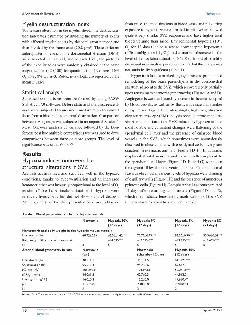
Table 1 Blood parameters in chronic hypoxia animals
Normoxia Hypoxia 10% (12 days)
Hypoxia 9% (12 days)
Hypoxia 8% (12 days)
Hypoxia 8% (23 days)
Hematocrit and body weight in the hypoxic mouse modelsHematocrit (%) 48.72±0.94 68.56±1.42*** 79.79±0.73*** 82.90±0.90*** 93.36±0.64***Body weight difference with normoxia – -14.33%*** -13.21%*** -13.25%*** -19.60%***n 5 5 5 5 5
Arterial blood gasometry in rats Normoxia (air)
Normoxia (chamber 12 days)
Hypoxia 10% (12 days)
Hematocrit (%) 48.5±1.1 48.1±1.5 61.3±2.3***O2 saturation (%) 95.5±0.4 95.7±0.6 67.6±7.3pO2 (mmHg) 108.2±2.9 104.6±3.5 50.0±1.9***pcO2 (mmHg) 44.0±1.5 40.7±0.3 44.9±2.7Hemoglobin (g/dl) 16.0±0.3 15.2±0.0 17.6±0.4*pH 7.35±0.02 7.38±0.00 7.28±0.03n 8 3 2
Note: *P,0.05 versus normoxia and ***P,0.001 versus normoxia; one-way analysis of variance and Bonferroni post hoc test.
Myelin destructuration indexTo measure alteration in the myelin sheets, the destructura-
tion index was estimated by dividing the number of axons
with affected myelin sheets by the total axon number and
then divided by the frame area (20.8 µm2). Three different
anteroposterior levels of the dorsomedial striatum (DMS)
were selected per animal, and at each level, ten pictures
of the axon bundles were randomly obtained at the same
magnification (×20,500) for quantification (Nx, n=6; 10%
O2, n=3; 8% O
2, n=3; ReNx, n=3). Data are reported as the
mean ± SEM.
statistical analysisStatistical comparisons were performed by using PASW
Statistics 17.0 software. Before statistical analysis, percent-
ages were subjected to arc-sine transformation to convert
them from a binomial to a normal distribution. Comparison
between two groups was subjected to an unpaired Student’s
t-test. One-way analysis of variance followed by the Bon-
ferroni post hoc multiple comparisons test was used to draw
comparisons between three or more groups. The level of
significance was set at P,0.05.
ResultsHypoxia induces nonreversible structural alterations in sVZAnimals acclimatized and survived well in the hypoxic
conditions, thanks to hyperventilation and an increased
hematocrit that was inversely proportional to the level of O2
tension (Table 1). Animals maintained in hypoxia were
relatively hypokinetic but did not show signs of distress.
Although most of the data presented here were obtained
from mice, the modifications in blood gases and pH during
exposure to hypoxia were estimated in rats, which showed
qualitatively similar SVZ responses and have higher total
blood volume than mice. Environmental hypoxia (10%
O2 for 12 days) led to a severe normocapnic hypoxemia
(∼50 mmHg arterial pO2) and a marked decrease in the
level of hemoglobin saturation (,70%). Blood pH slightly
decreased in animals exposed to hypoxia, but the change was
not statistically significant (Table 1).
Hypoxia induced a marked angiogenesis and pronounced
remodeling of the brain parenchyma in the dorsomedial
striatum adjacent to the SVZ, which recovered only partially
upon returning to normoxia (renormoxia) (Figure 1A and B).
Angiogenesis was manifested by increase in the area occupied
by blood vessels, as well as by the average size and number
of capillaries (Figure 1C). Interestingly, high-magnification
electron microscope (EM) analysis revealed profound ultra-
structural alterations at the SVZ induced by hypoxemia. The
most notable and consistent changes were flattening of the
ependymal cell layer and the presence of enlarged blood
vessels in the SVZ, which sometimes were anomalously
observed in close contact with ependymal cells, a very rare
situation in normoxic animals (Figure 1D–F). In addition,
displaced striatal neurons and axon bundles adjacent to
the ependymal cell layer (Figure 1D, E, and G) were seen
throughout all levels in the ventricular area. Other abnormal
features observed at various levels of hypoxia were thinning
of capillary walls (Figure 1H) and the presence of numerous
pyknotic cells (Figure 1I). Ectopic striatal neurons persisted
12 days after returning to normoxia (Figure 1D and E),
which may indicate long-lasting modifications of the SVZ
in individuals exposed to sustained hypoxia.

Hypoxia 2015:3 submit your manuscript | www.dovepress.com
Dovepress
Dovepress
19
Hypoxia and adult brain neurogenesis
A
C
D E
F G H
LV
LV
LVLVLV
Cap
LV
0.14
0.105
0.090
0.075
0.045
0.060
0.030
0.015
0
900
St
750
450
600
300
150
Nx
10%
8%
ReNx
0
120
LV
+1.10 mm < bregma < −0.50 mm
60
90
30
00
2
4
6
8
0.12
0.10
0.08
0.06
0.04
0.02
0Nx
Neu
ron
s/µm
Ap
pla
nat
ion
ind
ex
Blo
od
ves
sels
nu
mb
er
Ave
rag
e si
ze (
µm2 )
Are
a (%
to
tal s
urf
ace)
8% ReNx
Nx 8% ReNx
*
*
*
*****
*****
*** ****** ***
******
*
Cap
8%8% 8%8% 8%8%
8% ReNx8% ReNx8%8%
NxNx 10%10% 8%8% 8% ReNx8% ReNx
NxNx
8%8%I
B
Figure 1 Ultrastructural appearance of the subventricular germinal zone in chronic hypoxia.Notes: (A) Localization of the SVZ in the mouse brain (from the Paxinos and Franklin’s Mouse Brain Atlas). The rostro-caudal limits of the SVZ considered for quantification, with respect to bregma, are indicated. scale bar =100 µm. (B) semithin sections of the striatal region (st) adjacent to the lateral ventricle (lV) illustrating the increased presence of blood vessels in animals maintained at 10% (12 days) and 8% (12 days) hypoxia. large blood vessels were still observed after animals were maintained in a normoxic (nx) atmosphere (21% O2) for 12 days (8% renx). (C) Angiogenesis quantification presents an increase in the surface (left bargraph; n=3) and the average size (middle bargraph; n=3) of the blood vessels at 10% and 8% O2 tension. after 12 days of renormoxia (renx), animals only partially recovered the angiogenesis observed in 8% hypoxia. Number of blood vessels (right bargraph) increased (nonsignificantly) in hypoxia and significantly decreased after renormoxia (n=3). (D) representative electron microphotographs of the sVZ showing diminution of the ependymal layer width in 8% hypoxia (between dotted and plain lines). Displaced neurons at 8% hypoxia and post-8% renormoxia are highlighted in yellow. scale bar =6 µm. (E) Quantitative analysis of ependymal layer flattening (applanation index; in arbitrary units, upper bargraph) (n=3) and the number of displaced neurons per micrometer in the sVZ layer (lower bargraph) (n=3). (F–I) electron microphotographs illustrating 8% hypoxia-induced sVZ alterations. (F) Direct contact between a blood capillary (cap) evidenced by the elongated shape nucleus of the endothelial cell (on the left-hand side) and ependymocyte (cuboidal nucleus with microvilli). scale bar =8 µm, inset =2 µm. (G) Displaced striatal bundle of myelinated axons (white arrows) near the ventricle. scale bar =6 µm. (H) Thinning of the endothelial membrane (horizontal black arrow). scale bar =8 µm, inset =80 nm. (I) Pyknotic cells are also observed in the sVZ (black arrow). scale bar =4 µm. *P,0.05, **P,0.01, and ***P,0.001.Abbreviations: cap, capillary; lV, lateral ventricle; sVZ, subventricular zone.
nscs and intermediate progenitors at the sVZ niche are resistant to hypoxiaThe ultrastructural abnormalities observed in the SVZ lead
us to further investigate a possible effect of hypoxia on the
proliferative germinal center. The SVZ contains four main
cell types defined by their morphology, ultrastructure, and
molecular markers: migrating neuroblasts (type A cells),
astrocytes (type B cells), proliferative precursors (type C
cells), and ependymal cells (type E cells). It has been shown
that a subpopulation of B-cells are the primary NSCs, which

Hypoxia 2015:3submit your manuscript | www.dovepress.com
Dovepress
Dovepress
20
d’anglemont de Tassigny et al
are converted to transient amplifying type C cells that gener-
ate neuroblasts and glial cells.18,19 Neuroblasts arranged in
chains migrate tangentially along the RMS to the OB, where
they differentiate into mature interneurons. The cell classes
characteristic of SVZ are illustrated by semithin sections in
Figure 2A (left) that also show blood vessels within the SVZ
at 8% O2 tension and the presence of abnormal intercellular
gaps in renormoxia. However, the number of the various
cell types in the SVZ identified in ultrathin EM sections was
not significantly affected even by severe (8% O2) hypoxia
(Figure 2A, right). The SVZ dissected from normoxic and
chronically hypoxic animals showed a similar ability to
generate primary and secondary neurospheres (Figure 2B),
thus further supporting the view that SVZ progenitors (B-
and C-type cells) are unaffected by hypoxia. Indeed, SVZ-
derived neurosphere diameter, an indication of proliferative
activity of progenitor cells,16 was unaffected by a broad range
of O2 tensions (1%, 3%, and 21%). Progenitor proliferation
only decreased significantly when the neurospheres were
exposed to extreme low pO2 values (0.5% or ∼3 mmHg)
(Figure 2C).
In parallel to the neurosphere experiments, we exam-
ined the in vivo proliferation activity of cells in the anterior
horn of SVZ by means of the administration of BrdU to
animals as well as the immunocytochemical detection of
the proliferating cell nuclear antigen (PCNA), a broader
marker of proliferating cells. Unbiased counting of stained
cells with the two methods revealed no difference between
normoxic and hypoxic mice (Figure 2D and E). The number
of BrdU+ cells remaining in SVZ 11 days after injection of
the marker (an indication of B-type cell number29) as well
as the intensity of GFAP+ staining at SVZ was unchanged
by hypoxia (Figure S1), thus supporting the data obtained by
direct counting of B-type cells in ultrathin EM sections. The
number of neuroblasts (A-type cells), as determined by the
specific marker doublecortin,30 was also similar in hypoxic
and normoxic animals (Figure 2F). Taken together, these data
suggest that in spite of the profound structural alterations
induced by hypoxia in the SVZ, the cellular components of
the germinal niche (stem cells, transient amplifying progeni-
tors, and neuroblasts) are insensitive to chronic severe (up to
8% O2 for 12 days) hypoxic treatment.
chronic hypoxia reduces survival of newborn neurons in vivo and in vitroNewborn neuroblasts in the anterior SVZ migrate toward the
OB, where they differentiate into young neurons in either
the granule cell layer (GCL) or periglomerular layer.31,32 EM
observations of the RMS indicated that neuroblast migration
was not altered after 12 days at 8% O2 (Figure 3A). This
was also confirmed by experiments in which after 12 days
in hypoxia, animals were injected with BrdU and sacrificed
11 days later. With this protocol, BrdU+ neuroblasts born at
the SVZ were located at the GCL in maturation stage 3 at the
moment of sacrifice.32,33 Hypoxic treatments (8% or 10% O2)
produced a decreasing trend in the number of BrdU+ cells
(most of them maturating neuroblasts) at OB; however, the
differences were not statistically significant (Figure 3B).
We further investigated the nature of BrdU+ cells at the OB
and found that the number of double BrdU+ and NeuN+
(neuronal nuclei, a neuronal marker) cells clearly decreased
in mice treated with either 10% or 8% O2 tensions, whereas
the number of BrdU+ and NeuN- cells remained unaltered
(Figure 3C). In normoxia, at least half of the BrdU+ cells at
GCL were in the process of neuronal maturation (NeuN+),
but during exposure to hypoxia, this occurred only to a third
of the cells (Figure 3D). In animals that stayed for 23 days
in hypoxia, cell counts in the GCL indicated that exposure to
8% O2 tension produced a selective decrease in the number
of neurons (NeuN+ cells) at the GCL in parallel with an
increase in the number of moon-shaped nuclei, presumably
endothelial cells, which confirms the hypoxic condition of
these mice (Figure 4A). Hypoxia also induced an increase in
cell apoptosis at the OB that was not seen in other parts of the
brain, as, for example, the striatum (Figure 4B). These results
suggest that SVZ newborn neuroblasts can migrate normally
through the RMS in severe chronic hypoxia, but once arrived
in the OB, their differentiation into neurons or survival of
newly differentiated neuronal cells is compromised.
The effect of lowering pO2 on differentiation of SVZ
progenitors was further investigated in vitro by culturing
neurospheres in differentiation media and plating on adher-
ent substrate. Moderate hypoxia (3% O2) had no effect on
the number of Tuj1+ cells generated from neural progenitors.
However, more severe hypoxia (1% O2), probably similar to
the O2 levels likely reached in the OB of animals maintained
in hypoxia, resulted in a dramatic decrease in the survival of
neurons once they were generated. Under the same in vitro
conditions, GFAP+ astrocytes were unaffected (Figure 5A).
Oligodendrocyte damage in chronic hypoxiaBesides neuroblasts and astrocytes, multipotent progenitor
cells in SVZ neurospheres were also able to generate oligo-
dendrocytes, the survival of which was also compromised
in extreme hypoxic conditions (Figure 5B). The inhibition
Hypoxia 2015:3 submit your manuscript | www.dovepress.com
Dovepress
Dovepress
21
Hypoxia and adult brain neurogenesis
A
30
30
40
50
60
LVLVLV
LV LV
LV
LV LV
NxNx
NxNx
Nx8%8%
8%8% 8% ReNx8% ReNx
8%
LVStSt St St StSt
PCNABrdu DCXDapiDapi Dapi
Brdu PCNA DCX
25
20
20
15
10
5
0Nx Nx10%P
osi
tive
cel
ls/S
VZ
mm
Po
siti
ve c
ells
/SV
Z m
m
Sta
inin
g in
ten
sity
(au
)
Sp
her
e d
iam
eter
(µm
)
Sp
her
e fo
rmin
g c
ells
(%
)
Cel
l nu
mb
er/m
m
Cell type
8% Nx Nx10% 8% Nx Nx10% 8%
Nx Nx10% 3% 1% 0.5%
201816141210
10
8
8 83 30 0
30
40
50
60
20
10
0
1.01.21.41.6
200
*
Nx8%ReNx
E
120
100
80
60
20
40
0ACB
150
100
500.05
0.1
0.15
0.2
0.25
008 8
0.80.60.40.20.0
1.01.21.41.6
0.80.60.40.20.0
24
4 4 4 4 4 4 44
6
E
CB
D F
Secondary neurospheresPrimary neurospheres
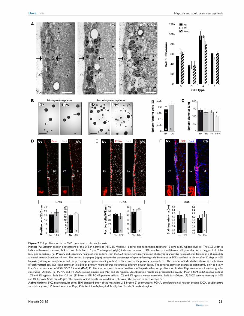
Figure 2 cell proliferation in the sVZ is resistant to chronic hypoxia.Notes: (A) semithin section photographs of the sVZ in normoxia (nx), 8% hypoxia (12 days), and renormoxia following 12 days in 8% hypoxia (renx). The sVZ width is indicated between the two black arrows. scale bar =10 µm. The bargraph (right) indicates the mean ± seM number of the different cell types that form the germinal niche (n=3 per condition). (B) Primary and secondary neurospheres culture from the SVZ region. Low-magnification photographs show the neurospheres formed in a 35 mm dish at clonal density. scale bar =1 mm. The vertical bargraphs (right) indicate the percentage of sphere-forming cells from mouse SVZ sacrificed in Nx or after 12 days at 10% hypoxia (primary neurospheres), and the percentage of sphere-forming cells after dispersion of the primary neurospheres. The number of individuals is shown at the bottom of each vertical bar. (C) Mean diameter (± SEM) of primary neurospheres cultured at different oxygen levels. The spheres diameter decreased significantly only at a very low O2 concentration of 0.5%. *P,0.05, n=4. (D–F) Proliferation markers show no evidence of hypoxia effect on proliferation in vivo. representative microphotographs illustrating (D) BrdU, (E) Pcna, and (F) DCX staining in normoxia (Nx) and 8% hypoxia. Quantification results are presented below. (D) Mean ± seM BrdU-positive cells at 10% and 8% hypoxia. scale bar =20 µm. (E) Mean ± seM Pcna-positive cells at 10% and 8% hypoxia versus normoxia. scale bar =20 µm. (F) DcX staining intensity at 10% and 8% hypoxia. scale bar =15 µm. The number of individuals per condition is shown at the bottom of each vertical bar.Abbreviations: sVZ, subventricular zone; seM, standard error of the mean; BrdU, 5-bromo-2’-deoxyuridine; Pcna, proliferating cell nuclear antigen; DcX, doublecortin; au, arbitrary unit; lV, lateral ventricle; Dapi, 4′,6-diamidino-2-phenylindole dihydrochloride; st, striatal region.

Hypoxia 2015:3submit your manuscript | www.dovepress.com
Dovepress
Dovepress
22
d’anglemont de Tassigny et al
A NxNx
Nx
8%8%
8%
Sacrifice(day 23)
SVZ
OBD
P
V
80706050
Po
siti
ve c
ells
/mm
2
40302010
454035302520151050
Nx 10%
**
* *
*
Nx 8%
Po
siti
ve c
ells
/mm
2
Nx
55 55 44 44
10%
10%
8% 8%Nx
Nx
49%
43%
35%
32%57%
65%
68%
51%
Nx
Brdu+NeuN+
Brdu+NeuN−
35
30
25
20
15
10
5
0
05 45 4
Nx Nx10% 8%
A
Brdu injection(day 12)
C
D
B
Proliferation
Brdu+
Brdu
Brdu+/NeuN+ Brdu+/NeuN−
RMS
Migration
NeuN
Figure 3 chronic hypoxia affects neuroblast differentiation in the OB.Notes: (A) electron microphotographs of neuroblasts chain in the rMs displaying similar morphology but dilated intracellular spaces in 8% hypoxia. squared areas (white dotted lined) are shown at greater magnification. Scale bar =10 µm, inset =2 µm. (B) illustration of the BrdU injection strategy. BrdU (3×50 mg/kg) was injected after 12 days in hypoxia or normoxia. BrdU-positive cells generated in the sVZ migrated for 11 days through the rMs to reach the OB. Vertical bars indicate the number of BrdU-positive cells in the gcl of the OB at 10% hypoxia versus normoxia (nx) or 8% hypoxia versus normoxia. The number of individuals per condition is shown at the bottom of each vertical bar. (C) Photomicrographs show BrdU+ (red) and neun+ (green) cells in the gcl in normoxia or 8% hypoxia. White arrows point at double-stained BrdU+/neun+ cells. scale bar =20 µm. Vertical bargraphs indicate the number of BrdU+ cells (± seM) that have differentiated into neun+ neurons (left bargraph), or that remain neun- (right bargraph). (D) circular diagrams illustrating the difference of BrdU+ cells differentiated into neun+ cells (red) or that remain neun- (green) between normoxia and 10% or 8% hypoxia, respectively, expressed as percentage of total BrdU+ cells. *P,0.05 and **P,0.01.Abbreviations: OB, olfactory bulb; rMs, rostral migratory stream; BrdU, 5-bromo-2’-deoxyuridine; sVZ, subventricular zone; gcl, granule cell layer; neun, neuronal nuclei; seM, standard error of the mean.
of neuronal or oligodendrocyte survival in severe hypoxia
(1% O2) in vitro was also observed in secondary neurospheres
regardless of whether the original progenitors came from
animals that had been maintained in normoxic (21% O2) or
hypoxic (10% O2) conditions (Figure S2).
We tested whether chronic hypoxia also damaged oli-
godendrocytes in vivo. Adult SVZ progenitors are known
to migrate to neighboring white matter bundles to generate
oligodendrocyte precursors.22,23,25 Hence, we analyzed oligo-
dendrocyte precursors, oligodendrocytes, and myelin bundles
in a region of the DMS adjacent to the SVZ within 300 µm
from the border of the lateral ventricle. Chronic exposure
to low pO2 (down to 8%) did not produce any difference in
the number of oligodendrocyte precursors (NG2-expressing
cells) in the DMS (Figure 6A). The number of NG2+ cells
in dorsolateral striatum and motor cortex also remained
unaffected by hypoxia (Figure S3). However, lowering pO2
resulted in a decrease in striatal oligodendrocyte (Olig+)
number, which was proportional to the severity of hypoxia.
The number of Olig+ cells decreased to half after 23 days of

Hypoxia 2015:3 submit your manuscript | www.dovepress.com
Dovepress
Dovepress
23
Hypoxia and adult brain neurogenesis
B
A NeuN
TUNEL
Dapi
Dapi
Total cells
Neurons (%)
Apoptosis
14,000 50 * ***
**
*
40
30
En
do
thel
ial c
ells
/m
m2
Dap
i+ c
ells
/mm
2
TU
NE
L+
cells
/mm
2
Neu
N+/
Dap
i+ (
%)
20
10
0
91.0
90.5
90.0
89.5
89.0
88.5
88.0
2.0
1.5
1.0
0.5
0
12,000
10,000
8,000
6,000
4,000
2,000
0Nx Nx10% 8% Nx Nx10% 8%
Nx Nx10% 8%
Nx NxNx10%
GCL Striatum
8% 8%
53 53 33 5
53
5
5
5 55 5
5 5
3
AngiogenesisNx
Nx
8%
8%
Figure 4 neuronal loss from apoptosis in the olfactory bulb in severe chronic hypoxia.Notes: (A) coronal sections of the olfactory bulb gcl indicating neun+ cells (red) and total cell number (Dapi, blue). Besides a general, but not significant, loss of cells (Dapi+), the number of neurons (neun+ cells) decreased after 23 days in 8% but not in 10% O2. note that angiogenesis (indicated by the increased number of endothelial cells with moon-shaped nuclei) is observed at both 10% and 8% hypoxia (white arrows). scale bar =50 µm. The insets show the indicated areas at higher magnification. The number of individuals per condition is shown at the bottom of each vertical bar. (B) Histological sections of the olfactory bulb gcl indicating TUnel+ cells (green) and total cell number (Dapi, blue) in normoxic animals (left) and in animals exposed to 8% O2 for 12 days (right). The vertical bargraph shows the mean number ± seM of TUnel+ cells per squared mm. The number of individuals per condition is shown at the bottom of each vertical bar. Significant increase in apoptotic cells is observed at 8% hypoxia versus normoxia (nx). TUnel+ cells are rarely found in the striatum of both normoxic and hypoxic (8% O2) animals. *P,0.05, **P,0.01, and ***P,0.001.Abbreviations: gcl, granule cell layer; neun, neuronal nuclei; Dapi, 4′,6-diamidino-2-phenylindole dihydrochloride; seM, standard error of the mean.
A 21% (7 d)
21% (7 d)
1% (7 d)
1% (7 d)B
12 21%
Tuj1+(neurons)
GFAP+(astrocytes)
3%
1%
21%
3%
1%
10
8
6
4
2
0
O4
GFAP
Dapi
DapiTuj1
60
50
40
Po
siti
ve c
ells
/to
tal
cells
(%
)
Po
siti
ve c
ells
/to
tal
cells
(%
)
30
20
10
0
8
10
6
4
2
07 days
O4+(oligodendrocytes)
7 days 7 days
**
**
3 days
Figure 5 in vitro sVZ progenitors differentiation and survival.Notes: (A) Microphotographs of sVZ neurospheres, cultured at variable levels of O2 tension, after 7 days in differentiation medium: neurons (Tuj1+, green), astrocytes (gFaP+, red), and nuclei (Dapi, blue). scale bar =30 µm. Bargraphs indicate the selective decrease of neurons after 7 days in culture at 1% O2. The number of gFaP+ cells (astrocytes) remains unchanged in the three different O2 tensions tested (n=4 per condition). (B) Microphotographs of sVZ neurospheres, cultured at variable levels of O2 tension, after 7 days in differentiation medium: oligodendrocytes (O4, red) and nuclei (Dapi, blue). scale bar =30 µm. Vertical bargraph shows the percentage of O4+ cells after 7 days in culture at three different levels of O2 tension. **P,0.01.Abbreviations: SVZ, subventricular zone; GFAP, glial fibrillary acidic protein; Dapi, 4′,6-diamidino-2-phenylindole dihydrochloride.
Hypoxia 2015:3submit your manuscript | www.dovepress.com
Dovepress
Dovepress
24
d’anglemont de Tassigny et al
A
B
C
Nx
Nx
Nx
8% (12 d)
8% (12 d)
8% (23 d)
NG2 Dapi
Olig Dapi
LV
LV
Olig
70
60
50
40
NG
2+ c
ells
/mm
2
Olig
+ ce
lls/m
m2
Olig
+ st
ain
ing
(op
tica
l in
ten
sity
, au
)
30
20
10
0Nx
Nx 10% Nx 8%
8%
55
44 99 55
100 ***
**
**80
60
40
20
0
60 10 Nx8%8
6
4
2
012 d 23 d 12 d 23 d
* **
Axon bundles Inter-bundles space
50403020100 55 55 55 55
(12 d)Nx
−23%
−27%−32%
−18%
−49%
8%(23 d)
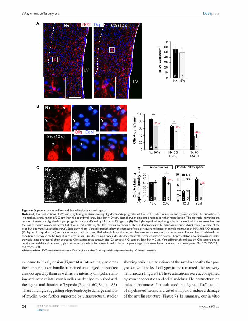
Figure 6 Oligodendrocytes cell loss and demyelination in chronic hypoxia.Notes: (A) coronal sections of sVZ and neighboring striatum showing oligodendrocyte progenitors (ng2+ cells, red) in normoxic and hypoxic animals. The discontinuous line marks a striatal region of 300 µm from the ependymal layer. scale bar =100 µm. Inset shows the indicated regions at higher magnification. The bargraph shows that the number of immature oligodendrocytes progenitors is not affected by 12 days in 8% hypoxia. (B) The high-magnification photographs in the medio-dorsal striatum illustrate the loss of mature oligodendrocytes (Olig+ cells, red) at 8% O2 (12 days) versus normoxia. Only oligodendrocytes with Dapi-positive nuclei (blue) located outside of the axon bundles were quantified (arrows). Scale bar =10 µm. Vertical bargraphs show the number of cells per square millimeter in animals maintained at 10% and 8% O2 tension (12 days or 23 days duration) versus their normoxic littermates. red values indicate the percent decrease from the normoxic counterparts. The number of individuals per condition is shown at the bottom of each vertical bar. (C) Olig staining optical density decreases with increased chronic hypoxia. representative photomicrographs (after grayscale image processing) show decreased Olig staining in the striatum after 23 days at 8% O2 tension. scale bar =40 µm. Vertical bargraphs indicate the Olig staining optical density inside (left) and between (right) the striatal axon bundles. Values in red indicate the percentage of decrease from the normoxic counterparts. *P,0.05, **P,0.01, and ***P,0.001.Abbreviations: sVZ, subventricular zone; Dapi, 4′,6-diamidino-2-phenylindole dihydrochloride; lV, lateral ventricle.
exposure to 8% O2 tension (Figure 6B). Interestingly, whereas
the number of axon bundles remained unchanged, the surface
area occupied by them as well as the intensity of myelin stain-
ing within the striatal axon bundles markedly diminished with
the degree and duration of hypoxia (Figures 6C, S4, and S5).
These findings, suggesting oligodendrocyte damage and loss
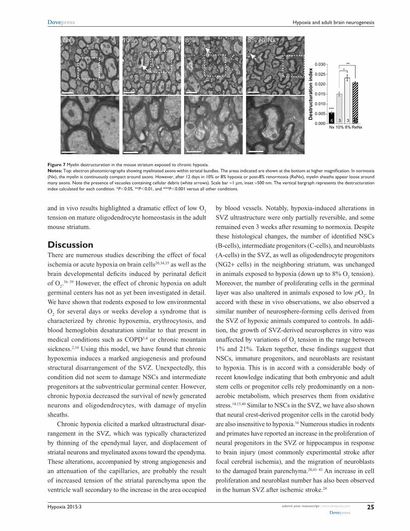
of myelin, were further supported by ultrastructural studies
showing striking disruptions of the myelin sheaths that pro-
gressed with the level of hypoxia and remained after recovery
in normoxia (Figure 7). These alterations were accompanied
by axon degeneration and cellular debris. The destructuration
index, a parameter that estimated the degree of affectation
of myelinated axons, indicated a hypoxia-induced damage
of the myelin structure (Figure 7). In summary, our in vitro
Hypoxia 2015:3 submit your manuscript | www.dovepress.com
Dovepress
Dovepress
25
Hypoxia and adult brain neurogenesis
8%8% 8% ReNx8% ReNx
0.030
0.025
0.020
Des
tru
ctu
rati
on
ind
ex
0.015
0.010
0.005
0.000Nx 10%
3
***
***
36 3
8% ReNx
10%10%NxNx
Figure 7 Myelin destructuration in the mouse striatum exposed to chronic hypoxia.Notes: Top: electron photomicrographs showing myelinated axons within striatal bundles. The areas indicated are shown at the bottom at higher magnification. In normoxia (nx), the myelin is continuously compact around axons. However, after 12 days in 10% or 8% hypoxia or post-8% renormoxia (renx), myelin sheaths appear loose around many axons. note the presence of vacuoles containing cellular debris (white arrows). scale bar =1 µm, inset =500 nm. The vertical bargraph represents the destructuration index calculated for each condition. *P,0.05, **P,0.01, and ***P,0.001 versus all other conditions.
and in vivo results highlighted a dramatic effect of low O2
tension on mature oligodendrocyte homeostasis in the adult
mouse striatum.
DiscussionThere are numerous studies describing the effect of focal
ischemia or acute hypoxia on brain cells20,34,35 as well as the
brain developmental deficits induced by perinatal deficit
of O2.36–39 However, the effect of chronic hypoxia on adult
germinal centers has not as yet been investigated in detail.
We have shown that rodents exposed to low environmental
O2 for several days or weeks develop a syndrome that is
characterized by chronic hypoxemia, erythrocytosis, and
blood hemoglobin desaturation similar to that present in
medical conditions such as COPD3,4 or chronic mountain
sickness.2,10 Using this model, we have found that chronic
hypoxemia induces a marked angiogenesis and profound
structural disarrangement of the SVZ. Unexpectedly, this
condition did not seem to damage NSCs and intermediate
progenitors at the subventricular germinal center. However,
chronic hypoxia decreased the survival of newly generated
neurons and oligodendrocytes, with damage of myelin
sheaths.
Chronic hypoxia elicited a marked ultrastructural disar-
rangement in the SVZ, which was typically characterized
by thinning of the ependymal layer, and displacement of
striatal neurons and myelinated axons toward the ependyma.
These alterations, accompanied by strong angiogenesis and
an attenuation of the capillaries, are probably the result
of increased tension of the striatal parenchyma upon the
ventricle wall secondary to the increase in the area occupied
by blood vessels. Notably, hypoxia-induced alterations in
SVZ ultrastructure were only partially reversible, and some
remained even 3 weeks after resuming to normoxia. Despite
these histological changes, the number of identified NSCs
(B-cells), intermediate progenitors (C-cells), and neuroblasts
(A-cells) in the SVZ, as well as oligodendrocyte progenitors
(NG2+ cells) in the neighboring striatum, was unchanged
in animals exposed to hypoxia (down up to 8% O2 tension).
Moreover, the number of proliferating cells in the germinal
layer was also unaltered in animals exposed to low pO2. In
accord with these in vivo observations, we also observed a
similar number of neurosphere-forming cells derived from
the SVZ of hypoxic animals compared to controls. In addi-
tion, the growth of SVZ-derived neurospheres in vitro was
unaffected by variations of O2 tension in the range between
1% and 21%. Taken together, these findings suggest that
NSCs, immature progenitors, and neuroblasts are resistant
to hypoxia. This is in accord with a considerable body of
recent knowledge indicating that both embryonic and adult
stem cells or progenitor cells rely predominantly on a non-
aerobic metabolism, which preserves them from oxidative
stress.14,15,40 Similar to NSCs in the SVZ, we have also shown
that neural crest-derived progenitor cells in the carotid body
are also insensitive to hypoxia.16 Numerous studies in rodents
and primates have reported an increase in the proliferation of
neural progenitors in the SVZ or hippocampus in response
to brain injury (most commonly experimental stroke after
focal cerebral ischemia), and the migration of neuroblasts
to the damaged brain parenchyma.20,41–43 An increase in cell
proliferation and neuroblast number has also been observed
in the human SVZ after ischemic stroke.24

Hypoxia 2015:3submit your manuscript | www.dovepress.com
Dovepress
Dovepress
26
d’anglemont de Tassigny et al
However, this response to ischemia, which is probably
due to the release of chemotactic pro-inflammatory agents
in the injured area, is transient and has only minor restorative
capacity as most of the newly generated cells are short-lived
and nonfunctional.44 In agreement with these observations,
it has recently been reported that short exposure to environ-
mental hypoxia (10% O2 for 6–72 hours) induces proliferation
(without increasing differentiation) of hippocampal NSCs,45
and we have observed a similar phenomenon in SVZ (data
not shown). However, these short-lasting (hours) exposures
to hypoxia are different from the chronic treatments lasting
weeks, in which angiogenesis and structural rearrangements
of brain structures are fully developed.
Although chronic hypoxia did not seem to affect the
maintenance and proliferation of neural progenitors in SVZ,
our in vivo and in vitro experiments indicate that survival of
neurons and oligodendrocytes was compromised, whereas
GFAP+ astrocytes were preserved. Exposure to hypoxia,
particularly to 8% O2, decreased the number of BrdU+
and NeuN+ cells and selectively increased apoptosis in the
OB in comparison with the striatum, thus suggesting the
loss of newly generated neurons. Under normal conditions
(21% O2), O
2 tension in most brain areas is estimated to be
2%–6%,5 a range of values considered as “mild hypoxia”.
These O2 levels are known to favor differentiation of neu-
ronal progenitors in vitro.46,47 However, O2 tension in some
brain regions surely decreases to near 1% or below in rodents
chronically exposed to severe low environmental pO2, and
whose arterial O2 tension drops from 100 mmHg to nearly
50 mmHg. Indeed, our data indicate that survival of dif-
ferentiated neurons and oligodendrocytes is unaffected in
SVZ-derived neurospheres cultured in 3% O2 but drastically
reduced in 1% O2. The O
2 level necessary to prevent cell
damage seems to be particularly high for oligodendrocytes,
since their number was significantly decreased in animals
exposed to relatively mild hypoxia (10% O2 for 12 days).
Hypoxia also markedly reduced myelin expression and
altered the ultrastructure of myelin sheaths. These observa-
tions fit with previous studies describing the high-energy
demands of oligodendrocytes and their particular sensitivity
to ischemic damage (for review and references, see Bradl
and Lassmann48). Oligodendrocytes are also highly vulner-
able to oxidative stress due to their elevated iron content
and relative lack of antioxidant defense.49 Hence, increased
mitochondrial production of ROS during chronic hypoxia
may be a major factor compromising oligodendrocyte
survival. The special vulnerability of newly generated OB
neurons to hypoxia could also result from a misbalance
between mitochondrial ROS production (increased during
hypoxia) and the maturation of the antioxidant defense. The
dependence of neuronal and oligodendrocyte survival on a
minimum level of O2 tension gives special significance to
the association between angiogenesis and neurogenesis.
During hypoxia, newly generated blood vessels could not
only help to minimize the effects of O2 deficiency but might
also contribute to paracrine maintenance of stem cells and
survival of newly differentiated neurons or glia by means
of the release of vascular endothelial trophic factors.50,51
Replacement of neurons and myelin-forming oligoden-
drocytes is essential for normal brain plasticity and repair,
and impairment of adult neurogenic centers can lead to
neuropsychiatric disorders in humans.12,52 Therefore, the
profound changes induced by chronic hypoxemia in the
SVZ and neighboring regions could help explain the neu-
rological symptoms described in chronically hypoxemic
patients. Cumulative evidence over the last 30 years has
confirmed that cognitive and sensorimotor alterations are
frequently seen in COPD patients and that they inversely
correlate with arterial pO2 and with compliance of O
2
therapy.4,6,7,53 Recent magnetic resonance imaging stud-
ies in the brains of COPD patients have shown regional
decreases in gray matter density and impairment of white
matter microstructural integrity associated with disease
severity.54 Interestingly, a sixfold higher risk of multiple
sclerosis, a demyelinating brain disorder, has been reported
among individuals (,60 years old) diagnosed with COPD,55
although this is an isolated observation that needs to be
confirmed. Similar to severe COPD, patients who develop
chronic mountain sickness due to “de-acclimatization”
to high altitude also present well-known neurological
disturbances in the form of paresthesias, loss of reflexes,
and cognitive impairment.1,2,10 In this regard, the thinning
of capillary walls near a flattened ependyma in the lateral
ventricles observed in chronically hypoxic mice might be a
fundamental pathophysiological factor in the development
of microhemorrhages characteristic of patients presenting
high-altitude cerebral edema.2
ConclusionThe findings in this report demonstrate that sustained hypox-
emia has a profound effect on the structure and function of
the brain SVZ neurogenic niche. They provide a solid foun-
dation for further research on the effects of chronic hypoxia
on the fate of newly generated cells in the adult brain and
their participation in the neurological alterations induced by
persistent hypoxemia.
Hypoxia 2015:3 submit your manuscript | www.dovepress.com
Dovepress
Dovepress
27
Hypoxia and adult brain neurogenesis
AcknowledgmentsThis research was supported by the Spanish “Instituto de
Salud Carlos III” (XdT, Miguel Servet grant CP12-03217
and PIE13/00004), The Botín Foundation, and The Spanish
Ministry of Science and Innovation (Plan Nacional, SAF
program). Ricardo Pardal received a Starting Grant from ERC.
We would like to thank Margarita Rubio and Rocío Duran for
technical assistance. We are grateful to members of the IBIS
Animal Facility Core for excellent care of the animals.
DisclosureThe authors report no conflicts of interest in this work.
References 1. Joseph V, Pequignot J-M. Breathing at high altitude. Cell Mol Life Sci.
2009;66:3565–3573. 2. Wilson MH, Newman S, Imray CH. The cerebral effects of ascent to
high altitudes. Lancet Neurol. 2009;8:175–191. 3. Sutherland ER, Cherniack RM. Management of chronic obstructive
pulmonary disease. N Engl J Med. 2004;350:2689–2697. 4. Schou L, Østergaard B, Rasmussen LS, Rydahl-Hansen S, Phanareth K.
Cognitive dysfunction in patients with chronic obstructive pulmonary disease – a systematic review. Respir Med. 2012;106:1071–1081.
5. Erecińska M, Silver I. Tissue oxygen tension and brain sensitivity to hypoxia. Respir Physiol. 2001;128:263–276.
6. Grant I, Heaton RK, McSweeny JA, Adams KM, Timms RM. Neuropsychologic findings in hypoxemic chronic obstructive pulmo-nary disease. Arch Intern Med. 1982;142:1470–1476.
7. Incalzi RA, Corsonello A, Trojano L, et al. Cognitive training is inef-fective in hypoxemic COPD: a six-month randomized controlled trial. Rejuvenation Res. 2008;11:239–250.
8. Singh B, Mielke M, Parsaik A, et al. A prospective study of chronic obstructive pulmonary disease and the risk for mild cognitive impairment. JAMA Neurol. 2014;71:581–588.
9. Monge C, Arregui A, León-Velarde F. Pathophysiology and epidemiology of chronic mountain sickness. Int J Sports Med. 1992;13: S79–S81.
10. Thomas PK, King RH, Feng SF, et al. Neurological manifestations in chronic mountain sickness: the burning feet-burning hands syndrome. J Neurol Neurosurg Psychiatry. 2000;69:447–452.
11. Zubieta-Castillo G, Zubieta-Calleja GR, Zubieta-Calleja L. Chronic mountain sickness: the reaction of physical disorders to chronic hypoxia. J Physiol Pharmacol. 2006;57(Suppl 4):431–442.
12. Zhao C, Deng W, Gage FH. Mechanisms and functional implications of adult neurogenesis. Cell. 2008;132:645–660.
13. Panchision DM. The role of oxygen in regulating neural stem cells in development and disease. J Cell Physiol. 2009;220:562–568.
14. Mohyeldin A, Garzón-Muvdi T, Quiñones-Hinojosa A. Oxygen in stem cell biology: a critical component of the stem cell niche. Cell Stem Cell. 2010;7:150–161.
15. Suda T, Takubo K, Semenza GL. Metabolic regulation of hematopoietic stem cells in the hypoxic niche. Cell Stem Cell. 2011;9:298–310.
16. Platero-Luengo A, González-Granero S, Durán R, et al. An O2-sensitive glomus cell-stem cell synapse induces carotid body growth in chronic hypoxia. Cell. 2014;156:291–303.
17. Macías D, Fernández-Agüera MC, Bonilla-Henao V, López-Barneo J. Deletion of the von Hippel-Lindau gene causes sympathoadrenal cell death and impairs chemoreceptor-mediated adaptation to hypoxia. EMBO Mol Med. 2014;6:1577–1592.
18. Doetsch F, Caillé I, Lim DA, García-Verdugo JM, Alvarez-Buylla A. Subventricular zone astrocytes are neural stem cells in the adult mam-malian brain. Cell. 1999;97:703–716.
19. Quiñones-Hinojosa A, Sanai N, Soriano-Navarro M, et al. Cellular com-position and cytoarchitecture of the adult human subventricular zone: a niche of neural stem cells. J Comp Neurol. 2006;494:415–434.
20. Arvidsson A, Collin T, Kirik D, Kokaia Z, Lindvall O. Neuronal replace-ment from endogenous precursors in the adult brain after stroke. Nat Med. 2002;8:963–970.
21. Picard-Riera N, Decker L, Delarasse C, et al. Experimental autoimmune encephalomyelitis mobilizes neural progenitors from the subventricular zone to undergo oligodendrogenesis in adult mice. Proc Natl Acad Sci U S A. 2002;99:13211–13216.
22. Menn B, Garcia-Verdugo JM, Yaschine C, Gonzalez-Perez O, Rowitch D, Alvarez-Buylla A. Origin of oligodendrocytes in the subventricular zone of the adult brain. J Neurosci. 2006;26:7907–7918.
23. Gonzalez-Perez O, Romero-Rodriguez R, Soriano-Navarro M, Garcia- Verdugo JM, Alvarez-Buylla A. Epidermal growth factor induces the progeny of subventricular zone type B cells to migrate and differentiate into oligodendrocytes. Stem Cells. 2009;27:2032–2043.
24. Martí-Fàbregas J, Romaguera-Ros M, Gómez-Pinedo U, et al. Proliferation in the human ipsilateral subventricular zone after ischemic stroke. Neurology. 2010;74:357–365.
25. Capilla-Gonzalez V, Guerrero-Cazares H, Bonsu JM, et al. The subven-tricular zone is able to respond to a demyelinating lesion after localized radiation. Stem Cells. 2014;32:59–69.
26. Pardal R, Ortega-Sáenz P, Durán R, López-Barneo J. Glia-like stem cells sustain physiologic neurogenesis in the adult mammalian carotid body. Cell. 2007;131(2):364–377.
27. Ferron SR, Andreu-Agullo C, Mira H, Sanchez P, Marques-Torrejon MA, Farinas I. A combined ex/in vivo assay to detect effects of exogenously added factors in neural stem cells. Nat Protoc. 2007;2:849–859.
28. Doetsch F, García-Verdugo JM, Alvarez-Buylla A. Cellular com-position and three-dimensional organization of the subventricular germinal zone in the adult mammalian brain. J Neurosci. 1997;17: 5046–5061.
29. Johansson CB, Momma S, Clarke DL, Risling M, Lendahl U, Frisén J. Identification of a neural stem cell in the adult mammalian central nervous system. Cell. 1999;96:25–34.
30. Brown JP, Couillard-Després S, Cooper-Kuhn CM, Winkler J, Aigner L, Kuhn HG. Transient expression of doublecortin during adult neurogenesis. J Comp Neurol. 2003;467:1–10.
31. Luskin MB. Restricted proliferation and migration of postnatally generated neurons derived from the forebrain subventricular zone. Neuron. 1993;11:173–189.
32. Lois C, Alvarez-Buylla A. Long-distance neuronal migration in the adult mammalian brain. Science. 1994;264:1145–1148.
33. Petreanu L, Alvarez-Buylla A. Maturation and death of adult-born olfactory bulb granule neurons: role of olfaction. J Neurosci. 2002;22: 6106–6113.
34. Jin K, Minami M, Lan JQ, et al. Neurogenesis in dentate subgranular zone and rostral subventricular zone after focal cerebral ischemia in the rat. Proc Natl Acad Sci U S A. 2001;98:4710–4715.
35. Zhang RL, Zhang ZG, Zhang L, Chopp M. Proliferation and differ-entiation of progenitor cells in the cortex and the subventricular zone in the adult rat after focal cerebral ischemia. Neuroscience. 2001;105: 33–41.
36. Back SA, Han BH, Luo NL, et al. Selective vulnerability of late oli-godendrocyte progenitors to hypoxia-ischemia. J Neurosci. 2002;22: 455–463.
37. Kako E, Kaneko N, Aoyama M, et al. Subventricular zone-derived oligodendrogenesis in injured neonatal white matter in mice enhanced by a nonerythropoietic erythropoietin derivative. Stem Cells. 2012;30: 2234–2247.
38. Oorschot DE, Voss L, Covey MV, et al. Spectrum of short- and long-term brain pathology and long-term behavioral deficits in male repeated hypoxic rats closely resembling human extreme prematurity. J Neurosci. 2013;33:11863–11877.
39. Salmaso N, Jablonska B, Scafidi J, Vaccarino FM, Gallo V. Neurobiology of premature brain injury. Nat Neurosci. 2014;17:341–346.
Hypoxia 2015:3submit your manuscript | www.dovepress.com
Dovepress
Dovepress
28
d’anglemont de Tassigny et al
40. Ezashi T, Das P, Roberts RM. Low O2 tensions and the prevention of differentiation of hES cells. Proc Natl Acad Sci U S A. 2005;102: 4783–4788.
41. Yamashita T, Ninomiya M, Hernández Acosta P, et al. Subventricular zone- derived neuroblasts migrate and differentiate into mature neurons in the post-stroke adult striatum. J Neurosci. 2006;26:6627–6636.
42. Yang Z, Levison SW. Hypoxia/ischemia expands the regenerative capac-ity of progenitors in the perinatal subventricular zone. Neuroscience. 2006;139:555–564.
43. Zhang ZG, Chopp M. Neurorestorative therapies for stroke: underlying mechanisms and translation to the clinic. Lancet Neurol. 2009;8: 491–500.
44. Snyder EY, Park KI. Limitations in brain repair. Nat Med. 2002;8: 928–930.
45. Varela-Nallar L, Rojas-Abalos M, Abbott AC, Moya EA, Iturriaga R, Inestrosa NC. Chronic hypoxia induces the activation of the Wnt/β-catenin signaling pathway and stimulates hippocampal neurogenesis in wild-type and APPswe-PS1∆E9 transgenic mice in vivo. Front Cell Neurosci. 2014;8:17.
46. Morrison SJ, Csete M, Groves AK, Melega W, Wold B, Anderson DJ. Culture in reduced levels of oxygen promotes clonogenic sympathoa-drenal differentiation by isolated neural crest stem cells. J Neurosci. 2000;20:7370–7376.
47. Studer L, Csete M, Lee SH, et al. Enhanced proliferation, survival, and dopaminergic differentiation of CNS precursors in lowered oxygen. J Neurosci. 2000;20:7377–7383.
48. Bradl M, Lassmann H. Oligodendrocytes: biology and pathology. Acta Neuropathol. 2010;119:37–53.
49. Thorburne S, Juurlink B. Low glutathione and high iron govern the susceptibility of oligodendroglial precursors to oxidative stress. J Neurochem. 1996;67:1014–1022.
50. Shen Q, Goderie SK, Jin L, et al. Endothelial cells stimulate self-renewal and expand neurogenesis of neural stem cells. Science. 2004;304: 1338–1340.
51. Porlan E, Perez-Villalba A, Delgado AC, Ferrón SR. Paracrine regu-lation of neural stem cells in the subependymal zone. Arch Biochem Biophys. 2013;534:11–19.
52. Ming G-L, Song H. Adult neurogenesis in the mammalian brain: significant answers and significant questions. Neuron. 2011;70: 687–702.
53. De Carolis A, Giubilei F, Caselli G, et al. Chronic obstructive pulmonary disease is associated with altered neuropsychological performance in young adults. Dement Geriatr Cogn Dis Extra. 2011;1:402–408.
54. Zhang H, Wang X, Lin J, et al. Grey and white matter abnormalities in chronic obstructive pulmonary disease: a case-control study. BMJ Open. 2012;2(2):e000844.
55. Egesten A, Brandt L, Olsson T, et al. Increased prevalence of mul-tiple sclerosis among COPD patients and their first-degree relatives: a population-based study. Lung. 2008;186:173–178.
Hypoxia 2015:3 submit your manuscript | www.dovepress.com
Dovepress
Dovepress
29
Hypoxia and adult brain neurogenesis
BrdU GFAP DapiA B 6
5
5
ns
ns
44 5
5 44 5
4
3
2
1
0
30
25
20
15
10
5GF
AP
inte
nsi
ty(a
rbit
rary
un
its)
Brd
U+
cells
/mm
2
0
Nx Nx10% 8%
Nx Nx10% 8%
C
Merged
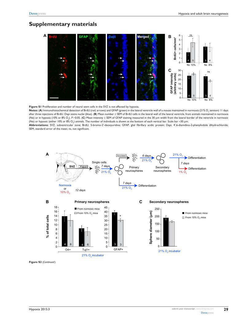
Figure S1 Proliferation and number of neural stem cells in the sVZ is not affected by hypoxia.Notes: (A) immunohistochemical detection of BrdU (red, arrows) and gFaP (green) in the lateral ventricle wall of a mouse maintained in normoxia (21% O2 tension) 11 days after three injections of BrdU. Dapi stains nuclei (blue). (B) Mean number ± seM of BrdU cells in the lateral wall of the lateral ventricle, from animals maintained in normoxia (nx) or in hypoxia (10% or 8% O2). P.0.05. (C) Mean intensity ± seM of gFaP staining measured in the 30 µm width from the lateral border of the ventricle in normoxic (nx) or hypoxic (either 10% or 8% O2) animals. The number of individuals is shown at the bottom of each vertical bar. scale bar =30 µm.Abbreviations: sVZ, subventricular zone; BrdU, 5-bromo-2′-deoxyuridine; GFAP, glial fibrillary acidic protein; Dapi, 4′,6-diamidino-2-phenylindole dihydrochloride; SEM, standard error of the mean; ns, not significant.
SVZ Papain
Single cells
Accutase
Primaryneurospheres
Secondaryneurospheres
Differentiation
Primary neurospheres Secondary neurospheres
Secondary neurospheres
O4+ Tuj1+ GFAP+
18 45 250
200
150
100
50
0
40
35 *30
25
20
15
10
5
0
16From normoxic mice
From 10% O2 mice From normoxic mice
From 10% O2 mice14
12
10
8
6
O4+ Tuj1+ GFAP+
Normoxicmice
Hypoxicmice
Normoxicmice
Normoxic mouse Hypoxic mouseO4
1% O2 1% O221% O2 21% O2
Tuj1 Dapi
Hypoxicmice
Normoxicmice
Hypoxicmice
6
% o
f to
tal c
ells
Sp
her
e d
iam
eter
(µm
)
6
4
4 4 3 32
0
25
20
1.6 60
50
40
30
20
10
0
1.4
1.2
1.0
0.8
0.6
0.4
0.2
0
% o
f to
tal c
ells
15
10
5***
*****
ns
366 6 6 6 6 6 6 3 3 30
B
A
C
D
Differentiation
Differentiation
21% O2
21% O2 incubator
21% O2
21% O2 incubator
Normoxiaor
10% O2
21% O2
1% O2
1% O2
21% O2
21% O2
7 days
12 days
7 days
7 days
6 days
Figure S2 (Continued )
Supplementary materials

Hypoxia 2015:3submit your manuscript | www.dovepress.com
Dovepress
Dovepress
30
d’anglemont de Tassigny et al
SVZ Papain
Single cells
Accutase
Primaryneurospheres
Secondaryneurospheres
Differentiation
Primary neurospheres Secondary neurospheres
Secondary neurospheres
O4+ Tuj1+ GFAP+
18 45 250
200
150
100
50
0
40
35 *30
25
20
15
10
5
0
16From normoxic mice
From 10% O2 mice From normoxic mice
From 10% O2 mice14
12
10
8
6
O4+ Tuj1+ GFAP+
Normoxicmice
Hypoxicmice
Normoxicmice
Normoxic mouse Hypoxic mouseO4
1% O2 1% O221% O2 21% O2
Tuj1 Dapi
Hypoxicmice
Normoxicmice
Hypoxicmice
6
% o
f to
tal c
ells
Sp
her
e d
iam
eter
(µm
)
6
4
4 4 3 32
0
25
20
1.6 60
50
40
30
20
10
0
1.4
1.2
1.0
0.8
0.6
0.4
0.2
0
% o
f to
tal c
ells
15
10
5***
*****
ns
366 6 6 6 6 6 6 3 3 30
B
A
C
D
Differentiation
Differentiation
21% O2
21% O2 incubator
21% O2
21% O2 incubator
Normoxiaor
10% O2
21% O2
1% O2
1% O2
21% O2
21% O2
7 days
12 days
7 days
7 days
6 days
Figure S2 in vitro sVZ secondary neurospheres formation and differentiation.Notes: (A) schematic diagram depicting the experimental design. single-cell suspension was obtained by papain digestion of sVZ tissue dissected from mice maintained either in normoxia (21% O2) or in hypoxia (10% O2) for 12 days. Primary neurospheres were obtained after 7 days and either placed in differentiation medium for 7 days in 21% O2 atmosphere, or dissociated by accutase digestion to obtain single cells that were placed in neurosphere culture conditions for 6 days. secondary neurospheres were then placed in differentiation conditions for 7 days in 21% or 1% O2 atmosphere. (B) Bargraphs indicate the percentage (± seM) of oligodendrocytes (O4+), neurons (Tuj1+), and astrocytes (gFaP+) in primary neurospheres from animals maintained in normoxia or hypoxia after 7 days of differentiation at 21% O2 atmosphere. (C) Mean diameter (± seM) of secondary sVZ neurospheres obtained from normoxic or hypoxic (10% O2) animals cultured at 21% O2 for 7 days. The data indicate that self-renewal of progenitors derived from hypoxic mice is not impaired. (D) Bargraphs indicate the percentage (± seM) of O4+, Tuj1+, and gFaP+ cells in secondary neurospheres derived from normoxic or hypoxic (10% O2) mice and differentiated for 7 days in 21% or 1% O2. a selective decrease in oligodendrocytic and neuronal population occurred at 1% O2 in comparison to 21% O2 atmosphere regardless of the previous normoxic or hypoxic status of the mice. The number of gFaP+ cells (astrocytes) remained unchanged. Microphotographs (bottom panel) illustrate the loss of O4+ (red) and Tuj1+ (green) in 1% O2 in both normoxic and hypoxic mice-derived secondary neurospheres. nuclei are stained in blue. scale bar =50 µm. The number of individuals per condition is indicated at the bottom of each vertical bar. *P,0.05, **P,0.01, and ***P,0.001.Abbreviations: SVZ, subventricular zone; SEM, standard error of the mean; GFAP, glial fibrillary acidic protein; ns, not significant; Dapi, 4′,6-diamidino-2-phenylindole dihydrochloride.
Motor cortex
DMSDLS
70
60
50 50
40 40
30 30
NG
2+ c
ells
/mm
2
20 20
10 10
0Nx
DMS DLS
NG2 Dapi
LV50 µm
Motor cortexNormoxia Normoxia Normoxia
Nx Nx8%
DMS DLS Motor cortex
8% 8%0
45 55 55
30
20
10
0
Figure S3 immature oligodendrocyte progenitors are not affected by hypoxia.Notes: immature oligodendrocyte progenitors (ng2+) were quantified in the DMS, DLS, and motor cortex as depicted in the mouse brain diagram (top left). Bargraphs show that the number (± seM) of ng2+ cells per mm2 in the DMs, Dls, and motor cortex is not affected after 12 days in 8% hypoxia. The number of individuals is shown at the bottom of each vertical bar. coronal sections in normoxic animals illustrate the ng2 (red) staining with Dapi-stained nuclei (blue) in the three regions analyzed. scale bar =50 µm. note that the DMs results are also presented in the main manuscript.Abbreviations: Dls, dorsolateral striatum; DMs, dorsomedial striatum; seM, standard error of the mean; Dapi, 4′,6-diamidino-2-phenylindole dihydrochloride; lV, lateral ventricle.
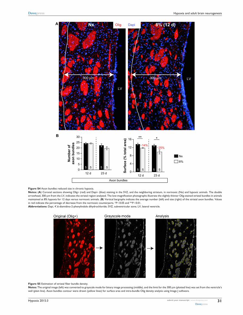
Hypoxia 2015:3 submit your manuscript | www.dovepress.com
Dovepress
Dovepress
31
Hypoxia and adult brain neurogenesis
30
LV
300 µm
NxA
B
8% (12 d)
300 µm
Olig Dapi
LV
25
20
15
16
12
8
4
0
10
55 5
12 d 23 d12 d
Su
rfac
e (%
to
tal a
rea)
Nu
mb
er o
fax
on
bu
nd
les
Axon bundles
23 d
Nx
8%
5 5
** *
5 5 5 5
−25%−14%
0
Figure S4 axon bundles reduced size in chronic hypoxia.Notes: (A) coronal sections showing Olig+ (red) and Dapi+ (blue) staining in the sVZ, and the neighboring striatum, in normoxic (nx) and hypoxic animals. The double arrowhead, 300 µm from the LV, indicates the striatal region analyzed. The low-magnification photographs illustrate the slightly thinner Olig-stained striatal bundles in animals maintained at 8% hypoxia for 12 days versus normoxic animals. (B) Vertical bargraphs indicate the average number (left) and size (right) of the striatal axon bundles. Values in red indicate the percentage of decrease from the normoxic counterparts. *P,0.05 and **P,0.01.Abbreviations: Dapi, 4′,6-diamidino-2-phenylindole dihydrochloride; sVZ, subventricular zone; lV, lateral ventricle.
Figure S5 Estimation of striatal fiber bundle density.Notes: The original image (left) was converted to grayscale mode for binary image processing (middle), and the limit for the 300 µm (plotted line) was set from the ventricle’s wall (plain line). axon bundles contour were drawn (yellow lines) for surface area and intra-bundle Olig density analysis using image J software.
Hypoxia 2015:3submit your manuscript | www.dovepress.com
Dovepress
Dovepress
32
d’anglemont de Tassigny et al
extended materials and methodsantibodies and special reagentsThe specificity of each antibody used in this study has been
extensively tested by other groups and showed specific and
expected staining in our hands. Primary antibodies were
used as follows: anti-5-bromo-2′-deoxyuridine (BrdU, clone
BU1/75 [ICR1] ab6326 from Abcam), 1:200; anti-proliferat-
ing cell nuclear antigen (PCNA, clone PC10, M0879, Dako),
1:1,000; anti-neuronal nuclei (NeuN, clone A60, MAB377
Millipore), 1:1,000; anti-glial fibrillary acidic protein (GFAP,
Z0334, Dako), 1:1,000; anti-doublecortin (DCX, sc-8066,
Santa Cruz Biotechnology), 1:500; anti-oligodendrocytes,
also known as RIP (Olig, MAB1580, Millipore), 1:1,000;
anti-chondroitin sulfate proteoglycan (NG2, AB5320,
Millipore), 1:100; anti-oligodendrocyte marker O4 (clone
81, MAB345, Millipore), 1:500; anti-beta III tubulin (Tuj1,
ab18207, Abcam), 1:500. We used secondary antibodies
anti-mouse IgG or IgM, anti-rabbit IgG, anti-goat IgG,
or anti-rat IgG conjugated with Alexa Fluor 488 or Alexa
Fluor 568 (1:500) (Invitrogen). Mouse on mouse M.O.M.
kit (BMK-2202, Vector Laboratories). Apoptotic cells were
revealed using a Click-iT® TUNEL Alexa Fluor® Imaging
Assay (C10245, Invitrogen). Nuclei were stained with 4′,6-
diamidino-2-phenylindole dihydrochloride (Dapi, D9542,
Sigma). The thymidine analog BrdU (B5002, Sigma) was
prepared in sterile saline and dissolved by sonication just
prior injection. Trypsin inhibitor solution contains 2.5 mg/
mL trypsin inhibitor (T9253, Sigma), 2.5 mg/mL bovine
serum albumin (A8022, Sigma), 4.5 mg/mL d-(+)-glucose
(G7021, Sigma), and 26 mM NaHCO3 in Earle’s balanced
salt solution (24010, Gibco).
Immunohistochemistry and immunocytofluorescenceMice were intracardially perfused with 0.1 M phosphate-
buffered saline (PBS) followed by 4% paraformaldehyde
in PBS pH 7.4. Following a 1-hour postfixation step, brains
were washed in PBS 0.1 M, cryoprotected in PBS with
30% sucrose, embedded in Tissue-Tek® O.C.T. Compound,
and frozen on dry ice. Using a cryostat (CM 1950, Leica,
Germany), 10 µm coronal cryosections were obtained in ten
series on Superfrost plus slides (Thermo Scientific). Each
series containing 8–9 sections was spaced 200 µm approxi-
mately ranging from +1.10 mm to -0.50 mm from bregma
in anteroposterior coordinates. For each immunoassay, one
complete series (ie, one slide) was used per animal. Sections
were stored at -20°C until use. After being brought back at
room temperature (RT), cryosections were rehydrated in PBS.
Some primary antibodies required specific pretreatment: to
uncover BrdU and PCNA, we used a solution of sodium citrate
10 mM, pH 6 with 0.05% Tween-20 at 95°C for 20 minutes.
For PCNA and Olig detection, a 1-hour pretreatment with
M.O.M. prevents background staining. Sections were then
washed with PBS and incubated for 1 hour at RT in a block-
ing solution (PBS 0.1 M with bovine serum albumin 1 mg/
mL, 10% fetal bovine serum, and 0.1% Triton X-100). Then,
sections were incubated with primary antibodies diluted
in the aforementioned blocking solution overnight at 4°C.
Sections were extensively washed in PBS with 0.1% Triton
X-100, and incubated with the appropriate secondary Alexa
488- or Alexa 568-conjugated antibodies in blocking buffer
for 1 hour at RT. Dapi 0.5 µg/mL was added in the last wash
for nuclear staining. Slides were coverslipped with Fluoro-gel
(17985-11, EMS, Hatfield, PA, USA). Fluorescence images
were obtained with a BX61 microscope equipped with a DP70
camera (Olympus). Given the inter-experimental groups dif-
ference that may occur, only animals from the same batch
were analyzed side by side. Thus, 10% hypoxia animals were
compared with their normoxic littermates, and 8% hypoxia
mice were compared with their normoxic littermates. Photos
were acquired at a 4,080×3,072 pixels resolution under the
aforementioned fluorescent microscope with same exposure
and contrast settings between different conditions in a given
staining. Alexa 488-labeled cells were detected with the
U-MWIBA2 filter (EX530-550/DM570/EM590, Olympus).
Alexa 568-labeled cells were examined with the U-N41021
filter (EX550-580/DM585/EM620). Dapi-labeled nuclei
were observed with the U-MNU2 filter (EX360-370/DM400/
EM420). An experimenter blinded to the treatment protocol
performed all photos acquisition, processing, and cell quanti-
fications. For BrdU and PCNA analysis in the subventricular
zone (SVZ), 10–14 photos of the SVZ for each staining and
Dapi were acquired per animal with the objective ×20. Images
were processed with Photoshop CS5 (Adobe) to merge BrdU
or PCNA staining with Dapi-positive nuclei. Only dual-
labeled nuclei that were lying along the wall of the lateral
ventricle were considered. Results were expressed as the
average number of BrdU or PCNA-positive cells ± standard
error of the mean (SEM) per SVZ mm.
For DCX analysis in the SVZ, 8–12 photos of the DCX
staining in the lateral wall of the SVZ were acquired. Analysis
was undertaken by converting color images to 8-bit grayscale
mode, and by measuring both the intensity and the surface
area of the region of interest using Image J 1.46r software
(NIH), and then multiplying the intensity by area values. This
method provides a solid estimation (in arbitrary units) of the
population of DCX-positive cells in each SVZ photos.

Hypoxia
Publish your work in this journal
Submit your manuscript here: http://www.dovepress.com/hypoxia-journal
Hypoxia is an international, peer-reviewed, open access journal that aims to improve understanding of the biological response to hypoxia. The journal will publish original research articles, reviews, methodological advances, clinical studies, and expert opinions that identify developments in the regulation of the physiological and pathological responses to
hypoxia and in the therapeutic targeting of hypoxia-responsive pathways. The manuscript management system is completely online and includes a very quick and fair peer-review system, which is all easy to use. Visit http://www.dovepress.com/testimonials.php to read real quotes from published authors.
Hypoxia 2015:3 submit your manuscript | www.dovepress.com
Dovepress
Dovepress
Dovepress
33
Hypoxia and adult brain neurogenesis
For BrdU and GFAP analysis in the SVZ of animals
sacrificed 11 days after BrdU injection (Figure S1), 9–13
photos of the SVZ for each staining plus Dapi were
acquired per animal with ×20 objective. Only dual-labeled
BrdU+/Dapi+ nuclei that were lying along the wall of the
lateral ventricle were considered. The GFAP intensity was
measured in the SVZ area corresponding to a 30 µm band
from the limit of the ventricular lumen. Color images were
converted to 8-bit mode and were quantified using Image J.
Results were expressed as the mean of the intensity ± SEM
in arbitrary unit.
For Olig analysis in the dorsomedial striatum (DMS),
8–12 photos per animal were acquired for Olig and Dapi
staining in the DMS, within a limit of 300 µm from the lat-
eral ventricle wall. Since Olig is a cytoplasmic marker, only
dual-labeled Dapi/Olig cells were considered as immunore-
active, and results are expressed as the average number of
Olig-positive cells ± SEM per mm2. To estimate the levels
of myelin, Olig staining images were converted to grayscale
to determine the axon bundles size and the optical density
(binary image processing of black and white) inside the axon
bundles and between them (inter-bundles space) (Figure S5 is
an example). Values for each animal were used to determine
mean counts, and these were used to generate mean ± SEM
values for each group. Immature oligodendrocytes were
detected with NG2/Dapi staining and quantified within the
same limits as for Olig staining.
For BrdU-positive cells migration to the olfactory bulb
(OB), mice were sacrificed 11 days after 3× BrdU 50 mg/kg
injections, and OB was removed and examined. BrdU and Dapi
staining was performed as described earlier. TUNEL/NeuN
staining was performed following the manufacturer instructions
with minor modifications. Intestine sections from the same
mice were used as a control for positive TUNEL staining,
since apoptosis is observed at the surface of the gastric pits.1
TUNEL- or BrdU-positive cells were counted in the granular
cell layer in six to ten photos per animal that were acquired
with a ×20 objective. Values are expressed as mean ± SEM
positive cells per mm2 for each group. The percent of neuronal
(NeuN positive) cells over the total cells in the OB was quanti-
fied in the same photos as those used for TUNEL quantifica-
tion. Dapi+ nuclei displaying a moon-like shape were not
taken into the total nuclei counting but were considered as
endothelial cells and counted as such.
For secondary neurosphere differentiation assay and
immunocytochemistry, glass coverslips in 24-well plates were
treated, prior to plating, with 0.5 mg/mL human fibronectin
(Biomedical Technologies) for adherence. Neurospheres
(6 days old) derived from primary neurospheres generated
from normoxic or hypoxic (10% O2 tension) animals were
plated in the mitogen-free medium as described in the main
manuscript and placed in 5% CO2 incubators with 21% or
1% O2 levels. After 7 days in differentiation conditions,
cells were fixed and treated for immunocytochemistry as
described in the “Materials and methods” section. Two
photos per condition were used for analysis of the follow-
ing staining: O4, Tuj1, and GFAP. The percentage of O4+,
Tuj1+, and GFAP+ cells was calculated over the total number
of Dapi+ nuclei.
Reference1. Frisch SM, Francis H. Disruption of epithelial cell-matrix interactions
induces apoptosis. J Cell Biol. 1994;124:619–626.
Related Documents