2011 Original Articles Korean Circulation J 1998;28( 12) :2011-2029 Hypoxia에 의한 혈관이완과 수축의 기전에 관한 연구 연세대학교 의과대학 생리학교실 강 복 순·이 영 호 Study on the Mechanism of Hypoxic Induced Vasodilatation and Vasoconstriction Bok Soon Kang, MD and Young Ho Lee, PhD Department of Physiology, Yonsei University College of Medicine, Seoul, Korea ABSTRACT Background:Although hypoxic pulmonary vasoconstriction ( HPC) and hypoxic coronary vasodilatation ( HCD) have been recognized by many researchers, the precise mechanism remains unknown. As isolated arteries will constrict or relax in vitro in response to hypoxia, the oxygen sensor/transduction mechanism must reside in the arterial smooth muscle, the endothelium, or both. Unfortunately, much of the current evidence is conflicting, especially concerning to the dependency of HPC and HCD on the endothelium and the role of the K + channel. Therefore, this experiment was attempted to clarify the dependency of HPC and HCD on the endothelium and the role of the K + channel on HPC and HCD. Methods:HPC was investigated in isolated main pulmonary arte- ries precontracted with norepinephrine ( NE) . HCD was investigated in isolated left circumflex coronary artery precontracted with prostaglandin F 2 α . Vascular rings were suspended for isometric tension recording in an organ chamber filled with Krebs-Henseleit solution. Hypoxia was induced by gassing the chamber with 95% N 2 +5% CO 2 , which was maintained for 15- 25 min. Results:1) Hypoxia elicited a vasoconstriction in NE- precontracted pulmonary arteries with endothelium, but a vasodilatation in PGF 2 α -precontracted coronary art- eries with and without endothelium. There was no difference between the amplitude of the HPC and HCD induced by two consecutive hypoxic challenges and the effect of normoxic and hyperoxic control Krebs-Henseleit sol- ution on subsequent response to hypoxia. 2) Inhibition of NO synthesis by the treatment with Nω w-nitro-L-argi- nine reduced HPC in pulmonary arteries, but inhibition of the cyclooxygenase pathway by treatment with indom- ethacin had no effect on HPC and HCD, respectively. 3) Blockades of the TEA-sensitive K + channel abolished HPC and HCD. 4) Apamin, a small conductance Ca 2 +-activated K + ( K Ca ) channel blocker, and iberiotoxin, a large conductance K Ca channel blocker, had no effect on the HCD. 5) Glibenclamide, an ATP-sensitive K + ( K ATP ) channel blocker, reduced HCD. 6) Cromakalim, an K ATP channel opener, relaxed the coronary artery precontr- acted with prostaglandin F 2 α . The degree of relaxation by cromakalim was similar to that by hypoxia and glib- enclamide reduced both hypoxia- and cromakalim-induced vasodilations. 7) Verapamil, a Ca 2+ entry blocker, caffeine, a Ca 2+ emptying drug;and ryanodine, an inhibitor of Ca 2+ release from SR, reduced HPC, respectively. Conclusion:HPC is dependent on the endothelium and is considered to be induced by inhibition of the mec- hanisms of NO-dependent vasodilation while HCD is independent of the endothelium and is considered to be induced by activation of the K ATP channel. ( Korean Circulation J 1998 ; 28( 12) : 2011-2029) KEY WORDS:Hypoxia·Nnitric oxide·Glibenclamide·K + channel·Pulmonary artery·Coronary Artery. 논문접수일:1998년 6월 10일 심사완료일:1999년 2월 5일 교신저자:강복순, 120-752 서울 서대문구 신촌동 134 연세대학교 의과대학 생리학교실 전화:(02) 361-5192·전송:(02) 393-0203 E-mail:[email protected]

Welcome message from author
This document is posted to help you gain knowledge. Please leave a comment to let me know what you think about it! Share it to your friends and learn new things together.
Transcript

2011
Original Articles Korean Circulation J 1998;;;;28((((12))))::::2011-2029
Hypoxia에 의한 혈관이완과 수축의 기전에 관한 연구
연세대학교 의과대학 생리학교실
강 복 순·이 영 호
Study on the Mechanism of Hypoxic Induced Vasodilatation and Vasoconstriction
Bok Soon Kang, MD and Young Ho Lee, PhD Department of Physiology, Yonsei University College of Medicine, Seoul, Korea ABSTRACT
Background:Although hypoxic pulmonary vasoconstriction (HPC) and hypoxic coronary vasodilatation (HCD) have been recognized by many researchers, the precise mechanism remains unknown. As isolated arteries will constrict or relax in vitro in response to hypoxia, the oxygen sensor/transduction mechanism must reside in the arterial smooth muscle, the endothelium, or both. Unfortunately, much of the current evidence is conflicting, especially concerning to the dependency of HPC and HCD on the endothelium and the role of the K+ channel. Therefore, this experiment was attempted to clarify the dependency of HPC and HCD on the endothelium and the role of the K+ channel on HPC and HCD. Methods:HPC was investigated in isolated main pulmonary arte-ries precontracted with norepinephrine (NE). HCD was investigated in isolated left circumflex coronary artery precontracted with prostaglandin F2α. Vascular rings were suspended for isometric tension recording in an organ chamber filled with Krebs-Henseleit solution. Hypoxia was induced by gassing the chamber with 95% N2+5% CO2, which was maintained for 15-25 min. Results:1) Hypoxia elicited a vasoconstriction in NE-precontracted pulmonary arteries with endothelium, but a vasodilatation in PGF2α-precontracted coronary art-eries with and without endothelium. There was no difference between the amplitude of the HPC and HCD induced by two consecutive hypoxic challenges and the effect of normoxic and hyperoxic control Krebs-Henseleit sol-ution on subsequent response to hypoxia. 2) Inhibition of NO synthesis by the treatment with Nωw-nitro-L-argi-nine reduced HPC in pulmonary arteries, but inhibition of the cyclooxygenase pathway by treatment with indom-ethacin had no effect on HPC and HCD, respectively. 3) Blockades of the TEA-sensitive K+ channel abolished HPC and HCD. 4) Apamin, a small conductance Ca2+-activated K+ (KCa) channel blocker, and iberiotoxin, a large conductance KCa channel blocker, had no effect on the HCD. 5) Glibenclamide, an ATP-sensitive K+ (KATP) channel blocker, reduced HCD. 6) Cromakalim, an KATP channel opener, relaxed the coronary artery precontr-acted with prostaglandin F2α. The degree of relaxation by cromakalim was similar to that by hypoxia and glib-enclamide reduced both hypoxia- and cromakalim-induced vasodilations. 7) Verapamil, a Ca2+ entry blocker, caffeine, a Ca2+ emptying drug;and ryanodine, an inhibitor of Ca2+ release from SR, reduced HPC, respectively. Conclusion:HPC is dependent on the endothelium and is considered to be induced by inhibition of the mec-hanisms of NO-dependent vasodilation while HCD is independent of the endothelium and is considered to be induced by activation of the K ATP channel. ((((Korean Circulation J 1998;28((((12)))):2011-2029)))) KEY WORDS:Hypoxia·Nnitric oxide·Glibenclamide·K+ channel·Pulmonary artery·Coronary Artery.
논문접수일:1998년 6월 10일
심사완료일:1999년 2월 5일
교신저자:강복순, 120-752 서울 서대문구 신촌동 134 연세대학교 의과대학 생리학교실 전화:(02) 361-5192·전송:(02) 393-0203
E-mail:[email protected]

Korean Circulation J 1998;28(12):2011-2029 2012
서 론
동맥내 산소분압이 40 mmHg 이하로 감소할 때(hy-
poxia) 관동맥을 포함한 대부분의 혈관에서 혈관이완이
발생된다. 이러한 혈관이완은 허혈(ischemia) 또는 증
가된 대사의 요구에 반응하기 위한 혈류량의 증가에 따
른 생리학적 조절과정이며, 이는 저산소증(hypoxia)에
노출된 조직으로부터 유리되는 혈관이완인자에 의해 주
로 조절되어진다. 그러나 이와는 달리 폐동맥에 있어서
는 동맥내 산소분압이 40 mmHg 이하로 감소할 때 혈
관수축이 발생되는데, 이 또한 환기(ventilation)와 혈액
관류량(perfusion)의 평형을 적절히 유지하기 위한 생리
학적 반응이다. 즉 저산소증때 혈관수축이 일어나므로서
폐내 환기가 되지 않는 부위에 혈액관류를 감소시키면서
동시에 환기가 잘되는 부위로 혈액관류를 전환시킨다.1)
따라서 저산소증에 대한 혈관의 반응은 조직의 요구에 따
라 결정된다.
많은 연구자들이 저산소증이 혈관의 긴장도에 미치는
영향을 규명하기 위해 분리된 혈관에서 저산소증의 효과
를 관찰하여 다음과 같은 결과를 얻었다. 여러 종류의 동
물로부터 분리된 폐동맥에서 저산소증 혈관수축(Hyp-
oxic pulmonary vasoconstriction;HPC)을 유발한다
는 것이 보고되었다.2)3) 그러나 HPC는 실험조건과 연
구자에 따라 그 양상이 다양하다. 즉 HPC가 유발될 때
그 수축 양상은 저산소증 초기에 빠른 수축이 나타나거
나,4) 점차적으로 수축이 증가하거나3) 또는 biphasic 형
태의 수축이 나타난다.5) 또한 사용된 혈관의 직경 및 근
육의 상태에 따라 혈관수축 양상이 다르게 나타난다고 보
고되었는데 Yuan등2)은 직경이 작은 폐동맥에서는 HPC
가 유발되나 직경이 큰 혈관에서는 HPC가 유발되지 않
는다고 보고하였으나, Bonnet등6)은 저산소증은 혈관직
경의 크기에 상관없이 모두 HPC를 유발한다고 하였다.
또한 HPC의 크기는 근육의 상태에 따라서도 다르게 나
타나는데 기초상태보다는 수축상태의 근육에서 더욱 더
현저하다고 보고되었다.2)
저산소증에 대한 관동맥의 반응은 기대와는 달리 분리
된 많은 관동맥에서 수축이 유발되는데,7)8) 이때 나타나
는 수축은 근육의 상태에 상관없이 저산소증 혈관수축의
정도는 거의 일정한 형태와 크기로 나타난다는 것이 보
고되었다.9) 그러나 관동맥에서 저산소증에 의해 혈관수
축이 유발된다는 보고와는 달리 사람의 관동맥10)이나 돼
지의 관동맥11)에서 저산소증은 혈관이완을 초래한다는
것이 보고되었다.
한편, 폐동맥과 관동맥에서의 저산소증에 의한 혈관
수축의 기전은 다양한 수축의 양상 및 상반되는 결과로
인해 매우 복잡하고 정립된 정설이 거의 없는 실정이다.
현재까지 보고된 기전으로서 가장 유력한 것은 내피세
포 및 K+ channel과 관련된 가설인데 이 또한 상반된
견해가 많다. 이중 내피세포와 관련된 것으로서는 저산
소증이 안정시에 내피세포로부터 nitric oxide의 유리를
억제하여 수축을 유발한다는 보고2)9)12-14)와 내피세포
로부터 수축물질(endothelium-derived contracting fa-
ctor;EDCF)을 유리시켜 혈관수축을 유발한다는 보
고15)16) 등이 있다. 이와는 달리 저산소증이 혈관 평활근
세포에 직접 작용하여 평활근 막에 존재하는 K+ cha-
nnel의 활성을 억제하여 수축을 유발한다는 보고도 있
다.3)17)18)
저산소증 혈관수축과 관련된 기전은 많은 연구자들의
실험에 의해 어느 정도 추구가 되고 있는 실정이나 저
산소증 혈관이완의 기전은 거의 밝혀지지 않고 있다. 다
만 indomethacin이 저산소증에 의한 혈관이완에 영향을
미치지 않는다는 보고,11) PGl2가 저산소증 혈관이완을
유발한다는 보고19)와 저산소증 상태에서 ATP의 감소
로 인해 ATP-sensitive K+ channel(K+ATP channel)
의 활성화로 막전위가 과분극되어 혈관이완이 초래된다
는 보고20) 등이 있다.
앞에서 기술하였듯이 많은 연구자들이 저산소증이 폐
동맥 및 관동맥의 긴장도에 미치는 영향을 관찰하여 HPC
및 HCD를 규명하였다고 하나 그 정확한 기전은 아직
불분명하다. In vitro에서 저산소증이 분리된 혈관을 수
축 및 이완을 유발하기 때문에 oxygen sensor가 평활
근이나 내피세포에 존재하는 것이 틀림없을 것이다. 그
러나 현재까지 보고된 대부분의 연구결과들은 일치된
견해가 없을 뿐 아니라, 특히 HPC 및 HCD의 내피세
포에 대한 의존성과 K+ channel의 역할에 대한 상반된
견해가 많다. 따라서 본 연구에서는 저산소증에 의해 수
축과 이완을 유발하는 폐동맥 및 관동맥에서 저산소증이
혈관의 긴장도에 미치는 영향을 근육의 상태(안정상태와
수축상태) 및 저산소증의 정도를 변화시키면서 관찰하여
저산소증 혈관수축과 저산소증 혈관이완의 기전을 규명
하고자 하였다.

2013
재료 및 방법
실험재료
폐동맥 및 관동맥환의 제조
폐동맥환
Sprague-Dawley rat을 참수(decapitation)한 후 흉
곽을 절개하여 심장과 폐를 동시에 적출하였다. 적출된
장기를 95% O2+5% CO2로 포화시킨 Krebs-Hens-
eleit 용액(KH;mM;NaCl 119, KCl 4.6, CaCl2 2.5,
NaHCO3 25, MgCl2 1.2, KH2PO4 1.2, glucose11)에 담
근 상태에서 심장을 제거한 후 폐를 실험용기(prepar-
ation chamber)에 핀을 이용하여 고정시켰다. 현미경하
에서 혈관 내면의 내피세포가 손상되지 않게 조심하면서
폐동맥 주위의 폐조직을 제거한 다음 결체조직 및 지방
조직을 안과용 미세가위 및 핀셋을 이용하여 조심스럽게
제거하였다.
관동맥환
체중 2.0∼2.5 kg의 토끼의 귀정맥(ear vein)에 pe-
ntobarbital sodium(60 mg/kg)과 heparin(2,000 IU/
kg)을 주사하여 마취시킨 후 경동맥을 절단하여 실혈시
킨 다음 심장을 적출하였다. 적출한 심장을 95% O2+
5% CO2로 포화시킨 KH 용액에 담근 상태에서 주위
의 심근 조직이 붙어 있는 상태로 관동맥을 분리한 후
실험용기에 핀을 이용하여 고정시킨 후 현미경하에서 관
동맥 주위의 심근 및 결체조직을 안과용 미세가위 및 핀
셋을 이용하여 조심스럽게 제거하였다. 분리된 main pu-
lmonary artery 및 left circumflex coronary artery
를 4∼5 mm 크기의 환(ring) 형태로 만들어 한쪽은 용
기(chamber) 내의 L-shaped rod에 고정하고 다른 한
쪽은 strain gauge transducer에 고정하였다. 이후 혈
관 환(ring)을 기체가 공급되고 37℃로 일정하게 유지
되는 KH 용액에서 1시간 정도 평형시켰다. 몇몇 실험에
서는 용액에 적신 면봉으로 혈관 내면을 가볍게 문질러
내피세포를 제거하였다. 내피세포의 제거여부는 10-7 M
NE 및 70 mM high K+ 용액(정상 KH 용액의 조성중
KCl의 농도가 70 mM 되게 NaCl을 감소시켜 만든 용
액)으로 혈관수축을 유도한 후 10-6 M acetylcholine
을 처치하였을 경우 이완이 되지 않는 것으로 확인하
였다.21)
실험이 진행되는 동안 실험기간별로 glass syringe를
사용하여 bath로부터 용액을 채취하여 blood gas ana-
lyzer(Radiometer, Cophenhagen, Denmark)를 이용해
산소분압(PO2)과 pH를 측정하였다.
약 물
본 실험에 사용된 주요약물은 L-norepinephrine bi-
tartrate(NE), prostaglandin F2α(PGF2α), acetylc-
holine chloride, Nω-nitro-L-arginine(L-NNA), in-
domethacin, apamin, iberiotoxin, tetraethylammo-
nium chloride(TEA), glibenclamide, cromakalim 및
verapamil로서 모두 Sigma Chemicals(St. Louis, MO,
USA)로부터 구하였다.
실험방법
1시간의 평형기간후 폐동맥의 경우 0.5 g의 기초장력
을 가한 상태에서 10-7 M NE로 혈관 수축을 유도하
는 과정을 1시간 간격으로 3회 반복하여 기초 상태 및
혈관의 반응이 일정하게 유지되도록 하였고, 실험시작 전
에 40 mM K+ 용액(정상 KH 용액의 조성중 KCl의 농
도가 40 mM되게 NaCl을 감소시켜 만든 용액)을 사용
하여 혈관의 수축고를 기록하였고 이를 세척한 후 본 실
험을 시행하였다. 관동맥의 경우 0.4 g의 안정장력을 가
한 상태에서 70 mM high-K+ 용액으로 혈관수축을
유도하는 과정을 1시간 간격으로 3회 반복하여 안정상
태 및 혈관의 반응이 일정하게 유지되도록 하였고, 실험
시작 전에 PGF2α(1.5×10⁻⁶ M)를 사용하여 혈관의
수축고를 기록하였고 이를 세척한 후 본 실험을 시행하
였다.
대부분의 실험에서 혈관 환(ring)이 고정된 용기(or-
gan bath)에 95% O2+5% CO2 기체(hyperoxic gas)
가 공급되는 KH 용액(PO2=543±8 mmHg, pH=7.
38±0.02)으로 공급하였으며, 일부 실험에서는 비교의
목적으로 20% O2+5% CO2+75% N2 기체로 구성된
압축공기(normoxic gas)가 공급되는 KH 용액(PO2=
134±12 mmHg, pH=7.39±0.02)을 사용하였다. 저
산소증은 상기의 두 기체가 공급되는 용액을 95% N2
Korean Circulation J 1998;28(12):2011-2029 2014
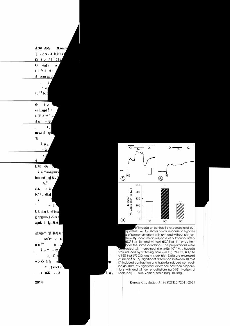
Fig. 1. Effect of hypoxia on contractile responses in rat pul-monary arteries. A1, A2:shows typical response to hypoxiain rings of pulmonary artery with (A1) and without (A2) en-dothelium. B:shows mean response of pulmonary arterywith (EC+;n=30) and without (EC-;n=11) endotheli-um under the same conditions. The preparations werecontracted with norepinephrine (NE;10-7 M). hypoxiawas induced by switching from 95% O2+5% CO2 (O2) toa 95% N2±5% CO2 gas mixture (N2). Data are expressedas mean±SE. *:significant difference between 40 mMK+-induced contraction and hypoxia-induced contract-ion (p<0.05). **:significant difference between prepara-tions with and without endothelium (p<0.05). Horizontalscale bar:10 min, Vertical scale bar:100 mg.
±5% CO2 기체(hypoxic gas)가 공급되는 KH 용액(PO2
=30.1±0.3 mmHg, pH=7.39±0.02)으로 바꾸어 유
발하였고 15∼25분간 유지시켰다. 저산소증 유발후 혈
관 환(ring)을 다시 95% O2+5% CO2 기체가 공급되는
KH 용액 또는 압축공기가 공급되는 KH 용액으로 바꾸
어 reoxygenation시켰고 다음 실험때까지 60분간 안
정화시켰다.
저산소증에 대한 반응은 10-7 M NE(폐동맥) 및 1.5×
10-6 M PGF2α 용액으로 수축된 혈관에서 실험하였다.
일정한 크기로 유지되는 수축하에서 저산소증의 영향을
관찰하였고 저산소증 기간이 끝난 후에 혈관은 reoxy-
genation되어졌는데 이때의 장력은 수축제에 의한 수축
고로 되돌아 왔으며 이후에 세척되었다. 몇몇 실험에 있
어서 저산소증에 대한 반응이 재생성을 가지는지 확인하
기 위해 상기의 실험방법과 동일하게 실험을 하면서 re-
oxygenation후 혈관의 장력이 수축제로 유도된 수축고
로 일정해졌을 때 다시 저산소증를 유발하는 과정을 시
행하였다.
저산소증 반응에 대한 여러가지 차단제의 효과는 적
절한 농도로 저산소증 전에 incubation하고 저산소증동
안 유지시키면서 관찰하였다. 즉 저산소증 반응에 대한
NO의 관여 여부는 L-NNA(10-5 M)를 사용하여 확인
하였고, cyclooxygenase pathway의 관여 여부는 in-
domethacin(10-5 M)을 사용하여 확인하였다. 세포막을
통한 Ca2+ 유입 및 세포내 Ca2+ 저장소로부터 Ca2+ 유
리가 저산소증 반응에 미치는 영향은 verapamil(10-5
M), caffeine(20 mM) 및 ryanodine(5 μM)을 사용
하여 확인하였다. 또한 저산소증에 대한 혈관 평활근 막
에 존재하는 K+ channel의 관여 여부는 tetraethyla-
mmonium chloride(TEA;10 mM), apamin(10-7 M),
iberiotoxin(5×10⁻⁸ M), glibenclamide(10-6 M) 및
cromakalim(5×10⁻⁶ M)을 사용하여 확인하였다.
결과분석 및 통계처리
실험결과는 40 mM K+ 용액(폐동맥) 및 PGF2α(관
동맥)에 의한 수축고의 백분율(%)로서 mean±SE로 표
시하였고, 저산소증에 의한 혈관 수축 및 이완은 수축제
에 의해 야기된 수축고에서부터 산정하였다. n은 실험에
사용된 동물의 수를 의미한다. 대조군과 약물 투여군간
의 차이는 Student’s paired 또는 unpaired t test를 실
시하여 p값이 0.05이하인 경우 유의한 것으로 간주하였다.
결 과
폐동맥 및 관동맥의 수축력에 대한 저산소증의 효과
Fig. 1 및 2는 내피세포의 존재 유무에 따라 저산소
증이 혈관 긴장도에 미치는 효과를 나타낸 실험결과이
다. Fig. 1의 전형적인 실험결과에서 보는 바와 같이 내
피세포가 존재하는 폐동맥의 경우 저산소증은 1∼2분 이
내에 혈관의 긴장도를 급격히 증가시키며 증가된 장력
은 저산소증 기간동안 유지되거나 약간 감소하는 양상
을 보였다. 20∼25분간의 저산소증 기간후 reoxyge-
nation을 실시한 경우 저산소증에 의해 증가된 장력은 급
격히 감소하여 NE에 의해 유도된 장력의 수준으로 유
지되었다. 그러나 내피세포가 존재하지 않는 경우 저산
소증에 의한 장력의 변화는 거의 관찰되지 않았다. Fig.
1B는 이들의 결과를 종합한 것으로 저산소증에 의한 장
력의 변화는 40 mM K+로 유도된 혈관 수축을 100%

2015
Fig. 2. Effect of hypoxia on the contractile responses in ra-bbit coronary artery. A, B:shows typical response to hyp-oxia in rings of coronary artery with (A) and without (B)endothelium. C:shows mean response of coronary arterywith (EC+;n=54) and without (EC-;n=11) endotheliumunder the same conditions. The preparations were contr-acted with prostaglandins F2α (PGF2α, 1.5×10⁻⁶ M).hypoxia was induced by switching from 95% O2+5% CO2(O2) to a 95% N2+5% CO2 gas mixture (N2). Data are exp-ressed as mean±SE. *:significant difference betweenPGF2α-induced contractility and hypoxia-induced contr-actility (p <0.05). EC+:ring with endothelium, EC-:ringwithout endothelium, ACh:acetylcholine.
Fig. 3. Reproducibility of two consecutive hypoxic challe-nges to pulmoanry arteries. A1, A2:shows typical response to two consecutive hypoxic challenges in precontracted (norepinephrine;NE, 10-7 M) pulmonary artery with (A1) and without (A2) endothelium. B:shows mean response of pulmonary artery with (EC+;n=10) and without (EC-;n=7) endothelium under the same conditions. Data are expressed as mean±SE. *:significant difference between 40 mM K+-induced contraction and hypoxia-induced co-ntraction (p<0.05). **:significant difference between preparations with and without endothelium (p<0.05). Hor-izontal scale bar:10 min, Vertical scale bar:100 mg.
로 하여 그 변화를 비교할 때, 내피세포가 존재하는 경우,
저산소에 의해 혈관 수축고는 222.4±13%(n=30, p<
0.05)으로 의의있게 증가하였고, 내피세포가 존재하지 않
는 경우의 수축고는 111.5±3%(n=11)이었다. 그러나
폐동맥과는 달리 관동맥에서 저산소증의 영향을 관찰해
본 결과(Fig. 2) 내피세포가 존재하는 경우 저산소증은
PGF2α(1.5×10⁻⁶ M)로 유도된 혈관수축을 이완시켰으
며 15∼25분간의 저산소증 기간후 reoxygenation을 실
시한 경우 저산소증에 의해 감소된 장력은 다시 PGF2α
에 의한 수축고로 회복되었다. 또한 내피세포가 존재하
지 않는 경우에도 동일한 반응을 관찰할 수 있었다. 이
들의 결과를 종합해 보면 저산소증에 의한 혈관이완 정
도는 PGF2α로 유도된 혈관이완을 100%로 하여 비교
할 때, 내피세포가 존재하는 경우는 62.7±10.1%(n=54),
내피세포가 존재하지 않는 경우는 63±5.4%(n=11)로
서 내피세포의 존재유무에 관계없이 거의 유사하였다.
Fig. 3 및 4는 저산소증에 의한 장력의 증가가 재생
성이 있는지를 확인하기 위해 NE 및 PGF2α로 각각
수축된 폐동맥 및 관동맥을 두번의 연속적인 저산소증
에 노출시켰을 때 반응을 관찰한 실험결과이다. Fig. 3
에서 보는 바와 같이 내피세포가 존재하는 폐동맥의 경
우 두 번의 연속적인 저산소증에 의해 야기된 장력의 증
가가 거의 유사함을 알 수 있었고(40 mM K+로 유도
된 혈관 수축을 100%로 하여 수축고의 변화를 비교할
때, 1st episode:230.9±16.2%, 2nd episode:232.
3±21. 9%;n=10), 내피세포가 존재하지 않는 폐동맥
의 경우 저산소증에 의한 장력의 증가는 거의 나타나지
않음을 알 수 있었다(1st episode:107.2±2.5%, 2nd
episode:102.3±1.2%;n=7). 관동맥에서 동일한 실
험을 실시한 결과 내피세포의 존재에 관계없이 두 번의
연속적인 저산소증에 의해 야기된 혈관이완의 정도는 거
의 유사함을 알 수 있었다. Fig. 4는 저산소증에 의한 장
력의 감소가 재현성이 있는지를 확인하기 위하여 PGF2

Korean Circulation J 1998;28(12):2011-2029 2016
Fig. 5. Effect of normoxic or hyperoxic control Krebs-Hens-eleit solution on subsequent response to hypoxia in pulm-onary arteries with endothelium. A1, A2:shows typical re-cording to effect of normoxic (A1) or hyperoxic (A2) con-trol Krebs-Henseleit solution on subsenquent response to hypoxia in precontracted (norepinephrine;NE, 10-7 M) pulmonary artery. B:shows mean response of pulmo-nary artery incubated with normoxic (20% O2) and hyper-oxic (95% O2) control Krebs-Henseleit solution under the same conditions (n=11). Data are expressed as mean±SE. *:significant difference between 40 mM K+-induced contraction and hypoxia-induced contraction (p<0.05).
: :
α(1.5×10⁻⁶ M)로 수축된 혈관을 두 번의 연속적인
저산소증에 노출시켰을 때의 반응을 관찰한 실험 결과
이다. 그림에서 보는 바와 같이 내피세포가 존재하는 경
우(Fig. 4A). 두 번의 연속적인 저산소증에 의해 야기된
혈관이완의 정도는 거의 유사함을 알 수 있었고, 내피
세포가 존재하지 않는 경우(Fig. 4B)에는 동일한 반응
을 보였다. Fig. 4C는 내피세포가 존재하지 않는 경우
의 실험결과를 종합한 것으로 PGF2α로 유도된 혈관이
완을 100%로 하여 비교할 때, 첫번째 저산소증 노출시
의 혈관이완은 64.6±10.1%;(n=9)이고, 두번째 저산
소증 노출시의 혈관이완은 70.6±8.7%(n=9)로서 두
번의 연속적인 저산소증에 의한 혈관이완 정도는 거의
유사하였다
Fig. 5 및 6은 폐동맥 및 관동맥을 normoxic gas와
hyperoxic gas가 각각 공급되는 KH 용액에서 incub-
ation한 후 저산소증의 효과를 관찰한 실험결과이다. 내
피세포가 존재하는 폐동맥의 경우 Fig. 5에서 보는 바와
같이 normoxic 기체와 hyperoxic 기체의 다른 혼합기
체에 incubation된 혈관에서 저산소증에 의한 장력의 증
가는 거의 일정하여 차이가 없었다(40 mM K+로 유도된
Fig. 4. Reproducibility of hypoxic coronary vasodilation fol-lowing two consecutive hypoxic challenges to coronaryartery. A, B:shows typical response to two consecutivehypoxic challenges in precontracted (prostaglandins F2α:PGF2α, 1.5×10⁻⁶ M) coronary artery with (A) and wi-thout (B) endothelium. C:shows mean response of coro-nary artery without endothelium under the same conditions(n=9). hypoxia was induced by switching from 95% O2+5% CO2 (O2) to a 95% N2+5% CO2 gas mixture (N2). Dataare expressed as mean±SE. *:significant difference bet-ween PGF2α-induced contractility and hypoxia-inducedcontractility (p<0.05). EC+:ring with endothelium, EC-:ring without endothelium, ACh:acetylcholine.

2017
Fig. 6. Effect of hyperoxic or normoxic control Krebs-Hens-eleit solution on subsequent response to hypoxia in coronaryartery without endothelium. A, B:shows typical recordingto effect of hyperoxic (A) or normoxic (B) control Krebs-Henseleit solution on subsequent response to hypoxia inprecontracted (prostaglandins F2α:PGF2α, 1.5×10⁻⁶ M)coronary artery. C:shows mean response of coronaryartery incubated with hyperoxic (95O2:n=20) and nor-moxic (20O2:n=7) control Krebs-Henseleit solution underthe same conditions. Hypoxia was induced by switchingfrom 95% O2+5% CO2 (O2) to a 95% N2+5% CO2 gas mixt-ure (N2). Data are expressed as mean±SE. *:significantdifference between PGF2α-induced contractility and hy-poxia-induced contractility (p<0.05).
Fig. 7. Effect of inhibiting NO synthesis by Nw-nitro-L-arginine on response to hypoxia in pulmonary arteries with endot-helium. A1, A2:shows typical response to hypoxia in precontracted (norepinephrine;NE, 10-7 M) pulmonary artery with (A2) and without (A1) Nω-nitro-L-arginine (L-NNA, 10-5 M). B:shows mean response of pulmonary artery with (L-NNA) and without (Cont.) Nω-nitro-L-arginine under the same conditions (n=9). Nω-nitro-L-arginine was applied 30-40 min before testing their efficacy. Data are expressed as mean±SE. *:significant difference bet-ween 40 mM K+-induced contraction and hypoxia-ind-uced contraction (p<0.05). **:significant difference bet-ween preparations with and without Nω-nitro-L-arginine (p< 0.05). Horizontal scale bar:10 min, Vertical scale bar:100 mg.
혈관수축을 100%로 하여 그 수축고를 비교할 때, nor-
moxic gas:228.4±13.5%, hyperoxic gas:253.0±
39.4%, n=11). 내피세포가 존재하지 않는 관동맥의 경
우 Fig. 6에서 보는 바와 같이 폐동맥의 경우와 유사하게
hyperoxic 기체와 normoxic 기체의 다른 혼합기체에 in-
cubation된 혈관에서 저산소증에 의한 혈관이완은 거의
일정하여 차이가 없었다(PGF2α로 유도된 혈관이완을
100%로 하여 그 변화를 비교할 때, hyperoxic gas:62.
8±6.3%, n=20, normoxic gas:67.3±7%, n=7).
HPC 및 HCD에 대한 NO 합성억제 및 cyclooxygenase
pathway의 차단 효과
Fig. 7은 내피세포가 존재하는 폐동맥에서 HPC에 NO
합성억제제인 L-NNA22)의 효과를 관찰한 실험결과이
다. 먼저 NE로 유도된 혈관 수축에 저산소증의 효과를
관찰한 후 1시간동안 세척한 다음 L-NNA 존재하에 동
일한 실험을 시행하였다. 그림에서 보는 바와 같이 NE
을 처치하기 10분전에 L-NNA(10-5 M)를 처치하였을
경우 안정시 기초장력이 증가되었고 저산소증에 의한 장력
의 증가는 소실(40 mM K+로 유도된 혈관수축을 100%
로 하여 그 수축고를 비교할 때, 대조군:250.5±30.6%,
L-NNA 처치군:111.3±5.6%, n=9, p<0.05)됨을 알
수 있었다.
Fig. 8 및 9는 HPC 및 HCD에 cyclooxygenase pa-
thway의 차단제인 indomethacin23)의 효과를 관찰한
실험결과이다. 먼저 내피세포가 존재하는 폐동맥에서 저
산소증의 효과를 관찰하여 HPC을 기록하였다(Fig. 8).
이후 indomethacin(10-5 M)을 전 처치한 결과 안정시
기초장력에 거의 영향을 미치지 않았으며 indometha-
cin 존재시 저산소증에 의한 장력은 오히려 다소 증가하

Korean Circulation J 1998;28(12):2011-2029 2018
는 경향(40 mM K+로 유도된 혈관수축을 100%로 하
여 그 수축고를 비교할 때, 대조군:228.4±38.5%, in-
domethacin 처치군:295.6±69.2%, n=8, Fig. 5B)
을 보였으나 통계적으로 유의하지는 않았다. 관동맥에
서도 동일한 실험을 실시한 결과(Fig. 9) 폐동맥과 유
사하게 indomethacin(10-5 M)은 안정시 기초장력에
거의 영향을 미치지 않았으며 저산소증에 의한 장력의
변화에도 거의 영향을 미치지 않았다(대조군:PGF2α
로 유도된 혈관이완을 100%로 하여 그 변화를 비교할
때, 68.7±7.9%, Indomethacin 처치군:54.3±7.5%,
n=6, Fig. 5C).
HPC 및 HCD에 대한 K+ channel blocker의 효과
HPC 및 HCD에 K+ channel의 관여여부를 규명하고자
하였다. 먼저 non-specific K+ channel 차단제인 TEA17)
의 존재유무에 따른 HPC 및 HCD의 변화를 관찰해 본
결과는 Fig. 10 및 11에 나태낸 바와 같다. Fig. 10에서
보는 바와 같이 폐동맥에서 NE로 혈관 수축을 유도한
후 저산소증의 효과를 관찰한 후 1시간동안 세척한 다음
1 mM TEA 존재하에 동일한 실험을 시행하였다. 그림
에서 보는 바와 같이 NE을 처치한 후 장력이 일정해졌
을 때 TEA를 처치하였을 경우 NE으로 유도된 장력이
현저히 증가하여 일정하게 유지되었고, 이후 저산소증을
유발하였을 경우 장력은 거의 변화되지 않았다(40 mM
K+로 유도된 혈관수축을 100%로 하여 그 수축고를 비
교할 때, 대조군:228.5±10.4%, TEA 처치군:114.1
±5.7%, n=8). Fig. 11은 관동맥에서 TEA의 효과를 관
찰한 것으로 먼저 PGF2α(1.5×10⁻⁶ M)로 유도된 혈
관수축에 저산소증의 효과를 관찰한 후 1시간 동안 세척
한 다음 10 mM TEA 존재하에 동일한 실험을 시행하
였다. 그림에서 보는 바와 같이 TEA를 전 처치하였을
경우 안정시 장력이 증가한 후 일정하게 유지되었으며,
Fig. 9. Effect of indomethacin on the response to hypoxia in coronary artery without endothelium. A, B:shows typi-cal response to hypoxia in the precontracted (prostagla-ndins F2α:PGF2α, 1.5×10⁻⁶ M) coronary artery without (A) and with (B) indomethacin (10-5 M). C:shows mean response of coronary artery with (Indomethacin) and wit-hout (Control) indomethacin under the same conditions (n=6). Indomethacin was applied 20-30 min before testing effect of hypoxia efficacy. Hypoxia was induced by swit-ching from 95% O2+5% CO2 (O2) to a 95% N2+5% CO2 gas mixture (N2). Data are expressed as mean±SE. *:signifi-cant difference between PGF2α-induced contractility and hypoxia-induced contractility (p<0.05). Indo:indo-methacin.
Fig. 8. Effect of blockading cyclooxygenase pathways byindomethacin on response to hypoxia in pulmonary arter-ies with endothelium. A1, A2:shows typical response tohypoxia in precontracted (norepinephrine;NE, 10-7 M)pulmonary artery with (A2) and without (A1) indometha-cin (10-5 M). B:shows mean response of pulmonary art-ery with (Indomethacin) and without (Cont.) indomet-hacin under the same conditions (n=8). Indomethacinwas applied 30-40 min before testing their efficacy. Dataare expressed as mean±SE. *:significant difference bet-ween 40 mM K+-induced contraction and hypoxia-indu-ced contraction (p<0.05). Horizontal scale bar:10 min,Vertical scale bar:100 mg.

2019
이후 PGF2α를 처치하여 장력이 증가하여 일정하게 유
지된 상태에서 저산소증을 유발하였을 경우 혈관의 장력
은 거의 변화되지 않아 저산소증에 의한 혈관이완은 거의
차단되었다(PGF2α로 유도된 혈관이완을 100%로 하
여 그 변화를 비교할 때, 대조군:13.3±11.8%, TEA 처
치군:83±5.3%, n=11).
Fig. 12 및 13은 내피세포가 존재하지 않는 관동맥 혈
관에서 small conductance KCa channel 차단제인 ap-
amin 및 large conductance KCa channel 차단제인 ibe-
riotoxin의 존재유무에 따른 저산소증의 효과를 관찰한
실험결과이다. 먼저 PGF2α(1.5×10⁻⁶ M)로 유도된 혈
관수축에 저산소증의 효과를 관찰한 후 1시간동안 세척
한 다음 10-7 M apamin 및 1.5×10⁻⁸ M iberiotoxin
이 각각 존재하는 조건에서 동일한 실험을 시행하였다.
Fig. 12에서 보는 바와 같이 apamin을 전 처치하였을 경
우에도 저산소증에 의해 혈관이완이 유발됨을 알 수 있었
고(PGF2α로 유도된 혈관이완을 100%로 하여 그 변화
를 비교할 때, 대조군:80.6±11.9%, Apamin 처치군:
74±12.1%, n=9), iberiotoxin을 전 처치하였을 경우 안
정시 장력은 증가한 후 일정하게 유지되었으나, 이후
PGF2α를 처치하여 장력이 증가하여 일정하게 유지된
상태에서 저산소증을 유발하였을 경우 iberiotoxin이 존
재하지 않는 경우와 유사하게 이완되었다.
Fig. 14는 내피세포가 존재하지 않는 관동맥에서 K+ATP
channel 차단제인 glibenclamide의 존재유무에 따른 저
산소증의 효과를 관찰한 실험결과이다. 먼저 PGF2α(1.5
×10-6 M)로 유도된 혈관수축에 저산소증의 효과를 관
찰한 후 1시간동안 세척한 다음 저산소증에 의한 혈관
이완에 glibenclamide의 효과를 관찰하였다. 그림에서
보는 바와 같이 10-6 M glibenclamide를 전 처치한 상
태에서 저산소증은 PGF2α로 유도된 장력을 거의 변화
시키지 않아 저산소증에 의한 혈관이완이 거의 차단됨을
Fig. 10. Effect of TEA on response to hypoxia in pulmonaryarteries with enodthelium. A1, A2:shows typical responseto hypoxia in precontracted (norepinephrine;NE, 10-7
M) pulmonary artery with (A2) and without (A1) TEA (1 mM).B:shows mean response of pulmonary artery with (TEA)and without (Cont.) TEA under the same conditions (n=8).TEA was applied after norepinephrine-induced precontr-action. Data are expressed as mean±SE. *:significantdifference between 40 mM K+-induced contraction andhypoxia- or TEA-induced contraction (p<0.05). Horizontalscale bar:10 min, Vertical scale bar:100 mg.
Fig. 11. Effect of tetraethylammonium chloride on the res-ponse to hypoxia in coronary artery without endothelium. A, B:shows typical response to hypoxia in precontracted (prostaglandins F2α:PGF2α, 1.5×10⁻⁶ M) coronary arte-ry without (A) and with (B) tetraethylammonium chloride (TEA, 10 mM). C:shows mean response of coronary art-ery with (TEA) and without (Control) TEA under the same conditions (n=11). TEA was applied 25 min before testing effect of hypoxia. Hypoxia was induced by switching from 95% O2+5% CO2 (O2) to a 95% N2+5% CO2 gas mixture (N2). Data are expressed as mean±SE. *:significant differe-nce between PGF2α-induced contractility and control (p<0.05). **:significant difference between control and TEA group (p<0.05).

Korean Circulation J 1998;28(12):2011-2029 2020
Fig. 14. Effect of glibenclamide on the response to hypoxia in coronary artery without endothelium. A, B:shows typi-cal response to hypoxia in precontracted (prostaglandins F2α:PGF2α, 1.5×10⁻⁶ M) coronary artery without (A) and with (B) glibenclamide (Gliben, 10-6 M). C:shows mean response of coronary artery with (Glibenclamide) and wi-thout (Control) glibenclamide under the same conditions (n=7). Glibenclamide was applied 25 min before testing effect of hypoxia. Hypoxia was induced by switching from 95% O2+5% CO2 (O2) to a 95% N2+5% CO2 gas mixture (N2). Data are expressed as mean±SE. *:significant difference between PGF2α-induced contractility and control (p<0.05). **:significant difference between control and glibencla-mide group (p<0.05).
알 수 있었다(PGF2n로 유도된 혈관이완을 100%로 하
여 그 변화를 비교할 때, 대조군:69±8.3%, Glibenc-
lamide 처치군:27.2±16.6%, n=7).
Fig. 15는 내피세포가 존재하지 않는 관동맥에서 K+ATP
channel 차단제인 glibenclamide가 저산소증 및 K+ATP
channel opener인 cromakalim에 의한 혈관이완에 미
치는 효과를 관찰한 실험결과이다. Fig. 15A에서 보는
바와 같이 먼저 PGF2α(1.5×10⁻⁶ M)로 유도된 혈관
수축에 저산소증에 의한 혈관이완을 관찰한 후, reox-
ygenation을 실시하여 이완된 혈관이 다시 회복되게 한
다음, 95% O2+5% CO2가 공급되는 기체상태에서 1.5
×10-6 M cromakalim을 처치한 결과 저산소증과 유사
한 혈관이완이 유발되었다. 한편, 10-6 M glibenclamide
를 전 처치한 실험조건(Fig. 15B)에서 동일한 실험을 실
시한 결과 저산소증에 의한 혈관이완과 cromakalim에
의한 혈관이완은 모두 억제되었다. 이들 결과를 종합하
여 Fig. 15C에 나타내었는데 저산소증 또는 cromaka-
lim에 의한 혈관이완은 각각 55.9±9.1%, 46±13%(n
=7)였으나, glibenclamide를 처치한 경우 이들에 의한
혈관이완은 각각 112.5±102%, 8.9±1.2%(n=7)로서
Fig. 12. Effect of apamin on the response to hypoxia in co-ronary artery without endothelium. A, B:shows typicalresponse to hypoxia in precontracted (prostaglandinsF2α:PGF2α, 1.5×10⁻⁶ M) coronary artery without (A) andwith (B) apamin (10-7 M). C:shows mean response of co-ronary artery with (Apamin) and without (Control) apa-min under the same conditions (n=9). Apamin wasapplied 20-25 min before testing effect of hypoxia.Hypoxia was induced by switching from 95% O2+5%CO2 (O2) to a 95% N2+5% CO2 gas mixture (N2). Dataare expressed as mean±SE. *:significant difference bet-ween PGF2α-induced contractility and hypoxiainducedcontractility (p<0.05).
Fig. 13. Effect of iberiotoxin on the response to hypoxia incoronary artery without endothelium. A, B:shows typicalresponse to hypoxia in precontracted (prostaglandinsF2α:PGF2α, 1.5×10⁻⁶ M) coronary artery without (A) andwith (B) iberiotoxin (Iberio T., 5×10⁻⁸ M). Iberiotoxin wasapplied 30 min before testing effect of hypoxia. Hypoxiawas induced by switching from 95% O2+5% CO2 (O2) toa 95% N2+5% CO2 gas mixture (N2).

2021
Fig. 15. Effect of glibenclamide on the response to hypoxia and cromakalim in coronary artery without endothelium. A, B:shows typical response to hypoxia and cromakalim (Cromak, 5×10⁻⁶ M) in precontracted (prostaglandins F2α:PGF2α, 1.5×10⁻⁶ M) coronary artery without (A) and with (B) glibenclamide (Gliben, 10-6 M). C:shows mean response of coronary artery with (Glibenclamide) and without (Control) glibenclamide under the same conditi-ons (n=7). Glibenclamide was applied 25 min before tes-ting effect of hypoxia. Hypoxia was induced by switching from 95% O2+5% CO2 (O2) to a 95% N2+5% CO2 gas mixt-ure (N2). Data are expressed as mean±SE. *:significant difference between PGF2α-induced contractility and hy-poxia or cromakalim-induced contractility (p<0.05). **:significant difference between control and glibenclamide group (p<0.05).
Fig. 16. Effect of verapamil on response to hypoxia in pu-lmonary arteries with endothelium. A1, A2:shows typicalresponse to hypoxia in precontracted (norepinephrine;NE, 10-7 M) pulmonary artery with (A2) and without (A1)verapamil (10-5 M). B:shows mean response of pulmon-ary artery with (Verapamil) and without (Cont.) verap-amil under the same conditions (n=6). Verapamil wasapplied 20 min before testing their efficacy. Data are ex-pressed as mean±SE. *:significant difference between40 mM K+-induced contraction and hypoxia-inducedcontraction (p<0.05). Horizontal scale bar:10 min, Verti-cal scale bar:100 mg.
혈관이완이 거의 억제됨을 알 수 있었다.
HPC에 대한 Ca2+ 유입 및 유리의 차단 효과
Fig. 16은 내피세포가 존재하는 폐동맥에서 HPC에
extracellular Ca2+ entry 차단제인 verapamil24)의 효과
를 관찰한 실험결과이다. 먼저 NE로 유도된 혈관 수축
에 저산소증의 효과를 관찰한 후 1시간동안 세척한 다
음 verapamil 존재하에 동일한 실험을 시행하였다. 그림
에서 보는 바와 같이 NE을 처치하기 10분 전에 vera-
pamil(10-5 M)을 처치하였을 경우 안정시의 기초장력
은 거의 변화되지 않았고 저산소증에 의한 장력의 증가
는 현저히 억제되었다(40 mM K+로 유도된 혈관수축을
100%로 하여 그 수축고를 비교할 때, 대조군:228.5±
10.4%, verapamil 처치군:114.1±5.7%, n=6).
Fig. 17은 내피세포가 존재하는 폐동맥에서 세포내 Ca2+
저장소내의 Ca2+을 고갈시키는 것으로 알려진25) caff-
eine의 존재 유무에 따른 HPC의 변화를 관찰한 실험이
다. 먼저 대조군으로 HPC을 기록한 후(Fig. 17A1), NE
로 혈관수축을 유도하고 caffeine(20 mM)을 처치하였
을 경우 일시적인 빠른 수축 후에 장력이 급격히 감소

Korean Circulation J 1998;28(12):2011-2029 2022
하였으나 NE 처치 전보다는 높은 장력을 유지하였고
(Fig. 17A2) 저산소증에 의한 장력의 증가는 완전히 소
실되었다(40 mM K+로 유도된 혈관수축을 100%로 하
여 그 수축고를 비교할 때, 대조군:203.1±8.6%, ca-
ffeine 처치군:101.2±0.3%, n=7, p<0.05).
Fig. 18은 내피세포가 존재하는 폐동맥에서 세포내
Ca2+ 저장소로부터 Ca2+ 유리를 억제하는 것으로 알
려진 ryanodine의 존재유무에 따른 HPC의 변화를 관
찰한 실험결과이다. 먼저 Fig. 18A1에서 보는 바와 같
이 HPC을 관찰하였다. 그러나 Fig. 18A2에서 보는 바
와 같이 ryanodine(5 μM)은 안정시 기초장력에 거의
영향을 미치지 않거나 다소 감소시켰으며 저산소증에 의
한 장력의 증가를 현저히 억제시켰다(40 mM K+로 유
도된 혈관수축을 100%로 하여 그 수축고를 비교할 때,
대조군:253.0±36.4%, ryanodine 처치군:144.4±
9.5%, n=9, p< 0.05).
고 찰
폐동맥에서 저산소증의 효과
저산소증이 분리된 폐동맥에 미치는 영향을 관찰한 이
전의 실험결과들은 일치되지 않고 특히 내피세포의 역
할 및 평활근 세포막에 존재하는 K+ channel의 역할에
대해서는 더욱 그러하다.2)3)5)26) 이러한 상반된 실험결과
들은 저산소증에 대한 반응이 실험조건에 매우 민감하기
때문에 실험조건의 차이에 따른 결과일 수 있다. 본 연구
에서는 쥐로부터 분리한 main pulmonary artery를 중
정도의 저산소증(30 mmHg)에 비교적 짧은 기간(20∼
25분) 노출하였을 경우 내피세포가 존재하는 혈관에서
저산소증 초기에 빠른 수축후에 지속적으로 유지되거나
약간 감소되는 전형적인 HPC의 양상을 보였다. 또한 이
러한 HPC의 크기는 두번의 연속적인 저산소증에 의해
Fig. 17. Effect of caffeine on response to hypoxia in pulm-onary arteries with endothelium. A1, A2:shows typicalresponse to hypoxia in precontracted (norepinephrine;NE, 10-7 M) pulmonary artery with (A2) and without (A1)caffeine (20 mM). B:shows mean response of pulmonaryartery with (Caffeine) and without (Cont.) caffeine underthe same conditions (n=7). Caffeine was applied afternorepinephrine-induced precontraction. Data are expre-ssed as mean±SE. *:significant difference between 40mM K+-induced contraction and hypoxia-induced cont-raction (p<0.05). **:significant difference between pre-parations with and without caffeine (p<0.05). Horizontalscale bar:10 min, Vertical scale bar:100 mg.
Fig. 18. Effect of ryanodine on response to hypoxia in pul-monary arteries with endothelium. A1, A2:shows typicalresponse to hypoxia in precontracted (norepinephrine;NE, 10-7 M) pulmonary artery with (A2) and without (A1)ryanodine (5 μM). B:shows mean response of pulmon-ary artery with (Ryanodine) and without (Cont.) ryano-dine under the same conditions (n=9). Ryanodine wasapplied 30-40 min before testing their efficacy. Data areexpressed as mean±SE. *:significant difference between40 mM K+-induced contraction and hypoxia-inducedcontraction (p<0.05). **:significant difference betweenpreparations with and without ryanodine (p<0.05). Horiz-ontal scale bar:10 min, Vertical scale bar:100 mg.

2023
거의 일정하여 재생성이 있음을 보여주었고, 혈관이 저산
소증에 노출되기 전의 기체상태인 normoxia 또는 hyp-
eroxia의 상태에 영향을 받지 않음을 알 수 있었다.
이러한 본 실험결과는 분리된 폐동맥 혈관에서 저산소
증이 monophasic contraction을 일으킨다는 보고와 잘
일치하나,2)27) 저산소증 초기에 빠른 수축 후 이완이 일
어난 다음 이차적으로 장력이 점진적으로 증가하는 bi-
phasic contraction과는 다른 양상이다.5)28) 그러나 저산
소증에 의한 biphasic contraction의 대부분이 저산소증
의 기간을 길게 하였을 경우 나타난 실험결과이므로 이
러한 HPC 양상의 차이는 저산소증에 노출된 기간의 차
이에 기인하는 것 같다.29) 한편, 혈관의 직경이 다를 경
우 저산소증의 효과가 다르게 나타날 수 있다.3) 비록 직
경이 작은 혈관일수록 pulmonary vascular resistance
의 주요 조절자인 것 같으나 이전의 대부분의 실험들이
main 또는 first branch의 폐동맥에 집중되어졌고,29) 본
실험결과에서도 HPC은 잘 유발되어 HPC의 기전을 연
구하는 데는 문제가 없을 것으로 생각된다.
HPC의 기전에 내피세포의 관여는 아직 불분명하다.
HPC에 내피세포의 존재가 필수적이라고 입증한 몇몇
보고2)26)가 있으나 내피세포를 제거한 혈관뿐만 아니라
심지어 분리된 폐동맥 단일 평활근세포에서도 HPC이 입
증되었다.3)27) 본 실험에서는 내피세포가 존재하는 혈관
에서만 HPC이 유발되고 내피세포가 존재하지 않는 혈
관에서는 저산소증에 의해 장력의 변화가 거의 없거나 다
소 감소하는 경향을 보여 내피세포가 HPC에 필수적임
을 알 수 있었다.
HPC의 기전으로 내피세포와 관련된 것은 2가지를 추
측해 볼 수 있다. 첫째, HPC은 endothelium-derived
relaxing factor(EDRF)인 NO의 생성 및 유리를 억제하
여 일어난다는 가설이다.30) 본 실험의 결과는 이 가설
을 뒷받침하는 결과이다. 즉 NO 생성 억제제인 L-NNA
를 전처치하여 basal NO 유리를 차단한 경우 안정시 장
력이 증가되면서 HPC은 완전히 소실되었다. 그러나 NO
의 합성 및 유리를 억제하였을 경우 HPC의 변화가 없
거나28) 오히려 증가된다27)31)는 사실이 보고되었는데 이
는 아마도 실험표본의 부위에 따른 차이로 생각된다. 즉
폐동맥의 근위부에서 저산소증은 EDRF의 합성을 감소
시키나 직경이 작은 폐동맥 혈관에서는 EDRF의 활성
을 증가시킨다는 보고가 있다.32)
두번째로 HPC은 endothelium-derived contracting
factor(EDCF)의 유리에 기인한다는 가설이다.33)34) 현
재까지 EDCF는 3가지 종류가 존재하는 것으로 알려지고
있다. 즉 저산소증 조건하에서 유리되는 EDCF1,33) mec-
hanical stretch에 의해 유리되는 EDCF235) 및 여러가
지 자극에 의해 유리되는 강력한 혈관 수축 peptide인
endothelin36)등이다. De Mey 및 Vanhoutte(1983)33)
가 수축된 개의 관동맥에서 저산소증은 EDRF의 합성을
억제하지 않고 수축을 증가시킨다는 것을 관찰하여 저
산소증에 의한 수축은 내피세포에서 모르는 수축인자의
유리에 기인하고 저산소증 동안에 EDCF가 유리된다고
처음 제안한 이래 이 EDCF는 EDCF1으로 명명되었으
나 이 물질의 본질 및 특성이 전혀 알려지지 않고 있다.
Vanhoutte(1987)35)에 의해 명명된 EDCF2는 pro-
staglandin H2임이 밝혀졌다.36) 또한 이 prostaglandin
H2가 HPC에 관여할 것이라는 실험보고가 있다.16)34) 즉
분리된 폐동맥에서의 HPC이 cyclooxygenase inhibitor
로 알려진 indomethacin의 처치에 의해 억제되고,15)16)
prostacyclin의 함량 변화가 혈관 긴장도의 변화와 잘
일치한다는 결과들38)로부터 HPC은 EDCF의 유리에 기
인할 것이라 추측하였다.34) 그러나 본 실험에서는 ind-
omethacin을 전 처치하였을 경우 NE에 의한 수축의 크
기는 감소되었으나 HPC은 거의 변화가 없거나 다소 증
가하는 경향을 보여 HPC과 prostaglandin의 대사산물
과의 관련성은 없는것으로 나타났다.
마지막으로 HPC과 EDCF의 하나인 endothelin과의
관련설로 endothelin이 HPC의 mediator로 제안되어졌
다.13) Endothelin은 폐동맥을 지속적으로 수축시키나 쉽
게 회복되지 않을 뿐 아니라29) endothelin의 유리에는
많은 시간이 소요되는 것으로 알려지고 있다.35) 그러나
일반적으로 HPC은 빠르게 나타나고 normoxia 상태로
전환시 회복이 잘 나타나므로 HPC과 endothelin의 관
련여부는 희박한 것으로 생각되고, 특히 본 실험과 같이
저산소증의 기간을 20∼25분으로 짧게 하였을 경우 en-
dothelin의 관련은 더욱 더 희박할 것으로 생각된다. 따
라서 본 실험에서 HPC은 내피세포로부터 수축인자의 유
리에 기인하기 보다는 오히려 basal NO 유리의 억제로 나
타난 결과라 사료된다.
최근의 실험에서 HPC의 기전으로 평활근 세포막에 존
재하는 K+ channel의 억제가 보고되었다.17)39) 즉 개의 폐
동맥에서 patch clamp의 실험결과 저산소증은 voltage-
dependent delayed rectifier K+ channel을 억제하고,40)

Korean Circulation J 1998;28(12):2011-2029 2024
Fig. 19. A possible mechanism of hypoxic pulmonary vas-oconstriction in rat pulmonary artery. EC:endothelial cell, SM:smooth muscle, SR:sarcoplasmic reticulum, NO:ni-tric oxide.
fetal pulmonary artery의 단일 평활근 세포에서는 KCa
channel을 억제한다고 보고되었다.41) 이들 실험결과들
은 폐동맥 단일 평활근 세포막에 존재하는 K+ channel
이 oxygen sensing에 중요한 역할을 할 수 있다는 것
을 의미한다.42) 만일 HPC이 K+ cahnnel의 억제에 기인
한다면 세포막의 탈분극이 HPC과 동반된다는 초기의 보
고43)와 잘 일치하고 평활근 세포의 탈분극은 voltage
dependent Ca2+ channel의 활성을 증가시켜 수축을 유
발할 수 있을 것이다.
본 실험에서도 non-specific K+ channel 차단제로 알
려진 TEA를 처치한 경우 장력이 증가되었으나 이후 저
산소증을 처치하였을 경우 장력은 변화되지 않아 TEA
존재하에서는 HPC가 완전히 소실되었다. 또한 voltage
dependent Ca2+ channel 차단제로 알려진 verapamil을
처치하였을 경우 HPC이 현저히 소실됨을 알 수 있었다.
이러한 실험결과들은 단일 평활근 세포에서 저산소증에
의한 K+ channel의 활성 억제를 TEA가 감소시키고,17)
verapamil이 저산소증에 의한 세포내 Ca2+ 농도의 증가
를 억제시킨다는 보고41)와 잘 일치하며, 실제 memb-
rane-sensitive dye와 Ca2+ sensitive dye를 동시에
축적시킨 단일 평활근 세포에서 저산소증은 세포막의 탈
분극을 유발하고 세포내 Ca2+ 농도를 증가시킨다는 것
이 보고되었다.41) 따라서 여러 연구자들의 실험결과들을
종합하여 볼 때 HPC은 저산소증이 평활근 세포막에 존
재하는 K+ channel을 직접 억제하여 세포막을 탈분극
시키고 그 후 Ca2+ 유입의 증가에 기인하는 것으로 생각
된다.
그러나 본 실험의 TEA나 verapamil의 결과는 여러
연구자들의 실험결과와 일치하나 이런 결과로부터 저산
소증이 직접 평활근 세포막에 존재하는 K+ channel의 활
성을 억제하여 HPC이 일어날 것이라 추정하기는 어렵다.
즉, 본 실험에서 내피세포가 존재하지 않는 혈관에서는
HPC이 전혀 나타나지 않으며 이러한 HPC이 L-NNA
의 처치에 의해 거의 억제되기 때문이다. 따라서 TEA나
verapamil의 실험결과는 오히려 NO의 유리와 관련된
것으로 추측된다. 즉 안정시에 내피세포로부터 유리된 NO
가 평활근 세포막에 존재하는 K+ channel의 활성(activ-
ity)를 증가시켜 이완을 초래하는데 저산소증이 NO의 유
리를 억제시킴으로서 이차적으로 NO에 의한 K+ channel
의 활성억제가 HPC의 기전일 것으로 추정된다(Fig. 19).
또한 내피세포로부터 유리된 NO가 평활근 세포막에 존
재하는 KCa channel을 직접 활성화시킨다는 보고44)와
NO가 ATP-sensitive K+ channel을 활성화시켜 세포
막을 과분극시키다는 보고45) 등이 NO와 K+ channel
과의 연관성을 더욱더 뒷받침한다.
한편, 본 실험의 결과 저산소증이 NO에 의한 K+ cha-
nnel 활성의 증가를 억제시킴으로서 HPC이 유발될 것
이라 추측되지만 TEA가 non-specific K+ channel 차
단제이므로 이에 관여하는 K+ channel의 종류는 명확
하게 밝혀지지 않았다. 다만 HPC의 기전으로 K+ cha-
nnel의 관여를 주장하는 몇몇 연구자들에 의하면 del-
ayed rectifier K+ channel40)과 KCa channel44)이 있다.
본 실험에서의 결과를 살펴보면 caffeine과 ryanodine
을 처치하여 세포내 Ca2+ 저장소내에 축적된 Ca2+을 고
갈시키거나 유리를 억제시킨 경우 HPC은 현저히 억제
됨을 알 수 있었다.
이러한 실험결과는 NO가 caffeine-sensitive 또는
ryanodine-sensitive 세포내 Ca2+ 저장소로부터 Ca2+을
유리시켜 간접적으로 KCa channel의 활성을 증가시키
는데 저산소증에 의해 이런 과정이 억제되어 HPC이 유
발될 수 있다는 것을 의미한다(Fig. 19). 따라서 앞에서
기술한 것처럼 NO가 직접 K+ channel을 활성화시킬 수
도 있고 한편으로 NO가 세포내 Ca2+ 저장소로부터 Ca2+
을 유리시켜 간접적으로 KCa channel을 활성화시킬 수
도 있다(Fig. 19). 그러나 본 실험의 결과로서는 NO에
의해 직접 활성화되는 K+ channel과 NO에 의해 Ca2+
저장소로부터 유리되는 Ca2+에 의해 활성화되는 K+ ch-
annel이 동일한 것인지 아니면 다른 channel인지 확인

2025
할 수는 없었고 추후 연구의 대상이다.
본 연구의 결과 내피세포가 존재할 때에만 HPC가 유
발되고 이 HPC가 L-NNA 전처치에 의해 억제되므로
HPC는 내피세포로부터 NO 유리의 억제에 기인하는 것
으로 생각된다. 또한 TEA, verapamil, caffeine 및 ry-
anodine의 약물을 사용하여 HPC의 기전을 규명하고자
실험한 결과 안정시 유리된 NO가 혈관 평활근 막에 존
재하는 K+ cahnnel을 활성화시켜서 또는 세포내 Ca2+
저장소로부터 Ca2+을 유리시켜 이차적으로 평활근을 이
완시키나 저산소증이 이 NO의 유리를 차단시키는 것으
로 생각된다. 그러나 본 실험에서 사용된 약물들이 혈관
평활근뿐만 아니라 내피세포에도 작용할 수 있어 이를
더욱 더 규명하여야 할 것으로 생각된다.
이상의 실험결과를 종합하여 볼때 HPC은 안정시 유
리되는 NO가 직접 또는 간접적으로 K+ channel의 활
성을 증가시켜 세포막의 과분극을 초래함으로서 세포막
을 통한 Ca2+ 유입을 억제하여 혈관이완을 일으키나 저
산소증 기간동안에는 이러한 basal NO의 합성 또는 유
리가 억제됨으로서 K+ channel의 활성 감소에 따른 세
포막의 탈분극이 초래되고 그 결과 voltage dependent
Ca2+ channel의 활성화에 의한 Ca2+ 유입의 증가에 기
인하는 것으로 사료된다.
관동맥에서의 저산소증의 효과
최근 patch clamp 방법을 사용하여 얻은 실험결과를
종합하면 정상 또는 normoxia 상태에서 혈관 평활근 세
포내 ATP 농도는 K+ATP channel을 닫힌 상태로 유지
하기에 충분하나, 허혈이나 저산소증 상태가 되면 세포
내 ATP 함량이 감소되므로 K+ATP channel의 억제가 감
소될 수 있다고 보고한 바 있다.46-48) K+ATP channel
이 활성화되면 K+의 유출이 일어나고 그 결과 막 전위
의 과분극 및 혈관 이완을 초래할 수 있다.49) 그러나 내
피세포를 포함한 외부 영향을 배제한 조건에서 혈관평
활근의 장력의 변화에 K+ATP channel의 역할을 명백하
게 규명한 실험적인 증거는 아직까지 보고되지 않았다.
다만 최근의 실험들 중 돼지의 관동맥에서 저산소증 혈관
이완이 유발되는데 이때 나타나는 혈관이완은 내피세포
의 존재유무에 관계없이 나타나고 glibenclamide에 의해
억제되는 부분이 존재한다는 것이 보고되었으나 이는 단
지 KH 용액내 glucose를 2-deoxygluoose로 바꾸었
을 때에만 나타난다는 것이다.50)
본 연구에서는 가토의 left circumflex coronary art-
ery를 glucose가 존재하는 정상적인 KH 용액에서 95%
N2+5% CO2로 노출시켜 중증도의 저산소증을 유발하
였을 경우 PGF2α로 유발된 수축이 이완되었다. 이러한
저산소증에 의한 혈관이완은 재현성이 있었으며, 저산소
증에 노출되기 전의 기체상태, 즉 hyperoxic gas나 nor-
moxic gas 상태에 노출된 혈관에서 거의 유사하였다. 본
연구에서 얻은 일련의 실험결과는 HCD에 K+ATP ch-
annel이 부분적으로 관여할 것이라는 것을 암시한다. 즉,
통상적인 방법에 따라 내피세포가 제거된 실험조건 및
cycloooxygenase inhibitor인 indomethacin23)을 처치
한 실험조건에서도 HCD가 유발되어 내피세포 및 pr-
ostaglandin이 HCD에 관여하지 않을 것이라는 것이 명백
하다. 또한 non-specific K+ channel 억제제인 TEA17)
가 HCD을 억제함을 알 수 있었다.
한편, 본 연구에서 저산소증에 의한 관동맥 평활근의
이완에 K+ATP channel이 관여한다는 것을 규명한 주요
한 실험적 증거는 sulfonylurea compound인 gliben-
clamide가 HCD을 현저히 억제한다는 것이다. Gliben-
clamide는 이미 antidiabetic sulfonylurea compound
중 가장 효력이 강력하고, K+ATP channel의 강력한 억
제제로 잘 알려져 있다.46)
혈관 평활근에 K+ATP channel이 존재한다는 것은 St-
anden등(1989)49)의 patch clamp의 실험으로 처음 보
고되었고, 이런 glibenclamide-sensitive channel들은
동맥조직에서 대사와 혈류를 연계시킬 수 있을 것이라 추
측하였다. 그 후 Daut등(1990)20)은 perfused guinea
pig heart에서 HCD의 초기 부분은 glibenclamide에 의
해 억제된다는 것을 보고하였다. 그러나 이 실험조건은
심근과 내피세포가 동시에 존재하기 때문에 저산소증이
K+ATP channel의 열림에 직접 관여한다고 말할 수는 없
다. 유사하게 쥐의 분리된 대동맥에서 glibenclamide-
sensitive K+ channel을 억제하였을 경우 저산소증에 의
한 혈관이완이 억제되지 않는다는 보고49)도 있고, 내피세
포가 제거된 가토 관동맥에서 저산소증에 의한 일시적인
이완에 glibenclamide-sensitive K+ channel이 관여하
지 않는다는 보고52)도 있다.
그러나 본 연구에서는 glucose가 함유된 정상적인 KH
용액 조건과 내피세포가 제거된 상태에서 HCD가 유발되
는데 이러한 혈관이완이 glibenclamide에 의해 현저히
억제됨으로서 K+ATP channel이 HCD에 관여할 것이라

Korean Circulation J 1998;28(12):2011-2029 2026
는 것을 강력히 시사한다. 또한 K+ATP channel을 열리
게 하는 antihypertensive drug인 cromakalim을 처치
시 혈관이완이 유발되는데 이때 유발되는 이완은 저산
소증에 의한 이완의 크기와 유사하고, glibenclamide는
HCD 및 cromakalim에 의한 혈관이완을 모두 억제함을
알 수 있었다.
분리된 관동맥 세포에서 저산소증이 K+ATP channel을
활성화시키는 기전은 다양할 수 있다. 그러나 가장 확실
한 기전은 저산소증이 평활근 세포내 에너지 대사의 변화
를 초래시키고 그 결과 막 주위의 ATP 농도가 감소되
어 K+ATP channel이 열리게 되는 것이다.53)54) 이러한
기전은 HCD가 glycolysis의 억제제인 2-deoxygluc-
ose 및 mitochondrial uncoupler인 dinitrophenol에 의
해 유발된다는 보고55)와 잘 일치한다. 또한 장간막 동맥
으로부터 분리된 평활근 세포에서 대사 억제물질을 사용
하여 세포의 대사를 억제하면 glibenclamide-sensitive
K+ current이 활성화되고,54)56) pipette 용액내 ATP의
농도를 감소시킨 조건에서 whole cell recording 실험시
K+ATP current가 활성화된다57)58)는 것이 입증되었다.
한편, 대사억제 또는 세포내 ATP의 감소가 K+ATP ch-
annel을 활성화시킬 수 있는 것은 명백하나 저산소증이
세포내 ATP 농도를 감소시키는지는 불분명하다. 즉, 대
동맥 평활근에서 저산소증은 ATP 양에 약간의 변화만 초
래한다는 것이 보고되었다.59)60) 그러나 관동맥과 같은 작
은 혈관의 대사가 큰 혈관의 대사와 다를 수 있고, 저산
소증에 의한 ATP 농도 감소는 전 세포내 ATP 감소보
다는 K+ATP channel이 존재하는 막 근처의 ATP 농도 감
소가 중요할 것이라 추측된다. 또한 일부 조직에서 K+ATP
channel은 pH와 같은 세포내 인자에 의해 조절되는 것
이 입증되어졌기 때문에59) 저산소증이 이들 인자중 하나
를 변화시킴으로서 K+ATP channel을 활성화시킬 수도
있다고 생각된다.
저산소증시 K+ATP channel 활성화에 의한 혈관이완
의 기전은 앞서 기술하였듯이 막전위의 과분극에 의한 세
포막에 존재하는 voltage-dependent Ca2+ channel의
open probability를 감소시킴으로서 평활근 세포내 유리
Ca2+ 농도의 감소에 의한 것으로 추측된다.20)49)
Voltage-dependent Ca2+ channel의 활성억제 이외
K+ 유출에 의한 혈관 이완의 기전으로 다음과 같은 기
전도 가능하리라 생각된다. 즉, 개의 관동맥에서 K+ 유출
은 근소포체(sarcoplasmic reticulum) 내로 Ca2+의 유
입을 변화시키는 것이 잘 알려져 있고,62) IP3 형성이 억
제된다는 것이 보고되었다.63) 또한 막전위의 과분극은 근
소포체 또는 다른 세포내 Ca2+ 저장소로부터 Ca2+의 유
리를 억제시킨다는 보고도 있다.64) 따라서 본 연구에서
와 같이 근소포체로부터 Ca2+을 유리시켜 혈관 평활근
을 수축시키는 PGF2α를 수축제로 사용하였을 경우에
는 저산소증에 의한 K+ ATP channel 활성화로 K+ 유
출이 증가되면 근소포체로부터 Ca2+의 유리가 억제되
어 혈관이완이 유발될 수도 있다.
한편, 저산소증에 의한 혈관이완이 glibenclamide에
의해 완전히 억제되지 않았기 때문에 HCD의 기전으로
K+ATP channel의 활성화이외 다른 추가적인 기전이 존
재할 것으로 생각되나 본 연구에서는 규명되지 못하였다.
그러나 glibenclamide에 의해 억제되지 않는 혈관이완
의 기전이 불분명하나 이것이 KCa channel에 의한 것은
아닌 것으로 생각된다. 즉, 본 연구에서 small 또는 large
conductance KCa channel의 억제제로 알려진 apamin1)
과 iberiotoxin65)의 경우 HCD에 아무런 영향을 미치지
못하였다.
이상의 실험결과들을 종합하여 볼 때 가토 관동맥은
중증도의 저산소증에 의해 혈관이완이 유발되는데 이러
한 혈관이완은 내피세포로부터 특정물질의 유리에 의한
것이 아니라 평활근 세포막에 존재하는 K+ATP channel
의 활성화에 기인하는 것으로 사료된다.
요 약
연구배경:
저산소증이 폐동맥과 관동맥의 긴장도에 미치는 영향을
관찰하여 hypoxic pulmonary vasoconstriction(HPC)
과 hypoxic coronary vasocodilation(HCD)을 규명하
였으나 그 기전은 불확실하다. In vitro에서 저사소증이
분리된 폐동맥과 관동맥을 수축 및 이완시키기 때문에
oxygen sensor가 폐동맥과 관동맥의 평활근이나 내피
세포에의 존재와 HPC 및 HCD에 대한 내피세포 및 K+
channel의 역할에 대해 상반된 견해가 많다. 따라서 HPC
및 HCD에 내피세포, K+ channel의 역할 및 그 기전을
규명하고자 하였다.
방 법:
저산소증이 폐동맥과 관동맥에 미치는 효과는 nore-
pinephrine으로 수축된 main pulmonary artery와 pro-

2027
staglandin F2α로 수축된 좌회전 관동맥에서 관찰하였고,
저산소증은 95%O2+5%CO2 기체를 95%N2+5%CO2
기체가 공급되는 Krebs-Henseleit 용액으로 교환하여
유발한 후 15∼25분가 유지시켰으며 여러가지 약물 존
재하에 실험을 시행하였다.
결 과:
1) 저산소증은 내피세포가 존재하는 폐동맥에서만 HPC
을 유발하였으며 내피세포가 존재하지 않는 경우 거의 영
향이 없었다. 그러나 관동맥에서 저산소증은 내피세포의
존재 유무에 관계없이 HCD를 유발하였다. 또한 HPC 및
HCD는 재생성이 있었으며, normoxic 또는 hyperoxic
gas로 공급되는 용액에 incubation된 혈관에서도 저산소
증의 효과는 거의 차이가 없었다.
2) Nω-nitro-L-arginine을 사용하여 NO의 합성
을 억제한 경우 HPC은 소실되었으나 cyclooxygenase
pathway의 차단제인 indomethacin의 경우 HPC 및 HCD
의 크기는 indomethacin이 존재하지 않은 경우에 비해
유사하거나 다소 증가되었다.
3) 폐동맥 및 관동맥에서 TEA를 사용하여 non-spe-
cific K+ channel의 활성을 억제한 경우 HPC 및 HCD
는 현저히 억제되었다.
4) 관동맥에서 apamin 및 iberiotoxin을 사용하여 sm-
all 및 large conductance calcium activated K+ cha-
nnel의 활성을 각각 억제하였을 경우 HCD의 크기는 거
의 영향을 받지 않았다.
5) 관동맥에서 glibenclamide를 사용하여 ATP-sen-
sitive K+ channel의 활성을 억제하였을 경우 HCD의 크
기는 현저히 억제되었다. 또한 ATP-sensitive K+ ch-
annel opener인 cromakalim은 저산소증에 의한 혈관이
완 정도와 유사하게 혈관을 이완시켰으며, 이러한 이완은
glibenclamide 전 처치에 의해 완전히 차단되었다.
6) 폐동맥에서 verapamil을 사용하여 세포외부로부터
의 Ca2+ 유입을 차단한 경우 HPC는 verapamil이 존
재하지 않는 경우에 비해 현저히 억제되었다. 또한 caf-
feine을 사용하여 세포내 Ca2+ 저장소내 Ca2+을 고갈시
키거나 ryanodine을 사용하여 세포내 Ca2+ 저장소로부
터 Ca2+ 유리를 억제시킨 경우 HPC는 이들 약물이 존
재하지 않는 경우에 비해 모두 현저히 억제되었다.
결 론:
이상의 실험결과들을 종합하여 볼때 쥐의 폐동맥에서
HPC는 전적으로 내피세포에 의존적임을 알 수 있었고,
이는 내피세포에서 이완물질인 nitric oxide의 유리를 억
제하여 nitric oxide에 의한 이완기전을 차단시킴으로서
일어난 결과일 것으로 추측된다. 그러나 가토의 관동맥
에서 HCD는 내피세포에 비의존적임을 알 수 있었고, 이
의 기전으로서는 K+ channel, 특히 ATP-sensitive K+
channel의 활성화가 중요한 역할을 할 것이라 추측된다.
중심 단어:저산소증·Nnitric oxide·Glibenclamide
·K+ channel·폐동맥·관동맥.
본 연구는 한국과학재단 핵심전문 연구비(961-0701-007-2) 지원으로 수행되었음.
REFERENCES 1) Wadsworth JD, Doorty KB, Ganellin CR, Strong PN. Ph-
otolabile derivatives of 125I-apamin: Defining the struct-ural criteria required for labeling high and low molecular mass polypeptides associated with small conductance Ca2+-activated K+ channels. Biochemistry 1996;35:7917-27.
2) Rodman DM, Yamaguchi T, O’Brien RF, McMurtry IF. Hypoxic contraction of isolated rat pulmonary artery. J Pharmacol Exp Ther 1989;248:952-9.
3) Yuan XJ, Tod ML, Rubin LJ, Blaustein MP. Contrasting effects of hypoxia on tension in rat pulmonary and mese-nteric arteries. Am J Physiol 1990;259:H281-9.
4) Rodman DM, Yamaguchi T, Hasunuma K, O’Brien RF, Mc-Murtry IF. Effects of hypoxia on endothelium-dependent relaxation of rat pulmonary artery. Am J Physiol 1990;258: L207-14.
5) Bennie RE, Packer CS, Power DR, Jin N, Rhoades RA. Biphasic contractile response of the pulmonary artery to hypoxia. Am J Physiol 1991;261:L156-63.
6) Bonnet P, Argibay JA, White E, Garnier D. Differences in the hypoxic contraction of small isolated pulmonary arte-ries of cat and rabbit. J Comp Physiol 1991;161:543-7.
7) Rubanyi GM, Vanhoutte PM. Hypoxia releases a vasoc-onstrictor substance from the canine vascular endothelium. J Physiol 1985;364:45-56.
8) Toda N, Matsumoto T, Yoshida K. Comparision of hypoxia-induced contraction in human, monkey, and dog coronary arteries. Am J Physiol 1992;262:H678-83.
9) Graser T, Vanhoutte PM. Hypoxic contraction of canine cor-onary arteries: Role of the endothelium and cGMP. Am J Physiol 1991;261:H1769-77.
10) Auch-Schwelk W, Basseller C, Nega N, Buscher U, Flek E. Role of endothelium-derived vasoactive factors in the vasomotor response of isolated human coronary arteries to hypoxia (abstract). J Vasc Res 1992;29:79.
11) Rubanyi G, Paul R. Two distinct effects of oxygen on va-scular tone in isolated porcine coronary arteries. Circ Res 1985;56:1-10.
12) Johns RA, Linden JM, Peach MJ. Endothelium-dependent relaxation and cyclic GMP accumulation in rabbit pulm-onary artery are selectively impaired by moderate hypoxia. Circ Res 1989;65:1508-15.
13) Holden WE, McCall E. Hypoxia-induced contractions of

Korean Circulation J 1998;28(12):2011-2029 2028
porcine pulmonary artery strips depend on intact endoth-elium. Exp Lung Res 1984;7:101-12.
14) Muramatsu M, Iwama Y, Shimizu K, Asano H, Toki Y, Mi-yazaki Y, et al. Hypoxia elicited contraction of aorta and coronary artery via removal of endothelium-derived nitric oxide. Am J Physiol 1992;263:H1339-47.
15) Madden MC, Vender RL, Friedman M. Effect of hypoxia on prostacyclin production in cultured pulmonary artery endothelium. Prostaglandins 1986;31:1049-62.
16) Rabinovitch M, Boudreau N, Vella G, Coceani F, Olley PM. Oxygen-related prostaglandins synthesis in ductus arteri-osus and other vascular cells. Pediatr Res 1989;26:330-35.
17) Post JM, Hume JR, Archer SL, Weir K. Direct role for pot-assium channel inhibition: A hypoxic pulmonary vasoco-nstriction. Am J Physiol 1992;262:C882-90.
18) Smirnov SV, Robertson TP, Ward JPT, Aaronson PI. Chro-nic hypoxia is associated with reduced delayed rectifier K+ current in rat pulmonary artery muscle cells. Am J Physiol 1994;266:H365-70.
19) Roberts AM, Messina EJ, Kaley G. Prostacyclin (PGI 2) mediates hypoxic relaxation of bovine coronary arterial st-rips. Prostaglandins 1981;21:555-69.
20) Daut J, Willibald MR, Nikolas VB, Gerhard M, Kerstin G, Liselotte GM. Hypoxic dilation of coronary artery is me-diated by ATP-sensitive potassium channels. Science 1990; 247:1341-4.
21) Furchgott RF, Zawadzki JV. The obligatory role of endo-thelial cells in the relaxation of arterial smooth muscle by acetylcholine. Nature 1980;288:373-6.
22) Palmer RMJ, Moncada S. A novel citrulline-forming en-zyme implicated in the formation of nitric oxide by vascu-lar endothelial cells. Biochem Biophys Res Commun 1989; 158:348-52.
23) Miller VM, Vanhoutte PM. Endothelium-dependent con-tractions to arachidonic acid are mediated by products of cyclooxygenase. Am J Physiol 1985;248:H432-8.
24) Archer SL, Yankovich RD, Chesler E, Weir EK. Comp-arative studies of nisoldipine, nifedipine and bepridil on experimental pulmonary hypertension. J Pharmacol Exp Ther 1985;233:12-7.
25) Leitzen PAA, Van Breemen C. The effects of caffeine on the noradrenaline-sensitive calcium store in rabbit aorta. J Physiol 1984;357:327-39.
26) Demiryurek AT, Wadsworth RM, Kane KA. Effects of hyp-oxia on isolated intrapulmonary arteries from sheep. Pulm Pharmacol 1991;4:158-64.
27) Ogata M, Phe M, Katayose D, Takishima T. Modulatory role of EDRF in hypoxic contraction of isolated porcine pulmonary arteries. Am J Physiol 1992;262:H691-7.
28) Jin N, Packer S, Rhoades RA. Pulmonary arterial hypoxic contraction: Signal transduction. Am J Physiol 1992;263: L73-8.
29) Leach RM, Robertson TP, Twort CHC, Ward JP. Hypoxic vasoconstriction in rat pulmonary and mesenteric arteries. Am J Physiol 1994;266:L223-31.
30) Robertson BE, Warren JB, Nye PCG. Inhibition of nitric oxide synthesis potentiates hypoxic vasoconstriction in iso-lated rat lungs. Exp Physiol 1990;75:255-7.
31) Ohe M, ogata M, Katayose D, Takishima T. Hypoxic co-ntraction of pre-stretched human pulmonary artery. Res Physiol 1992;87:105-14.
32) Kovitz KL, Aleskowitch TD, Sylvester JT, Flavahan NA. Endothelium-derived contracting and relaxing factors con-tribute to hypoxic responses of pulmonary arteries. Am J Physiol 1993;266:L1139-48.
33) De Mey JG, Vanhoutte PM. Anoxia and endothelium-de-pendent reactivity of the canine femoral artery. J Physiol 1983;335:65-74.
34) North AJ, Brannon TS, Wells LB, Campbell WB, Shaul PW. Hypoxia stimulates prostacyclin synthesis in newborn pulmonary artery endothelium by increasing cyclooxyge-nase-1 protein. Circ Res 1994;75:33-40.
35) Vanhoutte PM. Endothelium-dependent contraction in art-eries and veins. Blood Vessels 1987;24:141-4.
36) Ito T, Kato T, Iwama Y, Muramatsu M, Shimizu K, Asano H, et al. Prostaglandin H2 as an endothelium-derived cont-racting factor and its interaction with endothelium derived nitric oxide. J Hypertens 1991;9:729-36.
37) Yanagisawa M, Kurihara H, Kimura S, Tomobe Y, Kob-ayashi M, Mitsui Y, et al. A novel potent vasoconstrictor peptide produced by vascular endothelial cells. Nature 1988; 332:411-5.
38) Green RS, Leffler CW. Hypoxia stimulates prostacyclin synthesis by neonatal lungs. Pediatr Res 1984;18:832-5.
39) Yuan XJ, Goldman WF, Tod ML, Rubin LJ, Blaustein MP. Hypoxia reduces potassium currents in cultured rat pulm-onary but not mesenteric arterial myocytes. Am J Physiol 1993;264:L116-23.
40) Post JM, Gelband CH, Hume JR. [Ca2+] i inhibition of Ca2+ channels in canine pulmonary artery. Novel mecha-nism for hypoxia-induced membrane depolarization. Circ Res 1995;77:131-9.
41) Cornfield DN, Steven T, McMurtry IF, Abman SH, Ro-dman DM. Acute hypoxia causes membrane depolarizat-ion and calcium influx in fetal pulmonary artery smooth muscle cells. Am J Physiol 1994;266:L469-75.
42) Kozlowski RZ. Ion channels, oxygen sensation and signal transduction in pulmonary arterial smooth muscle. Card-iovasc Res 1995;30:318-25.
43) Harder DR, Madden JA, Dawson C. A membrane electr-ical mechanism for hypoxic vasoconstriction of small pu-lmonary arteries from the cat. Chest 1985;88:233-5.
44) Najibi S, Cohen RA. Enhanced role of K+ channels in re-laxations of hypercholesterolemic rabbit carotid artery to NO. Am J Physiol 1995;269:H805-11.
45) Murphy ME, Brayden JE. Nitric oxide hyperpolarizes rab-bit mesenteric arteries via ATP-sensitive potassium chan-nels. J Physiol 1995;486:47-58.
46) Standen NB. Potassium channels, metabolism and muscle. Exp Physiol 1992;77:1-25.
47) Edwards G, Weston AH. The pharmacology of ATP-sen-sitive potassium channels. Annu Rev Pharmacol Toxicol 1993;33:597-637.
48) McPherson GA. Current trends in the study of potassium channel openers. Gen Pharmacol 1993;24:275-81.
49) Standen NB, Quayle JM, Davies NW, Brayden JE, Huang Y, Nelson MT. Hyperpolarizing vasodilators activate ATP-sensitive K+ channels in arterial smooth muscle. Science 1989;245:177-80.
50) Mellemkjaer S, Nielsen-Kudsk JE. Glibenclamide inhibits hypoxic relaxation of isolated porcine coronary arteries under conditions of impaired glycolysis. Eur J Pharmacol
2029
Environ Toxicol Pharmacol Section 1994;270:307-12. 51) Jiang C, Collins P. Inhibition of hypoxia-induced relaxation
of rabbit isolated coronary arteries by N G-monomethyl-L-arginine but not glibenclamide. Br J Pharmacol 1994; 111:711-6.
52) Rodman DM, Hasunuma K, Peach JL, McMurtry IF. In-hibitor of ATP-sensitive K+ channel alters neither hypoxic contraction nor relaxation of rat aorta. Blood Vessels 1990; 27:365-8.
53) Kajioka S, Kitamura K, Kuriyama H. Guanosine dipho-s-phate activates and adenosine-5’-triphosphate-sensitive K+ channel in the rabbit portal vein. J Physiol 1991;444: 397-418.
54) Beech DJ, Zhang H, Nakao K, Bolton TB. K channel acti-vation by nucleotide diphosphates and its inhibition by glibenclamide in vascular smooth muscle cells. Br J Ph-armacol 1993;110:573-82.
55) von Beckerath N, Cyrys S, Dischner A, Daut J. Hypoxic vasodilatation in isolated, perfused guineapig heart: An analysis of the underlying mechanisms. J Physiol 1991; 442:297-319.
56) Silberberg SD, van Breemen C. A potassium current act-ivated by lemakalim and metabolic inhibition in rabbit me-senteric artery. Pflgers Arch 1992;420:118-20.
57) Clapp LH, Gurney AM. ATP-sensitive K+ channels regu-late resting membrane potential of pulmonary arterial sm-ooth muscle cells. Am J Physiol 1992;262:H916-20.
58) Quayle JM, Boney AD, Brayden JE, Nelson MT. Calcium gene-related peptide activated ATP-sensitive K+ currents in
the rabbit arterial smooth muscle via protein kinase. Am J Physiol 1994;475:9-13.
59) Namm DH, Zucker JL. Biochemical alterations caused by hypoxia in the isolated rabbit aorta. Can J Physiol Phar-macol 1976;55:882-7.
60) Post JM, Jones AW. Stimulation of arterial K efflux by ATP depletion and cromakalim is antagonized by glyburide. Am J Physiol 1991;260:H848-54.
61) Davies NW, Standen NB, Stanfield PR. The effect of int-racellular pH on ATP-dependent potassium channels of frog skeletal muscle. J Physiol 1992;445:549-68.
62) Chopra LC, Twort CHC, Ward JPT. Direct action of BRL 38227 and glibenclamide on intracellular calcium stores in cultured airway smooth muscle of rabbit. Br J Pharm-acol 1992;105:259-60.
63) Yamagishi T, Yanigasawa T, Taira N. K channel openers, cromakalim and Ki4032, inhibit agonist-induced Ca rele-ase in canine coronary artery. Naunyn-Schmiedeberg’s Arch Pharmacol 1992;346:691-700.
64) Cook NS, Quast U. Potassium channel pharmacology. In Cook NS, ed. Potassium Channels: Structure, Classificat-ion, Function and Therapeutic Potential. Chichester, Ellis Horwood Limited 1990;8:181-255.
65) Giangiacomo KM, Garcia-Calvo M, Hans-Gunther K, Mu-llmann TJ, Garcia ML, McManus O. Functional recons-titution of the large-conductance, calcium- activated pot-assium channel purified from bovine aortic smooth muscle. Biochemistry 1995;34:15849-62.
Related Documents