Citation: Coregliano-Ring, L.; Goia-Nishide, K.; Rangel, É.B. Hypokalemia in Diabetes Mellitus Setting. Medicina 2022, 58, 431. https://doi.org/10.3390/ medicina58030431 Academic Editor: Domenico Sergi Received: 24 January 2022 Accepted: 1 March 2022 Published: 16 March 2022 Publisher’s Note: MDPI stays neutral with regard to jurisdictional claims in published maps and institutional affil- iations. Copyright: © 2022 by the authors. Licensee MDPI, Basel, Switzerland. This article is an open access article distributed under the terms and conditions of the Creative Commons Attribution (CC BY) license (https:// creativecommons.org/licenses/by/ 4.0/). medicina Review Hypokalemia in Diabetes Mellitus Setting Lucas Coregliano-Ring 1 , Kleber Goia-Nishide 1,2 and Érika Bevilaqua Rangel 1,2, * 1 Department of Medicine, Nephrology Division, Federal University of São Paulo, São Paulo 04038-901, Brazil; [email protected] (L.C.-R.); [email protected] (K.G.-N.) 2 Instituto Israelita de Ensino e Pesquisa, Albert Einstein Hospital, São Paulo, São Paulo 05652-900, Brazil * Correspondence: [email protected] Abstract: Diabetes mellitus is a public health problem that affects millions of people worldwide regardless of age, sex, and ethnicity. Electrolyte disturbances may occur as a consequence of disease progression or its treatment, in particular potassium disorders. The prevalence of hypokalemia in diabetic individuals over 55 years of age is up to 1.2%. In patients with acute complications of diabetes, such as diabetic ketoacidosis, this prevalence is even higher. Potassium disorders, either hypokalemia or hyperkalemia, have been associated with increased all-cause mortality in diabetic individuals, especially in those with associated comorbidities, such as heart failure and chronic kidney disease. In this article, we discuss the main conditions for the onset of hypokalemia in diabetic individuals, briefly review the pathophysiology of acute complications of diabetes mellitus and their association with hypokalemia, the main signs, symptoms, and laboratory parameters for the diagnosis of hypokalemia, and the management of one of the most common electrolyte disturbances in clinical practice. Keywords: diabetes mellitus; hypokalemia; kidney and heart disease 1. Introduction According to the World Health Organization, diabetes mellitus (DM) contributes to 11.3% of deaths globally and an estimated 4.2 million deaths among 20–79-year-old adults are attributable to that chronic condition [1]. DM-attributed deaths possess regional disparity, ranging from 6.8% (lowest) in Africa to 16.2% (highest) in the Middle East and North Africa. About half (46.2%) of the deaths attributable to DM occur in people under the age of 60 years. Africa has the highest (73.1%) proportion of deaths attributable to DM in people under the age of 60 years, while Europe has the lowest (31.4%) [1]. Hypokalemia is one of the most common electrolyte disturbances in clinical practice and is usually secondary to poor glycemic control associated with polydipsia/polyuria, in particular diabetic ketoacidosis (DKA) and hyperglycemic hyperosmolar state (HHS), gastro-intestinal loss combined with hypomagnesemia, and diuretic use for controlling edema in chronic kidney disease (CKD) or heart failure (HF) due to cardio-renal syndrome. A higher risk of atrial fibrillation, respiratory muscle impairment, Q-T interval increase, torsade des pointes, and ventricular fibrillation, and, ultimately, higher morbidity and mortality in diabetic individuals with HF and CKD are clinical conditions associated with hypokalemia [2]. Therefore, there is an association between baseline serum K + levels and mortality in these patients (Figure 1). All three conditions (CKD, HF, and DM) can be associated with higher mortality rates over an 18-month follow-up when compared with control individuals. All-cause mortality was also higher at the extremes, both in K + values below 4.0 mEq/L and in values above 6.0 mEq/L [2]. Medicina 2022, 58, 431. https://doi.org/10.3390/medicina58030431 https://www.mdpi.com/journal/medicina

Welcome message from author
This document is posted to help you gain knowledge. Please leave a comment to let me know what you think about it! Share it to your friends and learn new things together.
Transcript
�����������������
Citation: Coregliano-Ring, L.;
Goia-Nishide, K.; Rangel, É.B.
Hypokalemia in Diabetes Mellitus
Setting. Medicina 2022, 58, 431.
https://doi.org/10.3390/
medicina58030431
Academic Editor: Domenico Sergi
Received: 24 January 2022
Accepted: 1 March 2022
Published: 16 March 2022
Publisher’s Note: MDPI stays neutral
with regard to jurisdictional claims in
published maps and institutional affil-
iations.
Copyright: © 2022 by the authors.
Licensee MDPI, Basel, Switzerland.
This article is an open access article
distributed under the terms and
conditions of the Creative Commons
Attribution (CC BY) license (https://
creativecommons.org/licenses/by/
4.0/).
medicina
Review
Hypokalemia in Diabetes Mellitus SettingLucas Coregliano-Ring 1 , Kleber Goia-Nishide 1,2 and Érika Bevilaqua Rangel 1,2,*
1 Department of Medicine, Nephrology Division, Federal University of São Paulo, São Paulo 04038-901, Brazil;[email protected] (L.C.-R.); [email protected] (K.G.-N.)
2 Instituto Israelita de Ensino e Pesquisa, Albert Einstein Hospital, São Paulo, São Paulo 05652-900, Brazil* Correspondence: [email protected]
Abstract: Diabetes mellitus is a public health problem that affects millions of people worldwideregardless of age, sex, and ethnicity. Electrolyte disturbances may occur as a consequence of diseaseprogression or its treatment, in particular potassium disorders. The prevalence of hypokalemiain diabetic individuals over 55 years of age is up to 1.2%. In patients with acute complications ofdiabetes, such as diabetic ketoacidosis, this prevalence is even higher. Potassium disorders, eitherhypokalemia or hyperkalemia, have been associated with increased all-cause mortality in diabeticindividuals, especially in those with associated comorbidities, such as heart failure and chronickidney disease. In this article, we discuss the main conditions for the onset of hypokalemia in diabeticindividuals, briefly review the pathophysiology of acute complications of diabetes mellitus andtheir association with hypokalemia, the main signs, symptoms, and laboratory parameters for thediagnosis of hypokalemia, and the management of one of the most common electrolyte disturbancesin clinical practice.
Keywords: diabetes mellitus; hypokalemia; kidney and heart disease
1. Introduction
According to the World Health Organization, diabetes mellitus (DM) contributesto 11.3% of deaths globally and an estimated 4.2 million deaths among 20–79-year-oldadults are attributable to that chronic condition [1]. DM-attributed deaths possess regionaldisparity, ranging from 6.8% (lowest) in Africa to 16.2% (highest) in the Middle East andNorth Africa. About half (46.2%) of the deaths attributable to DM occur in people underthe age of 60 years. Africa has the highest (73.1%) proportion of deaths attributable to DMin people under the age of 60 years, while Europe has the lowest (31.4%) [1].
Hypokalemia is one of the most common electrolyte disturbances in clinical practiceand is usually secondary to poor glycemic control associated with polydipsia/polyuria,in particular diabetic ketoacidosis (DKA) and hyperglycemic hyperosmolar state (HHS),gastro-intestinal loss combined with hypomagnesemia, and diuretic use for controllingedema in chronic kidney disease (CKD) or heart failure (HF) due to cardio-renal syndrome.
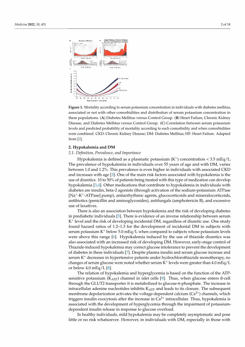
A higher risk of atrial fibrillation, respiratory muscle impairment, Q-T interval increase,torsade des pointes, and ventricular fibrillation, and, ultimately, higher morbidity andmortality in diabetic individuals with HF and CKD are clinical conditions associated withhypokalemia [2]. Therefore, there is an association between baseline serum K+ levels andmortality in these patients (Figure 1). All three conditions (CKD, HF, and DM) can beassociated with higher mortality rates over an 18-month follow-up when compared withcontrol individuals. All-cause mortality was also higher at the extremes, both in K+ valuesbelow 4.0 mEq/L and in values above 6.0 mEq/L [2].
Medicina 2022, 58, 431. https://doi.org/10.3390/medicina58030431 https://www.mdpi.com/journal/medicina
Medicina 2022, 58, 431 2 of 18
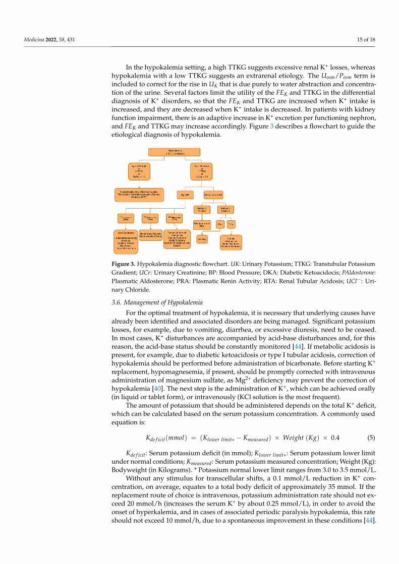
Figure 1. Mortality according to serum potassium concentration in individuals with diabetes mellitus,associated or not with other comorbidities and distribution of serum potassium concentration inthese populations. (A) Diabetes Mellitus versus Control Group. (B) Heart Failure, Chronic KidneyDisease, and Diabetes Mellitus versus Control Group. (C) Correlation between serum potassiumlevels and predicted probability of mortality according to each comorbidity and when comorbiditieswere combined. CKD: Chronic Kidney Disease; DM: Diabetes Mellitus; HF: Heart Failure. Adaptedfrom [2].
2. Hypokalemia and DM2.1. Definition, Prevalence, and Importance
Hypokalemia is defined as a plasmatic potassium (K+) concentration < 3.5 mEq/L.The prevalence of hypokalemia in individuals over 55 years of age and with DM, variesbetween 1.0 and 1.2%. This prevalence is even higher in individuals with associated CKDand increases with age [3]. One of the main risk factors associated with hypokalemia is theuse of diuretics: 10 to 50% of patients being treated with this type of medication can develophypokalemia [3,4]. Other medications that contribute to hypokalemia in individuals withdiabetes are insulin, beta-2 agonists (through activation of the sodium-potassium ATPase[Na+-K+-ATPase] pump), antiarrhythmic agents, glucocorticoids and mineralocorticoids,antibiotics (penicillin and aminoglycosides), antifungals (amphotericin B), and excessiveuse of laxatives.
There is also an association between hypokalemia and the risk of developing diabetesin prediabetic individuals [5]. There is evidence of an inverse relationship between serumK+ level and the risk of developing incidental DM, regardless of diuretic use. One studyfound hazard ratios of 1.2–1.3 for the development of incidental DM in subjects withserum potassium K+ below 5.0 mEq/L when compared to subjects whose potassium levelswere above this range [6]. Hypokalemia induced by the use of thiazide diuretics wasalso associated with an increased risk of developing DM. However, early-stage control ofThiazide-induced hypokalemia may correct glucose intolerance to prevent the developmentof diabetes in these individuals [7]. Despite plasma insulin and serum glucose increase andserum K+ decreases in hypertensive patients under hydrochlorothiazide monotherapy, nochanges of serum glucose were noted whether serum K+ levels were greater than 4.0 mEq/Lor below 4.0 mEq/L [8].
The relation of hypokalemia and hyperglycemia is based on the function of the ATP-sensitive potassium (KATP) channel in islet cells [9]. Thus, when glucose enters ß-cellthrough the GLUT2 transporter it is metabolized to glucose-6-phosphate. The increase inintracellular adenine nucleotides inhibits KATP and leads to its closure. The subsequentmembrane depolarization activates the voltage-dependent calcium (Ca2+) channels, whichtriggers insulin exocytosis after the increase in Ca2+ intracellular. Thus, hypokalemia isassociated with the development of hyperglycemia through the impairment of potassium-dependent insulin release in response to glucose overload.
In healthy individuals, mild hypokalemia may be completely asymptomatic and poselittle or no risk whatsoever. However, in individuals with DM, especially in those with
Medicina 2022, 58, 431 3 of 18
cardiovascular comorbidities, mild to moderate hypokalemia may pose a high risk ofmorbidity and mortality.
2.2. Main Causes of Hypokalemia in Individuals with DM
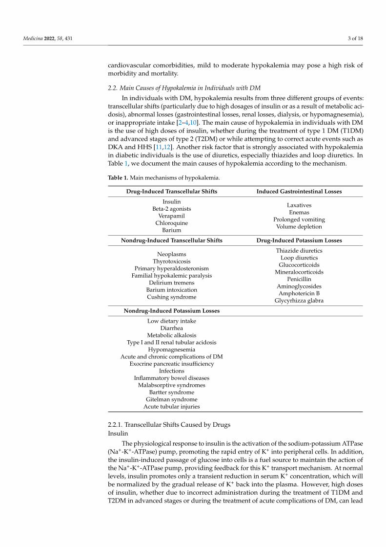
In individuals with DM, hypokalemia results from three different groups of events:transcellular shifts (particularly due to high dosages of insulin or as a result of metabolic aci-dosis), abnormal losses (gastrointestinal losses, renal losses, dialysis, or hypomagnesemia),or inappropriate intake [2–4,10]. The main cause of hypokalemia in individuals with DMis the use of high doses of insulin, whether during the treatment of type 1 DM (T1DM)and advanced stages of type 2 (T2DM) or while attempting to correct acute events such asDKA and HHS [11,12]. Another risk factor that is strongly associated with hypokalemiain diabetic individuals is the use of diuretics, especially thiazides and loop diuretics. InTable 1, we document the main causes of hypokalemia according to the mechanism.
Table 1. Main mechanisms of hypokalemia.
Drug-Induced Transcellular Shifts Induced Gastrointestinal Losses
InsulinBeta-2 agonists
VerapamilChloroquine
Barium
LaxativesEnemas
Prolonged vomitingVolume depletion
Nondrug-Induced Transcellular Shifts Drug-Induced Potassium Losses
NeoplasmsThyrotoxicosis
Primary hyperaldosteronismFamilial hypokalemic paralysis
Delirium tremensBarium intoxicationCushing syndrome
Thiazide diureticsLoop diuretics
GlucocorticoidsMineralocorticoids
PenicillinAminoglycosidesAmphotericin B
Glycyrhizza glabra
Nondrug-Induced Potassium Losses
Low dietary intakeDiarrhea
Metabolic alkalosisType I and II renal tubular acidosis
HypomagnesemiaAcute and chronic complications of DM
Exocrine pancreatic insufficiencyInfections
Inflammatory bowel diseasesMalabsorptive syndromes
Bartter syndromeGitelman syndrome
Acute tubular injuries
2.2.1. Transcellular Shifts Caused by DrugsInsulin
The physiological response to insulin is the activation of the sodium-potassium ATPase(Na+-K+-ATPase) pump, promoting the rapid entry of K+ into peripheral cells. In addition,the insulin-induced passage of glucose into cells is a fuel source to maintain the action ofthe Na+-K+-ATPase pump, providing feedback for this K+ transport mechanism. At normallevels, insulin promotes only a transient reduction in serum K+ concentration, which willbe normalized by the gradual release of K+ back into the plasma. However, high dosesof insulin, whether due to incorrect administration during the treatment of T1DM andT2DM in advanced stages or during the treatment of acute complications of DM, can lead
Medicina 2022, 58, 431 4 of 18
to hypokalemia [13], which is the most common cause of low serum K+ concentrations inindividuals with DM [4].
Beta-2 Agonists
Sympathomimetic Beta-2 agents are another important group of drugs that promotethe activation of the Na+-K+-ATPase pump. The main examples of Beta-2 agonists that caninduce hypokalemia are drugs used to treat asthma, such as antispasmodic agents and bron-chodilators (albuterol, terbutaline, ephedrine, metaproterenol, isoproterenol, fenoterol, pir-buterol), decongestants (pseudoephedrine), tocolytics (ritodrine and nylidrin), dopamine,and HF treatment (especially dobutamine). Hypokalemia induced by these agents canpersist for several hours and can reach levels as low as 2.5 mEq/L depending on dosageand route of administration. Although theophylline is not classified as an α–adrenergicagonist, it is an antispasmodic agent that also stimulates Na+-K+-ATPase activity. Toxiclevels of this agent can rapidly induce extreme hypokalemia [4].
Other Agents
Therapeutic doses of antiarrhythmic agents, such as verapamil, do not present anincreased risk for the development of hypokalemia in diabetic individuals. However,intoxication with high doses of verapamil can induce severe hypokalemia. High doses ofchloroquine and barium can also induce hypokalemia by inhibiting the release of K+ bycells [4].
2.2.2. Abnormal Potassium Losses Caused by DrugsDiuretics
One of the most common causes of hypokalemia in individuals with DM is the use ofdiuretics. Through different pathways, both loop diuretics and thiazides increase the Na+
supply to the collecting duct. The increase in Na+ concentration in this segment inducesits reabsorption, creating an electrochemical gradient that favors eliminating potassium.The degree of hypokalemia depends both on the dosage of diuretic used and on theNa+ concentration in the distal segments of the nephron. Moreover, the combinationof more than one class of diuretics, such as a loop with a thiazide or a thiazide analog,potentiates K+ secretion, which may facilitate the onset of hypokalemia. Drug-inducedhypokalemia may be associated with both metabolic acidosis and alkalosis, either byretaining bicarbonate (HCO3
−) or by inhibiting the antiporters Na+ and hydrogen (H+),induced, e.g., by acetazolamide.
Loop diuretics act by inhibiting the sodium-potassium-chloride (Na+-K+-2Cl−) co-transporter located in the thick ascending limb of Henle’s loop [14]. These drugs alsoinhibit the reabsorption of magnesium (Mg2+) and Ca2+, by compromising the potentialdifference between the tubular lumen and the interstitium (the main driving force for thereabsorption of these ions in this segment). The increase in urinary K+ excretion after loopdiuretic use is due to various mechanisms: (1) The increase in Na+ supply, especially tothe collecting duct, intensifies the excretion of K+ and H+ (Na+ reabsorption by the maincells creates an electrochemical gradient that enables the secretion of K+ into the tubularlumen, through the ROMK [renal outer medullary potassium] channels); (2) Activation ofthe renin-angiotensin-aldosterone system (RAAS) due to volume depletion and a decreasein sodium chloride (NaCl) transport at the dense macula; (3) Release of vasopressin, inresponse to Na+ and volume depletion. An increase in the urinary excretion of H+ and K+
can lead to hypochloremic alkalosis states and hypokalemia, especially if an inadequateintake of K+ is associated.
Thiazide diuretics inhibit Na+-Cl− cotransporter (ENCC1 or TSC) located in the apicalmembrane of cells in the convoluted distal tubule [14]. The expression of this protein isclosely regulated by aldosterone. The proximal tubule may be a secondary target for theaction of these drugs. Inhibitors of the Na+-Cl− symport increase intratubular K+ andH+ by the same mechanisms as loop diuretics. In the case of the diuretics (loop diuretics
Medicina 2022, 58, 431 5 of 18
and thiazide diuretics), the increased amount of intra-luminal Na+ delivered to the distalsegments of the nephrons enters the principal cells via the epithelial Na+ channel (ENaC)channel. This creates a negative electrical charge inside the lumen. Activation of Na+-K+-ATPase causes it to pump Na+ into the blood and exchange for K+. Drawn by thenegative electrical charge in the lumen, K+ will leave the cell to enter the lumen via theROMK channel, leading to hypokalemia. Inside the intercalated cell of the collecting ducts,the stimulation of the H+-K+-ATPase pump, which along with the negative charge inthe collecting duct lumen, causes H+ to exit intercalated cells into the lumen. Therefore,alkalosis metabolic develops (serum HCO3
− concentration, 28–36 mmol/L) and leads tohypokalemia as well [4]. Thiazide diuretics can reduce glucose tolerance and exacerbatelatent DM [15]. The mechanism of impaired glucose tolerance apparently involves changesin glucose metabolism and impaired insulin secretion. Thiazide-induced DM, however,does not appear to offer the same cardiovascular risk as incident DM [16].
Administration of K+ with the thiazide diuretic can prevent hyperglycemic events. Inhypertensive patients, Thiazide-induced hypokalemia may compromise treatment efficacy.Thiazide diuretics can also affect the lipid profile of diabetic patients, raising plasmalevels of total cholesterol, low-density lipoprotein, and triglycerides [14]. In hypertensiveindividuals, chlorthalidone was associated with an increased risk of hypokalemia (hazardratio [HR], 2.7), CKD (HR, 1.24), acute kidney failure (HR, 1.37), and DM [HR, 1.24]without offering a reduction in the risk of cardiovascular events, when compared withhydrochlorothiazide [17].
Glucocorticoids and Mineralocorticoids
Glucocorticoids, such as hydrocortisone, prednisone, and prednisolone do not di-rectly interfere with K+ excretion in the kidneys. Importantly, glucocorticoids are usuallygiven at doses that produce minimal mineralocorticoid stimulation (comparing to cortisol,prednisone and prednisolone have 0.8 mineralocorticoid potency, whereas hydrocorti-sone mineralocorticoid potency is equal to cortisol, fludrocortisone has 125–150 potencymineralocorticoid when compared to cortisol, and dexamethasone does not have mineralo-corticoid potency) to avoid the side effects associated with activation of the aldosteronepathway, which leads to hypokalemia, volume expansion, and hypertension [18].
Mineralocorticoids, such as fludrocortisone, can induce K+ depletion in the distalnephron, through their action on mineralocorticoid receptors, located in the apical mem-brane of tubular cells, which stimulate the expression and activity of Na+-K+-ATPasepump, ENaC, and ROMK channels, inducing Na+ reabsorption and K+ secretion [4]. Othersubstances with glucocorticoid action, especially licorice derivatives (Glycyrrhiza glabra),such as carbenoxolone or cottonseed derivatives (gossypol), can also induce hypokalemiadue to their inhibitory effects on 11-hydroxysteroid dehydrogenase [19].
Antibiotics
Beta-lactam antibiotics, such as penicillin, can lead to renal K+ losses, when adminis-tered intravascularly and in high doses, by increasing the Na+ supply to the distal segmentsof the nephron. Aminoglycosides and amphotericin B can cause hypokalemia due todisturbances in electrolyte homeostasis. Treatment with amphotericin B can lead to hy-pokalemia and hypomagnesemia in up to 90% of cases, depending on the dose. Morethan one mechanism has been related to amphotericin B-induced electrolyte disturbancessuch as the induction of pore formation in the membrane of renal tubular cells, changesin the H+-K+-ATPase pump in the distal tubule, leading to renal tubular acidosis (RTA)type I, and increased absorption of Na+ in the gastrointestinal tract, with resultant excre-tion of K+ in feces [20]. Aminoglycoside-induced hypokalemia may be associated withhypomagnesemia [21]. Due to their positive charge, aminoglycosides can bind to polyvalent cation receptors in the distal tubule, inhibiting Mg2+ reabsorption. Another suggestedmechanism for renal K+ loss is the stimulation of sodium and chloride channels, leading tohypokalemic metabolic alkalosis [21].
Medicina 2022, 58, 431 6 of 18
Oral Anti-Diabetics and Potassium
In overweight individuals with T2DM, linagliptin (a type 4 dipeptidyl-peptidaseinhibitor) increased the renal excretion of Na+ and K+ when compared to sulfonylureaglimepiride [22]. The use of glycosuric agents (sodium-glucose cotransporter-2 inhibitors),especially empagliflozin and dapagliflozin, is increasingly common in diabetic patientswith CKD and HF. These drugs promote an excellent improvement in volume through theincrease in urinary output and natriuresis, although they have not shown significant effectson K+ wastage [23–25].
2.2.3. Induced Gastrointestinal Losses
Despite being often disregarded when investigating hypokalemia, gastrointestinallosses due to excessive use of laxatives or enemas can also lead to K+ wastage, and shouldalways be considered during an investigation, especially in individuals with weight lossand dehydration. Prolonged vomiting leads to K+ losses, as K+ is found on gastric secretions(10 mEq/L), and when volume depletion occurs, RAAS is activated and promotes ultimatelyK+ renal losses [10].
2.2.4. Nondrug-Induced Transcellular Shifts
Acute anabolic states, in which a strong stimulus from growth factors, colony-stimulatingfactors, and other mediators, promotes cell proliferation, and potassium shifts from theextracellular environment to the interior of cells where formation can occur. Some condi-tions that can induce hypokalemia in this context are high-grade lymphomas, treatmentfor anemia due to B12 deficiency, and acute leukemias [4]. Other conditions that can in-duce severe hypokalemia (<3.0 mmol/L) are: Graves’ disease thyrotoxicosis (hypokalemiaoccurs due to a rapid and massive shift of K+ from the extracellular to the intracellularcompartment, as thyroid hormone stimulates Na+-K+ ATPase transcription, and enhancesthe activity and membrane of this pump in skeletal muscle cells, which can be associatedwith signs and symptoms of muscle weakness); primary hyperaldosteronism (due to excessmineralocorticoids); familial hypokalemic paralysis (an autosomal dominant mutation inthe gene encoding the dihydropyridine receptor); delirium tremens (related to the highserum concentration of catecholamines), and barium intoxication (due to blockage in theoutput of K+ from the cells) [4].
2.2.5. Nondrug-Induced Potassium Losses
Inadequate K+ intake is a very rare cause of hypokalemia when not associated withanother condition. For reductions in serum potassium concentration, dietary intake shouldbe below 1 g per day (25 mmol). Considering the high concentration of potassium in theintracellular environment, even in severe starvation scenarios, tissue lysis, especially in themusculature, releases potassium into the plasma, attenuating the potassium depletion [4].Diarrheal scenarios, in which there is a massive loss of water and electrolytes through thegastrointestinal tract, can result in symptomatic hypokalemia. In diabetic individuals, anycondition that increases the predisposition to diarrhea can lead to potassium losses, suchas acute and chronic complications of DM, exocrine pancreatic insufficiency, infections,neoplasms, inflammatory bowel diseases, malabsorption syndromes, and others [4,10].Another common cause of metabolic alkalosis in individuals with DM is excessive Na+
reabsorption in the distal nephron, associated with K+ spoliation, which may be secondaryto mineralocorticoid activity or abnormalities in renal transport. The prevalence of primaryaldosteronism in individuals with new-onset T2DM and hypertension is at least 20% [26].Among individuals with primary hyperaldosteronism, there is evidence that the prevalenceof DM is close to 21%, higher than an estimated 12% prevalence of DM in the generalpopulation [27]. In type I renal tubular acidosis, due to inadequate H+ secretion in thedistal tubule, hypokalemia can occur and is usually corrected with the administration ofsodium bicarbonate. In cases of type II acidosis, due to inadequate reabsorption of HCO3
−
in the proximal tubule, hypokalemia is uncommon, but it can be aggravated with the
Medicina 2022, 58, 431 7 of 18
administration of HCO3− [28]. Hypomagnesemia can also lead to hypokalemia or hinder
its treatment, by increasing renal K+ excretion [29]. As stated earlier, drugs that inducehypomagnesemia, such as amphotericin B and loop or thiazide diuretics, or conditionssuch as hyperaldosteronism and diarrhea can lead to hypokalemia, both directly and due tomagnesium loss. Other causes of hypomagnesemia include alcoholism, intrinsic renal tubu-lar transport disorders, such as Bartter and Gitelman syndromes, and tubular injuries fromother nephrotoxic drugs, in particular aminoglycosides and cisplatin. Hypomagnesemia isvery common in hypokalemic individuals with T2DM [30,31]. Metabolic alkalosis is oftenassociated with the development of hypokalemia (hypokalemic metabolic alkalosis). Oneof the main causes of metabolic alkalosis is vomiting, a situation in which hypokalemia issecondary to chloride depletion and reduced renal K+ reabsorption. In this scenario, chlo-ride replacement resolves both alkalosis and hypokalemia. Thus, K+ depletion stimulatesH+ secretion in the collecting duct, through H+-K+-ATPase and HCO3
− reabsorption inthe ascending limb of the loop of Henle. Potassium depletion also downregulates bothNa+-K+-2Cl− and Na+-Cl− cotransporters, increasing Na+ delivery to the collecting ductand further stimulating the collecting duct H+ secretion. When K+ deficiency is severe, thiseffect results in measurable Cl− excretion despite Cl− depletion and sustained metabolicalkalosis even when sodium chloride is administered. Additionally, K+ depletion stimu-lates ammonium production, facilitating the acid excretion needed to sustain metabolicalkalosis [31,32]. From a cellular point of view, the mechanism by which Mg2+ deficiencyleads to refractoriness of potassium correction is [32]:
(i) In the late distal tubular and cortical collecting duct cells, Na+-K+-ATPase in thebasolateral membrane pumps K+ into the cells and then K+ is secreted into the lumen viaapical K+ channels (ROMK and maxi-K/BK [Big potassium]/Slo1/KCa1.1 channels). Tonote, ROMK is an inward-rectifying K+ channel responsible for basal (not stimulated byflow) K+ secretion, so that inward rectification indicates that K+ enters the cells faster thanK+ is pumped out the cells. The Maxi-K channel is a large-conductance calcium-activatedK+ channel.
(ii) Na+ reabsorption via ENaC depolarizes the apical membrane potential, providingthe driving force for K+ secretion, as the lumen presents a negative charge. Aldosteroneincreases Na+ reabsorption via ENaC to stimulate K+ secretion through the ROMK channeland Na+-K+ ATPase, whereas Maxi-K channels are responsible for flow-stimulated K+
secretion. Mg2+ binds and blocks the pore of the ROMK channel, which leads to its inwardrectification, thereby limiting K+ efflux. Potassium influx can displace intracellular Mg2+
from the pore and release the block. The concentration of intracellular Mg2+ required forinhibiting ROMK relies on the membrane voltage and extracellular concentration of K+. Atzero intracellular Mg2+, K+ ions move in or out of the cell through the ROMK channel freely.At intra- (140 mM) and extracellular (5 mM) K+ concentrations, the chemical gradientsdrive K+ outwards. When the membrane potential is negative inside the cells, it drivesK+ inward.
(iii) Inward and outward movement of K+ ions reach an equilibrium at −86 mVequilibrium potential [Ek] = −60 × log 140/5). When the membrane potential is morenegative than Ek (−100 mV), K+ ions move into the cells. On the other hand, at a membranepotential higher than Ek (−50 mV), K+ moves out of the cells. At the physiologic Mg2+
intracellular concentration (1.0 mM), ROMK inward rectifies K+, as K+ efflux is blockedwhen intracellular Mg2+ binds to ROMK. The influx of K+ displaces intracellular Mg2+,allowing maximal K+ entry. These ROMK properties mediate K+ secretion in the distalnephron, which is regulated by intracellular Mg2+. Notably, when inward conductance isgreater than outward, K+ influx does not occur, as membrane potential is more positivethan Ek.
Therefore, at physiological conditions, ROMK is inhibited by the intracellular con-centration of Mg2+ which ranges from 0.1 to 10.0 mM, with the median at approximately1.0 mM [26]. Thus, intracellular Mg2+ is a critical determinant of ROMK-mediated K+ secre-tion in the distal nephron. Changes in intracellular Mg2+ concentration could significantly

Medicina 2022, 58, 431 8 of 18
affect K+ secretion. Acute complications of DM can also evolve with hypokalemia, eitherduring crises or during insulin treatment.
2.2.6. Genetic Causes of Hypokalemia
Hypokalemia may also be found in a broad set of genetic disorders. Briefly, from apathophysiologic perspective, hypokalemia may be divided into disorders with a low urineK+ excretion or disorders with a high urine K+ excretion, as reviewed elsewhere [33].
In the genetic disorders associated with low urine K+ excretion rate, three mechanismsare involved:
(a) increased K+ shift, including familial hypokalemia periodic paralysis or FPP (anautosomal dominant disorder caused by mutations on ion channel genes encodingthe dihydropyridine-sensitive voltage-gated Ca2+ channel α1-subunit (CACNA1S)[FPP type I] and tetrodotoxin-sensitive voltage-gated Na+ channel α-subunit [SCN4A][FPP type II] of skeletal muscle) and Andersen-Tawil syndrome (associated withmutations in the gene (KCNJ2) encoding a pore-forming subunit of the inward rectifierK+ channel protein, Kir2.1, which is expressed in heart and skeletal muscles);
(b) defects in the intestinal tract (characterized by a mutation in the downregulated inadenoma (DRA) gene encoding a Cl−-OH− (HCO3
−) exchanger expressed in the api-cal membranes of the colon and ileum, which leads to watery diarrhea, hypochloremicmetabolic acidosis, and hypokalemia);
(c) defects in exocrine glands (cystic fibrosis is associated with defective chloride reab-sorption by the dysfunctional CFTR [cystic fibrosis transmembrane regulator] in thesweat ducts which is responsible for excessive Cl− and Na+ loss in sweat, leading toECF volume depletion and, ultimately, to secondary hyperaldosteronism). To note, infamilial hypokalemia periodic paralysis, attacks can be induced not only by rest afterexercise, carbohydrate-rich meals, or exposure to cold but also by the administrationof glucose or insulin or glucocorticoid, which can put diabetic patients at greater risk.
In the genetic disorders associated with high urine K+ excretion rate, two mechanismsare involved in:
(a) increased urine flow rate to cortical collecting ducts;(b) increased K+ concentration in the cortical collecting ducts.
An increased urine flow rate to cortical collecting ducts is caused by increased excretionof electrolytes due to diuretic use or when a tubular defect is found or the increase in osmoleexcretion is due to non-electrolytes, such as mannitol, glucose, or urea. Therefore, a diabeticpatient with higher levels of plasma glucose may present hypokalemia which is secondaryto an increase in urine flow rate to cortical collecting ducts due to glycosuria.
When increased K+ concentration in the cortical collecting ducts is observed, we mayspeculate that a disorder characterized by fast Na+ reabsorption in the cortical collectingducts is present. The most common disorders found in this setting are genetic hypokalemiaassociated with mineralocorticoid excess state, including glucocorticoid-remediable al-dosteronism (a disorder caused by mutations in 11β-hydroxylase (CYP11B1) gene whichencodes the aldosterone synthase gene for aldosterone biosynthesis in the adrenal zonaglomerulosa and is regulated by angiotensin II and aldosterone synthase (CYP11B2) whichencodes 11β-hydroxylase gene for cortisol biosynthesis in the adrenal zona fasciculate andis regulated by adrenocorticotrophic hormone [ACTH]), congenital adrenal hyperplasiadue to 11β-hydroxylase or 17α-hydroxylase deficiencies [11β-OHD and 17α-OHD], Lid-dle’s syndrome (mutations in both β and γ subunits of ENaC associated with a deletionor alteration in their cytoplasmic C termini, which leads to a lack of its internalizationvia clathrin-coated pits pathway or its degraded via Nedd4 pathway, and therefore ENaCremains in an activated form on the cell surface), and apparent mineralocorticoid excess (arare disorder that is caused by mutations in the gene (HSD11B2) encoding renal-specific11β-hydroxysteroid dehydrogenase type 2 (11β-HSD2), which is responsible for convertingcortisol to cortisone in the principal cells of distal tubules and crucial for protecting the
Medicina 2022, 58, 431 9 of 18
mineralocorticoid receptor from being occupied by cortisol). In all these disorders, the ex-cess of mineralocorticoid, either by augmented aldosterone secretion or production of othersteroids, upregulates ENaC activity and leads to sodium reabsorption and hypertensionassociated with hypokalemia.
The second group of disorders associated with increased K+ concentration in thecortical collecting ducts includes a mechanism in which Cl− reabsorption in this segmentis diminished, which includes disorders such as hypochloremic metabolic alkalosis, inparticular Bartter´s syndrome (an autosomal recessive renal tubular disorder characterizedby defective reabsorption of NaCl in the Henle´s loop, including five subtypes of mutationsin genes encoding the Na+-K+-2Cl− cotransporter [NKCC2], K+ channel [ROMK], kidney-specific Cl− channel [CLCNKB], barttin [BSND], and calcium-sensing receptors [CaSR]) andGitelman´s syndrome (secondary to defective reabsorption of NaCl in the distal convolutedtubule due to mutations in the SLC12A3 gene, which encodes the thiazide-sensitive Na+-Cl− cotransporter [NCC] on the apical membrane of that tubule). In both syndromes,hypokalemia is associated with renal K+ and Na+ wasting, with low or normal bloodpressure and secondary hyperreninemia and hyperaldosteronism. Whereas in Bartter´ssyndrome, a high urine Ca2+ and Mg2+ excretion are found, in Gitelman´s syndrome a lowurine Ca2+ and high Mg2+ excretion are invariably found.
Importantly, Gitelman´s syndrome patients may be at greater risk of developinginsulin resistance and type 2 DM [34], as chronic hypokalemia and hypomagnesemiaimpair insulin secretion and sensitivity, whereas hyperaldosteronism increases insulinresistance. From a molecular and cellular point of view, when glucose enters the pancreaticß cell via GLUT2 transporter, it is metabolized to glucose-6-phosphate leading to changesin the intracellular concentration of adenine nucleotides that inhibit the KATP channel andcause its closure. When it occurs, the membrane depolarization activates subsequently thevoltage-dependent calcium channels, leading to an increase in intracellular calcium, whichtriggers insulin exocytosis. Mutations in the ATP-Sensitive Potassium-Channel SubunitKir6.2 lead to permanent neonatal DM [9].
In addition, hypomagnesemia found in Gitelman´s syndrome patients is implicatedin a reduced tyrosine kinase activity at the insulin receptor level, and dysregulates K+ -ATPand L-type Ca2+ channels in the ß cells, which impairs insulin activity and secretion. Hy-peraldosteronism can increase reactive oxygen species, accelerate endothelial remodeling,which can reduce the delivery of insulin for glucose metabolism, and promote insulin resis-tance by reducing insulin receptor substrate-1 expression, and by blocking the downstreamprotein kinase B signaling in the vascular smooth muscles [35].
The second cause of increased K+ concentration in the cortical collecting ducts com-prises hyperchloremic metabolic acidosis, in particular, inherited isolated proximal RTA(renal tubular acidosis associated with defects in the electroneutral Na+-H+ exchanger 3[NHE3] in the luminal membrane or the electrogenic Na+-HCO3
− cotransporter [NBC1]in the basolateral membrane, which is associated with a reduction in the reabsorption offiltered HCO3
−), inherited distal RTA (mutation in the genes encoding two H+-ATPasesubunits specific to intercalated cells, including ATP6V1B1 and ATP6V0A, or mutations inH+-K+-ATPase and AE1 Cl−-HCO3
− exchanger [SLC4A1], leading to an impaired abilityto excrete H+ in distal tubules), and inherited proximal and distal RTA (mutation in thegene encoding carbonic anhydrase II which generates H+ in distal convoluted tubules) [33].
In proximal RTA, the initial insult is associated with an excess of HCO3− excretion
and the urine pH is higher than 6.5. However, over time, HCO3− serum levels drop
and an impaired absorption is again sufficient to acidify urine to lower than 5.5. Thus,proximal HCO3
− excess loss in the urine causes an increase in urine flow rate to the distalnephron, leading to potassium wasting and activation of RAAS from mild hypovolemiaand, therefore, to hypokalemia. Other features associated with RTA include the augment inglucose, uric acid, phosphate, and amino acids in urine (Fanconi syndrome) [33].
In distal RTA, the decreased H+ in tubule lumen draws out K+ causing hypokalemia.In addition, these patients present calcium phosphate kidney stones caused by decreased
Medicina 2022, 58, 431 10 of 18
citrate excretion and hypercalciuria, as well as by the fact that salts are more likely toprecipitate at higher urine pH [33].
2.3. DM-Related Acute Complications: Diabetic Ketoacidosis (DKA), HyperglycemicHyperosmolar State (HHS), and Euglycemic Diabetic Ketoacidosis (EDKA)
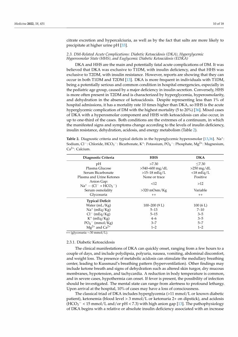
DKA and HHS are the main and potentially fatal acute complications of DM. It wasbelieved that DKA was exclusive to T1DM, with insulin deficiency, and that HHS wasexclusive to T2DM, with insulin resistance. However, reports are showing that they canoccur in both T1DM and T2DM [13]. DKA is more frequent in individuals with T1DM,being a potentially serious and common condition in hospital emergencies, especially inthe pediatric age group, caused by a major deficiency in insulin secretion. Conversely, HHSis more often present in T2DM and is characterized by hyperglycemia, hyperosmolarity,and dehydration in the absence of ketoacidosis. Despite representing less than 1% ofhospital admissions, it has a mortality rate 10 times higher than DKA, so HHS is the acutehyperglycemic complication of DM with the highest mortality (5 to 20%) [36]. Mixed casesof DKA with a hyperosmolar component and HHS with ketoacidosis can also occur, inup to one-third of the cases. Both conditions are the extremes of a continuum, in whichthe manifested signs and symptoms change according to the levels of insulin deficiency,insulin resistance, dehydration, acidosis, and energy metabolism (Table 2).
Table 2. Diagnostic criteria and typical deficits in the hyperglycemic hyperosmolar [13,36]. Na+:Sodium, Cl−: Chloride, HCO3
−: Bicarbonate, K+: Potassium, PO4−: Phosphate, Mg2+: Magnesium,
Ca2+: Calcium.
Diagnostic Criteria HHS DKA
pH >7.30 ≤7.30Plasma Glucose >540–600 mg/dL >250 mg/dL
Serum Bicarbonate >15–18 mEq/L <18 mEq/LPlasma and Urine Ketones None or trace Positive
Anion Gap:Na+ − (Cl− + HCO3
−) <12 >12
Serum osmolality >320 mOsm/Kg VariableGlycosuria ++ ++
Typical DeficitWater (mL/Kg) 100–200 (9 L) 100 (6 L)Na+ (mEq/Kg) 5–13 7–10Cl− (mEq/Kg) 5–15 3–5K+ (mEq/Kg) 4–6 3–5
PO4− (mmol/Kg) 3–7 5–7
Mg2+ and Ca2+ 1–2 1–2++ (glycosuria ∼30 mmol/L).
2.3.1. Diabetic Ketoacidosis
The clinical manifestations of DKA can quickly onset, ranging from a few hours to acouple of days, and include polydipsia, polyuria, nausea, vomiting, abdominal discomfort,and weight loss. The presence of metabolic acidosis can stimulate the medullary breathingcenter, leading to Kussmaul’s breathing pattern (hyperventilation). Other findings mayinclude ketone breath and signs of dehydration such as altered skin turgor, dry mucousmembranes, hypotension, and tachycardia. A reduction in body temperature is common,and in severe cases, hypothermia can onset. If fever is present, the possibility of infectionshould be investigated. The mental state can range from alertness to profound lethargy.Upon arrival at the hospital, 10% of cases may have a loss of consciousness.
The classical triad of DKA includes hyperglycemia (>11 mmol/L or known diabeticpatient), ketonemia (blood level > 3 mmol/L or ketonuria 2+ on dipstick), and acidosis(HCO3
− < 15 mmol/L and/or pH < 7.3) with high anion gap [13]. The pathophysiologyof DKA begins with a relative or absolute insulin deficiency associated with an increase
Medicina 2022, 58, 431 11 of 18
in counterregulatory hormones, especially glucagon, glucocorticoids, and catecholamines.This state of hormonal imbalance leads to an increase in blood glucose and stimulation ofhepatic gluconeogenesis, glycogenolysis, and insulin resistance, with low tissue glucoseconsumption. Glucocorticoids, especially cortisol, increase the supply of amino acids atthe expense of stimulating protein catabolism. Glucagon and catecholamines, in turn,induce the activation of the enzyme glycogen phosphorylase, increasing glycogenolysis.Furthermore, an increase in the glucagon/insulin ratio inhibits precursors of the glycolyticpathway, decreasing glycolysis. The depletion of electrolytes (especially potassium) andacidosis also interfere with insulin action in target tissues. Glycosuria secondary to hyper-glycemia results in osmotic diuresis, leading to water and electrolytes loss, hypovolemia,dehydration, and reduced GFR, further aggravating the hyperglycemia and creating avicious cycle [13].
Ketogenesis is also a result of this hormonal imbalance state, with insulin deficiencyand excess of counter-regulatory hormones. In this state, hormone-sensitive lipase isactivated in the adipose tissue, resulting in free fatty acid (FFA) debt into the blood. Inthe liver, lipid metabolism is shifted to oxidation of these FFAs, which are transformed inketone bodies. Glucagon, in turn, inhibits glycolysis at the hepatic level, by reducing thesynthesis of malonyl-CoA. This process also deviates the metabolism towards the oxidationof fatty acids in ketone bodies—acetone, acetoacetate, and ß-hydroxybutyrate—and therelease of H+ ions. The entry of K+ in the cells is decreased due to both insulin deficiencyand metabolic acidosis. An increase in extracellular osmolarity, as a result of osmoticdiuresis and electrolyte translocation, is present in both DKA and HHS, and inducesvolume outflow from the intra to extracellular space, with consequent cellular dehydrationand a small dilution in plasma Na+ concentration. Important renal K+ losses occur due toosmotic diuresis, with reduced NaCl reabsorption, and ketonuria. Other factors that canalso aggravate dehydration and electrolyte imbalance are the use of diuretics, vomiting,diarrhea, and reduced water intake.
Despite the total body K+ deficiency ranging between 3–5 mEq/kg in DKA, patientsoften have elevated plasma K+ concentrations, with normokalemia or hypokalemia beingimportant indications of K+ deficiency. During insulin treatment, plasma K+ levels willinvariably fall, possibly leading to hypokalemia [13]. Recent studies have shown that theincidence of hypokalemia in DKA may be lower than 4%, appearing, in most cases, onlyafter insulin administration [37]. In emergency department patients, however, the preva-lence of hypokalemia associated with DKA can be as high as 11% [38]. Rhabdomyolysis isanother possible complication of DKA, occurring more frequently in cases of prolongedacidosis, high serum creatinine, hyperkalemia, and ketonuria. Rhabdomyolysis per se canalso aggravate hyperkalemia, by releasing potassium from the cytoplasm [39].
2.3.2. Hyperglicemic Hyperosmolar State
Usually, HHS has a more insidious onset than DKA, and it may take several daysor even weeks for the first symptoms to appear. The main clinical manifestations arepolyuria, polydipsia, weakness, and blurred vision. Sensory changes are common in HHSand mental status can range from fully alert to coma. A seizure can occur in up to 20%of patients [36]. Signs of dehydration are reduced turgor, dry mucous membranes, coldextremities, hypotension, and reflex tachycardia [27]. Diagnostic criteria of HHS includeplasma glucose 30–33.3 mmol/L (540–600 mg/dL), pH > 7.30, HCO3
− > 15–18 mmol/L,urine acetoacetate negative or low positive, osmolality 320 mmol/kg and presentation withstupor or coma, severe dehydration, and feeling unwell [13,36].
Similar to DKA, in the HHS there is also hyperglycemia secondary to glycogenolysis,increased gluconeogenesis, and decreased entry of glucose into peripheral tissues. In thiscase, however, the insulin deficiency is relative. Insulinopenia is also accompanied by anincrease in counter-regulatory hormones (glucagon, catecholamines, and glucocorticoids).However, in HHS the concentrations of FFAs, cortisol, and glucagon are lower whencompared to those values found in DKA. In this case, it is believed that insulin deficiency

Medicina 2022, 58, 431 12 of 18
is sufficient to compromise the adequate use of glucose by tissues, but insufficient to shiftenergy metabolism towards lipolysis, ketogenesis, and metabolic acidosis [13]. In HHS,osmotic diuresis due to glycosuria is also present, resulting in loss of water and electrolytesand dehydration [39]. However, as this scenario settles in a longer period of time than inDKA, usually the volume deficit is greater. Hypovolemia also leads to dehydration anddecreased GFR, aggravating hyperglycemia. This “snowball effect” leads to higher bloodglucose and osmolality values than those found in DKA.
Analogous to diabetic ketoacidosis, the main acute complication of HHS treatment ishypokalemia secondary to insulin administration [36].
2.3.3. Euglycemic Diabetic Ketoacidosis
EDKA is a situation in which blood glucose remains below 200 mg/dL and is associ-ated with alcohol intoxication, pregnancy, prolonged fasting, and depression in patientswith T1DM, acute pancreatitis, and salicylate poisoning [13]. In recent years, it has reap-peared with the ascension of sodium-glucose cotransporter-2 (SGLT-2) inhibitors for thetreatment of DM and cardiovascular diseases. These drugs prevent the renal reabsorptionof sodium and glucose in the proximal tubule and increase glucosuria in patients with DM,decreasing glycemia. At the same time, these drugs induce an increase in plasma glucagon.This increase is not able to raise blood glucose concentration due to glycosuria. However,high glucagon induces a reduction in plasma insulin levels (reducing the insulin/glucagonratio), promotes lipolysis (20% enhanced lipid oxidation), decreases carbohydrate oxida-tion (falls by 60%), and increases the production of FFAs, substrates for the productionof ketones. Although recommended for patients with T2DM only, gliflozins have seenoff-label use as an adjunct to insulin therapy for T1DM. Recent studies show EDKA as acomplication of SGLT2 inhibitors in both groups of diabetic patients [13].
In addition to the use of SGLT-2 inhibitors, EDKA usually has a triggering event—situations of metabolic stress such as surgery, myocardial infarction, stroke, prolongedfasting, and strenuous exercise. Patients using SGLT-2 inhibitors should avoid alcoholicbeverages, ketogenic diets, maintain a good level of hydration and discontinue medicationbefore any stressors, such as surgery. Other precipitating factors of EDKA comprise areduction in carbohydrate intake, reduction of insulin dose in the context of good glycemiccontrol, cocaine use, and pregnancy [13]. Therefore, K+ should be monitored and cor-rected accordingly.
3. Symptoms, Exams, and Diagnosis of Hypokalemia
As the ion K+ plays a large role in the physiology of various tissues, organs, andsystems, its deficiency can lead to changes in cardiovascular functioning, in skeletal muscles,in the kidneys, and even in the release and effect of certain hormones [10]. The directcorrelation between K+ levels and the appearance of signs and symptoms is not linear,depending on intrinsic factors and clinical status of each individual, highlighting diabeticpatients, in which it may vary according to both K+ levels and the presence of other pre-existing comorbidities. Nevertheless, mild hypokalemia can be often asymptomatic [40].
Although chronic or persistent hypokalemia may be asymptomatic in some individu-als, patients with DM may have this condition worsened by diarrhea or vomiting, whichcan occur during acute complications of DM. Nocturia and polyuria can also be exacerbated,especially in individuals predisposed to persistent hypokalemia, as in Bartter and Gitelmansyndromes. Hypokalemia-induced polyuria is related to an impairment of vasopressinaction in collecting ducts. In addition, insulin treatment can also promote K+ shift intocells. Therefore, hypokalemia can have worse consequences in diabetic patients, whichputs these individuals at greater risk of chronic hypokalemia. In this group of patients,cardiovascular diseases are found more often, making them more vulnerable to cardiacarrhythmias, fluid depletion, and worsening neuropathy from muscle weakness [41,42]. Tonote, KATP channels may not function properly in the DM setting because its expression isreduced in myocardium cells and aortic smooth muscle cells, resulting in impaired heart
Medicina 2022, 58, 431 13 of 18
and vascular function [43]. Consequently, hypokalemia may affect the membrane potentialand pose a decreased response to stress conditions, such as hypoxia and oxidative stress.Importantly, as hypokalemia may lead to hyperglycemia due to the impairment of insulinsecretion and peripheral glucose utilization, a vicious circle is triggered where hypokalemiaworsens glucose control and vice-versa.
3.1. Cardiovascular Effects
The main cardiovascular changes caused by hypokalemia are cardiac arrhythmias [10].Low K+ concentration increases cardiac muscle excitability and delays its repolarization,which can induce both atrial and ventricular arrhythmias [44]. The most commonly ob-served ECG changes are shown in Figure 2, which include T wave flattening, ST-T segmentdepression, an extension of the QT interval [44], presence of U waves, and multiple ven-tricular extrasystoles, which can be seen in up to 20% of patients with severe hypokalemia(>2.6 mmol/L) [_bookmark3138]. Patients at greatest risk for developing life-threateningarrhythmias are the elderly or those with underlying ischemic heart disease. Hyperten-sive patients using hydrochlorothiazide seem to have a higher risk for the incidence ofsudden death [10]. The main serious arrhythmias induced by hypokalemia are ventricularfibrillation, ventricular tachycardia, and torsades des pointes.
Figure 2. Drawing of an ECG showing the main changes during hypokalemia: Extension of QTinterval, T wave flattening with ST-T depression, and U waves.
3.2. Muscular Effects
In contrast to cardiac musculature, hypokalemia can induce hyperpolarization ofskeletal muscle, compromising its ability to depolarize and contract. Additionally, dehy-dration (e.g., during diabetic ketoacidosis) can reduce blood supply to the musculatureand induce rhabdomyolysis. Together, these processes can lead to muscle weakness andfatigue. In severe cases, hypokalemia can cause respiratory muscles weakness and evenlead to respiratory acidosis [44].
3.3. Kidney Effects
The most common renal complication of hypokalemia is metabolic alkalosis, which canoccur through multiple pathways: The low serum K+ concentration promotes H+ secretionthrough the H+-K+-ATPase pump in the collecting ducts. Furthermore, it stimulates theabsorption of HCO3
− in the proximal tubule, NH4+ synthesis, and reduction in urinarycitrate secretion. Another effect of hypokalemia in the kidneys is the impairment of theurinary concentration capacity, apparently through defective activation of the enzymeadenylate cyclase in the tubular cells of the distal nephron, preventing the activity of theantidiuretic hormone. In addition, fluid intake is stimulated due to an increase in the levelof angiotensin II in the central nervous system. This hypokalemic-induced nephrogenicdiabetes insipidus can lead to polyuria, with loss of up to 3 L of water per day. Whenassociated with hyperaldosteronism, hypokalemia can also lead to cystic kidney disease,originating from the collecting duct epithelium [10].
3.4. Hormonal Effects
In diabetic patients, the effects of low K+ concentration on insulin have great impor-tance. Hypokalemia leads to both a reduction in pancreatic insulin release and its activity
Medicina 2022, 58, 431 14 of 18
in target cells. The combination of these effects can worsen hyperglycemia and diabeticcontrol [44], having devastating effects in individuals in DKA or HHS state.
3.5. Diagnosis of Hypokalemia
In the presence of the aforementioned signs and symptoms and after the identificationof serum K+ < 3 mmol/L, it is important to perform a sequential analysis of the possiblecauses and mechanisms behind hypokalemia. The first step is to assess any possiblerenalK+ losses, differentiating them from possible gastrointestinal losses. Some measurementscan be used to identify whether the causes are of renal or extrarenal origin, such as thetranstubular potassium gradient (TTKG), the urinary potassium excretion fraction, orthe potassium value obtained in an isolated urine sample, which can be normalized bycreatinine (K/Cr ratio) [45]. It is important to keep in mind that each of these measurementshas its due limitations, for example, not very sensitive to losses due to mineralocorticoidactivity. Additionally, because they are fixed values, they can be influenced by othervariables, such as volume and electrolyte intake, urinary flow, and GFR. Furthermore,TTKG is more sensitive in detecting inappropriate K+ secretion in hyperkalemia [44].
3.5.1. Fractional Excretion of Potassium (FEK)
FEK is the percent of K+ filtered into the proximal tubule that appears in the urine.For an individual with normal kidney function with an average dietary K+ intake, theFEK is approximately 10%. When hypokalemia is the result of extrarenal causes (low K+
intake, increased K+ shifts into cells, and gastrointestinal loss), the kidney conserves K+ and,consequently, FEK is low. Conversely, hypokalemia secondary to renal losses is associatedwith an increase in FEK. In contrast, in the setting of hyperkalemia, a high FEK suggests anextrarenal etiology, whereas a low FEK is consistent with a renal etiology.
FEK =ClKClCr
=
UKSK
UCrSCr
× 100% (1)
ClK =(V × UK)
SK(2)
ClCr =(V × UK)
SCr(3)
FEK: Fractional excretion of potassium; ClK: Clearence of potassium; ClCr: Clearence ofcreatinine; UK: Urinary potassium; SK: Serum potassium; UCr: Urinary creatinine; SCr:Serum creatinine; V: Urinary volume.
If a urine creatinine measurement is not available, one can often use UK alone, ina random urine specimen, to differentiate between renal and extrarenal causes of hy-pokalemia: UK > 20 mEq/L suggests a renal etiology, whereas UK < 20 mEq/L suggestsextrarenal etiology.
3.5.2. Transtubular Potassium Gradient (TTKG)
The transtubular potassium gradient estimates the potassium gradient between theurine and the blood in the distal nephron. TTKG is a measurement of net K+ secretion bythe distal nephron, after correcting for changes in urine osmolality. In a normal individualunder normal circumstances, the TTKG is about 6 to 12.
TTKG =UK/ UOsm
POsm
PK(4)
UK: Urinary potassium; UOsm: Urinary osmolality; POsm: Plasma osmolality; PK: Plasmaticpotassium; UCr: Urinary Creatinine.
Medicina 2022, 58, 431 15 of 18
In the hypokalemia setting, a high TTKG suggests excessive renal K+ losses, whereashypokalemia with a low TTKG suggests an extrarenal etiology. The Uosm/Posm term isincluded to correct for the rise in UK that is due purely to water abstraction and concentra-tion of the urine. Several factors limit the utility of the FEK and TTKG in the differentialdiagnosis of K+ disorders, so that the FEK and TTKG are increased when K+ intake isincreased, and they are decreased when K+ intake is decreased. In patients with kidneyfunction impairment, there is an adaptive increase in K+ excretion per functioning nephron,and FEK and TTKG may increase accordingly. Figure 3 describes a flowchart to guide theetiological diagnosis of hypokalemia.
Figure 3. Hypokalemia diagnostic flowchart. UK: Urinary Potassium; TTKG: Transtubular PotassiumGradient; UCr: Urinary Creatinine; BP: Blood Pressure; DKA: Diabetic Ketoacidocis; PAldosterone:Plasmatic Aldosterone; PRA: Plasmatic Renin Activity; RTA: Renal Tubular Acidosis; UCl−: Uri-nary Chloride.
3.6. Management of Hypokalemia
For the optimal treatment of hypokalemia, it is necessary that underlying causes havealready been identified and associated disorders are being managed. Significant potassiumlosses, for example, due to vomiting, diarrhea, or excessive diuresis, need to be ceased.In most cases, K+ disturbances are accompanied by acid-base disturbances and, for thisreason, the acid-base status should be constantly monitored [44]. If metabolic acidosis ispresent, for example, due to diabetic ketoacidosis or type I tubular acidosis, correction ofhypokalemia should be performed before administration of bicarbonate. Before starting K+
replacement, hypomagnesemia, if present, should be promptly corrected with intravenousadministration of magnesium sulfate, as Mg2+ deficiency may prevent the correction ofhypokalemia [40]. The next step is the administration of K+, which can be achieved orally(in liquid or tablet form), or intravenously (KCl solution is the most frequent).
The amount of potassium that should be administered depends on the total K+ deficit,which can be calculated based on the serum potassium concentration. A commonly usedequation is:
Kde f icit(mmol) = (Klower limit∗ − Kmeasured) × Weight (Kg) × 0.4 (5)
Kde f icit: Serum potassium deficit (in mmol); Klower limit∗: Serum potassium lower limitunder normal conditions; Kmeasured: Serum potassium measured concentration; Weight (Kg):Bodyweight (in Kilograms). * Potassium normal lower limit ranges from 3.0 to 3.5 mmol/L.
Without any stimulus for transcellular shifts, a 0.1 mmol/L reduction in K+ con-centration, on average, equates to a total body deficit of approximately 35 mmol. If thereplacement route of choice is intravenous, potassium administration rate should not ex-ceed 20 mmol/h (increases the serum K+ by about 0.25 mmol/L), in order to avoid theonset of hyperkalemia, and in cases of associated periodic paralysis hypokalemia, this rateshould not exceed 10 mmol/h, due to a spontaneous improvement in these conditions [44].
Medicina 2022, 58, 431 16 of 18
If faster replacement is required, 20 or 40 mmol/h can be given via a central venouscatheter due to the risk of phlebitis if a peripheral vein is cannulated for this purpose.Importantly, continuous ECG monitoring should be used under these circumstances. InDKA and HHS, serum K+ can be normal or elevated on admission despite total body K+
depletion, which is more severe in HHS compared to DKA (Table 1) [13,36]. Osmotic-induced intracellular dehydration results in K+ efflux from the cells. Since insulin causesa shift of K+ into the cell, via an indirect effect on Na+-K+ ATPase, one should correctthe K+ level to >3.3 mEq/L before starting insulin therapy. In that case, insulin must beheld. If K+ is between 3.3 and 5.3 mEq/L, 20–30 mEq of K+ should be given in each literof intravenous fluid to keep serum K+ between 4 to 5 mEq/L [37]. Potassium should bemonitored if >5.3 mEq/L. Magnesium should be checked and given intravenously whethernecessary, as this approach is important to prevent renal wasting of K+ with exacerbation ofhypokalemia. Routine administration of phosphate is not recommended. However, carefulphosphate replacement can be considered in patients with very low levels (<1 mEq/L) dueto the risk of cardiac dysfunction or respiratory distress [46].
In the DKA setting, major guidelines for K+ replacement emphasize the importance ofblood gas and renal function tests for profiling replacement [47–50]. Initial rehabilitationwith saline solution is recommended until serum K+ levels normalize. Insulin should bewithheld if blood K+ is below 3.3 mmol/L to avoid insulin-induced hypokalemia [46].
There are four main types of potassium-containing preparations: potassium chloride(KCl), potassium bicarbonate, potassium citrate, and potassium phosphate. Potassiumphosphate solution is particularly useful when hypophosphatemia is associated, and citrateor bicarbonate solutions, when acidosis is installed [40]. In most situations, however, thesolution of choice is potassium chloride. An adverse effect of oral KCl tablets (usuallycontaining 8 mmol K+) is the irritation of the gastrointestinal tract mucosa, which can evenlead to ulcerations or bleeding. For this reason, tablet ingestion must be accompanied bya large volume of fluid. The use of potassium-sparing diuretics during K+ replacementtreatment can ease the onset of hyperkalemia, especially in diabetic patients with reducedGFR, using non-steroidal anti-inflammatory drugs, ACEi, or ARBs [44]. An interestingapproach in diabetic patients prone to hypokalemia is to encourage the intake of potassium-rich foods, such as bananas, tomatoes, lentils, nuts, fish meat, etc., always keeping in mindthe glycemic load of each item.
4. Conclusions
DM has a growing prevalence worldwide regardless of age, sex and ethnicity. Patientswith this condition, if not well managed, are more susceptible to developing a series ofother conditions, such as electrolyte disturbances, which can be lethal, especially in patientswith other comorbidities, such as HF and CKD.
In this review, we presented the main reasons why diabetic patients are so vulnerable todeveloping hypokalemia, the mechanisms behind it, and the current methods of treatmentand management of this potentially lethal condition.
Rapid identification of hypokalemia can prevent the occurrence of serious,life-threatening complications, such as cardiac arrhythmias and respiratory muscle impair-ment. Therefore, indications for urgent treatment include severe or symptomatic changesin K+ levels, electrocardiography changes, or the presence of certain comorbid conditions.Collectively, these data indicate that, as with all diagnostic aids, clinical correlation isindicated and K+ intake should be addressed.
Author Contributions: Writing-original draft: L.C.-R., K.G.-N., É.B.R.; Writing—reviewing andediting: L.C.-R., É.B.R.; Funding acquisition: É.B.R. All authors have read and agreed to the publishedversion of the manuscript.
Funding: This work was supported by grants from FAPESP (Fundação de Amparo à Pesquisa doEstado de São Paulo/São Paulo Research Foundation; No. 2021/02216-7) and EFSD (EuropeanFoundation for the Study of Diabetes)/Sanofi to Rangel, É.B.R.
Medicina 2022, 58, 431 17 of 18
Conflicts of Interest: The authors declare no conflict of interest.
References1. Saeedi, P.; Salpea, P.; Karuranga, S.; Petersohn, I.; Malanda, B.; Gregg, E.W.; Unwin, N.; Wild, S.H.; Williams, R. Mortality
attributable to diabetes in 20–79 years old adults, 2019 estimates: Results from the International Diabetes Federation DiabetesAtlas, 9th edition. Diabetes Res. Clin. Pract. 2020, 162, 108086. [CrossRef] [PubMed]
2. Collins, A.J.; Pitt, B.; Reaven, N.; Funk, S.; McGaughey, K.; Wilson, D.; Bushinsky, D.A. Association of serum potassium withall-cause mortality in patients with and without heart failure, chronic kidney disease, and/or diabetes. Am. J. Nephrol. 2017, 46,213–221. [CrossRef] [PubMed]
3. Jiménez-Marrero, S.; Cainzos-Achirica, M.; Monterde, D.; Garcia-Eroles, L.; Enjuanes, C.; Yun, S.; Garay, A.; Moliner, P.; Alcoberro,L.; Corbella, X.; et al. Real-world epidemiology of potassium derangements among chronic cardiovascular, metabolic and renalconditions: A population-based analysis. Clin. Epidemiol. 2020, 12, 941–952. [CrossRef] [PubMed]
4. Gennari, F.J. Hypokalemia. N. Engl. J. Med. 1998, 339, 451–458. [CrossRef] [PubMed]5. Chatterjee, R.; Colangelo, L.A.; Yeh, H.C.; Anderson, C.A.; Daviglus, M.L.; Liu, K.; Brancati, F.L. Potassium intake and risk of
incident type 2 diabetes mellitus: The Coronary Artery Risk Development in Young Adults (CARDIA) Study. Diabetologia 2012,55, 1295–1303. [CrossRef]
6. Chatterjee, R.; Yeh, H.-C.; Shafi, T.; Selvin, E.; Andersen, C.; Pankow, J.S.; Miller, E.; Brancati, F. Serum and Dietary Potassium andRisk of Incident Type 2 Diabetes Mellitus: The Atherosclerosis Risk in Communities (ARIC) Study. Arch. Intern. Med. 2010, 170,1745–1751. [CrossRef]
7. Zillich, A.J.; Garg, J.; Basu, S.; Bakris, G.L.; Carter, B.L. Thiazide diuretics, potassium, and the development of diabetes: Aquantitative review. Hypertension 2006, 48, 219–224. [CrossRef]
8. Smith, S.M.; Anderson, S.D.; Wen, S.; Gong, Y.; Turner, S.T.; Cooper-Dehoff, R.M.; Schwartz, G.L.; Bailey, K.; Chapman, A.; Hall,K.L.; et al. Lack of correlation between thiazide-induced hyperglycemia and hypokalemia: Subgroup analysis of results from thepharmacogenomic evaluation of antihypertensive responses (PEAR) study. Pharmacotherapy 2009, 29, 1157–1165. [CrossRef]
9. Gloyn, A.L.; Pearson, E.; Antcliff, J.F.; Proks, P.; Bruining, G.J.; Slingerland, A.S.; Howard, N.; Srinivasan, S.; Silva, J.M.C.L.;Molnes, J.; et al. Activating Mutations in the Gene Encoding the ATP-Sensitive Potassium-Channel Subunit Kir6.2 and PermanentNeonatal Diabetes. N. Engl. J. Med. 2014, 350, 1838–1849. [CrossRef]
10. Weiner, I.D.; Wingo, C.S. Hypokalemia-Consequences Causes, and correction. J. Am. Soc. Nephrol. 1997, 8, 1179–1188. [CrossRef]11. Viera, A.J.; Wouk, N. Potassium disorders: Hypokalemia and hyperkalemia. Am. Fam. Physician 2015, 92, 487–495. [PubMed]12. Eslam, R.B.; Öztürk, B.; Panzer, S.; Qin, H.; Duca, F.; Binder, C.; Rettl, R.; Dachs, T.M.; Alasti, F.; Vila, G.; et al. Low serum
potassium levels and diabetes—An unfavorable combination in patients with heart failure and preserved ejection fraction. Int. J.Cardiol. 2020, 317, 121–127. [CrossRef] [PubMed]
13. Muneer, M.; Akbar, I. Acute Metabolic Emergencies in Diabetes: DKA, HHS and EDKA. Exp. Med. Biol. 2021, 1307, 85–114.14. Ellison, D.H. Clinical pharmachology in diuretic use. Clin. J. Am. Soc. Nephrol. 2019, 14, 1248–1257. [CrossRef] [PubMed]15. Palmer, B.F. Metabolic complications associated with use of diuretics. Semin. Nephrol. 2011, 31, 542–552. [CrossRef]16. Barzilay, J.I.; Davis, B.R.; Pressel, S.L.; Cutler, J.A.; Einhorn, P.T.; Black, H.R.; Cushman, W.C.; Ford, C.E.; Margolis, K.L.; Moloo, J.;
et al. ALLHAT Collaborative Research Group. Long-term effects of incident diabetes mellitus on cardiovascular outcomes inpeople treated for hypertension: The ALLHAT Diabetes Extension Study. Cardiovasc. Qual. Outcomes 2012, 5, 153–162. [CrossRef]
17. Hripcsak, G.; Suchard, M.A.; Shea, S.; Chen, R.; You, S.C.; Pratt, N.; Madigan, D.; Krumholz, H.M.; Ryan, P.B.; Schuemie, M.J.Comparison of Cardiovascular and Safety Outcomes of Chlorthalidone vs Hydrochlorothiazide to Treat Hypertension. JAMAIntern. Med. 2020, 180, 542–551. [CrossRef]
18. De Lucena, D.D.; Rangel, É.B. Glucocorticoids use in kidney transplant setting. Expert Opin. Drug Metab. Toxicol. 2018, 14,1023–1041. [CrossRef]
19. Nazari, S.; Rameshrad, M.; Hosseinzadeh, H. Toxicological Effects of Glycyrrhiza glabra (Licorice): A Review. Phytother. Res. 2017,31, 1635–1650. [CrossRef]
20. Kim, Y.W. Antimicrobial-induced Electrolyte and Acid-Base Disturbances. Electrolyte Blood Press. 2008, 5, 111–115. [CrossRef]21. Kang, H.S.; Kerstan, D.; Dai, L.; Ritchie, G.; Quamme, G.A. Aminoglycosides inhibit hormone-stimulated Mg2+ uptake in mouse
distal convoluted tubule cells. Can. J. Physiol. Pharmacol. 2000, 78, 595–602. [CrossRef] [PubMed]22. Muskiet, M.H.; Tonneijck, L.; Smits, M.M.; Kramer, M.H.; Ouwens, D.M.; Hartmann, B.; Holst, J.J.; Touw, D.J.; Danser, A.J.; Joles,
J.A.; et al. Effects of DPP-4 Inhibitor Linagliptin Versus Sulfonylurea Glimepiride as Add-on to Metformin on Renal Physiology inOverweight Patients with Type 2 Diabetes (RENALIS): A Randomized, Double-Blind Trial. Diabetes Care 2020, 43, 2889–2893.[CrossRef] [PubMed]
23. Ibrahim, A.; Ghaleb, R.; Mansour, H.; Hanafy, A.; Mahmoud, N.M.; Elsharef, M.A.; Salama, M.K.; Elsaughier, S.M.; Abdel-Wahid, L.; Mohamed, M.E.; et al. Safety and Efficacy of Adding Dapagliflozin to Furosemide in Type 2 Diabetic Patients withDecompensated Heart Failure and Reduced Ejection Fraction. Front. Cardiovasc. Med. 2020, 7, 602251. [CrossRef] [PubMed]
24. Griffin, M.; Rao, V.S.; Ivey-Miranda, J.; Fleming, J.; Mahoney, D.; Maulion, C.; Suda, N.; Siwakoti, K.; Ahmad, T.; Jacoby, D.; et al.Empagliflozin in Heart Failure: Diuretic and Cardiorenal Effects. Circulation 2020, 142, 1028–1039. [CrossRef] [PubMed]
Medicina 2022, 58, 431 18 of 18
25. Cox, Z.L.; Collins, S.P.; Aaron, M.; Hernandez, G.A.; McRae, A.T., III; Davidson, B.T.; Fowler, M.; Lindsell, C.J.; Harrell, F.E., Jr.;Jenkins, C.A.; et al. Efficacy and safety of dapagliflozin in acute heart failure: Rationale and design of the DICTATE-AHF trial.Am. Heart J. 2021, 232, 116–124. [CrossRef]
26. Hu, Y.; Zhang, J.; Liu, W.; Su, X. Determining the Prevalence of Primary Aldosteronism in Patients with New-Onset Type 2Diabetes and Hypertension. J. Clin. Endocrinol. Metab. 2020, 105, dgz293. [CrossRef]
27. Akehi, Y.; Yanase, T.; Motonaga, R.; Umakoshi, H.; Tsuiki, M.; Takeda, Y.; Yoneda, T.; Kurihara, I.; Itoh, H.; Katabami, T.;et al. TJapan Primary Aldosteronism Study Group. High Prevalence of Diabetes in Patients with Primary Aldosteronism(PA) Associated With Subclinical Hypercortisolism and Prediabetes More Prevalent in Bilateral Than Unilateral PA: A Large,Multicenter Cohort Study in Japan. Diabetes Care 2019, 42, 938–945.
28. Sebastian, A.; McSherry, E.; Morris, R.C.J. Renal potassium wasting in renal tubular acidosis (RTA): Its occurrence in types 1 and 2RTA despite sustained correction of systemic acidosis. J. Clin. Investig. 1971, 50, 667–678. [CrossRef]
29. Kobrin, S.M.; Goldfarb, S. Magnesium deficiency. Semin. Nephrol. 1990, 10, 525–535.30. Shardha, A.K.; Vaswani, A.S.; Faraz, A.; Alam, M.T.; Kumar, P. Frequency and risk factors associated with hypomagnesaemia in
hypokalemic type-2 diabetic patients. J. Coll. Physicians Surg. Pak. 2014, 24, 830–835.31. González, W.; Altieri, P.I.; Alvarado, S.; Banchs, H.L.; Escobales, N.; Crespo, M.; Borges, W. Magnesium: The forgotten electrolyte.
Bol. Asoc. Med. Puerto Rico 2013, 105, 17–20.32. Huang, C.-L.; Kio, E. Mechanism of hypokalemia in magnesium deficiency. J. Am. Soc. Nephrol. 2007, 18, 2649–2652. [CrossRef]
[PubMed]33. Lin, S.-H.; Yang, S.-S.; Chau, T. A practical approach to genetic hypokalemia. Electrolyte Blood Press 2010, 8, 38–50. [CrossRef]
[PubMed]34. He, G.; Gang, X.; Sun, Z.; Wang, P.; Wang, G.; Guo, W. Type 2 diabetes mellitus caused by Gitelman syndrome-related hypokalemia
A case report. Medicine 2020, 99, e21123. [CrossRef]35. Hitomi, H.; Kiyomoto, H.; Nishiyama, A.; Hara, T.; Moriwaki, K.; Kaifu, K.; Ihara, G.; Fujita, Y.; Ugawa, T.; Kohno, M. Aldosterone
suppresses insulin signaling via the downregulation of insulin receptor substrate-1 in vascular smooth muscle cells. Hypertension2007, 50, 750–755. [CrossRef] [PubMed]
36. Milanesi, A.; Weinreb, J.E. Hyperglycemic Hyperosmolar State. In Comprehensive FREE Online Endocrinology Book; Feingold, K.R.F.,Anawalt, B., Eds.; MDText.com, Inc.: South Dartmouth, MA, USA, 2018.
37. Jang, T.B.; Chauhan, V.; Morchi, R.; Najand, H.; Naunheim, R.; Kaji, A.H. Hypokalemia in diabetic ketoacidosis is less commonthan previously reported. Intern. Emerg. Med. 2015, 10, 177–180. [CrossRef] [PubMed]
38. Marti, G.; Schwarz, C.; Leichtle, A.B.; Fiedler, G.-M.; Arampatzis, S.; Exadaktylos, A.K.; Lindner, G. Etiology and symptoms ofsevere hypokalemia in emergency department patients. Eur. J. Emerg. Med. 2014, 21, 46–51. [CrossRef] [PubMed]
39. Al-Azzawi, O.F.N.; Razak, M.K.A.; Al-Hammady, S.J. Rhabdomyolysis; is it an overlooked DKA complication. Diabetes Metab.Syndr. 2019, 13, 3047–3052. [CrossRef]
40. Lim, S. Approach to hypokalemia. Acta Med. Indones 2007, 39, 56–64.41. Kardalas, E.; Paschou, S.A.; Anagnostis, P.; Muscogiuri, G.; Siasos, G.; Vryonidou, A. Hypokalemia: A clinical update. Endocr.
Connect. 2018, 7, R135–R146. [CrossRef]42. Liamis, G.; Liberopoulos, E.; Barkas, F.; Elisaf, M. Diabetes mellitus and electrolyte disorders. World J. Clin. Cases 2014, 2, 488–496.
[CrossRef] [PubMed]43. Ren, Y.; Xu, X.; Wang, X. Altered mRNA Expression of ATP-Sensitive and Inward Rectifier Potassium Channel Subunits in
Streptozotocin-Induced Diabetic Rat Heart and Aorta. J. Pharmacol. Sci. 2003, 93, 478–483. [CrossRef] [PubMed]44. Unwin, R.J.; Luft, F.C.; Shirley, D.G. Pathophysiology and management of hypokalemia: A clinical perspective. Nat. Rev. Nephrol.
2011, 7, 75–84. [CrossRef] [PubMed]45. Assadi, F. Diagnosis of hypokalemia: A problem-solving approach to clinical cases. Iran. J. Kidney Dis. 2008, 2, 115–122. [PubMed]46. Usman, A. Initial potassium replacement in diabetic ketoacidosis: The unnoticed area of gap. Front. Endocrinol. 2018, 9, 109.
[CrossRef]47. National Collaborating Centre for Women’s and Children’s Health (UK). Type 1 Diabetes: Diagnosis and Management of Type 1
Diabetes in Children and Young People; RCOG Press: London, UK, 2004.48. Kitabchi, A.E.; Umpierrez, G.E.; Murphy, M.B.; Kreisberg, R.A. Hyperglycemic crises in adult patients with diabetes: A consensus
statement from the American Diabetes Association. Diabetes Care 2006, 29, 2739–2748. [CrossRef]49. Savage, M.W.; Dhatariya, K.K.; Kilvert, A.; Rayman, G.; Rees, J.A.; Courtney, C.H.; Hilton, L.; Dyer, P.H.; Hamersley, M.S. Joint
British Diabetes Societies guideline for the management of diabetic ketoacidosis. Diabet. Med. 2011, 28, 508–515. [CrossRef]50. Malaysia MoH. Clinical Practice Guideline Management of Type 2 Diabetes Mellitus, 5th ed.; MOH/P/PAK/184.09 (GU). 2015.
Available online: https://www.moh.gov.my/moh/resources/Penerbitan/CPG/Endocrine/3a.pdf (accessed on 13 October 2021).
Related Documents


