Hypersensitiv ity: Types & Features Under some circumstances, immune response produce damaging or fatal results. Such deleterious reactions are known collectively as Hypersensitivity. IR are damaging rather than helpful to the host. Inappropriate/overr eaction/immunopathol ogy

Welcome message from author
This document is posted to help you gain knowledge. Please leave a comment to let me know what you think about it! Share it to your friends and learn new things together.
Transcript

8/6/2019 Hypersensitivity Types & Features
http://slidepdf.com/reader/full/hypersensitivity-types-features 1/30
Hypersensitivity: Types & Features
Under some circumstances, immune responseproduce damaging or fatal results.
Such deleterious reactions are known collectivelyas Hypersensitivity. IR are damaging rather thanhelpful to the host.
Inappropriate/overreaction/immunopathology
8/6/2019 Hypersensitivity Types & Features
http://slidepdf.com/reader/full/hypersensitivity-types-features 2/30
Types
Coombs and Gell (1960): Reactions 4 types:
Type I > 2-30 min IgE Abs mediated
Type II 5-8 hr Abs (IgG & IgM) +
Complement (cytotoxic)
Type III 2-8 hr Ag/Ab Immune Complex
Type IV 24-72 hr T-cell mediated (DTH)

8/6/2019 Hypersensitivity Types & Features
http://slidepdf.com/reader/full/hypersensitivity-types-features 3/30
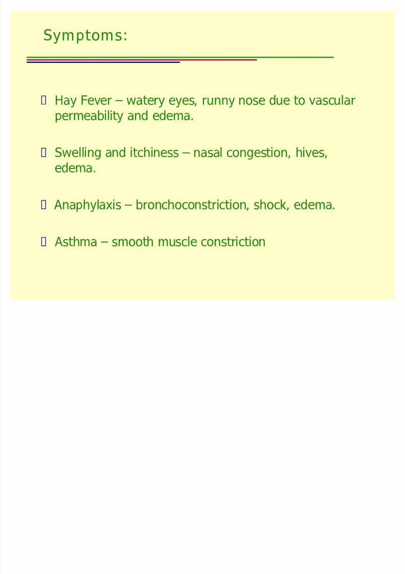
Type I: Ig E-Mediated Hypersensitivity
Most common type of hypersensitivity reaction.
Mediated by IgE & causes hay fever to life
threatening (bee stings) clinical manifestations.
Reactions are stimulated by binding of IgE (viaits Fc region) to high affinity IgE specificreceptors (FcRI) expressed on mast cells &basophils.

8/6/2019 Hypersensitivity Types & Features
http://slidepdf.com/reader/full/hypersensitivity-types-features 4/30
When cross linked by Ags, IgE triggers mast cells & basophils to release inflammatorymediators that lead to clinical features
(allergic reactions) eg. rhinitis, asthma,allergy, hay fever, bee sting, etc.
These reactions are very rapid, occur within
min after challenge-that is re-exposure to Ag.
Allergic reactions are called ITH or Type Ihypersensitivity.

8/6/2019 Hypersensitivity Types & Features
http://slidepdf.com/reader/full/hypersensitivity-types-features 5/30
Sequence of Events involved:
Sensitization Phase = Activation Phase =
Effector Phase
8/6/2019 Hypersensitivity Types & Features
http://slidepdf.com/reader/full/hypersensitivity-types-features 6/30
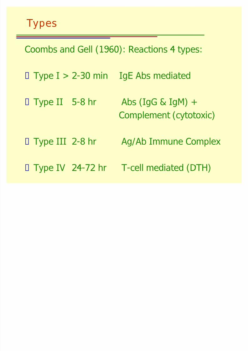
Sensitization Phase
Initial exposure to an allergen (Ag) induces IgEformation, following the primary antibodymediated immune response.
IgE binds via Fc portion to specific receptors onbasophils (in blood) and mast cells (in tissues),
(both cell types have histamine granules)
(No effect on mast cells directly up to this stage)

8/6/2019 Hypersensitivity Types & Features
http://slidepdf.com/reader/full/hypersensitivity-types-features 7/30
Activation Phase
Second and subsequent exposure to sameallergen (specific Ag):
-allergen (specific Ag) binds to V region of Fab and crosslinks two adjacent IgEs onmast cell/basophil surface.
8/6/2019 Hypersensitivity Types & Features
http://slidepdf.com/reader/full/hypersensitivity-types-features 8/30
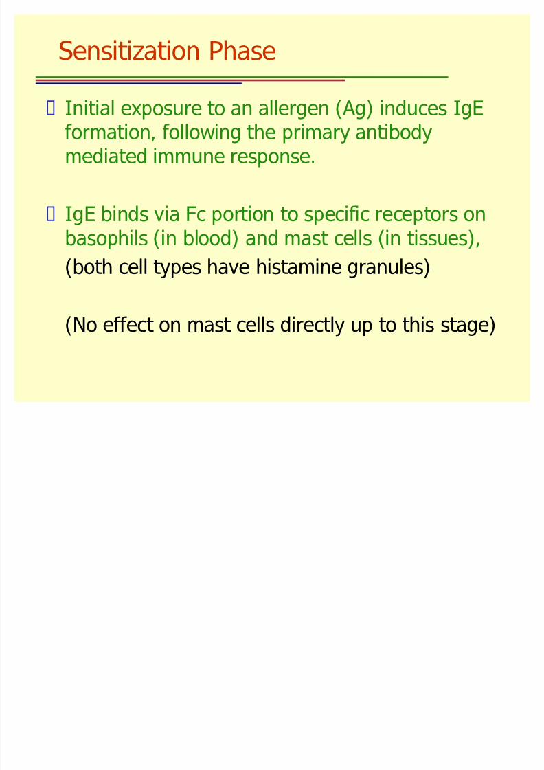
Effector Phase
Crosslinking of two adjacent IgEs on mast
cell/basophil surface triggers these cells to
degranulate immediately and release histamine
and many other mediators.
Immediate response (min after exposure)
mediators release due to an increase in cGMPlevels and/or decrease in cAMP levels
Leads to clinical manifestations, allergy,
asthma, etc.

8/6/2019 Hypersensitivity Types & Features
http://slidepdf.com/reader/full/hypersensitivity-types-features 9/30
Sensitization-skin contact, ingestion, injection,inhalation.
~50% generates IgE response to airborne
Ags but ~20% develops clinical symptoms(those with high IL-4).
Low levels of IgE in non-allergic due tosuppressor effect of IFN- (Th1 produced)
dowregulates Th2.
8/6/2019 Hypersensitivity Types & Features
http://slidepdf.com/reader/full/hypersensitivity-types-features 10/30

8/6/2019 Hypersensitivity Types & Features
http://slidepdf.com/reader/full/hypersensitivity-types-features 11/30
Key Mediators
Histamine preformed molecules in granules.
-binds receptors on target cells (lungs, skin, bloodvessels)
-causes vasodilation, capillary permeability, smooth
muscle constriction
-antihistamines block histamine receptors.
Prostaglandins from mast cells, causes bronchial
constriction, edema.
Leukotriene adhesion of leukocytes to capillaryvenules, degranulation.

8/6/2019 Hypersensitivity Types & Features
http://slidepdf.com/reader/full/hypersensitivity-types-features 12/30
SRS-A (Slow Reacting Substance of Anaphylaxis)
-produced after exposure to allergen, nor preformed
-slow release
-bronchoconstricton: implicated in asthma
ECF-A (Eosinophil Chemotactic Factor of Anaphylaxis)
-preformed in mast cell granules
-quick release
-attracts eosinophils which release histaminase and
arylsulfate from their granules
-histaminase degrades excess histamine
8/6/2019 Hypersensitivity Types & Features
http://slidepdf.com/reader/full/hypersensitivity-types-features 13/30
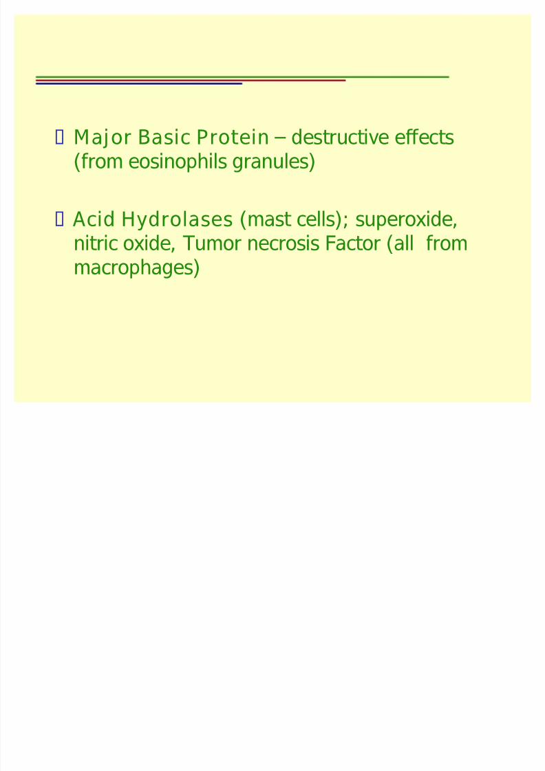
Major Basic Protein destructive effects(from eosinophils granules)
Acid Hydrolases (mast cells); superoxide,nitric oxide, Tumor necrosis Factor (all frommacrophages)
8/6/2019 Hypersensitivity Types & Features
http://slidepdf.com/reader/full/hypersensitivity-types-features 14/30
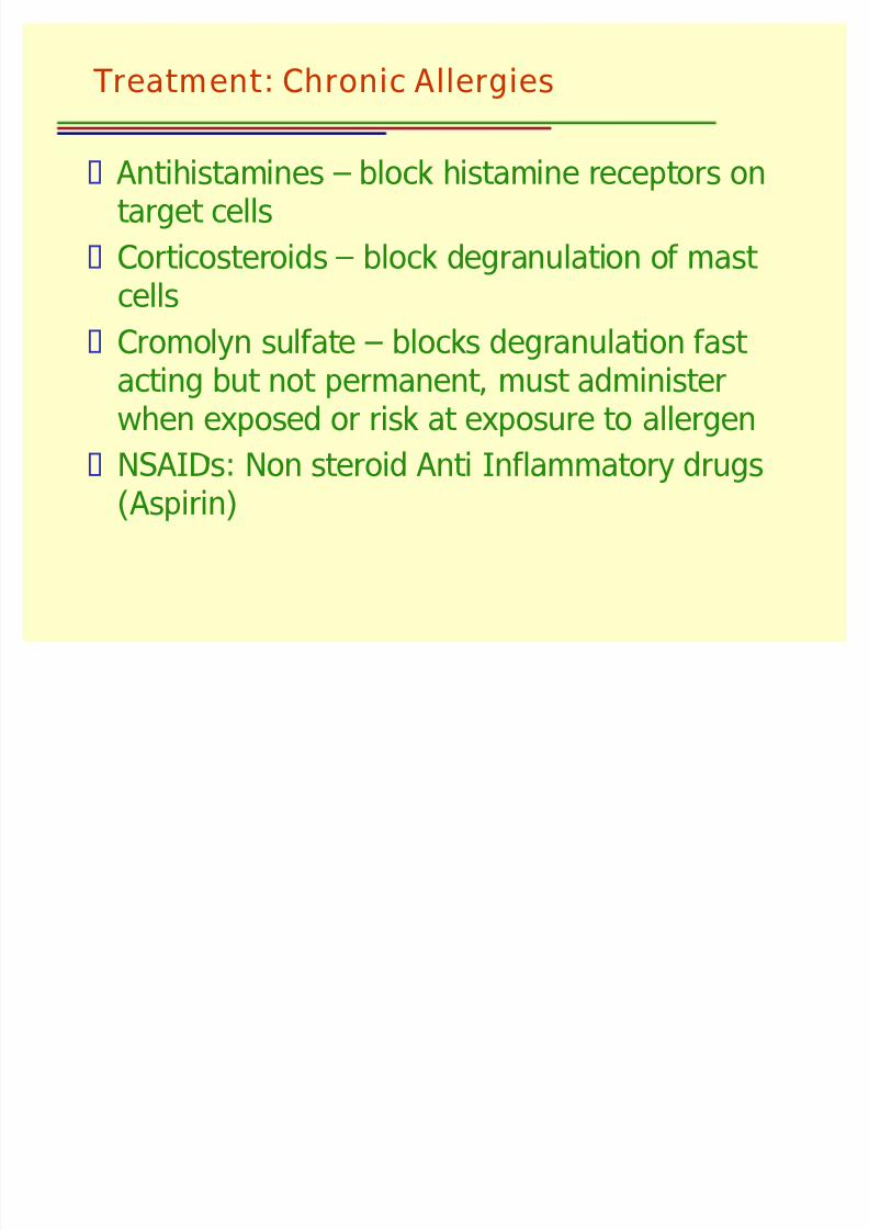
Treatment: Chronic Allergies
Antihistamines block histamine receptors ontarget cells
Corticosteroids block degranulation of mast
cells Cromolyn sulfate blocks degranulation fast
acting but not permanent, must administerwhen exposed or risk at exposure to allergen
NSAIDs: Non steroid Anti Inflammatory drugs(Aspirin)
8/6/2019 Hypersensitivity Types & Features
http://slidepdf.com/reader/full/hypersensitivity-types-features 15/30
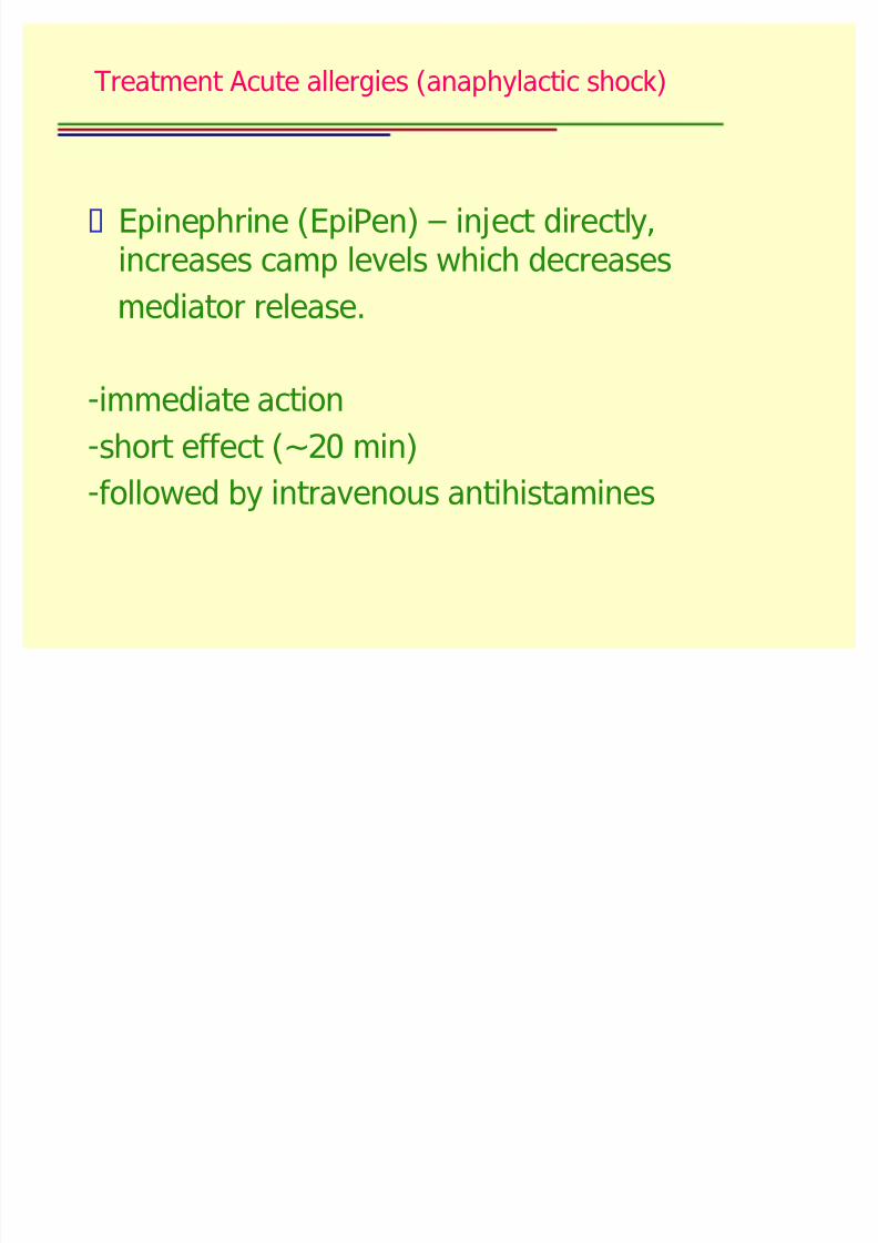
Treatment Acute allergies (anaphylactic shock)
Epinephrine (EpiPen) inject directly,increases camp levels which decreases
mediator release.
-immediate action
-short effect (~20 min)-followed by intravenous antihistamines

8/6/2019 Hypersensitivity Types & Features
http://slidepdf.com/reader/full/hypersensitivity-types-features 16/30
Type-II Reactions
Hypersensitivity resulting from Abs mistakenly reactingwith normal self-Ags on body cells.
Binding of Abs to these normal cells result in immunedestruction.

8/6/2019 Hypersensitivity Types & Features
http://slidepdf.com/reader/full/hypersensitivity-types-features 17/30
Mechanism
Either IgG or IgM made against normal self-Ags as aresult of a failure in immune tolerance or a foreign Agresembling some molecules on the surface of host
cells enters the body and IgG or IgM made against that Ag then cross-reacts with the host cell surface.
8/6/2019 Hypersensitivity Types & Features
http://slidepdf.com/reader/full/hypersensitivity-types-features 18/30
Binding of IgG/IgM to host cell surface leads to:
Opsonization of host cell.
Activation of classical complement pathway causing
MAC mediated lysis of cells.
ADCC destruction of host cells: NK cells attachment >
release pore-forming proteins perforins & proteolytic
enzymes granzymes. Granyzymes pass throughpores & activates enzymes that leads to apoptosis of infected cell by means of destruction of its structuralcytoskelton proteins & by chromosomal degradation.
8/6/2019 Hypersensitivity Types & Features
http://slidepdf.com/reader/full/hypersensitivity-types-features 19/30
Ab & Rh blood group reactions.
Autoimmune diseases (Graves/multiple sclerosis)
Good pastures disease, auto Ab to lung & kidney
basement membrane cause hemorrhage at site of
binding.
Breakdown of tolerance to self:
Abs to Ach receptors, loss of receptors, reducing orinhibiting nerve impulses across neuro-muscular
junctions (myasthenia gravis).

8/6/2019 Hypersensitivity Types & Features
http://slidepdf.com/reader/full/hypersensitivity-types-features 20/30
Hypersensitivity Reactions III
Normally Immune complexes removed byphagocytic cells & there is no tissue damage.
However, when large amounts of I.Cs.persists in tissues they activate complement cascade and cause damage to tissues.(Arthus reaction).
Ags responsible: Microbial Ags, auto Ags,foreign serum components.
8/6/2019 Hypersensitivity Types & Features
http://slidepdf.com/reader/full/hypersensitivity-types-features 21/30
Removal of Ag Ab Complex
Ag-Ab complex binds to Fc receptors of IgG,RBCs have C3b receptors, binds tocomplexes, that have fixed complement &transport them to liver, where complexes areremoved by phagocytic kupffer cells.
Ag-Ag complexes are clusters of interlocking
Ags-Abs; rapidly removed by blood stream byMacrophages (spleen), kupffer cells (liver).

8/6/2019 Hypersensitivity Types & Features
http://slidepdf.com/reader/full/hypersensitivity-types-features 22/30
When large quantities of ICs of a certain sizeare formed in circulation, they can bedeposited in tissues and trigger a variety of pathogenic events, called hypersensitivityreactions- III. (Kidneys, skin, joints, eye).
8/6/2019 Hypersensitivity Types & Features
http://slidepdf.com/reader/full/hypersensitivity-types-features 23/30
Ag-Ab complex activates classicalcomplement pathway. This may causeMassive inflammation: complement proteinC5a.
Influx of neutophils: Due to complement protein C5a, neutrophils discharge theirlysosomal enzymes & cause tissue damage,further inflammation.
MAC lysis of surrounding tissue cells. Aggregation of platlets: Blockage of
capillaries.

8/6/2019 Hypersensitivity Types & Features
http://slidepdf.com/reader/full/hypersensitivity-types-features 24/30
Under some circumstances ICs continue tocirculate, and eventually become trapped intissues of kidneys (glomerulonephritis), liver,skin (skin lesions), joints (thematoid arthritis),blood vessels & lead to inflammation & tissuedamage.
8/6/2019 Hypersensitivity Types & Features
http://slidepdf.com/reader/full/hypersensitivity-types-features 25/30
Localized: inhaled> bacterial fungal spores,pigeons serum (farmers lung disease).Systemic: Micobes (Streptococcus)Streptococcal nephritis. Serum sickness(fever, skin eruption, lympadenopathy.

8/6/2019 Hypersensitivity Types & Features
http://slidepdf.com/reader/full/hypersensitivity-types-features 26/30
Type-IV- Delayed Type of Hypersensitivity DTH
Mediated by T-cells together with dendriticcells, macrophages & cytokines.
The normal events associated with cell-
mediated immunity are a crucial mode of immunologic reactivity for protection against intracellular parasites (bacteria, viruses etc).However, the nature of reaction and its
mediators also cause DTH reactions.

8/6/2019 Hypersensitivity Types & Features
http://slidepdf.com/reader/full/hypersensitivity-types-features 27/30
When activated by contact with an Ag presented byantigen-presenting cell the responding T cells releaseinappropriately large amount of cytokines, some of which attract and activate other mononuclear cells
that are not antigen specific (such as monocytes andmacrophages).
Recruitment and activation of these antigen-
nonspecific mononuclear cells are mainly responsiblefor the eventual damaging outcome of the reactions.
8/6/2019 Hypersensitivity Types & Features
http://slidepdf.com/reader/full/hypersensitivity-types-features 28/30
Major events: 3 steps
Activation of antigen-specific inflammatory Th- 1cells in a previously sensitized individual.
Elaboration of proinflammatory cytokines by the
antigen-specific Th-1 cells.
Recruitment and activation of antigen non-specificinflammatory leukocytes.
These events typically occur over a period of several days (24-72 hrs) hence known as DTH

8/6/2019 Hypersensitivity Types & Features
http://slidepdf.com/reader/full/hypersensitivity-types-features 29/30
Causative Agents
Skin contact, small molecules (chemicals,plant molecules).
Contact Sensitivity (contact dermatitis): eg.
some cosmetics, Formaldehyde.
Poison Ivy Dermatitis: Offending substance iscontained in an oil secreted by leaves of
poison-ivy vine & other plants. Theypenetrate skin & cause blister formation.
8/6/2019 Hypersensitivity Types & Features
http://slidepdf.com/reader/full/hypersensitivity-types-features 30/30
Mantoux or PPD Test
Small amounts of purified protein derivative(PPD) of tuberculin derived from M.tuberculosis organisms injected into skin &site examined after 72 hrs. +ve skin test Ist red swelling, maximal at 48-72 hrs post injection. This is caused by influx of T-cells ¯ophages at site of injection.
PPD test is useful for public healthsurveillance of TB.
Related Documents