Hyperbaric oxygen results in increased vascular endothelial growth factor (VEGF) protein expression in rabbit calvarial critical-sized defects Tommy C. O. Fok, BSc(PT), a Ahmed Jan, DDS, b Sean A. F. Peel, PhD, c A. Wayne Evans, MD, d Cameron M. L. Clokie, DDS, PhD, FRCDC, e George K. B. Sándor, MD, DDS, PhD, FRCDC, FRCSC, FACS, f London and Toronto, Canada, Tampere and Oulu, Finland UNIVERSITY OF WESTERN ONTARIO, UNIVERSITY OF TORONTO, UNIVERSITY OF TAMPERE, AND UNIVERSITY OF OULU Background. Hyperbaric oxygen therapy (HBO) promotes osseous healing, however the mechanism by which this occurs has not been elucidated. HBO may promote angiogenesis, which is vital for bone healing. Vascular endothelial growth factor (VEGF) is one of the key factors that stimulates angiogenesis. Objective. The objective of this study was to investigate whether HBO altered VEGF expression during bone healing. Methods and materials. Archived samples from calvarial defects of rabbits exposed to HBO (2.4 ATA, 90 minutes a day, 5 days a week for 4 weeks) and normobaric oxygen controls (NBO) were analyzed by immunohistochemistry. Results. VEGF expression in 6-week HBO samples was elevated compared to NBO (P .012). Staining of the 12- week HBO samples was reduced compared to 6-week HBO (P .008) and was similar to 6- and 12-week NBO control samples. Conclusion. HBO therapy resulted in increased VEGF expression in the defects even 2 weeks after the termination of treatment (6 weeks postsurgery). (Oral Surg Oral Med Oral Pathol Oral Radiol Endod 2008;105:417-22) A critical-sized osseous defect is defined as the mini- mum dimension of a bony lesion that cannot repair itself to its preinjured state without intervention during an individual’s life span. 1 In the rabbit calvarium this is defined as a defect 15 mm in diameter. Critical-sized osseous defects may lead to numerous complications including fracture, non-union and pseudo-arthrosis. 2 Surgical treatments prevent further complications, which may involve the use of a fixation device and autogenous bone graft material to bridge the gap in the defect. All such reconstructive procedures that require a second surgical site for the harvesting of tissue are associated with potential morbidity. 3,4 Synthetic bio- materials have been used in place of autogenous bone grafts. 5 Recently, Jan et al., 6 using the rabbit critical- sized calvarial defect model, showed that 20 treatments of 90 minutes of hyperbaric oxygen (HBO) at 2.4 atmospheres could heal both critical-sized and supra- critical-sized calvarial defects when compared to nor- mobaric oxygen controls (NBO). This suggests that HBO has the potential to augment bony healing. HBO’s mode of action in the treatment of decompres- sion sickness and carbon monoxide poisoning is well understood based on its effects on reducing gas emboli a Resident in Oral and Maxillofacial Surgery, Schulich School of Medicine and Dentistry, University of Western Ontario. b Resident in Oral and Maxillofacial Surgery and Anesthesia, Univer- sity of Toronto. c Assistant Professor, Orthobiologics Group, University of Toronto. d Hyperbaric Medicine Unit, Department of Anesthesia, Faculty of Medicine, University of Toronto. e Professor, Discipline Head, Oral and Maxillofacial Surgery and Anesthesia, Director, Orthobiologics Group, University of Toronto. f Professor, Oral and Maxillofacial Surgery and Anesthesia, Univer- sity of Toronto; Regea Institute, University of Tampere, Tampere, Finland; and Dosent, University of Oulu, Oulu, Finland. Received for publication Dec 7, 2006; returned for revision Jul 13, 2007; accepted for publication Jul 13, 2007. 1079-2104/$ - see front matter © 2008 Mosby, Inc. All rights reserved. doi:10.1016/j.tripleo.2007.07.015 417 Vol. 105 No. 4 April 2008 ORAL AND MAXILLOFACIAL SURGERY Editor: James R. Hupp

Welcome message from author
This document is posted to help you gain knowledge. Please leave a comment to let me know what you think about it! Share it to your friends and learn new things together.
Transcript
Vol. 105 No. 4 April 2008
ORAL AND MAXILLOFACIAL SURGERY Editor: James R. Hupp
Hyperbaric oxygen results in increased vascular endothelialgrowth factor (VEGF) protein expression in rabbitcalvarial critical-sized defectsTommy C. O. Fok, BSc(PT),a Ahmed Jan, DDS,b Sean A. F. Peel, PhD,c
A. Wayne Evans, MD,d Cameron M. L. Clokie, DDS, PhD, FRCDC,e
George K. B. Sándor, MD, DDS, PhD, FRCDC, FRCSC, FACS,f London and Toronto, Canada,Tampere and Oulu, FinlandUNIVERSITY OF WESTERN ONTARIO, UNIVERSITY OF TORONTO, UNIVERSITY OF TAMPERE, ANDUNIVERSITY OF OULU
Background. Hyperbaric oxygen therapy (HBO) promotes osseous healing, however the mechanism by which thisoccurs has not been elucidated. HBO may promote angiogenesis, which is vital for bone healing. Vascular endothelialgrowth factor (VEGF) is one of the key factors that stimulates angiogenesis.Objective. The objective of this study was to investigate whether HBO altered VEGF expression during bone healing.Methods and materials. Archived samples from calvarial defects of rabbits exposed to HBO (2.4 ATA, 90 minutes aday, 5 days a week for 4 weeks) and normobaric oxygen controls (NBO) were analyzed by immunohistochemistry.Results. VEGF expression in 6-week HBO samples was elevated compared to NBO (P � .012). Staining of the 12-week HBO samples was reduced compared to 6-week HBO (P � .008) and was similar to 6- and 12-week NBOcontrol samples.Conclusion. HBO therapy resulted in increased VEGF expression in the defects even 2 weeks after the termination of
treatment (6 weeks postsurgery). (Oral Surg Oral Med Oral Pathol Oral Radiol Endod 2008;105:417-22)A critical-sized osseous defect is defined as the mini-mum dimension of a bony lesion that cannot repairitself to its preinjured state without intervention duringan individual’s life span.1 In the rabbit calvarium this is
aResident in Oral and Maxillofacial Surgery, Schulich School ofMedicine and Dentistry, University of Western Ontario.bResident in Oral and Maxillofacial Surgery and Anesthesia, Univer-sity of Toronto.cAssistant Professor, Orthobiologics Group, University of Toronto.dHyperbaric Medicine Unit, Department of Anesthesia, Faculty ofMedicine, University of Toronto.eProfessor, Discipline Head, Oral and Maxillofacial Surgery andAnesthesia, Director, Orthobiologics Group, University of Toronto.fProfessor, Oral and Maxillofacial Surgery and Anesthesia, Univer-sity of Toronto; Regea Institute, University of Tampere, Tampere,Finland; and Dosent, University of Oulu, Oulu, Finland.Received for publication Dec 7, 2006; returned for revision Jul 13,2007; accepted for publication Jul 13, 2007.1079-2104/$ - see front matter© 2008 Mosby, Inc. All rights reserved.
doi:10.1016/j.tripleo.2007.07.015defined as a defect 15 mm in diameter. Critical-sizedosseous defects may lead to numerous complicationsincluding fracture, non-union and pseudo-arthrosis.2
Surgical treatments prevent further complications,which may involve the use of a fixation device andautogenous bone graft material to bridge the gap in thedefect. All such reconstructive procedures that require asecond surgical site for the harvesting of tissue areassociated with potential morbidity.3,4 Synthetic bio-materials have been used in place of autogenous bonegrafts.5 Recently, Jan et al.,6 using the rabbit critical-sized calvarial defect model, showed that 20 treatmentsof 90 minutes of hyperbaric oxygen (HBO) at 2.4atmospheres could heal both critical-sized and supra-critical-sized calvarial defects when compared to nor-mobaric oxygen controls (NBO). This suggests thatHBO has the potential to augment bony healing.
HBO’s mode of action in the treatment of decompres-sion sickness and carbon monoxide poisoning is well
understood based on its effects on reducing gas emboli417
OOOOE418 Fok et al. April 2008
and hastening carboxyhemoglobin dissociation.7 How-ever, it has also demonstrated effectiveness in the treat-ment of necrotizing soft tissue infections, soft tissue radi-ation necrosis, diabetic wound healing, and now osseousdefect repair where other mechanisms are believed toinvolved.8,9 It has been well established that the formationof new blood vessels (angiogenesis) is essential in theprocess of soft tissue and bone repair.10,11 Vascular dis-ruption, caused by traumatic injury has been shown tolead to the formation of a hypoxic zone. Wound hypoxiais necessary to stimulate angiogenesis and revasculariza-tion. HBO increases the amount of oxygen dissolved inthe blood (oxygen tension) which can in turn increase theamount of oxygen delivered to these hypoxic tissues re-ducing the effects of the hypoxia.7 While this is helpful incases of chronic hypoxia, which blunts the repair process,it is not so clear as to how this would stimulate the normalrepair process.
Vascular endothelial growth factor (VEGF) has beenidentified as one of the primary growth factors respon-sible for neovascularization during wound healing andembryonic development.12 Oxygen tension is a keyregulator of VEGF expression in vitro and in vivo.13-15
We therefore wished to investigate the effect of HBOon VEGF expression in bone healing.
METHODS AND MATERIALS
Experimental designThis investigation used archived tissue from a pre-
vious study.6 The surgical protocol for the study wasapproved by the University of Toronto Animal Careand Ethics Committee (Protocol number 20005145). Atotal of 21 skeletally mature male New Zealand whiterabbits were divided into 2 groups (n � 10 for the6-week group, n � 11 for the 12-week group). Fiveanimals in the 6-week group received hyperbaric oxy-gen treatment (HBO) and 5 control rabbits were kept ina normobaric environment (NBO). Similarly, 5 rabbitsin the 12-week group received hyperbaric treatment(HBO) while 6 control animals were exposed to anormobaric environment (NBO). Critical-sized calvar-ial defects of 15 mm and supra critical-sized defects of18 mm were randomly assigned to the right and leftparietal bones of the rabbits and created using a straightfissured bur guided by a template. For the HBO treat-ment group, each rabbit was placed into an animalhyperbaric oxygen chamber and exposed to 100% O2
under 2.4 atmospheres of pressure, for 90 minutes aday, 5 days a week for 4 weeks (20 treatments). Therabbits were sacrificed 6 weeks or 12 weeks postsur-gery and the parietal bones were harvested. These sam-ples were fixed with 10% formalin and decalcified in a
solution of 45% formic acid in 0.2 M sodium citrate.Qualitative analysis: histologyFollowing fixation and decalcification, the midpoint
of the defect region was identified and served as thecoronal reference plane of section prior to embeddingin paraffin. Multiple 6-�m sections were cut andstained with hematoxylin and eosin (H&E) for conven-tional light-microscopy. The defect region was visual-ized in all samples and the appearance of new bonyregenerate was noted.
Quantitative analysis: immunohistochemicalanalysis
Multiple 6-�m sections cut from the same paraffinblock were used in the histological analysis. Thesesections were incubated with mouse monoclonal anti-human VEGF121 antibody (clone JH-21, Lab VisionCorp, Fremont, CA), with known rabbit cross-reactiv-ity, as a primary antibody. Then an avidin-biotin com-plex (Lab Vision Corp) was incubated to label theprimary antibody, and a color reagent was added at theend to allow the horseradish peroxidase reaction to takeplace.
Analysis of VEGF expressionUsing the image capturing software Image Pro Plus
4.1 for Windows (Media Cybernetics, Carlsbad, CA), 6random fields from each section were captured at �40magnification using an RT Color digital camera (Diag-nostic Instruments Inc, Sterling Heights, MI). A total of3 sections were used for each defect resulting in a totalof 18 random images for each right and 18 of each leftdefect. The area stained for VEGF in each field wasmeasured by setting a threshold intensity above whicha pixel is counted using the Image Pro Plus software.
To determine whether there was any difference inVEGF expression in the center of the defects comparedto the margins of the defects, 2 images were taken fromthe central one third of the defect and 4 images weretaken from the margins and their VEGF staining mea-sured.
Statistical analysisOne-way analysis of variance (ANOVA) was em-
ployed for analysis within and between the groups.When differences were found, the Student-Newman-Keuls method was used as a post hoc test to determinewhich groups were significantly different.
Comparisons of VEGF staining between the15-mm and 18-mm defects in the same rabbits, andbetween the margins and central regions within eachdefect, were made using the paired t test. Statisticalsignificance was set at P � .05. All statistical anal-yses were performed using Sigma Stat software
(v3.0, SYSTAT, San Jose, CA).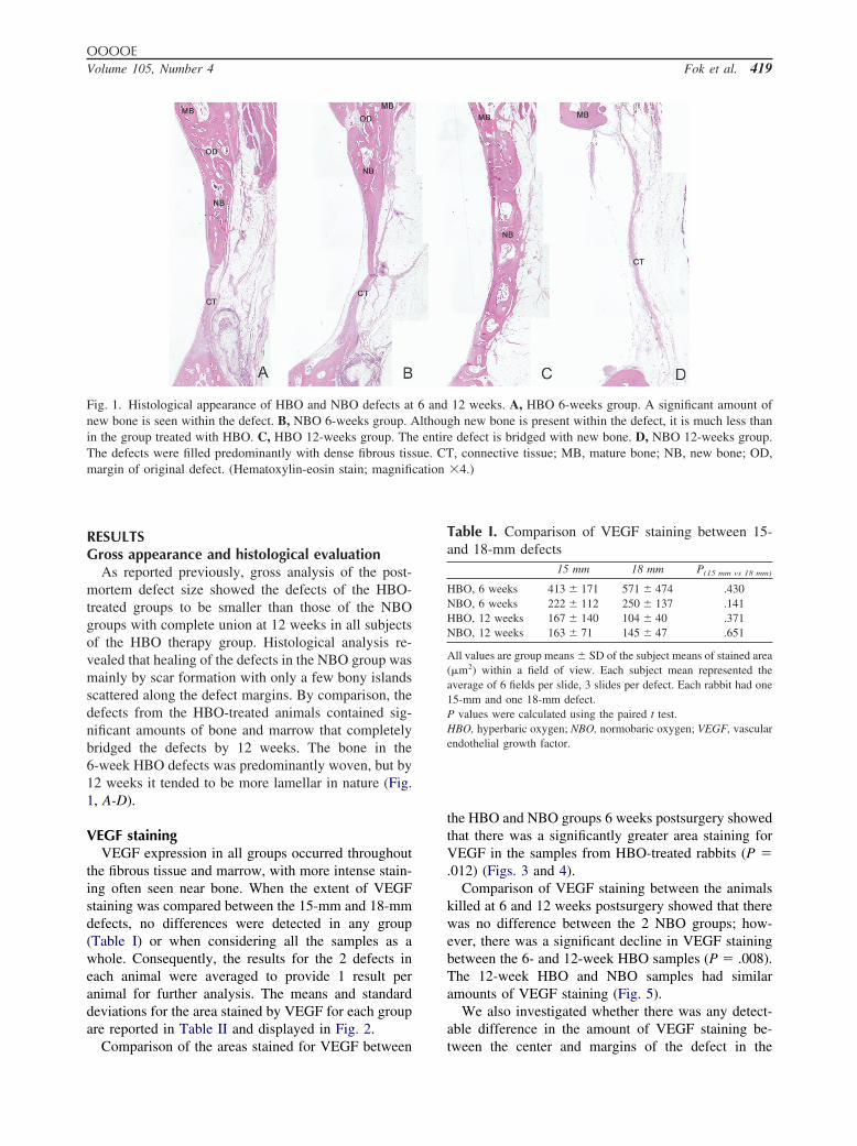
cation
OOOOEVolume 105, Number 4 Fok et al. 419
RESULTSGross appearance and histological evaluation
As reported previously, gross analysis of the post-mortem defect size showed the defects of the HBO-treated groups to be smaller than those of the NBOgroups with complete union at 12 weeks in all subjectsof the HBO therapy group. Histological analysis re-vealed that healing of the defects in the NBO group wasmainly by scar formation with only a few bony islandsscattered along the defect margins. By comparison, thedefects from the HBO-treated animals contained sig-nificant amounts of bone and marrow that completelybridged the defects by 12 weeks. The bone in the6-week HBO defects was predominantly woven, but by12 weeks it tended to be more lamellar in nature (Fig.1, A-D).
VEGF stainingVEGF expression in all groups occurred throughout
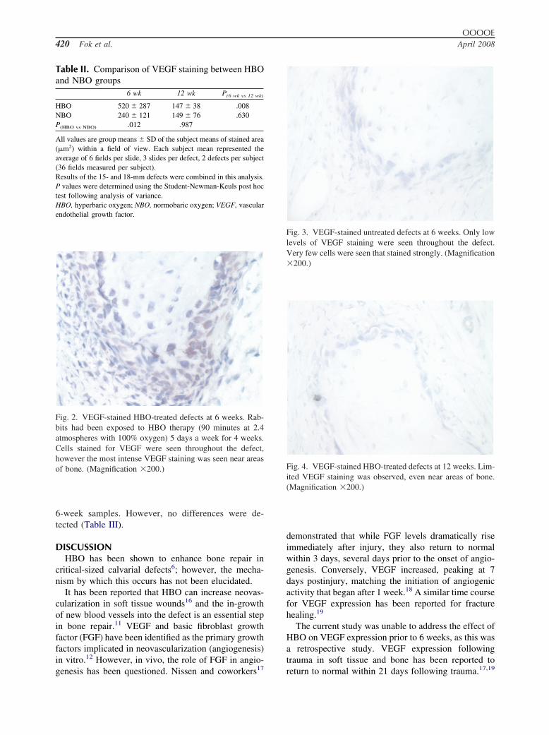
the fibrous tissue and marrow, with more intense stain-ing often seen near bone. When the extent of VEGFstaining was compared between the 15-mm and 18-mmdefects, no differences were detected in any group(Table I) or when considering all the samples as awhole. Consequently, the results for the 2 defects ineach animal were averaged to provide 1 result peranimal for further analysis. The means and standarddeviations for the area stained by VEGF for each groupare reported in Table II and displayed in Fig. 2.
Fig. 1. Histological appearance of HBO and NBO defects atnew bone is seen within the defect. B, NBO 6-weeks group. Ain the group treated with HBO. C, HBO 12-weeks group. ThThe defects were filled predominantly with dense fibrous tismargin of original defect. (Hematoxylin-eosin stain; magnifi
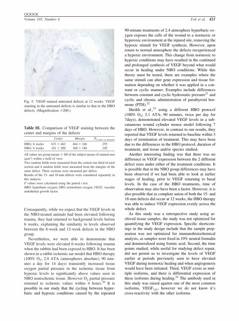
Comparison of the areas stained for VEGF between
the HBO and NBO groups 6 weeks postsurgery showedthat there was a significantly greater area staining forVEGF in the samples from HBO-treated rabbits (P �.012) (Figs. 3 and 4).
Comparison of VEGF staining between the animalskilled at 6 and 12 weeks postsurgery showed that therewas no difference between the 2 NBO groups; how-ever, there was a significant decline in VEGF stainingbetween the 6- and 12-week HBO samples (P � .008).The 12-week HBO and NBO samples had similaramounts of VEGF staining (Fig. 5).
We also investigated whether there was any detect-able difference in the amount of VEGF staining be-
12 weeks. A, HBO 6-weeks group. A significant amount ofgh new bone is present within the defect, it is much less thane defect is bridged with new bone. D, NBO 12-weeks group.
, connective tissue; MB, mature bone; NB, new bone; OD,�4.)
Table I. Comparison of VEGF staining between 15-and 18-mm defects
15 mm 18 mm P(15 mm vs 18 mm)
HBO, 6 weeks 413 � 171 571 � 474 .430NBO, 6 weeks 222 � 112 250 � 137 .141HBO, 12 weeks 167 � 140 104 � 40 .371NBO, 12 weeks 163 � 71 145 � 47 .651
All values are group means � SD of the subject means of stained area(�m2) within a field of view. Each subject mean represented theaverage of 6 fields per slide, 3 slides per defect. Each rabbit had one15-mm and one 18-mm defect.P values were calculated using the paired t test.HBO, hyperbaric oxygen; NBO, normobaric oxygen; VEGF, vascularendothelial growth factor.
6 andlthou
e entirsue. CT
tween the center and margins of the defect in the
OOOOE420 Fok et al. April 2008
6-week samples. However, no differences were de-tected (Table III).
DISCUSSIONHBO has been shown to enhance bone repair in
critical-sized calvarial defects6; however, the mecha-nism by which this occurs has not been elucidated.
It has been reported that HBO can increase neovas-cularization in soft tissue wounds16 and the in-growthof new blood vessels into the defect is an essential stepin bone repair.11 VEGF and basic fibroblast growthfactor (FGF) have been identified as the primary growthfactors implicated in neovascularization (angiogenesis)in vitro.12 However, in vivo, the role of FGF in angio-
Table II. Comparison of VEGF staining between HBOand NBO groups
6 wk 12 wk P(6 wk vs 12 wk)
HBO 520 � 287 147 � 38 .008NBO 240 � 121 149 � 76 .630P(HBO vs NBO) .012 .987
All values are group means � SD of the subject means of stained area(�m2) within a field of view. Each subject mean represented theaverage of 6 fields per slide, 3 slides per defect, 2 defects per subject(36 fields measured per subject).Results of the 15- and 18-mm defects were combined in this analysis.P values were determined using the Student-Newman-Keuls post hoctest following analysis of variance.HBO, hyperbaric oxygen; NBO, normobaric oxygen; VEGF, vascularendothelial growth factor.
Fig. 2. VEGF-stained HBO-treated defects at 6 weeks. Rab-bits had been exposed to HBO therapy (90 minutes at 2.4atmospheres with 100% oxygen) 5 days a week for 4 weeks.Cells stained for VEGF were seen throughout the defect,however the most intense VEGF staining was seen near areasof bone. (Magnification �200.)
genesis has been questioned. Nissen and coworkers17
demonstrated that while FGF levels dramatically riseimmediately after injury, they also return to normalwithin 3 days, several days prior to the onset of angio-genesis. Conversely, VEGF increased, peaking at 7days postinjury, matching the initiation of angiogenicactivity that began after 1 week.18 A similar time coursefor VEGF expression has been reported for fracturehealing.19
The current study was unable to address the effect ofHBO on VEGF expression prior to 6 weeks, as this wasa retrospective study. VEGF expression followingtrauma in soft tissue and bone has been reported to
Fig. 3. VEGF-stained untreated defects at 6 weeks. Only lowlevels of VEGF staining were seen throughout the defect.Very few cells were seen that stained strongly. (Magnification�200.)
Fig. 4. VEGF-stained HBO-treated defects at 12 weeks. Lim-ited VEGF staining was observed, even near areas of bone.(Magnification �200.)
return to normal within 21 days following trauma.17,19
OOOOEVolume 105, Number 4 Fok et al. 421
Consequently, while we expect that the VEGF levels inthe NBO-treated animals had been elevated followingtrauma, they had returned to background levels before6 weeks, explaining the similarity in levels observedbetween the 6-week and 12-week defects in the NBOgroup.
Nevertheless, we were able to demonstrate thatVEGF levels were elevated 6-weeks following traumawhen the rabbits had been exposed to HBO. It has beenshown in a rabbit ischemic ear model that HBO therapy(100% O2; 2.0 ATA (atmospheres absolute); 90 min-utes a day for 14 days) transiently increased tissueoxygen partial pressure in the ischemic tissue fromhypoxic levels to significantly above values seen inNBO nonischemic tissue. However O2 partial pressurereturned to ischemic values within 4 hours.20 It ispossible in our study that the cycling between hyper-
Fig. 5. VEGF-stained untreated defects at 12 weeks. VEGFstaining in the untreated defects is similar to that in the HBOdefects. (Magnification �200.)
Table III. Comparison of VEGF staining between thecenter and margins of the defects
Center Margin P(center vs margin)
HBO, 6 weeks 633 � 462 464 � 246 .255NBO, 6 weeks 181 � 200 269 � 188 .105
All values are group means � SD of the subject means of stained area(�m2) within a field of view.Two random fields were measured from the central one third of eachsection and 4 random fields were measured from the margins of thesame defect. Three sections were measured per defect.Results of the 15- and 18-mm defects were considered separately inthis analysis.P values were calculated using the paired t test.HBO, hyperbaric oxygen; NBO, normobaric oxygen; VEGF, vascularendothelial growth factor.
baric and hypoxic conditions caused by the repeated
90-minute treatments of 2.4 atmosphere hyperbaric ox-ygen exposes the cells of the wound to a normoxic orhyperoxic environment at the injured site, removing thehypoxic stimuli for VEGF synthesis. However, uponreturn to normal atmosphere the defects reexperienceda hypoxic environment. This change from normoxic tohypoxic conditions may have resulted in the continuedand prolonged synthesis of VEGF beyond what wouldoccur in healing under NBO conditions. While thistheory must be tested, there are examples where thesame stimuli can alter gene expression and tissue for-mation depending on whether it was applied in a con-stant or cyclic manner. Examples include differencesbetween constant and cyclic hydrostatic pressure21 andcyclic and chronic administration of parathyroid hor-mone (PTH).22
Sheikh et al.,23 using a different HBO protocol(100% O2; 2.1 ATA; 90 minutes, twice per day for7days), demonstrated elevated VEGF levels in a sub-cutaneous wound cylinder mouse model following 7days of HBO. However, in contrast to our results, theyreported that VEGF levels returned to baseline within 3days of termination of treatment. This may have beendue to the differences in the HBO protocol, duration oftreatment, and tissue and/or species studied.
Another interesting finding was that there was nodifference in VEGF expression between the 2 differentdefect sizes under either of the treatment conditions. Itis possible that in the NBO group differences may havebeen observed if we had been able to look at earlierstages of healing, prior to VEGF returning to basallevels. In the case of the HBO treatments, time ofobservation may also have been a factor. However, it isalso possible that as complete union of both the 15- and18-mm defects did occur at 12 weeks, the HBO therapywas able to induce VEGF expression evenly across thewhole defect.
As this study was a retrospective study using ar-chived tissue samples, the study was not optimized forquantifying the VEGF expression. Specific shortcom-ings in the study design include that the sample prep-aration was not optimized for immunohistochemicalanalysis, as samples were fixed in 10% neutral formalinand demineralized using formic acid. Second, the timepoints studied, while useful for studying defect repair,did not permit us to investigate the levels of VEGFearlier at periods previously seen to have elevatedVEGF during normoxic healing and when angiogenesiswould have been initiated. Third, VEGF exists as mul-tiple isoforms, and there is differential expression ofthese isoforms during healing.24 The antibody used inthis study was raised against one of the most commonisoforms, VEGF121, however we do not know it’s
cross-reactivity with the other isoforms.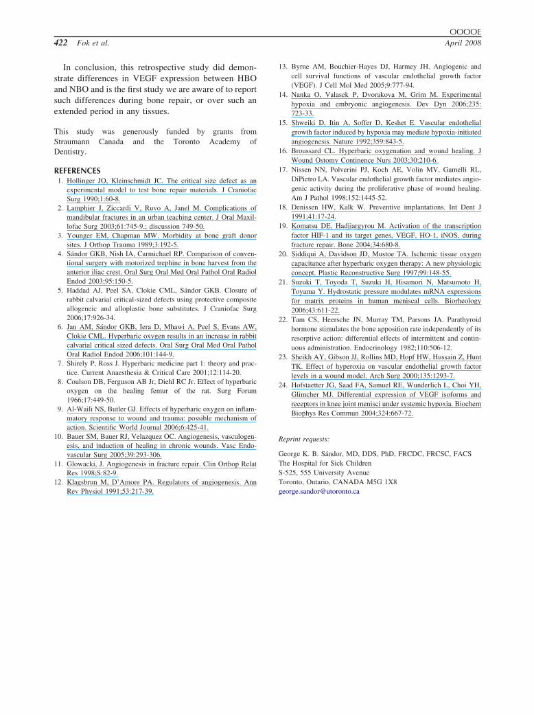
OOOOE422 Fok et al. April 2008
In conclusion, this retrospective study did demon-strate differences in VEGF expression between HBOand NBO and is the first study we are aware of to reportsuch differences during bone repair, or over such anextended period in any tissues.
This study was generously funded by grants fromStraumann Canada and the Toronto Academy ofDentistry.
REFERENCES1. Hollinger JO, Kleinschmidt JC. The critical size defect as an
experimental model to test bone repair materials. J CraniofacSurg 1990;1:60-8.
2. Lamphier J, Ziccardi V, Ruvo A, Janel M. Complications ofmandibular fractures in an urban teaching center. J Oral Maxil-lofac Surg 2003;61:745-9.; discussion 749-50.
3. Younger EM, Chapman MW. Morbidity at bone graft donorsites. J Orthop Trauma 1989;3:192-5.
4. Sándor GKB, Nish IA, Carmichael RP. Comparison of conven-tional surgery with motorized trephine in bone harvest from theanterior iliac crest. Oral Surg Oral Med Oral Pathol Oral RadiolEndod 2003;95:150-5.
5. Haddad AJ, Peel SA, Clokie CML, Sándor GKB. Closure ofrabbit calvarial critical-sized defects using protective compositeallogeneic and alloplastic bone substitutes. J Craniofac Surg2006;17:926-34.
6. Jan AM, Sándor GKB, Iera D, Mhawi A, Peel S, Evans AW,Clokie CML. Hyperbaric oxygen results in an increase in rabbitcalvarial critical sized defects. Oral Surg Oral Med Oral PatholOral Radiol Endod 2006;101:144-9.
7. Shirely P, Ross J. Hyperbaric medicine part 1: theory and prac-tice. Current Anaesthesia & Critical Care 2001;12:114-20.
8. Coulson DB, Ferguson AB Jr, Diehl RC Jr. Effect of hyperbaricoxygen on the healing femur of the rat. Surg Forum1966;17:449-50.
9. Al-Waili NS, Butler GJ. Effects of hyperbaric oxygen on inflam-matory response to wound and trauma: possible mechanism ofaction. Scientific World Journal 2006;6:425-41.
10. Bauer SM, Bauer RJ, Velazquez OC. Angiogenesis, vasculogen-esis, and induction of healing in chronic wounds. Vasc Endo-vascular Surg 2005;39:293-306.
11. Glowacki, J. Angiogenesis in fracture repair. Clin Orthop RelatRes 1998;S:82-9.
12. Klagsbrun M, D’Amore PA. Regulators of angiogenesis. Ann
Rev Physiol 1991;53:217-39.13. Byrne AM, Bouchier-Hayes DJ, Harmey JH. Angiogenic andcell survival functions of vascular endothelial growth factor(VEGF). J Cell Mol Med 2005;9:777-94.
14. Nanka O, Valasek P, Dvorakova M, Grim M. Experimentalhypoxia and embryonic angiogenesis. Dev Dyn 2006;235:723-33.
15. Shweiki D, Itin A, Soffer D, Keshet E. Vascular endothelialgrowth factor induced by hypoxia may mediate hypoxia-initiatedangiogenesis. Nature 1992;359:843-5.
16. Broussard CL. Hyperbaric oxygenation and wound healing. JWound Ostomy Continence Nurs 2003;30:210-6.
17. Nissen NN, Polverini PJ, Koch AE, Volin MV, Gamelli RL,DiPietro LA. Vascular endothelial growth factor mediates angio-genic activity during the proliferative phase of wound healing.Am J Pathol 1998;152:1445-52.
18. Denissen HW, Kalk W. Preventive implantations. Int Dent J1991;41:17-24.
19. Komatsu DE, Hadjiargyrou M. Activation of the transcriptionfactor HIF-1 and its target genes, VEGF, HO-1, iNOS, duringfracture repair. Bone 2004;34:680-8.
20. Siddiqui A, Davidson JD, Mustoe TA. Ischemic tissue oxygencapacitance after hyperbaric oxygen therapy: A new physiologicconcept. Plastic Reconstructive Surg 1997;99:148-55.
21. Suzuki T, Toyoda T, Suzuki H, Hisamori N, Matsumoto H,Toyama Y. Hydrostatic pressure modulates mRNA expressionsfor matrix proteins in human meniscal cells. Biorheology2006;43:611-22.
22. Tam CS, Heersche JN, Murray TM, Parsons JA. Parathyroidhormone stimulates the bone apposition rate independently of itsresorptive action: differential effects of intermittent and contin-uous administration. Endocrinology 1982;110:506-12.
23. Sheikh AY, Gibson JJ, Rollins MD, Hopf HW, Hussain Z, HuntTK. Effect of hyperoxia on vascular endothelial growth factorlevels in a wound model. Arch Surg 2000;135:1293-7.
24. Hofstaetter JG, Saad FA, Samuel RE, Wunderlich L, Choi YH,Glimcher MJ. Differential expression of VEGF isoforms andreceptors in knee joint menisci under systemic hypoxia. BiochemBiophys Res Commun 2004;324:667-72.
Reprint requests:
George K. B. Sándor, MD, DDS, PhD, FRCDC, FRCSC, FACSThe Hospital for Sick ChildrenS-525, 555 University AvenueToronto, Ontario, CANADA M5G 1X8
[email protected]
Related Documents